implausibility of the vibrational theory of...
TRANSCRIPT

Implausibility of the vibrational theory of olfactionEric Blocka,1, Seogjoo Jangb,1, Hiroaki Matsunamic,1, Sivakumar Sekharand, Bérénice Dethiera, Mehmed Z. Ertemd,e,Sivaji Gundalaa, Yi Panf, Shengju Lif, Zhen Lif, Stephene N. Lodgea, Mehmet Ozbild, Huihong Jiangf, Sonia F. Penalbaa,Victor S. Batistad, and Hanyi Zhuangf,g,1
aDepartment of Chemistry, University at Albany, State University of New York, Albany, NY 12222; bDepartment of Chemistry and Biochemistry, QueensCollege, and Graduate Center, City University of New York, Flushing, NY 11367; cDepartment of Molecular Genetics and Microbiology and Departmentof Neurobiology, Duke Institute for Brain Sciences, Duke University Medical Center, Durham, NC 27710; dDepartment of Chemistry, Yale University,New Haven, CT 06520; eChemistry Department, Brookhaven National Laboratory, Upton, NY 11973; fDepartment of Pathophysiology, Key Laboratoryof Cell Differentiation and Apoptosis of Ministry of Education, Shanghai Jiaotong University School of Medicine, Shanghai 200025, China; and gInstituteof Health Sciences, Shanghai Jiao tong University School of Medicine/Shanghai Institutes for Biological Sciences of Chinese Academy of Sciences, Shanghai200031, China
Edited by Jerrold Meinwald, Cornell University, Ithaca, NY, and approved March 31, 2015 (received for review February 20, 2015)
The vibrational theory of olfaction assumes that electron transferoccurs across odorants at the active sites of odorant receptors(ORs), serving as a sensitive measure of odorant vibrationalfrequencies, ultimately leading to olfactory perception. A previousstudy reported that human subjects differentiated hydrogen/deuterium isotopomers (isomers with isotopic atoms) of the muskcompound cyclopentadecanone as evidence supporting the theory.Here, we find no evidence for such differentiation at the molecularlevel. In fact, we find that the human musk-recognizing receptor,OR5AN1, identified using a heterologous OR expression system androbustly responding to cyclopentadecanone and muscone, fails todistinguish isotopomers of these compounds in vitro. Furthermore,themouse (methylthio)methanethiol-recognizing receptor, MOR244-3,as well as other selected human and mouse ORs, responded similarlyto normal, deuterated, and 13C isotopomers of their respectiveligands, paralleling our results with the musk receptor OR5AN1.These findings suggest that the proposed vibration theory doesnot apply to the human musk receptor OR5AN1, mouse thiol recep-tor MOR244-3, or other ORs examined. Also, contrary to the vibra-tion theory predictions, muscone-d30 lacks the 1,380- to 1,550-cm−1 IRbands claimed to be essential for musk odor. Furthermore, our the-oretical analysis shows that the proposed electron transfer mecha-nism of the vibrational frequencies of odorants could be easilysuppressed by quantum effects of nonodorant molecular vibrationalmodes. These and other concerns about electron transfer at ORs,together with our extensive experimental data, argue against theplausibility of the vibration theory.
olfaction | isotopomers | cyclopentadecanone | muscone | electron transfer
In 1870, the British physician William Ogle wrote: “As in theeye and the ear the sensory impression is known to result not
from the contact of material particles given off by the object seenor heard, but from waves or undulations of the ether or the air,one cannot but suspect that the same may be true in theremaining sense, and that the undulatory theory of smell. . . [maybe] the true one” (1, 2). Of the 29 different “theories of odour”listed in the 1967 edition of The Chemical Senses (3), nine as-sociate odor with vibrations, particularly those theories cham-pioned by Dyson (4, 5) and Wright (6–8). However, the premisethat olfaction involves detection of vibrational frequencies ofodorants remains highly speculative because neither the struc-tures of the odorant receptors (ORs) nor the binding sites or theactivation mechanisms triggered upon odorant binding to ORshave been established. In 1996–1997, Turin (9–12) elaborated onthe undulatory theory of smell, as considered in more detailbelow, and suggested that a mechanism analogous to inelasticelectron tunneling spectroscopy (13) may be involved, wheretunneling electrons in the receptor probe the vibrational fre-quencies of odorants. In 2013, Gane et al. (14) commented that“whether olfaction recognizes odorants by their shape, theirmolecular vibrations, or both remains an open and controversial
question” and that “a convenient way to address [this question] isto test for odor character differences between deuterated andnondeuterated odorant isotopomers since these have identicalground-state conformations but different vibrational modes.”Gane et al. (14) also stated that a particularly appropriate testcase would involve odorants containing “more CH group. . .[such as] musks [which] are among the largest odorants andtypically contain 15–18 carbons and 28 or more hydrogens.”In judging the plausibility of the vibration theory, we use a
multipronged approach:
i) We consider the concepts of shape vs. vibration theory andodorant perception vs. reception.
ii) As a test of the vibration theory, we have prepared a series ofisotopomers of musks and other compounds, containing up to30 C–H or C–D bonds as test odorants, which are evaluatedusing in vitro activation of receptors identified by us and othergroups as being highly responsive to these isotopomers.
iii) We consider the confounding effects of impurities and iso-tope effects in interpreting odorant perception, as well asthe validity of requirements for specific IR bands for recog-nition of musks by their receptors.
Significance
The vibrational theory of olfaction posits detection of odorantsthrough their vibrational frequencies rather than solely through“hand-in-glove” substrate/enzyme-like odorant–odorant receptor(OR) interactions. To test the theory, we compare responsesof different human and mouse ORs toward deuterated andundeuterated isotopomers (isotopic atom isomers) of receptor-responsive odorants because isotopomers should differ in theirmolecular vibrational frequencies. However, no differences inreceptor response are seen with any tested labeled/unlabeledodorant/receptor pairs. Because published behavioral studieshave shown that humans can distinguish isotopomers, peri-receptor events or impurities, rather than receptor-level vi-brational effects, are suggested. Because theoretical aspectsof the vibration theory are also found wanting, the vibrationtheory is deemed implausible in the absence of compellingreceptor-level experimental evidence to the contrary.
Author contributions: E.B., S.J., H.M., V.S.B., and H.Z. designed research; E.B., S.J., H.M.,S.S., B.D., M.Z.E., S.G., Y.P., S.L., Z.L., S.N.L., M.O., H.J., S.F.P., V.S.B., and H.Z. performedresearch; E.B. contributed new reagents/analytic tools; E.B., S.J., H.M., S.S., B.D., M.Z.E.,S.G., Y.P., S.N.L., M.O., S.F.P., V.S.B., and H.Z. analyzed data; and E.B., S.J., H.M., V.S.B., andH.Z. wrote the paper.
The authors declare no conflict of interest.
This article is a PNAS Direct Submission.
Freely available online through the PNAS open access option.1To whom correspondence may be addressed. Email: [email protected], [email protected], [email protected], or [email protected].
This article contains supporting information online at www.pnas.org/lookup/suppl/doi:10.1073/pnas.1503054112/-/DCSupplemental.
www.pnas.org/cgi/doi/10.1073/pnas.1503054112 PNAS Early Edition | 1 of 9
BIOPH
YSICSAND
COMPU
TATIONALBIOLO
GY
CHEM
ISTR
YPN
ASPL
US
iv) We examine the physical validity of the models developed tosupport the vibration theory.
v) We consider the specific limitations of our in vitro approachusing isotopomers to evaluate the vibration theory, basedprimarily on results obtained with a single identified humanmusk OR, in addition to other OR/ligand pairs.
vi) We consider plausible nonvibration theory models for dock-ing of musks to the humanmusk receptor, OR5AN1, where themusk carbonyl group functions as a hydrogen bond acceptor.
Gane et al. (14) have framed the argument for olfactory dis-crimination of hydrogen isotopomers as one of “shape” vs. “vi-bration.” However, neither the binding modes of isotopomersnor their activation mechanisms are known. ORs belong to thesuperfamily of class A G protein-coupled receptors (GPCRs),which are known to be activated through allosteric conforma-tional changes induced upon ligand binding even without trig-gering any kind of electron transfer processes. Ligand–receptorinteractions can be both attractive and repulsive, involving hy-drogen bonding, van der Waals, cation–π, π–π, ion–ion, dipole–dipole, steric, and hydrophobic interactions with the receptor,with water channels and bridging water molecules mediatinghydrogen bonds, as well as metal–ion coordination, as we haverecently demonstrated in the latter case (15, 16). Therefore,molecular shape can be considered a “straw-man” alternative tothe vibration theory when describing the differing affinities ofligands bound to GPCRs (17, 18), including isotopomers (19,20). Some of these attractive and repulsive interactions wereidentified in 1940 by Pauling and Delbrück (21), who note thatinteracting biomolecules “must have complementary surfaces,like die and coin, and also a complementary distribution of activegroups.” In addition, shape-related features are misrepresentedby vibration theory proponents. For example, Franco et al. (17)stated: “Given that proteins are chiral, a shape-only theorycannot account for the identical odors of most enantiomericpairs,” echoing similar comments by Turin (22): “One wouldtherefore generally expect enantiomers to have completely dif-ferent smells. This is emphatically not the case.” However, theseassertions are clearly at odds with the highly developed abilityof mice and other mammals to discriminate an array of non-pheromonal chiral odorant enantiomeric pairs (23–25), withthe divergent in vitro responses to enantiomers by differentcombinations of ORs (26) and, in particular, with the highlyselective response of the musk-sensitive mouse receptor, MOR215-1,to (R)-muscone (“l-muscone”) compared with (S)-muscone(“d-muscone”) (27).In addition to our concerns regarding shape, a second issue
relates to describing how different smells are perceived, that is,the perception of an odorant. It is known that in vivo perceptionof odorants reflects the totality of perireceptor events as well asodorant–OR interactions (reception). Volatile odorants enterthe nasal passage, where they dissolve in the nasal mucus over-lying the olfactory epithelium and are then rapidly detected byORs on the cilia of the olfactory sensory neurons, ultimatelyleading to signaling (28, 29). It is the mechanism of odorant–ORinteractions, the reception of the odorant, that we seek to ex-amine with isotopomers to determine whether the vibrationtheory is plausible, displaying isotope effects, because perceptioncould be influenced by isotope effects due to the perireceptorevents involving mucosal components, such as enzymes, muco-polysaccharides, salts, and antibodies.Whether deuterated and nondeuterated odorant isotopomers
can be distinguished by smell and, even if they can, whether thisdistinction validates the vibration theory is a matter of conten-tion. A 2001 paper by Haffenden et al. (30) reported thatbenzaldehyde-d6 gave a statistically significant difference in odorperception relative to normal benzaldehyde, in support of thevibration theory. However, this study has been criticized for
lacking double-blind controls to eliminate bias and because itused an anomalous version of the duo-trio test (31). Further-more, the study failed to account for perireceptor events,namely, the enzyme-mediated conversion of odorants that hasbeen shown to occur in nasal mucus. For example, benzaldehydeis converted to benzoic acid (32), a reaction potentially subject tosignificant primary isotope effects (2, 33, 34), which could ex-plain the difference in odor perception for the benzaldehydeisotopomers. Earlier claims that human subjects can distinguishodors of acetophenone isotopomers (9, 35) have been shown tobe untrue (14, 31). Recent studies indicate that Drosophilamelanogaster can distinguish acetophenone isotopomers (36, 37)and that Apis mellifera L., the honey bee, can be trained to dis-criminate pairs of isotopomers (38). These studies differ fromearlier insect studies in which isotopomer discrimination wasnot found. For example, systematic deuteration of 4-(p-hydroxy-phenyl)-2-butanone acetate, a Dacus cucurbitae Coquillett (themale melon fly) attractant, did not affect the attractiveness of thecompound to the fly (39); deuteration of alarm pheromonesfailed to alter the response toward these compounds by Pogo-nomyrmex badius worker ants (40); and honey bees could notdistinguish between deuterated and nondeuterated nitroben-zene (41).Concerns have been raised (42) about aspects of the Dro-
sophila study (36), which is “behavioural and not at the receptorlevel” (2) (also a concern with the Apis study). Also, given thatthe ORs and their downstream signaling in Drosophila and hu-mans are completely unrelated, the Drosophila study should notbe considered predictive of the ability of humans to distinguishisotopomers (2, 17). In view of the above discussion, it is in-teresting that in a blinded behavioral study, smell panelists dis-tinguished between deuterated and nondeuterated isotopomers ofcyclopentadecanone (1; Fig. 1A) and other musk odorants (14).Here, we study the response of human musk-sensitive OR5AN1,
identified through screening of heterologously expressed humanORs, to cyclopentadecanone (1) and muscone (4) isotopomers. Wealso present pharmacological data on the response of mouse ORsto deuterated and nondeuterated acetophenone and benzaldehyde,as well as selected 13C isotopomers. In addition, we present relatedstudies on the response of various human and mouse ORs to otherdeuterated and nondeuterated odorants, including (methylthio)-methanethiol (MTMT, 8; Fig. 1C) and bis(methylthiomethyl)disulfide (9), studied in connection with our investigation of therole of copper coordination in the recognition of both sulfur-con-taining odorants by the mouse (methylthio)methanethiol receptor,MOR244-3 (15, 16). Insofar as the ability to distinguish odors ofisotopomers directly tests the predictions of the vibration theory,the comparative response of human and mouse ORs to iso-topomers of these selected ligands in the heterologous OR ex-pression system constitutes a robust test of the vibration theory.Finally, we discuss the basis for recent vibration theories of olfac-tion and supporting computational evidence (37, 43–47) in light ofwell-established electron transfer theories (48). We point out thatkey assumptions underlying the vibration theory lack experimentalsupport and are missing important physical features expected forbiological systems.
Experimental ResultsResponse of a Human Musk OR to Deuterated and NondeuteratedMuscone and Other Musk Compounds. Because human subjectsare reported to discriminate between fully deuterated and non-deuterated cyclopentadecanone (1; Exaltone) (14), we sought toperform a corresponding receptor activation assay in vitro.First, from a commercial sample of cyclopentadecanone (1,
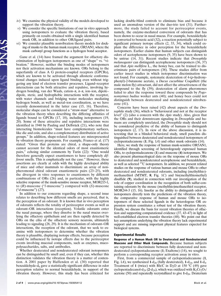
Fig. 1A), we synthesized 1-d28 (fully deuterated), 1-d24, and 1-d4.Treatment of 1 (twice) with Rh/C in D2O at 150 °C (49) gavecyclopentadecanol-d29 (2-d29), which was oxidized with K2Cr2O7/acetone (50) and repeatedly recrystallized to give 1-d28. Deuterium
2 of 9 | www.pnas.org/cgi/doi/10.1073/pnas.1503054112 Block et al.
was selectively introduced into 1, or selectively removedfrom 1-d28, adjacent to the carbonyl group using D2O/K2CO3 orH2O/K2CO3 (51), giving 1-d4 and 1-d24, respectively, at 130 °C. Asham sample of 1, which underwent all of the same procedures as1-d28, but with H2O instead of D2O, was also included as a negativecontrol. Both 2/2-d29 and 3-d30, a byproduct in the catalytic reductionof 1, and nondeuterated 3 were also tested for receptor activation.Similarly, (R,S)-muscone [4; (R,S)-3-methylcyclopentadecanone] wasconverted to 4-d30 by way of cis/trans-3-methylcyclopentadecanol-d31(5-d31) (Fig. 1B). Compounds 4 and 4-d30 are baseline-resolved byGC and show very different IR spectra (Fig. 2; experimental detailsof synthesis and characterization of deuterated compounds 1–5 areprovided in SI Appendix). Contrary to statements by Gane et al. (14)that a musk receptor “detects vibrations in the 1,380–1,550 cm−1
range,” and that musk odor requires that “the molecule has intensebands in that region,” the IR spectrum of 4-d30 is devoid of 1,380- to1,550-cm−1 absorption (Fig. 2).Second, using a heterologous OR expression system (52, 53),
we performed parallel screenings of all deuterated and non-deuterated versions of 1 on the human OR repertoire. Among all330 human ORs screened, we identified one OR, OR5AN1, thatis a bona fide receptor for 1 and its isotopomers (SI Appendix, Fig.S3.1). OR5AN1 also responds strongly to other related muskanalogs, including muscone, cyclopentadecanol, and ω-pentade-calactone (Exatolide) (SI Appendix, Fig. S3.3). This response isconsistent with a recent report (27), in which OR5AN1 was iden-tified as a human muscone OR, based on homology to the mouseOR MOR215-1, functionally cloned from muscone-responsiveglomeruli; a second report on OR5AN1 as the only functionalhuman homolog of mouse muscone ORs in vivo (54); and a thirdreport that only a small number of receptors are thought to beinvolved in sensing musk odor (55). Our screening and the fol-lowing confirmation experiments did not reveal any human OR thatresponded to only one, two, or three of the four isotopomers of 1.Third, we tested whether or not OR5AN1 responded similarly
to isotopomers of the different musk analogs. We found that all
four different isotopomers of 1 gave highly similar responses andthe EC50 values of the respective dose–response curves were notsignificantly different (Fig. 3A, Left, and SI Appendix, Table S3.2A).In addition, we tested 2 alongside fully C–D deuterated isotopomer2-d29 and found that even though this compound evoked a muchsmaller response, similar response levels were seen between thedeuterated and nondeuterated versions of this compound (SI Ap-pendix, Fig. S3.2B and Table S3.2B). We also found the hydrocar-bon analogs cyclopentadecane (3) and 3-d30 to be inactive (SIAppendix, Fig. S3.2B). We also tested whether or not OR5AN1responded similarly to isotopomers of (R,S)-muscone (4). Again,we found similar responses between the undeuterated (R,S)-muscone and its fully deuterated d30 isotopomer (4-d30) (Fig.3A, Right, and SI Appendix, Table S3.2A).
Mouse ORs for Acetophenone and Benzaldehyde Show Similar Re-sponses to All Isotopomers Tested. We assayed isotopomers ofacetophenone (6) and benzaldehyde (7) in our system usingcognate mouse ORs. Similar to the case of cyclopentadecanone,no significant difference was seen between nondeuterated anddeuterated versions for all of the ORs tested (Fig. 4 A and B andSI Appendix, Table S3.2A). In addition, 13C-labeled isotopomersmay present a good test for validating/invalidating the vibrationtheory because they do not significantly alter vibrational fre-quencies (42). We also included 6-α,β-13C2 and 6-13C8 as wellas 13CHO-7-13C1 to test against their 12C counterparts in our odorantpanel. We again found no significant differences among all iso-topomers tested (Fig. 4 A and B and SI Appendix, Table S3.2A),indicating that neither vibrational frequencies nor other factors,such as association/dissociation rates, are contributing to the levelof OR activation.
Additional ORs Respond Similarly to Isotopomeric Ligands in Heter-ologous Cells. We investigated whether other ORs respond dif-ferently to isotopomer pairs by assaying 14 other known receptor/ligand pairs using 10 human and mouse ORs and 10 odorous
Fig. 1. (A) Preparation of deuterated 1–3. Deuterium could be selectively introduced, or selectively removed, adjacent to the carbonyl group usingD2O/K2CO3 or H2O/K2CO3, respectively, at 130 °C; global replacement of all hydrogens could be achieved with Rh/C in D2O at 150 °C. Repetition led tomore complete deuteration as well as reduction of 1 to 3 and 2; oxidation of 2 gave 1 with ∼98% deuteration. Chromatography of deuterated 1 withfreshly distilled pentane followed by repeated recrystallization from methanol/water to constant melting point gave samples showing no new peaks intheir 1H NMR spectra, other than very weak peaks corresponding to those peaks seen in undeuterated 1. (B) Deuterated (97%) muscone 4 was pre-pared via alcohol 5 as above. (C ) 8-d5 and 2,4,5,7-tetrathiaoctane-d10, (9-d10; 98% deuterium) were prepared as shown. Details of these syntheses areprovided in SI Appendix.
Block et al. PNAS Early Edition | 3 of 9
BIOPH
YSICSAND
COMPU
TATIONALBIOLO
GY
CHEM
ISTR
YPN
ASPL
US

ligands with purchased or synthesized H/D isotopomers, includingoctanol and octanol-d17, discriminated by Drosophila in the afore-mentioned study (36). Our assay included MTMT (8) and MTMT-d5 (8-d5) (Fig. 1C), as well as bis(methylthiomethyl) disulfide (9)and 9-d10, ligands for mouse receptor MOR244-3, which are no-table for requiring copper for ligand binding and whose active sitewe have modeled (15, 16). Deuterated compound (8-d5) was pre-pared in several steps from dimethyl sulfoxide-d6 and then oxidizedto 9-d10; these compounds had ∼100% d5 and d10 deuterium in-corporation, respectively, according to GC-MS. When 8 and 9 weretested in the presence of 30 μM Cu2+, no differential receptor ac-tivity was seen, which was also the case for the other isotopomericpairs tested (Fig. 3B and SI Appendix, Fig. S3.2B and Table S3.2 Aand B).
Concerns Involving Impurities. When comparing odors of iso-topomers, it is essential to ensure that what is being measured isthe odor of pure isotopomers devoid of impurities, because traceimpurities could lead to a differential response at the organismor receptor level. For example, despite Turin’s claims of differentodors for deuterated and nondeuterated dimethyl sulfide (35),
no differences were seen in the OR response to dimethyl sulfide(10) and 10-d6 (Fig. 3B and SI Appendix, Table S3.2A). Theseresults are consistent with reports that the odor of commercialsamples of dimethyl sulfide is due to impurities, which can beremoved by washing with aqueous HgCl2 (56). We suggest thatcommercial samples of 10-d6 are of much higher purity thansamples of dimethyl sulfide; the former may have lower levels ofthese impurities. In general, this difference in purity is anticipatedbetween undeuterated and deuterated isotopomers, based on themultistep procedures involved in isotopic labeling and expecta-tions based on the much higher cost of the deuterated compounds.In a more pertinent example, Gane et al. (14) report the
1H NMR data for deuterated cyclopentadecanone (1), purifiedby silica gel chromatography using 9:1 hexane/ether, as “δ 2.37(m, 0.2H), 1.59 (m, 0.22H), 1.30–1.20 (m, 1.72H), 0.84–0.87(m, 0.25H) [m = multiplet; nH = relative number of protonsfound by NMR integration, e.g., 0.2H].” Notably, the 1H NMRspectrum of pure, commercial 1 (fig. 1 of ref. 14 and SI Appendix,Fig. S2.7) shows the highest field peak at δ 1.30–1.20, with noevidence of absorption at δ 0.84–0.87, which leads us to questionthe assertion of Gane et al. (14) for their deuterated 1 that “Noimpurities are seen in the spectra.” In our hands, the 1H NMRspectra (SI Appendix, Fig. S2.6) for chromatographed and re-peatedly recrystallized samples of 1-d28 lack the unidentifiedimpurity peak at δ 0.84–0.87 in the deuterated 1 of Gane et al.(14), which was not seen in the 1H NMR spectrum of their [orour (SI Appendix)] commercial 1. This impurity peak, seen in ourdeuterated samples when commercial, unpurified chromatogra-phy grade hexanes were used for chromatography, but not withredistilled pentane, could possibly have compromised the smelltesting performed by Gane et al. (14), given that for odor eval-uation in their study, “after silica gel purification, aliquots of thedeuterated musks were diluted in ethanol and their odor char-acter assessed on smelling strips,” and that the δ 0.84–0.87 im-purity peak constituted 10% by integration of all residual protonsignals. With regard to GC purification before additional smelltesting, it is not known whether or not the compound(s)
Fig. 2. Superimposed IR spectra of 4-d30 (red trace) and undeuteratedmuscone (4; black trace) showing that 4-d30 is devoid of IR absorption in the1,380- to 1,550-cm−1 region.
Fig. 3. Dose–response curves of OR5AN1 to isotopomers of cyclopentadecanone (1) and muscone (4) (A) and MOR244-3 to isotopomers of MTMT (8),bis(methylthiomethyl) disulfide (9), and dimethyl sulfide (10) (B). Best-fit logEC50 values of the curves are shown alongside the graph legends (placedbelow the graphs). Scatter plots with 95% confidence interval logEC50 values and indicating statistical significances between the logEC50 values amongisotopomers are also shown below the corresponding graphs. In B, 30 μM of copper was added upon odorant stimulation. “SHAM” indicates non-deuterated cyclopentadecanone subjected to the same chemical synthetic procedures as the deuterated samples without D2O addition. NS, not significant. For alldose–response curve graphs, the chemical structures of the respective compounds are shown within the graphs and normalized responses are shown asmean ± SEM (n = 3).
4 of 9 | www.pnas.org/cgi/doi/10.1073/pnas.1503054112 Block et al.

responsible for the additional δ 0.84–0.87 impurity peak, or de-composition products of the compound(s) in the hot injection port,coelute with deuterated musks.
Concerns Involving Isotope Effects. Although differences in per-ception of hydrogen/deuterium isotopomers have been invokedas evidence supporting the vibration theory, it is important torecognize that changing H to D not only changes vibration butalso intermolecular interactions, due to the lowering of zeropoint energy of bonds to D compared with H. Thus, the acidity ofD2O and H2O are different, hydrogen bonding of O–H and O–Dbonds are different, boiling points and freezing points are dif-ferent, etc. In particular, the gas chromatographic retention timesof isotopomeric pairs in the present study are significantly differ-ent in all cases examined.The lack of isotope effects of isotopomers 1–8 when interacting
with the corresponding receptors is not unexpected, given thatC–H/C–D bonds are not likely to be broken during docking withthe receptor. Comparative isotopomer–receptor interactions canbe probed computationally. In fact, we have reported a quantum
mechanics/molecular mechanics (QM/MM) model for the mouseORMOR244-3 in complex with the organosulfur odorant MTMT(8) (16). The proposed binding site consists of a copper ion co-ordinated to the thioether sulfur atom of MTMT as well as to theN, S, and O atoms of H105, C109, and N202 residues. The QM/MM calculations indicate that both the deuterated odorant(8-d5) and nondeuterated odorant (8) have similar binding af-finities, and that no difference in response is predicted upondeuteration, consistent with the experimental observations(SI Appendix).In addition, we point out that isotope effects in odorant re-
sponse at the behavioral/organismal level are not necessarilyevidence in favor of the vibration theory. Perireceptor eventsand/or psychophysical processes are known to be important inolfaction (2, 57) and may result in different olfactory percept ofisotopomers. For example, it has been proposed that the naso-pharyngeal mucus “behaves like a polar chromatographic col-umn” (58), with differential diffusion rates, air/mucus partitioncoefficients (59), and solubility toward dissolved odorants (60), po-tentially leading to separation of isotopomers. Because 1 and 1-d28
Fig. 4. Dose–response curves of various mouse receptors to isotopomers of acetophenone (6) (A) and benzaldehyde (7) (B). Best-fit logEC50 values of thecurves are shown alongside the graph legends (placed below the graphs). Scatter plots with 95% confidence interval logEC50 values and indicating statisticalsignificances between the logEC50 values among isotopomers are also shown below the corresponding graphs.
Block et al. PNAS Early Edition | 5 of 9
BIOPH
YSICSAND
COMPU
TATIONALBIOLO
GY
CHEM
ISTR
YPN
ASPL
US

are separated by several minutes on a gas chromatographic column(14), and HPLC separation of H/D isotopomers is well known (61),isotopomer fractionation could contribute to perceived differences.Furthermore, as noted by Brookes et al. (46), biotransformation
enzymes reside within the mucus layer and “comparisons of odorscould well be affected even by small differences in metabolism, forinstance from reaction rates depending on isotope[s].” Becausethe Baeyer–Villager (B-V) reaction is known to be mediated (62)by oxidative enzymes (e.g., cytochrome P450), which are present inthe nasopharyngeal mucus layer (2, 32, 63, 64), and the B-V re-action of deuterated cyclic ketones forming deuterated lactones isknown to show an isotope effect (62), such a reaction might affectodor perception of pairs such as 1 and 1-d28. Indeed, we haveconfirmed that 1-d28 undergoes peracid-mediated B-V oxidationfaster than nondeuterated 1, in accord with literature results (62),although a full kinetic analysis in the case of 1/1-d28 was notpossible due to partial overlap of ketone and lactone peaks underGC-MS conditions. Deuterium substitution is well known to affectdrug pharmacokinetics [e.g., for drugs metabolized by aldehydeoxidase (65)], and can change many intermolecular interactions.
Critique of Current Theoretical Proposals. Turin’s idea that electrontransfer occurs at ORs and that these ORs can detect odorantvibrational frequencies has gained traction in recent years (37,43–47). These theoretical works (37, 43, 47) are in support of thevibration theory but remain largely tentative because they ad-mittedly rely on unconfirmed assumptions, lacking experimentalevidence, to make the proposal appear to be feasible. Electrontransfer in biological environments is not uncommon. There is asubstantial amount of literature reporting various experimentaland theoretical studies. However, no evidence exists that GPCRsrequire electron transfer for their activation. The proposedmechanism (43) of delivering electrons to ORs is also too un-reliable to set the stage for a failsafe mechanism of detectingodorants’ vibrational frequencies. In addition, biological electrontransfer processes are sensitive to chemical bonding character-istics, local molecular environments, and dynamic fluctuations,which can affect the transfer rate by orders of magnitudes. ORsites are floppy and open to numerous and diverse-sized odor-ants, and they are susceptible to these effects. Current theoret-ical proposals supporting the vibration theory, as summarizedbelow, are oblivious to these complex issues.Brookes et al. (43) proposed a model based on the standard
spin-boson type of Hamiltonian for electron transfer (66), usingJortner’s expression (67) of electron transfer rate for quantumvibrational modes. Two electron transfer times, τ0 for electrontransfer without odorant and τ1 for electron transfer exciting onevibrational quantum of the odorant, were introduced. Assumingthat all vibrational modes coupled to electron transfer are clas-sical, except for the odorant oscillator, they obtain Marcus’sexpression (68) for 1/τ0 and Jortner’s expression (67) for 1/τ1 (SIAppendix). Approximating the odorant oscillator as a classicalpoint dipole, they estimated the Huang–Rhys factor of theodorant oscillator to be S ≈ 0.01. This value is very small, andwould be difficult to detect unless high-quality samples andsensitive spectroscopic techniques are used. Brookes et al. (43)recognize this issue and propose that (i) an OR site is finelytuned so that the energy difference between the electron donorand acceptor matches the vibrational quantum of the odorantoscillator, ED − EA = ZΩ, and (ii) the reorganization energy ofthe protein environment is assumed to be very small, ca. λ = 30meV. Under these conditions, they conclude that electrontransfer could detect odorant vibrational frequencies.Although Brookes et al. (43) bring the vibration theory to a
more concrete theoretical level, none of the key assumptions hassupporting experimental evidence. Furthermore, their estimatefor the reorganization energy of the electron transfer-coupledprotein environment is unusually small even compared with
other confirmed biological electron transfer processes (69). Thereorganization energy for electron transfer in well-secured hy-drophobic pockets of proteins can be small (70), but the assumedvalue (43) relies on old literature data (71), which is smaller byan order of magnitude than more recent estimates (72). Al-though the restriction on the reorganization energy can be re-lieved somewhat by modification of the resonance condition (SIAppendix), Solov’yov et al. (47) estimate that the reorganizationenergy needs to be smaller than 0.1 eV for the vibration theory tobe feasible. This value is still substantially smaller than com-monly known values (69, 70). Clear experimental or computa-tional evidence supporting such estimate is lacking.Another fundamental issue with the proposed theoretical
models (43, 47) is the neglect of quantum contributions frommolecular vibrational modes other than those molecular vibra-tional modes of the odorant oscillator, leaving the window ofvibrational frequencies open only for odorant molecules. This istantamount to neglect of molecular-level structural informationon OR sites available from homology models (47). The metal–ligand bonds and peptide bonds in the postulated electron–donor or acceptor sites could have similarly high-frequency modeswith inelastic effects at least comparable to those high-frequencymodes of odorants. Full consideration of such modes can easilyalter the qualitative nature of electron transfer (73, 74) andcould mask the vibrational frequencies of odorants, as illustratedin Fig. 5, making a cursory analysis of the vibration theoryhighly unreliable (a more detailed description is provided inSI Appendix).The resulting values of detection efficiency τ0/τ1, plotted in
Fig. 5, show that a modest amount of coupling to quantum vi-brational modes of the environment could be sufficient to sup-press the proposed odor detection mechanism of the vibrationtheory (43). In addition, considering the prevalence of C–Hbonds in protein environments, it is unclear how the effects ofdeuterating the odorants, as proposed by Gane et al. (14), canstand out even if the proposed electron transfer mechanism were
Fig. 5. Natural logarithms of the ratios of τ0/τ1 vs. −ΔG0/λ (negative Gibbsfree energy of reaction in the unit of reorganization energy) correspondingto the results in SI Appendix, Figs. S1.1–S1.3. The term “Classical” refers tothe classical limit of Ohmic bath, SI Appendix, Eq. 18, with parameters for SIAppendix, Fig. S1.1. The term “Quantum-1” refers to the quantum regime ofthe Ohmic bath with parameters for SI Appendix, Fig. S1.2. The term“Quantum-2” refers to the case of classical Ohmic bath plus one quantummode in the bath with parameters for SI Appendix, Fig. S1.3. Each columnrepresents a different value of ZωO/λ, where ωO is the angular frequency ofthe odorant oscillator and λ is the reorganization energy of the proteinenvironments. SO = 0.01 (Upper) and SO = 0.05 (Lower), where SO is theHuang–Rhys factor for the odorant oscillator.
6 of 9 | www.pnas.org/cgi/doi/10.1073/pnas.1503054112 Block et al.

true. Although Solov’yov et al. (47) made significant improve-ments to the work by Brookes et al. (43) by including the effectsof more than one vibrational mode of odorants, calculating theHuang–Rhys factor in the presence of the field, and recognizingthe importance of structural fitting of odorants, they also omittedconsideration of nonodorant quantum vibrational modes. Thus,the issues raised above remain unresolved.Bittner et al. (37) proposed a model where the electron
transfer can occur only through the odorant as a bridge. Thesensitivity to molecular vibration in this model also originatesfrom resonance effects, assuming that internal modes of theodorant are excited impulsively during hole transfer from a do-nor site to acceptor site on the OR, along the direction of thegradient of the Born–Oppenheimer potential for its oxidizedform. These assumptions lead to an interesting expression for thedetection efficiency with direct correlation to IR signals and withsome predictive capability. However, the final rate expressiondoes not depend on the electronic energy of the odorant, which isat odds with most known multistate electron transfer processes(75). Most importantly, the model does not include the re-organization energy in the resonance condition, let alone the ef-fects of quantum vibrational modes of donor and acceptor sites.Other important issues that are not considered by all current
theoretical proposals are the effects of disorder, dynamicalfluctuations, and the sensitivity of electron couplings to bondingcharacteristics. For example, it is well established (75) that ef-fective donor–acceptor electronic coupling is very sensitive tochemical characteristics and conformational details of the bridge(odorant) molecules [e.g., as shown by recent single-moleculeconductance measurements, where substantial fluctuations ofconductance were seen even when metallic electrodes are usedunder well-controlled bias potentials (76)]. Dynamical modula-tion of these electronic couplings is also very likely, resulting infundamentally different kinetics (77, 78). Therefore, all of thesefactors can easily alter the electron transfer rate by orders ofmagnitude, becoming as important or more so, than the pro-posed Jortner-type vibronic effect (43). A theory that can gen-uinely support Turin’s idea needs to demonstrate that electrontransfer can indeed amplify small vibrational contributions ofodorants despite all of these complicating effects typical ofelectron transfer in biological environments.
DiscussionIn the absence of OR structural models, theoretical work islimited to the construction of phenomenological models consis-tent with available experimental observations. The principal ex-perimental evidence supporting the vibrational theory has beenthe deuterium isotope effect at the perceptual (behavioral) level(14, 36, 38). However, we find no experimental evidence sup-porting the theory at the molecular level. We focused on thefunctional analysis of a human OR tuned to the same muskcompounds that were recently promoted as important experi-mental evidence, with the aim of specifically testing the electrontunneling mechanism at the receptor level. However, the ex-perimental data reported in our study show a dramatic lack ofcorrelation between OR-level signals and isotope effects over anextended set of 26 receptor/ligand pairs, with at least one deu-terated counterpart to each of these ligands. In addition, we findthat the assumptions of current theoretical models lack experi-mental support and do not necessarily fit into the general pictureof typical electron transfer processes in biological environments.Thus, our combined experimental results and theoretical analysispresent a comprehensive set of observations questioning thevalidity of the vibration theory as a plausible description ofodor detection.Gane et al. (14), finding that cyclopentadecanone (1) and
1-d28 can be distinguished by human smell, speculated that “a smallnumber of receptors, possibly just one, are involved in sensing
musk odor,” in accord with an earlier similar conclusion (55). Arecent study (27) using heterologous cell assays and a c-fos in-duction assay in the olfactory bulb identified MOR215-1 as astrong musk-responding mouse OR. This study observes that“6% of humans are muscone anosmic. . .; therefore, musconemay be recognized by only a small set of ORs, includingOR5AN1 in humans, and genetic variation in these receptorsmay cause muscone anosmia” (27). McClintock et al. (54)identified five highly related ORs, including MOR215-1, that arelikely to be activated by muscone in freely behaving mice, sup-porting multiple muscone receptors. Importantly, however, theonly functional human counterpart of these ORs is OR5AN1.Here, we identify OR5AN1 through a thorough screening of thehuman OR repertoire using all four isotopomers of 1 and findthat this OR responds similarly to these isotopomers. In sum-mary, despite extensive screening, multiple research groups haveidentified only OR5AN1 as a human musk receptor. Neverthe-less, failure to identify other human musk receptors in additionto OR5AN1 still leaves open the possibility that there are otherhuman musk ORs. Future studies with genetic association withthe OR5AN1 locus and/or development of OR5AN1-specificantagonist(s) could show whether OR5AN1 is the only OR thatmediates behavioral responses to the musk compounds.We supplement our study of the response of OR5AN1 to
isotopomers with the analysis of the response of copper-requiring mouse receptor MOR244-3 to isotopomers of its mostactive ligands, as well as with the study of several other humanand mouse receptors responding to cognate ligands. The con-sistent lack of difference found in the responses of all of thesehuman and mouse receptors to isotopomers lessens our concernabout possibly missing key receptors that are differentially responsiveto isotopomers. Furthermore, we note it would be unusual for some,but not all, ORs strongly responsive to a particular ligand to dem-onstrate isotopomer discrimination.In addition to OR5AN1, we describe here OR/ligand pairs of
isotopomers of compounds 6–9. We found that none of thetested receptors exhibits different responses to isotopomers, al-though the IR spectra of the nondeuterated parents are strik-ingly different from the fully deuterated analogs (Fig. 2 and SIAppendix), and the inelastic electron-tunneling vibration fre-quencies would also be expected to differ significantly. Our ex-periments sought to examine the validity of the vibration theoryat the receptor level by comparing the differential response toisotopomers, using a cell-based OR expression system, comparedwith differential responses found from animal behavioral studiesor human odorant perception. So far, many studies have beenable to correlate functional responses from intact neurons tofunctional responses of in vitro OR pharmacology. For example,by comparing the response profiles of several mouse ORs tocognate ligands using functional imaging of the olfactory bulbagainst heterologous ORs, Oka et al. (79) showed that ligandselectivity of the ORs is comparable, although the responses varyin efficacy. In addition, in vitro activity of human receptors inheterologous cells has been shown to predict human perceptionfor several different ORs, also suggesting that the heterologoussystem at least partly mimics in vivo function (80, 81). None-theless, it should be noted that the current in vitro method is notwithout limitations. The experimental setup may lack the sourceof electrons assumed in the vibration theory. We cannot excludethe possibility that some ORs simply may not function in oursystem, thus reflecting only a fraction of OR responses that maybe present at the perceptual level. One possibility is that theactivation of certain ORs may lead to alternative signalingpathways and that our cAMP-based assay may not be able todetect such activation. In addition, the absence of a nasal mu-cosal environment prevents the evaluation of the significance ofperireceptor events.
Block et al. PNAS Early Edition | 7 of 9
BIOPH
YSICSAND
COMPU
TATIONALBIOLO
GY
CHEM
ISTR
YPN
ASPL
US

With the limitations noted above, given the absence of aneffect of deuteration on OR response, as demonstrated in thepresent work, and the lack of experimental evidence supportingthe fundamental assumptions of current theoretical models ofthe vibration theory, we conclude that the perceived differencesin smell and olfactory response are likely due to perireceptorprocesses or impurity of the tested odorants and not to inelasticelectron tunneling assisted by vibrational modes.
ConclusionSince Ogle’s original proposal (1) for the vibration theory morethan 140 y ago, the idea has been embraced by Dyson (4, 5),Wright (6–8), and Turin (9, 10). However, we find that it doesnot apply to the human musk receptor OR5AN1 or the mousethiol receptor MOR244-3, as shown by the clear absence ofisotope effects with deorphaned human and mouse ORs on ex-posure to the specific deuterated, 13C, and nonlabeled ligands forthese ORs. Our testing included OR5AN1, which strongly re-sponds identically to both muscone (4) and 4-d30. We also findthat 4-d30 lacks IR absorption in the 1,380- to 1,550-cm−1 range(Fig. 2), which is clearly at odds with the claims of Gane et al.(14) that a musk receptor “detects vibrations in the 1,380–1,550 cm−1
range,” and that musk odor requires that “the molecule has intensebands in that region.” Muscone-d30 has even more C–D bonds thanfound in ligands previously tested by Gane et al. (14), who claim thatit is the number of hydrogen vibrational modes that is “essential fordetecting the difference between isotopomers.”Our experimental results are consistent with the ability of mice
(and other mammals) to discriminate between a large array ofnonpheromonal chiral odorant enantiomeric pairs, as well aswith the ability of mouse receptor MOR215-1 to discriminate(R)- and (S)-muscone (27). Although it is known that the mus-cone enantiomers “differ from each other with regard to odorquality and the odor detection threshold in humans” (27), dataare not yet available on the response of OR5AN1 to musconeenantiomers. We agree with the suggestion that the “musconereceptor is specific to C15 and C16 macrocyclic ketone com-pounds and that the ketone moiety may function as a hydrogenbond acceptor” (27). Although QM/MM and mutagenesis stud-ies should elucidate the nature of the interaction of musks withOR5AN1, such a suggestion would be in accord with specifichydrogen bonding interactions as observed for the mouse euge-nol receptor mOR-EG (82), as well as our observation that hy-drocarbon analog cyclopentadecane (3), which lacks the carbonylgroup of 1, is inactive toward OR5AN1 (SI Appendix, Fig. S3.2B).Although some insect and human behavioral/psychophysical
studies showed perceptual differences for isotopomers, peri-receptor events or trace impurities may be sufficient to explainany isotope effect (2). Finally, with regard to the plausibility ofthe vibration theory, it has been argued that rather than beingcausal, any nonisotopic relationship between vibrational fre-quency and odor may come about indirectly as a consequence of
“similar molecules having similar properties” (83) and because“the vibration spectrum of a molecule reflects its structure” (84).Our findings that the vibration theory is not supported by rig-orous analysis of the response of OR5AN1 to diverse isotopomersreinforce Sell’s recommendation (85) that those individuals“wishing to study the nature of odorant-receptor recognitionshould use receptor activation rather than odor as input data.”
Materials and MethodsChemicals. All odorants were purchased from Sigma–Aldrich, J&K, or Chemsky,or were synthesized in-house. Deuterium incorporation into compounds1–4, 8, and 9 was accomplished by methods reported in the literature fromundeuterated or deuterated commercially available starting materials, asdescribed in SI Appendix, with full characterization of all compounds,following purification by chromatography and recrystallization to a constantmelting point (when possible), by 1H and 13C NMR, IR spectroscopy, and GC-MS.Spectra and GC-MS traces are included in SI Appendix. The chemicals were dis-solved in DMSO or ethanol and diluted further into working concentrationsbefore experiments.
Heterologous Expression of ORs. A HEK 293T-derived Hana3A cell line wasgrown in Minimum Essential Medium (HyClone) containing 10% (vol/vol) FBSat 37 °C with 5% (vol/vol) CO2. Lipofectamine 2000 (Invitrogen) was used fortransfection. Luciferase assays were performed as previously described. After18–24 h, OR, the accessory OR protein, mRTP1S, and constructs for fireflyluciferase and Renilla luciferase expression were transfected into cells.Twenty-four hours after transfection, the cells were stimulated with odor-ants [plus 30 μM Cu2+ ions when the ligands were MTMT-, bis(methyl-thiomethyl) disulfide, and dimethyl sulfide-dissolved in CD293 (Invitrogen)].We used the Dual-Glo Luciferase Assay System (Promega) and followed themanufacturer’s instructions for measuring chemiluminescence.
Statistical Analyses. One-way ANOVA or an unpaired Student’s t test wasused to compare the 95% confidence interval logEC50 values among iso-topomers for each receptor/odorant pair in Figs. 3 and 4. The level of sig-nificance was *P < 0.05. An F test was used to compare the best-fit values ofEC50, Hill slope, and top of the dose–response curves between the originalhydrogenated odorant and its isotopomers in Figs. 3 and 4 and SI Appendix,Fig. S3.2. Bonferroni correction was applied to the F tests to account formultiple comparisons. The level of significance was *P < 0.00076 beforecorrection and *P < 0.05 after correction.
ACKNOWLEDGMENTS. We thank Marshall Newton, Leslie Vosshall, AveryGilbert, and Andreas Keller for their valuable comments on the manuscript.The authors acknowledge support from the National Science Foundation(Grant CHE-1265679 to E.B. and Grant CHE-1362926 to S.J.); the NationalScience Foundation Faculty Early Career Development Program (CAREERGrant CHE-0846899 to S.J. and Grant CHE-1213742 to V.S.B.); the ChineseAcademy of Sciences for a Visiting Professorship for Senior InternationalScientists (to E.B.); the Camille Dreyfus Teacher Scholar Award (to S.J.); theProgram for Innovative Research Team of Shanghai Municipal EducationCommission (H.Z.); the Shanghai Eastern Scholar Program (Grant J50201 toH.Z.); the National Basic Research Program of China (Grant 2012CB910401 toH.Z.); a Computational Materials and Chemical Sciences project under Con-tract DE-AC02-98CH10886 with the US Department of Energy and supportedby its Division of Chemical Sciences, Geosciences, and Biosciences, Office ofBasic Energy Sciences (to M.Z.E.); and the NIH (Grants DC005782 andDC012095 to H.M.).
1. Ogle W (1870) Anosmia, or cases illustrating the physiology and pathology of thesense of smell. Med Chir Trans 53:263–290.
2. Sell CS (2014) Chemistry and the Sense of Smell (Wiley, Hoboken, NJ).3. Moncrieff RW (1967) The Chemical Senses (Leonard Hill, London), 3rd Ed.4. Dyson GM (1938) The scientific basis of odor. Chem Ind 57(28):647–651.5. Dyson GM (1928) Some aspects of the vibration theory of odor. Perfumery and Es-
sential Oil Record 19:456–459.6. Wright RH, Reid C, Evans HGV (1956) Odor and molecular vibration. III. A new theory
of olfactory stimulation. Chem Ind (37):973–977.7. Wright RH (1977) Odor and molecular vibration: Neural coding of olfactory in-
formation. J Theor Biol 64(3):473–502.8. Wright RH (1961) Odour and molecular vibration. Nature 190(4781):1101–1102.9. Turin L (1996) A spectroscopic mechanism for primary olfactory reception. Chem
Senses 21(6):773–791.10. Turin L (1997) The nose as spectroscopist. Chem Ind (21):866–870.11. Burr C (2002) The Emperor of Scent: A Story of Perfume, Obsession, and the Last
Mystery of the Senses (Random House, New York).12. Gilbert AN (2003) The emperor’s new theory. Nat Neurosci 6(4):335.
13. Wolf EL (1985) Principles of Electronic Tunneling Spectroscopy (Oxford Univ Press, New York).14. Gane S, et al. (2013) Molecular vibration-sensing component in human olfaction. PLoS
ONE 8(1):e55780.15. Duan X, et al. (2012) Crucial role of copper in detection of metal-coordinating
odorants. Proc Natl Acad Sci USA 109(9):3492–3497.16. Sekharan S, et al. (2014) QM/MM model of the mouse olfactory receptor MOR244-3
validated by site-directed mutagenesis experiments. Biophys J 107(5):L5–L8.17. Franco MI, Turin L, Mershin A, Skoulakis EMC (2011) Reply to Hettinger: Olfaction is a
physical and a chemical sense in Drosophila. Proc Natl Acad Sci USA 108(31):E350 (lett).18. Hettinger TP (2011) Olfaction is a chemical sense, not a spectral sense. Proc Natl Acad
Sci USA 108(31):E349 (lett).19. Wade D (1999) Deuterium isotope effects on noncovalent interactions between
molecules. Chem Biol Interact 117(3):191–217.20. Schramm VL (2007) Binding isotope effects: Boon and bane. Curr Opin Chem Biol
11(5):529–536.21. Pauling L, Delbrück M (1940) The nature of the intermolecular forces operative in
biological processes. Science 92(2378):77–79.
8 of 9 | www.pnas.org/cgi/doi/10.1073/pnas.1503054112 Block et al.
22. Turin L (2005) Rational odorant design. Chemistry and Technology of Flavors andFragrances, ed Rowe DJ (Blackwell, Oxford), pp 261–273.
23. Rizvanovic A, Amundin M, Laska M (2013) Olfactory discrimination ability of Asianelephants (Elephas maximus) for structurally related odorants. Chem Senses 38(2):107–118.
24. Laska M, Shepherd GM (2007) Olfactory discrimination ability of CD-1 mice for a largearray of enantiomers. Neuroscience 144(1):295–301.
25. Rubin BD, Katz LC (2001) Spatial coding of enantiomers in the rat olfactory bulb. NatNeurosci 4(4):355–356.
26. Saito H, Chi Q, Zhuang H, Matsunami H, Mainland JD (2009) Odor coding by aMammalian receptor repertoire. Sci Signal 2(60):ra9.
27. Shirasu M, et al. (2014) Olfactory receptor and neural pathway responsible for highlyselective sensing of musk odors. Neuron 81(1):165–178.
28. Axel R (2005) Scents and sensibility: A molecular logic of olfactory perception (Nobellecture). Angew Chem Int Ed Engl 44(38):6110–6127.
29. Buck LB (2005) Unraveling the sense of smell (Nobel lecture). Angew Chem Int Ed Engl44(38):6128–6140.
30. Haffenden LJW, Yaylayan VA, Fortin J (2001) Investigation of vibrational theory ofolfaction with variously labelled benzaldehydes. Food Chem 73(1):67–72.
31. Keller A, Vosshall LB (2004) A psychophysical test of the vibration theory of olfaction.Nat Neurosci 7(4):337–338.
32. Nagashima A, Touhara K (2010) Enzymatic conversion of odorants in nasal mucusaffects olfactory glomerular activation patterns and odor perception. J Neurosci30(48):16391–16398.
33. Raju VS, Sharma PK, Banerji KK (2000) Kinetics and mechanism of the oxidationof substituted benzaldehydes by benzyltrimethylammonium chlorobromate. J OrgChem 65(11):3322–3325.
34. Seok WK, Meyer TJ (2005) Mechanism of oxidation of benzaldehyde by polypyridyloxo complexes of Ru(IV). Inorg Chem 44(11):3931–3941.
35. Turin L, Yoshii F (2003) Structure-odor relations: A modern perspective. Handbook ofOlfaction and Gustation, ed Doty RL (Marcel Dekker, New York), pp 275–294.
36. Franco MI, Turin L, Mershin A, Skoulakis EM (2011) Molecular vibration-sensingcomponent in Drosophila melanogaster olfaction. Proc Natl Acad Sci USA 108(9):3797–3802.
37. Bittner ER, Madalan A, Czader A, Roman G (2012) Quantum origins of molecularrecognition and olfaction in Drosophila. J Chem Phys 137(22):22A551.
38. Gronenberg W, et al. (2014) Honeybees (Apis mellifera) learn to discriminate thesmell of organic compounds from their respective deuterated isotopomers. Proc BiolSci 281(1778):20133089.
39. Doolittle RE, Beroza M, Keiser I, Schneider EL (1968) Deuteration of the melon flyattractant, cue-lure, and its effect on olfactory response and infra-red absorption.J Insect Physiol 14(12):1697–1712.
40. Blum MS, Doolittle RE, Beroza M (1971) Alarm pheromones: Utilization in evaluationof olfactory theories. J Insect Physiol 17(12):2351–2361.
41. Barker RJ, Berdel RL, Waller GD (1973) The molecular basis for scent discrimination:Response to nitrobenzene-d5 of honey bees (Apis mellifera L.) conditioned with ni-trobenzene. Experientia 29(4):418–419.
42. Klika KD (2013) The potential of 13C isotopomers as a test for the vibrational theory ofolfactory sense recognition. ISRN Org Chem 2013:515810.
43. Brookes JC, Hartoutsiou F, Horsfield AP, Stoneham AM (2007) Could humans recog-nize odor by phonon assisted tunneling? Phys Rev Lett 98(3):038101.
44. Brookes JC (2010) Science is perception: What can our sense of smell tell us aboutourselves and the world around us? Philos Trans A Math Phys Eng Sci 368(1924):3491–3502.
45. Brookes JC (2011) Olfaction: The physics of how smell works? Contemp Phys 52(5):385–402.
46. Brookes JC, Horsfield AP, Stoneham AM (2012) The swipe card model of odorantrecognition. Sensors (Basel) 12(11):15709–15749.
47. Solov’yov IA, Chang PY, Schulten K (2012) Vibrationally assisted electron transfermechanism of olfaction: Myth or reality? Phys Chem Chem Phys 14(40):13861–13871.
48. Winkler JR, Gray HB (2014) Long-range electron tunneling. J Am Chem Soc 136(8):2930–2939.
49. Maegawa T, et al. (2008) Mild and efficient H/D exchange of alkanes based on C-Hactivation catalyzed by rhodium on charcoal. Angew Chem Int Ed Engl 47(29):5394–5397.
50. Harding KE, May LM, Dick KF (1975) Selective oxidation of allylic alcohols withchromic acid. J Org Chem 40(11):1664–1665.
51. Wesslen B (1968) Aldol reactions of formaldehyde in non-aqueous media. 4. Mech-anism of acid-catalyzed reaction of 2-butanone with formaldehyde. Acta Chem Scand22(7):2085–2110.
52. Saito H, Kubota M, Roberts RW, Chi Q, Matsunami H (2004) RTP family membersinduce functional expression of mammalian odorant receptors. Cell 119(5):679–691.
53. Zhuang H, Matsunami H (2008) Evaluating cell-surface expression and measuringactivation of mammalian odorant receptors in heterologous cells. Nat Protoc 3(9):1402–1413.
54. McClintock TS, et al. (2014) In vivo identification of eugenol-responsive and muscone-responsive mouse odorant receptors. J Neurosci 34(47):15669–15678.
55. Nara K, Saraiva LR, Ye X, Buck LB (2011) A large-scale analysis of odor coding in theolfactory epithelium. J Neurosci 31(25):9179–9191.
56. Morton TH (2000) Archiving odors. Of Molecules and Mind, eds Bhushan N,Rosenfeld S (Oxford Univ Press, Oxford), pp 251–272.
57. Schilling B, Kaiser R, Natsch A, Gautschi M (2010) Investigation of odors in the fra-grance industry. Chemoecology 20:135–147.
58. Mozell MM (1970) Evidence for a chromatographic model of olfaction. J Gen Physiol56(1):46–63.
59. Hahn I, Scherer PW, Mozell MM (1994) A mass transport model of olfaction. J TheorBiol 167(2):115–128.
60. Wilkes FJ, Laing DG, Hutchinson I, Jinks AL, Monteleone E (2009) Temporal processingof olfactory stimuli during retronasal perception. Behav Brain Res 200(1):68–75.
61. Turowski M, et al. (2003) Deuterium isotope effects on hydrophobic interactions: Theimportance of dispersion interactions in the hydrophobic phase. J Am Chem Soc125(45):13836–13849.
62. Renz M, Meunier B (1999) 100 years of Baeyer-Villager oxidations. European J OrgChem 1999(4):737–750.
63. Zhuo X, et al. (1999) Biotransformation of coumarin by rodent and human cyto-chromes P-450: Metabolic basis of tissue-selective toxicity in olfactory mucosa of ratsand mice. J Pharmacol Exp Ther 288(2):463–471.
64. Thiebaud N, et al. (2013) Odorant metabolism catalyzed by olfactory mucosal en-zymes influences peripheral olfactory responses in rats. PLoS ONE 8(3):e59547.
65. Sharma R, et al. (2012) Deuterium isotope effects on drug pharmacokinetics. I. Sys-tem-dependent effects of specific deuteration with aldehyde oxidase cleared drugs.Drug Metab Dispos 40(3):625–634.
66. Leggett AJ, et al. (1987) Dynamics of the dissipative two-state system. Rev Mod Phys59(1):1–85.
67. Jortner J (1976) Temperature-dependent activation-energy for electron-transfer be-tween biological molecules. J Chem Phys 64(12):4860–4867.
68. Marcus RA (1965) On theory of electron-transfer reactions. 6. Unified treatment forhomogeneous and electrode reactions. J Chem Phys 43(2):679–701.
69. Moser CC, Keske JM, Warncke K, Farid RS, Dutton PL (1992) Nature of biologicalelectron transfer. Nature 355(6363):796–802.
70. Gray HB, Winkler JR (1996) Electron transfer in proteins. Annu Rev Biochem 65:537–561.
71. Jia Y, et al. (1993) Primary charge separation in mutant reaction centers of Rhodo-bacter capsulatus. J Phys Chem 97(50):13180–13191.
72. Wang H, et al. (2007) Protein dynamics control the kinetics of initial electron transferin photosynthesis. Science 316(5825):747–750.
73. Ulstrup J, Jortner J (1975) Effect of intramolecular quantum modes on free-energyrelationships for electron-transfer reactions. J Chem Phys 63(10):4358–4368.
74. Jang S, Newton MD (2006) Closed-form expressions of quantum electron transfer ratebased on the stationary-phase approximation. J Phys Chem B 110(38):18996–19003.
75. Jortner J (1999) Electron Transfer—From Isolated Molecules to Biomolecules, edBixon M (Wiley, Hoboken, NJ), Vol 106.
76. Venkataraman L, Klare JE, Nuckolls C, Hybertsen MS, Steigerwald ML (2006) De-pendence of single-molecule junction conductance on molecular conformation. Na-ture 442(7105):904–907.
77. Medvedev ES, Stuchebrukhov AA (1997) Inelastic tunneling in long-distance bi-ological electron transfer reactions. J Chem Phys 107(10):3821–3831.
78. Jang S, Newton MD (2005) Theory of torsional non-Condon electron transfer: Ageneralized spin-boson Hamiltonian and its nonadiabatic limit solution. J Chem Phys122(2):024501.
79. Oka Y, et al. (2006) Odorant receptor map in the mouse olfactory bulb: In vivo sen-sitivity and specificity of receptor-defined glomeruli. Neuron 52(5):857–869.
80. Keller A, Zhuang H, Chi Q, Vosshall LB, Matsunami H (2007) Genetic variation in ahuman odorant receptor alters odour perception. Nature 449(7161):468–472.
81. Menashe I, et al. (2007) Genetic elucidation of human hyperosmia to isovaleric acid.PLoS Biol 5(11):e284.
82. Baud O, et al. (2011) The mouse eugenol odorant receptor: Structural and functionalplasticity of a broadly tuned odorant binding pocket. Biochemistry 50(5):843–853.
83. Takane SY, Mitchell JB (2004) A structure-odour relationship study using EVA de-scriptors and hierarchical clustering. Org Biomol Chem 2(22):3250–3255.
84. Gabler S, Soelter J, Hussain T, Sachse S, Schmuker M (2013) Physicochemical vs. vi-brational descriptors for prediction of odor receptor responses. Mol Inform 32(9-10):855–865.
85. Triller A, et al. (2008) Odorant-receptor interactions and odor percept: A chemicalperspective. Chem Biodivers 5(6):862–886.
Block et al. PNAS Early Edition | 9 of 9
BIOPH
YSICSAND
COMPU
TATIONALBIOLO
GY
CHEM
ISTR
YPN
ASPL
US
Supporting Information
Implausibility of the vibrational theory of olfaction
Eric Block, Seogjoo Jang, Hiroaki Matsunami, Sivakumar Sekharan, Bérénice Dethier, Mehmed Z. Ertem,
Sivaji Gundala, Yi Pan, Shengju Li, Zhen Li, Stephene N. Lodge, Mehmet Ozbil, Huihong Jiang, Sonia
Flores Penalba, Victor S. Batista and Hanyi Zhuang
Table of Contents Part 1. Detailed theoretical analysis of the vibrational theory of olfaction [Seogjoo Jang]
Figure S1.1 Figure S1.2 Figure S1.3
Part 2. Synthesis and characterization of deuterated ligands [Eric Block, Bérénice Dethier, Sivaji Gundala, Stephene N. Lodge, Sonia Flores Penalba]
Figure S2.1 Cyclopentadecanol-d29 (2-d29) GC-MS. Figure S2.2 Cyclopentadecanol-d29 (2-d29) IR spectrum. Figure S2.3 Cyclopentadecane-d30 (3-d30) GC-MS. Figure S2.4 Cyclopentadecane-d30 (3-d30) IR spectrum. Figure S2.5 Cyclopentadecanone-d28 (1-d28) GC-MS. Figure S2.6 Cyclopentadecanone-d28 (1-d28) 1H NMR spectrum. Figure S2.7 Commercial cyclopentadecanone (1) 1H NMR spectrum. Figure S2.8 Cyclopentadecanone-d28 (1-d28) IR spectrum. Figure S2.9 Cyclopentadecanone (commercial; 1) IR spectrum.
1
Figure S2.10 Cyclopentadecanone-2,2,15,15-d4 (1-d4) 1H (top) and 13C (bottom) NMR spectra. Figure S2.11 Cyclopentadecanone-2,2,15,15-d4 (1-d4) IR spectrum. Figure S2.12 d,l-3-Methylcyclopentadecanone-d30 (muscone-d30; 4-d30) GC-MS.
Figure S2.13 d,l-3-Methylcyclopentadecanone (muscone; 4) GC-MS.
Figure S2.14 d,l-3-Methylcyclopentadecanone (muscone; 4; top) and d,l-3-methyl-cyclopentadecanone-d30 (muscone-d30; 4-d30; bottom) 1H NMR spectra.
Figure S2.15 Chloromethyl methyl sulfide-d5. Figure S2.16 (Methylthio)methanethiol-d5.
Figure S2.17 2,4,5,7-Tetrathiaoctane-d10 (9-d10).
Part 3. Screening for human cyclopentadecanone receptor and the response of various ORs to isotopomers [Hiroaki Matsunami, Huihong Jiang, Shengju Li, Zhen Li, Yi Pan, Hanyi Zhuang]
Figure S3.1. Screening for human ORs for all four isotopomers of cyclopentadecanone done using luciferase assays of 330 unique human odorant receptors.
Figure S3.2. Dose-response curves of A) mouse and B) human ORs to various isotopomeric groups of compounds.
Figure S3.3. Response of human OR5AN1 to representative musk compounds
Table S3.1. A list of musk and other compounds and their isotopomers used in Figures 2 and S3.2. Table S3.2. Pairwise comparisons between the dosage response curves for the original odorant and its deuterated or 13C isotopomers with an F test.
Part 4. QM/MM binding affinity calculations of deuterated and non-deuterated MTMT [Sivakumar Sekharan, Mehmed Z. Ertem, Mehmet Ozbil, Victor S. Batista]
Table S4.1. ONIOM enthalpies and Gibbs free energies in units of Hartrees for the optimized MOR244-3 model in complex with deuterated and non-deuterated MTMT. Table S4.2. Gibbs free energies in units of Hartrees for undeuterated and deuterated ligand as itself and bound in receptor active site with associated deuterium isotope effects.
2
Supporting Information: Part 1
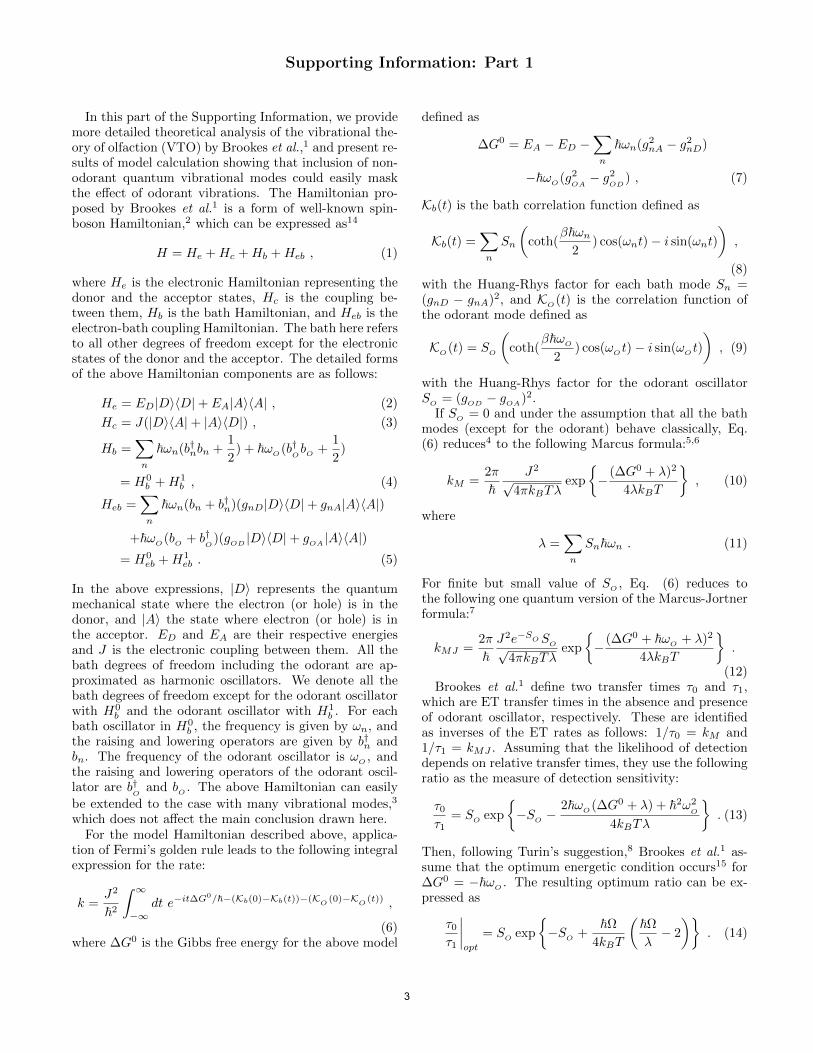
In this part of the Supporting Information, we providemore detailed theoretical analysis of the vibrational the-ory of olfaction (VTO) by Brookes et al.,1 and present re-sults of model calculation showing that inclusion of non-odorant quantum vibrational modes could easily maskthe effect of odorant vibrations. The Hamiltonian pro-posed by Brookes et al.1 is a form of well-known spin-boson Hamiltonian,2 which can be expressed as14
H = He +Hc +Hb +Heb , (1)
where He is the electronic Hamiltonian representing thedonor and the acceptor states, Hc is the coupling be-tween them, Hb is the bath Hamiltonian, and Heb is theelectron-bath coupling Hamiltonian. The bath here refersto all other degrees of freedom except for the electronicstates of the donor and the acceptor. The detailed formsof the above Hamiltonian components are as follows:
He = ED|DD|+ EA|AA| , (2)
Hc = J(|DA|+ |AD|) , (3)
Hb =
n
ωn(b†nbn +
1
2) + ω
O(b†
ObO+
1
2)
= H0b +H
1b , (4)
Heb =
n
ωn(bn + b†n)(gnD|DD|+ gnA|AA|)
+ωO(b
O+ b
†O)(g
OD|DD|+ g
OA|AA|)
= H0eb +H
1eb . (5)
In the above expressions, |D represents the quantummechanical state where the electron (or hole) is in thedonor, and |A the state where electron (or hole) is inthe acceptor. ED and EA are their respective energiesand J is the electronic coupling between them. All thebath degrees of freedom including the odorant are ap-proximated as harmonic oscillators. We denote all thebath degrees of freedom except for the odorant oscillatorwith H
0band the odorant oscillator with H
1b. For each
bath oscillator in H0b, the frequency is given by ωn, and
the raising and lowering operators are given by b†n and
bn. The frequency of the odorant oscillator is ωO, and
the raising and lowering operators of the odorant oscil-lator are b
†O
and bO. The above Hamiltonian can easily
be extended to the case with many vibrational modes,3
which does not affect the main conclusion drawn here.For the model Hamiltonian described above, applica-
tion of Fermi’s golden rule leads to the following integralexpression for the rate:
k =J2
2
∞
−∞dt e
−it∆G0/−(Kb(0)−Kb(t))−(K
O(0)−K
O(t))
,
(6)where ∆G
0 is the Gibbs free energy for the above model
defined as
∆G0 = EA − ED −
n
ωn(g2nA − g
2nD)
−ωO(g2
OA− g
2OD
) , (7)
Kb(t) is the bath correlation function defined as
Kb(t) =
n
Sn
coth(
βωn
2) cos(ωnt)− i sin(ωnt)
,
(8)with the Huang-Rhys factor for each bath mode Sn =(gnD − gnA)2, and K
O(t) is the correlation function of
the odorant mode defined as
KO(t) = S
O
coth(
βωO
2) cos(ω
Ot)− i sin(ω
Ot)
, (9)
with the Huang-Rhys factor for the odorant oscillatorS
O= (g
OD− g
OA)2.
If SO= 0 and under the assumption that all the bath
modes (except for the odorant) behave classically, Eq.(6) reduces4 to the following Marcus formula:5,6
kM =2π
J2
√4πkBTλ
exp
− (∆G
0 + λ)2
4λkBT
, (10)
where
λ =
n
Snωn . (11)
For finite but small value of SO, Eq. (6) reduces to
the following one quantum version of the Marcus-Jortnerformula:7
kMJ =2π
J2e−S
OSO√
4πkBTλexp
− (∆G
0 + ωO+ λ)2
4λkBT
.
(12)Brookes et al.1 define two transfer times τ0 and τ1,
which are ET transfer times in the absence and presenceof odorant oscillator, respectively. These are identifiedas inverses of the ET rates as follows: 1/τ0 = kM and1/τ1 = kMJ . Assuming that the likelihood of detectiondepends on relative transfer times, they use the followingratio as the measure of detection sensitivity:
τ0
τ1= S
Oexp
−S
O−
2ωO(∆G
0 + λ) + 2ω2O
4kBTλ
. (13)
Then, following Turin’s suggestion,8 Brookes et al.1 as-sume that the optimum energetic condition occurs15 for∆G
0 = −ωO. The resulting optimum ratio can be ex-
pressed as
τ0
τ1
opt
= SOexp
−S
O+
Ω4kBT
Ωλ
− 2
. (14)
3
2
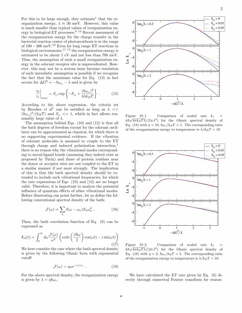
For this to be large enough, they estimate1 that the re-organization energy, λ ≈ 30 meV. However, this valueis much smaller than typical values of reorganization en-ergy in biological ET processes.9–13 Recent assessment ofthe reorganization energy for the charge transfer in thebacterial reaction center of photosynthesis is in the rangeof 100− 300 meV.10 Even for long range ET reactions inbiological environments,11–13 the reorganization energy isestimated to be about 1 eV and not less than 700 meV.Thus, the assumption of such a small reorganization en-ergy in the odorant receptor site is unprecedented. How-ever, this may not be a serious issue because resolutionof such unrealistic assumption is possible if we recognizethe fact that the maximum value for Eq. (13) in factoccurs for ∆G
0 = −ωO− λ and is given by
τ0
τ1
max
= SOexp
−S
O+
(ωO)2
4λkBT
. (15)
According to the above expression, the criteria setby Brookes et al1 can be satisfied as long as λ <<
(ωO)2/(kBT ) and S
O<< 1, which in fact allows rea-
sonably large value of λ.The assumption behind Eqs. (10) and (12) is that all
the bath degrees of freedom except for the odorant oscil-lator can be approximated as classical, for which there isno supporting experimental evidence. If the vibrationof odorant molecules is assumed to couple to the ETthrough charge and induced polarization interaction,1
there is no reason why the vibrational modes correspond-ing to metal-ligand bonds (assuming they indeed exist asproposed by Turin) and those of protein residues nearthe donor or acceptor sites are not coupled to the ET ina similar manner if not more strongly. The implicationof this is that the bath spectral density should be ex-tended to include such vibrational frequencies, for whichthe rate expressions of Eqs. (10) and (12) are no longervalid. Therefore, it is important to analyze the potentialinfluence of quantum effects of other vibrational modes.Before illustrating our point further, let us define the fol-lowing conventional spectral density of the bath:
J (ω) =
n
δ(ω − ωn)Snω2n . (16)
Then, the bath correlation function of Eq. (8) can beexpressed as
Kb(t) =
∞
0dω
J (ω)
ω2
coth
βω2
cos(ωt)− i sin(ωt)
(17)We here consider the case where the bath spectral densityis given by the following Ohmic form with exponentialcutoff:
J (ω) = ηωe−ω/ωc . (18)
For the above spectral density, the reorganization energyis given by λ = ηωc.
-8
-4
0
SO
= 0
SO
= 0.01
SO
= 0.05
-8
-4
0
Ln k
s
-1 0 1 2 3
!"G0/ #
-8
-4
0
h_ $
O/# = 0.5
h_ $
O/# = 1
h_ $
O/# = 2
Figure S1.1. Comparison of scaled rate ks =k
√4πkBTλ/(2πJ
2) for the Ohmic spectral density ofEq. (18) with η = 10, ωc/kBT = 1. The corresponding ratioof the reorganization energy to temperature is λ/kBT = 10.
-8
-4
0
SO
= 0
SO
= 0.01
SO
= 0.05
-8
-4
0
Ln k
s
-1 0 1 2 3
!"G0/ #
-8
-4
0
h_ $
O/# = 0.5
h_ $
O/# = 1
h_ $
O/# = 2
Figure S1.2. Comparison of scaled rate ks =k
√4πkBTλ/(2πJ
2) for the Ohmic spectral density ofEq. (18) with η = 2, ωc/kBT = 5. The corresponding ratioof the reorganization energy to temperature is λ/kBT = 10.
We have calculated the ET rate given by Eq. (6) di-rectly through numerical Fourier transform for reason-
4

3
able value of λ = 10kBT and for different values of η,ωc, and S
O. The results in Fig. S1.1 correspond to
the case where all the bath modes are classical or nearlyclassical. As implied by Eq. (15), we find the effectof coupling to odorant oscillator becomes significant forω
O≥ λ. However, even such enhancement becomes
obscure for quantum mechanical bath as shown in Fig.S1.2. The cutoff frequency of the bath in this case is fivetimes that of the thermal energy. Thus, assuming 300 K,such cutoff frequency amounts to a vibration of about1, 000 cm−1. Even for this moderate value of the cutofffrequency, Fig. S1.2 shows that the effect of odorant os-cillator does not stand out significantly even up to thevalue of ω
O≈ 4, 000 cm−1.
-8
-4
0
SO
= 0
SO
= 0.01
SO
= 0.05
-8
-4
0
Ln k
s
-1 0 1 2 3 4
!"G0/ #
-8
-4
0
h_ $
O/# = 0.5
h_ $
O/# = 1
h_ $
O/# = 2
Figure S1.3. Comparison of scaled rate ks =k
√4πkBTλ/(2πJ
2) for the spectral density of Eq. (19)with η = 10, ωc/kBT = 1, Sq = 0.1, and ωq/ωc = 15.The corresponding ratio of the reorganization energy totemperature is λ/kBT = 10.
Actual spectral densities are in general much morecomplex and, often, there are quantum modes well sepa-rated from classical modes. As a simple representation ofsuch case, we considered the following spectral density:
J (ω) = ηωe−ω/ωc + Sqω
2qδ(ω − ωq) . (19)
The total reorganization energy (but without includ-ing the effects of the odorant) in this case is given byλT = λ + Sqωq, where λ has been defined below Eq.(18). We have calculated the ET rates for the above spec-tral density once again by direct numerical calculationof Eq. (6) through Fourier transform. We have chosenSq = 0.1 and ωq/ωc = 15, while keeping all other param-eters the same as those for Fig. S1.1. The results shownin Fig. S1.3 demonstrate that the sensitivity observed inFig. S1.1 has disappeared in this case because of the ad-ditional quantum mode in the bath. Any actual spectraldensity in biological environments is expected to be muchmore complex than Eq. (19). Detecting the frequency ofthe odorant through different ETs, as proposed by theVTO, in such environment appears to be much more dif-ficult. In addition, such hypothetical detection efficiencyis likely to degrade further due to thermal fluctuationsand disorder.
In the main text, Fig. 3 provides the results of τ0/τ1,which is calculated by taking the ratio of the rate forfinite value of SO to that with SO = 0. These resultsconfirm that the proposed mechanism of VTO is effec-tive only for sufficiently large frequency of the odorantoscillator, ωO, for a given value of reorganization energy.Most importantly, even such enhancement can easily beoverridden once quantum vibrational modes in the bath(the donor/acceptor sites or other environments) becomeactive even by modest amount.
The importance of high frequency vibrational modesin the inverted regime of ET, where the proposed mech-anism of VTO is active, is a well established fact, in par-ticular, in biological environments. On the other hand,the key assumption of VTO1 as demonstrated here is thatsuch molecular vibrations of the environment do not con-tribute to the ET so that the spectral range of the bathis cleared for the detection of odorant frequency only.While such assumption may be reasonable for conven-tional inelastic tunneling spectroscopy involving metalelectrodes as the donor and the acceptor of the electron,it lacks clear justification in biological molecular envi-ronments. Moreover, even if the proposed ET involvedmetal atomic centers and the transferring electron (orhole) were significantly delocalized, the effects of non-odorant molecular environments near the electron (hole)centers would be more important than those of weaklybound odorant molecule.
1 Brookes JC, Hartoutsiou F, Horsfield AP, & Stoneham,AM (2007) Could humans recognize odor by phonon as-sisted tunneling? Phys. Rev. Lett. 98:038101.
2 Leggett AJ, Chakravarty S, Dorsey AT, Fisher MPA, GargA, & Zwerger W (1987) Dynamics of the dissipative 2-statesystem. Rev. Mod. Phys. 59:1.
3 Solov’yov IA, Chang PY, & Schulten K (2012) Vibra-tionally assisted electron transfer mechanism of olfaction:myth or reality? Phys. Chem. Chem. Phys. 14:13861.
4 Jang S & Newton MD (2006) Closed form expressionsof quantum electron transfer rate based on the stationaryphase approximation. J. Phys. Chem. B 110:18996.
5
4
5 Marcus RA (1956) On the theory of oxidation-reductionreactions involving electron transfer. I. J. Chem. Phys.24:966.
6 Marcus RA (1965) Theory of electron-transfer rates ofsolvated electrons. J. Chem. Phys. 43:679.
7 Jortner J (1976) Temperature dependent activation en-ergy for electron transfer between biological molecules. J.Chem. Phys. 64:4860.
8 Turin L (1996) A spectroscopic mechanism for primaryolfactory reception. Chem. Senses 21:773.
9 Moser CC, Keske JM, Warncke K, Faird RS, & DuttonPL (1992) Nature of biological electron transfer Nature796:355.
10 LeBard DN, Matyushov DV (2009) Energetics of Bacterial
Photosynthesis. J. Phys. Chem. B 113:12424.11 Gray HB and Winkler JR (1996) Electron transfer in pro-
teins Annu. Rev. Biochem. 65:537.12 Gray HB and Winkler JR (2005) Long-range electron
transfer Proc. Natl. Acad. Sci., USA 102:3534.13 Skourtis SS, Balabin IA, Kawatsu T, & Beratan DN (2005)
Protein dynamics and electron transfer: Electronic deco-herence and non-Condon effects Proc. Natl. Acad. Sci.,USA 102:3552.
14 We here adopt a different notation than that used byBrookes et al.
15 In fact, the energy difference EA−ED rather than the freeenergy of reaction is used in Ref. 1
6

Supporting Information. Part 2. Synthesis and Characterization of Deuterated Ligands
Cyclopentadecanol-d29 (2-d29). Cyclopentadecanone (1; 98%; 1 g, 4.46 mmol), rhodium on
carbon (200 mg), cyclohexane (0.8 mL) and D2O (27 mL) was stirred in a sealed tube under H2
atmosphere at 150 °C during 15 h (48). After filtration of the rhodium on Celite, the mixture was
diluted in saturated aqueous NH4Cl and extracted with CH2Cl2 ( 3). The organic layer was dried
and concentrated. GC-MS analysis showed the presence of 10:10:1 cyclopentadecanone:
cyclopentadecanol:cyclopentadecane (1:2:3; 1.2 g; 91% yield calculated for alcohol 2). The
material was then stirred without further purification in a sealed tube with fresh D2O (27 mL),
cyclohexane (0.8 mL) and rhodium on carbon (200 mg) under H2 atmosphere at 150 °C during
15 h. After dilution in saturated aqueous NH4Cl, the products are extracted by CH2Cl2. The
organic layer was dried, concentrated, and analyzed by GC-MS, revealing a mixture of cyclopen-
tadecanone-d28 (1-d28; 2%), cyclopentadecanol-d29 (2-d29; 94%) and cyclopentadecane-d30 (3-d30;
4%). The mixture was purified by column chromatography (hexanes:Et2O, 10:1), affording
cyclopentadecanol-d29 (2-d29) as a colorless solid (332 mg, 30% yield), mp 80-81 °C (compare
mp 2, 79-80 °C), with an overall deuterium content of 98.5% by GC-MS; m/z 253 (M+, 1%), m/z
236 (M+-DHO, 100%) (Fig. S2.1), IR 3279 (br, OH), 2992 (w, residual C–H), 2195, 2097 (C–D)
(Fig. S2.2), 1H NMR (400 MHz, CDCl3) 1.27 (d, J = 8.4 Hz); and cyclopentadecane-d30 (3-d30),
a colorless solid (87 mg, 8% yield), mp 52-55 °C (compare 3, mp 60-63 °C), 98% deuterated by
GC-MC, m/z 240 (Fig. S2.3), IR 2918, 2851 (w, residual C–H), 2194, 2097 (s, C–D) (Fig. S2.4); 1H NMR (400 MHz, CDCl3) 1.26; 13C NMR (100 MHz, CDCl3) 25.97, 25.78, 25.59, 25.41,
25.22.
Cyclopentadecanone-d28 (1-d28). Cyclopentadecanol-d29 (2-d29; 120 mg, 0.468 mmol) was
dissolved in 2:1 acetone:H2O (2:1; 6 mL). Then, a solution of K2Cr2O7 (65 mg/mL; 8.8 mL,
0.656 mmol, 1.4 eq) in aqueous H2SO4 (12.5% v/v) was added dropwise to the mixture with
stirring (49). The mxture was stirred at RT for 3 h, until a dark green coloration appeared. The
mixture was extracted by hexanes ( 3). The combined organic layers were dried (MgSO4) and
concentrated in vacuo affording cyclopentadecanone-d28 (1-d28), a colorless solid (91 mg; 77 %
yield) containing 98.9% deuterium as indicated by GC-MS m/z 252 (Fig. S2.5). Recrystallization
[H2O/MeOH (1:1)] gave colorless crystals, mp 61-62 °C (compare 1, mp 62-63 °C); weak 1H
NMR (400 MHz, CDCl3) 2.38 (d, J = 10.2 Hz, 5H), and 1.25 (s, 9H); a peak due to water at
7
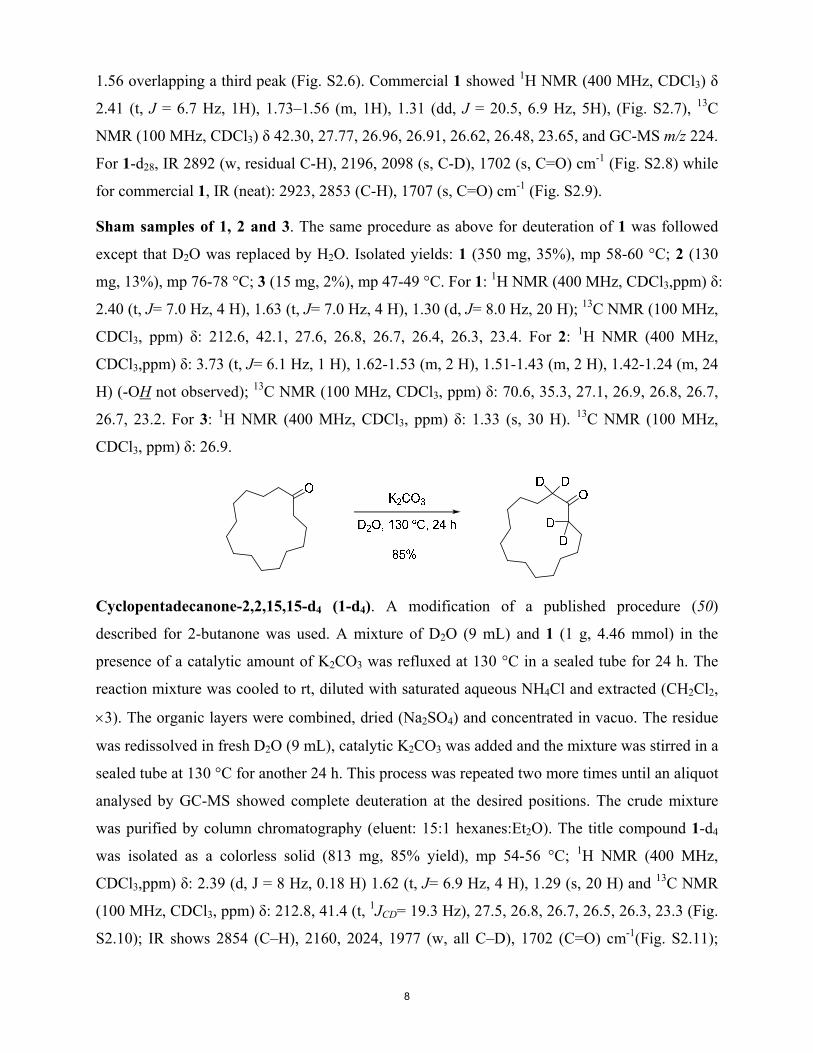
1.56 overlapping a third peak (Fig. S2.6). Commercial 1 showed 1H NMR (400 MHz, CDCl3)
2.41 (t, J = 6.7 Hz, 1H), 1.73–1.56 (m, 1H), 1.31 (dd, J = 20.5, 6.9 Hz, 5H), (Fig. S2.7), 13C
NMR (100 MHz, CDCl3) 42.30, 27.77, 26.96, 26.91, 26.62, 26.48, 23.65, and GC-MS m/z 224.
For 1-d28, IR 2892 (w, residual C-H), 2196, 2098 (s, C-D), 1702 (s, C=O) cm-1 (Fig. S2.8) while
for commercial 1, IR (neat): 2923, 2853 (C-H), 1707 (s, C=O) cm-1 (Fig. S2.9).
Sham samples of 1, 2 and 3. The same procedure as above for deuteration of 1 was followed
except that D2O was replaced by H2O. Isolated yields: 1 (350 mg, 35%), mp 58-60 °C; 2 (130
mg, 13%), mp 76-78 °C; 3 (15 mg, 2%), mp 47-49 °C. For 1: 1H NMR (400 MHz, CDCl3,ppm) :
2.40 (t, J= 7.0 Hz, 4 H), 1.63 (t, J= 7.0 Hz, 4 H), 1.30 (d, J= 8.0 Hz, 20 H); 13C NMR (100 MHz,
CDCl3, ppm) : 212.6, 42.1, 27.6, 26.8, 26.7, 26.4, 26.3, 23.4. For 2: 1H NMR (400 MHz,
CDCl3,ppm) : 3.73 (t, J= 6.1 Hz, 1 H), 1.62-1.53 (m, 2 H), 1.51-1.43 (m, 2 H), 1.42-1.24 (m, 24
H) (-OH not observed); 13C NMR (100 MHz, CDCl3, ppm) : 70.6, 35.3, 27.1, 26.9, 26.8, 26.7,
26.7, 23.2. For 3: 1H NMR (400 MHz, CDCl3, ppm) : 1.33 (s, 30 H). 13C NMR (100 MHz,
CDCl3, ppm) : 26.9.
Cyclopentadecanone-2,2,15,15-d4 (1-d4). A modification of a published procedure (50)
described for 2-butanone was used. A mixture of D2O (9 mL) and 1 (1 g, 4.46 mmol) in the
presence of a catalytic amount of K2CO3 was refluxed at 130 °C in a sealed tube for 24 h. The
reaction mixture was cooled to rt, diluted with saturated aqueous NH4Cl and extracted (CH2Cl2,
3). The organic layers were combined, dried (Na2SO4) and concentrated in vacuo. The residue
was redissolved in fresh D2O (9 mL), catalytic K2CO3 was added and the mixture was stirred in a
sealed tube at 130 °C for another 24 h. This process was repeated two more times until an aliquot
analysed by GC-MS showed complete deuteration at the desired positions. The crude mixture
was purified by column chromatography (eluent: 15:1 hexanes:Et2O). The title compound 1-d4
was isolated as a colorless solid (813 mg, 85% yield), mp 54-56 °C; 1H NMR (400 MHz,
CDCl3,ppm) : 2.39 (d, J = 8 Hz, 0.18 H) 1.62 (t, J= 6.9 Hz, 4 H), 1.29 (s, 20 H) and 13C NMR
(100 MHz, CDCl3, ppm) : 212.8, 41.4 (t, 1JCD= 19.3 Hz), 27.5, 26.8, 26.7, 26.5, 26.3, 23.3 (Fig.
S2.10); IR shows 2854 (C–H), 2160, 2024, 1977 (w, all C–D), 1702 (C=O) cm-1(Fig. S2.11);
8
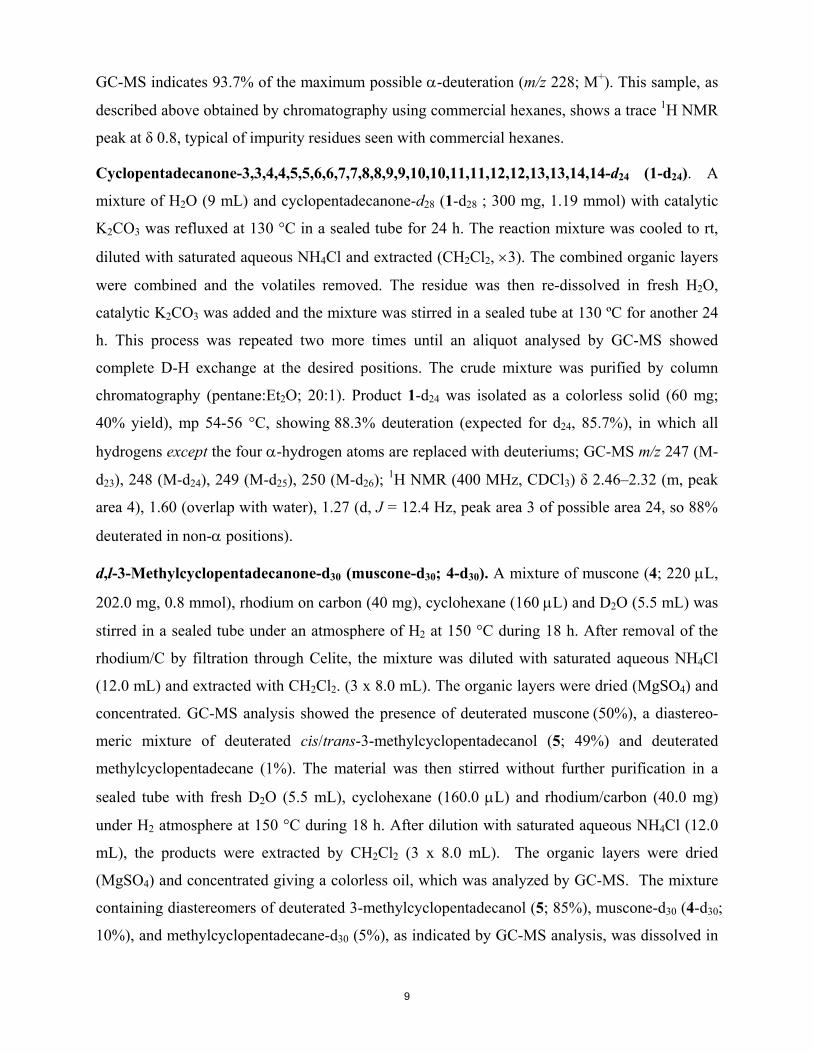
GC-MS indicates 93.7% of the maximum possible -deuteration (m/z 228; M+). This sample, as
described above obtained by chromatography using commercial hexanes, shows a trace 1H NMR
peak at 0.8, typical of impurity residues seen with commercial hexanes.
Cyclopentadecanone-3,3,4,4,5,5,6,6,7,7,8,8,9,9,10,10,11,11,12,12,13,13,14,14-d24 (1-d24). A
mixture of H2O (9 mL) and cyclopentadecanone-d28 (1-d28 ; 300 mg, 1.19 mmol) with catalytic
K2CO3 was refluxed at 130 °C in a sealed tube for 24 h. The reaction mixture was cooled to rt,
diluted with saturated aqueous NH4Cl and extracted (CH2Cl2, 3). The combined organic layers
were combined and the volatiles removed. The residue was then re-dissolved in fresh H2O,
catalytic K2CO3 was added and the mixture was stirred in a sealed tube at 130 ºC for another 24
h. This process was repeated two more times until an aliquot analysed by GC-MS showed
complete D-H exchange at the desired positions. The crude mixture was purified by column
chromatography (pentane:Et2O; 20:1). Product 1-d24 was isolated as a colorless solid (60 mg;
40% yield), mp 54-56 °C, showing 88.3% deuteration (expected for d24, 85.7%), in which all
hydrogens except the four -hydrogen atoms are replaced with deuteriums; GC-MS m/z 247 (M-
d23), 248 (M-d24), 249 (M-d25), 250 (M-d26); 1H NMR (400 MHz, CDCl3) 2.46–2.32 (m, peak
area 4), 1.60 (overlap with water), 1.27 (d, J = 12.4 Hz, peak area 3 of possible area 24, so 88%
deuterated in non- positions).
d,l-3-Methylcyclopentadecanone-d30 (muscone-d30; 4-d30). A mixture of muscone (4; 220 L,
202.0 mg, 0.8 mmol), rhodium on carbon (40 mg), cyclohexane (160 L) and D2O (5.5 mL) was
stirred in a sealed tube under an atmosphere of H2 at 150 °C during 18 h. After removal of the
rhodium/C by filtration through Celite, the mixture was diluted with saturated aqueous NH4Cl
(12.0 mL) and extracted with CH2Cl2. (3 x 8.0 mL). The organic layers were dried (MgSO4) and
concentrated. GC-MS analysis showed the presence of deuterated muscone (50%), a diastereo-
meric mixture of deuterated cis/trans-3-methylcyclopentadecanol (5; 49%) and deuterated
methylcyclopentadecane (1%). The material was then stirred without further purification in a
sealed tube with fresh D2O (5.5 mL), cyclohexane (160.0 L) and rhodium/carbon (40.0 mg)
under H2 atmosphere at 150 °C during 18 h. After dilution with saturated aqueous NH4Cl (12.0
mL), the products were extracted by CH2Cl2 (3 x 8.0 mL). The organic layers were dried
(MgSO4) and concentrated giving a colorless oil, which was analyzed by GC-MS. The mixture
containing diastereomers of deuterated 3-methylcyclopentadecanol (5; 85%), muscone-d30 (4-d30;
10%), and methylcyclopentadecane-d30 (5%), as indicated by GC-MS analysis, was dissolved in
9

acetone:H2O (6 mL; 2:1 ratio). A solution of K2Cr2O7 (65 mg/mL) in H2O/H2SO4 (6 mL; 12.5 %
v/v) was then added dropwise to the mixture. The flask was stirred at 45 °C for 2 h, until the
reaction mixture became dark green. After cooling to RT, the mixture was extracted by hexanes
(3 x 8.0 mL). The combined organic layers are dried (MgSO4) and evaporated under reduced
pressure. Muscone-d30, 4-d30, is obtained as a colorless oil (90 mg; 42% yield) after column
chromatography (10:1 hexanes:Et2O); 96.7% deuterated according to GC-MS analysis, m/z 268
(Fig. S2.12; compare m/z 238 for muscone 4, Fig. S2.13); IR (neat) 2197, 2099 (C–D), 1704
(C=O) cm-1 which can be compared with the IR (neat) 2925, 2856 (C–H), 1710 (C=O) cm-1 for
undeuterated 4 (Fig. 2)]; NMR similar to, but much weaker, than that of undeuterated 4 (Fig.
S2.14).
Chloromethyl methyl sulfide-d5. Dimethyl sulfoxide-d6 (25.0 g, 0.3 mol) was dissolved in
CH2Cl2 (20 mL) and was slowly added to a heated solution of thionyl chloride (23.7 mL, 0.33
mmol, 1.1 eq) in CH2Cl2 (60 mL). The reaction mixture was stored overnight at room
temperature and distilled to give the title compound as a light yellow liquid (30 g, 98%), bp 104-
106 °C ; 13C-NMR (100 MHz, CDCl3) 51.68, 14.27; GC-MS m/z 101 (C2D5S35Cl), 103
(C2D5S37Cl); GC-MS indicated ~100% deuteration (Fig. S2.15).
(Methylthio)methanethiol-d5 (8-d5). Thiourea (9.3 g, 126 mmol) was added to a solution of
chloromethyl methyl sulfide-d5 (12.5 g, 126 mmol) in dry acetone (100 mL). Cloudiness began
to appear in 5 min. The solution was stored overnight at room temperature, and then cooled in
ice, whereupon an oil that separated turned into a white solid. The solid was filtered and used
without any further purification in the next step. (Methylthiomethyl)-d5 isothiuronium chloride
(12.5 g, 70.6 mmol) was hydrolyzed by stirring with NaOH (5 N, 2.25 eq) at room temperature
for 4 h. The oil that separated as an upper layer was extracted with several portions of peroxide-
free ether. The combined ether layers were washed (H2O) and dried (Na2SO4). After filtration,
the solvent was removed by distillation through a Vigreux column in a nitrogen atmosphere at a
bath temperature of 45-50 °C affording the product as a clear, mobile liquid with a sharp, pene-
trating garlic-like in 40% yield; 1H-NMR (400 MHz, CDCl3) 1.94 (s); 13C-NMR (100 MHz,
CDCl3) 23.92, 13.98; m/z 99.0 (C2D5HS2); GC-MS showed ~100% d5 deuteration (Fig. S2.16).
2,4,5,7-Tetrathiaoctane-d10 (9-d10). Iodine (388 mg, 1.33 mmol) in benzene (10 mL) was added
to a solution of (methylthio)methanethiol-d5 (8-d5; 250 mg, 2.66 mmol) and triethylamine (0.37
mL, 2.66 mmol) in benzene (2 mL) at 0 °C. The solution was stirred for 12 h at rt. The reaction
10

mixture was filtered and the filtrate was washed (H2O) and concentrated to give 9-d10 (200 mg); 13C NMR (100 MHz, CDCl3) 45.32, 15.40; m/z 196.10 (C4D10S4); GC-MS indicated ~100%
deuteration (Fig. S2.17).
11
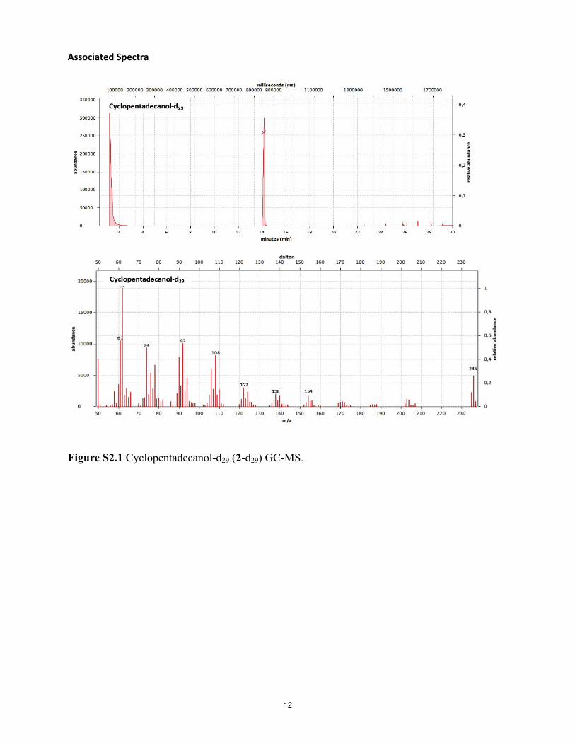
Associated Spectra
Figure S2.1 Cyclopentadecanol-d29 (2-d29) GC-MS.
12

Figure S2.2 Cyclopentadecanol-d29 (2-d29) IR spectrum.
13

Figure S2.3 Cyclopentadecane-d30 (3-d30) GC-MS.
14
Figure S2.4 Cyclopentadecane-d30 (3-d30) IR spectrum.
15

Figure S2.5 Cyclopentadecanone-d28 (1-d28) GC-MS.
16
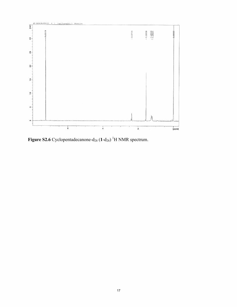
Figure S2.6 Cyclopentadecanone-d28 (1-d28) 1H NMR spectrum.
17
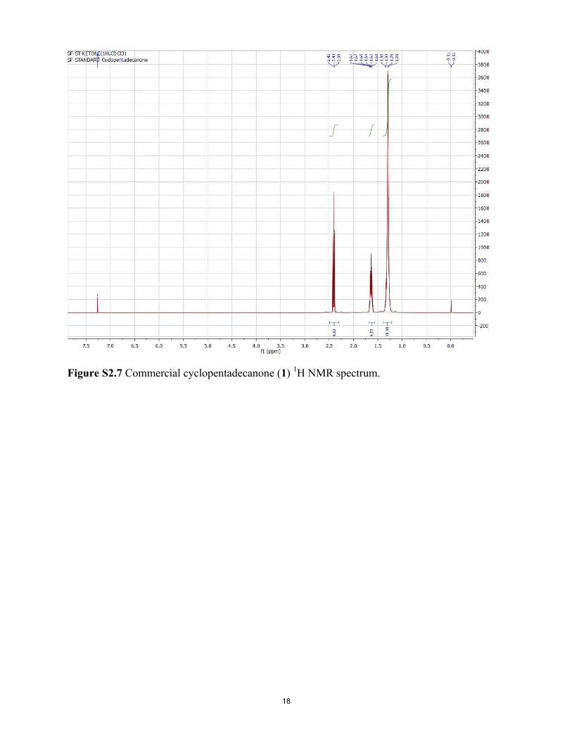
Figure S2.7 Commercial cyclopentadecanone (1) 1H NMR spectrum.
18
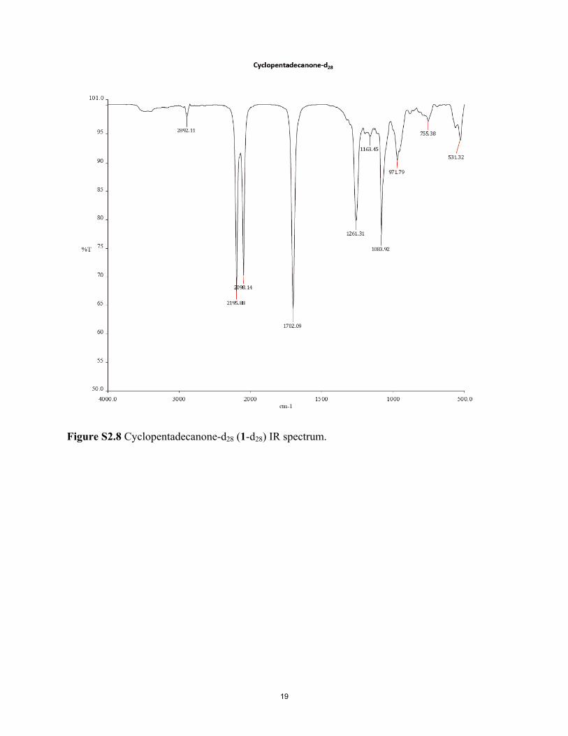
Figure S2.8 Cyclopentadecanone-d28 (1-d28) IR spectrum.
19

Figure S2.9 Cyclopentadecanone (commercial; 1) IR spectrum.
20
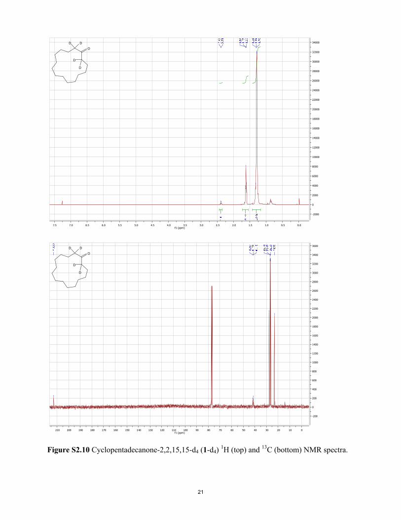
0.00.51.01.52.02.53.03.54.04.55.05.56.06.57.07.5f1 (ppm)
-2000
0
2000
4000
6000
8000
10000
12000
14000
16000
18000
20000
22000
24000
26000
28000
30000
32000
34000
O
DD
D
D
0102030405060708090100110120130140150160170180190200210f1 (ppm)
-200
0
200
400
600
800
1000
1200
1400
1600
1800
2000
2200
2400
2600
2800
3000
3200
3400
3600
O
DD
D
D
Figure S2.10 Cyclopentadecanone-2,2,15,15-d4 (1-d4) 1H (top) and 13C (bottom) NMR spectra.
21

Figure S2.11 Cyclopentadecanone-2,2,15,15-d4 (1-d4) IR spectrum.
22
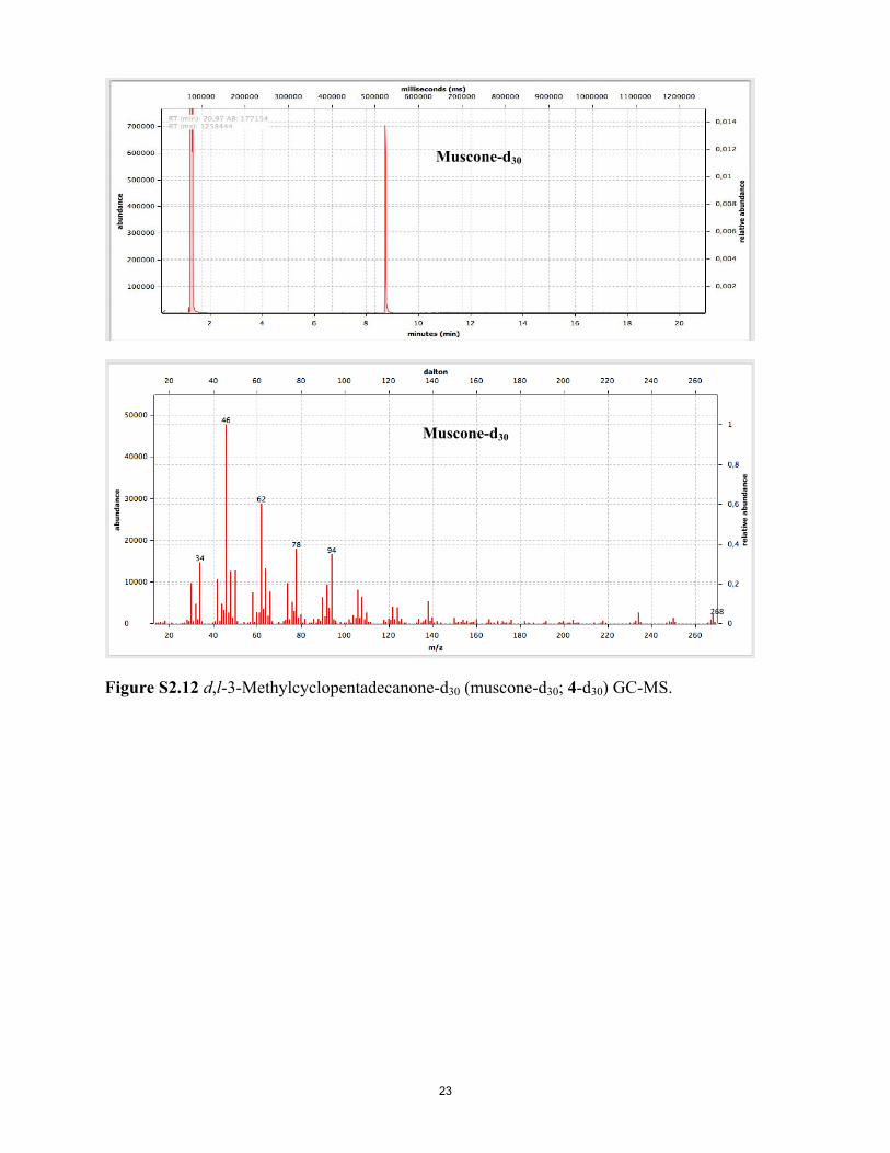
Muscone-d30
Muscone-d30
268
Figure S2.12 d,l-3-Methylcyclopentadecanone-d30 (muscone-d30; 4-d30) GC-MS.
23
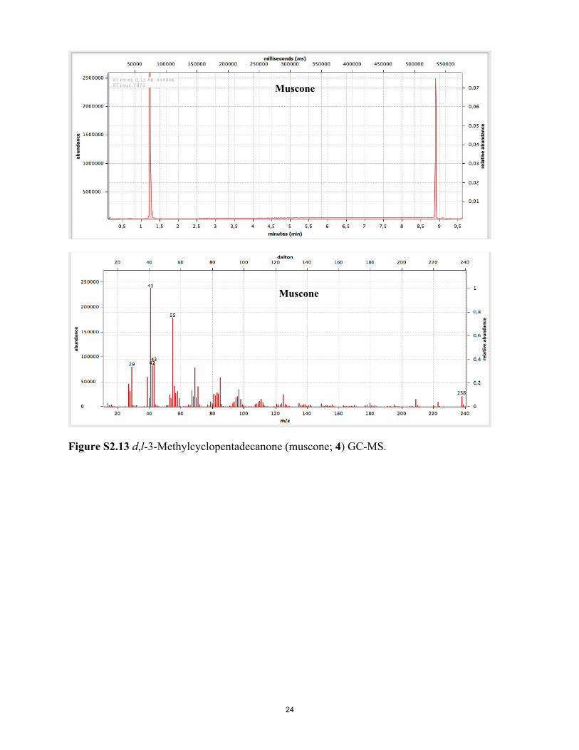
Muscone
Muscone
238
Figure S2.13 d,l-3-Methylcyclopentadecanone (muscone; 4) GC-MS.
24
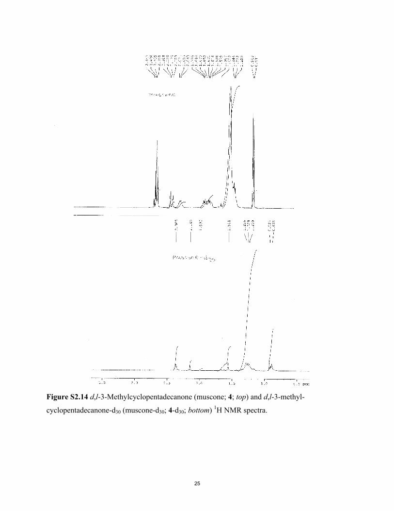
Figure S2.14 d,l-3-Methylcyclopentadecanone (muscone; 4; top) and d,l-3-methyl-
cyclopentadecanone-d30 (muscone-d30; 4-d30; bottom) 1H NMR spectra.
25
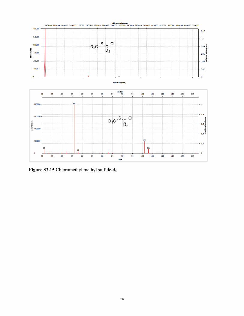
Figure S2.15 Chloromethyl methyl sulfide-d5.
26
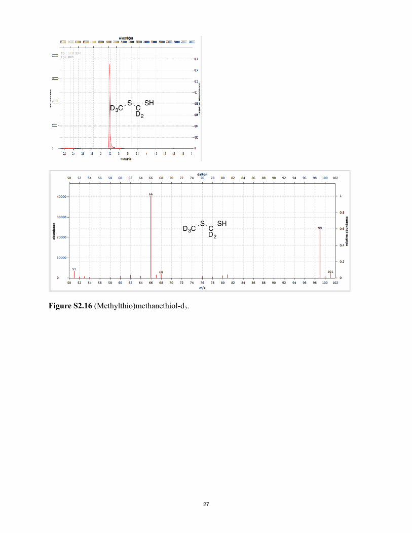
Figure S2.16 (Methylthio)methanethiol-d5.
27
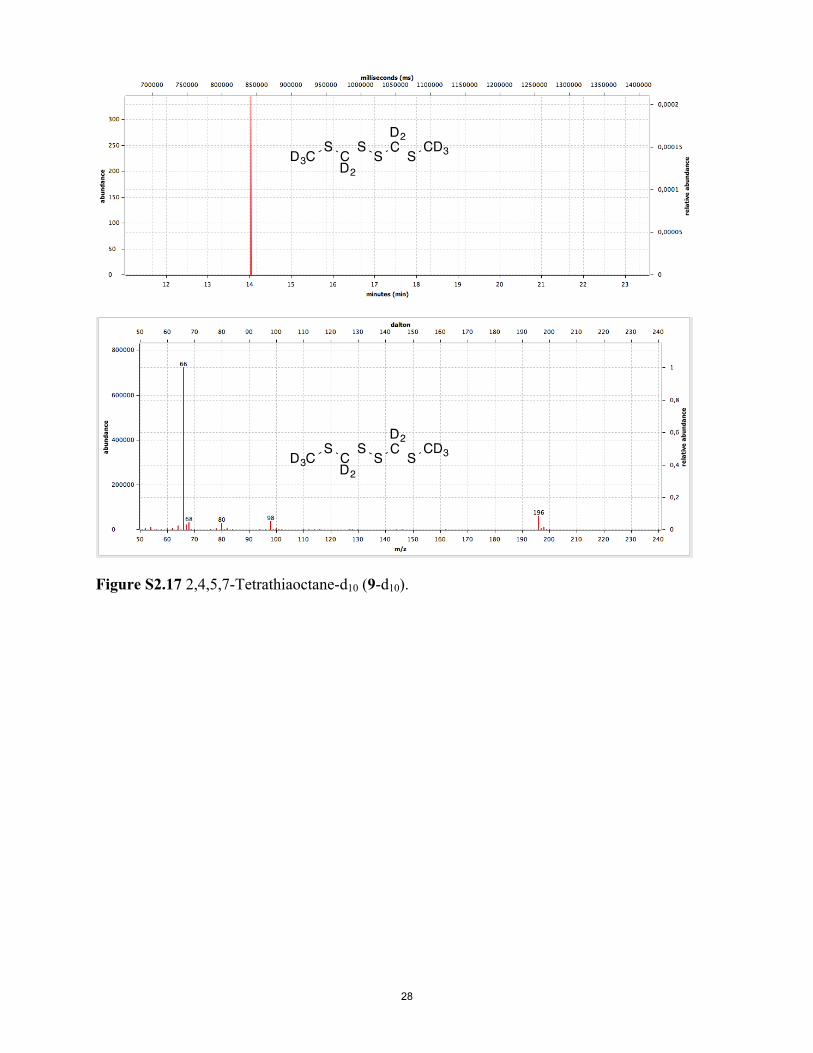
Figure S2.17 2,4,5,7-Tetrathiaoctane-d10 (9-d10).
28

Supporting Information. Part 3. Screening for human cyclopentadecanone receptor and
the response of various ORs to isotopomers
Figure S3.1. Screening for human ORs for all four isotopomers of cyclopentadecanone done using luciferase assays of 330 unique human odorant receptors. Normalized responses are shown as mean ±S.E.M. (N = 3). OR51E1 was determined to be a non-responsive OR in the follow-up confirmation experiments due to a high receptor background.
29
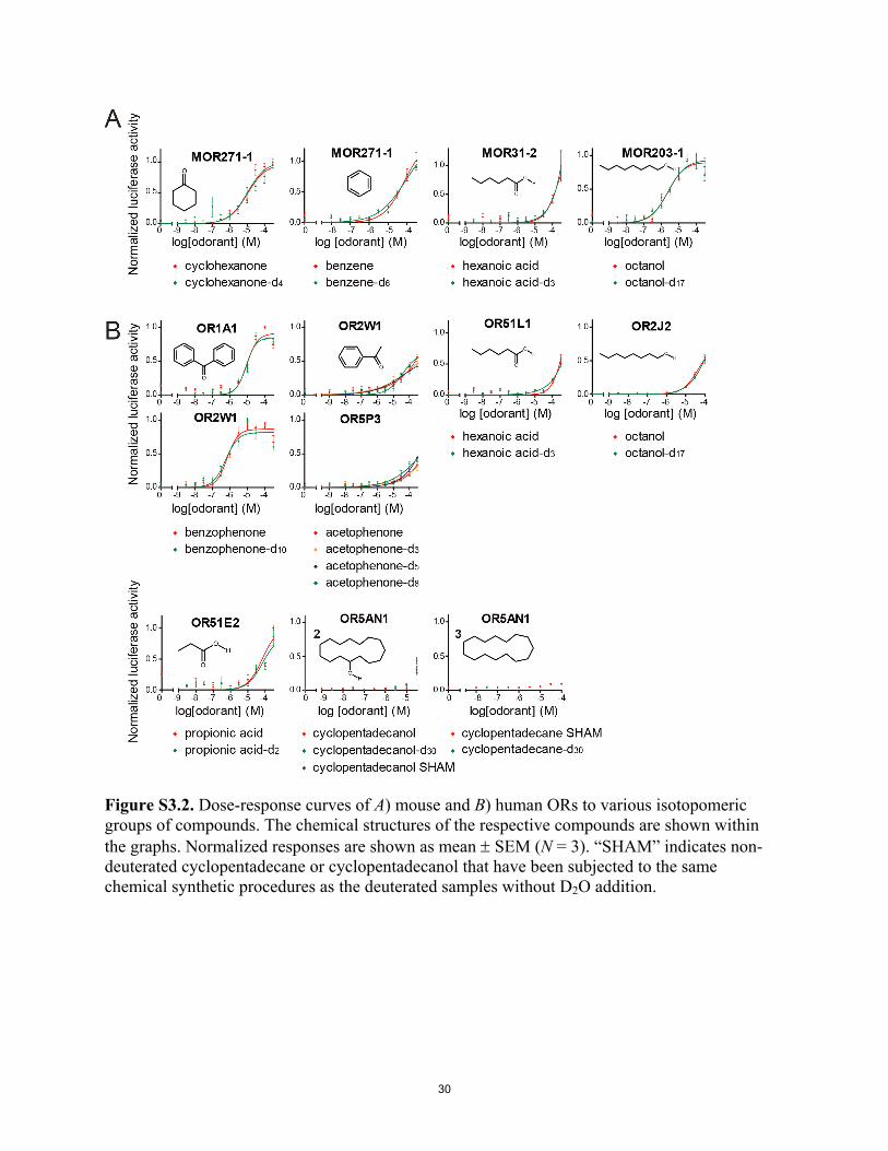
Figure S3.2. Dose-response curves of A) mouse and B) human ORs to various isotopomeric groups of compounds. The chemical structures of the respective compounds are shown within the graphs. Normalized responses are shown as mean SEM (N = 3). “SHAM” indicates non-deuterated cyclopentadecane or cyclopentadecanol that have been subjected to the same chemical synthetic procedures as the deuterated samples without D2O addition.
30

Figure S3.3. Response of human OR5AN1 to representative musk compounds, including cyclopentadecanone (1), cyclopentadecanol (2), muscone (4), and -pentadecalactone. Normalized responses are shown as mean SEM (N = 3).
31
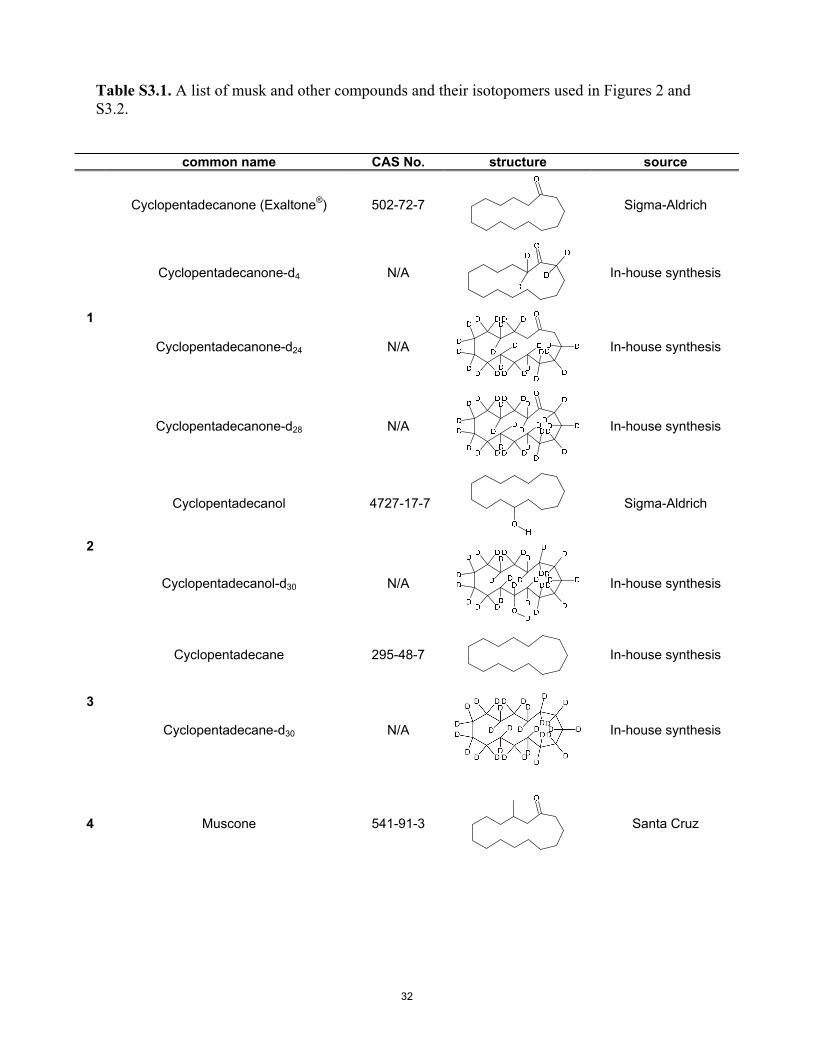
Table S3.1. A list of musk and other compounds and their isotopomers used in Figures 2 and S3.2.
common name CAS No. structure source
Cyclopentadecanone (Exaltone®) 502-72-7
Sigma-Aldrich
Cyclopentadecanone-d4 N/A
In-house synthesis
Cyclopentadecanone-d24 N/A
In-house synthesis
1
Cyclopentadecanone-d28 N/A
In-house synthesis
Cyclopentadecanol
4727-17-7 Sigma-Aldrich
2
Cyclopentadecanol-d30 N/A
In-house synthesis
Cyclopentadecane 295-48-7
In-house synthesis
3
Cyclopentadecane-d30 N/A
In-house synthesis
4 Muscone 541-91-3
Santa Cruz
32
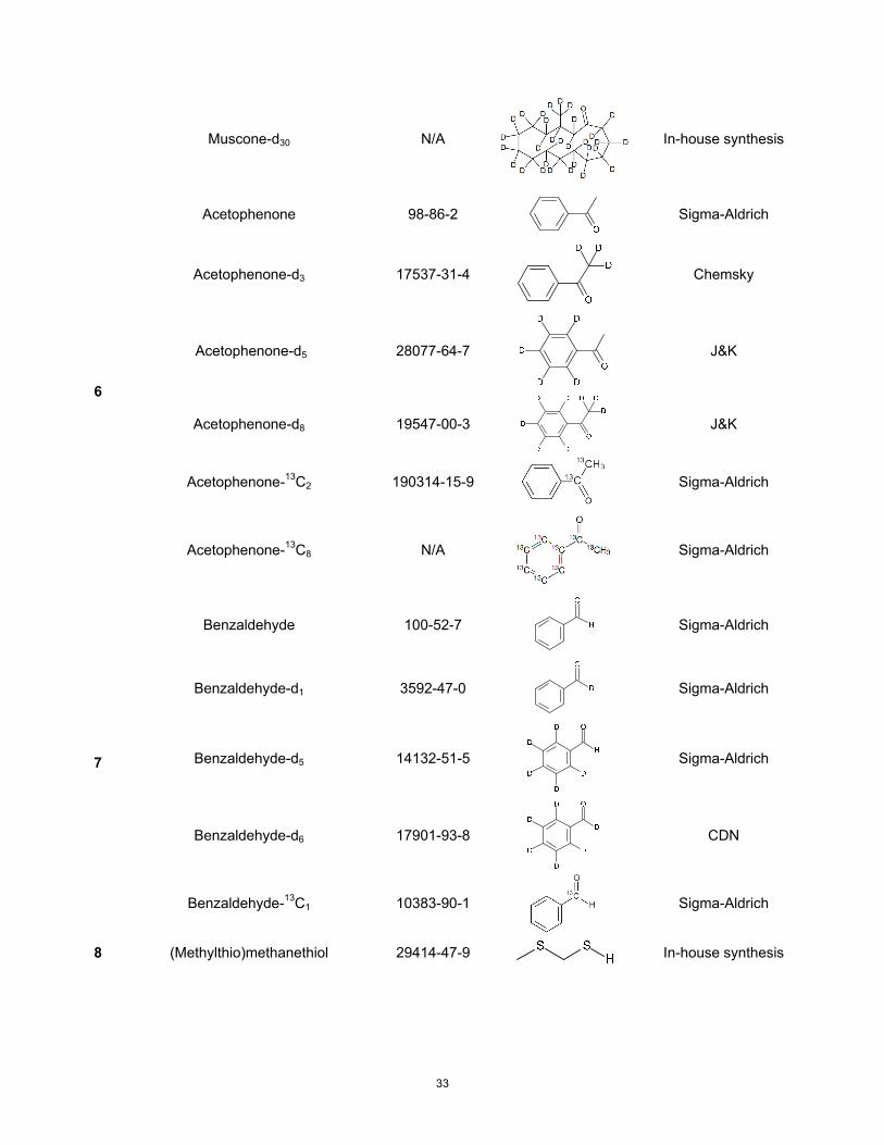
Muscone-d30 N/A
In-house synthesis
Acetophenone 98-86-2
Sigma-Aldrich
Acetophenone-d3 17537-31-4
Chemsky
Acetophenone-d5 28077-64-7
J&K
Acetophenone-d8 19547-00-3
J&K
Acetophenone-13C2 190314-15-9
Sigma-Aldrich
6
Acetophenone-13C8 N/A
Sigma-Aldrich
Benzaldehyde 100-52-7
Sigma-Aldrich
Benzaldehyde-d1 3592-47-0
Sigma-Aldrich
Benzaldehyde-d5 14132-51-5
Sigma-Aldrich
Benzaldehyde-d6 17901-93-8
CDN
7
Benzaldehyde-13C1 10383-90-1
Sigma-Aldrich
8 (Methylthio)methanethiol 29414-47-9
In-house synthesis
33
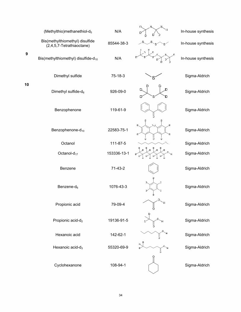
(Methylthio)methanethiol-d5 N/A
In-house synthesis
Bis(methylthiomethyl) disulfide (2,4,5,7-Tetrathiaoctane) 85544-38-3 In-house synthesis
9 Bis(methylthiomethyl) disulfide-d10 N/A
In-house synthesis
Dimethyl sulfide 75-18-3
Sigma-Aldrich
10
Dimethyl sulfide-d6 926-09-0
Sigma-Aldrich
Benzophenone 119-61-9
Sigma-Aldrich
Benzophenone-d10 22583-75-1
Sigma-Aldrich
Octanol 111-87-5 Sigma-Aldrich
Octanol-d17 153336-13-1
Sigma-Aldrich
Benzene 71-43-2
Sigma-Aldrich
Benzene-d6 1076-43-3
Sigma-Aldrich
Propionic acid 79-09-4
Sigma-Aldrich
Propionic acid-d2 19136-91-5
Sigma-Aldrich
Hexanoic acid 142-62-1
Sigma-Aldrich
H 3 5532
exanoic acid-d 0-69-9
Sigma-Aldrich
Cyclohexanone 108-94-1
Sigma-Aldrich
34

C yclohexanone-d4 1006-03-7
Sigma-Aldrich
Boldface numbers in the leftmost column represent numbers used in the text.
35

Table S3.2. Pairwise comparisons between the dosage response curves for the original odorant and its deuterated or 13C isotopomers. A. Comparisons of dosage response curves in Figures 3 and 4.
OR odorant EC50 F value P value OR5AN1 Cyclopentadecanone-H 3.49
Cyclopentadecanone-d4 4.29 1.448 17.556 Cyclopentadecanone-d24 3.90 0.7283 36.2934 Cyclopentadecanone-d28 3.62 0.1661 60.5616 Cyclopentadecanone-SHAM 3.11 0.1030 63.1686
OR5AN1 Muscone-H 9.62 Muscone-d30 17.53 5.588 1.551
MOR244-3 (Methylthio)methanethiol 1.58 (Methylthio)methanethiol-d5 1.61 1.594 16.005
MOR244-3 Bis(methylthiomethyl)disulfide 0.21 Bis(methylthiomethyl)disulfide-d10 1.60 2.183 9.4314
MOR244-3 Dimethyl sulfide 4.31 Dimethyl sulfide-d6 5.55 0.6118 36.6036
MOR162-1 Acetophenone-H 6.17 Acetophenone-d3 3.99 1.722 13.4772 Acetophenone-d5 5.14 0.5139 39.9894 Acetophenone-d8 5.40 3.717 2.7984 Acetophenone-13C2 9.15 9.221 0.099 Acetophenone-13C8 6.13 1.357 18.4932
MOR41-1 Acetophenone-H 5.46 Acetophenone-d3 5.47 1.581 15.378 Acetophenone-d5 3.27 0.8486 29.337 Acetophenone-d8 3.41 2.909 5.3064 Acetophenone-13C2 7.70 0.7576 31.8912 Acetophenone-13C8 9.68 1.483 16.7178
MOR129-1 Acetophenone-H 2.29 Acetophenone-d3 2.51 0.2094 53.6448 Acetophenone-d5 1.49 0.8262 29.8386 Acetophenone-d8 0.60 7.136 0.3036 Acetophenone-13C2 0.86 3.912 2.4288 Acetophenone-13C8 1.19 1.431 17.325
MOR271-1 Acetophenone-H 32.43 Acetophenone-d3 33.54 0.04510 63.096 Acetophenone-d5 43.91 1.022 24.9348 Acetophenone-d8 40.23 0.5906 37.1778 Acetophenone-13C2 57.28 4.311 1.8348 Acetophenone-13C8 45.04 1.859 11.9988
MOR106-1 Acetophenone-H 59.35 Acetophenone-d3 44.32 0.8513 29.1588 Acetophenone-d5 22.79 4.121 2.0922 Acetophenone-d8 39.84 0.9542 26.532 Acetophenone-13C2 65.61 0.04723 62.964 Acetophenone-13C8 55.63 0.1664 55.9614
36
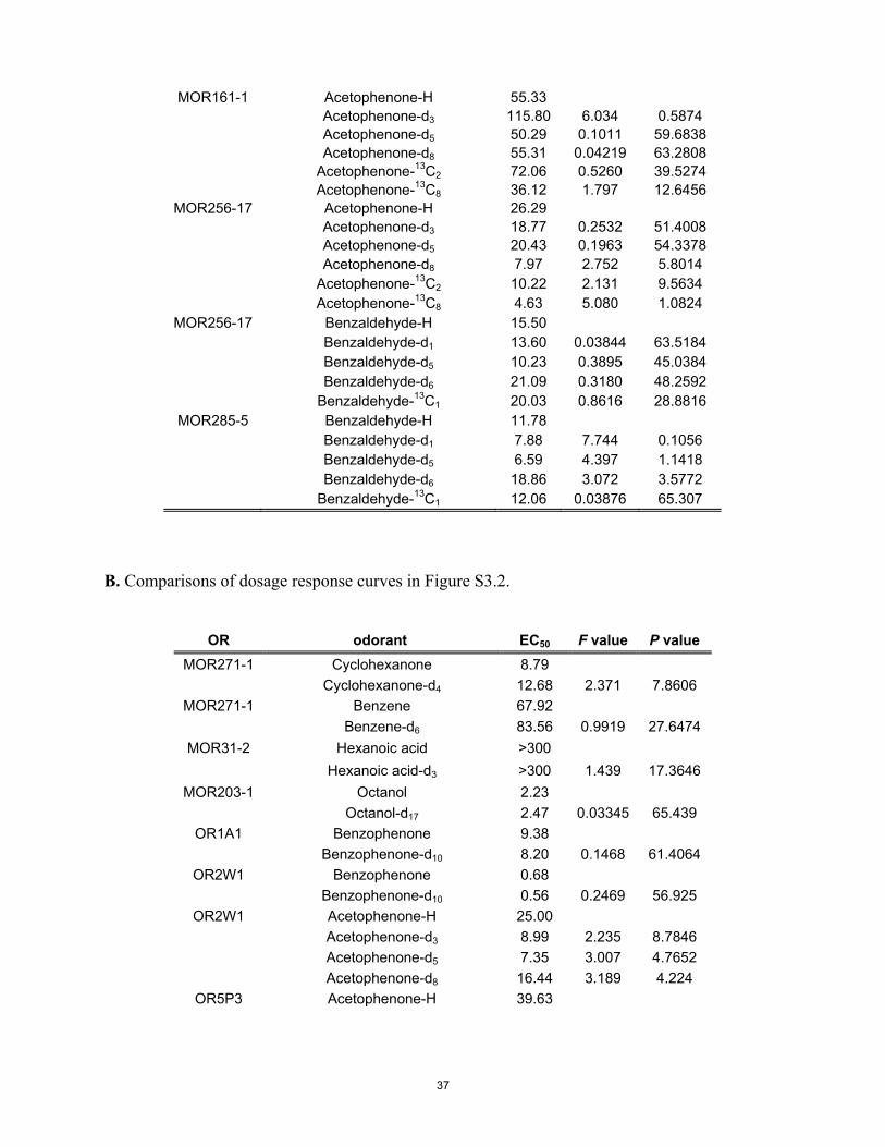
MOR161-1 Acetophenone-H 55.33 Acetophenone-d3 115.80 6.034 0.5874 Acetophenone-d5 50.29 0.1011 59.6838 Acetophenone-d8 55.31 0.04219 63.2808 Acetophenone-13C2 72.06 0.5260 39.5274 Acetophenone-13C8 36.12 1.797 12.6456
MOR256-17 Acetophenone-H 26.29 Acetophenone-d3 18.77 0.2532 51.4008 Acetophenone-d5 20.43 0.1963 54.3378 Acetophenone-d8 7.97 2.752 5.8014 Acetophenone-13C2 10.22 2.131 9.5634 Acetophenone-13C8 4.63 5.080 1.0824
MOR256-17 Benzaldehyde-H 15.50 Benzaldehyde-d1 13.60 0.03844 63.5184 Benzaldehyde-d5 10.23 0.3895 45.0384 Benzaldehyde-d6 21.09 0.3180 48.2592 Benzaldehyde-13C1 20.03 0.8616 28.8816
MOR285-5 Benzaldehyde-H 11.78 Benzaldehyde-d1 7.88 7.744 0.1056 Benzaldehyde-d5 6.59 4.397 1.1418 Benzaldehyde-d6 18.86 3.072 3.5772 Benzaldehyde-13C1 12.06 0.03876 65.307
B. Comparisons of dosage response curves in Figure S3.2.
OR odorant EC50 F value P value MOR271-1 Cyclohexanone 8.79
Cyclohexanone-d4 12.68 2.371 7.8606 MOR271-1 Benzene 67.92
Benzene-d6 83.56 0.9919 27.6474 MOR31-2 Hexanoic acid >300
Hexanoic acid-d3 >300 1.439 17.3646 MOR203-1 Octanol 2.23
Octanol-d17 2.47 0.03345 65.439 OR1A1 Benzophenone 9.38
Benzophenone-d10 8.20 0.1468 61.4064 OR2W1 Benzophenone 0.68
Benzophenone-d10 0.56 0.2469 56.925 OR2W1 Acetophenone-H 25.00
Acetophenone-d3 8.99 2.235 8.7846 Acetophenone-d5 7.35 3.007 4.7652 Acetophenone-d8 16.44 3.189 4.224
OR5P3 Acetophenone-H 39.63
37

Acetophenone-d3 25.53 0.5257 39.5406 Acetophenone-d5 48.87 0.2926 49.467 Acetophenone-d8 24.60 1.530 15.8928
OR51L1 Hexanoic acid >300 Hexanoic acid-d3 >300 2.302 8.316
OR2J2 Octanol >300 Octanol-d17 >300 2.474 6.2436
OR51E2 Propionic acid 72.44 Propionic acid-d2 62.37 0.04077 63.3666
OR5AN1 Cyclopentadecane SHAM Cyclopentadecane-d30
No curve fit
OR5AN1 Cyclopentadecanol-H Cyclopentadecanol-d30 Cyclopentadecanol SHAM
No curve fit
EC50 values are given in M. F values and P values are shown for the isotopmers compared.
Before Bonferroni correction: * P < 0.00076. After Bonferroni correction: * P < 0.05.
38
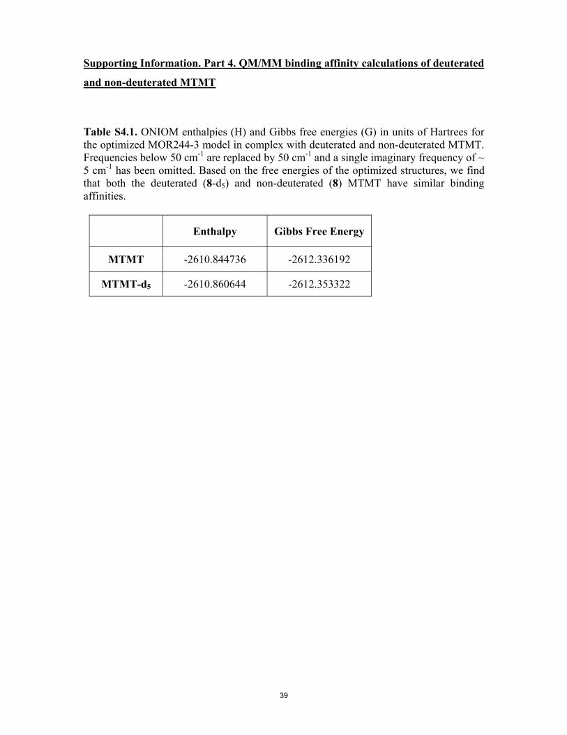
Supporting Information. Part 4. QM/MM binding affinity calculations of deuterated
and non-deuterated MTMT
Table S4.1. ONIOM enthalpies (H) and Gibbs free energies (G) in units of Hartrees for the optimized MOR244-3 model in complex with deuterated and non-deuterated MTMT. Frequencies below 50 cm-1 are replaced by 50 cm-1 and a single imaginary frequency of ~ 5 cm-1 has been omitted. Based on the free energies of the optimized structures, we find that both the deuterated (8-d5) and non-deuterated (8) MTMT have similar binding affinities.
Enthalpy Gibbs Free Energy
MTMT -2610.844736 -2612.336192
MTMT-d5 -2610.860644 -2612.353322
39

Calculating Isotope Effects (EIEs)
Deuterium Isotope Effect
= KH/KD
= exp [- GH/RT] / exp [- GD/RT]
= exp [( GD- GH)/RT]
= exp [((GD-GH)bound -(GD-GH)free)/RT]
= 0.94 for MTMT
where “bound” refers to substrate bound in receptor active site and “free” refers to
molecule as itself in gas phase. The relevant Gibbs free energy values employed for EIE
calculations are presented in Table S4.2.
Table S4.2. Gibbs free energies (G) in units of Hartrees for undeuterated (GH) and deuterated (GD) ligand as itself (free) and bound in receptor active site (bound) with associated deuterium isotope effects (DIEs).
GH GD
Free Bound Free Bound DIE
MTMT -876.088348 -2612.336192 -876.105415 -2612.353322 0.94
40