impact of the complement system and gab3 on natural killer
TRANSCRIPT

From the INSTITUTE FOR SYSTEMIC INFLAMMATION RESEARCH
OF THE UNIVERSITY OF LÜBECK
Director: PROF. DR. med. JÖRG KÖHL
Impact of the complement system and Gab3 on
natural killer cells during Toxoplasma gondii
infection
Dissertation
for Fulfillment of the Requirements for the Doctoral Degree
of the University of Lübeck
From the Department of Natural Sciences
Submitted by:
Fabian Mey
From Minden
Lübeck 2019

First referee: Prof. Dr. med. Jörg Köhl
Second referee: Prof. Dr. rer. nat. Ulrich Schaible
Date of oral examination: 9th October 2019
Approved for printing: 9th October 2019

I
List of contents
Summary............................................................................................................................................ V
Zusammenfassung ........................................................................................................................... VII
1 Introduction ................................................................................................................ 1
1.1 The immune system ....................................................................................................... 1
1.1.1 Innate and adaptive immunity ....................................................................................................... 1
1.1.2 The complement system ................................................................................................................ 4
1.1.2.1 Activation of the complement system ................................................................................... 5
1.1.2.2 Regulation of the complement system .................................................................................. 8
1.1.2.3 The anaphylatoxin C5a and its two receptors ........................................................................ 8
1.2 IL-12 cytokine family .................................................................................................... 11
1.3 Natural killer cells: key players during early innate immune responses.......................... 12
1.3.1 Natural killer cell activation and pathogen clearance ................................................................... 12
1.3.2 Dendritic cell – mediated NK cell activation via stimulatory synapse formation ........................... 14
1.4 Toxoplasma gondii: an intracellular parasite ................................................................ 15
1.4.1 Biology and life cycle of Toxoplasma gondii ................................................................................. 15
1.4.2 The innate immune response against T. gondii ............................................................................ 17
1.4.3 The role of NK cells during T. gondii infection .............................................................................. 21
1.4.4 The adaptive immune response against T. gondii infection .......................................................... 21
1.4.5 The role of the complement system during T. gondii infections ................................................... 22
1.5 The growth factor receptor-bound protein 2 (Grb-2)-associated binder (Gab) protein
family ................................................................................................................................... 24
1.5.1 Role and function in signal transduction ...................................................................................... 24
1.5.2 PH-domain – associated recruitment of Gab proteins .................................................................. 26
1.6 Hypothesis and specific aims ........................................................................................ 27
2 Material and Methods............................................................................................... 29
2.1 Material ...................................................................................................................... 29
2.1.1 Mouse strains .............................................................................................................................. 29
2.1.2 Chemicals and reagents ............................................................................................................... 29
2.1.3 Buffers, solutions and media ........................................................................................................ 31
2.1.4 Antibodies for flow cytometry ..................................................................................................... 32
2.1.5 Plastic ware and disposable items ................................................................................................ 34
2.1.6 Commercially available kits .......................................................................................................... 35
2.1.7 Laboratory equipment ................................................................................................................. 35

II
2.1.8 Computer software ...................................................................................................................... 37
2.2 Methods ...................................................................................................................... 38
2.2.1 Animals ........................................................................................................................................ 38
2.2.1.1 Generation of floxed reporter mice ..................................................................................... 38
2.2.1.2 ENU (N-ethyl-N-nitrosourea) mutagenesis .......................................................................... 38
2.2.1.3 Generation of mixed bone marrow chimeric mice ............................................................... 39
2.2.2 Toxoplasma gondii mouse infection models ................................................................................ 40
2.2.2.1 Generating a stock of T. gondii cysts for infections .............................................................. 40
2.2.2.2 Determination of T. gondii brain cysts in infected mice ....................................................... 41
2.2.2.3 Oral infection model ............................................................................................................ 41
2.2.2.4 Intraperitoneal infection model ........................................................................................... 42
2.2.2.5 Acute and chronic T. gondii infection models ...................................................................... 42
2.2.2.6 Evaluation of T. gondii-induced disease severity in mice...................................................... 43
2.2.3 Immune cell isolation from mouse tissue ..................................................................................... 43
2.2.3.1 Cell number determination ................................................................................................. 45
2.2.4 Histological sections .................................................................................................................... 45
2.2.4.1 Wright-Giemsa staining using Diff-Quik ............................................................................... 45
2.2.4.2 Evaluation of histology sections........................................................................................... 45
2.2.5 Measurement of cytokine production in sera and supernatants .................................................. 46
2.2.5.1 Enzyme-linked immunosorbent assay (ELISA)-based measurement ..................................... 46
2.2.5.2 AYOXXA LUNARISTM 12-Plex Cytokine Kit ............................................................................. 46
2.2.6 In vitro NK cell stimulation ........................................................................................................... 46
2.2.7 Flow cytometry ............................................................................................................................ 47
2.2.7.1 Surface staining and general gating strategy ....................................................................... 47
2.2.7.2 Intracellular stainings .......................................................................................................... 48
2.2.8 Statistical analyses ....................................................................................................................... 48
3 Results ...................................................................................................................... 50
3.1 The survival after oral T. gondii infection is higher in C5aR1-/- and C5aR2-/- mice than in
WT mice ................................................................................................................................. 50
3.2 Potential protective role of C5a receptors in the intestine during T. gondii infections .... 52
3.3 Impact of the parasite infection dose on disease development in the oral T. gondii
infection model ...................................................................................................................... 53
3.4 Assessment of C5aR1/2 expression during acute i.p. T. gondii infection using floxed
GFP-C5aR1 and floxed tdTomato-C5aR2 mice .......................................................................... 57
3.5 Functional roles of C5aR2 on NK cells and DCs .............................................................. 78

III
3.5.1 Frequencies and cell numbers of DCs and NK cells of WT, C5aR1-/- and C5aR2-/- mice in the spleen .
.................................................................................................................................................... 78
3.5.2 The C5a/C5aR2 axis controls IFN- production from NK cells after IL-12/IL-18 stimulation ........... 79
3.5.3 C5aR2 stimulation results in phosphorylation of p38 mitogen-activated protein kinase............... 80
3.5.4 C5aR2-/- NK cells show elevated NKp46 expression compared to WT NK cells .............................. 81
3.5.5 tdTomato-C5aR2+ NK cells express higher numbers of NKp46 receptors than tdTomato-C5aR2- NK
cells from the spleen and peritoneal cavity. .............................................................................................. 83
3.5.6 Increased CD11a and VCAM1 expression in DCs and NK cells from the spleen of C5aR2-/- as
compared to WT mice .............................................................................................................................. 83
3.6 Gab3R27C mice suffer from increased susceptibility and cyst burden during T. gondii
infection ................................................................................................................................. 85
3.6.1 Gab3R27C and WT mice show similar cytokine and chemokine profiles in serum ........................... 86
3.6.2 Impaired cell differentiation of myeloid cells from Gab3R27C mice in mixed BM-chimeric mice ..... 88
4 Discussion ................................................................................................................. 90
4.1 C5a receptors are critical for controlling intestinal inflammation during early T. gondii
infection ................................................................................................................................. 90
4.2 Uncoupled co-expression of C5aR1 and C5aR2 as an important immune regulatory
mechanism ............................................................................................................................. 91
4.3 C5aR2 controls NK cell-derived IFN- production in response to IL-12/IL-18 stimulation. 96
4.4 The C5a/C5aR2 axis may control NKp46 expression as one mechanism of NK cell
regulation ............................................................................................................................... 97
4.5 Control of cell-cell interaction between NK cells and DCs as a mechanism of C5aR2-
mediated regulation of NK cell activation ................................................................................ 98
4.6 Gab3 is crucially important for cell differentiation and influences T. gondii susceptibility ..
................................................................................................................................... 99
5 References................................................................................................................ 102
6 APPENDIX................................................................................................................. 120
6.1 Abbreviations ............................................................................................................. 121
6.2 Figures ....................................................................................................................... 124
6.3 Tables ........................................................................................................................ 133
6.4 List of publications ...................................................................................................... 134
6.4.1 Congress contributions .............................................................................................................. 134

IV
7 Acknowledgements .................................................................................................. 136
8 Curriculum Vitae....................................................................................................... 138

Introduction
V
Summary
Toxoplasma gondii (T. gondii) is an obligate intracellular parasite which is able to infect every nucleated
cell in humans and animals. It is globally spread, and infections are usually asymptomatic. However, in
immunocompromised patients, the infection can cause live-threatening symptoms. Furthermore,
primary infections during pregnancy can seriously harm the unborn and result in abortion or death
shortly after birth.
In mice, the immune response against T. gondii is mainly driven by the recognition of the Toll-like
receptor (TLR) 11 and 12 ligand profilin. TLR11 and TLR12 are, besides other cells, expressed on CD8+
dendritic cells (DCs), which produce pro-inflammatory cytokines like IL-12, IL-15 and IL-18 in response
to T. gondii sensing. These cytokines are necessary to prime other effector cells like natural killer (NK)
cells, which are responsible for early production of IFN-. Previous results from our institute suggested
that C5aR2-/- NK cells respond with increased IFN- production after IL-12 and IL-18 stimulation and
that C5aR1-/- mice suffer from increased susceptibility during T. gondii infections.
Based on these data, I hypothesized that regulation of C5aR1 and C5aR2 plays an important role during
T. gondii infections on different cell types and that regulation of both C5a receptors might be a key
feature in controlling the parasite.
To test my hypotheses, I used floxed GFP-C5aR1 and floxed tdTomato-C5aR2 reporter mice which allow
to analyze cell-specific C5a receptor expression. I found that C5aR1 and C5aR2 are independently
regulated on several immune cell populations like NK cells, macrophages and neutrophils. Importantly,
C5aR2 but not C5aR1 was expressed in subpopulations of NK cells in different compartments.
Furthermore, I showed that C5aR1 and C5aR2 were independently expressed and regulated in the
blood, spleen and lung.
The function of C5aR2 is still enigmatic as both, anti- and pro-inflammatory properties have been
reported. To determine potential functions of C5aR2, I focused on NK cell that exclusively expressed
C5aR2 but not C5aR1. I found that the lack of C5aR2 on NK cells results in hyperresponsiveness in
response to IL-12 / IL-18 stimulation resulting in massive IFN- production. Mechanistically, my findings
suggest that C5aR2-mediated p38 MAPK phosphorylation and NKp46 expression may account for the
suppressive effect of C5aR2 on NK cell activation.
As DC and NK cell interaction is indispensable for the induction of an early immune response upon
T. gondii infections, I investigated the expression of important cell-cell-interaction molecules like
VCAM1 and CD11a on these two cell types. I could show that VCAM1 as well as CD11a expression is
altered on NK cells and DCs from C5aR1-/- and C5aR2-/- mice compared to WT mice.

Introduction
VI
In addition to C5aR2, I assessed the role of the growth factor receptor-bound protein 2 (Grb-2)
associated binder (Gab) Gab3, another protein highly expressed in NK cells. In the IRTG partner lab,
Dr. Hoebe identified an ENU-generated germline mutant with a point mutation in the gab3 gene
resulting in an amino acid change from arginine to cysteine at position27. I found that non-functional
Gab3R27C resulted in increased susceptibility to T. gondii infection characterized by increased mortality
and parasite burden in the brain. Furthermore, the Gab3R27C mutations resulted in impaired
cell-differentiation of myeloid cells in the peritoneal cavity, but not in spleen or bone marrow.
My findings show that the complement system and more specifically the anaphylatoxin C5a and its
two receptors C5aR1 and C5aR2 play important roles during T. gondii infections. Especially my findings
of the independent regulation of C5aR1 and C5aR2 on different immune cell population and the unique
expression of C5aR2 on NK cells provide new insights into the regulation and activation of the
complement system. Furthermore, my results provide new evidence for the important function of
Gab3 during T. gondii infections, which may result from the impact on cell differentiation.

Introduction
VII
Zusammenfassung
Toxoplasma gondii (T. gondii) ist ein intrazellulärer Parasit, der jede Zellkern-haltige Zelle in Menschen
und Tieren befallen kann. Er ist auf der ganzen Welt zu finden und Infektionen verlaufen in der Regel
asymptomatisch. In immungeschwächten Patienten kann eine Infektion allerdings zu
lebensbedrohlichen Symptomen führen. Außerdem können Primärinfektionen während der
Schwangerschaft zu Fehlgeburten oder massiven gesundheitlichen Schädigungen des Ungeborenen,
bis hin zum Tod, führen.
In Mäusen erfolgt die Erkennung des Parasiten über die Toll-like-Rezeptoren (TLR) 11 und 12, welche
das T. gondii- produzierte Protein Profilin erkennen. TLR11 und 12 sind unter anderem auf CD8+
dendritischen Zellen (DCs) exprimiert, welche als Antwort auf die Erkennung des Parasiten
pro-inflammatorische Zytokine wie IL-12, IL-15 und IL- 18 produzieren. Diese Zytokine sind notwendig
um weitere Effektorzellen, wie natürliche Killer (NK) Zellen, zu aktiveren, damit diese IFN-
produzieren. NK Zellen sind maßgeblich für die Produktion von IFN- in der Frühphase der Infektion
verantwortlich. Vorherige Ergebnisse aus unserem Institut haben gezeigt, dass C5aR2-/- NK Zellen mit
signifikant erhöhter IFN- Produktion auf Stimulation mit IL-12 und IL-18 reagieren. Außerdem wurde
gezeigt, dass C5aR1-/- Mäuse eine höhere Suszeptibilität währende einer T. gondii Infektion aufweisen.
Basierend auf diesen Ergebnissen vermute ich, dass die Regulation von C5aR1 und C5aR2 auf
verschiedenen Zelltypen während der Infektion eine wichtige Rolle spielt. Außerdem könnte eine
Regulation der Expression der beiden Rezeptoren eine wichtige Rolle spielen.
Um meine Hypothesen zu testen, habe ich floxed GFP-C5aR1 und floxed tdTomato-C5aR2
Reportermäuse verwendet, welche eine genaue Analyse der Expression der beiden C5a Rezeptoren
auf einzelnen Zelltypen erlauben. Ich konnte zeigen, dass beide Rezeptoren in unterschiedlicher
Ausprägung exprimiert und außerdem nicht immer im gleichen Maße reguliert werden. So konnte eine
unterschiedliche Regulation zum Beispiel auf NK Zellen, Makrophagen und Neutrophilen
nachgewiesen werden. Außerdem konnte ich zeigen, dass C5aR2, aber nicht C5aR1, auf
Subpopulationen von NK Zellen in verschiedenen Organen exprimiert ist.
Die Funktion von C5aR2 ist immer noch nicht endgültig geklärt, da sowohl pro- als auch anti-
inflammatorische Funktionen beschrieben sind. Um potenzielle Funktionen des C5aR2 zu ermitteln,
habe ich vermehrt NK Zellen analysiert, da diese exklusiv den C5aR2 exprimieren. Ich konnte zeigen,
dass das Fehlen von C5aR2 auf NK Zellen für eine Hypersensibilität nach IL-12 und IL-18 Stimulation
sorgt, welche durch eine erhöhten IFN- Produktion charakterisiert ist. Mechanistisch deuten meine
Ergebnisse darauf hin, dass C5aR2-bedingte p38 MAPK Phosphorylierung und NKp46 Expression für
den reduzierten C5aR2-Effekt auf NK Zellen verantwortlich sind.

Introduction
VIII
Da die direkte Interaktion von DCs und NK Zellen für eine Aktivierung der NK Zellen unerlässlich ist, um
eine Immunantwort nach einer T. gondii Infektion einzuleiten, habe ich mir die Expression von
Molekülen für die Zell-Zell-Interaktion, wie VCAM1 und CD11a, auf diesen beiden Zelltypen angesehen.
Ich konnte zeigen, dass die Expression von VCAM1 und CD11a auf NK Zellen und DCs von C5aR1-/- und
C5aR2-/- Mäusen im Vergleich zu WT Mäusen unterschiedlich ist.
Zusätzlich zu den Untersuchungen am C5aR2, habe ich mir die Funktion vom growth factor receptor-
bound protein 2 (Grb-2) associated binder 3 (Gab 3) angeguckt, welches ebenfalls stark auf NK Zellen
exprimiert ist. Im IRTG-Partnerlabor von Dr. Hoebe wurde eine ENU-generierte Mauslinie erstellt,
welche eine Punktmutation im gab3 Gen aufweist. Diese Mutation sorgt für einen
Aminosäureaustausch von Arginin zu Cystein an der 27. Position.
Ich konnte zeigen, dass ein nicht-funktionales Gab3 Protein für eine erhöhte Suszeptibilität während
der T. gondii Infektion sorgt, welche durch erhöhte Sterblichkeit und Parasitenlasten im Gehirn
charakterisiert ist. Außerdem führt die Gab3 Mutation zu einer Beeinträchtigung der
Zelldifferenzierung von myeloischen Zellen in der Bauchhöhle, nicht aber in der Milz oder im
Knochenmark.
Meine Ergebnisse zeigen, dass das Komplementsystem und vor allem das Anaphylatoxin C5a und die
beiden Rezeptoren C5aR1 und C5aR2 wichtige Rollen während der T. gondii Infektion spielen. Vor
allem meine Ergebnisse zur unterschiedlichen Regulierung der Expression der beiden Rezeptoren auf
verschiedenen Zellpopulationen und die spezifische Expression von C5aR2 auf NK Zellen erlaubt neue
Rückschlüsse auf die Regulation und Aktivität des Komplementsystems. Außerdem konnte ich neue
Beweise für die Wichtigkeit und Funktion von Gab3 während einer T. gondii Infektion herausarbeiten,
welche möglicherweise auf eine Beeinträchtigung bei der Zelldifferenzierung zurückzuführen sind.

Introduction
1
1 INTRODUCTION
1.1 The immune system
1.1.1 Innate and adaptive immunity
To protect the host from pathogens, different defense strategies have developed during evolution. The
first defense mechanisms include outer physical barriers like the skin on which anti-microbial peptides
are secreted, while the inner defense mechanisms comprise the innate and adaptive immune
responses (Figure 1). These defense mechanisms protect the body from pathogens and also eliminate
damaged host cells and maintain body homeostasis. If pathogens enter the host, immediate innate
immune responses prevent uncontrolled spreading of the pathogen throughout the host. After this
initial phase, the innate immune system instructs the adaptive immune system to mount
pathogen-specific immune response to eliminate the threat. Here, antigen-specific receptors on T- and
B-cells recognize specific pathogen structures resulting in the induction of a pathogen-specific immune
response subsequently leading to the elimination of the specific pathogen (Murphy and Weaver,
2016).
The innate immune system has evolved early in multicellular organisms. It consists of anatomical and
chemical barriers as well as cellular and humoral defense mechanisms. Important mechanisms involve
phagocytic cells like macrophages or granulocytes, dendritic cells (DCs), mast cells, natural killer (NK)
cells, and innate lymphoid cells (ILCs) as well as the complement system. As the complement system
and NK cells are of major importance for this thesis, I will describe them in detail in chapter 1.1.2. and
1.3., respectively.
An innate immune response occurs within minutes after recognition of pathogen components known
as pathogen-associated molecular patterns (PAMPs) like lipopolysaccharides (LPS), bacterial flagellin
or nucleic acids including RNA and DNA. PAMPs have been conserved during evolution, hence making
them a perfect target for recognition. Additionally, release of molecules from damaged or dying cells
of the host known as damage-associated molecular patterns (DAMPs) like for example ATP, which are
normally not found in extracellular compartments, are also recognized and can induce innate immune
responses. Cells from the innate immune system express a wide range of pathogen recognition
receptors (PRRs) which detect PAMPs and DAMPs and trigger an immune response (Thompson et al.,
2011).
DCs play crucial roles in the induction of immune responses and are indispensable for a proper NK cell
activation. On the one hand, they are able to detect PAMPS and DAMPS via the expression of many

Introduction
2
different TLRs. In response to that, they start to produce pro-inflammatory cytokines like IL-2, IL-12,
IL-15 or IL-18 and activate effector cells like NK cells. On the other hand, DCs are potent
antigen-presenting cells (APCs) that are able to take up, process and finally present antigens via major
histocompatibility complexes (MHC) to cells of the adaptive immune system (Summerfield and
McCullough, 2009). Thus, DCs act as a bridge between the innate and adaptive immune system.
In comparison to an innate immune response, the induction of an adaptive immune response takes
days after the first contact with the pathogen. Furthermore, in contrast to innate immunity, adaptive
immune responses are pathogen-specific and can induce the formation of an immunological memory.
B lymphocytes (B cells) and T lymphocytes (T cells) are the two major cell types of the adaptive immune
response which express distinct types of antigen-specific receptors, the so-called B-cell antigen
receptor (BCR) and T-cell antigen receptor (TCR).
After recognition of an antigen by the BCR, the B cell starts to proliferate and differentiate into plasma
cells, which are the effector form of B cells. Plasma cells start to produce and secrete antibodies with
the same antigen specificity as their BCR and hence produce antibodies which target the antigen that
has been recognized before (Murphy and Weaver, 2016). Those antibodies can opsonize pathogens to
induce elimination by phagocytes, activate the complement system or neutralize the pathogen.
In contrast to B cells, T cells can only recognize antigens which are presented by an APC. However,
when T cells recognize an antigen they also start to proliferate and cytotoxic CD8+ T cells are activated,
while CD4+ helper T cells differentiate into different subsets (TH1, TH2, TH17, Treg). CD8+ cytotoxic T cells
kill infected host cells, whereas CD4+ helper T cells produce cytokines to activate or regulate other cells
of the immune system. Regulatory T cells (Treg) protect the host from developing an overwhelming
immune response characterized by elevated cytokine levels (Alberts et al., 2002).
Another specialty of the adaptive immune system is the capability of some B and T cells to differentiate
into memory cells that confer long-lasting specific immunity. Those memory cells will differentiate into
effector cells when the organism is exposed to a specific antigen for the second time and allow very
quick elimination of pathogens by faster proliferation and activation compared to the first encounter
with the specific antigen.

Introduction
3
Figure 1: Overview of innate and adaptive immune responses. Shown are major cell types and humoral components of both parts. The fast reacting innate immune system (left) consists of humoral factors (complement) and cell components, that act as a first line defense system. The adaptive immune response (right) is characterized by a slower but individual response and comprises B cells, T cells and antibodies. DCs and NK cells show characteristics of both parts and the complement system is able to regulate cells from the adaptive immune system. From Playfair and Chain, 2012

Introduction
4
1.1.2 The complement system
The complement system was first described in the 1890s as a heat-labile protein in serum.
Complement proteins appeared early in evolution and it is highly likely that it already played a central
role in innate immunity before the evolution of the adaptive immunity. Complement activation can
either occur via canonical activation by three different pathways: the classical, lectin and alternative
pathway (Fujita et al., 2004; Walport, 2001a, 2001b) or non-canonically by local production or cleavage
of complement fragments from immune cells (Bröker et al., 2018; Huber-Lang et al., 2002a). Another
non-canonical mechanism serves as a cross-talk between the coagulation and complement cascade
and is mediated by thrombin, plasmin, factors Xa/Xia, factor VII-activating protease or kallikrein-
related peptidase 14 (Amara et al., 2010; Kanse et al., 2012; Oikonomopoulou et al., 2013).
Furthermore, release of specific proteases is one of the strategies known from pathogens like
Salmonella enterica (Ramu et al., 2007) or Staphylococcus aureus (Jusko et al., 2014) to evade
complement-mediated killing (Jusko et al., 2012). This can also lead to release of biologically active C3a
and C5a peptides (Lambris et al., 2008).
As already mentioned in the previous chapter, the complement system is part of the innate immune
system and has the capability of regulating and inducing adaptive immune responses. With its over 50
soluble and membrane bound components, the complement system is a key element in the prevention
and control of diseases by opsonizing pathogens and the recruiting of immune cells (Ricklin et al.,
2016). Furthermore, the complement system is involved in coagulation processes (Wiegner et al.,
2016) and bridges innate and adaptive immunity by its ability to boost the activation of B cells (Rickert,
2005) and induce T cell differentiation (Liszewski et al., 2013).
The traditional view is that the complement-associated plasma proteins are mainly produced in the
liver, but recent data suggest that local production from immune cells, like mast cells, macrophages or
DCs is more important than initially thought (Cole et al., 1980; Fukuoka et al., 2013; Li et al., 2011).
Most of the plasma proteins are enzymes and circulate in inactive forms as proenzymes or zymogens.
Complement activation leads to distinct effector pathways like inflammation, phagocytosis, cell
migration or membrane attack (Murphy and Weaver, 2016).

Introduction
5
1.1.2.1 Activation of the complement system
As described above, canonical complement activation can occur after pathogen recognition via three
different pathways: the classical, lectin or alternative pathway (Figure 2).
Figure 2: Complement activation and regulation. Complement activation can occur via three different pathways: classical, lectin or alternative pathway. All pathways lead to the cleavage of C3 and C5 via C3 (C4bC2a, C3bBb) or C5 (C4bC2aC3b, C3bBbC3b) convertases. The cleavage products C3a and C5a are important anaphylatoxins inducing cell recruitment or activation. C3b is necessary for opsonization of pathogens. C3b and its further degradation products can bind various complement receptors (CRs). At the end of the complement cascade the membrane attack complex (MAC) (C5b-9) is formed to induce pore formation in the pathogen. MBL=mannose binding lectin; MASP=MBL-associated serine protease; FB=factor B; FP=factor P; FD=factor D; FH=factor H; C1INH=C1 inhibitor; FI=factor I; C4BP=C4 binding protein; CR=complement receptor; Modified from Lubbers et al., 2017.
One important mechanism of classical pathway activation is the formation by
antibody-antigen-immune complexes. Those immune complexes can be recognized by C1 which is
binding to the Fc part of the antibodies. C1 consists of the hexameric recognition protein C1q and the
proenzymes C1r and C1s. Antibodies differ in their potency to activate the complement system via the
classical pathway. C1q predominantly binds to IgM and to certain IgG-subclasses. In humans IgG1 and

Introduction
6
IgG3 are the most potent IgG-subclasses that activate the complement system, whereas IgG4 lacks
that ability and IgG2 just shows minor reactivity (Diebolder et al., 2014). In mice mainly IgG2a/c and
IgG2b activate the classical pathway, whereas IgG1 does not activate the complement system via this
pathway (Karsten and Köhl, 2012). Other immunoglobulin subtypes do not play a role in the activation
of the complement system via the classical pathway. Binding of C1q to immune complexes results in
conformational changes in the C1r-C1s-complex and leads to activation of C1r via autocatalytic
enzymatic activity. C1r then cleaves C1s to form an active serine protease. Activated C1s then acts on
the next two components of the classical pathway, C4 and C2 to generate two small fragments - C4a
and C2b - and two large fragments (C4b and C2a) which together form the classical pathway C3
convertase C4bC2a. C4a binds to protease-activated receptor (PAR)1 and 4 (Wang et al., 2017a). The
C3 convertase cleaves C3 into C3a and C3b. C3a binds to its receptor C3aR and exerts its properties as
an anaphylatoxin, while C3b binds either to the pathogen surface to opsonize it or to the convertase
itself to form the C5 convertase C4bC2aC3b. Cleavage of C5 leads to production of C5a and C5b. Like
C3a, C5a functions as an anaphylatoxin and chemoattractant which can bind to its two cognate
receptors (C5aR1, C5aR2) (Lee et al., 2008). Together with C6, C7, C8 and several molecules of C9, C5b
binds to the surface of the pathogen to form the membrane attack complex (MAC) or terminal
complement complex (TCC) (Kolev et al., 2014). The MAC induces pore formation within the cell
membrane and can directly destroy pathogens.
Activation of the lectin pathway is dependent on mannose binding lectin (MBL) and ficolins. These
molecules form complexes with MBL-associated serine proteases (MASP)-1, MASP-2 and MASP-3 and
can bind to carbohydrate residues on microbial surfaces or of glycosylated antibodies (Cestari et al.,
2013). MASP-2 is then able to cleave C4 and C2 into the two cleaving products each. From here, the
lectin pathway follows the same route as the classical pathway.
Surveillance of the alternative pathway is achieved by a low level of spontaneous activation mediated
by the hydrolysis of C3 into C3(H2O). Binding of factor B, which was previously cleaved and activated
by factor D, results in the formation of the fluid-phase C3 convertase. This convertase cleaves C3 to
form C3a and C3b, which can covalently bind to nearby hydroxyl or amine groups on a cell surface to
form a surface-bound C3 convertase (C3bBb). Due to the fact that C3b is both a part of the convertase
and the product of the reaction, a positive feedback-loop is formed resulting in opsonization of the
surface with C3b which enhances phagocytosis (Du Clos and Mold, 2013). The C3 convertase is
stabilized by properdin (factor P) which allows binding of more Bb to form the alternative pathway C5
convertase C3bBbC3b, which is also stabilized by properdin. Together with C6, C7 C8 and C9, C5b forms
the MAC which results in pathogen clearance. Additionally, to the described process, it was shown that

Introduction
7
the alternative pathway can be activated by activation of pro-factor D through MASP-3 from the lectin
pathway (Dobó et al., 2016).
In contrast to the described canonical activation of the complement system via the three pathways,
non-canonical activation via local production of complement fragments from immune cells becomes
more and more important. It is described that several immune cells can produce complement factors
upon TLR or Fc gamma receptor (FcR) stimulation (Morgan and Gasque, 1997). Additionally, in human
CD4+ T cells the lysosomal endopeptidase cathepsin L cleaves C3 and allows C3a to bind its receptor
C3aR which is linked to a survival mechanism mediated by mTOR (Liszewski et al., 2013). Furthermore,
C5 cleavage in T cells mediates inflammasome activity and induction of TH1 immune responses (Arbore
et al., 2016) and phagocytic cells, in this case rat alveolar macrophages and human blood neutrophils,
also produce local C5a, which was chemotactically active, and contribute to increased local
complement factor concentrations (Huber-Lang et al., 2002a). Besides alveolar macrophages, also
macrophages from the peritoneal cavity are able to produce C5 and cleave it into C5a after TLR2 and
IL-10 receptor activation (Bröker et al., 2018). Already in the 1970s it was described that trypsin can
cleave C5 to generate C5a (Minta and Man, 1977). Summarized, local complement production is
already described for many immune cells like mast cells, neutrophils, monocytes, macrophages, DCs
as well as B cells and T cells (Lubbers et al., 2017), but not for NK cells.
Taken together, complement activation occurs by canonical or non-canonical pathways and is not only
limited to the well-established classical, lectin and alternative pathways. Also, local production of
complement factors and cell-specific activation play important roles in cell recruitment, immune
responses and inflammatory conditions.

Introduction
8
1.1.2.2 Regulation of the complement system
Even in the absence of pathogens, the complement system is activated spontaneously at low levels.
Via the alternative pathway, C3b is generated by spontaneous hydrolysis of C3 and can bind to host
cell surfaces. To avoid unwanted and detrimental complement activation and potential damage to host
cells, a strong regulation is indispensable. Such regulatory proteins can be soluble or membrane-bound
factors and regulate complement activity at different stages including the initiation and the formation
of C3 convertase, the amplification and formation of C5 convertase, and the assembly of the MAC
(Figure 2). Dysregulation can result in disease or pathogen replication when the complement system
activation is actively inhibited by pathogens (Wong and Kavanagh, 2018). For example, more than 50%
of atypical hemolytic uremic syndrome (aHUS) cases are associated with defective regulation of the
alternative pathway resulting in systemic thrombotic microangiopathy (Goodship et al., 2017). One
reason are mutations in factor H and consequently impaired decay of the C3 convertase resulting in
increased C3 cleavage (Kavanagh et al., 2013). However, also mutations in other regulatory proteins
like membrane cofactor protein (MCP/CD46) (Richards et al., 2007), factor B (Goicoechea de Jorge et
al., 2007) or C3 (Frémeaux-Bacchi et al., 2008) facilitate the development of aHUS. Other diseases
which are associated with dysregulation of complement activation are, among others, C3
glomerulopathy (Master Sankar Raj et al., 2016) and paroxysmal nocturnal hemoglobinuria (PNH)
(Risitano, 2013).
1.1.2.3 The anaphylatoxin C5a and its two receptors
Besides the formation of the MAC, complement activation results in the production of the
anaphylatoxins C3a and C5a which can bind to their receptors C3aR, C5aR1 or C5aR2. Because of their
importance for this thesis, I will outline the role of C5a and its two receptors in detail in this chapter.
C5a consists of 74-79 amino acids and is released upon C5 cleavage (Klos et al., 2013). After its
generation, C5a is quickly modified by serum carboxypeptidases N, which removes the C-terminal
arginine residue creating C5a des-Arg (Campbell et al., 2002; Gerard and Hugli, 1981). Initially, C5a
des-Arg was considered to have a reduced potency to activate cells and therefore the modification
from C5a into C5a des-Arg was thought to be a regulatory mechanism (Monk et al., 2007). This view
was challenged by the fact that physiological concentrations of C5a des-Arg are able to induce C5aR1,
but not C5aR2-dependent cell activation (Reis et al., 2012). Interestingly, C5aR1 binds C5a des-Arg with
a 10-100-fold lower affinity as C5a, whereas C5aR2 binds both ligands with nearly equal affinity (Scola
et al., 2007).

Introduction
9
A strong regulation of C5a is necessary as it is the most potent pro-inflammatory peptide among all
complement cleavage fragments and has a broad spectrum of biologic functions (Table 1). Initially, C5a
was considered as an anaphylaxis-inducing molecule, as it causes muscle contraction and Ca2+ influx in
smooth muscle cells (Regal et al., 1983; Scheid et al., 1983). However, C5a also works as a strong
chemoattractant for neutrophils (Snyderman and Pike, 1984), eosinophils (DiScipio et al., 1999), mast
cells (Hartmann et al., 1997), microglia (Yao et al., 1990) as well as monocytes and macrophages (Scola
et al., 2007). Furthermore, C5a can modulate cytokine expression from various cell types and reduce
neutrophil apoptosis (Perianayagam et al., 2002; Riedemann et al., 2002). C5a also plays a role in the
coagulation pathway and contributes to several immunological diseases, like autoimmune bullous
disorders (Karsten et al., 2018), sepsis (Yan and Gao, 2012), rheumatoid arthritis (Hornum et al., 2017)
or allergic asthma (Köhl et al., 2006).
Table 1: Overview of C5a-mediated effects on immune cells via C5aR1 and C5aR2. Modified from (Lee et al., 2008).
Cell type Activity
Neutrophils
chemotaxis
release of granule enzymes
enhanced expression of adhesion molecules
oxidative burst
phagocytosis
delayed apoptosis
Eosinophils chemotaxis
release of granule enzymes
Basophils histamine release
Mast cells chemotaxis
histamine release
Plasmacytoid dendritic cells chemotaxis
Macrophages / monocytes chemotaxis
cytokine release
Thymocytes enhanced apoptosis
Endothelium vasodilation
chemokine release
Hepatocytes enhanced regeneration
Microglia chemotaxis
Introduction
10
C5a exerts its functions via binding to its two cognate receptors: C5aR1 (CD88) and C5aR2 (former:
C5L2). Both receptors are seven-transmembrane receptors and are expressed on a wide range of cells,
mainly from the myeloid origin (Dunkelberger et al., 2012; Karsten et al., 2015, 2017; Laumonnier et
al., 2017).
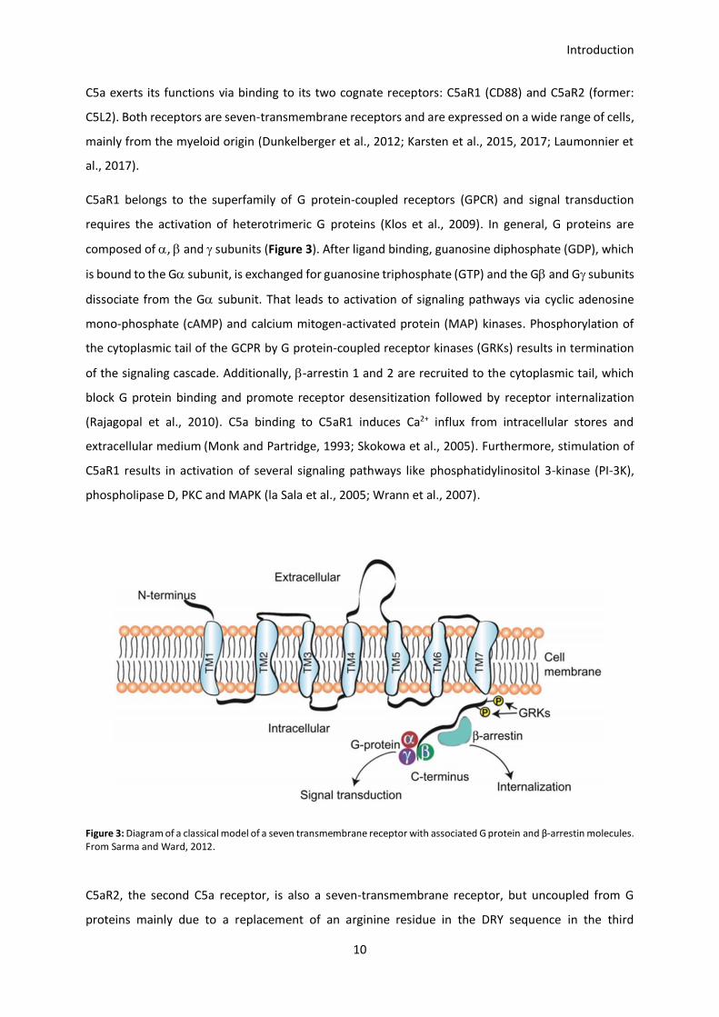
C5aR1 belongs to the superfamily of G protein-coupled receptors (GPCR) and signal transduction
requires the activation of heterotrimeric G proteins (Klos et al., 2009). In general, G proteins are
composed of , and subunits (Figure 3). After ligand binding, guanosine diphosphate (GDP), which
is bound to the G subunit, is exchanged for guanosine triphosphate (GTP) and the G and G subunits
dissociate from the G subunit. That leads to activation of signaling pathways via cyclic adenosine
mono-phosphate (cAMP) and calcium mitogen-activated protein (MAP) kinases. Phosphorylation of
the cytoplasmic tail of the GCPR by G protein-coupled receptor kinases (GRKs) results in termination
of the signaling cascade. Additionally, -arrestin 1 and 2 are recruited to the cytoplasmic tail, which
block G protein binding and promote receptor desensitization followed by receptor internalization
(Rajagopal et al., 2010). C5a binding to C5aR1 induces Ca2+ influx from intracellular stores and
extracellular medium (Monk and Partridge, 1993; Skokowa et al., 2005). Furthermore, stimulation of
C5aR1 results in activation of several signaling pathways like phosphatidylinositol 3-kinase (PI-3K),
phospholipase D, PKC and MAPK (la Sala et al., 2005; Wrann et al., 2007).
Figure 3: Diagram of a classical model of a seven transmembrane receptor with associated G protein and β-arrestin molecules. From Sarma and Ward, 2012.
C5aR2, the second C5a receptor, is also a seven-transmembrane receptor, but uncoupled from G
proteins mainly due to a replacement of an arginine residue in the DRY sequence in the third

Introduction
11
transmembrane domain and a change in the NPXXY sequence in the seventh transmembrane domain
(He et al., 2001; Ohno et al., 2000; Okinaga et al., 2003). Consequently, the receptor is not able to
induce Ca2+ influx. Therefore, C5aR2 was initially described as a decoy receptor to control and regulate
C5aR1 functions (Scola et al., 2009). In line with that, publications suggest that C5aR2 has
anti-inflammatory properties mediated via -arrestin signaling (Bamberg et al., 2010). However, also
pro-inflammatory properties mediated via modulation of C5aR1-induced ERK phosphorylation have
been described (Chen et al., 2007; Hsu et al., 2014). Besides the controversial discussion about pro- or
anti-inflammatory roles of this receptor, scientists also debate about the cellular location of the
receptor. For human granulocytes and peripheral blood monocytes it was shown that C5aR2 is
localized predominantly intracellularly (Bamberg et al., 2010). A similar expression was observed for
murine neutrophils and macrophages (Karsten et al., 2017).
1.2 IL-12 cytokine family
IL-12 family cytokines include IL-12, IL-23, IL-27 and IL-35 and play important roles on the induction of
innate immune responses (de Saint-Vis et al., 1998). Cytokines which belong to this family form
heterodimers that consist of an -chain (p19, p28, p35) and a -chain (p40 or Ebi3) (Figure 4). Pairing
of p40 chain with p35 or p19 forms IL-12 and IL-23, respectively, whereas heterodimers of Ebi3 with
p28 and p35 result in IL-27 and IL-35. The IL-12 family cytokine receptors consist of heterodimeric
structures sharing receptor subunits such as IL-12Rs1 or gp130.
The pro-inflammatory cytokine IL-12 and IL-23 have key roles in the development of the TH1 and TH17
subsets of helper T cells. IL-12 is also a crucial pro-stimulatory cytokine for NK cells in the induction of
innate immune responses (Ferlazzo et al., 2004). IL-27 is reported to have mainly inhibitory activity
and is often generated during the resolution phase of an immune response by APCs. IL-35 is a potent
anti-inflammatory cytokine expressed by Treg cells. It controls the development of TH1 and TH17 cells
and inhibits TH1 cell proliferation (Vignali and Kuchroo, 2012).

Introduction
12
Figure 4: IL-12 cytokine family, receptors and signaling components. The IL-12 family comprises the heterodimeric cytokines
IL-12, IL-23, IL-27 and IL-35. They consist of an -chain (p19, p28, p35) and a -chain (p40 or Ebi3). These cytokines exert their functions upon binding to heterodimeric receptors and involve distinct JAK-STAT signaling partners. The bottom bar reflects their functional spectrum ranging from pro-inflammatory (IL-23, IL-12) to inhibitory (IL-35). From Vignali and Kuchroo, 2012.
1.3 Natural killer cells: key players during early innate immune
responses
NK cells develop in the thymus and liver (Sojka et al., 2014) and belong to the first defense line of the
organism against pathogens. Under naïve conditions, NK cells are found mainly in the spleen, but in
lower numbers also in blood, liver, lymph nodes, in the uterus during gestation as well as in the
peritoneal cavity. During development, NK cells undergo different maturation steps to fulfill effector
functions like recognition and killing of virally-infected cells or the production of pro-inflammatory
cytokines like IFN- (Orr and Lanier, 2010; Vivier et al., 2008). To promote and control their function
and development, NK cells rely on cytokines like IL-2, IL-12, IL-15 or IL-18 and on transcription factors
like Nfil3 or PU.1 (Colucci et al., 2001; Kamizono et al., 2009; Mandal and Viswanathan, 2015).
1.3.1 Natural killer cell activation and pathogen clearance
As already mentioned above, circulating NK cells require activation by cytokines to allow their
recruitment to the site of infection (Fogler et al., 1996). NK cells in the lymph nodes or spleen receive
their activation signals via DCs, which form a stimulatory synapse and start to secrete cytokines like
IL-2, IL-12, IL-15 and IL-18 to induce NK cell activity (Borg et al., 2004). Furthermore, NK cell activity is
controlled by a wide range of receptors, which are expressed on the surface and have inhibitory or

Introduction
13
activating properties. Killer immunoglobulin-like receptors (KIR), Ig-like receptors (CD158), the C-type
lectin receptor (NKG2A) and leukocyte inhibitory receptors (LIR1, LAIR-1) belong to the inhibitory
receptor family. Activating receptors are the natural cytotoxicity receptors (NKp46, NKp44), C-type
lectin receptors (NKG2D) and Ig-like receptors (2B4) (Carrillo-Bustamante et al., 2016). The ability of
NK cells to respond to a variety of stimuli and to participate in immune responses under different
pathological conditions is based on their different expression of inhibitory and activating receptors.
Under physiological conditions, NK cells express two to four inhibitory receptors and an array of
activation receptors which results in a heterogeneity within the NK cell population (Mandal and
Viswanathan, 2015). With these receptors, NK cells are able to regulate cytotoxicity (Figure 5).
Inhibitory receptors recognize self-MHC class I molecules on the surface of the cell. This signal prevents
activation of the NK cell. Tumor cells or virus-infected cells tend to downregulate MHC class I on the
surface of the cells to escape recognition by cytotoxic T lymphocytes (CTL) making them a target for
NK cells (Malnati et al., 1993). Once the NK cell is activated, the cytotoxic ability is mediated via
different pathways: Perforin, a membrane-disrupting protein, and granzymes are secreted by
exocytosis. Both substances work together to induce apoptosis of the target cell. The second pathway
depends on a caspase-dependent apoptosis. Here, death receptors (e.g. Fas) on target cells are
associated with their equivalent ligand (FasL) and tumor necrosis factor-related apoptosis-inducing
ligand (TRAIL) on NK cells which results in caspase-dependent apoptosis (Zamai et al., 1998). A third
pathway includes the antibody-dependent cellular cytotoxicity (ADCC) as NK cells express the
low-affinity FcRIII. Here, FcRIII recognizes antibodies bound to the surface of a target cell which
induces the release of cytotoxic factors (Wang et al., 2015). Interestingly, ADCC requires processes of
the adaptive immunity as it results from binding of antibodies and therefore emphasizes the role of
NK cells in innate and adaptive immune responses.

Introduction
14
Figure 5: The balance of inhibitory and stimulatory signals received by NK cells. Cells under naïve conditions are protected from killing when stimulatory signals are counter-balanced by inhibitory signals delivered by MHC I. Missing MHC I expression results in activation of the NK cell and killing of the target cell (missing-self recognition). Modified from Raulet and Vance, 2006.
1.3.2 Dendritic cell – mediated NK cell activation via stimulatory synapse
formation
DCs are professional APCs and are necessary to induce immune responses by recognizing pathogens
via PRRs. Besides their role in activating and regulating adaptive immune responses, they are also
involved in the activation of NK cells. Here, soluble and contact-dependent activation by DCs is
necessary to induce cytokine production, proliferation and cytotoxicity of NK cells (Figure 6).
Contact-dependent activation is mediated via the ligation of NKp46, NKp30, NKG2D or 2B with MHC I
or IL-15 receptor provided on the DC side. Soluble factors, which mediate NK cell activation include
IL-2 (Granucci et al., 2004), IL-12 (Lehmann et al., 2001), IL-15, IL-18 (Srivastava et al., 2013), IFN- and
IFN- (Swann et al., 2007) produced by DCs. IL-15 has a dual function and can act as a soluble factor
after secretion and a contact-dependent factor via presentation through the IL-15 receptor in the
stimulatory synapse (Viaud et al., 2009). Borg et al showed that a direct cell-cell interaction between
DCs and NK cells is necessary to induce IFN- production (Borg et al., 2004). Following the activation,
NK cells become cytotoxic and secrete granzymes and perforins, start to proliferate or produce high
amounts of the pro-inflammatory cytokines TNF- and IFN-. Reciprocally, NK cell-derived IFN- is

Introduction
15
important to induce DC maturation and polarization of T cells into the TH1 phenotype (Elssen et al.,
2014). Besides specific NK cell receptors, other adhesion molecules like vascular cell adhesion protein
1 (VCAM1) or very late antigen-4 (VLA4) play important roles in mediating NK cell activity via DCs
(Fogler et al., 1996).
Figure 6: DC-induced NK cell activation. DCs can affect NK cell function by augmentation of cytotoxicity, cytokine secretion (IFN-γ and TNF-α), and proliferation. This depends on contact-dependent (ligation of NKp46, NKp30, NKG2D, 2B4) as well as soluble factors (IL-2, IL- 12, IL-15, IL-18, IFN-α, and IFN-β). IFN-γ secretion by NK cells is, in turn, responsible for DC maturation and Th1 polarization, whereas augmentation of NK cell cytotoxicity contributes to tumor cell lysis. From Elssen et al., 2014.
1.4 Toxoplasma gondii: an intracellular parasite
1.4.1 Biology and life cycle of Toxoplasma gondii
T. gondii is an intracellular parasite, which is able to infect virtually all nucleated cells and therefore, is
present in a wide range of hosts. Depending on age and environment, up to 50 % of the human
population is infected with T. gondii (Flegr et al., 2014). The parasite has a sexual life cycle in the
definitive host and a two-staged asexual life cycle in secondary hosts (Figure 7). T. gondii can be divided
into three different strains (I, II, III), which differ in their virulence, ability to cause encephalitis and
cytokine induction (Araujo and Slifer, 2003; Haque et al., 1999). For my experiments, I used ME49 type
II strain parasites.

Introduction
16
Within the definitive host, T. gondii undergoes sexual reproduction in the gut. Members of the
definitive host family (cats) can be infected by ingesting infected animals like mice. Once taken up, the
cyst form of the parasite survives the passage through the stomach and starts to infect epithelial cells
of the cat’s small intestine (Dubey et al., 2011a). Within these cells the parasite undergoes sexual
reproduction that ends up in the production of millions of zygote-containing cysts known as oocysts.
After massive reproduction inside the epithelial cell, oocysts are released into the intestinal lumen and
are shed in the cat’s feces. Due to the sexual replication and as a result of meiosis, many of the
generated oocysts contain different genotypes and thus a huge number of genetically distinct parasites
can be generated from one single cat. In the environment, oocysts can spread to soil, water, food or
anything else potentially contaminated with the feces. Due to its structure, oocysts can survive and
remain infectious for many months in cold and dry climates (Dubey et al., 2011b). Humans are infected
with parasites by consuming raw meat or contaminated water. Farmed animals like cow, sheep or pigs
are infected by consuming contaminated water or food contaminated with cat feces. In
immunocompetent humans, symptoms of the infection are flu-like, often the infection is
asymptomatic. However, in immunocompromised patients, e.g. HIV patients, the parasite can cause
blindness or seriously harm the host, causing life-threatening symptoms like brain or heart damage
(Wang et al., 2017b). Furthermore, during pregnancy, a primary T. gondii infection can result in serious
symptoms up to stillbirth or death of the newborn shortly after birth (Endris et al., 2014).
Introduction
17
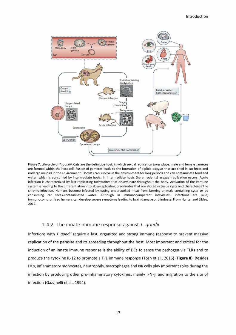
Figure 7: Life cycle of T. gondii. Cats are the definitive host, in which sexual replication takes place: male and female gametes are formed within the host cell. Fusion of gametes leads to the formation of diploid oocysts that are shed in cat feces and undergo meiosis in the environment. Oocysts can survive in the environment for long periods and can contaminate food and water, which is consumed by intermediate hosts. In intermediate hosts (here: rodents) asexual replication occurs. Acute infection is characterized by fast replicating tachyzoites that disseminate throughout the body. Activation of the immune system is leading to the differentiation into slow-replicating bradyzoites that are stored in tissue cysts and characterize the chronic infection. Humans become infected by eating undercooked meat from farming animals containing cysts or by consuming cat feces-contaminated water. Although in immunocompetent individuals, infections are mild, immunocompromised humans can develop severe symptoms leading to brain damage or blindness. From Hunter and Sibley, 2012.
1.4.2 The innate immune response against T. gondii
Infections with T. gondii require a fast, organized and strong immune response to prevent massive
replication of the parasite and its spreading throughout the host. Most important and critical for the
induction of an innate immune response is the ability of DCs to sense the pathogen via TLRs and to
produce the cytokine IL-12 to promote a TH1 immune response (Tosh et al., 2016) (Figure 8). Besides
DCs, inflammatory monocytes, neutrophils, macrophages and NK cells play important roles during the
infection by producing other pro-inflammatory cytokines, mainly IFN- and migration to the site of
infection (Gazzinelli et al., 1994).

Introduction
18
Figure 8: Immune response to T. gondii infection. IFN-γ is crucial for survival of the host during Toxoplasma gondii infection. Production of this cytokine by NK-cells is dependent on TLR11-mediated recognition of T. gondii profilin by DCs. Neutrophils provide an important innate source for IFN-γ; the mechanisms that regulate neutrophil-derived IFN-γ are not well understood
because this IFN-γ is not regulated by TLRs or by IL-12. Macrophages are primed with the IFN- to destruct the parasite and
limit replication. In later stages of the infection, DCs also prime T cells to produce IFN-. From Yarovinsky, 2014.
As already mentioned, TLRs play a key role in sensing the parasite as MyD88-deficient mice are highly
susceptible to toxoplasmosis and succumb to death early after an infection (Sukhumavasi et al., 2008).
TLRs which are involved in T. gondii sensing are TLR 2, 4, 7, 9 and especially 11 and 12 which play a
central role (Yarovinsky and Sher, 2006). Quadruple knockout mice for TLRs 3, 7, 9, 11 show complete
loss of resistance to T. gondii infections emphasizing the important role for an early and well-organized
immune response induction (Kim et al., 2008). TLR11 and TLR12 recognize the T. gondii-derived protein
profilin, a conserved molecule among protozoan parasites, via different ways: the direct interaction of
TLR11 with profilin within endolysosomes results in the recruitment of MyD88 and the initiation of
downstream immune signaling cascades. Furthermore, TLR11 and TLR12 heterodimerization or TLR12
homodimerization and subsequent profilin detection also results in MyD88 recruitment and induction
of DC responsiveness (Koblansky et al., 2013; Raetz et al., 2013; Yarovinsky et al., 2005). Besides TLR11
and TLR12, other TLRs play important roles in detecting T. gondii-derived substrates and induce an
immune response. TLR2 functions as a sensor for glycosylphosphatidylinositol (GPI) anchors which
cover the cell surface of many protozoan parasites (Campos et al., 2004; Krishnegowda et al., 2005). It
is expressed on macrophages, where it is responsible for the induction of tumor necrosis factor (TNF)
and CC-chemokine ligand 2 (CCL2) production. On epithelial cells TLR2 triggers the production of
chemokines during the infection (Debierre-Grockiego et al., 2007; Ju et al., 2009). Besides TLR2, TLR4
can also detect T. gondii GPIs in vitro. Furthermore, both TLRs can also be activated by T. gondii-derived
heat shock protein (HSP) 70 (Debierre-Grockiego et al., 2007; Mun et al., 2005). T. gondii can also be

Introduction
19
detected in a TLR-independent manner via inflammasomes. This intracellular sensor system can
cooperate with TLRs in pathogen recognition, but primarily functions to detect microbial ligands in the
cytosolic compartment. Here, it is involved in processing of the pro-inflammatory cytokines IL-1 and
IL-18, which are involved in host defense against T. gondii. Deficiency in inflammasome-associated
intracellular sensor NLRP1 results in increased susceptibility to T. gondii, but the exact mechanism how
it is involved remains elusive (Ewald et al., 2014).
Finally, Andrade et al published that, in case of insufficient MyD88 activation via TLR11 and TLR12,
heterodimers of TLR7 and TLR9, which sense T. gondii-derived nucleic acids, compensate and induce
DC responsiveness (Andrade et al., 2013). That is an interesting observation, as TLR12 is not present in
the human genome and therefore the TLR11/12-dependent recognition is central in host defense in
mice or rodents and not applicable to the human innate immune system. Similar to mice, data support
that a TH1/TH17-based immune response and T-cell-derived IFN- are critical for parasite control in
humans (Meira et al., 2014). Nevertheless, a detailed model of T. gondii recognition by the human
immune system is still missing. Analyses showed that human TLR5 is evolutionary close to the mouse
trl11 family which suggests that TLR5 in humans mimics murine TLR11 function (Gonzalez et al., 2014).
Like many parasites, T. gondii promotes host survival to allow maximum parasite replication and
spreading through the body. As already described, T. gondii infections result in IL-12-mediated IFN-
production from innate immune cells and a TH1 response by CD4+ and CD8+ T cells. Both is critical for
host survival. Blocking of IL-12 or genetically ablating the IL-12 subunits IL-12p35 or IL-12p40 results in
increased susceptibility to T. gondii, comparable to that of MyD88-deficient mice (Scanga et al., 2002).
Injection with soluble tachyzoite antigen (STAg) revealed that splenic CD8+ DCs are the main source
of IL-12 (Sousa et al., 1997). However, other DC subsets and also macrophages are able to produce
IL-12 after priming with IFN- (Trinchieri, 2003). Analysis of mice lacking the transcription factor
IFN-regulatory factor 8 (IRF8) resulted in blocked CD8+ DC development, lack of early IL-12 production
and decreased survival during T. gondii infections (Scharton-Kersten et al., 1997a). It is published that
IRF8 regulates both IL-12p40 and IL-12p35 expression downstream of TLR-11 and MyD88, which
explains the increased susceptibility of IRF8-deficient mice (Raetz et al., 2013). The first cells which
respond to TLR-mediated IL-12 production from CD8+ DCs are NK cells, which start to produce IFN-
and therefore are responsible for the induction of a potent immune response.
Besides DCs, neutrophils are an important source for IL-12 as they are able to secrete pre-stored IL-12
upon T. gondii detection in vivo and in vitro (Bliss et al., 1999a, 1999b, 2000). The importance of
neutrophils during infections is shown by decreased IL-12 levels and increased parasite replication
after neutrophil depletion (Bliss et al., 2001). However, neutrophil depletion using Gr-1, which is a
combined antibody of the neutrophil-specific Ly6g and monocyte and neutrophil-expressing Ly6c, also

Introduction
20
affects other cells like inflammatory monocytes, due to the fact the both cell types share the same
surface marker Ly6c. That complicates interpretation of the obtained data (Dunay et al., 2010). In
addition to IL-12 secretion, neutrophils migrate in large numbers to the site of infection and start to
produce IFN- to support NK cells in limiting parasite spreading, although the amount of IFN- is low
compared to that produced by NK cells and T cells (Biswas et al., 2017). Moreover, neutrophils are
involved in direct parasite killing via phagocytosis, release of reactive oxygen species and the formation
of DNA containing extracellular traps (NETs) (Abi Abdallah et al., 2012; Chtanova et al., 2008; Konishi
and Nakao, 1992; Nakao and Konishi, 1991). Therefore, it is not surprising that mice which lack the
recruitment receptor CXCR2 result in a defect in neutrophil recruitment, suffer from higher parasite
levels in the central nervous system (CNS) (Del Rio et al., 2001).
In addition to neutrophils, also impaired migration of monocytes due to a CCR2 deficiency leads to
increased susceptibility to T. gondii infection (Benevides et al., 2008; Dunay et al., 2010; Robben et al.,
2005). Monocytes contribute to the control of T. gondii in a direct and an indirect manner. They
produce IL-1 and IL-12 after stimulation with the parasite to enhance IFN- production and contribute
to parasite killing through the production of nitric oxide (NO) and inducible nitric oxide synthase (iNOS)
(Dunay and Sibley, 2010; Mordue and Sibley, 2003; Robben et al., 2005). CCR2-deficient mice succumb
to death during T. gondii infections after 3-4 weeks due to decreased expression of iNOS and increased
parasite burden in the CNS (Benevides et al., 2008). Monocytes can develop into monocyte-derived
DCs (moDCs) or macrophages. As moDCs they can induce adaptive immune responses (Domínguez and
Ardavín, 2010) and as macrophages they migrate to the site of infection and control the infection by
pathogen clearance or cytokine production. For a more detailed discussion see the review from
(Gordon and Taylor, 2005).
In macrophages the fate of T. gondii depends on the mechanism of uptake into the cell. If macrophages
phagocytose dead or opsonized parasites, the parasite is degraded in lysosomal compartments (Sibley
et al., 1985), whereas live parasites that actively invade the cell hide in parasitophorous vacuoles (PV)
(Mordue and Sibley, 1997). Macrophages which have been primed with IFN- are capable of controlling
parasite replication in PVs through upregulation of NO and iNOS (Adams et al., 1990). iNOS-/- mice
survive the acute infection phase, but are highly susceptible to toxoplasmic encephalitis (Khan et al.,
1997; Scharton-Kersten et al., 1997b). IFN- also induces upregulation of 47-48 kDa immune GTPases
(IGTP,LRG47, IRG47) which are essential for resistance in the acute and chronic infection and limit
parasite growth (Collazo et al., 2001; Taylor et al., 2000). Independent from IFN- and NO, TNF- and
CD40/CD40L signaling results in anti-microbial activity and increases survival during the infection
(Andrade et al., 2003, 2005). Additionally, activated macrophages contribute to IL-12 production

Introduction
21
regulated by TRAF6-dependent phosphorylation of the MAPK family member p38 and ERK1/2 (Mason
et al., 2004).
1.4.3 The role of NK cells during T. gondii infection
NK cells are an essential and early source for IFN- in T. gondii infection that compensates for the lack
of CD8+ T cells (Denkers et al., 1993; Hunter et al., 1994; Johnson et al., 1993). As the NK cell activity
peaks early during the acute phase of infection, NK cells do not appear to be significant in the chronic
phase (Kang and Suzuki, 2001). Hence, most studies concentrated on early NK cell activity and IFN-
production mainly regulated via IL-12, which is produced by other innate immune cells (DCs,
monocytes, neutrophils; as described earlier) (Gazzinelli et al., 1993; Hunter et al., 1994). NK cells can
also be cytotoxic for cells infected with T. gondii (Hauser and Tsai, 1986; Subauste et al., 1992).
However, it was shown that NK cells become infected with the parasite when lysing infected cells. In
consequence, the parasite can disseminate within the NK cell. It was also proposed that infected NK
cells show a higher motility that facilitates spreading of the parasite throughout the organism (Ueno
et al., 2015).
1.4.4 The adaptive immune response against T. gondii infection
Three to five days after the infection, cells from the adaptive immune system start to induce effector
mechanisms that help controlling the parasite. Here, B and T cells occupy important functions as mice
with deficiencies in B cell, CD4+ T cell or CD8+ T cell functions survive the acute phase of the infection,
but succumb to death during the chronic stage of infection (Johnson and Sayles, 2002; Kang et al.,
2000).
B cells contribute to parasite control by the production of specific antibodies. Parasite-specific IgM,
IgA, IgE and IgG2 have been isolated from human patients. Detection of specific antibodies of which
certain subtypes help to distinguish between freshly (IgM) and chronically (IgG) infected patients
(Correa et al., 2007; Remington, 1969; Remington et al., 1968, 2004). Mice lacking B cells develop
normal IFN- responses, but succumb to infection within 3-4 weeks, suffering from a high parasite
burden in the CNS (Kang et al., 2000). As passive transfer of antibodies confers protection to B
cell-deficient mice, it is likely that the lack of antibodies is responsible for the increased susceptibility
(Johnson and Sayles, 2002).

Introduction
22
The importance of T cells becomes clear as HIV patients develop severe toxoplasmosis associated with
reduced T cell numbers (Israelski and Remington, 1988). The initiation of proper T cell responses
requires that naïve CD4+ or CD8+ T cells encounter APCs bearing cognate T. gondii antigens.
Additionally, co-stimulatory signals via CD28 and inducible co-stimulator (ICOS) and cytokines like IL-12
are required for T cell activation, which is characterized by proliferation, differentiation and IFN-
production (Curtsinger et al., 1999, 2003; Harding et al., 1992). Presentation of antigens occurs mainly
via DCs, but also B cells and macrophages are capable of presenting antigens to CD4+ T cells via MHC
class II (Jenkins et al., 2001). Presentation of T. gondii antigens requires phagocytosis of the parasite,
infected cells, parasitic debris or is achieved through endocytosis of antigens secreted by T. gondii
itself. Alternatively, antigens are presented on APCs after active infection with the parasite (Koshy et
al., 2010).
CD8+ T cells are specialized to recognize cells infected with viral, bacterial or parasitic organisms, hence
it is not surprising that these cells play an important role in maintaining resistance to T. gondii
infections. CD8+ T cells control infection by production of IFN-, CD40/CD40L interactions and through
perforin-mediated cytolysis of infected cells (Denkers et al., 1997; Gazzinelli et al., 1992; Reichmann et
al., 2000). Depletion of CD8+ T cells in mice results in increased susceptibility to toxoplasmosis and
mice succumb to death approximately 50 days post infection (p.i.) (Denkers et al., 1997). Additionally,
adoptive transfer of CD8+ T cells from chronically infected mice or mice vaccinated with an attenuated
strain of T. gondii, is sufficient to maintain resistance (Gigley et al., 2009; Parker et al., 1991). Some
epitopes of T. gondii presented on MHC class I alleles can be recognized by CD8+ T cells. These include
peptides which are conserved across multiple T. gondii strains and that are actively secreted by the
parasite (Blanchard et al., 2008; Frickel et al., 2008; Wilson et al., 2010).
1.4.5 The role of the complement system during T. gondii infections
Assessment of T. gondii-specific antibody production to distinguish between freshly and chronically
infected patients has already been discovered in 1948 by Sabin and Feldman (Sabin and Feldman,
1948). In 1980, the classical complement pathway was identified as the most important part of the
complement system in controlling T. gondii, as depletion of alternative pathway complement factors
B, D and P did not impair parasite killing (Schreiber and Feldman, 1980). The minor importance of the
alternative pathway can be explained with the reduced capacity of C3b to bind to the cellular
membrane of tachyzoites and the fast degradation into its inactive form iC3b (Fuhrman and Joiner,
1989). During chronic stage of infection, rupture of brain cysts causes C1q production and activation
of the classical complement pathway (Xiao et al., 2016). In vivo blocking of complement protein C3 in

Introduction
23
mice results in acute susceptibility to toxoplasmosis due to impaired CD4+ and CD8+ T cell responses
(Johnson et al., 1996). Furthermore, C3aR- and C5aR1-double deficiency in mice leads to increased
susceptibility due to decreased IL-12 and IFN- levels (Strainic et al., 2008). Experiments from Daria
Briukhovetska in the Institute for Systemic Inflammation Research (Lübeck, Germany) showed that
C5aR1 deficiency leads to increased susceptibility in mice characterized by increased weight loss and
mortality rate (Briukhovetska, 2017). Additionally, C5aR1- and C5aR2-deficient mice suffer from
increased parasite burdens in the brain 30 days p.i. In both strains altered IL-12 and IFN- levels seem
to be at least one critical factor for the increased susceptibility emphasizing the importance of
complement activation for the immune response to T. gondii infection.

Introduction
24
1.5 The growth factor receptor-bound protein 2
(Grb-2)-associated binder (Gab) protein family
1.5.1 Role and function in signal transduction
As demonstrated in the previous chapters, the complement system and especially NK cells play crucial
and important roles during T. gondii infection. For further investigation of the specific NK cell function,
I focused on another protein which is highly expressed on NK cells: Gab3.
The growth factor receptor-bound protein 2 (Grb-2) associated binder (Gab) protein family describes
three different docking and scaffolding proteins (Gab1, Gab2, Gab3) which contribute to specific signal
transduction pathways. Although the overall sequence homology between all three proteins is only
40 %-50 %, they share similar topology. Each protein contains an N-terminal pleckstrin homology (PH)
domain, proline-rich motifs and multiple potential tyrosyl and seryl/threonyl phosphorylation sites
(Nishida and Hirano, 2003) (Figure 9). Although the overall topology is similar, the three Gab proteins
are expressed in different cells and expression deficiency has different consequences (Figure 10). Gab1
and Gab2 are ubiquitously expressed but are found only in low levels in lymphoid tissues. In contrast,
Gab3 is highly expressed in lymphoid tissues. The key mechanism to induce activation of Gab proteins
is the phosphorylation of tyrosine residues in response to growth factors or cytokines. Phosphorylation
occurs, besides other factors, in response to IL-3, IL-6 and oncoproteins in case of Gab1 and Gab2 and
in response to M-CSF in the case of Gab3 (Nishida and Hirano, 2003). Grb-2 is described as an adaptor
protein which is involved in signal transduction and cell communication. The protein contains one Src
Homology (SH) 2 and two SH3 domains which form complexes with the phosphorylated tyrosine
residues from the Gab proteins and therefore allow transport of Grb2 to the membrane to bring it in
close proximity to associated receptors (Berry et al., 2002).
Figure 9: Schematic structures of Gab family docking proteins. Shown are the schematic domain structures of three human Gab proteins (Gab1–3). All Gab proteins consist of a highly conserved N-terminal PH domain that is involved in membrane targeting. The central proline-rich regions mediate the association with SH3 domain-containing adaptor proteins such as Grb2. PH=Pleckstrin homology; P=Proline From Nakaoka and Komuro, 2013.

Introduction
25
The downstream signaling partner of all Gab proteins are largely unknown, but experiments with mice
lacking either Gab1, Gab2 or Gab3 revealed first hints for the involvement of Gab proteins in several
cell signaling pathways. Gab1-deficient embryos die between gestational day 12.5 and 18.5 with
multiple defects in the heart, placenta and liver (Itoh et al., 2000). Furthermore, ERK activation in
response to epidermal growth factor (EGF), platelet-derived growth factor (PDGF) and hepatocyte
growth factor (HGF) in Gab1-deficient primary fibroblasts was strongly reduced. These results indicate
that Gab1 acts downstream of receptor tyrosine kinases (RTK), but upstream of ERK. Additionally, Cai
et al showed that Gab1 promotes Ras/MAPK signaling to enhance epidermal cell proliferation and
differentiation (Cai et al., 2002).
In contrast to Gab1-deficient mice, Gab2-deficient mice are viable, generally healthy and have a normal
lifespan. The only differences compared to WT mice are decreased mast cells numbers and impaired
mast cell activation in the skin and stomach of Gab2-deficient mice (Nishida et al., 2002). The defective
activation is due to impaired PI-3 kinase and ERK activation.
The biological function of Gab3 is poorly understood. Mice with targeted disruption of the Gab3 gene
have been generated and these mice develop normally, are generally healthy and immunocompetent
(Seiffert et al., 2003). Data from the exAC gene database showed no loss of function mutations in
humans indicating a role of Gab3 for survival, reproduction or cell differentiation.
Targeted disruption in Gab1, Gab2 and/or Gab3 in double and triple knockouts have not been studied
yet but will answer open questions regarding signaling partners and the involvement in cell
differentiation and activation.

Introduction
26
Figure 10: Overview of Gab1, Gab2 and Gab3 regarding molecular weight, expression, tyrosine phosphorylation and consequences of expression deficiency. Reviewed in (Nishida and Hirano, 2003)
1.5.2 PH-domain – associated recruitment of Gab proteins
Recruitment of Gab proteins to activate receptors can be achieved via direct and indirect mechanisms
(Figure 11). The interaction of Gab1 with c-Met, the receptor for HGF, is described as the only direct
recruitment among Gab proteins (Weidner et al., 1996). In all other signaling pathways, the
recruitment of Grb2-Gab complexes require additional molecules and is described as an indirect
mechanism. One of these indirect mechanisms includes the Gab protein PH domain. This domain can
bind to phospholipids and therefore allows recruitment of the Grb2-Gab complex to the receptor. As
the PH domain is highly conserved among all three Gab proteins it is likely that all Gab proteins are
able to bind to the cell membrane via its PH domain. The role of the PH domain is best understood for
EGFR signaling in the context with Gab1 (Rodrigues et al., 2000). Here, Gab1 is initially targeted to Grb2
leading to phosphorylation of tyrosyl residues in the Grb2 protein, activation of PI3K and generation
of 3-phosphoinositide lipids. The Gab1 PH domain is then able to bind to these lipids and creates a
positive feedback loop allowing more sustained signaling. In general, the PH domain is important and
required for several recruitment mechanisms as point mutations in the PH domain result in impaired
recruitment of Gab proteins (Schutzman et al., 2001).

Introduction
27
Figure 11: Gab protein recruitment mechanisms. a) Recruitment via MBS and Grb2–Gab1 complexes. Shown is a schematic of Gab1 recruitment to the HGF receptor. Gab1 can bind directly to the HGF receptor (HGFR) through its Met-binding sequence (MBS), as well as indirectly, as part of a Grb2–Gab1 complex, to a Grb2 SH2 domain binding site in the HGFR. b) Recruitment via Shc–Grb2–Gab2. Gab2 is recruited to IL3/GM-CSF/IL-5 receptor family by means of a Shc–Grb2–Gab2 complex. c) Recruitment via another scaffolding adaptor. In FGF receptor signaling, Gab1 is recruited to the receptor complex via Grb2 binding to the scaffolding adaptor FRS2. d) Recruitment via Gab family PH domains. In some signaling pathways, binding of the Gab PH domain to appropriate phosphoinositides is required for initial recruitment to receptor complexes or for sustained signaling (e.g. EGFR). e) Recruitment of Gab proteins to oncoproteins. Gab proteins are phosphorylated in response to several oncogenic stimuli. Recruitment of Gab2 to Y177 of Bcr–Abl as part of a Grb2–Gab2 complex. Abbreviations: BCR, B-cell receptor; EGFR, epidermal growth factor receptor; GM-CSF, granulocyte–macrophage colony-stimulating factor; IL, interleukin; PH, pleckstrin-homology; PI3K, phosphoinositide 3-kinase; SH2, Src-homology 2; Yp, phosphotyrosine. From Gu and Neel, 2003.
1.6 Hypothesis and specific aims
Previous experiments have shown that C5aR1-/- mice suffer from increased susceptibility and parasite
burden in the brain during T. gondii infections. C5aR2-/- mice also showed increased parasite burden in
the brain compared to WT mice, but not as distinct as C5aR1-/- mice (Briukhovetska, 2017). It has been
shown that C5a is critical for the development of an appropriate CD4+ T cell response during T. gondii
infections (Strainic et al., 2008). However, the underlying mechanisms regarding the signaling via
C5aR1 and C5aR2 in DCs and especially in NK cells in the acute phase of the infection remain elusive.
Using novel floxed reporter mice for C5aR1 and C5aR2, the Köhl laboratory already identified NK cells
as C5aR2, but not C5aR1 expressing cells what makes them a unique cell type to investigate the
functions of the still poorly understood C5aR2 during T. gondii infections. Besides the unique C5aR2
expression as an extrinsic factor influencing NK cell function, the Hoebe laboratory showed that NK
cells are highly expressing Gab3 cells, which is a Grb-2-associated binder protein, the function of which
is also poorly understood. Therefore, NK cells are a suitable cell type to investigate and define the role
of C5aR2 and Gab3 during early T. gondii infections. Besides the undisputed role of NK cells in T. gondii
infections, the regulation of C5aR1 and C5aR2 expression on other innate and adaptive immune cells
in naïve mice and during the early phase of T. gondii infection has never been investigated before.

Introduction
28
Based on previous data, I hypothesized that C5aR1/C5aR2 expression and regulation on immune cells
and especially C5aR2 expression on NK cells play important roles during early T. gondii infection.
Further, I proposed that potential independent co-expression of both anaphylatoxin receptors may
play a key role in controlling the parasite. Finally, as Gab3 is highly expressed on NK cells, I hypothesized
that a point mutation in the gab3 gene results in altered susceptibility toward T. gondii infection. To
test these hypotheses, I pursued the following specific aims:
1. Define the role of C5aR1 and C5aR2 signaling in DC-dependent and -independent activation of
NK cells.
2. Delineate the role of C5aR1 and C5aR2 signaling in NK cell-mediated control of T. gondii
infection.
3. Define the regulation of C5aR1 and C5aR2 expression on innate and adaptive immune cells
during T. gondii infection.
4. Define the potential role of Gab3 and C5aR1/2 signaling pathways in NK cells for the induction
of T. gondii-specific immune responses.
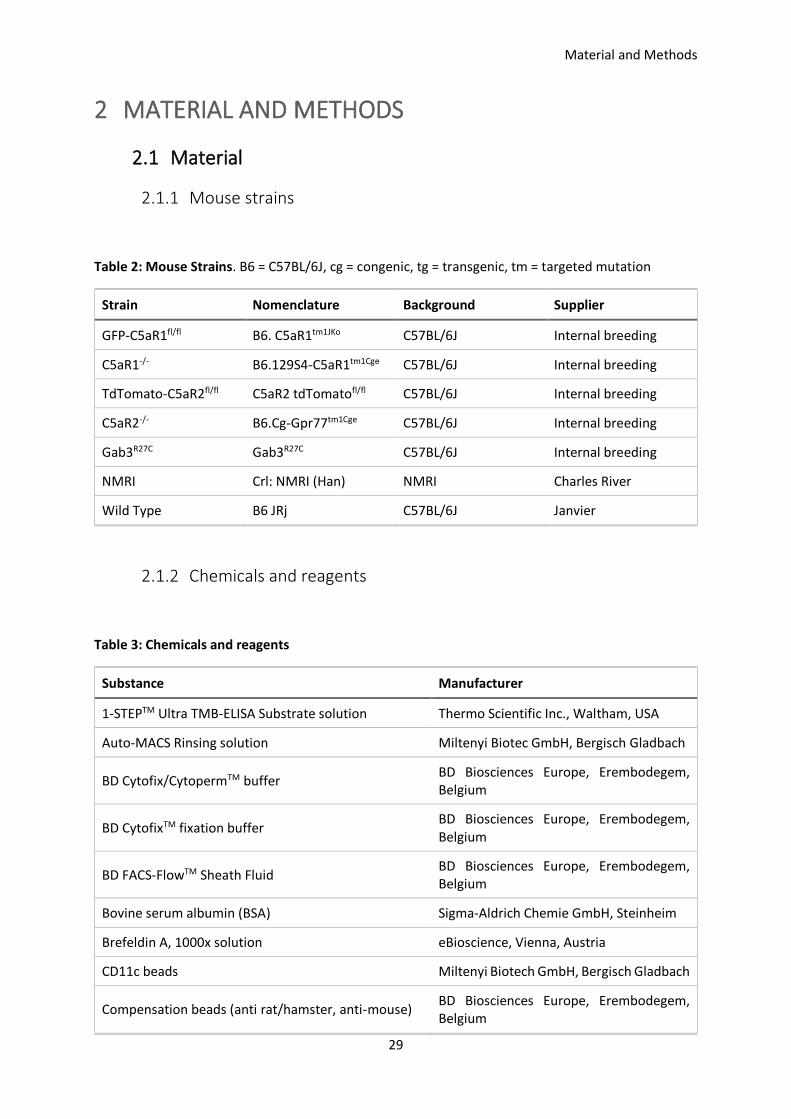
Material and Methods
29
2 MATERIAL AND METHODS
2.1 Material
2.1.1 Mouse strains
Table 2: Mouse Strains. B6 = C57BL/6J, cg = congenic, tg = transgenic, tm = targeted mutation
Strain Nomenclature Background Supplier
GFP-C5aR1fl/fl B6. C5aR1tm1JKo C57BL/6J Internal breeding
C5aR1-/- B6.129S4-C5aR1tm1Cge C57BL/6J Internal breeding
TdTomato-C5aR2fl/fl C5aR2 tdTomatofl/fl C57BL/6J Internal breeding
C5aR2-/- B6.Cg-Gpr77tm1Cge C57BL/6J Internal breeding
Gab3R27C Gab3R27C C57BL/6J Internal breeding
NMRI Crl: NMRI (Han) NMRI Charles River
Wild Type B6 JRj C57BL/6J Janvier
2.1.2 Chemicals and reagents
Table 3: Chemicals and reagents
Substance Manufacturer
1-STEPTM Ultra TMB-ELISA Substrate solution Thermo Scientific Inc., Waltham, USA
Auto-MACS Rinsing solution Miltenyi Biotec GmbH, Bergisch Gladbach
BD Cytofix/CytopermTM buffer BD Biosciences Europe, Erembodegem, Belgium
BD CytofixTM fixation buffer BD Biosciences Europe, Erembodegem, Belgium
BD FACS-FlowTM Sheath Fluid BD Biosciences Europe, Erembodegem, Belgium
Bovine serum albumin (BSA) Sigma-Aldrich Chemie GmbH, Steinheim
Brefeldin A, 1000x solution eBioscience, Vienna, Austria
CD11c beads Miltenyi Biotech GmbH, Bergisch Gladbach
Compensation beads (anti rat/hamster, anti-mouse) BD Biosciences Europe, Erembodegem, Belgium

Material and Methods
30
Substance Manufacturer
Dimethyl sulfoxide (DMSO) Sigma-Aldrich Chemie GmbH, Steinheim
DNase I recombinant, RNase-free Sigma-Aldrich Chemie GmbH, Steinheim
Dulbecco’s phosphate buffered saline (DPBS) Gibco® by Life Technologies Corporation, Carlsbad, USA
Ethanol, 70 % denaturated Carl Roth GmbH & Co. KG, Karlsruhe
Ethanol, 96 % denaturated Carl Roth GmbH & Co. KG, Karlsruhe
Ethanol, absolute J. T. Baker, Deventer, Netherlands
Ethylenediaminetetraacetic acid (EDTA) Sigma-Aldrich Chemie GmbH, Steinheim
Fetal calf serum (FCS) PAA Laboratories GmbH, Pasching, Austria
Formaldehyde solution, 37 % Sigma-Aldrich Chemie GmbH, Steinheim
HEPES (4-(2-hydroxyethyl)-1-piperazineethanesulfonic acid) buffer solution 1M
Life technologies Corporation, Carlsbad, USA
Isopropanol Otto Fisher GmbH & Co. KG, Saarbrücken
Ketamine Sigma-Aldrich Chemie GmbH, Steinheim
L-glutamine (200mM concentrate) Life technologies Corporation, Carlsbad, USA
Liberase™ TL Research Grade Sigma-Aldrich Chemie GmbH, Steinheim
MACS BSA stock solution Miltenyi Biotec GmbH, Bergisch Gladbach
Methanol (CH3OH), 99.8 % Sigma-Aldrich Chemie GmbH, Steinheim
Paraffin Sigma-Aldrich Chemie GmbH, Steinheim
Penicillin-Streptomycin, 100x liquid Gibco® by Life Technologies Corporation, Carlsbad, USA
Potassium bicarbonate (KHCO3) Sigma-Aldrich Chemie GmbH, Steinheim
Potassium carbonate (K2CO3) Sigma-Aldrich Chemie GmbH, Steinheim
Potassium chloride (KCL) Carl Roth GmbH & Co. KG, Karlsruhe
Potassium dihydrogen phosphate (KH2PO4) Carl Roth GmbH & Co. KG, Karlsruhe
RPMI 1640 Gibco® by Life Technologies Corporation, Carlsbad, USA
Sodium chloride (NaCl) Carl Roth GmbH & Co. KG, Karlsruhe
Sodium dihydrogen phosphate (NaH2PO4) Carl Roth GmbH & Co. KG, Karlsruhe
Sodium hydrogen phosphate (Na2HPO4) Sigma-Aldrich Chemie GmbH, Steinheim
Sodium pyruvate Life technologies Corporation, Carlsbad, USA

Material and Methods
31
Substance Manufacturer
Tris-aminomethane (Tris) Sigma-Aldrich Chemie GmbH, Steinheim
Tris-HCL Sigma-Aldrich Chemie GmbH, Steinheim
Trypan blue Life technologies Corporation, Carlsbad, USA
Trypsin-EDTA 0.5 %, 10x Life technologies Corporation, Carlsbad, USA
Tween® 20 Sigma-Aldrich Chemie GmbH, Steinheim
Xylazine Sigma-Aldrich Chemie GmbH, Steinheim
Table 4: Stimulants
Substance Manufacturer
C5a, human recombinant Hycult Biotech, Uden, Netherlands
CSF-2 (GM-CSF), murine recombinant Peprotech Corporation, Rocky Hill, USA
P32 C5aR2-specific agonist Trent M. Woodruff, University of Queensland, Brisbane, Australia
2.1.3 Buffers, solutions and media
Table 5: Buffers, solutions and media
Buffer/Solution/Medium Formulation
Complete RPMI medium
RPMI 1640
10 % FCS, heat-inactivated
100 units/ml Penicillin
100 µg/ml Streptomycin
2mM L-Glutamine
Dulbecco’s Phosphate-Buffered Saline (DPBS)
(self-made)
13.7 mM NaCl
0.27 mM KCl
0.81 mM Na2HPO4
0.15 mM KH2PO4
pH 7.2-7.4
Aqua destillata
Diff-Quik solution B Thiazine dye (blue colour)

Material and Methods
32
Buffer/Solution/Medium Formulation
Phosphate buffer
Diff-Quik solution C Eosin dye (red colour)
Phosphate buffer
ELISA wash buffer (self-made) 0.05 % Tween® 20 in DPBS
Flow cytometer buffer, ELISA block buffer 1 % BSA in DPBS
MACS buffer Auto MACS Rinsing-solution
MACS BSA stock solution 1:20
Red blood cell (RBC) lysis buffer
155 mM NH4Cl
10 mM KHCO3
0.1 mM EDTA
Aqua destillata
pH 7.2, sterile
Tris buffer
500 mM NaCl
20 mM Tris-HCl
Aqua destillata
pH 7.2
2.1.4 Antibodies for flow cytometry
Table 6: Antibodies. AF = Alexa Fluor®, APC = Allophycocyanin, BV = Brilliant Violet, Cy = Cyanin, FITC = Fluorescein isothiocyanate, PE = Phycoerythrin, PerCP = Peridinin-chlorophyll-protein complex, hamster = Armenian hamster
Target Dye Clone Species Manufacturer Concentration
(µg/ml)
CD103 PerCP-Cy5.5 E7 hamster Biolegend 0.25
CD11b
BV510
M1/70 rat
Biolegend 0.125
AF700 eBioscience 0.5
PerCP-Cy5.5 Biolegend 1
CD11c APC
N418 hamster eBioscience 0.5
PE Biolegend 1
CD16/32 - 93 rat eBioscience 10
CD19 eFluor450 eBio1D3 rat eBioscience 1.25
CD23 PacificBlue B3B4 rat Biolegend 1.25

Material and Methods
33
Target Dye Clone Species Manufacturer Concentration
(µg/ml)
CD27 PE LG-3A10 hamster BD Biosciences 0.5
CD3 eFluor450 17A2 rat eBioscience 0.5
CD3e PerCP-Cy5.5 145-2C11 hamster BD Biosciences 1.25
CD4 PE-Cy7 RM4-5 rat eBioscience 0.5
CD4 PE-Cy5 GK1.5 rat Biolegend 0.5
CD4 BV421 RM4-5 rat Biolegend 1
CD43 BV421 S7 rat BD Biosciences 0.5
CD45R
APC
RA3-6B2 rat
eBioscience 1.25
APC-Cy7 Biolegend 0.5
PerCP-Cy5.5 Biolegend 1
CD49b eF450 DX5 rat eBioscience 0.5
CD5 PerCP 53-7.3 rat Biolegend 0.5
CD64 BV711 10.1 mouse BD Biosciences 0.5
CD8
PerCP-Cy5.5
53-6.7
rat Biolegend 1
FITC rat
Biolegend 2.5
BV510 Biolegend, 0.125
F4/80 BV510
BM8 rat Biolegend, 0.5
APC Biolegend 1
IFN- BV605
XMG1.2 rat Biolegend 10
PE eBioscience 2
IgD AF700 11-26c.2a rat Biolegend 1.25
IgM PE-Cy7 Il/41 rat eBioscience 0.5
IL-12p40/70 PE C15.6 rat BD Biosciences 2
Ly49c/i FITC 5E6 mouse BD Biosciences 2.5
Ly49g2 PerCP-eFluor710 4D11 rat Thermo Fisher 1
Ly6C PerCP-Cy5.5 HK1.4 rat eBioscience 0.5
Ly6G eF450 RB6-8C5
rat Thermo Fisher 0.5
FITC 1A8 Biolegend 2.5
MHCII APC-eFluor780
M5/114.15.2 rat eBioscience 0.2
PE-Cy7 Biolegend 0.5

Material and Methods
34
Target Dye Clone Species Manufacturer Concentration
(µg/ml)
APC Miltenyi 3
NK1.1 AF700
PK136 rat Biolegend 1.25
APC mouse Biolegend 0.5
NKp46 APC
29A1.4 rat Biolegend 0.5
eFluor450 rat Thermo Fisher 0.5
Siglec-F BV421 E50-2440 rat BD Biosciences 0.25
2.1.5 Plastic ware and disposable items
Table 7: Plastic ware and disposable items
Material Manufacturer
BD Microtainer tube BD Biosciences Europe, Erembodegem, Belgium
Blunt fill needle, 18G BD Biosciences Europe, Erembodegem, Belgium
Cell strainer 40 µm BD Biosciences Europe, Erembodegem, Belgium
Cover glass, 20x20 mm, thickness 1 Glaswarenfabrik Karl Hecht GmbH & Co. KG, Sondheim/Rhön
Cover glass, circular, 25 mm, borosilicate glass thickness 1
VWR, international GmbH, Darmstadt
ELISA reservoir, 25 ml VWR, international GmbH, Darmstadt
Falcon Tube 15 ml, 50 ml Sarstedt AD & Co., Nümbrecht
Filter tip 10 µl, 100 µl, 1000 µl Sarstedt AD & Co., Nümbrecht
Lancet B. Braun Melsungen AG, Melsungen
MACS Separation MS Column Miltenyi Biotech GmbH, Bergisch Gladbach
Microscope slide, 76x26 mm Gerhard Menzel GmbH, Braunschweig
Microtiter plate, 6-well Sarstedt AD & Co., Nümbrecht
Microtiter plate, 96-well, high bind Corning, Corning, USA
Microtiter plate, 96-well, with lid (F bottom) Greiner Bio-One GmbH, Frickenhausen
Microtiter plate, 96-well, with lid (U buttom) Greiner Bio-One GmbH, Frickenhausen
Needle hypodermic, 26G BD Biosciences Europe, Erembodegem, Belgium
Nitrile powder-free examination gloves Ansell Healthcare GmbH, Munich

Material and Methods
35
Material Manufacturer
Pipette 5 ml, 10 ml, 25 ml Greiner Bio-One GmbH, Frickenhausen
Pipette tips 10 µl, 100 µl, 1000 µl Sarstedt AD & Co., Nümbrecht
Reaction tube 0.5 ml, 1.5 ml, 2.0 ml (low-binding)
Sarstedt AD & Co., Nümbrecht
Reaction tube 0.5 ml, 1.5 ml, 2.0 ml, 5.0 ml Eppendorf AG, Hamburg
Single use cannula 21G, blunt B. Braun Melsungen AG, Melsungen
Spatula VWR International GmbH, Darmstadt
Syringe 1 ml, 5 ml, 10 ml, Luer-Lok™ BD Biosciences Europe, Erembodegem, Belgium
Syringe 5 ml, 10 ml BD Biosciences Europe, Erembodegem, Belgium
Syringe, 1 ml B. Braun Melsungen AG, Melsungen
Tube 5 ml, 75x12 mm, unsterile for flow cytometry
Sarstedt AD & Co., Nümbrecht
Weighing dish Greiner Bio-One GmbH, Frickenhausen
2.1.6 Commercially available kits
Table 8: Commercially available kits
Kit Manufacturer
AYOXXA LUNARIS Multiplex 12-plex Ayoxxa Biosystems GmbH, Cologne
Fixable Viability Dye eFluor 780 eBioscience, Vienna, Austria
LEGENDplex Mouse Inflammation Panel Biolegend Europe, London, UK
Mouse IFN-gamma DuoSet® ELISA R&D Systems, Minneapolis, USA
Mouse IL12/IL-23p40 DuoSet® ELISA R&D Systems, Minneapolis, USA
Mouse IL-12p70 DuoSet® ELISA R&D Systems, Minneapolis, USA
2.1.7 Laboratory equipment
Table 9: Laboratory equipment
Equipment Manufacturer
Biological safety cabinets Nuaire Inc., Plymouth, USA
Cell sorter FACS ARIAIII Beckton Dickinson GmbH, Heidelberg

Material and Methods
36
Equipment Manufacturer
Centrifuge 5424 Eppendorf AG, Hamburg
Centrifuge 5424R Eppendorf AG, Hamburg
Centrifuge 5810R Eppendorf AG, Hamburg
Centrifuge Rotofix 32A Hettich Lab Technology, Tuttlingen
Chemical hood Waldner Laboreinrichtungen GmbH & Co KG, Wangen
Confocal microsope FluoView1000 Olympus, Hamburg
Dissecting scissors WPI Deutschland GmbH, Berlin
ELISA reader Fluostar Omega 0415 BMG Labtech GmbH, Ortenberg
ELISA washer Nunc-ImmunoTM wash 12 Thermo Fisher Scientific Inc., Waltham, USA
Flow cytometer BD LSRII Beckton Dickinson GmbH, Heidelberg
Forceps WPI Deutschland GmbH, Berlin
Fridge, 4°C and -20°C combined Liebherr International GmbH, Biberach an der Riß
Ice machine AF-10 Scotsman, Milan, Italy
MACS Magnetic Cell Separator Miltenyi Biotech GmbH, Bergisch Gladbach
Microscope BA310 LED Motic, Wetzlar
Microscope camera Leica EC3 Leica Mikrosystem Vertrieb GmbH, Wetzlar
Microscope camera Moticam Motic, Wetzlar
Microscope Fluovert FS Leica Mikrosystem Vertrieb GmbH, Wetzlar
Microscope Leica DM IL LED Leica Mikrosystem Vertrieb GmbH, Wetzlar
Multichannel pipette Biohit M300 Sartorius Biohit Liquid Handling Oy, Helsinki, Finland
Neubauer counting chamber, improved VWR International GmbH, Darmstadt
pH-Meter Seven Easy PH S20-K Mettler Toledo, Schwerzenbach, Switzerland
Pipetboy pipette controller Integra Biosciences AG, Zizers, Switzerland
Pipette (0.1-2.5 µl, 0.5-10 µl, 10-100 µl, 20-200 µl, 100-1000 µl)
Eppendorf AG, Hamburg
Precision balance EMB 1000-2 Kern & Sohn, Balingen
Precision balance LC6200S Sartorius AG, Göttingen
Pump, VacuGene Pharmacia, Belgium
Pure water system Nanopure Diamond D11931
Thermo Fisher Scientific GmbH, Bremen
Shaker Polymax 1040 Heidolph Instruments GmbH & Co. KG, Schwabach

Material and Methods
37
Equipment Manufacturer
Suction system Vacusafe 158310 Integra Biosciences GmbH, Fernwald
Table centrifuge Carl Roth GmbH & Co. KG, Karlsruhe
Tablet Acer Iconia Tab Acer, Xizhi, New Taipei, Taiwan
Thermomixer 5436 Eppendorf, Hamburg
Ultra-low temperature freezer, -80°C SANYO Electrics Co., Japan
Vortex-Genie 2 Scientific Industries Inc., New York, USA
Water bath GFL, Burgwedel
2.1.8 Computer software
Table 10: Computer Software
Software Developer
Adobe Acrobat Reader DC Adobe Systems Inc., San Jose, USA
BD FACSDiva V8.0 BD Biosciences, San Jose, USA
FlowJO X FlowJo, LLD, Ashland, USA
FluoView FV1000 2.0b Olympus, Hamburg
GraphPad Prism 7 Graph Pad Software Inc., LaJolla, USA
Imaris 7.2.3 Bitplane AG, Zurich, Switzerland
Leica Aquire 1.0 Leica Microsystems CMS GmbH, Wetzlar
Microsoft Office 365 Microsoft Corporation, Redmond, USA
MARS – Data Analysis Software 2.30 R2 BMG Labtech GmbH, Ortenberg
Omega 1.30 BMG Labtech GmbH, Ortenberg

Material and Methods
38
2.2 Methods
2.2.1 Animals
WT C57BL/6J and NMRI mice were purchased from JANVIER. C5aR1-/-, C5aR2-/-, floxed GFP-C5aR1 and
floxed tdTomato-C5aR2 mice on the C57BL/6J background were bred and maintained in an SPF animal
facility of the University of Lübeck. Gab3R27C mice on the C57BL/6J background were bred and
maintained in an SPF animal facility of the CCHMC in Cincinnati, USA. All animal experiments were
conducted according to protocols (number 39(36-3/15) and 39_2017-03-02) approved by the ethical
committee (Ministerium für Energiewende, Landwirtschaft, Umwelt, Natur und Digitalisierung, Kiel,
Germany). Animals used were male or female and between 8 and 12 weeks old.
2.2.1.1 Generation of floxed reporter mice
The Köhl laboratory (Lübeck, Germany) generated two floxed reporter mice to track C5aR1 or C5aR2
expression. These strains allow to track C5aR1 and C5aR2 expression without the need of antibodies
and to generate cell- or tissue-specific C5aR1 or C5aR2 knockouts.
Floxed GFP-C5aR1 reporter mice were generated as described in Karsten et al. 2015 (Karsten et al.,
2015). Floxed td-Tomato-C5aR2 reporter mice were generated as described in Karsten et al. 2017
(Karsten et al., 2017).
2.2.1.2 ENU (N-ethyl-N-nitrosourea) mutagenesis
ENU-mutation experiments were conducted at Cincinnati Children’s Hospital Medical Center (CCHMC)
in the laboratory of Kasper Hoebe.
N-ethyl-N-nitrosourea (ENU) is reported to be the most potent germline mutagen in mice (Russell et
al., 1979). At optimal concentrations (100 mg/kg) it causes one new mutation per gene in every 700
gametes. ENU primarily induces point mutations through alkylation of nucleic acids resulting in
mispairing and base-pair substitutions during the next DNA replication round. The most frequently
induced mutations are AT to TA transversions and AT to GC transitions (Popp et al., 1983) that primarily
lead to missense mutations (64 % missense, 26 % splicing errors, 10 % non-sense mutations) (Salinger
and Justice, 2008). As high doses of ENU are toxic, application protocols need to be precise to allow
mutations and avoid toxicity. Besides its toxicity, ENU is carcinogenic and can lead to various types of
tumors in treated mice (Keller et al., 2016). When used as a mutagen, it induces mutations in male

Material and Methods
39
spermatogonial stem cells. Mutations in female germ cells and postspermatogonial stem cells are one
magnitude below that of male stem cell spermatogonia.
To initiate mutations, 5-6 weeks old C57BL/6 male mice were anesthetized with isoflurane. ENU was
injected i.p. in three weekly doses (100 mg/kg body weight).
After the last dose, mice were separated for 12 weeks to allow recovery of fertility. Then, treated males
were bred with untreated C57BL/6 female mice to generate G1 offspring. These animals were either
used for phenotypic screening or to produce G2 offspring to allow backcrossing (Figure 12).
Figure 12: ENU treatment of C57BL/6 mice. Each G1 male is bred with 2 G2 females. Subsequently from each G2 female 3 G3 offspring are tested in recessive screens. From Hoebe, 2009.
2.2.1.3 Generation of mixed bone marrow chimeric mice
Mixed bone marrow (BM) chimeric mice experiments were conducted at Cincinnati Children’s Hospital
Medical Center (CCHMC) in the laboratory of Kasper Hoebe.
BM chimeric mice were generated by lethally irradiating mice and replacing the BM and all BM-derived
cells. Such mice are a perfect tool to study the contribution of various immune cell types in the course
of an immune response with respect to their hematopoietic origin (Holl, 2013).
The common leukocyte antigen CD45 is often used in generating mixed BM chimeric mice, because it
is expressed by most hematopoietic cells and therefore allows to distinguish between cells from
different donors and the host in mixed BM mice (Shen et al., 1985). Recipient mice (here: CD45.1+ WT,

Material and Methods
40
CD45.2+ WT) were irradiated with a first dose of 700 rad followed by a second dose of 475 rad 3 h later.
The next day, BM cells from donor mice (here: CD45.1+ WT, CD45.2+ Gab3R27C) were harvested and
diluted to 5x10⁶ cells/100 µl in PBS. The BM of donors was mixed 1:1 and transferred intravenously
(i.v.) into the irradiated recipients the same day. On day 29 post transplantation, recipients were
euthanized and cells from different compartments were isolated and analyzed by flow cytometry with
respect to CD45.1 and CD45.2 expression (Figure 13).
Figure 13: Generation of mixed BM chimeric mice. Mixed BM were created by combining BM cells from two different donors: CD45.1+ WT mice and CD45.2+ Gab3R27C mice. Mixed BM cells were then i.v. injected into two different recipients (CD45.1+ WT mice and CD45.2+ WT mice) which had been UV-irradiated before to deplete immune cells. After 29 days of recovery, recipient mice were euthanized to analyze cell composition in different compartments.
2.2.2 Toxoplasma gondii mouse infection models
2.2.2.1 Generating a stock of T. gondii cysts for infections
To ensure generation of a homogeneous stock of T. gondii ME49 cysts, we used NMRI mice and
infected them i.p. with 10 cysts of T. gondii ME49 (provided by D. Schlüter, Institute for Medical
Microbiology and Hospital Hygiene, University Magdeburg). NMRI mice do not develop acute
toxoplasmosis and serve as the perfect vehicle to generate cyst stocks (Lu et al., 2005). Expansion of
the cyst stock was achieved by i.p. infection of NMRI or WT C57BL/6J mice with 10 cysts harvested
from long-time infected NMRI mice. After at least 4 months, cysts from brains of C57BL/6J or NMRI

Material and Methods
41
mice were used for the intraperitoneal (i.p.) infection model (see below). After the first experiments, I
noticed that the harvested cysts from infected WT C57BL/6J mice lack homogeneity regarding cyst size
and infectivity and therefore I decided to use cysts from long-time infected NMRI mice only.
2.2.2.2 Determination of T. gondii brain cysts in infected mice
T. gondii cysts were harvested from brains of NMRI or C57BL/6 mice which were infected at least for
4 months. For this purpose, mice were euthanized with CO2 followed by cervical dislocation, before
wetting the head with 70 % ethanol and removing the skin using scissors. The skull was opened with
scissors and the brain was removed. The brain was then placed in a 5 ml reaction tube filled with 1 ml
ice-cold and sterile DPBS. To homogenize the tissue, it was repeatedly pumped through a 5 ml luer lock
syringe which was connected to different needles (18G, 21G, 26G; 5-10 times each needle). To
determine the tissue cyst count, 20 µl of the brain suspension was placed on a microscope slide,
covered with a 20x20 mm coverslip and cysts were counted using a phase-contrast microscope with a
40-fold magnification (Figure 14). Cysts were counted on the entire slight to calculate the total number
of cysts in the brain suspension using the following formula:
𝑐𝑦𝑠𝑡 𝑐𝑜𝑢𝑛𝑡𝑡𝑜𝑡𝑎𝑙 = (𝑉𝑜𝑙𝑢𝑚𝑒𝑛𝑡𝑜𝑡𝑎𝑙
20µ𝑙) ∗ 𝑐𝑦𝑠𝑡 𝑐𝑜𝑢𝑛𝑡20µ𝑙
Figure 14: T. gondii cyst in brain suspension at 40-fold magnification under a phase contrast microscope.
2.2.2.3 Oral infection model
T. gondii cysts were harvested from NMRI or C57BL/6 mice, the cyst numbers were determined as
described in 2.2.2.2. and the brain solution was diluted with DPBS. Mice were infected by oral gavage

Material and Methods
42
with 1-10 cysts in 0.2 ml DPBS. The gavage needle was connected to a 1 ml syringe containing the brain
cyst solution. The needle was then placed through the mouth in the esophagus into the stomach where
0.2 ml of the solution was injected. Afterwards, mice were observed for at least 15 min to exclude the
development of any discomfort in response to life-threatening rupture of the stomach or esophagus.
If any suspicious behavior, like apathy, bleeding from mouth or nose or any other criteria mentioned
in 2.2.2.6, was observed, the mice were immediately euthanized as described in 2.2.2.2.
2.2.2.4 Intraperitoneal infection model
For i.p. injections of T. gondii cysts, brain cysts from NMRI or C57BL/6 mice were harvested and the
cyst load was determined as described above. Brain cysts were diluted to 250 cysts/ml, of which 200 µl
were injected directly into the peritoneal cavity using a 27G needle connected to a 1 ml syringe.
2.2.2.5 Acute and chronic T. gondii infection models
After infecting mice orally or i.p., survival, the disease score and changes in relative and absolute
weight were determined daily. Leukocyte distribution was analyzed using flow cytometry between
days 1 and 7 p.i. in spleen, lung, blood and peritoneal cavity. In some experiments, mice were sacrificed
after 30 days p.i. to determine the brain cyst count. Serum and peritoneal lavage supernatants were
used to measure cytokine levels (Figure 15).
Figure 15: T. gondii infection models. Mice were infected with either 10 cysts i.p. or 1-10 cysts orally. The immune reaction was analyzed during the acute phase of the infection between days 1 and 7 p.i. in spleen, blood, peritoneal cavity and lung or during the chronic phase 30 days p.i. in the brain.

Material and Methods
43
2.2.2.6 Evaluation of T. gondii-induced disease severity in mice
After infection, the health status of infected mice was controlled daily. In case that any termination
criteria were reached, mice were anesthetized and euthanized by inhalation of CO2 followed by cervical
dislocation. Termination criteria were defined as follows:
1. Weight loss of more than 20 % of the initial weight (before infection). Relative weight was
calculated using the following formula:
𝑟𝑒𝑙𝑎𝑡𝑖𝑣𝑒 𝑤𝑒𝑖𝑔ℎ𝑡, % =𝑐𝑢𝑟𝑟𝑒𝑛𝑡 𝑤𝑒𝑖𝑔ℎ𝑡, 𝑔
𝑖𝑛𝑖𝑡𝑖𝑎𝑙 𝑤𝑒𝑖𝑔ℎ𝑡, 𝑔𝑥 100%
2. Disease score ≥ 4.
Disease scores were defined by the animal protection ordinance
(Tierschutz-Versuchstierordnung) as followed:
▪ Score 1: coat, appetite, gait, posture is normal. Mouse is alert and responds normally
to external stimuli (escape behavior, reactivity).
▪ Score 2: Mouse is active, but moves slower than normal, back slightly humped, fur
starts to become dull
▪ Score 3: Score 2 plus tilted head, ataxia and delayed righting reflex
▪ Score 4: Mouse is quiet, alert and responsive, but shows a strongly curved back, coarse
fur, reacts with movement only to external stimuli
▪ Score 5: Mouse no longer responds to stimuli and is apathetic, lays on the side.
2.2.3 Immune cell isolation from mouse tissue
Cell isolation from peritoneal cavity
For cell isolation from peritoneal cavity, mice were anesthetized with CO2 and euthanized by cervical
dislocation. A small incision was made in the outer skin of the peritoneum followed by pulling back the
skin. One ml (to determine cytokine concentrations) or 5 ml (to analyze immune cell composition) of
ice-cold DPBS was injected in the peritoneal cavity using a 26G needle and the abdomen was gently
massaged to detach peritoneal cells. Then, the injected DPBS was removed from the cavity, transferred
into a 15 ml Falcon tube and stored on ice. To isolate the cells, the Falcon tube was centrifuged for
5 min at 500 g, 4°C. The supernatant was either discarded or in case of a 1 ml lavage kept for cytokine
analysis. The cell pellet was used for further analysis.
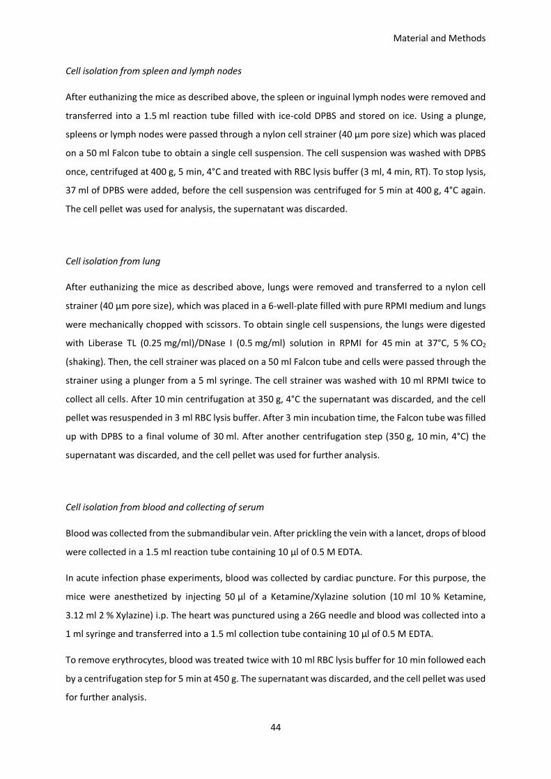
Material and Methods
44
Cell isolation from spleen and lymph nodes
After euthanizing the mice as described above, the spleen or inguinal lymph nodes were removed and
transferred into a 1.5 ml reaction tube filled with ice-cold DPBS and stored on ice. Using a plunge,
spleens or lymph nodes were passed through a nylon cell strainer (40 µm pore size) which was placed
on a 50 ml Falcon tube to obtain a single cell suspension. The cell suspension was washed with DPBS
once, centrifuged at 400 g, 5 min, 4°C and treated with RBC lysis buffer (3 ml, 4 min, RT). To stop lysis,
37 ml of DPBS were added, before the cell suspension was centrifuged for 5 min at 400 g, 4°C again.
The cell pellet was used for analysis, the supernatant was discarded.
Cell isolation from lung
After euthanizing the mice as described above, lungs were removed and transferred to a nylon cell
strainer (40 µm pore size), which was placed in a 6-well-plate filled with pure RPMI medium and lungs
were mechanically chopped with scissors. To obtain single cell suspensions, the lungs were digested
with Liberase TL (0.25 mg/ml)/DNase I (0.5 mg/ml) solution in RPMI for 45 min at 37°C, 5 % CO2
(shaking). Then, the cell strainer was placed on a 50 ml Falcon tube and cells were passed through the
strainer using a plunger from a 5 ml syringe. The cell strainer was washed with 10 ml RPMI twice to
collect all cells. After 10 min centrifugation at 350 g, 4°C the supernatant was discarded, and the cell
pellet was resuspended in 3 ml RBC lysis buffer. After 3 min incubation time, the Falcon tube was filled
up with DPBS to a final volume of 30 ml. After another centrifugation step (350 g, 10 min, 4°C) the
supernatant was discarded, and the cell pellet was used for further analysis.
Cell isolation from blood and collecting of serum
Blood was collected from the submandibular vein. After prickling the vein with a lancet, drops of blood
were collected in a 1.5 ml reaction tube containing 10 µl of 0.5 M EDTA.
In acute infection phase experiments, blood was collected by cardiac puncture. For this purpose, the
mice were anesthetized by injecting 50 µl of a Ketamine/Xylazine solution (10 ml 10 % Ketamine,
3.12 ml 2 % Xylazine) i.p. The heart was punctured using a 26G needle and blood was collected into a
1 ml syringe and transferred into a 1.5 ml collection tube containing 10 µl of 0.5 M EDTA.
To remove erythrocytes, blood was treated twice with 10 ml RBC lysis buffer for 10 min followed each
by a centrifugation step for 5 min at 450 g. The supernatant was discarded, and the cell pellet was used
for further analysis.

Material and Methods
45
To obtain serum, blood samples were transferred to BD microtainer SST blood collection tubes and
centrifuged for 2 min at 10000 g. Serum was transferred to 0.5 ml collection tubes and stored at -20°C
for further analysis.
2.2.3.1 Cell number determination
To determine the cell number of different cell suspensions, cell pellets were dissolved in PBS+1 % BSA
and mixed 1:1 with trypan blue to stain dead cells. Ten µl of this mixture were used in an improved
Neubauer chamber under the microscope to count living cells. The cell number was calculated using
the following formula:
𝑐𝑒𝑙𝑙 𝑐𝑜𝑢𝑛𝑡 𝑥104 =𝑐𝑒𝑙𝑙𝑠 𝑐𝑜𝑢𝑛𝑡𝑒𝑑 𝑥 𝑑𝑖𝑙𝑢𝑡𝑖𝑜𝑛 𝑓𝑎𝑐𝑡𝑜𝑟 𝑥 𝑣𝑜𝑙. 𝑖𝑛 𝑚𝑙
# 𝑜𝑓 𝑐𝑜𝑢𝑛𝑡𝑒𝑑 𝑠𝑞𝑢𝑎𝑟𝑒𝑠
2.2.4 Histological sections
2.2.4.1 Wright-Giemsa staining using Diff-Quik
Histology sections of the ileum and jejunum of infected WT, C5aR1-/-and C5aR2-/- mice were fixed with
methanol (10 sec.), then dipped 5 times in solution C containing thiazine dye, followed by 5 dips in
solution B containing eosin dye. After each staining process, the remaining staining-solution was
washed-off with water and sections were air-dried before use.
2.2.4.2 Evaluation of histology sections
To analyze and evaluate histology sections from the intestine of infected mice, Dr. Sebastian Jendrek
(Department of Internal Medicine, University of Lübeck) and Prof. Dr. Christian Sina (Institute for
Nutritional Medicine, University of Lübeck) provided their expertise. They scored all sections according
to Erben et al (Erben et al., 2014) evaluating tissue destruction (score 0-3) and inflammatory cell
infiltration (score 0-3). Each score ranges from 0 (no damage or cell infiltration) to 3 (massive damage
and cell infiltration) and the sum of both scores (max. score 6) allows a precise evaluation of the
severity of the infection. The scoring was blinded as Dr. Jendrek and Prof. Dr. Sina did not know which
section came from which mice.
Material and Methods
46
2.2.5 Measurement of cytokine production in sera and supernatants
2.2.5.1 Enzyme-linked immunosorbent assay (ELISA)-based measurement
Enzyme-linked immunosorbent assay (ELISA) was used to determine the cytokine concentrations of
IFN- (detection limit: 30 pg/ml), IL-12p40 (detection limit: 39 pg/ml) and IL-12p70 (detection limit:
39 pg/ml) in serum and peritoneal lavage supernatants. Furthermore, cytokine production from in vitro
cell culture supernatants was analyzed. R&D DuoSet ELISA-Kits were used according to the
manufacturers’ recommendations. The reaction volume was reduced to 50 µl without loss of
sensitivity and reproducibility.
2.2.5.2 AYOXXA LUNARISTM 12-Plex Cytokine Kit
The LUNARISTM multiplex system from AYOXXA (AYOXXA Biosystems GmbH, Cologne, Germany) was
used to measure cytokines from serum and peritoneal lavage supernatants. The following cytokines
were measured simultaneously: IL-1, IL-2, IL-4, IL-5, IL-6, IL-10, IL-12p70, IL-13, IL-17A, IFN-, TNF,
GM-CSF. The multiplex system which determines cytokine concentrations from only 3 µl per sample,
is a protein assay format, based on antibody-coated beads immobilized on a solid surface. Every well
on a LUNARISTM BioChip has a unique biomarker-specific antibody-bead distribution which is saved in
a chip-specific datafile. The reaction principle follows a classical sandwich ELISA and all antibodies are
tagged with a fluorophore to analyze cytokine production via the mean fluorescence intensity (MFI).
The LUNARISTM BioChip was analyzed using the AYOXXA LUNARISTM Reader.
2.2.6 In vitro NK cell stimulation
Stimulation with IL-12 and IL-18
Splenic NK cells were purified using either the MoFlo Legacy or FACS ARIA III cell sorter. Prior to the
cell sorting, the splenic cells were stained with CD3, NKp46 or NK1.1 to identify NK cells as NK1.1+
CDe3- or NK1.1+ NKp46+ cells (Figure 16).
After sorting, 1x10⁵ NK cells were cultured in a 6-well plate in 200µl medium and stimulated with
10 ng/ml IL-12 and 10 ng/ml IL-18 for 24 h. In some experiments, NK cells were also stimulated with
100µM of the C5aR2-specific agonist P32. After 24h, the supernatant was collected, and IFN-
production was measured by ELISA.

Material and Methods
47
Figure 16: Gating strategy for sorting NK cells from the spleen depicted as dot plots. A) Excluding cell debris. B) + C) excluding cell doublets. D) Gating on living cells. E) Gating on NK cells as NK1.1+ NKp46+ cells (left) or NK1.1+ CDe3- cells (right).
Stimulation with C5a
NK cells were purified and cultured as described before and 12.5nM C5a was used for the stimulation.
The stimulation was stopped by the addition of 200µl 2 % PFA after 1 min, 2 min, 3 min, 5 min, 10 min
and 30 min. The NK cells were then used for intracellular staining and analysis of MAP-kinase
phosphorylation.
2.2.7 Flow cytometry
2.2.7.1 Surface staining and general gating strategy
Identification and phenotypical analysis of different immune cells from distinct organs was achieved
by flow cytometry. Before staining with specific antibodies, Fc-block (CD16/CD32) was used to block
Fc receptors preventing unspecific binding of antibodies. Fc block was used at 10 µg/ml for 15 min at
4°C. Master mixes were prepared with specific antibodies from Table 6 using the indicated
concentrations. After Fc block incubation, cells were washed with 1 ml of PBS+1 %BSA and centrifuged
at 500 g, 5 min, 4°C before the cell pellet was resuspended in 300 µl of master mix solution and
incubated for 15 min at 4°C again. Following another washing step with 1 ml of PBS+1 %BSA, the cell
pellet was resuspended in BD CytofixTM buffer and incubated for 10 min at 4°C. After another washing
step with 1 ml PBS+1 %BSA and centrifugation at 500 g, 4°C the cell pellet was resuspended in 350 µl
PBS+1 %BSA and analyzed on the BD LSRII cell analyzer or BD FACS ARIAIII cell sorter. For reliable
results, I recorded up to 2x10⁶ cells per sample. To exclude small particles and cell debris, the FSC

Material and Methods
48
threshold was set to 10,000-15,000. Furthermore, FSC-A/SSC-A, FSC-A/FSC-H and SSC-A/SSC-H gating
was used to exclude cell debris and cell doublets (Figure 17).
Figure 17: Gating strategy depicted as dot plots. A) Gate setting to exclude small particles and cell debris. Gating for single cells to exclude cell doublets using B) FSC-A and FSC-H and C) SSC-A and SSC-H.
2.2.7.2 Intracellular stainings
Intracellular staining for different phosphorylated MAP kinases required fixation and permeabilization
with paraformaldehyde (PFA) and methanol. Cells were stained for surface markers before stimulation
as described above. After stimulation (1 min, 2 min, 3 min, 5 min, 10 min, 30 min) 200 µl 2 % PFA was
added to 2x10⁵ cells in 100 µl. Cells where transferred to 1.5 ml reaction tubes containing 1 ml 2 % PFA
to fix the cells. After 10 min incubation at room temperature, pelleting of cells was achieved by
centrifugation at 500 g, 5 min, 4°C. After carefully removing the supernatant, the cell pellet was
resuspended in 1 ml pre-cooled (-20°C) 100 % methanol and vortexed to initiate permeabilization.
After 20 min of incubation on ice and centrifugation at 500 g, 5 min, 4°C, cells were resuspended in
100 µl PBS+1 %BSA and stained with the respective antibodies for intracellular targets (45 min, 4°C)
(Table 6). Finally, after washing cells with PBS+1 %BSA, cells were resuspended in 300µl PBS+1 %BSA
and analyzed on the BD LSRII flow cytometer.
2.2.8 Statistical analyses
Statistical analyses were performed using GraphPad Prism 7 (GraphPad Software, La Jolla, USA). The
presented data show the individual values and the mean ± standard error of the mean (SEM). Two
group comparisons were performed with unpaired students t-test (if not mentioned otherwise) and
multiple group comparisons were performed with one-way ANOVA (analysis of variance) with Tukey
multiple comparison test. Multiple comparisons with more than one independent variable were
performed using two-way ANOVA with Sidak multiple comparison test. To compare survival curves of

Material and Methods
49
two groups log-rank (Mantel-Cox) test was used. P-values ≤ 0.05 were considered as statistically
significant (*p≤0.05; **p<0.01; ***p<0.001; ****p<0.0001).

Results
50
3 RESULTS
3.1 The survival after oral T. gondii infection is higher in C5aR1-/-
and C5aR2-/- mice than in WT mice
Due to the life cycle of the parasite, oral infection of mice with T. gondii cysts is the natural way how
mice become infected with the parasite (Dubey, 1998) and 10 cysts have been considered to be an
appropriate amount of cysts according to the literature (Dias et al., 2014; Liesenfeld, 2002) as well as
personal communication with Prof. Schlüter (Institute for Medical Microbiology, Magdeburg).
To investigate whether C5aR1-/- and C5aR2-/- mice develop different disease severities in comparison
to WT mice, I infected these strains with 10 cysts T. gondii orally.
To evaluate the disease progression, I weighed and scored the mice every day. Mice that fulfilled one
of the termination criteria (weight loss ≥ 20 %, disease score ≥ 4) were sacrificed.
I observed that WT mice were more susceptible compared to C5aR1-/- and C5aR2-/- regarding weight
loss, disease score and survival (Figure 18). However, also C5aR1-/- and C5aR2-/- mice developed severe
inflammation characteristics and were sacrificed due to weight loss of more than 20 % at latest on day
20 p.i.
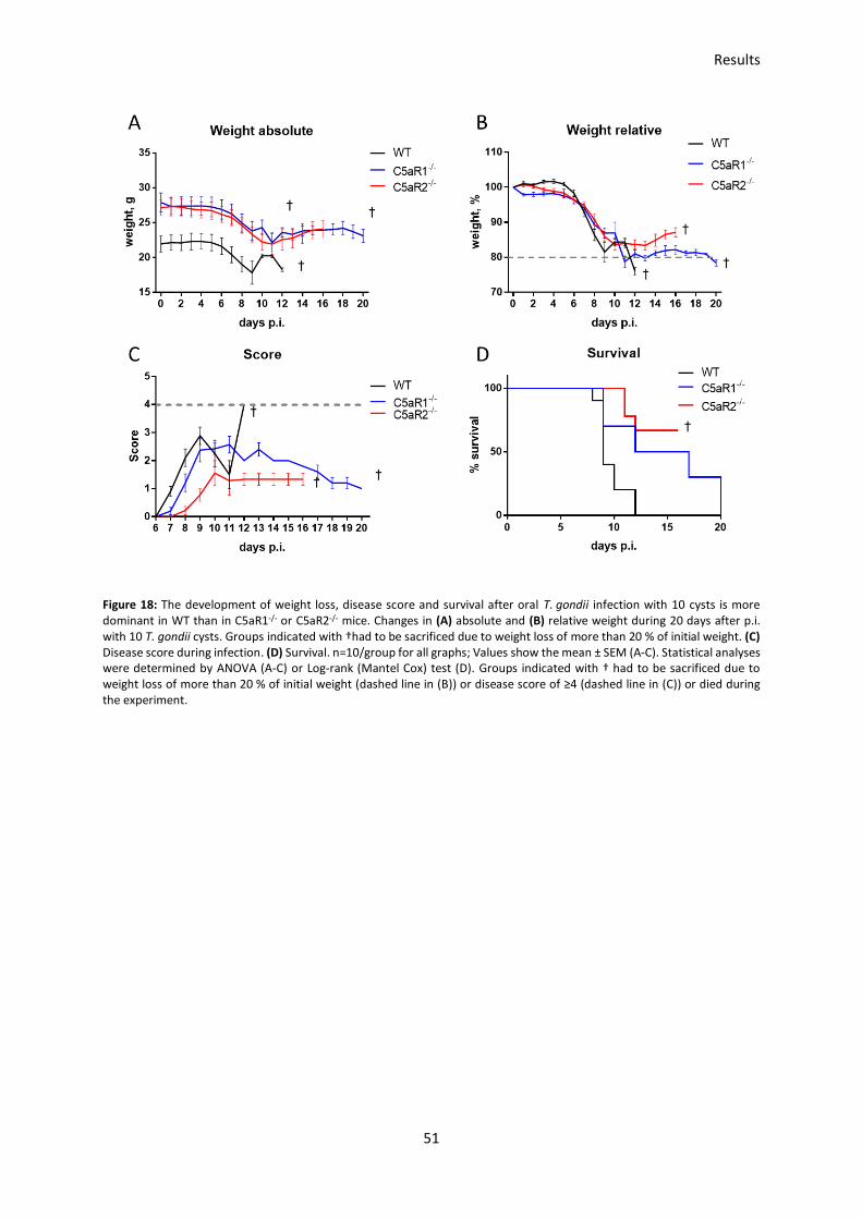
Results
51
Figure 18: The development of weight loss, disease score and survival after oral T. gondii infection with 10 cysts is more dominant in WT than in C5aR1-/- or C5aR2-/- mice. Changes in (A) absolute and (B) relative weight during 20 days after p.i. with 10 T. gondii cysts. Groups indicated with †had to be sacrificed due to weight loss of more than 20 % of initial weight. (C) Disease score during infection. (D) Survival. n=10/group for all graphs; Values show the mean ± SEM (A-C). Statistical analyses were determined by ANOVA (A-C) or Log-rank (Mantel Cox) test (D). Groups indicated with † had to be sacrificed due to weight loss of more than 20 % of initial weight (dashed line in (B)) or disease score of ≥4 (dashed line in (C)) or died during the experiment.

Results
52
3.2 Potential protective role of C5a receptors in the intestine
during T. gondii infections
To evaluate whether C5aR1 or C5aR2 control the immune response after T. gondii infection and cell
infiltration into the intestine, I infected WT, C5aR1-/- and C5aR2-/- mice orally with 10 cysts and after 7
days I analyzed tissue destruction and cell infiltration into the jejunum and ileum.
The qualitative evaluation was performed in collaboration with Dr. Sebastian Jendrek (Department of
Internal Medicine, University of Lübeck) and Prof. Dr. Christian Sina (Institute for Nutritional Medicine,
University of Lübeck) who analyzed all tissue samples according to Erben et al. (Erben et al., 2014) and
scored them for inflammatory cell infiltration and tissue damage. Both scores can range from 0 (no
tissue destruction or cell infiltration) to 3 (massive tissue destruction and cell infiltration) resulting in
a total score ranging from 0 to 6.
The scoring results from the ileum clearly indicated that C5aR2-/- mice are protected from developing
severe inflammation during the early phase of T. gondii infection (Figure 19). The score of C5aR2-/- (1.3)
was much lower than the score of C5aR1-/- (3.6) or WT (4) mice.
In jejunum sections, WT and C5aR2-/- mice showed an average score of 1.6 and 2, respectively and
were protected from developing severe inflammation, whereas C5aR1-/- mice showed a higher score
reaching an average of 4.3, confirming the observed tissue destruction and massive cell infiltration
(Figure 19).
The restriction in C5aR1-/- and C5aR2-/- mice resulting in only a small sample size did not allow a
conclusive statistical evaluation of the data. In future experiments it will be important to increase the
number of mice to obtain more robust results.

Results
53
Figure 19: Inflammatory cell infiltration and tissue damage score in WT, C5aR1-/- and C5aR2-/- mice after seven days of oral T. gondii infection. Scoring results from paraffin-embedded tissue sections of the ileum (left) and jejunum (right) from orally infected WT, C5aR1-/- and C5aR2-/- mice 7 days p.i. Scoring includes tissue destruction and cell infiltration. Statistical analyses were determined by ANOVA. Values show the mean ± SEM. n=3/group
3.3 Impact of the parasite infection dose on disease development
in the oral T. gondii infection model
My previous results showed that the oral infection with 10 cysts resulted in a high mortality as
evidenced by the fulfillment of termination criteria such as massive weight loss in all treatment groups
(Figure 18). In light of this finding, I decided to evaluate the impact of a lower number of cysts on
disease development. I conducted a titration experiment with different numbers of cysts and
specifically infected them with 1, 3, 5, 7, and 10 cysts. Uninfected WT mice were used as a control. To
evaluate the disease progression, I weighed the mice and determined their disease scores every day.
All infected mice started to lose weight after day four p.i. (Figure 20A). All mice infected with ≥3 cysts
reached the threshold of 20 % weight loss and had to be sacrificed before the end of the 15-day
experiment (Figure 20B). In line with the reduced weight, mice showed strong disease symptoms as
indicated by their disease scores (Figure 20C). Here, comparable with weight loss, mice infected with
higher numbers of T. gondii cysts developed a more severe disease score. Mice infected with 10 cysts
already showed first symptoms 7 days p.i., whereas mice infected with 3, 5 and 7 cysts started to
develop symptoms between days 8 and 9. Mice infected with a minimum of one cyst showed first
disease signs between days 10 and 15 and lost a maximum of 15 % of their initial weight. Mice infected
with one cyst were able to recover after the initial infection phase with the exception of one mouse,
while all mice infected with ≥3 cysts died (Figure 20D).
I le u mS
co
re
WT
C5aR
1- /
-
C5aR
2- /
-
0
2
4
6
J e ju n u m
Sc
ore
WT
C5aR
1- /
-
C5aR
2- /
-
0
2
4
6

Results
54
Figure 20: Impact of parasite infection dose on disease development in oral T. gondii infection. Oral infection of WT mice with 1, 3, 5, 7, 10 T. gondii cysts. Absolute (A) and relative weight (B) over 15 days p.i. (C) Disease score during infection. Groups indicated with † had to be sacrificed due to weight loss of more than 20 % of initial weight or died during the experiment. (D) Animal survival; n=5/group for all graphs. Values show the mean ± SEM (A-C). Statistical analyses were determined by ANOVA (A-C) and by Log-rank (Mantel Cox) test (D). Groups indicated with † had to be sacrificed due to termination criteria weight loss of more than 20 % of initial weight (dashed line in B)) or disease score of ≥4 (dashed line in C)) or died during the experiment.
IL12-p40 and IFN- production during infection
As already described in the introduction, IL-12 and IFN- play important roles for the induction of an
immune response after T. gondii infection (1.4.2). DCs are able to recognize T. gondii-derived proteins
and start to produce IL-12 and other pro-inflammatory cytokines. IL-12, besides others, is necessary to
activate NK cells following the production of IFN- to limit the parasite spreading throughout the host.
Therefore, I measured serum-levels of IL-12p40 and IFN- on day 5 and day 7 p.i. as an indicator for
the early immune response.
The results show that IL-12p40 levels raised with increasing parasite load from 2000 pg/ml (1 cyst) up
to 6000 pg/ml (10 cysts) and revealed significant differences between the infection with 1 cyst and
10 cysts 5 days p.i. (Figure 21A). IFN- levels followed the same trend and showed significant
differences, but the concentrations were lower and ranged from 1000 pg/ml (1 cyst) up to 3000 pg/ml

Results
55
(10 cysts) (Figure 21B). Interestingly, analyses from day 7 showed different results, as the IL-12p40
production increased from 6000 pg/ml (1 cyst) up to 10000 pg/ml (5 cysts) and slightly dropped to
8000 pg/ml after the infection with 7 or 10 cysts (Figure 21C). On the other hand, the IFN- production
significantly increased up to 6000 pg/ml (3-10 cysts) compared to 3000 pg/ml (1 cyst) on day 7 p.i.
(Figure 21D).
The comparison of IL12-40 and IFN- production between day 5 and 7 showed that IL-12p40 levels
ranged from 2000 pg/ml (1 cyst) up to 6000 pg/ml (10 cysts) 5 days p.i. (Figure 22A). Furthermore,
IL-12p40 levels significantly increased between day 5 and 7 in all groups, except for mice infected with
10 cysts, which reached their plateau already after 5 days.
IFN- levels significantly increased between day 5 and day 7 after infection in all groups (Figure 22B).
Interestingly, one cyst was enough to induce a strong IFN- response ranging from 1000 pg/ml after 5
days p.i. to 3000 pg/ml after seven days p.i. Noticeable, IFN- levels reached a plateau at around
6000 pg/ml after infection with 3 or more cysts 5 days p.i.
Figure 21: Impact of parasite infection dose on IL-12p40 and IFN- production on day 5 (A, B) and day 7 (C, D) p.i. to compare
the cytokine production relative to the parasite infection dose. IL-12p40 (A) and IFN- (B) production significantly increases after the infection with 1 cyst and 10 cysts 5 days p.i. IL-12p40 production (C) significantly increases after infection with 5
cysts compared to 1 cyst 7 days p.i., whereas the IFN- production (D) significantly increases after infection with 3, 5, 7 and 10 cysts compared to the infection with 1 cyst. Cytokine concentrations from uninfected mice were below the detection limit and are not shown. Values show the mean ± SEM; n=5/group for all graphs. *p<0.05; **p<0.01; ***p<0.001 (ANOVA).

Results
56
Figure 22: Impact of parasite infection dose on IL-12p40 and IFN- production to compare the difference between day 5 and
day 7 p.i. Serum cytokine levels for (A) IL-12p40 and (B) IFN- in response to infection using 1, 3, 5, 7, 10 cysts on day 5 and 7 p.i. Values show the mean ± SEM. n=5/group; asterisks indicate significance between day 5 and day 7; *p<0.05; **p<0.01; ***p<0.001; ****p<0.0001 (ANOVA).
In summary, the results from the oral infection model show that already one cyst is enough to induce
a strong immune response characterized by massive weight loss, a high disease score and high IL12p40
and IFN- levels 5 and 7 days p.i. Although the oral infection is the natural route of infection with the
parasite, my results indicate that this model has a high variability. The reliable administration of only
one cyst is technically difficult.

Results
57
3.4 Assessment of C5aR1/2 expression during acute i.p. T. gondii
infection using floxed GFP-C5aR1 and floxed tdTomato-C5aR2
mice
The results from the oral infection experiments revealed that infection with already one cyst induced
a strong infection characterized by weight loss, the development of a disease score and solid IL-12p40
and IFN- levels. Due to the high variability of parasites within one cyst and the difficult preparation
and precise application of only one cyst, I decided to use the i.p. infection model to obtain more reliable
results. Previous experiments from Daria Briukhovetska (Briukhovetska, 2017) revealed that
C5aR1-/- mice suffered from increased susceptibility during i.p. T. gondii infections characterized by
increased cyst burden in the brain 30 days p.i. as well as increased mortality. Mainly responsible for
the altered susceptibility were decreased systemic IL-12 and IFN- levels in C5aR1-/- mice in the early
phase of the infection.
Recently the Koehl laboratory generated floxed GFP-C5aR1 and floxed tdTomato-C5aR2 knock-in mice
to analyze and evaluate the expression of complement receptors C5aR1 and C5aR2, respectively. The
mice were already used to characterize C5aR1 and C5aR2 expression in different immune cells isolated
from different organs under steady state conditions (Karsten et al., 2015, 2017). My data obtained with
the oral infection model and the previous data from Daria Briukhovetska (Briukhovetska, 2017)
showed that C5aR1 and C5aR2 exert important roles during T. gondii infection. Here, I aimed to use
floxed GFP-C5aR1 and floxed tdTomato-C5aR2 mice to evaluate the expression of both receptors under
steady state conditions and seven days after i.p. infection with T gondii.
Expression and regulation of C5aR1 on immune cells under steady state and infectious conditions
To define the background signal caused by autofluorescent cells, I used WT mice as controls (Figure
23A, left panel). Samples from uninfected and infected GFP-C5aR1fl/fl reporter mice showed for all
tissues a clear and distinct GFP-C5aR1 expression. The immune cell population was pre-gated as
described in chapter 2.2.7. The comparison of uninfected and infected reporter mice showed that
significantly more cells express GFP-C5aR1 in the peritoneal cavity, spleen and lung under infectious
conditions (Figure 23B). The comparison of uninfected and infected reporter mice in blood showed no
significant differences in cell numbers or the frequency of C5aR1+ cells (Figure 23B). In the peritoneal
cavity, GFP-C5aR1 expressing cells significantly increased from 1x10⁵ cells (6 %) under steady state
conditions to 2.5x10⁵ cells (15 %) under infectious conditions. Cells in the spleen also significantly
increased GFP-C5aR1 expression under infectious conditions from 1x10⁶ cells (2 %) up to 3x10⁶ cells
(4 %). In the lung, immune cells expressing GFP-C5aR1 increased from 1x10⁶ cells (5 %) under steady

Results
58
state conditions up to 3x10⁶ cells (13 %) under infectious conditions (Figure 23B). The significant
changes in C5aR1 expression were also reflected by the relative increase in C5aR1+ cells (Figure 23C).
Taken together, these results showed that more cells in the peritoneal cavity, spleen and lung
expressed GFP-C5aR1 during an acute T. gondii infection. Furthermore, steady state levels of
GFP-C5aR1 expression in the blood, peritoneal cavity and lung were comparable in all tissues (3-6 % of
all cells were GFP-C5aR1+). Under infectious conditions, cells expressing GFP-C5aR1 increased to a
different extent.
Figure 23: The number and the frequency of GFP-C5aR1+ mice is increased in the peritoneal cavity, spleen and lung, but not in blood seven days after peritoneal T. gondii infection. GFP-C5aR1fl/fl mice were infected i.p. with 50 T. gondii cysts and sacrificed on day 7 p.i. Uninfected WT mice served as controls. (A) Flow cytometric analysis of the GFP-C5aR1 signal in cells from blood, peritoneal cavity, spleen and lung. Dot plots depict the GFP-C5aR1 signal in WT, uninfected GFP-C5aR1fl/fl and infected GFP-C5aR1fl/fl mice. (B) Cell counts of GFP-C5aR1 positive cells in blood, peritoneal cavity, spleen and lung. (C) Graphs show the percentage of GFP-C5aR1 positive cells in blood, peritoneal cavity, spleen and lung. Values show the mean ± SEM; n = 5-10. Results are from three independent experiments. Statistical differences between groups were assessed by student t test. *p<0.05, **p<0.01, ****p<0.0001

Results
59
Expression and regulation of C5aR2 on immune cells under steady state and infectious conditions
In addition to C5aR1, C5a is able to bind to C5aR2. In comparison to C5aR1, our knowledge regarding
C5aR2 expression and function is less profound. To reduce the gap, I used tdTomato-C5aR2fl/fl mice to
analyze the expression of C5aR2 in different tissues under steady state conditions and after peritoneal
T. gondii infection.
For tdTomato-C5aR2fl/fl reporter mice, I used the same experimental design to define and analyze
tdTomato-C5aR2 expressing cells and regulation of this receptor as for GFP-C5aR1. Comparable with
the results obtained with GFP-C5aR1fl/fl mice, cells from tdTomato-C5aR2fl/fl mice showed a distinct
tdTomato-C5aR2+ population in all analyzed compartments under steady state and infectious
conditions (Figure 24A). Comparing blood cells from uninfected and infected reporter mice revealed
that the cell number significantly increased from 0.5x10⁶ (10 %) to 1.5x10⁶ (40 %) cells (Figure 24B+C).
In the peritoneal cavity around 2.5x10⁵ cells (13 %) showed tdTomato-C5aR2 expression under steady
state and infectious conditions. Spleen cells significantly increased tdTomato-C5aR2 expression from
1x10⁷ (10 %) to 1.8x10⁷ (20 %). The number of tdTomato C5aR2-expressing cells in the lung did not
change after infection and was around 1.2x10⁶ cells (5 %) (Figure 24B+C).
Interestingly, the number of tdTomato-C5aR2+ cells in the spleen under steady state and infectious
conditions was 10-times higher than the number of GFP-C5aR1+ cells.
These results show that more cells in the blood and spleen express tdTomato-C5aR2 in response to i.p.
T. gondii infection. The cells in the peritoneal cavity and lung did not change their tdTomato-C5aR2
expression pattern under infectious conditions.

Results
60
Figure 24: The number and frequency of td-Tomato-C5aR2+ cells increased in the blood and the spleen, but not in the peritoneal cavity and the lung after peritoneal T. gondii infection. Td-Tomato-C5aR2fl/fl mice infected i.p. with 50 T. gondii cysts and sacrificed on day 7 p.i. Uninfected WT mice served as controls. (A) Flow cytometric analysis of the tdTomato-C5aR2 signal in cells from blood, peritoneal cavity, spleen and lung. Dot plots depict the tdTomato-C5aR2 signal in WT, uninfected tdTomato-C5aR2fl/fl and infected tdTomato-C5aR2fl/fl mice. (B) Cell counts of tdTomato-C5aR2-positive cells in blood, peritoneal cavity, spleen and lung. (C) Frequency of tdTomato-C5aR2-positive cells in blood, peritoneal cavity, spleen and lung as measured by flow cytometry. Values show the mean ± SEM; n = 5-10. Results are from three independent experiments. Statistical differences between groups were assessed by students t-test; *p<0.05, **p<0.01, ***p<0.001
GFP-C5aR1 and tdTomato-C5aR2 are not always regulated in a similar manner in blood, spleen and
lung
Since the discovery of C5aR2 in 2000, several reports have shown that C5aR2 is co-expressed and
co-regulated with C5aR1 (Bamberg et al., 2010; Gerard et al., 2005). Further, it has been shown that
C5aR2 can function independent of C5aR1, or even counter-act C5aR1 signaling (Huber-Lang et al.,
2005; Xu et al., 2016). Therefore, and after analyzing the two receptors independently from each other
(Figure 23 and Figure 24), I aimed to analyze the regulation of both receptors in relation to each other.
Using the two reporter mice, I found that C5aR1 and C5aR2 are differentially expressed during T. gondii
infection. In blood, a higher frequency of cells expressed tdTomato-C5aR2 under infectious conditions,
while no change in GFP-C5aR1 expression was detectable (Figure 25A). This trend is also reflected by
cell numbers (Figure 26A). In the peritoneal cavity, the frequencies (Figure 25B) and numbers (Figure

Results
61
26B) of GFP-C5aR1+ and tdTomato-C5aR2+ cells were similar under infectious conditions. In contrast, I
found a higher frequency of cells expressing tdTomato-C5aR2 under steady state conditions. The
results from the spleen showed that a higher frequency (Figure 25C) and a higher number of cells
(Figure 26C) expressed tdTomato-C5aR2 under steady state conditions. Even more cells expressed
tdTomato-C5aR2 under infectious conditions. Frequencies from lung cells showed significant
differences between GFP-C5aR1+ and tdTomato-C5aR2+ cells, as the frequency for GFP-C5aR1+ cells
was 3-times higher under infectious conditions (Figure 25D) than that of tdTomato-C5aR2+ cells. I
found a similar trend regarding the cell numbers (Figure 26D).
Figure 25: Frequency of GFP-C5aR1 and tdTomato-C5aR2-expressing cells in different tissues under steady state conditions and infectious conditions are regulated differently. Frequencies of C5aR1-GFP+ and C5aR2-tdTomato+ cells in (A) blood, (B) peritoneal cavity, (C) spleen and (D) lung in samples from uninfected (black) or infected (red) mice. Values show the mean ± SEM. n=5-11. Results are from three independent experiments. Statistical differences between groups were assessed by ANOVA. ***p<0.001; ****p<0.0001.

Results
62
Figure 26: Cell numbers of GFP-C5aR1 and tdTomato-C5aR2-expressing cells in different tissues under steady state conditions and infectious conditions also regulated differently. Cell numbers of C5aR1-GFP+ and C5aR2-tdTomato+ cells in (A) blood, (B) peritoneal cavity, (C) spleen and (D) lung in samples from uninfected (black) or infected (red) mice. Values show the mean ± SEM. n=5-11. Results are from three independent experiments. Statistical differences between groups were assessed by ANOVA. ***p<0.001;
In the next set of experiments, I determined the expression of C5aR1 and C5aR2 in different immune
cells including NK cells, neutrophils, inflammatory monocytes, cDCs and eosinophils using flow
cytometry.
Regulation of C5aR1 and C5aR2 expression on natural killer cells
NK cells are one of the most important innate immune cell population during early T. gondii infections.
As the main producer of IFN-, NK cells play a crucial role in controlling the spreading of the parasite
by activation of other innate immune cells (Denkers et al., 1993). IFN- production from NK cells is
dependent on DC-derived cytokines like IL-12, IL-15 or IL-18 (Une et al., 2003). Additionally, NK cells

Results
63
regulate the immune response during T. gondii infection by producing anti-inflammatory cytokines like
IL-10 (Gigley, 2016).
For flow cytometric analysis, NK cells were identified as NK1.1+ NKp46+ cells (Figure 27A). Under naïve
conditions, the blood, spleen and lung are the tissues with the highest frequency of NK cells with
around 3 % of total cells, followed by the peritoneal cavity with around 1 % of total cells (Figure 27B).
Most NK cells reside in the spleen (~20x105 cells), followed by the lung (4x10⁵ cells), the blood (1x10⁵
cells) and the peritoneal cavity (0.2x10⁵ cells) (Figure 27C). Under infectious conditions NK cell
frequencies dropped in blood, spleen and lung and increased in the peritoneal cavity, which is the first
compartment to encounter the parasite during the infection (Figure 27B). Similar to the cell
frequencies, cell numbers decreased in the peritoneal cavity and the spleen. In contrast, I observed no
drop of cell numbers in blood and lung (Figure 27C). As shown in Figure 27D+E, NK cells did not express
GFP-C5aR1 in any of the examined organs. In contrast, NK cells showed a clear and comparable signal
of tdTomato-C5aR2 expression under steady state and infectious conditions (Figure 27D+F). In the
blood, around 2 % of cells from non-infected and 5 % from infected mice and in the lung around 2 %
cells from non-infected and 3% of infected mice were tdTomato-C5aR2+. Twelve percent of naïve NK
cells in the peritoneal cavity were tdTomato-C5aR2+ and around 10 % of NK cells under infectious
conditions. Thus, the peritoneal cavity was the compartment with the highest frequency of
tdTomato-C5aR2+ NK cells. NK cells from the spleen also expressed tdTomato-C5aR2. Here, 7 % of cells
from non-infected and around 10 % of cells from infected mice were tdTomato-C5aR2+. The
frequencies for tdTomato-C5aR2-expressing NK cells did not change between steady state and
infectious conditions in all tissues. Interestingly, the total number of NK cells, which express tdTomato-
C5aR2 in the spleen, dropped significantly under infectious conditions (Figure 27H).

Results
64

Results
65
Figure 27: NK cells from blood, peritoneal cavity, spleen and lung express tdTomato-C5aR2, but not GFP-C5aR1 under steady state conditions and seven days after i.p. infection with T. gondii. (A) NK cells were identified as NK1.1+NKp46+ cells. Depicted is the dot plot from an uninfected spleen sample. Frequencies (B) and cell counts (C) of NK cells in non-infected and infected reporter mice shown as percentage of living cells. (D) Flow cytometric analysis of GFP-C5aR1 (upper histograms) and tdTomato-C5aR2 (lower histograms) expression in NK cells from blood, peritoneal cavity, spleen and lung of non-infected WT mice (gray histograms), non-infected (dashed line) and infected (solid line) C5aR1/2 reporter mice. (E+F) Frequencies of GFP-C5aR1+ and tdTomato-C5aR2+ NK cells in blood, peritoneal cavity, spleen and lung under steady state conditions and upon i.p. infection with T. gondii. (G+H) cell counts of GFP-C5aR1+ and tdTomato-C5aR2+ NK cells in blood, peritoneal cavity, spleen and lung under steady state conditions and upon i.p. infection with T. gondii. Values show the mean ± SEM; n = 5-14. Results are from three independent experiments. Statistical differences between groups were assessed by student t test. ***p<0.001; ****p<0.0001
Regulation of C5aR1 and C5aR2 expression on macrophages
Macrophages are not only able to phagocytose dead parasites, but can also be infected by living
parasites, which can actively invade immune cells (Mordue and Sibley, 1997; Sibley et al., 1985). For
flow cytometric analysis, macrophages were identified by expression of the macrophage surface
marker F4/80 (Dos Anjos Cassado, 2017) (Figure 28A). Under naïve conditions, macrophages were a
major cell population in the peritoneal cavity with around 30 % of all cells, while only 4 % in the spleen
and 2 % in the lung were identified as macrophages (Figure 28B). I found that the spleen was the
compartment with the highest number of macrophages (30x105) under naïve conditions, followed the
peritoneal cavity (4x10⁵) and the spleen (1,5x10⁵) (Figure 28C). Under infectious conditions, the
frequencies significantly dropped in the peritoneal cavity and lung, while the frequency in the spleen
significantly increased (Figure 28B). Interestingly, the cell numbers only increased in the spleen, but
did not significantly change in the peritoneal cavity or lung during T. gondii infection. As visible in the
histograms, macrophages from different compartments expressed both C5aRs with different
intensities under steady state and infectious conditions (Figure 28D).
Between 40 to 80 % of macrophages in the peritoneal cavity, spleen and lung expressed GFP-C5aR1
and tdTomato-C5aR2 under steady state conditions. (Figure 28G+H).The histograms showed that, in
contrast to the homogeneous expression levels in the spleen, macrophages in the peritoneal cavity
and the lung expressed GFP-C5aR1 in a heterogenous manner under steady state conditions (Figure
28D). Furthermore, only a slight change in expression occurred after infection. Around 80 % of naïve
and 65 % of peritoneal macrophages under infectious conditions were GFP-C5aR1+ (Figure 28G). The
high variance of data and low cell frequency under steady state conditions may also be responsible for
the reduced GFP-C5aR1 expression results.
Interestingly, cell numbers for GFP-C5aR1-expressing macrophages dropped significantly (Figure 28I).
In the spleen, nearly all naïve macrophages expressed GFP-C5aR1, but the frequencies and cell
numbers of GFP-C5aR1+ cells significantly dropped under infectious conditions (Figure 28I). In the lung,
the frequency of GFP-C5aR1 expressing macrophages increased from 40 % in uninfected to 55 % in

Results
66
macrophages from infected mice. In contrast, I observed no change in the number of GFP-C5aR1+ cells
(Figure 28I). Interestingly, the expression of GFP-C5aR1 on macrophages from lung but not from the
peritoneal cavity or spleen significantly decreased during T. gondii infection (Figure 28E).
Furthermore, the overall frequencies and the number of tdTomato-C5aR2-expressing macrophages
decreased in spleen and peritoneal cavity. In contrast, frequencies were unchanged whereas cell
numbers of tdTomato-C5aR2+ macrophages were significantly reduced in the lung (Figure 28H+J). The
reduced frequencies and numbers of tdTomato-C5aR2+ macrophages in the peritoneal cavity and
spleen were associated with a significant downregulation of tdTomato-C5aR2 on macrophages found
in the two compartments (Figure 28F).

Results
67

Results
68
Figure 28: Regulation of GFP-C5aR1 and tdTomato-C5aR2 expression in macrophages is different in the peritoneal cavity, spleen and lung under steady state conditions and seven days after i.p. infection with T. gondii. (A) Macrophages were identified as F4/80+ cells. Depicted is the dot plot from an uninfected spleen sample. Frequencies (B) and cell counts (C) of F4/80+ macrophages in non-infected and infected reporter mice shown as percentage of living cells. (D) Flow cytometric analysis of GFP-C5aR1 (upper histograms) and tdTomato-C5aR2 (lower histograms) expression in macrophages from peritoneal cavity, spleen and lung from non-infected WT mice (grey histograms), non-infected (dashed line) and infected
(solid line) C5aR1/2 reporter mice. (E+F) MFI values for GFP-C5aR1 and tdTomato-C5aR2 expression in macrophages from the peritoneal cavity, spleen and lung under steady state conditions and upon i.p. infection with T. gondii. (G+H) Frequencies of GFP-C5aR1+ and tdTomato-C5aR2+ macrophages in peritoneal cavity, spleen and lung under steady state conditions and upon i.p. infection with T. gondii. (I+J) cell counts of GFP-C5aR1+ and tdTomato-C5aR2+ macrophages in peritoneal cavity, spleen and lung under steady state conditions and upon i.p. infection with T. gondii. Values show the mean ± SEM; n = 5-14. Results are form three independent experiments. Statistical differences between groups were assessed by student t test. *p<0.05; **p<0.01; ***p<0.001; ****p<0.0001
Regulation of C5aR1 and C5aR2 expression on neutrophils
For flow cytometric analysis, neutrophils were identified as Ly6g+ SSC-Ahigh cells (Figure 29A).
Comparison of neutrophil frequencies showed that less than 10 % of the cells in the blood, spleen or
lung were neutrophils under steady state conditions (Figure 29B). Neutrophils are normally stored in
the bone-marrow and start migrating to the site of infection after activation or recognition of
chemoattractants like C5a (Swamydas and Lionakis, 2013). In the peritoneal cavity, no neutrophils
could be detected under steady state conditions. Under infectious conditions, around 40 % of the cells
in the blood, 15 % of cells in the spleen and lung and 10 % of cells in the peritoneal cavity were
neutrophils. This increase in frequencies is associated with an increase in the neutrophil cell count
(Figure 29C). I found the highest number of neutrophils (around 100x105 cells) in the spleen, followed
by the lung (40x105), the blood (20x105) and the peritoneal cavity (3x10⁵). As shown in the histograms,
neutrophils expressed both C5a receptors under steady state and infectious conditions (Figure 29D).
Between 80 % and 90 % of neutrophils expressed GFP-C5aR1 in the blood, spleen and lung under
steady state conditions (Figure 29G). No expression could be determined for neutrophils in the
peritoneal cavity due to the absence of neutrophils under steady state conditions. Under infectious
conditions between 75 % and 85 % of neutrophils in all compartments expressed GFP-C5aR1. The GFP-
C5aR1 expression was significantly reduced in neutrophils located in the blood or the lung but not in
neutrophils located in the spleen (Figure 29E). The numbers of GFP-C5aR1+ neutrophils in the spleen
significantly increased (Figure 29I). In the blood 95 % of neutrophils expressed tdTomato-C5aR2, while
only 60 % to 65 % expressed the receptor in the spleen and lung under steady state conditions (Figure
29H). During infection, the expression in the blood dropped to 70 %, while no change was observed in
the spleen and lung. In the peritoneal cavity, around 60 % expressed tdTomato-C5aR2 under infectious
conditions. From the MFI it was visible, that the tdTomato-C5aR2 expression on neutrophils in the
blood and lung, but not in spleen decreased upon infection (Figure 29F). Similarly, the frequency of
tdTomato-C5aR2 significantly decreased in the blood following infection, while I found no change in

Results
69
the other compartments. The cell number of tdTomato-C5aR2+ neutrophils increased in all
compartments (Figure 29J).

Results
70

Results
71
Figure 29: Regulation of GFP-C5aR1 and tdTomato-C5aR2 expression in neutrophils from the blood, peritoneal cavity, spleen and lung under steady state conditions and seven days after i.p. infection with T. gondii. (A) Neutrophils were identified as Ly6g+ SSC-Ahigh cells. Depicted is the dot plot from an uninfected spleen sample. Frequencies (B) and cell counts (C) of neutrophils in non-infected and infected reporter mice shown as percentage of living cells. (D) GFP-C5aR1 (upper histograms) and tdTomato-C5aR2 (lower histograms) expression in neutrophils from blood, peritoneal cavity, spleen and lung of non-infected WT mice (grey histogram), non-infected (dashed line) and infected (solid line) C5aR1/2 reporter mice. (E+F) MFI values for GFP-C5aR1 and tdTomato-C5aR2 in neutrophils from the blood, peritoneal cavity, spleen and lung under steady state conditions and upon i.p. infection with T. gondii. (G+H) Frequencies of GFP-C5aR1+ and tdTomato-C5aR2+ neutrophils in blood, peritoneal cavity, spleen and lung under steady state and upon i.p. infection with T. gondii. (I+J) Cell counts of GFP-C5aR1+ and tdTomato-C5aR2+ neutrophils in blood, peritoneal cavity, spleen and lung under steady state conditions and upon i.p. infection with T. gondii. Values show the mean ± SEM; n = 5-14. Results are from three independent experiments. Statistical differences between groups were assessed by student t test. p<0.05; **p<0.01; ***p<0.001; ****p<0.0001
Regulation of C5aR1 and C5aR2 expression on inflammatory monocytes
During T. gondii infections, inflammatory monocytes are recruited to the site of infection through a
mechanism that involves the TLR11 and TLR12 ligand profilin (Neal and Knoll, 2014). Mice deficient in
inflammatory monocytes or with impaired ability to recruit such cells succumb to death during
T. gondii infections due to an uncontrolled pro-inflammatory response (Dunay et al., 2010). For flow
cytometric analysis, inflammatory monocytes were defined as Ly6G- Ly6Chigh cells (Figure 30A). Under
naïve conditions around 10 % of the cells in the blood and spleen and less than 5 % in the peritoneal
cavity were inflammatory monocytes (Figure 30B). The only significant increase to 25 % during
T. gondii infection occurred in the peritoneal cavity. The number of GFP-C5aR1+ monocytes
significantly increased under infectious conditions in the peritoneal cavity and the spleen (Figure 30C).
Investigating GFP-C5aR1 and tdTomato-C5aR2 expression revealed at best minor expression for
GFP-C5aR1, but a solid expression of tdTomato-C5aR2 in the spleen under both conditions. Further,
tdTomato-C5aR2 was expressed on a small population of blood monocytes under steady state
conditions which was absent following infection (Figure 30D+F). A clear increase in frequencies of
GFP-C5aR1+ and tdTomato-C5aR2+ inflammatory monocytes under infectious conditions was
detectable in the peritoneal cavity but not in the blood or spleen (Figure 30E+F). In contrast, the cell
numbers of GFP-C5aR1+ or tdTomato-C5aR2+ monocytes in the different compartments did not change
upon infection (Figure 30H+I). Interestingly, up to 20 % of inflammatory monocytes expressed
tdTomato-C5aR2 during infection, but only 7 % expressed GFP-C5aR1. The frequency of
tdTomato-C5aR2+ inflammatory monocytes in the spleen was around 20 %-25 % and did not change in
response to infection. Interestingly, the number of tdTomato-C5aR2+ inflammatory monocytes
increased significantly which was in line with the increased number of total inflammatory monocytes
in the spleen (Figure 30C+I).

Results
72

Results
73
Figure 30: Regulation of GFP-C5aR1 and tdTomato-C5aR2 expression in inflammatory monocytes from the blood, peritoneal cavity and spleen under steady state conditions and seven days after i.p. infection with T. gondii. (A) Inflammatory monocytes were identified as Ly6g+ Ly6c+ cells. Depicted is the dot plot from an uninfected spleen sample. Frequencies (B) and cell counts (C) of inflammatory monocytes in non-infected and infected reporter mice as percentage of living cells. (D) Flow cytometric analysis of GFP-C5aR1 (upper histograms) and tdTomato-C5aR2 (lower histograms) expression in inflammatory monocytes from blood, peritoneal cavity and spleen of non-infected WT mice (grey histogram), non-infected (dashed line) and infected
(solid line) C5aR1/2 reporter mice. (E) MFI values for tdTomato-C5aR2 in inflammatory monocytes from the blood, peritoneal cavity and spleen under steady state conditions and upon i.p. infection with T. gondii. (F+G) Frequencies of GFP-C5aR1+ and tdTomato-C5aR2+ inflammatory monocytes in blood, peritoneal cavity and spleen under steady state conditions and upon i.p. infection with T. gondii. (H+I) Cell counts of GFP-C5aR1+ and tdTomato-C5aR2+ inflammatory monocytes in blood, peritoneal cavity and spleen under steady state conditions and upon i.p. infection with T. gondii. Values show the mean ± SEM; n = 5-14. Results are from three independent experiments. Statistical differences between groups were assessed by student t test. p<0.05; **p<0.01; ***p<0.001; ****p<0.0001
Regulation of C5aR1 and C5aR2 expression on conventional dendritic cells
During T. gondii infections, DCs play important roles, as they are the main producers of IL-12 and
therefore responsible for the activation of other immune cells and especially NK cells during the early
phase of the infection (Liu et al., 2006).
For flow cytometric analysis, DCs were identified as MHCIIhi CD11c+ cells (Figure 31A). Under naïve
conditions, around 1.5 % of cells in the blood, peritoneal cavity and spleen were identified as DCs
(Figure 31B). Under infectious conditions the frequency and the number of DCs significantly increased
in the peritoneal cavity and spleen (Figure 31B+C). As shown in the histograms, DCs expressed GFP-
C5aR1, but not tdTomato-C5aR2 (Figure 31D). The GFP-C5aR1 expression in DCs from the blood,
peritoneum or spleen from uninfected and infected mice was similar (Figure 31E). The frequencies and
numbers of GFP-C5aR1+ DCs in the peritoneum (50 %, 1x10⁵ cells) and spleen (10 %, 3x10⁵ cells)
increased significantly upon infection (Figure 31F+G).
DCs showed no expression of tdTomato-C5aR2 in the different compartments under steady state
conditions (Figure 31D). Under infectious conditions the expression of tdTomato-C5aR2 increased, at
best, minor. The number of tdTomato-C5aR2+ DC in the blood and the spleen but not in the peritoneal
cavity increased significantly. The significant increase in tdTomato-C5aR2+ blood DC numbers was
associated with a significant increase in the frequency of tdTomato-C5aR2+ blood DCs whereas the
frequency of tdToamto-C5aR2+ spleen DCs did not change upon infection (Figure 31G+I).

Results
74

Results
75
Figure 31: Regulation of GFP-C5aR1 and tdTomato-C5aR2 expression in cDCs from the blood, peritoneal cavity and spleen under steady state conditions and seven days after i.p. infection with T. gondii. (A) cDCs were identified as MHCII+ CD11C+ cells. Depicted is the dot plot from an uninfected spleen sample. Frequencies (B) and cell counts (C) of cDCs in non-infected and infected reporter mice shown as percentage of living cells. (D) Flow cytometric analysis of GFP-C5aR1 (upper histograms) and tdTomato-C5aR2 (lower histograms) expression in cDCs from blood, peritoneal cavity and spleen of non-infected WT
mice (grey histogram), non-infected (dashed line) and infected (solid line) C5aR1/2 reporter mice. (E) MFI values for GFP-C5aR1 in cDCs from the blood, peritoneal cavity and spleen under steady state conditions and upon i.p. infection with T. gondii. (F+G) Frequencies of GFP-C5aR1+ and tdTomato-C5aR2+ cDCs in blood, peritoneal cavity and spleen under steady state conditions and upon i.p. infection with T. gondii. (H+I) Cell counts of GFP-C5aR1+ and tdTomato-C5aR2+ cDCs in blood, peritoneal cavity and spleen under steady state conditions and upon i.p. infection with T. gondii. Values show the mean ± SEM; n = 5-14. Results are from three independent experiments. Statistical differences between groups were assessed by student t test. p<0.05; **p<0.01; ***p<0.001; ****p<0.0001
Regulation of C5aR1 and C5aR2 expression on eosinophils
Previous findings suggest that eosinophils control T. gondii infections mainly in the lung (Fenoy et al.,
2009). Therefore, I only analyzed GFP-C5aR1 and tdTomato-C5aR2 expression on eosinophils from the
lung.
For flow cytometric analysis, eosinophils in the lung were defined as Siglec-F+ CD11c- cells (Figure 32A).
The frequencies of eosinophils in the lung were low under steady state conditions and further
decreased under infectious conditions (Figure 32B). The number of lung eosinophils did not change
upon infection (Figure 32C).
As shown in Figure 32D, eosinophils expressed both C5a receptors under both conditions. The
expression intensity was similar on eosinophils from non-infected and infected mice as shown by the
MFI (Figure 32E+F). The comparison of the frequencies and cell numbers of GFP-C5aR1+ or tdTomato-
C5aR2+ eosinophils in the pulmonary compartment showed no differences between steady state or
infectious conditions (Figure 32G-J).

Results
76

Results
77
Figure 32: Regulation of GFP-C5aR1 and tdTomato-C5aR2 expression in eosinophils from the lung C5aR1 under steady state conditions and seven days after i.p. infection with T. gondii. (A) eosinophils were identified as Siglec-F+ CD11c- cells. Depicted is the dot plot from an uninfected lung sample. Frequencies (B) and cell counts (C) of eosinophils in non-infected and infected reporter mice as percentage of living cells. (D) Flow cytometric analysis of GFP-C5aR1 (upper histograms) and tdTomato-C5aR2 (lower histograms) expression in eosinophils from the lung of non-infected WT mice (grey histogram), non-
infected (dashed line) and infected (solid line) C5aR1/2 reporter mice. (E+F) MFI values for GFP-C5aR1 and tdTomato-C5aR2 in eosinophils from the lung under steady state conditions and upon i.p. infection with T. gondii. (G+H) Frequencies of GFP-C5aR1+ and tdTomato-C5aR2+ eosinophils in the lung under steady state conditions and upon i.p. infection with T. gondii. (I+J) Cell counts of GFP-C5aR1+ and tdTomato-C5aR2+ eosinophils in the lung under steady state conditions and upon i.p. infection with T. gondii. Values show the mean ± SEM; n = 5-14. Results are from three independent experiments. Statistical differences between groups were assessed by student t test. ***p<0.001

Results
78
3.5 Functional roles of C5aR2 on NK cells and DCs
3.5.1 Frequencies and cell numbers of DCs and NK cells of WT, C5aR1-/- and
C5aR2-/- mice in the spleen
The results obtained from the reporter mice showed that NK cells express tdTomato-C5aR2, but not
GFP-C5aR1. The exclusive expression of C5aR2 in NK cells makes them a suitable cell population to
investigate the function and potential consequences of the absence of C5aR2.
Splenic DCs are important and indispensable for activation of NK cells. The formation of a stimulatory
synapse between DCs and NK cells with cell-cell contact is the basis for a strong NK cell-driven immune
response (Borg et al., 2004). In response to stimulatory synapse formation, DCs produce IL-12, IL-15
and IL-18 and deliver these cytokines directly to the NK cells subsequently leading to IFN- production.
To determine whether WT, C5aR1-/- and C5aR2-/- DCs or NK cell numbers and frequencies differ in the
spleen, I performed flow cytometric analyses. Interestingly, the frequency of cDCs in C5aR2-/- was
significantly lower compared to WT mice (Figure 33A). Further, the number of cDCs in C5aR2-/- mice
was, in trend, lower than in WT mice. Also, the frequencies of NK cells were reduced in C5aR1-/- and
C5aR2-/- mice compared to WT mice. This reduction was not reflected by the cell counts (Figure 33B),
which might be due to the small sample size.
Figure 33: Impact of C5aR1 or C5aR2 deficiency on the relative and absolute cell numbers of cDCs and NK cells in the spleen. (A) cDC and (B) NK cell frequencies (left) and cell numbers (right) in the spleen of WT, C5aR1-/- and C5aR2-/- mice. Values show the mean ± SEM. n=3-15. *p<0.05; **p<0.01. Statistical differences between WT, C5aR1-/- and C5aR2-/- were determined by ANOVA.

Results
79
3.5.2 The C5a/C5aR2 axis controls IFN- production from NK cells after
IL-12/IL-18 stimulation
To test whether C5aR2 regulates IFN- production from NK cells, I purified NK cells from WT and
C5aR2-/- mice via FACS and stimulated them with or without IL-12 and IL-18 for 20h at 37°C and 5 %
CO2. After 24h, I collected the supernatant from the NK cell cultures and measured IFN- by ELISA. In
some experiments, I pre-stimulated the purified NK cells with the specific C5aR2 agonist P32
30 minutes before IL-12/IL-18 stimulation. P32 (Croker et al., 2016) was kindly provided by Trent
Woodruff (The University of Queensland, Australia).
Stimulation of WT NK cells with IL-12 and IL-18 resulted in strong production of IFN- (Figure 34A).
Strikingly, purified NK cells from C5aR2-/- mice produced significantly higher amounts of IFN- than NK
cells from WT mice under the same stimulatory conditions, suggesting that C5aR2 controls IL12/18-
driven IFN- production from NK cells. To test whether the C5aR2 is directly involved in the regulation
of the production of IFN-, I pre-stimulated purified NK cells with the C5aR2-specific agonist P32. The
IFN- production from NK cells pre-stimulated with the agonist P32 was significantly reduced
compared to samples stimulated only with IL-12 and IL-18 (Figure 34B). This effect was not visible in
C5aR2-/- NK cells.
Taken together, the results suggest a novel and important regulatory function for C5aR2 in NK cells, as
specific C5aR2 activation suppresses IL-12/IL-18-driven IFN- production in NK cells.
Figure 34: C5aR2 controls IFN- produced by NK cells. (A) IFN-γ production from sorted splenic NK cells of WT and C5aR2−/− mice 24 h after stimulation with IL-12/IL-18. (B) IFN-γ production from sorted WT splenic NK cells 24 h after stimulation with IL-12/IL-18 in the presence or the absence of the C5aR2 agonist P32. Shown is the relative decrease in IL-12/IL-18–mediated IFN-γ production in response to C5aR2 stimulation by the C5aR2 agonist P32. Values show the mean ± SEM. n=5-7 *p<0.05; ***p<0.001. Data were analyzed using unpaired student t-test (A) or paired student t-test (B).

Results
80
3.5.3 C5aR2 stimulation results in phosphorylation of p38
mitogen-activated protein kinase
After unveiling the novel role for C5aR2 in controlling the IFN- production from NK cells, I aimed to
identify intracellular pathways which are involved in the C5aR2 signaling and might mechanistically
explain the reduced IFN- production after activation of C5aR2. It is well known that p38 is critically
involved in IFN- production by stabilizing IFN- mRNA through IL-18-dependent activation of p38
MAPK (Mavropoulos et al., 2005). Therefore, with the help of Caroline Hennig (undergraduate student
at the Institute for Systemic Inflammation Research), we started to investigate p38 phosphorylation
levels in WT and C5aR2-/- NK cells after time-dependent C5a stimulation and analyzed the samples via
flow cytometry. In WT NK cells, p38 phosphorylation showed a relative increase of around 8 % 2 min
after C5a stimulation. The p38 levels dropped below the initial level during the next 8 min (Figure 35B).
No change in p38 phosphorylation was observed using C5aR2-/- NK cells after C5a stimulation (Figure
35C).
Taken together, the results show that p38 MAPK is involved in C5aR2 signaling in response to C5a
signaling.
Figure 35: Stimulation of NK cells with C5a results in reduced phosphorylation levels of p38 in WT NK cells within 10 min. (A) Histogram data from WT NK cells (left) and C5aR2-/- NK cells (right) unstimulated (grey histograms), after 2 min C5a stimulation (dashed lines) and after 10 min C5a stimulation (bold dashed lines). Relative change of phosphorylated p38 in unstimulated or C5a-stimulated purified NK cells from (B) WT or (C) C5aR2-/-mice. Values show the mean ± SEM. n=6; *p<0.05; ***p<0.001 (ANOVA)

Results
81
3.5.4 C5aR2-/- NK cells show elevated NKp46 expression compared to WT
NK cells
To test whether the absence of C5aR2 expression on NK cells influences the expression of other cell
surface receptors, I investigated the expression of the NK cell receptors NK1.1 and NKp46. An
important function of NKp46 is to recognize the hemagglutinin proteins of influenza viruses (Draghi et
al., 2007) or poxviruses (Jarahian et al., 2011). Furthermore, it can recognize self and tumor ligands,
although the identity of these ligands remains mostly unknown (Glasner et al., 2017). Additionally, It
has been reported, that the absence of NKp46 on NK cells results in increased IFN- production when
co-cultured with YAC1 cells (Narni-Mancinelli et al., 2012). Furthermore, NKp46 deficiency leads to
significantly increased susceptibility to murine cytomegalovirus infections. The same group also
published, that NKp46 is a receptor for complement factor P (properdin). However, binding of
properdin to NKp46 leads to activation of different genetic programs and does not induce classical NK
cell activation. mRNA sequencing revealed that stimulation of NK cells with anti-NKp46 monoclonal
antibodies results in upregulation of Ifn- and Ccl3 transcripts, which was not observed after
stimulation with properdin (Narni-Mancinelli et al., 2017).
As shown in section 3.5.2, C5aR2-/- NK cells produce significantly more IFN- after stimulation with
IL-12/IL-18 (Figure 34). To investigate whether the increased production is associated with altered
NKp46 expression, I prepared single cell suspensions from spleen and peritoneal lavage fluid of WT,
C5aR1-/- and C5aR2-/- mice and analyzed NKp46 and NK1.1 expression on NK cells using flow cytometry.
Results from the experiments showed that - compared to WT cells - NKp46 expression is significantly
upregulated on NK cells in the spleen and peritoneal cavity of C5aR2-/-, but not of C5aR1-/- mice (Figure
36). The expression level for NK1.1 was similar in all three mouse strains (data not shown).

Results
82
Figure 36: NKp46 expression levels in NK cells from spleen and peritoneal cavity of WT, C5aR1-/- and C5aR2-/- mice. (A) NKp46 expression in NK cells from the spleen (left) and peritoneal cavity (right) of WT (grey histograms), C5aR1-/- (solid line) and C5aR2-/- mice (dashed line). (B) Shown is the NKp46 expression as mean fluorescence intensity (MFI) on NK cells from WT, C5aR1-/- and C5aR2-/- mice in the spleen (left) and peritoneal cavity (right). Values show the mean ± SEM. n=5. **p<0.01, ****p<0.0001 (ANOVA)

Results
83
3.5.5 tdTomato-C5aR2+ NK cells express higher numbers of NKp46
receptors than tdTomato-C5aR2- NK cells from the spleen and
peritoneal cavity.
To further investigate whether C5aR2 is involved in NKp46 expression, I used the tdTomato-C5aR2
reporter mice and checked whether I can link NKp46 expression to C5aR2 expression. Interestingly, I
found higher NKp46 expression in tdTomato-C5aR2+ NK cells as compared to tdTomato-C5aR2- NK cells
in the spleen (Figure 37C) and peritoneal cavity (Figure 37B), but not in the blood (Figure 37A).
Figure 37: NKp46 expression is higher in tdTomato-C5aR2+ NK cells than in tdTomato-C5aR2- NK cells from spleen and
peritoneal cavity, but not in the blood. Shown is the MFI of NKp46 expression on tdTomato-C5aR2+ and tdTomato-C5aR2- NK
cells in (A) blood, (B) peritoneal cavity and (C) spleen. MFI = MFItdTomato-C5aR2 – MFIWT; Values show the mean ± SEM. n=5-9. ****p<0.0001 (unpaired student t test)
3.5.6 Increased CD11a and VCAM1 expression in DCs and NK cells from the
spleen of C5aR2-/- as compared to WT mice
To establish a stable stimulatory synapse between NK cells and DCs, the expression of co-stimulatory
molecules is of major importance. Besides other co-stimulatory molecules, the binding partner VCAM1
and VLA4 as well as LFA-1 and ICAM1 play important roles in this progress. VLA4 is expressed on the
surface of NK cells and, after NK cell activation, it can bind VCAM1. Besides VCAM1 binding, it is
important to drive NK cell infiltration to the site of infection where they can promote inflammatory
responses (Gan et al., 2012; Lin and Castro, 1998). VCAM1 is expressed on lymphocytes, monocytes

Results
84
and eosinophils. It mediates the adhesion of those cells to the vascular endothelium and also functions
as a co-stimulatory molecule for stimulatory synapses by binding to VLA4 (Cook-Mills, 2002). LFA-1 is
composed of two subunits, CD18 and CD11a and binds ICAM1 (Choi et al., 2008), among other ligands.
To investigate whether NK cells and DCs from WT, C5aR1-/- and C5aR2-/- mice differ in the expression
of VCAM1 and VLA4, I analyzed those molecules via flow cytometry together with Lilit Grigoryan (RISE
student).
Splenic DCs showed an increased expression for VCAM1 in C5aR2-/- mice when compared to WT DCs,
which was associated with a higher frequency of VCAM+ DCs in C5aR2-/- mice as compared with WT
mice (Figure 38A, B). Furthermore, CD11a expression was increased on C5aR1-/- but not on C5aR2-/-
DCs when compared to WT DCs (Figure 38A). The increased expression of CD11a in DCs from C5aR1-/-
was associated with an increased frequency of CD11a+ DCs in C5aR1-/- mice as compared with WT mice.
Of note, the frequency of CD11a+ DCs was also higher in C5aR2-/- mice (Figure 38B).
In NK cells I observed only minor regulatory differences in VCAM1 and CD11a expression. The
expression of VCAM1 was significantly upregulated on C5aR2-/- NK cells when compared to NK cells
from WT mice (Figure 38C). CD11a expression was similar in NK cells from all mouse strains. The cell
frequencies did not show any difference for VCAM1 and CD11a when comparing NK cells from the
three strains (Figure 38D).
Figure 38: VCAM1 and CD11a expression in DCs and NK cells and frequencies of VCAM1+ or CD11a+ DCs or NK cells in WT, C5aR1-/- and C5aR2-/- mice. Shown is the MFI (A, C) or frequency (B, D) of DCs (A, B) or NK cells (C, D). Values show the mean ± SEM. n≥13-16; *p<0.05; **p<0.01; ***p<0.001 (ANOVA)

Results
85
3.6 Gab3R27C mice suffer from increased susceptibility and cyst
burden during T. gondii infection
In addition to the important role of C5aR2 for NK cell function during T. gondii infections, I focused on
another molecule called Gab3, which was found to be highly expressed on NK cells and therefore
potentially exerts important roles in controlling T. gondii infection.
Gab3 belongs to the Grb-2-associated binder (Gab) protein family and works as a docking and
scaffolding protein. In contrast to Gab1 and Gab2, relatively little is known about the function and
possible intracellular interaction partners of Gab3. Interestingly, the Hoebe lab in Cincinnati found that
NK cells express Gab3. The Hoebe lab induced a point mutation in the gab3 gene by ENU mutagenesis.
Using this mouse strain allowed me to investigate whether the mutation affects the ability of the innate
and adaptive immune system to control T. gondii infections in the acute and chronic phase.
I infected female Gab3R27C and WT mice with 50 cysts of T. gondii ME49 i.p. I recorded the weight and
health condition daily for 30 days. Relative weight loss was comparable between the two groups. Both
strains lost up to 15 % of their initial weight during the acute phase of the infection until day 12 (Figure
39A). After initial recovery between day 12 and 16 p.i. all mice started losing weight until day 22 p.i.
At the end of the experiment both groups ended up with 85 % of the initial weight. Although WT and
Gab3R27C mice did not differ in weight loss, Gab3R27C were more susceptible to infection as shown by
lower survival rates between days 22 and 30 p.i. (Figure 39C). Interestingly, both mouse strains
behaved similar in the acute phase of the infection, but Gab3R27C mice showed increased susceptibility
from day 22 p.i. when the survival rate dropped to 33 % at the end of the experiment. In addition to
the increased susceptibility, Gab3R27C mice suffered from significantly more brain cysts 30 days p.i. as
compared to WT mice (Figure 39C). The number of T. gondii cysts found in the brain of surviving
Gab3R27C mice almost doubled as compared to WT mice. Besides the weight, I also recorded the overall
health score daily during the experiment, but no change or difference in the general health condition
was observed (data not shown).
The decreased survival (Figure 39C) and the elevated parasite burden in the brain (Figure 39D) showed
that Gab3 is crucially involved in the control of the parasite.

Results
86
Figure 39: Gab3R27C mice showed increased susceptibility and higher cyst counts in the brain during T. gondii infections. (A) Relative weight loss. (B) Survival of WT and Gab3R27C mice during first 30 days after infection. (C) Survival of WT and Gab3R27C mice between day 22 and day 30 after infection. (D) Gab3R27C mice show significantly increased parasite burden in the brain 30 days p.i. The data are from three independent experiments; values show the mean ± SEM. n=10-15; Data were analyzed by ANOVA (A), by Log-rank (Mantel-Cox) (B+C) and by unpaired student t-test (D). *p≤0.05 ***p<0.001
3.6.1 Gab3R27C and WT mice show similar cytokine and chemokine profiles
in serum
For the induction of an appropriate immune response during T. gondii infections, the production of
pro-inflammatory cytokines, mainly IFN-, TNF- or IL-12, is indispensable. Here, DCs are mainly
responsible for the production of IL-12, IL-15 or IL-18, which is necessary for an appropriate activation
of NK cells. After activation, NK cells produce IFN-, which is important to limit parasite spreading.
Without an appropriate production of IL-12 or IFN-, the parasite would infect multiple cells of the host
and irreversibly damage the tissue. However, to balance the immune response and to prevent an
overwhelming pro-inflammatory response, the production of anti-inflammatory cytokines, e.g. IL-10,
is of equal importance (Dogruman-Al et al., 2011). To investigate whether the increased susceptibility
and parasite brain cyst count in Gab3R27C mice was related to altered serum cytokine levels, I collected
blood samples from WT and Gab3R27C mice on days 1, 3, 5 and 7 p.i., isolated the serum und performed
a multiplex analysis with the Bio-Plex® multiplex system (Bio-Rad Laboratories GmbH, Munich). The
multiplex analysis showed that WT and Gab3R27C mice only differ slightly in their serum cytokine profile
(Figure 40). I measured higher IL-12p70 levels in Gab3R27C mice on day 5 p.i. However, the overall
concentration of IL-12p70 was still very low with 0.5 ng/ml up to 1.0 ng/ml (Figure 40). The kinetics for
all other cytokines and the chemokine MCP-1 were comparable between WT and Gab3R27C mice. The
IFN-, TNF- and IL-6 levels significantly increased until day 7 p.i., while MCP-1 already reached the

Results
87
highest concentration 3 days p.i. Interestingly, the anti-inflammatory cytokine IL-10 also significantly
increased during the first 7 days p.i.
Figure 40: Cytokine and chemokine levels from WT and Gab3R27C mice in serum were comparable between day 1 and day 7 p.i. with 50 cysts of T. gondii i.p. Cytokine levels were analyzed using Bio-Plex® multiplex system (Bio-Rad Laboratories GmbH, Munich). Values show the mean ± SEM. n=3; * indicates significant differences during the observation period within the WT group, # indicates significant differences during the observation period within the Gab3R27C group, + indicates differences during the observation period between the WT and Gab3R27C group; # *p≤0.05; **p<0.01; ***p<0.001 ****p<0.001. Data were analyzed by ANOVA.
IFN
- (
ng
/ml)
1 3 5 7
0
5 0
1 0 0
1 5 0* * ** * *
* *
##
#
IL-1
2p
70
(n
g/m
l)
1 3 5 7
0
1
2
W T
G ab3R 2 7 C
+
*#
# # # ## # # #
TN
F-
(n
g/m
l)
1 3 5 7
0
1
2
3
* ** *
*
#
MC
P-1
(n
g/m
l)
1 3 5 7
0
1
2
3
4
5
* * * *
* *
* *
# # # #
# # #
##
IL-1
0 (
ng
/ml)
1 3 5 7
0
1
2
3
4
* *
* *
*
d a y s p .i.
IL-6
(n
g/m
l)
1 3 5 7
0 .0
0 .5
1 .0
1 .5
2 .0
* * * * # # # #
* * #
* *
d a y s p .i.
IL-2
7 (
ng
/ml)
1 3 5 7
0
5
1 0
1 5
IL-1
(
ng
/ml)
1 3 5 7
0
1
2

Results
88
3.6.2 Impaired cell differentiation of myeloid cells from Gab3R27C mice in
mixed BM-chimeric mice
After figuring out that the cytokine and chemokine profiles are comparable for most of the analyzed
cytokines and chemokine and therefore could not be the explanation for the different survival after
the T. gondii infection, I investigated the ability of cells from Gab3R27C mice to differentiate compared
to WT mice. For this purpose, I performed a mixed BM-chimeric experiment. I collected BM cells from
CD45.1+ WT mice and CD45.2+ Gab3R27C mice, mixed them in a 1:1 ratio and injected the mixed BM-cells
i.v. into CD45.1+ and CD45.2+ WT recipients, that had been irradiated by 700 rad before. Thirty days
after the cell transfer, mice were sacrificed and the cell composition in the peritoneal cavity was
analyzed using flow cytometry. With the help of CD45.1 and CD45.2 markers it was possible to
distinguish cells originating from CD45.1+ WT or CD45.2+ Gab3R27C BM-cells.
A comparison of total lymphocytes isolated from the peritoneal cavity revealed a shift towards CD45.1+
WT cells in both recipients (Figure 41A). The percentage of CD45.1+ cells was 86 % and 73 % in CD45.1+
and CD45.2+ recipients, respectively. Additionally, I analyzed single cell types in both recipients to
evaluate if only specific cell linages were affected by the impaired cell differentiation.
Results from CD45.1+ WT recipients uncovered that all analyzed cell types mainly differentiated from
CD45.1+ WT donors and only a minor fraction from CD45.2+ Gab3R27C donors (Figure 41B, left). In line
with that, analysis of CD45.2+ WT recipients showed the same results, as most of the cells are CD45.1+
and therefore originated from WT BM cells (Figure 41B, right). Interestingly, CD4+ and CD8+ T cells
mainly differentiated from CD45.2+ Gab3R27C donors.
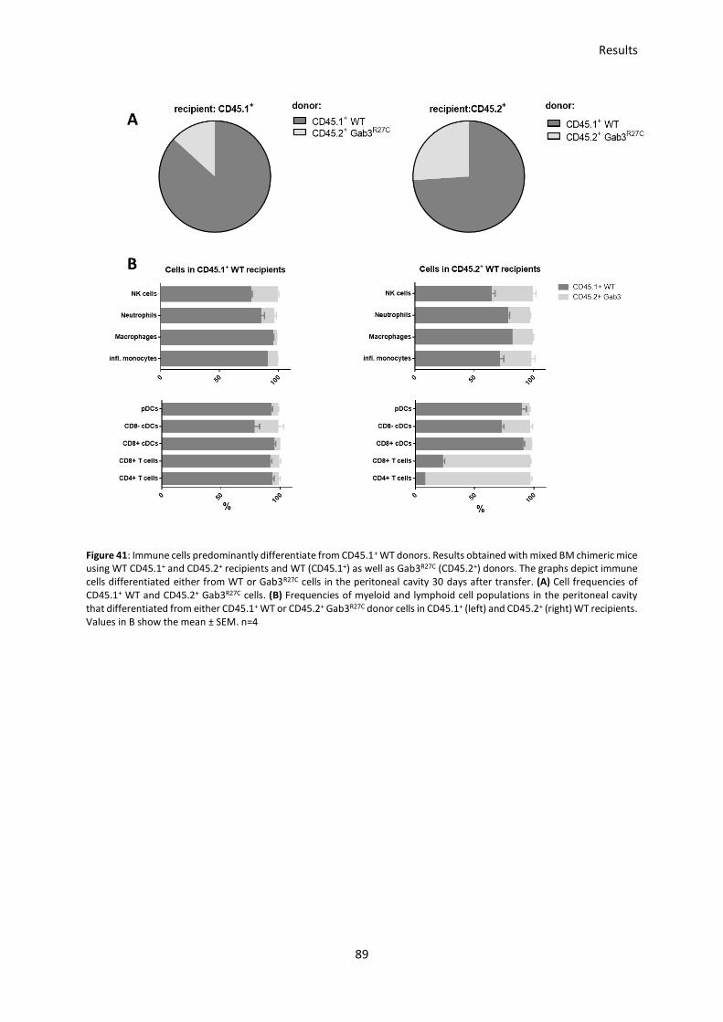
Results
89
Figure 41: Immune cells predominantly differentiate from CD45.1+ WT donors. Results obtained with mixed BM chimeric mice using WT CD45.1+ and CD45.2+ recipients and WT (CD45.1+) as well as Gab3R27C (CD45.2+) donors. The graphs depict immune cells differentiated either from WT or Gab3R27C cells in the peritoneal cavity 30 days after transfer. (A) Cell frequencies of CD45.1+ WT and CD45.2+ Gab3R27C cells. (B) Frequencies of myeloid and lymphoid cell populations in the peritoneal cavity that differentiated from either CD45.1+ WT or CD45.2+ Gab3R27C donor cells in CD45.1+ (left) and CD45.2+ (right) WT recipients. Values in B show the mean ± SEM. n=4

Discussion
90
4 DISCUSSION
4.1 C5a receptors are critical for controlling intestinal
inflammation during early T. gondii infection
By infecting WT mice with 1, 3, 5, 7 or 10 cysts orally, I showed that infection with only 1 cyst is
sufficient to induce a massive inflammation characterized by weight loss, increase of disease scores
and mortality. Additionally, higher systemic IFN- and IL-12 levels after 5 and 7 days p.i. were observed.
Interestingly, oral infection of C5aR2-/- mice with 10 cysts did not result in massive tissue inflammation
in the gut as cell infiltration into the intestine was significantly lower than in WT and C5aR1-/- mice.
C5aR2 was discovered in the year 2000 and initially described as a decoy receptor for C5a (Ohno et al.,
2000). As already mentioned, different publications suggest an anti-inflammatory role for C5aR2
during lung injury (Gerard et al., 2005) and in contrast to that, pro-inflammatory roles as C5aR2 is
involved in ERK1/2 or p38 MAPK phosphorylation mediated via -arrestin recruitment in human
monocyte-derived macrophages (HMDMs) (Croker et al., 2014). The importance of C5aR2 is
emphasized by in vivo experiments showing that -arrestin 2 knockout mice show decreased survival
and increased release of pro-inflammatory cytokines during sepsis-induced inflammation (Fan et al.,
2010). Additionally, C5aR2 is involved in neutrophil mobilization as specific stimulation of C5aR2
inhibits neutrophil migration in WT, but not in C5aR2-/- mice (Croker et al., 2016). In summary, it was
already shown that C5aR2 has both, pro- and anti-inflammatory properties, depending on the cell type,
tissue and signaling pathway. Interestingly, in a dextran sulfate sodium (DSS)- induced mouse model
of acute ulcerative colitis (UC), C5aR2-/- mice are less susceptible characterized by reduced weight loss
and a lower disease score in comparison to WT mice (Hsu et al., 2014). As a result of the massive
destruction of tissue and crypt architecture in WT and C5aR1-/- mice it is likely that these mice do not
only suffer from intestinal inflammation caused by the parasite, but also from sepsis caused by
breaking up the barrier and leaking of bacteria and other pro-inflammatory mediators into the blood
stream. It has been shown that a combined blockade of C5aR1 and C5aR2 during sepsis is necessary to
improve the survival of mice (Rittirsch et al., 2008), again underlining the functional role of C5aR2.
These results are in line with my observations and indicate that C5aR2 plays an important role in
mediating C5a-induced inflammation during acute T. gondii infections in the intestine. However, the
exact mechanism how C5aR2 regulates C5a-mediated signaling during acute T. gondii infections
remains elusive. It is possible that C5aR2 forms dimers with C5aR1 as already described previously to
regulate the pro-inflammatory signaling through C5aR1 and induce the production of
anti-inflammatory cytokines like IL-10 (Hsu et al., 2014). The potential lack of anti-inflammatory
cytokines in C5aR1-/- mice would explain the severe inflammation induced by both, elevated levels of

Discussion
91
pro-inflammatory cytokines like IFN- due to the missing control of C5aR1-mediated cytokine
production and the lack of anti-inflammatory cytokines like IL-10. On the other hand, lack of C5aR2
would also result in missing interaction of C5aR1 and C5aR2 and should result in the same cytokine
production, if the interaction of C5aR1 and C5aR2 is the key mechanism here. However, C5aR2-/- mice
are protected from developing severe inflammation in the gut which indicates that missing C5aR2
results in a better controlled immune response regarding production of pro- and anti-inflammatory
cytokines. A potential explanation might be the missing transfer of intracellularly expressed C5aR2 to
the surface of the cell to counterbalance C5aR1-mediated pro-inflammatory immune responses, but
not due to missing C5aR1-C5aR2 interaction at a first step. Another potential crosstalk partner for
C5aR2 could involve TLRs, as it was already described that C5aR1 can form dimers with TLR4 in which
C5a negatively regulates TLR4-induced immune responses (Hawlisch et al., 2005). As C5aR1 and C5aR2
are structurally similar, it is possible that also C5aR2 forms dimers with different TLRs to control
pro- and anti-inflammatory immune responses. In this case, my results indicate that missing interaction
of C5aR2 and TLRs results in a better controlled immune response with potentially lower levels of pro-
inflammatory cytokines.
Taken together, I hypothesize that C5aR2 has significant roles in regulation of T. gondii-induced
inflammation in the intestine and it seems unlikely that C5aR2 only acts as a decoy receptor for C5a,
because the obtained results regarding the development of inflammation in the gut of WT, C5aR1-/- and
C5aR2-/- mice are too diverse. Further experiments unveiling better insights into locally produced
cytokines and complement activation, would help to better understand the role of C5aR2 in the
induction of T. gondii-dependent inflammation in the intestine.
4.2 Uncoupled co-expression of C5aR1 and C5aR2 as an important
immune regulatory mechanism
The institute for systemic inflammation research recently published the expression pattern of C5aR1
and C5aR2 on innate and adaptive immune cells under naïve conditions. With the help of floxed
GFP-C5aR1 and floxed tdTomato-C5aR2 reporter mice it was possible to reliably determine the
expression pattern of both C5a receptors (Karsten et al., 2015, 2017). To follow this up, I investigated
the regulation of C5aR1 and C5aR2 expression during early T. gondii infection. So far, nothing is
published regarding a potential uncoupled co-expression of C5aR1 and C5aR2 under specific
inflammatory conditions. I could show that during early T. gondii infections this specific regulation of
C5aR1 and C5aR2 might play an important role. Previous experiments from our institute showed that
C5aR1-/- mice suffered from increased susceptibility and parasite burden in the brain and C5aR2-/- mice

Discussion
92
also showed increased parasite burden 30 days p.i. (Briukhovetska, 2017). Together with my results
showing that C5aR2-/- mice are protected from developing severe inflammation in the gut, the available
results emphasize the importance of both C5aRs and their potential cross-talk. Analyzing C5aR1 and
C5aR2 expression in the blood showed that significantly more cells express C5aR2 under infectious
conditions. Interestingly, cells expressing C5aR1 did not change. In the spleen more cells express C5aR1
and C5aR2 during the infection. Interestingly, under steady state conditions more cells express C5aR2
compared to C5aR1. In the lung is was the other way around, as C5aR1 was expressed on more immune
cells under infectious conditions compared to C5aR2. However, the C5a receptor expression on
immune cells in the peritoneal cavity did not change during T. gondii infection at all, but under steady
state condition more cells expressed C5aR2. As the peritoneal cavity is the first tissue which
encountered the parasite in the i.p. infection model, no regulation of the expression of both receptors
on immune cells in the peritoneal cavity might indicate that C5aR1 and C5aR2 are not crucially involved
in cell activation, what is unlikely in light of several publications (Croker et al., 2014; Denk et al., 2017),
or the parasites infiltrated and killed cells before they were able to respond to the infection. Although,
no regulation on total immune cells was detectable, single cell populations showed regulatory
differences. Macrophages, which are highly positive for C5aR1 and C5aR2 expression under naïve
conditions, show the same regulation in the spleen and peritoneal cavity as less cells are C5aR2+ under
infectious conditions. Interestingly, the expression results indicate the C5aR2, but not C5aR1 is
downregulated on macrophages. Furthermore, the cell frequency for macrophages in the peritoneal
cavity drops significantly under infectious conditions, indicating that macrophages which remain inside
the peritoneal cavity downregulate C5aR2, while those who migrate to other tissues keep high C5aR2
expression levels. This regulation suggests that downregulation of C5aR2 is necessary to activate
macrophages, as C5aR2 acts to counter-balance C5aR1-mediated cell activation (Bamberg et al., 2010).
In the presence of C5a, a downregulation of C5aR2 would result in higher potency of C5a to bind to its
pro-inflammatory receptor C5aR1 and activate the cells as the counter-balancing via C5aR2 is missing.
The reduction in macrophage frequency in the peritoneal cavity is surprising as those cells play an
important role during early T. gondii infections by phagocytosing free parasites, but they also get
actively infected by tachyzoites. The fate of the macrophage depends on priming with IFN- after the
infection with T. gondii, but before the parasite enters the cell (Nathan et al., 1983). If macrophages
are primed with IFN-, they are able to induce effector mechanisms like upregulation of iNOS and NO
which leads to parasite killing. However, if IFN- is missing because neutrophils or NK cells, which are
responsible for early IFN- production, are already infected with the parasite, lack activation or
migration to the site of infection, the parasite can reproduce within the macrophage and spread
throughout the organism. For NK cells it was already shown that infected NK cells develop a
hypermobility after infection with T. gondii facilitating parasite spreading throughout the body (Ueno

Discussion
93
et al., 2015). It is possible that also infected macrophages tend to migrate out of the peritoneal cavity
to facilitate parasite spreading. Another possibility is that insufficient IFN- levels inhibit macrophage
activation and therefore, T. gondii actively infects macrophages and kills them after intracellular
tachyzoite reproduction.
As an early IFN- producer, neutrophils are of main importance in controlling the infection.
Unfortunately, it was not able to analyze the C5aRs regulation on neutrophils under naïve conditions
in the peritoneal cavity, because neutrophils are absent under steady state conditions. However,
compared to other analyzed tissues and the fact that neutrophils, like macrophages, are highly
expressing C5aRs cells (Dunkelberger et al., 2012; Karsten et al., 2015, 2017) and the fact the C5a is a
strong chemoattractant for neutrophils and enhances phagocytosis and release of granule enzymes
(Goldstein and Weissmann, 1974; Mollnes et al., 2002), it seems that less cells express C5aR2 under
infectious conditions, whereas C5aR1 expression remains stable. Hence, the expression of C5aR2 on
neutrophils is comparable to the expression on macrophages. The importance of neutrophil-derived
IFN- during T. gondii infections is shown by Hunter et al. (Hunter et al., 1994) who published that NK
cell and T cell-deficient mice suffer from increased susceptibility during T. gondii infections, but are not
as susceptible as IFN- deficient mice, due to the fact that IFN- produced by neutrophils compensates
the lack of NK cell- and T cell-derived IFN-. The frequency of neutrophils expressing C5aR2 and the
expression intensity is downregulated under infectious conditions in the blood. Therefore, it seems
that neutrophils which migrate into the blood downregulate C5aR2. Downregulation of C5aR2 allows
more C5a to bind to C5aR1 and that could alter the migration behavior of neutrophils and increase
migration to the sites of infection. On the other hand, publications showed that high doses of C5a can
lead to nonspecific chemotactic migration and neutrophil dysfunction when bound to C5aR1 (Huber-
Lang et al., 2002b). Therefore, maintaining high expression levels of C5aR2 could be necessary to
prevent neutrophil dysfunction and counter-balance C5aR1-mediated neutrophil migration and
activation. Based on my results I hypothesize that downregulation of C5aR2 facilitates neutrophil
activation and migration as well as it improves parasite killing or phagocytosis what is necessary to
limit parasite spreading and the development of systemic inflammation. Consequently, C5aR2-/- mice
would survive T. gondii infections significantly better than C5aR1-/- mice. Unfortunately, we could not
observe such a prominent difference between these two KO mice as both strains showed similar
survival (Briukhovetska, 2017). Although these results limit the assumed anti-inflammatory role of the
C5aR2, it is still tempting to speculate that the receptor is important to maintain cells inactive under
naïve conditions. For a better understanding, further experiments are necessary to investigate
whether local cytokine production is altered in cells from WT and C5aR2-/- mice. Furthermore,

Discussion
94
migration experiments with uninfected and infected neutrophils or macrophages will help to
understand if T. gondii actively influences mobility.
Besides neutrophils, eosinophils belong to the group of granulocytes. However, they show significantly
reduced expression intensity for both receptors compared to neutrophils. They are known to be
important in fighting parasites and have important roles during allergic asthma (Gleich and Loegering,
1984; Weller, 1994). After recruitment to the site of infection they start to produce pro-inflammatory
cytokines like IL-2, IL-4, IL-5 and IL-12 besides others (Kita, 1996). In the induction phase of allergic
asthma, eosinophil and neutrophil influx as well as production of pro-inflammatory cytokines are the
main reasons for establishing a maladaptive immune response (Verstraelen et al., 2008). Therefore, it
is surprising that T. gondii infections lead to significant reduction of the eosinophil population in the
lung. It is tempting to speculate that eosinophils are either phagocytosing free tachyzoites or are
actively infected by the parasite. In both cases the parasite would replicate inside of the eosinophil
and that results in killing of the cell. As the lung is one of the first organs where T. gondii aims to migrate
into, the observed reduction and nearly complete loss of eosinophil population demonstrates the
severity of the infection. Regarding C5aR1 and C5aR2 expression in eosinophils, no change between
non-infectious and infectious conditions was observed. Eosinophils express both receptors at
moderate levels with slightly increased expression levels for C5aR2. It is important to notice, that the
eosinophil population is nearly absent under infectious conditions what exacerbates the flow
cytometric analysis and reduces the validity of the obtained data regarding the expression of C5aR1
and C5aR2. However, as no regulation was detected, C5a receptors seem to have just minor roles in
regulating eosinophil activation and migration. Interestingly, Fenoy et al (Fenoy et al., 2009) showed
that T. gondii infections protect BALB/c mice from the development of allergic airway inflammation by
blocking eosinophil and neutrophil migration to the lung. Due to the missing eosinophils and
neutrophils, the establishment of a maladaptive immune response is unlikely. Although, T. gondii
infections do not block eosinophil migration into the lung, it seems that the infection leads to increased
eosinophil death and therefore to diminished frequencies in the lung, what has the same effect as
blocking the migration and preventing the induction of a maladaptive immune response. On the other
hand, missing neutrophils and eosinophils in the lung also results in reduced pro-inflammatory
cytokines and consequently facilitates spreading and reproduction of parasites like T. gondii.
Besides the described high-expressing C5aR1 and C5aR2 cells, low-expressing inflammatory monocytes
were detected. In contrast to the described high-expressing C5aR1 and C5aR2 cells, these cells only
show minor regulatory mechanisms. The cell frequency of C5aR1 and C5aR2 expressing cells is
upregulated after infection in the peritoneal cavity. They are known to be recruited to the site of
infection following profilin detection via TLR11 in mice (Neal and Knoll, 2014), what is also reflected by

Discussion
95
increased frequencies in the peritoneal cavity. Furthermore, they produce iNOS and TNF- as essential
mediators for controlling parasite spreading (Dunay et al., 2008; Mordue and Sibley, 2003).
Consequently, depletion of inflammatory monocytes leads to increased susceptibility and mice
succumb to death early during the acute phase of the infection (Dunay et al., 2010). However, only
little is known about the role of anaphylatoxins for regulation and activation of inflammatory
monocytes. That is not surprising as my results indicate that inflammatory monocytes at best show
minor C5aR1 or C5aR2 expression in all investigated organs. Although inflammatory monocytes do not
express or regulate C5a receptors as distinct as neutrophils or macrophages, their role during T. gondii
infections is undisputed. In contrast to macrophages and neutrophils, which downregulate C5aRs
under infectious conditions, cells expressing C5aR1 or C5aR2 on DCs either do not change or increase,
as visible for C5aR1 in the peritoneal cavity and spleen and C5aR2 in the blood. Interestingly, the
expression intensity did not change. DCs in general and especially cDCs play an indispensable role
during T. gondii infections as they are the main source for IL-12 after recognizing profilin via TLR11 and
TLR12 in mice (Plattner et al., 2008). IL-12 is necessary to activate other effector cells to induce a potent
immune response and limit parasite growth and spreading. Additionally, DCs are known to be
important in regulating TH1 and TH17 differentiation. Furthermore, is has previously been shown that
exposure to LPS and C5a resulted in enhanced production of cytokines, indicating synergism between
TLRs and C5aR1 (Riedemann et al., 2002, 2004). Another synergism between TLRs and C5a receptors
was shown by a negative regulation of TLR4- and CD40-induced synthesis of IL-12 family cytokines from
inflammatory macrophages via C5a (Hawlisch et al., 2005). Additionally, it is published that TLR
modulates C5a-induced responses by negatively modulating C5aR2 to reduce the counter-balancing
effect and increase pro-inflammatory responses (Raby et al., 2011). The published results are in line
with my results as cDCs massively upregulate C5aR1 expression in the peritoneal cavity and spleen
under infectious conditions which could lead to increased TLR- and C5a-mediated activation and
cytokine production. Additionally, the assumed anti-inflammatory role of C5aR2 is reduced, because
increased expression of C5aR1 results in higher potential for C5a to bind to its pro-inflammatory
receptor. The fact that cDCs in the peritoneal cavity show the most distinct difference in C5aR1
expression comparing steady state and infectious conditions is due to the i.p. infection model which
results in the encounter of immune cells with the parasite in this compartment first.
To obtain more detailed information about the function of C5aR1 and C5aR2 on single cell populations
during early T. gondii infections, it will be necessary to generate cell-specific C5aR1 or C5aR2 knockouts
using the Cre/lox system. With this opportunity it will be possible to investigate the impact of C5aR1
and C5aR2 on individual immune responses regarding parasite susceptibility and cytokine production
in vivo and to draw precise conclusions about the role of both receptors.
Discussion
96
4.3 C5aR2 controls NK cell-derived IFN- production in response
to IL-12/IL-18 stimulation
One of the key result of the C5aR1- and C5aR2-expression studies is that NK cells and B cells express
C5aR2 but not C5aR1 (Karsten et al., 2015, 2017). This unique expression pattern makes them a
suitable cell type to directly investigate C5aR2 functions and downstream signaling events.
Furthermore, the data show that only a subpopulation of NK cells expresses C5aR2. The expression
pattern was confirmed using PCR, GFP-C5aR1 and tdTomato-C5aR2 reporter mice. Functionally I was
able to show that C5aR2-/- NK cells produce significant more IFN- after IL-12 and IL-18 stimulation than
WT NK cells. In line with this observation, pre-stimulation of WT NK cells with the specific C5aR2 agonist
P32 (Croker et al., 2016) resulted in reduced IFN- production. Thus, C5aR2 on NK cells negatively
regulated IFN- production in NK cells upon cytokine stimulation demonstrating a crucial and novel
regulatory function for the C5a/C5aR2-axis in IL-12/IL-18-driven production of IFN-. As IFN- is an
important cytokine to control infections, it is not surprising that some T. gondii strains developed
mechanisms to block IFN- production from effector cells by increasing the association of STAT1 with
DNA (Rosowski et al., 2014). As the in vitro results indicate a regulatory function for C5aR2, I aimed to
identify intracellular signaling pathways of C5aR2 and stimulated NK cells with C5a and stained for
phosphorylated p38 MAPK, which is a signaling pathway downstream of IL-12 and IL-18 (Zhang and
Kaplan, 2000). Experiments showed that C5a stimulation in WT NK cells results in phosphorylation of
p38 after 2 min and desensitization after 10 min after stimulation. This effect was C5aR2-specific, since
stimulation of C5aR2-/- NK cells did not result in any change in p38 phosphorylation. These results
indicate that C5aR2 is involved in the regulation of p38 phosphorylation as it can counter-balance
phosphorylation of p38 upon IL-12 or IL-18 stimulation to prevent an overwhelming cytokine storm
(Figure 42). Considering the fact that complement factors like C5a can also be produced locally during
inflammation by various cell types (Bröker et al., 2018; Huber-Lang et al., 2002a; Lubbers et al., 2017),
the unique expression of C5aR2 on NK cells becomes even more important. Locally produced C5a can
regulate NK cell activation by binding to the anti-inflammatory C5aR2 to prevent NK cells from
excessive activation resulting in the massive production of pro-inflammatory cytokines what could
harm the host. As it is important during an inflammatory response to keep the balance between an
adequate inflammatory response that limits parasite growth or spreading and prevention of
overproduction of pro-inflammatory cytokines that would harm the host, C5aR2-controlled NK cell
activation seems to play an important role in this process of local inflammation. In future experiments
it will be necessary to identify other downstream signaling pathways of C5aR2 to better understand,
how C5aR2 controls NK cell activation. Additionally, stimulating NK cells with IL-12/IL-18 in the

Discussion
97
presence or absence of C5a could help to understand how C5aR2 signaling influences IFN- production
in vitro.
Figure 42: C5aR2 stimulation leads to blockade of p38 phosphorylation. Schematic figure illustrating the function of C5aR2 on (A) WT and (B) C5aR2-/- NK cells. C5a stimulation of C5aR2 results in decreased phosphorylation of p38 MAPK and
consequently in reduced IFN- production after IL-12/18 stimulation in WT NK cells. C5aR2-/- NK cells lack this regulatory
mechanism and are hyperresponsive to IL-12/18 stimulation and produce more IFN-.
4.4 The C5a/C5aR2 axis may control NKp46 expression as one
mechanism of NK cell regulation
NKp46 is known as an activating receptor on NK cells and able to recognize self and tumor ligands
(Glasner et al., 2017). The absence of the receptor results in increased IFN- production in the presence
of YAC1 cells (Narni-Mancinelli et al., 2012). The same group also published, that NKp46 is a receptor
for complement factor P (properdin) (Narni-Mancinelli et al., 2017). Interestingly, binding of factor P
to NKp46 leads to activation of different genetic programs and does not induce classical NK cell
activation. Additionally, it was shown that NKp46 is involved in the expression of TNF-related apoptosis
inducing ligand (TRAIL), which is important for anti-tumor, anti-viral and immunoregulatory functions
(Sheppard et al., 2018). Interestingly, NKp46 is the only cytotoxicity receptor, which is shared by
primates and rodents, emphasizing an essential role during evolution (Kruse et al., 2014). Therefore, I
investigated the expression of NKp46 on NK cells of WT, C5aR1-/- and C5aR2-/- mice. My experiments
showed that C5aR2-/- NK cells from the spleen and peritoneal cavity express significantly higher levels
of NKp46 as compared to NK cells from WT or C5aR1-/- mice. Further, I found that C5aR2+ NK cells from
reporter mice in the spleen and peritoneal cavity have a higher NKp46 expression compared to
C5aR2- NK cells. I expected reverse results regarding the expression of NKp46 on C5aR2+ NK cells, as
NKp46 is significantly higher expressed on C5aR2-/- NK cells, but it still allows to speculate about the
NKp46 function in connection with C5aR2 on NK cells. In vitro results showed that the lack of C5aR2

Discussion
98
on NK cells resulted in increased production of IFN- after IL-12/18 stimulation, suggesting an
anti-inflammatory role for C5aR2 on NK cells. Since NKp46 is described as an activating receptor, I
expected regulation of C5aR2 and NKp46 in the same manner to control NK cell activity and prevent
excessive activation which could result in host tissue damage. In this case, C5aR2 would
counter-balance NKp46-dependent NK cell activation. Using the tdTomato-C5aR2 reporter mice, I
showed that C5aR2- NK cells express less NKp46 than WT cells, supporting the view that
NKp46-mediated NK cell activation is controlled via C5aR2 and lower NKp46 expression requires lower
C5aR2 expression. Interestingly, on C5aR2-/- NK cells the expression is not regulated in the same
manner. Here, lack of C5aR2 results in massively increased NKp46 expression levels. If NKp46 and
C5aR2 expression is linked, these results explain the increased IFN- production from C5aR2-/- NK cells,
as increased NKp46 expression results in enhanced NK cell activation, which cannot be counter-
regulated due to the lack of suppressive C5aR2. Additionally, it was shown that NKp46 ligand
complement factor P can bind to tumor cell lines and potentially work as a NK cell activator (Kemper
et al., 2010), which would further increase NKp46-mediated NK cell activation. Taken together, NK cells
from C5aR2-/- mice might exert an increased cytotoxicity and/or IFN- production as the
anti-inflammatory C5aR2 is missing and simultaneously, the activating receptor NKp46 is
overexpressed. Another reason for the overexpressed NKp46 might be genomic changes resulting from
the knockout of the c5ar2 gene in C5aR2-/- mice, as interference of genomic equipment not only affects
the targeted gene c5ar2 but can also have an influence on closely located or structurally related genes.
4.5 Control of cell-cell interaction between NK cells and DCs as a
mechanism of C5aR2-mediated regulation of NK cell activation
Besides the direct regulation of NK cells by receptors like C5aR2, NK cells need further signals,
mediated via other immune cells like DCs, to become fully activated. To achieve that, NK cells need
priming by various factors like IL-12, IL-15 or IL-18 which are mainly produced by DCs or macrophages
(Chaix et al., 2008; Guia et al., 2008; Lucas et al., 2007). Borg et al. (Borg et al., 2004) showed that
activation of NK cells via DCs requires the formation of a stimulatory synapse resulting in a direct
cell-cell-contact between NK cells and DCs. Furthermore, they showed that DC-derived IL-12 and IL-18
is directly released into the synapse to allow rapid NK cell activation and that IL-15 is presented to the
NK cells via the IL-15R. Besides directed cytokine release, cell adhesion molecules like VCAM-1 and
LFA-1 play in important role in stabilizing the stimulatory synapse. In experiments investigating splenic
DC and NK cell frequencies, total cell numbers and VCAM1 and CD11a expression, I found differences
between WT, C5aR1-/- and C5aR2-/- which might explain altered NK cell responsiveness to cytokines, as

Discussion
99
C5aR2-/- cells express higher levels of interaction molecules and therefore might respond to cytokines
in a more sensitive way. I observed that total splenic DC numbers in C5aR2-/- mice were reduced
compared to WT mice. Furthermore, C5aR2-/- DCs expressed higher levels of VCAM1 and more DCs
were positive for CD11a when compared to WT DCs. For C5aR2-/- NK cells, significant differences for
cell frequencies, but not for total cell numbers were observed. Furthermore, C5aR2-/- NK cells
expressed higher levels of VCAM1 as compared to WT NK cells. The increased expression of VCAM1
could result in a lowered threshold for NK cell activation and might explain the hyperresponsiveness
of C5aR2-/- NK cells in stimulation experiments and the increased IFN- production in in vitro
experiments. Interestingly, I did not find systemic differences for IFN- between WT and C5aR2-/- mice,
which might be explained by the reduced numbers of DCs and NK cells in C5aR2-/- mice as cells from
C5aR2-/- mice have a lower threshold for activation due to increased expression of important adhesion
molecules, but reduced cell numbers. One reasons for the same amount of measured IFN- might be
that the increased production of IFN- by each cell was overlaid by the reduced cell numbers.
Furthermore, the reduced DC numbers could result in less effective NK cell activation due to missing
interaction partner. However, as the relative number of both populations was reduced, the ratio
between DCs and NK cells remains the same. Increased expression of co-stimulatory adhesion
molecules could work as a compensation mechanism and allow potent NK cell activation by DCs, as
formation of the stimulatory synapse is facilitated by increased expression of co-stimulatory adhesion
molecules.
4.6 Gab3 is crucially important for cell differentiation and
influences T. gondii susceptibility
In addition to unique expression pattern for C5aR2 on NK cells, I uncovered high expression of Gab3
while I was in the IRTG partner laboratory of Dr. Hoebe at the CCHMC. Gab3 belongs to the scaffolding
protein family of Grb-2 associated proteins together with Gab1 and Gab2. They contribute to specific
signal transduction pathways and all three molecules share a similar topology. However, relatively little
is known about the function, interaction partners and the role of Gab3 as reflected in the low number
of publications dealing with Gab3. It is known that Gab3 deficient mice develop normal and are
generally healthy and immunocompetent, suggesting that Gab3 functions can be compensated under
steady state conditions (Seiffert et al., 2003). In contrast to Gab1 and Gab2, Gab3 is highly expressed
in the lymphoid tissue. Furthermore, experiments from the Hoebe lab revealed a strong expression on
NK cells, mast cells and CD8+ T cells (unpublished data). To investigate the function of Gab3, the Hoebe
lab generated an ENU-mutated germline mouse strain with a missense mutation in the pleckstrin

Discussion
100
homology (PH) domain of Gab3 (Gab3R27C). As NK cells are crucially involved during early T. gondii
infections, I chose this model to further investigate the role and function of Gab3. I.p. infection with
50 T. gondii cysts showed that Gab3R27C mice suffered from increased susceptibility to the infection
and elevated parasite burden after 30 days p.i. compared to WT mice. Interestingly, no substantial
differences in disease-associated systemic cytokine levels were observed during the first seven days
p.i. However, all measured cytokines showed only very low concentrations ranging from 0 ng/ml to
10 ng/ml, except for IFN- which was present in higher concentrations. These results indicate that the
Gab3 protein is not involved in the regulation of cytokine production and therefore it is unlikely that
Gab3 is associated in pathways downstream of TLRs, the IL-12-family or IFN- receptors. Interestingly,
experiments investigating the cell composition in the peritoneal cavity of Gab3R27C mice showed that
under steady state conditions cell frequencies of NK cells, neutrophils and macrophages were reduced
compared to WT mice. Additionally, mixed BM-chimeric mice experiments revealed impaired ability of
BM cells from Gab3R27C to differentiate into innate and adaptive immune cells in the peritoneal cavity,
but not in the spleen. The slight heterogeneity in CD45.1+ and CD45.2+ WT donors, as well as the shift
towards GabR27C-differentiated T cells might be explained by insufficient irradiation at the beginning of
the experiment, which resulted in increased CD45.2+ cells already before i.v. injection of the mixed
BM. Especially the reduced macrophage population and the impaired differentiation ability of
macrophages in the context of T. gondii infections seems to be a major reason for the increased
susceptibility of Gab3R27C mice. Although macrophages represent the major cell population in the
peritoneal cavity with up to 35 % of all cells, I could not detect differences in disease-associated
systemic cytokine levels on different days p.i. This might be explained by the local impaired
differentiation ability of Gab3R27C cells, which is limited to the peritoneal cavity. Therefore, potential
lack of cytokines produced in the peritoneal cavity are compensated or overlaid by unaffected cells in
the spleen or blood. However, macrophages are one of the first cell populations which interact with
T. gondii after i.p. infections and play an important role in limiting the reproduction and spreading of
the parasite (Masek and Hunter, 2013). This massive and significant reduction in the macrophage
population may result in less effector cells, local cytokines or phagocytosis during the early state of
infection and facilitate parasite spreading throughout the organism. Hence, it is tempting to speculate
that reduced macrophages numbers and impaired cell differentiation results in delayed or insufficient
activation or migration into the peritoneal cavity early after i.p. infection (Figure 43). Additionally, also
the local reduction of neutrophil and NK cell numbers in the peritoneal cavity can contribute to
increased susceptibility as the lack of local produced cytokines, mainly IFN- (Johnson et al., 1993),
facilitates T. gondii reproduction and spreading throughout the body. For a better understanding how
Gab3 influences cell activation or migration, further experiments to identify potential downstream

Discussion
101
target proteins of Gab3 need to be done. Additionally, measurement of local produced cytokines in
the peritoneal cavity would help to understand the role of Gab3.
Figure 43: Schematic figure showing the impact of the Gab mutation Gab3R27C during the acute and chronic phase of T. gondii infection. (A) In the WT mice immediate innate immune cell activation drives effective elimination of tachyzoites. The development of an appropriate adaptive immune response converts the remaining tachyzoites into tissue cysts residing preferentially in the brain. (B) In the Gab3R27C mice, a delayed or impaired innate immune response results in ineffective elimination of tachyzoites in the acute phase of the infection which leads to increased tissue cyst production that cannot be efficiently controlled by the adaptive immune system.

References
102
5 REFERENCES
Abi Abdallah, D.S., Lin, C., Ball, C.J., King, M.R., Duhamel, G.E., and Denkers, E.Y. (2012). Toxoplasma gondii triggers release of human and mouse neutrophil extracellular traps. Infect. Immun. 80, 768–777.
Adams, L.B., Hibbs, J.B., Taintor, R.R., and Krahenbuhl, J.L. (1990). Microbiostatic effect of murine-activated macrophages for Toxoplasma gondii. Role for synthesis of inorganic nitrogen oxides from L-arginine. J. Immunol. Baltim. Md 1950 144, 2725–2729.
Alberts, B., Johnson, A., Lewis, J., Raff, M., Roberts, K., and Walter, P. (2002). The Adaptive Immune System. Mol. Biol. Cell 4th Ed.
Amara, U., Flierl, M.A., Rittirsch, D., Klos, A., Chen, H., Acker, B., Brückner, U.B., Nilsson, B., Gebhard, F., Lambris, J.D., et al. (2010). Molecular intercommunication between the complement and coagulation systems. J. Immunol. Baltim. Md 1950 185, 5628–5636.
Andrade, R.M., Wessendarp, M., and Subauste, C.S. (2003). CD154 activates macrophage antimicrobial activity in the absence of IFN-gamma through a TNF-alpha-dependent mechanism. J. Immunol. Baltim. Md 1950 171, 6750–6756.
Andrade, R.M., Portillo, J.-A.C., Wessendarp, M., and Subauste, C.S. (2005). CD40 signaling in macrophages induces activity against an intracellular pathogen independently of gamma interferon and reactive nitrogen intermediates. Infect. Immun. 73, 3115–3123.
Andrade, W.A., Souza, M. do C., Ramos-Martinez, E., Nagpal, K., Dutra, M.S., Melo, M.B., Bartholomeu, D.C., Ghosh, S., Golenbock, D.T., and Gazzinelli, R.T. (2013). Combined action of nucleic acid-sensing Toll-like receptors and TLR11/TLR12 heterodimers imparts resistance to Toxoplasma gondii in mice. Cell Host Microbe 13, 42–53.
Araujo, F.G., and Slifer, T. (2003). Different Strains of Toxoplasma gondii Induce Different Cytokine Responses in CBA/Ca Mice. Infect. Immun. 71, 4171–4174.
Arbore, G., West, E.E., Spolski, R., Robertson, A.A.B., Klos, A., Rheinheimer, C., Dutow, P., Woodruff, T.M., Yu, Z.X., O’Neill, L.A., et al. (2016). T helper 1 immunity requires complement-driven NLRP3 inflammasome activity in CD4+ T cells. Science 352, aad1210.
Bamberg, C.E., Mackay, C.R., Lee, H., Zahra, D., Jackson, J., Lim, Y.S., Whitfeld, P.L., Craig, S., Corsini, E., Lu, B., et al. (2010). The C5a Receptor (C5aR) C5L2 Is a Modulator of C5aR-mediated Signal Transduction. J. Biol. Chem. 285, 7633–7644.
Benevides, L., Milanezi, C.M., Yamauchi, L.M., Benjamim, C.F., Silva, J.S., and Silva, N.M. (2008). CCR2 receptor is essential to activate microbicidal mechanisms to control Toxoplasma gondii infection in the central nervous system. Am. J. Pathol. 173, 741–751.
Berry, D.M., Nash, P., Liu, S.K.-W., Pawson, T., and McGlade, C.J. (2002). A High-Affinity Arg-X-X-Lys SH3 Binding Motif Confers Specificity for the Interaction between Gads and SLP-76 in T Cell Signaling. Curr. Biol. 12, 1336–1341.
Biswas, A., French, T., Düsedau, H.P., Mueller, N., Riek-Burchardt, M., Dudeck, A., Bank, U., Schüler, T., and Dunay, I.R. (2017). Behavior of Neutrophil Granulocytes during Toxoplasma gondii Infection in the Central Nervous System. Front. Cell. Infect. Microbiol. 7.

References
103
Blanchard, N., Gonzalez, F., Schaeffer, M., Joncker, N.T., Cheng, T., Shastri, A.J., Robey, E.A., and Shastri, N. (2008). Immunodominant, protective response to the parasite Toxoplasma gondii requires antigen processing in the endoplasmic reticulum. Nat. Immunol. 9, 937–944.
Bliss, S.K., Marshall, A.J., Zhang, Y., and Denkers, E.Y. (1999a). Human polymorphonuclear leukocytes produce IL-12, TNF-alpha, and the chemokines macrophage-inflammatory protein-1 alpha and -1 beta in response to Toxoplasma gondii antigens. J. Immunol. Baltim. Md 1950 162, 7369–7375.
Bliss, S.K., Zhang, Y., and Denkers, E.Y. (1999b). Murine neutrophil stimulation by Toxoplasma gondii antigen drives high level production of IFN-gamma-independent IL-12. J. Immunol. Baltim. Md 1950 163, 2081–2088.
Bliss, S.K., Butcher, B.A., and Denkers, E.Y. (2000). Rapid recruitment of neutrophils containing prestored IL-12 during microbial infection. J. Immunol. Baltim. Md 1950 165, 4515–4521.
Bliss, S.K., Gavrilescu, L.C., Alcaraz, A., and Denkers, E.Y. (2001). Neutrophil Depletion during Toxoplasma gondii Infection Leads to Impaired Immunity and Lethal Systemic Pathology. Infect. Immun. 69, 4898–4905.
Borg, C., Jalil, A., Laderach, D., Maruyama, K., Wakasugi, H., Charrier, S., Ryffel, B., Cambi, A., Figdor, C., Vainchenker, W., et al. (2004). NK cell activation by dendritic cells (DCs) requires the formation of a synapse leading to IL-12 polarization in DCs. Blood 104, 3267–3275.
Briukhovetska, D. (2017). The roles of C5a receptor 1 and chemokine receptor 5 in Toxoplasma gondii infection.
Bröker, K., Figge, J., Magnusen, A.F., Manz, R.A., Köhl, J., and Karsten, C.M. (2018). A Novel Role for C5a in B-1 Cell Homeostasis. Front. Immunol. 9.
Cai, T., Nishida, K., Hirano, T., and Khavari, P.A. (2002). Gab1 and SHP-2 promote Ras/MAPK regulation of epidermal growth and differentiation. J. Cell Biol. 159, 103–112.
Campbell, W.D., Lazoura, E., Okada, N., and Okada, H. (2002). Inactivation of C3a and C5a octapeptides by carboxypeptidase R and carboxypeptidase N. Microbiol. Immunol. 46, 131–134.
Campos, M.A., Closel, M., Valente, E.P., Cardoso, J.E., Akira, S., Alvarez-Leite, J.I., Ropert, C., and Gazzinelli, R.T. (2004). Impaired production of proinflammatory cytokines and host resistance to acute infection with Trypanosoma cruzi in mice lacking functional myeloid differentiation factor 88. J. Immunol. Baltim. Md 1950 172, 1711–1718.
Carrillo-Bustamante, P., Keşmir, C., and de Boer, R.J. (2016). The evolution of natural killer cell receptors. Immunogenetics 68, 3–18.
Cestari, I., Evans-Osses, I., Schlapbach, L.J., de Messias-Reason, I., and Ramirez, M.I. (2013). Mechanisms of complement lectin pathway activation and resistance by trypanosomatid parasites. Mol. Immunol. 53, 328–334.
Chaix, J., Tessmer, M.S., Hoebe, K., Fuséri, N., Ryffel, B., Dalod, M., Alexopoulou, L., Beutler, B., Brossay, L., Vivier, E., et al. (2008). Cutting edge: Priming of NK cells by IL-18. J. Immunol. Baltim. Md 1950 181, 1627–1631.
Chen, N.-J., Mirtsos, C., Suh, D., Lu, Y.-C., Lin, W.-J., McKerlie, C., Lee, T., Baribault, H., Tian, H., and Yeh, W.-C. (2007). C5L2 is critical for the biological activities of the anaphylatoxins C5a and C3a. Nature 446, 203–207.

References
104
Choi, E.Y., Orlova, V.V., Fagerholm, S.C., Nurmi, S.M., Zhang, L., Ballantyne, C.M., Gahmberg, C.G., and Chavakis, T. (2008). Regulation of LFA-1–dependent inflammatory cell recruitment by Cbl-b and 14-3-3 proteins. Blood 111, 3607–3614.
Chtanova, T., Schaeffer, M., Han, S.-J., van Dooren, G.G., Nollmann, M., Herzmark, P., Chan, S.W., Satija, H., Camfield, K., Aaron, H., et al. (2008). Dynamics of neutrophil migration in lymph nodes during infection. Immunity 29, 487–496.
Cole, F.S., Matthews, W.J., Marino, J.T., Gash, D.J., and Colten, H.R. (1980). Control of complement synthesis and secretion in bronchoalveolar and peritoneal macrophages. J. Immunol. Baltim. Md 1950 125, 1120–1124.
Collazo, C.M., Yap, G.S., Sempowski, G.D., Lusby, K.C., Tessarollo, L., Vande Woude, G.F., Sher, A., and Taylor, G.A. (2001). Inactivation of LRG-47 and IRG-47 reveals a family of interferon gamma-inducible genes with essential, pathogen-specific roles in resistance to infection. J. Exp. Med. 194, 181–188.
Colucci, F., Samson, S.I., DeKoter, R.P., Lantz, O., Singh, H., and Di Santo, J.P. (2001). Differential requirement for the transcription factor PU.1 in the generation of natural killer cells versus B and T cells. Blood 97, 2625–2632.
Cook-Mills, J.M. (2002). VCAM-1 signals during lymphocyte migration: role of reactive oxygen species. Mol. Immunol. 39, 499–508.
Correa, D., Cañedo-Solares, I., Ortiz-Alegría, L.B., Caballero-Ortega, H., and Rico-Torres, C.P. (2007). Congenital and acquired toxoplasmosis: diversity and role of antibodies in different compartments of the host. Parasite Immunol. 29, 651–660.
Croker, D.E., Halai, R., Kaeslin, G., Wende, E., Fehlhaber, B., Klos, A., Monk, P.N., and Cooper, M.A. (2014). C5a2 can modulate ERK1/2 signaling in macrophages via heteromer formation with C5a1 and β-arrestin recruitment. Immunol. Cell Biol. 92, 631–639.
Croker, D.E., Monk, P.N., Halai, R., Kaeslin, G., Schofield, Z., Wu, M.C., Clark, R.J., Blaskovich, M.A., Morikis, D., Floudas, C.A., et al. (2016). Discovery of functionally selective C5aR2 ligands: novel modulators of C5a signalling. Immunol. Cell Biol. 94, 787–795.
Curtsinger, J.M., Schmidt, C.S., Mondino, A., Lins, D.C., Kedl, R.M., Jenkins, M.K., and Mescher, M.F. (1999). Inflammatory cytokines provide a third signal for activation of naive CD4+ and CD8+ T cells. J. Immunol. Baltim. Md 1950 162, 3256–3262.
Curtsinger, J.M., Johnson, C.M., and Mescher, M.F. (2003). CD8 T cell clonal expansion and development of effector function require prolonged exposure to antigen, costimulation, and signal 3 cytokine. J. Immunol. Baltim. Md 1950 171, 5165–5171.
Debierre-Grockiego, F., Campos, M.A., Azzouz, N., Schmidt, J., Bieker, U., Resende, M.G., Mansur, D.S., Weingart, R., Schmidt, R.R., Golenbock, D.T., et al. (2007). Activation of TLR2 and TLR4 by glycosylphosphatidylinositols derived from Toxoplasma gondii. J. Immunol. Baltim. Md 1950 179, 1129–1137.
Del Rio, L., Bennouna, S., Salinas, J., and Denkers, E.Y. (2001). CXCR2 deficiency confers impaired neutrophil recruitment and increased susceptibility during Toxoplasma gondii infection. J. Immunol. Baltim. Md 1950 167, 6503–6509.

References
105
Denk, S., Taylor, R.P., Wiegner, R., Cook, E.M., Lindorfer, M.A., Pfeiffer, K., Paschke, S., Eiseler, T., Weiss, M., Barth, E., et al. (2017). Complement C5a-induced changes in neutrophil morphology during inflammation. Scand. J. Immunol. 86, 143–155.
Denkers, E.Y., Gazzinelli, R.T., Martin, D., and Sher, A. (1993). Emergence of NK1.1+ cells as effectors of IFN-gamma dependent immunity to Toxoplasma gondii in MHC class I-deficient mice. J. Exp. Med. 178, 1465–1472.
Denkers, E.Y., Yap, G., Scharton-Kersten, T., Charest, H., Butcher, B.A., Caspar, P., Heiny, S., and Sher, A. (1997). Perforin-mediated cytolysis plays a limited role in host resistance to Toxoplasma gondii. J. Immunol. Baltim. Md 1950 159, 1903–1908.
Dias, R.R.F., de Carvalho, E.C.Q., Leite, C.C. da S., Tedesco, R.C., Calabrese, K. da S., Silva, A.C., DaMatta, R.A., and de Fatima Sarro-Silva, M. (2014). Toxoplasma gondii Oral Infection Induces Intestinal Inflammation and Retinochoroiditis in Mice Genetically Selected for Immune Oral Tolerance Resistance. PLoS ONE 9.
Diebolder, C.A., Beurskens, F.J., de Jong, R.N., Koning, R.I., Strumane, K., Lindorfer, M.A., Voorhorst, M., Ugurlar, D., Rosati, S., Heck, A.J.R., et al. (2014). Complement Is Activated by IgG Hexamers Assembled at the Cell Surface. Science 343, 1260–1263.
DiScipio, R.G., Daffern, P.J., Jagels, M.A., Broide, D.H., and Sriramarao, P. (1999). A comparison of C3a and C5a-mediated stable adhesion of rolling eosinophils in postcapillary venules and transendothelial migration in vitro and in vivo. J. Immunol. Baltim. Md 1950 162, 1127–1136.
Dobó, J., Pál, G., Cervenak, L., and Gál, P. (2016). The emerging roles of mannose-binding lectin-associated serine proteases (MASPs) in the lectin pathway of complement and beyond. Immunol. Rev. 274, 98–111.
Dogruman-Al, F., Fidan, I., Celebi, B., Yesilyurt, E., Erdal, B., Babur, C., and Kustimur, S. (2011). Cytokine profile in murine toxoplasmosis. Asian Pac. J. Trop. Med. 4, 16–19.
Domínguez, P.M., and Ardavín, C. (2010). Differentiation and function of mouse monocyte-derived dendritic cells in steady state and inflammation. Immunol. Rev. 234, 90–104.
Dos Anjos Cassado, A. (2017). F4/80 as a Major Macrophage Marker: The Case of the Peritoneum and Spleen. Results Probl. Cell Differ. 62, 161–179.
Draghi, M., Pashine, A., Sanjanwala, B., Gendzekhadze, K., Cantoni, C., Cosman, D., Moretta, A., Valiante, N.M., and Parham, P. (2007). NKp46 and NKG2D recognition of infected dendritic cells is necessary for NK cell activation in the human response to influenza infection. J. Immunol. Baltim. Md 1950 178, 2688–2698.
Du Clos, T.W., and Mold, C. (2013). 20 - Complement in host deficiencies and diseases. In Clinical Immunology (Fourth Edition), R.R. Rich, T.A. Fleisher, W.T. Shearer, H.W. Schroeder, A.J. Frew, and C.M. Weyand, eds. (London: Content Repository Only!), pp. 252–269.
Dubey, J.P. (1998). Advances in the life cycle of Toxoplasma gondii. Int. J. Parasitol. 28, 1019–1024.
Dubey, J.P., Velmurugan, G.V., Rajendran, C., Yabsley, M.J., Thomas, N.J., Beckmen, K.B., Sinnett, D., Ruid, D., Hart, J., Fair, P.A., et al. (2011a). Genetic characterisation of Toxoplasma gondii in wildlife from North America revealed widespread and high prevalence of the fourth clonal type. Int. J. Parasitol. 41, 1139–1147.
References
106
Dubey, J.P., Ferreira, L.R., Martins, J., and Jones, J.L. (2011b). Sporulation and Survival of Toxoplasma gondii Oocysts in Different Types of Commercial Cat Litter. J. Parasitol. 97, 751–754.
Dunay, I.R., and Sibley, L.D. (2010). Monocytes mediate mucosal immunity to Toxoplasma gondii. Curr. Opin. Immunol. 22, 461–466.
Dunay, I.R., DaMatta, R.A., Fux, B., Presti, R., Greco, S., Colonna, M., and Sibley, L.D. (2008). Gr1+ Inflammatory Monocytes Are Required for Mucosal Resistance to the Pathogen Toxoplasma gondii. Immunity 29, 306–317.
Dunay, I.R., Fuchs, A., and Sibley, L.D. (2010). Inflammatory monocytes but not neutrophils are necessary to control infection with Toxoplasma gondii in mice. Infect. Immun. 78, 1564–1570.
Dunkelberger, J., Zhou, L., Miwa, T., and Song, W.-C. (2012). C5aR expression in a novel GFP reporter gene knock-in mouse: implications for the mechanism of action of C5aR signaling in T cell immunity. J. Immunol. Baltim. Md 1950 188, 4032–4042.
Elssen, C.H.M.J.V., Oth, T., Germeraad, W.T.V., Bos, G.M.J., and Vanderlocht, J. (2014). Natural Killer Cells: The Secret Weapon in Dendritic Cell Vaccination Strategies. Clin. Cancer Res. 20, 1095–1103.
Endris, M., Belyhun, Y., Moges, F., Adefiris, M., Tekeste, Z., Mulu, A., and Kassu, A. (2014). Seroprevalence and Associated Risk Factors of Toxoplasma gondii in Pregnant Women Attending in Northwest Ethiopia. Iran. J. Parasitol. 9, 407–414.
Erben, U., Loddenkemper, C., Doerfel, K., Spieckermann, S., Haller, D., Heimesaat, M.M., Zeitz, M., Siegmund, B., and Kühl, A.A. (2014). A guide to histomorphological evaluation of intestinal inflammation in mouse models. Int. J. Clin. Exp. Pathol. 7, 4557–4576.
Ewald, S.E., Chavarria-Smith, J., and Boothroyd, J.C. (2014). NLRP1 Is an Inflammasome Sensor for Toxoplasma gondii. Infect. Immun. 82, 460–468.
Fan, H., Bitto, A., Zingarelli, B., Luttrell, L.M., Borg, K., Halushka, P.V., and Cook, J.A. (2010). Beta-arrestin 2 negatively regulates sepsis-induced inflammation. Immunology 130, 344–351.
Fenoy, I., Giovannoni, M., Batalla, E., Martin, V., Frank, F.M., Piazzon, I., and Goldman, A. (2009). Toxoplasma gondii infection blocks the development of allergic airway inflammation in BALB/c mice. Clin. Exp. Immunol. 155, 275–284.
Ferlazzo, G., Pack, M., Thomas, D., Paludan, C., Schmid, D., Strowig, T., Bougras, G., Muller, W.A., Moretta, L., and Münz, C. (2004). Distinct roles of IL-12 and IL-15 in human natural killer cell activation by dendritic cells from secondary lymphoid organs. Proc. Natl. Acad. Sci. U. S. A. 101, 16606–16611.
Flegr, J., Prandota, J., Sovičková, M., and Israili, Z.H. (2014). Toxoplasmosis – A Global Threat. Correlation of Latent Toxoplasmosis with Specific Disease Burden in a Set of 88 Countries. PLoS ONE 9.
Fogler, W.E., Volker, K., McCormick, K.L., Watanabe, M., Ortaldo, J.R., and Wiltrout, R.H. (1996). NK cell infiltration into lung, liver, and subcutaneous B16 melanoma is mediated by VCAM-1/VLA-4 interaction. J. Immunol. 156, 4707–4714.
Frémeaux-Bacchi, V., Miller, E.C., Liszewski, M.K., Strain, L., Blouin, J., Brown, A.L., Moghal, N., Kaplan, B.S., Weiss, R.A., Lhotta, K., et al. (2008). Mutations in complement C3 predispose to development of atypical hemolytic uremic syndrome. Blood 112, 4948–4952.

References
107
Frickel, E.-M., Sahoo, N., Hopp, J., Gubbels, M.-J., Craver, M.P.J., Knoll, L.J., Ploegh, H.L., and Grotenbreg, G.M. (2008). Parasite stage-specific recognition of endogenous Toxoplasma gondii-derived CD8+ T cell epitopes. J. Infect. Dis. 198, 1625–1633.
Fuhrman, S.A., and Joiner, K.A. (1989). Toxoplasma gondii: mechanism of resistance to complement-mediated killing. J. Immunol. Baltim. Md 1950 142, 940–947.
Fujita, T., Matsushita, M., and Endo, Y. (2004). The lectin-complement pathway--its role in innate immunity and evolution. Immunol. Rev. 198, 185–202.
Fukuoka, Y., Hite, M.R., Dellinger, A.L., and Schwartz, L.B. (2013). Human skin mast cells express complement factors C3 and C5. J. Immunol. Baltim. Md 1950 191, 1827–1834.
Gan, Y., Liu, R., Wu, W., Darin, O., Bomprezzi, R., and Shi, F.-D. (2012). Antibody to α4 Integrin Suppresses Natural Killer Cells Infiltration in Central Nervous System in Experimental Autoimmune Encephalomyelitis. J. Neuroimmunol. 247, 9–15.
Gazzinelli, R., Xu, Y., Hieny, S., Cheever, A., and Sher, A. (1992). Simultaneous depletion of CD4+ and CD8+ T lymphocytes is required to reactivate chronic infection with Toxoplasma gondii. J. Immunol. Baltim. Md 1950 149, 175–180.
Gazzinelli, R.T., Hieny, S., Wynn, T.A., Wolf, S., and Sher, A. (1993). Interleukin 12 is required for the T-lymphocyte-independent induction of interferon gamma by an intracellular parasite and induces resistance in T-cell-deficient hosts. Proc. Natl. Acad. Sci. U. S. A. 90, 6115–6119.
Gazzinelli, R.T., Wysocka, M., Hayashi, S., Denkers, E.Y., Hieny, S., Caspar, P., Trinchieri, G., and Sher, A. (1994). Parasite-induced IL-12 stimulates early IFN-gamma synthesis and resistance during acute infection with Toxoplasma gondii. J. Immunol. Baltim. Md 1950 153, 2533–2543.
Gerard, C., and Hugli, T.E. (1981). Identification of classical anaphylatoxin as the des-Arg form of the C5a molecule: Evidence of a modulator role for the oligosaccharide unit in human des-Arg74-C5a. Proc. Natl. Acad. Sci. 78, 1833–1837.
Gerard, N.P., Lu, B., Liu, P., Craig, S., Fujiwara, Y., Okinaga, S., and Gerard, C. (2005). An Anti-inflammatory Function for the Complement Anaphylatoxin C5a-binding Protein, C5L2. J. Biol. Chem. 280, 39677–39680.
Gigley, J.P. (2016). The Diverse Role of NK Cells in Immunity to Toxoplasma gondii Infection. PLOS Pathog. 12, e1005396.
Gigley, J.P., Fox, B.A., and Bzik, D.J. (2009). Cell-mediated immunity to Toxoplasma gondii develops primarily by local Th1 host immune responses in the absence of parasite replication. J. Immunol. Baltim. Md 1950 182, 1069–1078.
Glasner, A., Isaacson, B., and Mandelboim, O. (2017). Expression and function of NKp46 W32R: the human homologous protein of mouse NKp46 W32R (Noé). Sci. Rep. 7, 40944.
Gleich, G.J., and Loegering, D.A. (1984). Immunobiology of Eosinophils. Annu. Rev. Immunol. 2, 429–459.
Goicoechea de Jorge, E., Harris, C.L., Esparza-Gordillo, J., Carreras, L., Arranz, E.A., Garrido, C.A., López-Trascasa, M., Sánchez-Corral, P., Morgan, B.P., and Rodríguez de Córdoba, S. (2007). Gain-of-function mutations in complement factor B are associated with atypical hemolytic uremic syndrome. Proc. Natl. Acad. Sci. U. S. A. 104, 240–245.

References
108
Goldstein, I.M., and Weissmann, G. (1974). Generation of C5-derived lysosomal enzyme-releasing activity (C5a) by lysates of leukocyte lysosomes. J. Immunol. Baltim. Md 1950 113, 1583–1588.
Gonzalez, R.M.S., Shehata, H., O’Connell, M.J., Yang, Y., Moreno-Fernandez, M.E., Chougnet, C.A., and Aliberti, J. (2014). Toxoplasma gondii-derived profilin triggers Human TLR5-dependent cytokine production. J. Innate Immun. 6, 685–694.
Goodship, T.H.J., Cook, H.T., Fakhouri, F., Fervenza, F.C., Frémeaux-Bacchi, V., Kavanagh, D., Nester, C.M., Noris, M., Pickering, M.C., Rodríguez de Córdoba, S., et al. (2017). Atypical hemolytic uremic syndrome and C3 glomerulopathy: conclusions from a “Kidney Disease: Improving Global Outcomes” (KDIGO) Controversies Conference. Kidney Int. 91, 539–551.
Gordon, S., and Taylor, P.R. (2005). Monocyte and macrophage heterogeneity. Nat. Rev. Immunol. 5, 953–964.
Granucci, F., Zanoni, I., Pavelka, N., Dommelen, S.L.H. van, Andoniou, C.E., Belardelli, F., Esposti, M.A.D., and Ricciardi-Castagnoli, P. (2004). A Contribution of Mouse Dendritic Cell–Derived IL-2 for NK Cell Activation. J. Exp. Med. 200, 287–295.
Gu, H., and Neel, B.G. (2003). The ‘Gab’ in signal transduction. Trends Cell Biol. 13, 122–130.
Guia, S., Cognet, C., de Beaucoudrey, L., Tessmer, M.S., Jouanguy, E., Berger, C., Filipe-Santos, O., Feinberg, J., Camcioglu, Y., Levy, J., et al. (2008). A role for interleukin-12/23 in the maturation of human natural killer and CD56+ T cells in vivo. Blood 111, 5008–5016.
Haque, S., Hanna, S., Gharbi, S., Franck, J., Dumon, H., and Haque, A. (1999). Infection of mice by a Toxoplasma gondii isolate from an AIDS patient: virulence and activation of hosts’ immune responses are independent of parasite genotype. Parasite Immunol. 21, 649–657.
Harding, F.A., McArthur, J.G., Gross, J.A., Raulet, D.H., and Allison, J.P. (1992). CD28-mediated signalling co-stimulates murine T cells and prevents induction of anergy in T-cell clones. Nature 356, 607–609.
Hartmann, K., Henz, B.M., Krüger-Krasagakes, S., Köhl, J., Burger, R., Guhl, S., Haase, I., Lippert, U., and Zuberbier, T. (1997). C3a and C5a stimulate chemotaxis of human mast cells. Blood 89, 2863–2870.
Hauser, W.E., and Tsai, V. (1986). Acute toxoplasma infection of mice induces spleen NK cells that are cytotoxic for T. gondii in vitro. J. Immunol. Baltim. Md 1950 136, 313–319.
Hawlisch, H., Belkaid, Y., Baelder, R., Hildeman, D., Gerard, C., and Köhl, J. (2005). C5a negatively regulates toll-like receptor 4-induced immune responses. Immunity 22, 415–426.
He, R., Browning, D.D., and Ye, R.D. (2001). Differential roles of the NPXXY motif in formyl peptide receptor signaling. J. Immunol. Baltim. Md 1950 166, 4099–4105.
Hoebe, K. (2009). Genetic dissection of Toll-like receptor signaling using ENU mutagenesis. Methods Mol. Biol. Clifton NJ 517, 239–251.
Holl, E.K. (2013). Generation of Bone Marrow and Fetal Liver Chimeric Mice. Methods Mol. Biol. Clifton NJ 1032, 315–321.
Hornum, L., Hansen, A.J., Tornehave, D., Fjording, M.S., Colmenero, P., Wätjen, I.F., Søe Nielsen, N.H., Bliddal, H., and Bartels, E.M. (2017). C5a and C5aR are elevated in joints of rheumatoid and psoriatic arthritis patients, and C5aR blockade attenuates leukocyte migration to synovial fluid. PLoS ONE 12.

References
109
Hsu, W.-C., Yang, F.-C., Lin, C.-H., Hsieh, S.-L., and Chen, N.-J. (2014). C5L2 is required for C5a-triggered receptor internalization and ERK signaling. Cell. Signal. 26, 1409–1419.
Huber-Lang, M., Younkin, E.M., Sarma, J.V., Riedemann, N., McGuire, S.R., Lu, K.T., Kunkel, R., Younger, J.G., Zetoune, F.S., and Ward, P.A. (2002a). Generation of C5a by Phagocytic Cells. Am. J. Pathol. 161, 1849–1859.
Huber-Lang, M., Sarma, J.V., Rittirsch, D., Schreiber, H., Weiss, M., Flierl, M., Younkin, E., Schneider, M., Suger-Wiedeck, H., Gebhard, F., et al. (2005). Changes in the Novel Orphan, C5a Receptor (C5L2), during Experimental Sepsis and Sepsis in Humans. J. Immunol. 174, 1104–1110.
Huber-Lang, M.S., Younkin, E.M., Sarma, J.V., McGuire, S.R., Lu, K.T., Guo, R.F., Padgaonkar, V.A., Curnutte, J.T., Erickson, R., and Ward, P.A. (2002b). Complement-Induced Impairment of Innate Immunity During Sepsis. J. Immunol. 169, 3223–3231.
Hunter, C.A., and Sibley, L.D. (2012). Modulation of innate immunity by Toxoplasma gondii virulence effectors. Nat. Rev. Microbiol. 10, 766–778.
Hunter, C.A., Subauste, C.S., Van Cleave, V.H., and Remington, J.S. (1994). Production of gamma interferon by natural killer cells from Toxoplasma gondii-infected SCID mice: regulation by interleukin-10, interleukin-12, and tumor necrosis factor alpha. Infect. Immun. 62, 2818–2824.
Israelski, D.M., and Remington, J.S. (1988). Toxoplasmic encephalitis in patients with AIDS. Infect. Dis. Clin. North Am. 2, 429–445.
Itoh, M., Yoshida, Y., Nishida, K., Narimatsu, M., Hibi, M., and Hirano, T. (2000). Role of Gab1 in heart, placenta, and skin development and growth factor- and cytokine-induced extracellular signal-regulated kinase mitogen-activated protein kinase activation. Mol. Cell. Biol. 20, 3695–3704.
Jarahian, M., Fiedler, M., Cohnen, A., Djandji, D., Hämmerling, G.J., Gati, C., Cerwenka, A., Turner, P.C., Moyer, R.W., Watzl, C., et al. (2011). Modulation of NKp30- and NKp46-mediated natural killer cell responses by poxviral hemagglutinin. PLoS Pathog. 7, e1002195.
Jenkins, M.K., Khoruts, A., Ingulli, E., Mueller, D.L., McSorley, S.J., Reinhardt, R.L., Itano, A., and Pape, K.A. (2001). In vivo activation of antigen-specific CD4 T cells. Annu. Rev. Immunol. 19, 23–45.
Johnson, L.L., and Sayles, P.C. (2002). Deficient humoral responses underlie susceptibility to Toxoplasma gondii in CD4-deficient mice. Infect. Immun. 70, 185–191.
Johnson, L.L., VanderVegt, F.P., and Havell, E.A. (1993). Gamma interferon-dependent temporary resistance to acute Toxoplasma gondii infection independent of CD4+ or CD8+ lymphocytes. Infect. Immun. 61, 5174–5180.
Johnson, L.L., Gibson, G.W., and Sayles, P.C. (1996). CR3-dependent resistance to acute Toxoplasma gondii infection in mice. Infect. Immun. 64, 1998–2003.
Ju, C.-H., Chockalingam, A., and Leifer, C.A. (2009). Early response of mucosal epithelial cells during Toxoplasma gondii infection. J. Immunol. Baltim. Md 1950 183, 7420–7427.
Jusko, M., Potempa, J., Karim, A.Y., Ksiazek, M., Riesbeck, K., Garred, P., Eick, S., and Blom, A.M. (2012). A metalloproteinase karilysin present in the majority of Tannerella forsythia isolates inhibits all pathways of the complement system. J. Immunol. Baltim. Md 1950 188, 2338–2349.

References
110
Jusko, M., Potempa, J., Kantyka, T., Bielecka, E., Miller, H.K., Kalinska, M., Dubin, G., Garred, P., Shaw, L.N., and Blom, A.M. (2014). Staphylococcal proteases aid in evasion of the human complement system. J. Innate Immun. 6, 31–46.
Kamizono, S., Duncan, G.S., Seidel, M.G., Morimoto, A., Hamada, K., Grosveld, G., Akashi, K., Lind, E.F., Haight, J.P., Ohashi, P.S., et al. (2009). Nfil3/E4bp4 is required for the development and maturation of NK cells in vivo. J. Exp. Med. 206, 2977–2986.
Kang, H., and Suzuki, Y. (2001). Requirement of non-T cells that produce gamma interferon for prevention of reactivation of Toxoplasma gondii infection in the brain. Infect. Immun. 69, 2920–2927.
Kang, H., Remington, J.S., and Suzuki, Y. (2000). Decreased resistance of B cell-deficient mice to infection with Toxoplasma gondii despite unimpaired expression of IFN-gamma, TNF-alpha, and inducible nitric oxide synthase. J. Immunol. Baltim. Md 1950 164, 2629–2634.
Kanse, S.M., Gallenmueller, A., Zeerleder, S., Stephan, F., Rannou, O., Denk, S., Etscheid, M., Lochnit, G., Krueger, M., and Huber-Lang, M. (2012). Factor VII-activating protease is activated in multiple trauma patients and generates anaphylatoxin C5a. J. Immunol. Baltim. Md 1950 188, 2858–2865.
Karsten, C.M., and Köhl, J. (2012). The immunoglobulin, IgG Fc receptor and complement triangle in autoimmune diseases. Immunobiology 217, 1067–1079.
Karsten, C.M., Laumonnier, Y., Eurich, B., Ender, F., Bröker, K., Roy, S., Czabanska, A., Vollbrandt, T., Figge, J., and Köhl, J. (2015). Monitoring and cell-specific deletion of C5aR1 using a novel floxed GFP-C5aR1 reporter knock-in mouse. J. Immunol. Baltim. Md 1950 194, 1841–1855.
Karsten, C.M., Wiese, A.V., Mey, F., Figge, J., Woodruff, T.M., Reuter, T., Scurtu, O., Kordowski, A., Almeida, L.N., Briukhovetska, D., et al. (2017). Monitoring C5aR2 Expression Using a Floxed tdTomato-C5aR2 Knock-In Mouse. J. Immunol. Baltim. Md 1950 199, 3234–3248.
Karsten, C.M., Beckmann, T., Holtsche, M.M., Tillmann, J., Tofern, S., Schulze, F.S., Heppe, E.N., Ludwig, R.J., Zillikens, D., König, I.R., et al. (2018). Tissue Destruction in Bullous Pemphigoid Can Be Complement Independent and May Be Mitigated by C5aR2. Front. Immunol. 9, 488.
Kavanagh, D., Goodship, T.H., and Richards, A. (2013). Atypical hemolytic uremic syndrome. Semin. Nephrol. 33, 508–530.
Keller, R.R., Gestl, S.A., Lu, A.Q., Hoke, A., Feith, D.J., and Gunther, E.J. (2016). Carcinogen-specific mutations in preferred Ras-Raf pathway oncogenes directed by strand bias. Carcinogenesis 37, 810–816.
Kemper, C., Atkinson, J.P., and Hourcade, D.E. (2010). Properdin: emerging roles of a pattern-recognition molecule. Annu. Rev. Immunol. 28, 131–155.
Khan, I.A., Schwartzman, J.D., Matsuura, T., and Kasper, L.H. (1997). A dichotomous role for nitric oxide during acute Toxoplasma gondii infection in mice. Proc. Natl. Acad. Sci. U. S. A. 94, 13955–13960.
Kim, Y.-M., Brinkmann, M.M., Paquet, M.-E., and Ploegh, H.L. (2008). UNC93B1 delivers nucleotide-sensing toll-like receptors to endolysosomes. Nature 452, 234–238.
Kita, H. (1996). The eosinophil: A cytokine-producing cell? J. Allergy Clin. Immunol. 97, 889–892.
Klos, A., Tenner, A.J., Johswich, K.-O., Ager, R.R., Reis, E.S., and Köhl, J. (2009). The role of the anaphylatoxins in health and disease. Mol. Immunol. 46, 2753–2766.

References
111
Klos, A., Wende, E., Wareham, K.J., and Monk, P.N. (2013). International Union of Basic and Clinical Pharmacology. [corrected]. LXXXVII. Complement peptide C5a, C4a, and C3a receptors. Pharmacol. Rev. 65, 500–543.
Koblansky, A.A., Jankovic, D., Oh, H., Hieny, S., Sungnak, W., Mathur, R., Hayden, M.S., Akira, S., Sher, A., and Ghosh, S. (2013). Recognition of profilin by Toll-like receptor 12 is critical for host resistance to Toxoplasma gondii. Immunity 38, 119–130.
Köhl, J., Baelder, R., Lewkowich, I.P., Pandey, M.K., Hawlisch, H., Wang, L., Best, J., Herman, N.S., Sproles, A.A., Zwirner, J., et al. (2006). A regulatory role for the C5a anaphylatoxin in type 2 immunity in asthma. J. Clin. Invest. 116, 783–796.
Kolev, M., Le Friec, G., and Kemper, C. (2014). Complement--tapping into new sites and effector systems. Nat. Rev. Immunol. 14, 811–820.
Konishi, E., and Nakao, M. (1992). Naturally occurring immunoglobulin M antibodies: enhancement of phagocytic and microbicidal activities of human neutrophils against Toxoplasma gondii. Parasitology 104 ( Pt 3), 427–432.
Koshy, A.A., Fouts, A.E., Lodoen, M.B., Alkan, O., Blau, H.M., and Boothroyd, J.C. (2010). Toxoplasma secreting Cre recombinase for analysis of host-parasite interactions. Nat. Methods 7, 307–309.
Krishnegowda, G., Hajjar, A.M., Zhu, J., Douglass, E.J., Uematsu, S., Akira, S., Woods, A.S., and Gowda, D.C. (2005). Induction of proinflammatory responses in macrophages by the glycosylphosphatidylinositols of Plasmodium falciparum: cell signaling receptors, glycosylphosphatidylinositol (GPI) structural requirement, and regulation of GPI activity. J. Biol. Chem. 280, 8606–8616.
Kruse, P.H., Matta, J., Ugolini, S., and Vivier, E. (2014). Natural cytotoxicity receptors and their ligands. Immunol. Cell Biol. 92, 221–229.
Lambris, J.D., Ricklin, D., and Geisbrecht, B.V. (2008). Complement evasion by human pathogens. Nat. Rev. Microbiol. 6, 132.
Laumonnier, Y., Karsten, C.M., and Köhl, J. (2017). Novel insights into the expression pattern of anaphylatoxin receptors in mice and men. Mol. Immunol. 89, 44–58.
Lee, H., Whitfeld, P.L., and Mackay, C.R. (2008). Receptors for complement C5a. The importance of C5aR and the enigmatic role of C5L2. Immunol. Cell Biol. 86, 153–160.
Lehmann, C., Zeis, M., and Uharek, L. (2001). Activation of natural killer cells with interleukin 2 (IL-2) and IL-12 increases perforin binding and subsequent lysis of tumour cells. Br. J. Haematol. 114, 660–665.
Li, K., Fazekasova, H., Wang, N., Sagoo, P., Peng, Q., Khamri, W., Gomes, C., Sacks, S.H., Lombardi, G., and Zhou, W. (2011). Expression of complement components, receptors and regulators by human dendritic cells. Mol. Immunol. 48, 1121–1127.
Liesenfeld, O. (2002). Oral infection of C57BL/6 mice with Toxoplasma gondii: a new model of inflammatory bowel disease? J. Infect. Dis. 185 Suppl 1, S96-101.
Lin, K.C., and Castro, A.C. (1998). Very late antigen 4 (VLA4) antagonists as anti-inflammatory agents. Curr. Opin. Chem. Biol. 2, 453–457.
References
112
Liszewski, M.K., Kolev, M., Le Friec, G., Leung, M., Bertram, P.G., Fara, A.F., Subias, M., Pickering, M.C., Drouet, C., Meri, S., et al. (2013). Intracellular complement activation sustains T cell homeostasis and mediates effector differentiation. Immunity 39, 1143–1157.
Liu, C.-H., Fan, Y., Dias, A., Esper, L., Corn, R.A., Bafica, A., Machado, F.S., and Aliberti, J. (2006). Cutting edge: dendritic cells are essential for in vivo IL-12 production and development of resistance against Toxoplasma gondii infection in mice. J. Immunol. Baltim. Md 1950 177, 31–35.
Lu, F., Huang, S., Hu, M.S., and Kasper, L.H. (2005). Experimental ocular toxoplasmosis in genetically susceptible and resistant mice. Infect. Immun. 73, 5160–5165.
Lubbers, R., van Essen, M.F., van Kooten, C., and Trouw, L.A. (2017). Production of complement components by cells of the immune system. Clin. Exp. Immunol. 188, 183–194.
Lucas, M., Schachterle, W., Oberle, K., Aichele, P., and Diefenbach, A. (2007). Dendritic cells prime natural killer cells by trans-presenting interleukin 15. Immunity 26, 503–517.
Malnati, M.S., Lusso, P., Ciccone, E., Moretta, A., Moretta, L., and Long, E.O. (1993). Recognition of virus-infected cells by natural killer cell clones is controlled by polymorphic target cell elements. J. Exp. Med. 178, 961–969.
Mandal, A., and Viswanathan, C. (2015). Natural killer cells: In health and disease. Hematol. Oncol. Stem Cell Ther. 8, 47–55.
Masek, K.S., and Hunter, C.A. (2013). Pro-Inflammatory Responses in Macrophages during Toxoplasma gondii Infection (Landes Bioscience).
Mason, N.J., Fiore, J., Kobayashi, T., Masek, K.S., Choi, Y., and Hunter, C.A. (2004). TRAF6-dependent mitogen-activated protein kinase activation differentially regulates the production of interleukin-12 by macrophages in response to Toxoplasma gondii. Infect. Immun. 72, 5662–5667.
Master Sankar Raj, V., Gordillo, R., and Chand, D.H. (2016). Overview of C3 Glomerulopathy. Front. Pediatr. 4.
Mavropoulos, A., Sully, G., Cope, A.P., and Clark, A.R. (2005). Stabilization of IFN-gamma mRNA by MAPK p38 in IL-12- and IL-18-stimulated human NK cells. Blood 105, 282–288.
Meira, C.S., Pereira-Chioccola, V.L., Vidal, J.E., de Mattos, C.C.B., Motoie, G., Costa-Silva, T.A., Gava, R., Frederico, F.B., de Mattos, L.C., and Toxoplasma Groups (2014). Cerebral and ocular toxoplasmosis related with IFN-γ, TNF-α, and IL-10 levels. Front. Microbiol. 5, 492.
Minta, J.O., and Man, D.P. (1977). Cleavage of human C5 by trypsin: characterization of the digestion products by gel electrophoresis. J. Immunol. Baltim. Md 1950 119, 1597–1602.
Mollnes, T.E., Brekke, O.-L., Fung, M., Fure, H., Christiansen, D., Bergseth, G., Videm, V., Lappegård, K.T., Köhl, J., and Lambris, J.D. (2002). Essential role of the C5a receptor in E coli-induced oxidative burst and phagocytosis revealed by a novel lepirudin-based human whole blood model of inflammation. Blood 100, 1869–1877.
Monk, P.N., and Partridge, L.J. (1993). Characterization of a complement-fragment-C5a-stimulated calcium-influx mechanism in U937 monocytic cells. Biochem. J. 295, 679–684.
Monk, P.N., Scola, A.-M., Madala, P., and Fairlie, D.P. (2007). Function, structure and therapeutic potential of complement C5a receptors. Br. J. Pharmacol. 152, 429–448.
References
113
Mordue, D.G., and Sibley, L.D. (1997). Intracellular fate of vacuoles containing Toxoplasma gondii is determined at the time of formation and depends on the mechanism of entry. J. Immunol. Baltim. Md 1950 159, 4452–4459.
Mordue, D.G., and Sibley, L.D. (2003). A novel population of Gr-1+-activated macrophages induced during acute toxoplasmosis. J. Leukoc. Biol. 74, 1015–1025.
Morgan, B.P., and Gasque, P. (1997). Extrahepatic complement biosynthesis: where, when and why? Clin. Exp. Immunol. 107, 1–7.
Mun, H.-S., Aosai, F., Norose, K., Piao, L.-X., Fang, H., Akira, S., and Yano, A. (2005). Toll-Like Receptor 4 Mediates Tolerance in Macrophages Stimulated with Toxoplasma gondii-Derived Heat Shock Protein 70. Infect. Immun. 73, 4634–4642.
Murphy, K., and Weaver, C. (2016). Janeway’s Immunobiology, 9th edition (Garland Science).
Nakao, M., and Konishi, E. (1991). Proliferation of Toxoplasma gondii in human neutrophils in vitro. Parasitology 103 Pt 1, 23–27.
Nakaoka, Y., and Komuro, I. (2013). Gab Docking Proteins in Cardiovascular Disease, Cancer, and Inflammation. Int. J. Inflamm. 2013.
Narni-Mancinelli, E., Jaeger, B.N., Bernat, C., Fenis, A., Kung, S., De Gassart, A., Mahmood, S., Gut, M., Heath, S.C., Estellé, J., et al. (2012). Tuning of natural killer cell reactivity by NKp46 and Helios calibrates T cell responses. Science 335, 344–348.
Narni-Mancinelli, E., Gauthier, L., Baratin, M., Guia, S., Fenis, A., Deghmane, A.-E., Rossi, B., Fourquet, P., Escalière, B., Kerdiles, Y.M., et al. (2017). Complement factor P is a ligand for the natural killer cell–activating receptor NKp46. Sci. Immunol. 2.
Nathan, C.F., Murray, H.W., Wiebe, M.E., and Rubin, B.Y. (1983). Identification of interferon-gamma as the lymphokine that activates human macrophage oxidative metabolism and antimicrobial activity. J. Exp. Med. 158, 670–689.
Neal, L.M., and Knoll, L.J. (2014). Toxoplasma gondii Profilin Promotes Recruitment of Ly6Chi CCR2+ Inflammatory Monocytes That Can Confer Resistance to Bacterial Infection. PLOS Pathog. 10, e1004203.
Nishida, K., and Hirano, T. (2003). The role of Gab family scaffolding adapter proteins in the signal transduction of cytokine and growth factor receptors. Cancer Sci. 94, 1029–1033.
Nishida, K., Wang, L., Morii, E., Park, S.J., Narimatsu, M., Itoh, S., Yamasaki, S., Fujishima, M., Ishihara, K., Hibi, M., et al. (2002). Requirement of Gab2 for mast cell development and KitL/c-Kit signaling. Blood 99, 1866–1869.
Ohno, M., Hirata, T., Enomoto, M., Araki, T., Ishimaru, H., and Takahashi, T.A. (2000). A putative chemoattractant receptor, C5L2, is expressed in granulocyte and immature dendritic cells, but not in mature dendritic cells. Mol. Immunol. 37, 407–412.
Oikonomopoulou, K., DeAngelis, R.A., Chen, H., Diamandis, E.P., Hollenberg, M.D., Ricklin, D., and Lambris, J.D. (2013). Induction of complement C3a receptor responses by kallikrein-related peptidase 14. J. Immunol. Baltim. Md 1950 191, 3858–3866.

References
114
Okinaga, S., Slattery, D., Humbles, A., Zsengeller, Z., Morteau, O., Kinrade, M.B., Brodbeck, R.M., Krause, J.E., Choe, H.-R., Gerard, N.P., et al. (2003). C5L2, a nonsignaling C5A binding protein. Biochemistry 42, 9406–9415.
Orr, M.T., and Lanier, L.L. (2010). Natural killer cell education and tolerance. Cell 142, 847–856.
Parker, S.J., Roberts, C.W., and Alexander, J. (1991). CD8+ T cells are the major lymphocyte subpopulation involved in the protective immune response to Toxoplasma gondii in mice. Clin. Exp. Immunol. 84, 207–212.
Perianayagam, M.C., Balakrishnan, V.S., King, A.J., Pereira, B.J.G., and Jaber, B.L. (2002). C5a delays apoptosis of human neutrophils by a phosphatidylinositol 3-kinase-signaling pathway. Kidney Int. 61, 456–463.
Plattner, F., Yarovinsky, F., Romero, S., Didry, D., Carlier, M.-F., Sher, A., and Soldati-Favre, D. (2008). Toxoplasma profilin is essential for host cell invasion and TLR11-dependent induction of an interleukin-12 response. Cell Host Microbe 3, 77–87.
Playfair, J.H.L., and Chain, B.M. (2012). Immunology at a Glance, 10th Edition.
Popp, R.A., Bailiff, E.G., Skow, L.C., Johnson, F.M., and Lewis, S.E. (1983). Analysis of a mouse alpha-globin gene mutation induced by ethylnitrosourea. Genetics 105, 157–167.
Raby, A.-C., Holst, B., Davies, J., Colmont, C., Laumonnier, Y., Coles, B., Shah, S., Hall, J., Topley, N., Köhl, J., et al. (2011). TLR activation enhances C5a-induced pro-inflammatory responses by negatively modulating the second C5a receptor, C5L2. Eur. J. Immunol. 41, 2741–2752.
Raetz, M., Kibardin, A., Sturge, C.R., Pifer, R., Li, H., Burstein, E., Ozato, K., Larin, S., and Yarovinsky, F. (2013). Cooperation of TLR12 and TLR11 in the IRF8-dependent IL-12 response to Toxoplasma gondii profilin. J. Immunol. Baltim. Md 1950 191, 4818–4827.
Rajagopal, S., Rajagopal, K., and Lefkowitz, R.J. (2010). Teaching old receptors new tricks: biasing seven-transmembrane receptors. Nat. Rev. Drug Discov. 9, 373–386.
Ramu, P., Tanskanen, R., Holmberg, M., Lähteenmäki, K., Korhonen, T.K., and Meri, S. (2007). The surface protease PgtE of Salmonella enterica affects complement activity by proteolytically cleaving C3b, C4b and C5. FEBS Lett. 581, 1716–1720.
Raulet, D.H., and Vance, R.E. (2006). Self-tolerance of natural killer cells. Nat. Rev. Immunol. 6, 520–531.
Regal, J.F., Hardy, T.M., Casey, F.B., and Chakrin, L.W. (1983). Effects of C5a on guinea pig lung: histamine release and mechanism of contraction. Immunopharmacology 5, 315–327.
Reichmann, G., Walker, W., Villegas, E.N., Craig, L., Cai, G., Alexander, J., and Hunter, C.A. (2000). The CD40/CD40 ligand interaction is required for resistance to toxoplasmic encephalitis. Infect. Immun. 68, 1312–1318.
Reis, E.S., Chen, H., Sfyroera, G., Monk, P.N., Köhl, J., Ricklin, D., and Lambris, J.D. (2012). C5a receptor-dependent cell activation by physiological concentrations of desarginated C5a: insights from a novel label-free cellular assay. J. Immunol. Baltim. Md 1950 189, 4797–4805.
Remington, J.S. (1969). The present status of the IgM fluorescent antibody technique in the diagnosis of congenital toxoplasmosis. J. Pediatr. 75, 1116–1124.

References
115
Remington, J.S., Miller, M.J., and Brownlee, I. (1968). IgM antibodies in acute toxoplasmosis. II. Prevalence and significance in acquired cases. J. Lab. Clin. Med. 71, 855–866.
Remington, J.S., Thulliez, P., and Montoya, J.G. (2004). Recent developments for diagnosis of toxoplasmosis. J. Clin. Microbiol. 42, 941–945.
Richards, A., Kathryn Liszewski, M., Kavanagh, D., Fang, C.J., Moulton, E., Fremeaux-Bacchi, V., Remuzzi, G., Noris, M., Goodship, T.H.J., and Atkinson, J.P. (2007). Implications of the initial mutations in membrane cofactor protein (MCP; CD46) leading to atypical hemolytic uremic syndrome. Mol. Immunol. 44, 111–122.
Rickert, R.C. (2005). Regulation of B lymphocyte activation by complement C3 and the B cell coreceptor complex. Curr. Opin. Immunol. 17, 237–243.
Ricklin, D., Reis, E.S., and Lambris, J.D. (2016). Complement in disease: a defence system turning offensive. Nat. Rev. Nephrol. 12, 383–401.
Riedemann, N.C., Guo, R.-F., Sarma, V.J., Laudes, I.J., Huber-Lang, M., Warner, R.L., Albrecht, E.A., Speyer, C.L., and Ward, P.A. (2002). Expression and Function of the C5a Receptor in Rat Alveolar Epithelial Cells. J. Immunol. 168, 1919–1925.
Riedemann, N.C., Guo, R.-F., Hollmann, T.J., Gao, H., Neff, T.A., Reuben, J.S., Speyer, C.L., Sarma, J.V., Wetsel, R.A., Zetoune, F.S., et al. (2004). Regulatory role of C5a in LPS-induced IL-6 production by neutrophils during sepsis. FASEB J. Off. Publ. Fed. Am. Soc. Exp. Biol. 18, 370–372.
Risitano, A.M. (2013). Paroxysmal nocturnal hemoglobinuria and the complement system: recent insights and novel anticomplement strategies. Adv. Exp. Med. Biol. 735, 155–172.
Rittirsch, D., Flierl, M.A., Nadeau, B.A., Day, D.E., Huber-Lang, M., Mackay, C.R., Zetoune, F.S., Gerard, N.P., Cianflone, K., Köhl, J., et al. (2008). Functional roles for C5a receptors in sepsis. Nat. Med. 14, 551–557.
Robben, P.M., LaRegina, M., Kuziel, W.A., and Sibley, L.D. (2005). Recruitment of Gr-1+ monocytes is essential for control of acute toxoplasmosis. J. Exp. Med. 201, 1761–1769.
Rodrigues, G.A., Falasca, M., Zhang, Z., Ong, S.H., and Schlessinger, J. (2000). A novel positive feedback loop mediated by the docking protein Gab1 and phosphatidylinositol 3-kinase in epidermal growth factor receptor signaling. Mol. Cell. Biol. 20, 1448–1459.
Rosowski, E.E., Nguyen, Q.P., Camejo, A., Spooner, E., and Saeij, J.P.J. (2014). Toxoplasma gondii Inhibits Gamma Interferon (IFN-γ)- and IFN-β-Induced Host Cell STAT1 Transcriptional Activity by Increasing the Association of STAT1 with DNA. Infect. Immun. 82, 706–719.
Russell, W.L., Kelly, E.M., Hunsicker, P.R., Bangham, J.W., Maddux, S.C., and Phipps, E.L. (1979). Specific-locus test shows ethylnitrosourea to be the most potent mutagen in the mouse. Proc. Natl. Acad. Sci. 76, 5818–5819.
Sabin, A.B., and Feldman, H.A. (1948). Dyes as Microchemical Indicators of a New Immunity Phenomenon Affecting a Protozoon Parasite (Toxoplasma). Science 108, 660–663.
de Saint-Vis, B., Fugier-Vivier, I., Massacrier, C., Gaillard, C., Vanbervliet, B., Aït-Yahia, S., Banchereau, J., Liu, Y.J., Lebecque, S., and Caux, C. (1998). The cytokine profile expressed by human dendritic cells is dependent on cell subtype and mode of activation. J. Immunol. Baltim. Md 1950 160, 1666–1676.
References
116
la Sala, A., Gadina, M., and Kelsall, B.L. (2005). G(i)-protein-dependent inhibition of IL-12 production is mediated by activation of the phosphatidylinositol 3-kinase-protein 3 kinase B/Akt pathway and JNK. J. Immunol. Baltim. Md 1950 175, 2994–2999.
Salinger, A.P., and Justice, M.J. (2008). Mouse Mutagenesis Using N-Ethyl-N-Nitrosourea (ENU). Cold Spring Harb. Protoc. 2008, pdb.prot4985.
Sarma, J.V., and Ward, P.A. (2012). New developments in C5a receptor signaling. Cell Health Cytoskelet. 4, 73–82.
Scanga, C.A., Aliberti, J., Jankovic, D., Tilloy, F., Bennouna, S., Denkers, E.Y., Medzhitov, R., and Sher, A. (2002). Cutting edge: MyD88 is required for resistance to Toxoplasma gondii infection and regulates parasite-induced IL-12 production by dendritic cells. J. Immunol. Baltim. Md 1950 168, 5997–6001.
Scharton-Kersten, T., Contursi, C., Masumi, A., Sher, A., and Ozato, K. (1997a). Interferon consensus sequence binding protein-deficient mice display impaired resistance to intracellular infection due to a primary defect in interleukin 12 p40 induction. J. Exp. Med. 186, 1523–1534.
Scharton-Kersten, T.M., Yap, G., Magram, J., and Sher, A. (1997b). Inducible nitric oxide is essential for host control of persistent but not acute infection with the intracellular pathogen Toxoplasma gondii. J. Exp. Med. 185, 1261–1273.
Scheid, C.R., Webster, R.O., Henson, P.M., and Findlay, S.R. (1983). Direct effect of complement factor C5a on the contractile state of isolated smooth muscle cells. J. Immunol. Baltim. Md 1950 130, 1997–1999.
Schreiber, R.D., and Feldman, H.A. (1980). Identification of the activator system for antibody to Toxoplasma as the classical complement pathway. J. Infect. Dis. 141, 366–369.
Schutzman, J.L., Borland, C.Z., Newman, J.C., Robinson, M.K., Kokel, M., and Stern, M.J. (2001). The Caenorhabditis elegans EGL-15 signaling pathway implicates a DOS-like multisubstrate adaptor protein in fibroblast growth factor signal transduction. Mol. Cell. Biol. 21, 8104–8116.
Scola, A.-M., Higginbottom, A., Partridge, L.J., Reid, R.C., Woodruff, T., Taylor, S.M., Fairlie, D.P., and Monk, P.N. (2007). The role of the N-terminal domain of the complement fragment receptor C5L2 in ligand binding. J. Biol. Chem. 282, 3664–3671.
Scola, A.-M., Johswich, K.-O., Morgan, B.P., Klos, A., and Monk, P.N. (2009). The human complement fragment receptor, C5L2, is a recycling decoy receptor. Mol. Immunol. 46, 1149–1162.
Seiffert, M., Custodio, J.M., Wolf, I., Harkey, M., Liu, Y., Blattman, J.N., Greenberg, P.D., and Rohrschneider, L.R. (2003). Gab3-deficient mice exhibit normal development and hematopoiesis and are immunocompetent. Mol. Cell. Biol. 23, 2415–2424.
Shen, F.W., Saga, Y., Litman, G., Freeman, G., Tung, J.S., Cantor, H., and Boyse, E.A. (1985). Cloning of Ly-5 cDNA. Proc. Natl. Acad. Sci. U. S. A. 82, 7360–7363.
Sheppard, S., Schuster, I.S., Andoniou, C.E., Cocita, C., Adejumo, T., Kung, S.K.P., Sun, J.C., Degli-Esposti, M.A., and Guerra, N. (2018). The Murine Natural Cytotoxic Receptor NKp46/NCR1 Controls TRAIL Protein Expression in NK Cells and ILC1s. Cell Rep. 22, 3385–3392.
Sibley, L.D., Weidner, E., and Krahenbuhl, J.L. (1985). Phagosome acidification blocked by intracellular Toxoplasma gondii. Nature 315, 416–419.

References
117
Skokowa, J., Ali, S.R., Felda, O., Kumar, V., Konrad, S., Shushakova, N., Schmidt, R.E., Piekorz, R.P., Nürnberg, B., Spicher, K., et al. (2005). Macrophages induce the inflammatory response in the pulmonary Arthus reaction through G alpha i2 activation that controls C5aR and Fc receptor cooperation. J. Immunol. Baltim. Md 1950 174, 3041–3050.
Snyderman, R., and Pike, M.C. (1984). Chemoattractant receptors on phagocytic cells. Annu. Rev. Immunol. 2, 257–281.
Sojka, D.K., Tian, Z., and Yokoyama, W.M. (2014). Tissue-resident natural killer cells and their potential diversity. Semin. Immunol. 26, 127–131.
Sousa, C.R. e, Hieny, S., Scharton-Kersten, T., Jankovic, D., Charest, H., Germain, R.N., and Sher, A. (1997). In Vivo Microbial Stimulation Induces Rapid CD40 Ligand–independent Production of Interleukin 12 by Dendritic Cells and their Redistribution to T Cell Areas. J. Exp. Med. 186, 1819–1829.
Srivastava, S., Pelloso, D., Feng, H., Voiles, L., Lewis, D., Haskova, Z., Whitacre, M., Trulli, S., Chen, Y.-J., Toso, J., et al. (2013). Effects of interleukin-18 on natural killer cells: costimulation of activation through Fc receptors for immunoglobulin. Cancer Immunol. Immunother. CII 62, 1073–1082.
Strainic, M.G., Liu, J., Huang, D., An, F., Lalli, P.N., Muqim, N., Shapiro, V.S., Dubyak, G.R., Heeger, P.S., and Medof, M.E. (2008). Locally Produced Complement Fragments C5a and C3a Provide Both Costimulatory and Survival Signals to Naive CD4+ T Cells. Immunity 28, 425–435.
Subauste, C.S., Dawson, L., and Remington, J.S. (1992). Human lymphokine-activated killer cells are cytotoxic against cells infected with Toxoplasma gondii. J. Exp. Med. 176, 1511–1519.
Sukhumavasi, W., Egan, C.E., Warren, A.L., Taylor, G.A., Fox, B.A., Bzik, D.J., and Denkers, E.Y. (2008). Toll-Like Receptor Adaptor MyD88 is Essential for Pathogen Control During Oral Toxoplasma gondii Infection but not Adaptive Immunity Induced by a Vaccine Strain of the Parasite. J. Immunol. Baltim. Md 1950 181, 3464–3473.
Summerfield, A., and McCullough, K.C. (2009). Dendritic Cells in Innate and Adaptive Immune Responses against Influenza Virus. Viruses 1, 1022–1034.
Swamydas, M., and Lionakis, M.S. (2013). Isolation, Purification and Labeling of Mouse Bone Marrow Neutrophils for Functional Studies and Adoptive Transfer Experiments. J. Vis. Exp. JoVE.
Swann, J.B., Hayakawa, Y., Zerafa, N., Sheehan, K.C.F., Scott, B., Schreiber, R.D., Hertzog, P., and Smyth, M.J. (2007). Type I IFN Contributes to NK Cell Homeostasis, Activation, and Antitumor Function. J. Immunol. 178, 7540–7549.
Taylor, G.A., Collazo, C.M., Yap, G.S., Nguyen, K., Gregorio, T.A., Taylor, L.S., Eagleson, B., Secrest, L., Southon, E.A., Reid, S.W., et al. (2000). Pathogen-specific loss of host resistance in mice lacking the IFN-gamma-inducible gene IGTP. Proc. Natl. Acad. Sci. U. S. A. 97, 751–755.
Thompson, M.R., Kaminski, J.J., Kurt-Jones, E.A., and Fitzgerald, K.A. (2011). Pattern Recognition Receptors and the Innate Immune Response to Viral Infection. Viruses 3, 920–940.
Tosh, K.W., Mittereder, L., Bonne-Annee, S., Hieny, S., Nutman, T.B., Singer, S.M., Sher, A., and Jankovic, D. (2016). The IL-12 response of primary human DC and monocytes to Toxoplasma gondii is stimulated by phagocytosis of live parasites rather than host cell invasion. J. Immunol. Baltim. Md 1950 196, 345–356.

References
118
Trinchieri, G. (2003). Interleukin-12 and the regulation of innate resistance and adaptive immunity. Nat. Rev. Immunol. 3, 133–146.
Ueno, N., Lodoen, M.B., Hickey, G.L., Robey, E.A., and Coombes, J.L. (2015). Toxoplasma gondii-infected natural killer cells display a hypermotility phenotype in vivo. Immunol. Cell Biol. 93, 508–513.
Une, C., Andersson, J., and Orn, A. (2003). Role of IFN-alpha/beta and IL-12 in the activation of natural killer cells and interferon-gamma production during experimental infection with Trypanosoma cruzi. Clin. Exp. Immunol. 134, 195–201.
Verstraelen, S., Bloemen, K., Nelissen, I., Witters, H., Schoeters, G., and Van Den Heuvel, R. (2008). Cell types involved in allergic asthma and their use in in vitro models to assess respiratory sensitization. Toxicol. Vitro Int. J. Publ. Assoc. BIBRA 22, 1419–1431.
Viaud, S., Terme, M., Flament, C., Taieb, J., André, F., Novault, S., Escudier, B., Robert, C., Caillat-Zucman, S., Tursz, T., et al. (2009). Dendritic cell-derived exosomes promote natural killer cell activation and proliferation: a role for NKG2D ligands and IL-15Ralpha. PloS One 4, e4942.
Vignali, D.A.A., and Kuchroo, V.K. (2012). IL-12 family cytokines: immunological playmakers. Nat. Immunol. 13, 722–728.
Vivier, E., Tomasello, E., Baratin, M., Walzer, T., and Ugolini, S. (2008). Functions of natural killer cells. Nat. Immunol. 9, 503–510.
Walport, M.J. (2001a). Complement. First of two parts. N. Engl. J. Med. 344, 1058–1066.
Walport, M.J. (2001b). Complement. Second of two parts. N. Engl. J. Med. 344, 1140–1144.
Wang, H., Ricklin, D., and Lambris, J.D. (2017a). Complement-activation fragment C4a mediates effector functions by binding as untethered agonist to protease-activated receptors 1 and 4. Proc. Natl. Acad. Sci. U. S. A. 114, 10948–10953.
Wang, W., Erbe, A.K., Hank, J.A., Morris, Z.S., and Sondel, P.M. (2015). NK Cell-Mediated Antibody-Dependent Cellular Cytotoxicity in Cancer Immunotherapy. Front. Immunol. 6.
Wang, Z.-D., Liu, H.-H., Ma, Z.-X., Ma, H.-Y., Li, Z.-Y., Yang, Z.-B., Zhu, X.-Q., Xu, B., Wei, F., and Liu, Q. (2017b). Toxoplasma gondii Infection in Immunocompromised Patients: A Systematic Review and Meta-Analysis. Front. Microbiol. 8.
Weidner, K.M., Di Cesare, S., Sachs, M., Brinkmann, V., Behrens, J., and Birchmeier, W. (1996). Interaction between Gab1 and the c-Met receptor tyrosine kinase is responsible for epithelial morphogenesis. Nature 384, 173–176.
Weller, P.F. (1994). Eosinophils: structure and functions. Curr. Opin. Immunol. 6, 85–90.
Wiegner, R., Chakraborty, S., and Huber-Lang, M. (2016). Complement-coagulation crosstalk on cellular and artificial surfaces. Immunobiology 221, 1073–1079.
Wilson, D.C., Grotenbreg, G.M., Liu, K., Zhao, Y., Frickel, E.-M., Gubbels, M.-J., Ploegh, H.L., and Yap, G.S. (2010). Differential regulation of effector- and central-memory responses to Toxoplasma gondii Infection by IL-12 revealed by tracking of Tgd057-specific CD8+ T cells. PLoS Pathog. 6, e1000815.
Wong, E.K.S., and Kavanagh, D. (2018). Diseases of complement dysregulation—an overview. Semin. Immunopathol. 40, 49–64.

References
119
Wrann, C.D., Tabriz, N.A., Barkhausen, T., Klos, A., van Griensven, M., Pape, H.C., Kendoff, D.O., Guo, R., Ward, P.A., Krettek, C., et al. (2007). The phosphatidylinositol 3-kinase signaling pathway exerts protective effects during sepsis by controlling C5a-mediated activation of innate immune functions. J. Immunol. Baltim. Md 1950 178, 5940–5948.
Xiao, J., Li, Y., Gressitt, K.L., He, H., Kannan, G., Schultz, T.L., Svezhova, N., Carruthers, V.B., Pletnikov, M.V., Yolken, R.H., et al. (2016). Cerebral complement C1q activation in chronic Toxoplasma infection. Brain. Behav. Immun.
Xu, R., Lin, F., Bao, C., Huang, H., Ji, C., Wang, S., Jin, L., Sun, L., Li, K., Zhang, Z., et al. (2016). Complement 5a receptor-mediated neutrophil dysfunction is associated with a poor outcome in sepsis. Cell. Mol. Immunol. 13, 103–109.
Yan, C., and Gao, H. (2012). New insights for C5a and C5a receptors in sepsis. Front. Immunol. 3.
Yao, J., Harvath, L., Gilbert, D.L., and Colton, C.A. (1990). Chemotaxis by a CNS macrophage, the microglia. J. Neurosci. Res. 27, 36–42.
Yarovinsky, F. (2014). Innate immunity to Toxoplasma gondii infection. Nat. Rev. Immunol. 14, 109–121.
Yarovinsky, F., and Sher, A. (2006). Toll-like receptor recognition of Toxoplasma gondii. Int. J. Parasitol. 36, 255–259.
Yarovinsky, F., Zhang, D., Andersen, J.F., Bannenberg, G.L., Serhan, C.N., Hayden, M.S., Hieny, S., Sutterwala, F.S., Flavell, R.A., Ghosh, S., et al. (2005). TLR11 activation of dendritic cells by a protozoan profilin-like protein. Science 308, 1626–1629.
Zamai, L., Ahmad, M., Bennett, I.M., Azzoni, L., Alnemri, E.S., and Perussia, B. (1998). Natural Killer (NK) Cell–mediated Cytotoxicity: Differential Use of TRAIL and Fas Ligand by Immature and Mature Primary Human NK Cells. J. Exp. Med. 188, 2375–2380.
Zhang, S., and Kaplan, M.H. (2000). The p38 mitogen-activated protein kinase is required for IL-12-induced IFN-gamma expression. J. Immunol. Baltim. Md 1950 165, 1374–1380.

APPENDIX
120
6 APPENDIX

APPENDIX
121
6.1 Abbreviations
Abbreviation Explanation
KO Knock-out
°C Degree Celsius
% Percent
AF Alexa Fluor®
APC Allophycocyanin
APC Antigen-presenting cell
BMDC Bone marrow derived DC
BSA Bovine serum albumin
C5aR1 C5a receptor 1
C5aR2 C5a receptor 2
CD Cluster of differentiation
cDC Conventional DC
CFSE Carboxyfluorescein succinimidyl ester
DAMP Danger associated molecular pattern
DC Dendritic cell
EDTA Ethylenediaminetetraacetate
ELISA Enzyme-linked immunosorbent assay
Et al At alii
FACS Fluorescence activated cell sorting
FITC Fluorescein isothiocyanate
FSC Forward scatter
g Gram (unit)
APPENDIX
122
Abbreviation Explanation
µl Microliter
ENU N-ethyl-N-nitrosourea
Flx Floxed (flanked by LoxP)
g Gravity
GAB Grb2-associated binder family
GFP Green fluorescent protein
GM-CSF Granulocyte-macrophage colony-stimulating factor
GPCR G-protein coupled receptor
h Hour
Hi High
i.e. In example
i.p. Intraperitoneal
Int Intermediate
IVC Individually ventilated cage
Lo Low
LPS Lipopolysaccharide
MACS Magnetic associated cell sorting
mg Milligram
MHC Major histocompatibility complex
min Minute
ml Milliliter
moDC Monocyte-derived DC
MyD88 Myeloid differentiation primary response gene 88

APPENDIX
123
Abbreviation Explanation
NK Natural killer
PAMP Pathogen-associated molecular pattern
PBS Phosphate buffered saline
PCR Polymerase chain reaction
PE Phycoerythrin
PerCP Peridinin chlorophyll
Rec Recombinant
Rpm Rounds per minute
RT Room temperature
SD Standard deviation
SEM Standard error of the mean
SSC Side scatter
STAg Solube toxoplasma antigen
T. gondii Toxoplasma gondii
Td-Tomato Tandem-Tomato
TLR Toll-like receptor
TM Trademark
TNF Tumor necrosis factor
USA United States of America
WT Wild type

APPENDIX
124
6.2 Figures
Figure 1: Overview of innate and adaptive immune responses. Shown are major cell types and humoral
components of both parts. The fast reacting innate immune system (left) consists of humoral factors
(complement) and cell components, that act as a first line defense system. The adaptive immune
response (right) is characterized by a slower but individual response and comprises B cells, T cells and
antibodies. DCs and NK cells show characteristics of both parts and the complement system is able to
regulate cells from the adaptive immune system. From Playfair and Chain, 2012................................. 3
Figure 2: Complement activation and regulation. Complement activation can occur via three different
pathways: classical, lectin or alternative pathway. All pathways lead to the cleavage of C3 and C5 via
C3 (C4bC2a, C3bBb) or C5 (C4bC2aC3b, C3bBbC3b) convertases. The cleavage products C3a and C5a
are important anaphylatoxins inducing cell recruitment or activation. C3b is necessary for opsonization
of pathogens. C3b and its further degradation products can bind various complement receptors (CRs).
At the end of the complement cascade the membrane attack complex (MAC) (C5b-9) is formed to
induce pore formation in the pathogen. MBL=mannose binding lectin; MASP=MBL-associated serine
protease; FB=factor B; FP=factor P; FD=factor D; FH=factor H; C1INH=C1 inhibitor; FI=factor I; C4BP=C4
binding protein; CR=complement receptor; Modified from Lubbers et al., 2017. ................................. 5
Figure 3: Diagram of a classical model of a seven transmembrane receptor with associated G protein
and β-arrestin molecules. From Sarma and Ward, 2012. ..................................................................... 10
Figure 4: IL-12 cytokine family, receptors and signaling components. The IL-12 family comprises the
heterodimeric cytokines IL-12, IL-23, IL-27 and IL-35. They consist of an -chain (p19, p28, p35) and a
-chain (p40 or Ebi3). These cytokines exert their functions upon binding to heterodimeric receptors
and involve distinct JAK-STAT signaling partners. The bottom bar reflects their functional spectrum
ranging from pro-inflammatory (IL-23, IL-12) to inhibitory (IL-35). From Vignali and Kuchroo, 2012. . 12
Figure 5: The balance of inhibitory and stimulatory signals received by NK cells. Cells under naïve
conditions are protected from killing when stimulatory signals are counter-balanced by inhibitory
signals delivered by MHC I. Missing MHC I expression results in activation of the NK cell and killing of
the target cell (missing-self recognition). Modified from Raulet and Vance, 2006. ............................. 14
Figure 6: DC-induced NK cell activation. DCs can affect NK cell function by augmentation of cytotoxicity,
cytokine secretion (IFN-γ and TNF-α), and proliferation. This depends on contact-dependent (ligation
of NKp46, NKp30, NKG2D, 2B4) as well as soluble factors (IL-2, IL- 12, IL-15, IL-18, IFN-α, and IFN-β).
IFN-γ secretion by NK cells is, in turn, responsible for DC maturation and Th1 polarization, whereas
augmentation of NK cell cytotoxicity contributes to tumor cell lysis. From Elssen et al., 2014............ 15
Figure 7: Life cycle of T. gondii. Cats are the definitive host, in which sexual replication takes place:
male and female gametes are formed within the host cell. Fusion of gametes leads to the formation of
diploid oocysts that are shed in cat feces and undergo meiosis in the environment. Oocysts can survive

APPENDIX
125
in the environment for long periods and can contaminate food and water, which is consumed by
intermediate hosts. In intermediate hosts (here: rodents) asexual replication occurs. Acute infection is
characterized by fast replicating tachyzoites that disseminate throughout the body. Activation of the
immune system is leading to the differentiation into slow-replicating bradyzoites that are stored in
tissue cysts and characterize the chronic infection. Humans become infected by eating undercooked
meat from farming animals containing cysts or by consuming cat feces-contaminated water. Although
in immunocompetent individuals, infections are mild, immunocompromised humans can develop
severe symptoms leading to brain damage or blindness. From Hunter and Sibley, 2012. ................... 17
Figure 8: Immune response to T. gondii infection. IFN-γ is crucial for survival of the host during
Toxoplasma gondii infection. Production of this cytokine by NK-cells is dependent on TLR11-mediated
recognition of T. gondii profilin by DCs. Neutrophils provide an important innate source for IFN-γ; the
mechanisms that regulate neutrophil-derived IFN-γ are not well understood because this IFN-γ is not
regulated by TLRs or by IL-12. Macrophages are primed with the IFN- to destruct the parasite and limit
replication. In later stages of the infection, DCs also prime T cells to produce IFN-. From Yarovinsky,
2014. .................................................................................................................................................... 18
Figure 9: Schematic structures of Gab family docking proteins. Shown are the schematic domain
structures of three human Gab proteins (Gab1–3). All Gab proteins consist of a highly conserved
N-terminal PH domain that is involved in membrane targeting. The central proline-rich regions mediate
the association with SH3 domain-containing adaptor proteins such as Grb2. PH=Pleckstrin homology;
P=Proline From Nakaoka and Komuro, 2013. ....................................................................................... 24
Figure 10: Overview of Gab1, Gab2 and Gab3 regarding molecular weight, expression, tyrosine
phosphorylation and consequences of expression deficiency. Reviewed in (Nishida and Hirano, 2003)
............................................................................................................................................................. 26
Figure 11: Gab protein recruitment mechanisms. a) Recruitment via MBS and Grb2–Gab1 complexes.
Shown is a schematic of Gab1 recruitment to the HGF receptor. Gab1 can bind directly to the HGF
receptor (HGFR) through its Met-binding sequence (MBS), as well as indirectly, as part of a Grb2–Gab1
complex, to a Grb2 SH2 domain binding site in the HGFR. b) Recruitment via Shc–Grb2–Gab2. Gab2 is
recruited to IL3/GM-CSF/IL-5 receptor family by means of a Shc–Grb2–Gab2 complex. c) Recruitment
via another scaffolding adaptor. In FGF receptor signaling, Gab1 is recruited to the receptor complex
via Grb2 binding to the scaffolding adaptor FRS2. d) Recruitment via Gab family PH domains. In some
signaling pathways, binding of the Gab PH domain to appropriate phosphoinositides is required for
initial recruitment to receptor complexes or for sustained signaling (e.g. EGFR). e) Recruitment of Gab
proteins to oncoproteins. Gab proteins are phosphorylated in response to several oncogenic stimuli.
Recruitment of Gab2 to Y177 of Bcr–Abl as part of a Grb2–Gab2 complex. Abbreviations: BCR, B-cell
receptor; EGFR, epidermal growth factor receptor; GM-CSF, granulocyte–macrophage

APPENDIX
126
colony-stimulating factor; IL, interleukin; PH, pleckstrin-homology; PI3K, phosphoinositide 3-kinase;
SH2, Src-homology 2; Yp, phosphotyrosine. From Gu and Neel, 2003. ................................................ 27
Figure 12: ENU treatment of C57BL/6 mice. Each G1 male is bred with 2 G2 females. Subsequently
from each G2 female 3 G3 offspring are tested in recessive screens. From Hoebe, 2009. .................. 39
Figure 13: Generation of mixed BM chimeric mice. Mixed BM were created by combining BM cells from
two different donors: CD45.1+ WT mice and CD45.2+ Gab3R27C mice. Mixed BM cells were then i.v.
injected into two different recipients (CD45.1+ WT mice and CD45.2+ WT mice) which had been
UV-irradiated before to deplete immune cells. After 29 days of recovery, recipient mice were
euthanized to analyze cell composition in different compartments. ................................................... 40
Figure 14: T. gondii cyst in brain suspension at 40-fold magnification under a phase contrast
microscope. .......................................................................................................................................... 41
Figure 15: T. gondii infection models. Mice were infected with either 10 cysts i.p. or 1-10 cysts orally.
The immune reaction was analyzed during the acute phase of the infection between days 1 and 7 p.i.
in spleen, blood, peritoneal cavity and lung or during the chronic phase 30 days p.i. in the brain. ..... 42
Figure 16: Gating strategy for sorting NK cells from the spleen depicted as dot plots. A) Excluding cell
debris. B) + C) excluding cell doublets. D) Gating on living cells. E) Gating on NK cells as NK1.1+ NKp46+
cells (left) or NK1.1+ CDe3- cells (right). ................................................................................................ 47
Figure 17: Gating strategy depicted as dot plots. A) Gate setting to exclude small particles and cell
debris. Gating for single cells to exclude cell doublets using B) FSC-A and FSC-H and C) SSC-A and SSC-H.
............................................................................................................................................................. 48
Figure 18: The development of weight loss, disease score and survival after oral T. gondii infection with
10 cysts is more dominant in WT than in C5aR1-/- or C5aR2-/- mice. Changes in (A) absolute and (B)
relative weight during 20 days after p.i. with 10 T. gondii cysts. Groups indicated with †had to be
sacrificed due to weight loss of more than 20 % of initial weight. (C) Disease score during infection. (D)
Survival. n=10/group for all graphs; Values show the mean ± SEM (A-C). Statistical analyses were
determined by ANOVA (A-C) or Log-rank (Mantel Cox) test (D). Groups indicated with † had to be
sacrificed due to weight loss of more than 20 % of initial weight (dashed line in (B)) or disease score of
≥4 (dashed line in (C)) or died during the experiment. ......................................................................... 51
Figure 19: Inflammatory cell infiltration and tissue damage score in WT, C5aR1-/- and C5aR2-/- mice
after seven days of oral T. gondii infection. Scoring results from paraffin-embedded tissue sections of
the ileum (left) and jejunum (right) from orally infected WT, C5aR1-/- and C5aR2-/- mice 7 days p.i.
Scoring includes tissue destruction and cell infiltration. Statistical analyses were determined by
ANOVA. Values show the mean ± SEM. n=3/group .............................................................................. 53
Figure 20: Impact of parasite infection dose on disease development in oral T. gondii infection. Oral
infection of WT mice with 1, 3, 5, 7, 10 T. gondii cysts. Absolute (A) and relative weight (B) over 15 days

APPENDIX
127
p.i. (C) Disease score during infection. Groups indicated with † had to be sacrificed due to weight loss
of more than 20 % of initial weight or died during the experiment. (D) Animal survival; n=5/group for
all graphs. Values show the mean ± SEM (A-C). Statistical analyses were determined by ANOVA (A-C)
and by Log-rank (Mantel Cox) test (D). Groups indicated with † had to be sacrificed due to termination
criteria weight loss of more than 20 % of initial weight (dashed line in B)) or disease score of ≥4 (dashed
line in C)) or died during the experiment. ............................................................................................ 54
Figure 21: Impact of parasite infection dose on IL-12p40 and IFN- production on day 5 (A, B) and day
7 (C, D) p.i. to compare the cytokine production relative to the parasite infection dose. IL-12p40 (A)
and IFN- (B) production significantly increases after the infection with 1 cyst and 10 cysts 5 days p.i.
IL-12p40 production (C) significantly increases after infection with 5 cysts compared to 1 cyst 7 days
p.i., whereas the IFN- production (D) significantly increases after infection with 3, 5, 7 and 10 cysts
compared to the infection with 1 cyst. Cytokine concentrations from uninfected mice were below the
detection limit and are not shown. Values show the mean ± SEM; n=5/group for all graphs. *p<0.05;
**p<0.01; ***p<0.001 (ANOVA). ......................................................................................................... 55
Figure 22: Impact of parasite infection dose on IL-12p40 and IFN- production to compare the
difference between day 5 and day 7 p.i. Serum cytokine levels for (A) IL-12p40 and (B) IFN- in response
to infection using 1, 3, 5, 7, 10 cysts on day 5 and 7 p.i. Values show the mean ± SEM. n=5/group;
asterisks indicate significance between day 5 and day 7; *p<0.05; **p<0.01; ***p<0.001;
****p<0.0001 (ANOVA). ...................................................................................................................... 56
Figure 23: The number and the frequency of GFP-C5aR1+ mice is increased in the peritoneal cavity,
spleen and lung, but not in blood seven days after peritoneal T. gondii infection. GFP-C5aR1fl/fl mice
were infected i.p. with 50 T. gondii cysts and sacrificed on day 7 p.i. Uninfected WT mice served as
controls. (A) Flow cytometric analysis of the GFP-C5aR1 signal in cells from blood, peritoneal cavity,
spleen and lung. Dot plots depict the GFP-C5aR1 signal in WT, uninfected GFP-C5aR1fl/fl and infected
GFP-C5aR1fl/fl mice. (B) Cell counts of GFP-C5aR1 positive cells in blood, peritoneal cavity, spleen and
lung. (C) Graphs show the percentage of GFP-C5aR1 positive cells in blood, peritoneal cavity, spleen
and lung. Values show the mean ± SEM; n = 5-10. Results are from three independent experiments.
Statistical differences between groups were assessed by student t test. *p<0.05, **p<0.01,
****p<0.0001 ...................................................................................................................................... 58
Figure 24: The number and frequency of td-Tomato-C5aR2+ cells increased in the blood and the spleen,
but not in the peritoneal cavity and the lung after peritoneal T. gondii infection. Td-Tomato-C5aR2fl/fl
mice infected i.p. with 50 T. gondii cysts and sacrificed on day 7 p.i. Uninfected WT mice served as
controls. (A) Flow cytometric analysis of the tdTomato-C5aR2 signal in cells from blood, peritoneal
cavity, spleen and lung. Dot plots depict the tdTomato-C5aR2 signal in WT, uninfected
tdTomato-C5aR2fl/fl and infected tdTomato-C5aR2fl/fl mice. (B) Cell counts of tdTomato-C5aR2-positive

APPENDIX
128
cells in blood, peritoneal cavity, spleen and lung. (C) Frequency of tdTomato-C5aR2-positive cells in
blood, peritoneal cavity, spleen and lung as measured by flow cytometry. Values show the mean ±
SEM; n = 5-10. Results are from three independent experiments. Statistical differences between groups
were assessed by students t-test; *p<0.05, **p<0.01, ***p<0.001 ..................................................... 60
Figure 25: Frequency of GFP-C5aR1 and tdTomato-C5aR2-expressing cells in different tissues under
steady state conditions and infectious conditions are regulated differently. Frequencies of C5aR1-GFP+
and C5aR2-tdTomato+ cells in (A) blood, (B) peritoneal cavity, (C) spleen and (D) lung in samples from
uninfected (black) or infected (red) mice. Values show the mean ± SEM. n=5-11. Results are from three
independent experiments. Statistical differences between groups were assessed by ANOVA.
***p<0.001; ****p<0.0001. ................................................................................................................. 61
Figure 26: Cell numbers of GFP-C5aR1 and tdTomato-C5aR2-expressing cells in different tissues under
steady state conditions and infectious conditions also regulated differently. Cell numbers of
C5aR1-GFP+ and C5aR2-tdTomato+ cells in (A) blood, (B) peritoneal cavity, (C) spleen and (D) lung in
samples from uninfected (black) or infected (red) mice. Values show the mean ± SEM. n=5-11. Results
are from three independent experiments. Statistical differences between groups were assessed by
ANOVA. ***p<0.001;............................................................................................................................ 62
Figure 27: NK cells from blood, peritoneal cavity, spleen and lung express tdTomato-C5aR2, but not
GFP-C5aR1 under steady state conditions and seven days after i.p. infection with T. gondii. (A) NK cells
were identified as NK1.1+NKp46+ cells. Depicted is the dot plot from an uninfected spleen sample.
Frequencies (B) and cell counts (C) of NK cells in non-infected and infected reporter mice shown as
percentage of living cells. (D) Flow cytometric analysis of GFP-C5aR1 (upper histograms) and
tdTomato-C5aR2 (lower histograms) expression in NK cells from blood, peritoneal cavity, spleen and
lung of non-infected WT mice (gray histograms), non-infected (dashed line) and infected (solid line)
C5aR1/2 reporter mice. (E+F) Frequencies of GFP-C5aR1+ and tdTomato-C5aR2+ NK cells in blood,
peritoneal cavity, spleen and lung under steady state conditions and upon i.p. infection with T. gondii.
(G+H) cell counts of GFP-C5aR1+ and tdTomato-C5aR2+ NK cells in blood, peritoneal cavity, spleen and
lung under steady state conditions and upon i.p. infection with T. gondii. Values show the mean ± SEM;
n = 5-14. Results are from three independent experiments. Statistical differences between groups were
assessed by student t test. ***p<0.001; ****p<0.0001 ....................................................................... 65
Figure 28: Regulation of GFP-C5aR1 and tdTomato-C5aR2 expression in macrophages is different in the
peritoneal cavity, spleen and lung under steady state conditions and seven days after i.p. infection with
T. gondii. (A) Macrophages were identified as F4/80+ cells. Depicted is the dot plot from an uninfected
spleen sample. Frequencies (B) and cell counts (C) of F4/80+ macrophages in non-infected and infected
reporter mice shown as percentage of living cells. (D) Flow cytometric analysis of GFP-C5aR1 (upper
histograms) and tdTomato-C5aR2 (lower histograms) expression in macrophages from peritoneal

APPENDIX
129
cavity, spleen and lung from non-infected WT mice (grey histograms), non-infected (dashed line) and
infected (solid line) C5aR1/2 reporter mice. (E+F) MFI values for GFP-C5aR1 and tdTomato-C5aR2
expression in macrophages from the peritoneal cavity, spleen and lung under steady state conditions
and upon i.p. infection with T. gondii. (G+H) Frequencies of GFP-C5aR1+ and tdTomato-C5aR2+
macrophages in peritoneal cavity, spleen and lung under steady state conditions and upon i.p. infection
with T. gondii. (I+J) cell counts of GFP-C5aR1+ and tdTomato-C5aR2+ macrophages in peritoneal cavity,
spleen and lung under steady state conditions and upon i.p. infection with T. gondii. Values show the
mean ± SEM; n = 5-14. Results are form three independent experiments. Statistical differences
between groups were assessed by student t test. *p<0.05; **p<0.01; ***p<0.001; ****p<0.0001 ... 68
Figure 29: Regulation of GFP-C5aR1 and tdTomato-C5aR2 expression in neutrophils from the blood,
peritoneal cavity, spleen and lung under steady state conditions and seven days after i.p. infection with
T. gondii. (A) Neutrophils were identified as Ly6g+ SSC-Ahigh cells. Depicted is the dot plot from an
uninfected spleen sample. Frequencies (B) and cell counts (C) of neutrophils in non-infected and
infected reporter mice shown as percentage of living cells. (D) GFP-C5aR1 (upper histograms) and
tdTomato-C5aR2 (lower histograms) expression in neutrophils from blood, peritoneal cavity, spleen
and lung of non-infected WT mice (grey histogram), non-infected (dashed line) and infected (solid line)
C5aR1/2 reporter mice. (E+F) MFI values for GFP-C5aR1 and tdTomato-C5aR2 in neutrophils from the
blood, peritoneal cavity, spleen and lung under steady state conditions and upon i.p. infection with
T. gondii. (G+H) Frequencies of GFP-C5aR1+ and tdTomato-C5aR2+ neutrophils in blood, peritoneal
cavity, spleen and lung under steady state and upon i.p. infection with T. gondii. (I+J) Cell counts of
GFP-C5aR1+ and tdTomato-C5aR2+ neutrophils in blood, peritoneal cavity, spleen and lung under
steady state conditions and upon i.p. infection with T. gondii. Values show the mean ± SEM; n = 5-14.
Results are from three independent experiments. Statistical differences between groups were
assessed by student t test. p<0.05; **p<0.01; ***p<0.001; ****p<0.0001 ......................................... 71
Figure 30: Regulation of GFP-C5aR1 and tdTomato-C5aR2 expression in inflammatory monocytes from
the blood, peritoneal cavity and spleen under steady state conditions and seven days after i.p. infection
with T. gondii. (A) Inflammatory monocytes were identified as Ly6g+ Ly6c+ cells. Depicted is the dot
plot from an uninfected spleen sample. Frequencies (B) and cell counts (C) of inflammatory monocytes
in non-infected and infected reporter mice as percentage of living cells. (D) Flow cytometric analysis
of GFP-C5aR1 (upper histograms) and tdTomato-C5aR2 (lower histograms) expression in inflammatory
monocytes from blood, peritoneal cavity and spleen of non-infected WT mice (grey histogram), non-
infected (dashed line) and infected (solid line) C5aR1/2 reporter mice. (E) MFI values for tdTomato-
C5aR2 in inflammatory monocytes from the blood, peritoneal cavity and spleen under steady state
conditions and upon i.p. infection with T. gondii. (F+G) Frequencies of GFP-C5aR1+ and tdTomato-
C5aR2+ inflammatory monocytes in blood, peritoneal cavity and spleen under steady state conditions
APPENDIX
130
and upon i.p. infection with T. gondii. (H+I) Cell counts of GFP-C5aR1+ and tdTomato-C5aR2+
inflammatory monocytes in blood, peritoneal cavity and spleen under steady state conditions and
upon i.p. infection with T. gondii. Values show the mean ± SEM; n = 5-14. Results are from three
independent experiments. Statistical differences between groups were assessed by student t test.
p<0.05; **p<0.01; ***p<0.001; ****p<0.0001 .................................................................................... 73
Figure 31: Regulation of GFP-C5aR1 and tdTomato-C5aR2 expression in cDCs from the blood,
peritoneal cavity and spleen under steady state conditions and seven days after i.p. infection with
T. gondii. (A) cDCs were identified as MHCII+ CD11C+ cells. Depicted is the dot plot from an uninfected
spleen sample. Frequencies (B) and cell counts (C) of cDCs in non-infected and infected reporter mice
shown as percentage of living cells. (D) Flow cytometric analysis of GFP-C5aR1 (upper histograms) and
tdTomato-C5aR2 (lower histograms) expression in cDCs from blood, peritoneal cavity and spleen of
non-infected WT mice (grey histogram), non-infected (dashed line) and infected (solid line) C5aR1/2
reporter mice. (E) MFI values for GFP-C5aR1 in cDCs from the blood, peritoneal cavity and spleen
under steady state conditions and upon i.p. infection with T. gondii. (F+G) Frequencies of GFP-C5aR1+
and tdTomato-C5aR2+ cDCs in blood, peritoneal cavity and spleen under steady state conditions and
upon i.p. infection with T. gondii. (H+I) Cell counts of GFP-C5aR1+ and tdTomato-C5aR2+ cDCs in blood,
peritoneal cavity and spleen under steady state conditions and upon i.p. infection with T. gondii. Values
show the mean ± SEM; n = 5-14. Results are from three independent experiments. Statistical
differences between groups were assessed by student t test. p<0.05; **p<0.01; ***p<0.001;
****p<0.0001 ...................................................................................................................................... 75
Figure 32: Regulation of GFP-C5aR1 and tdTomato-C5aR2 expression in eosinophils from the lung
C5aR1 under steady state conditions and seven days after i.p. infection with T. gondii. (A) eosinophils
were identified as Siglec-F+ CD11c- cells. Depicted is the dot plot from an uninfected lung sample.
Frequencies (B) and cell counts (C) of eosinophils in non-infected and infected reporter mice as
percentage of living cells. (D) Flow cytometric analysis of GFP-C5aR1 (upper histograms) and
tdTomato-C5aR2 (lower histograms) expression in eosinophils from the lung of non-infected WT mice
(grey histogram), non-infected (dashed line) and infected (solid line) C5aR1/2 reporter mice. (E+F)
MFI values for GFP-C5aR1 and tdTomato-C5aR2 in eosinophils from the lung under steady state
conditions and upon i.p. infection with T. gondii. (G+H) Frequencies of GFP-C5aR1+ and tdTomato-
C5aR2+ eosinophils in the lung under steady state conditions and upon i.p. infection with T. gondii. (I+J)
Cell counts of GFP-C5aR1+ and tdTomato-C5aR2+ eosinophils in the lung under steady state conditions
and upon i.p. infection with T. gondii. Values show the mean ± SEM; n = 5-14. Results are from three
independent experiments. Statistical differences between groups were assessed by student t test.
***p<0.001 .......................................................................................................................................... 77

APPENDIX
131
Figure 33: Impact of C5aR1 or C5aR2 deficiency on the relative and absolute cell numbers of cDCs and
NK cells in the spleen. (A) cDC and (B) NK cell frequencies (left) and cell numbers (right) in the spleen
of WT, C5aR1-/- and C5aR2-/- mice. Values show the mean ± SEM. n=3-15. *p<0.05; **p<0.01. Statistical
differences between WT, C5aR1-/- and C5aR2-/- were determined by ANOVA. .................................... 78
Figure 34: C5aR2 controls IFN- produced by NK cells. (A) IFN-γ production from sorted splenic NK cells
of WT and C5aR2−/− mice 24 h after stimulation with IL-12/IL-18. (B) IFN-γ production from sorted WT
splenic NK cells 24 h after stimulation with IL-12/IL-18 in the presence or the absence of the C5aR2
agonist P32. Shown is the relative decrease in IL-12/IL-18–mediated IFN-γ production in response to
C5aR2 stimulation by the C5aR2 agonist P32. Values show the mean ± SEM. n=5-7 *p<0.05;
***p<0.001. Data were analyzed using unpaired student t-test (A) or paired student t-test (B). ........ 79
Figure 35: Stimulation of NK cells with C5a results in reduced phosphorylation levels of p38 in WT NK
cells within 10 min. (A) Histogram data from WT NK cells (left) and C5aR2-/- NK cells (right) unstimulated
(grey histograms), after 2 min C5a stimulation (dashed lines) and after 10 min C5a stimulation (bold
dashed lines). Relative change of phosphorylated p38 in unstimulated or C5a-stimulated purified NK
cells from (B) WT or (C) C5aR2-/-mice. Values show the mean ± SEM. n=6; *p<0.05; ***p<0.001
(ANOVA) ............................................................................................................................................... 80
Figure 36: NKp46 expression levels in NK cells from spleen and peritoneal cavity of WT, C5aR1-/- and
C5aR2-/- mice. (A) NKp46 expression in NK cells from the spleen (left) and peritoneal cavity (right) of
WT (grey histograms), C5aR1-/- (solid line) and C5aR2-/- mice (dashed line). (B) Shown is the NKp46
expression as mean fluorescence intensity (MFI) on NK cells from WT, C5aR1-/- and C5aR2-/- mice in the
spleen (left) and peritoneal cavity (right). Values show the mean ± SEM. n=5. **p<0.01, ****p<0.0001
(ANOVA) ............................................................................................................................................... 82
Figure 37: NKp46 expression is higher in tdTomato-C5aR2+ NK cells than in tdTomato-C5aR2- NK cells
from spleen and peritoneal cavity, but not in the blood. Shown is the MFI of NKp46 expression on
tdTomato-C5aR2+ and tdTomato-C5aR2- NK cells in (A) blood, (B) peritoneal cavity and (C) spleen. MFI
= MFItdTomato-C5aR2 – MFIWT; Values show the mean ± SEM. n=5-9. ****p<0.0001 (unpaired student t test)
............................................................................................................................................................. 83
Figure 38: VCAM1 and CD11a expression in DCs and NK cells and frequencies of VCAM1+ or CD11a+
DCs or NK cells in WT, C5aR1-/- and C5aR2-/- mice. Shown is the MFI (A, C) or frequency (B, D) of DCs
(A, B) or NK cells (C, D). Values show the mean ± SEM. n≥13-16; *p<0.05; **p<0.01; ***p<0.001
(ANOVA) ............................................................................................................................................... 84
Figure 39: Gab3R27C mice showed increased susceptibility and higher cyst counts in the brain during
T. gondii infections. (A) Relative weight loss. (B) Survival of WT and Gab3R27C mice during first 30 days
after infection. (C) Survival of WT and Gab3R27C mice between day 22 and day 30 after infection. (D)
Gab3R27C mice show significantly increased parasite burden in the brain 30 days p.i. The data are from

APPENDIX
132
three independent experiments; values show the mean ± SEM. n=10-15; Data were analyzed by
ANOVA (A), by Log-rank (Mantel-Cox) (B+C) and by unpaired student t-test (D). *p≤0.05 ***p<0.001
............................................................................................................................................................. 86
Figure 40: Cytokine and chemokine levels from WT and Gab3R27C mice in serum were comparable
between day 1 and day 7 p.i. with 50 cysts of T. gondii i.p. Cytokine levels were analyzed using Bio-Plex®
multiplex system (Bio-Rad Laboratories GmbH, Munich). Values show the mean ± SEM. n=3; * indicates
significant differences during the observation period within the WT group, # indicates significant
differences during the observation period within the Gab3R27C group, + indicates differences during the
observation period between the WT and Gab3R27C group; # *p≤0.05; **p<0.01; ***p<0.001
****p<0.001. Data were analyzed by ANOVA. ..................................................................................... 87
Figure 41: Immune cells predominantly differentiate from CD45.1+ WT donors. Results obtained with
mixed BM chimeric mice using WT CD45.1+ and CD45.2+ recipients and WT (CD45.1+) as well as Gab3R27C
(CD45.2+) donors. The graphs depict immune cells differentiated either from WT or Gab3R27C cells in
the peritoneal cavity 30 days after transfer. (A) Cell frequencies of CD45.1+ WT and CD45.2+ Gab3R27C
cells. (B) Frequencies of myeloid and lymphoid cell populations in the peritoneal cavity that
differentiated from either CD45.1+ WT or CD45.2+ Gab3R27C donor cells in CD45.1+ (left) and CD45.2+
(right) WT recipients. Values in B show the mean ± SEM. n=4 ............................................................. 89
Figure 42: C5aR2 stimulation leads to blockade of p38 phosphorylation. Schematic figure illustrating
the function of C5aR2 on (A) WT and (B) C5aR2-/- NK cells. C5a stimulation of C5aR2 results in decreased
phosphorylation of p38 MAPK and consequently in reduced IFN- production after IL-12/18 stimulation
in WT NK cells. C5aR2-/- NK cells lack this regulatory mechanism and are hyperresponsive to IL-12/18
stimulation and produce more IFN-. ................................................................................................... 97
Figure 43: Schematic figure showing the impact of the Gab mutation Gab3R27C during the acute and
chronic phase of T. gondii infection. (A) In the WT mice immediate innate immune cell activation drives
effective elimination of tachyzoites. The development of an appropriate adaptive immune response
converts the remaining tachyzoites into tissue cysts residing preferentially in the brain. (B) In the
Gab3R27C mice, a delayed or impaired innate immune response results in ineffective elimination of
tachyzoites in the acute phase of the infection which leads to increased tissue cyst production that
cannot be efficiently controlled by the adaptive immune system. .................................................... 101

APPENDIX
133
6.3 Tables
Table 1: Overview of C5a-mediated effects on immune cells via C5aR1 and C5aR2. Modified from (Lee
et al., 2008). ........................................................................................................................................... 9
Table 2: Mouse Strains. B6 = C57BL/6J, cg = congenic, tg = transgenic, tm = targeted mutation ........ 29
Table 3: Chemicals and reagents ......................................................................................................... 29
Table 4: Stimulants .............................................................................................................................. 31
Table 5: Buffers, solutions and media ................................................................................................. 31
Table 6: Antibodies. AF = Alexa Fluor®, APC = Allophycocyanin, BV = Brilliant Violet, Cy = Cyanin,
FITC = Fluorescein isothiocyanate, PE = Phycoerythrin, PerCP = Peridinin-chlorophyll-protein complex,
hamster = Armenian hamster .............................................................................................................. 32
Table 7: Plastic ware and disposable items ........................................................................................ 34
Table 8: Commercially available kits ................................................................................................... 35
Table 9: Laboratory equipment ........................................................................................................... 35
Table 10: Computer Software ............................................................................................................. 37
Oral Presentations
NDI3 - New Developments in Immunology, Inflammation and Infection, Borstel (2017)
“C5aR2 is expressed in NK cells during acute Toxoplasma gondii infection and controls NK cell function
in vitro”

APPENDIX
134
6.4 List of publications
“Monitoring C5aR2 Expression Using a Floxed tdTomato-C5aR2 Knock-In Mouse.” Karsten CM, Wiese
AV, Mey F, Figge J, Woodruff TM, Reuter T, Scurtu O, Kordowski A, Almeida LN, Briukhovetska D, Quell
KM, Sun J, Ender F, Schmudde I, Vollbrandt T, Laumonnier Y, Köhl J.
J Immunol. 2017 Nov 1;199(9):3234-3248. doi: 10.4049/jimmunol.1700710
6.4.1 Congress contributions
Poster Presentations
International Cluster Symposium, Kiel (2015)
“The role of C5a-mediated cross-talk between natural killer cells and dendritic cells in Toxoplasma
gondii infection”
1st International Symposium on Immunoregulation in Allergy and Infection, Luebeck (2016)
“Role of C5a/C5aR in the activation of natural killer cells”
16th European Meeting on Complement in Human Disease, Copenhagen (2017)
“C5aR2 is expressed in NK cells during acute Toxoplasma gondii infection and controls NK cell function
in vitro”
47th Annual Meeting German Society for Immunology, Erlangen (2017)
“C5aR2 is expressed in NK cells during acute Toxoplasma gondii infection and controls NK cell function
in vitro”
Annual Immunology Retreat Cincinnati Childrens Hospital Medical Center, Cincinnati
(2017)
“C5aR2 is expressed in NK cells during acute Toxoplasma gondii infection and controls NK cell
function in vitro”

APPENDIX
135
NDI3 - New Developments in Immunology, Inflammation and Infection, Borstel (2017)
“C5aR2 is expressed in NK cells during acute Toxoplasma gondii infection and controls NK cell function
in vitro”
Acknowledgements
136
7 ACKNOWLEDGEMENTS
This thesis wouldn’t be possible without the help and support of several people who I would like to
express my gratitude:
Prof. Dr. med. Jörg Köhl for the opportunity to work in his institute for the past 3,5 years and for taking
over supervision for my thesis.
Dr. Christian Karsten and Dr. Julia Figge for all the time they invested during the progress of this thesis,
for inspiring discussions during the past years and the great atmosphere in the lab and outside. Thanks
for great Christmas parties, “Why am I (un)happy”-meetings, lab meetings with lots of cake and all the
HSV jokes.
PhD Kasper Hoebe and PhD Edith Janssen for a great time I spent in Cincinnati, where I learned so
many things and enjoyed being there. Special thanks to Kasper for all his time for our – more or less –
regular skype meetings and the helpful discussions. Also, thanks to all team members of both labs.
You all made my stay in Cincinnati memorable!
Prof. Dr. Norbert Tautz for being head of the examination board.
Prof. Dr. Tamas Laskay for taking part in the yearly reports as the 3rd supervisor of my project.
Prof. Dr. med. Dirk Schlüter for sharing his expertise regarding T. gondii experiments and providing of
parasite cultures.
Prof. Dr. Ulrich Schaible for being the second referee for my thesis.
Dr. Tillman Vollbrandt for his patience while sorting millions of NK cells for me and for some great
Squash lessons.
All the students I was allowed to work with: Birte Ohm, Lilit Grygorjan, Sebastian Wenzel, Caroline
Hennig, Lucia Grandison. I really enjoyed working with all of you and I hope that I could awaken your
interest in research, the complement system and at least a little bit in soccer .
Lana Pohle for her excellent work and help during long infection experiment days, also on weekends.
Also, thanks to Gabriele Köhl and Esther Strerath for assistance regarding animal management and
ordering. Claudia Delfs for assistance with organizing and paperwork.
To all present and past ISEF colleagues for a great working atmosphere, coffee breaks, birthday cakes
and legendary “Sekt”-Fridays.

Acknowledgements
137
Special thanks to my parents Heidi and Udo Mey who always supported me during my study, work
and in every situation in my life. It makes everything easier, if you know that you have your support
from home. And home is where your heart is!
Another special thanks to Melissa! You were “stupid” enough to choose a scientist, but for over 7 years
you are always at my side to support, motivate or solace me. That’s not taken for granted and I really
appreciate that!

Curriculum Vitae
138
8 CURRICULUM VITAE
FABIAN MEY
Königstr. 69
32427 Minden, Germany
+49 151 70068044
Personal Profile:
Young PhD with expertise in biomedicine and infection models; skills in molecular methods and techniques;
advanced experience in supervision and project management, ability to work in a team with good self-organization
and high motivation.
Professional Experience
12/2018 – now Project Manager: Quality control at the LOMAPHARM GmbH, Emmerthal
02/2015 – 08/2018 PhD Student at the Institute for systemic inflammation research (ISEF), Luebeck
Project B2 - The role of C5a-mediated cross-talk between natural killer cells and
dendritic cells in Toxoplasma gondii infection
02/2016 – 08/2016 Exchange student at Division of Immunology, laboratory of Kasper Hoebe, PhD
Cincinnati Children’s Hospital Medical Center and University of Cincinnati College
of Medicine
Role of Gab3 proteins during Toxoplasma gondii infections
05/2012 Assistant at the University of Osnabrueck, department of animal physiology
Responsible for the students during the practical course, correct protocols and
conduct oral exams
Education:
10/2012 – 10/2014 Master of Science in Cell and Molecular Biology at University Osnabrueck
Final mark: 1.3
Master’s thesis: “Localisation of protein trap transposons within the
„Morin“-collection: Lines 110-707, 110-710, 110-726, 110-853“ (mark: 1.5)
10/2009 – 9/2012 Bachelor of Science in Cell and Molecular Biology at University Osnabrueck
Final mark: 2.2
Bachelor’s thesis: “Electrophysical investigations of the membrane potential on the
marine ciliate Euplotes vannus“(mark: 1.8)
07/1999 – 05/2008 Abitur at Staedtisches Gymnasium Petershagen, Petershagen

Curriculum Vitae
139
Abitur, final mark: 2.7
Focus subjects: Biology, History
Further examination subjects: English, Sports
Voluntary Activities:
04/2010 – 09/2014 Active member of the student association,
Department: Biology/Chemistry of the University of Osnabrueck
04/2011 – 03/2013 Member of the study commission,
Department: Biology/Chemistry of the University of Osnabrueck
01/2011 – 12/2011 Member of the council of the student association,
Department: Biology/Chemistry of the University of Osnabrueck
06/2008 – 09/2009 Coach and responsible person of the U19-Soccerteam
TuS Petershagen Ovenstaedt
Skills:
Animal handling: Surgical dissection, harvesting of tissues, blood sampling, oral gavage, i.p. and s.c. injections,
Toxoplasma gondii infection models
Flow cytometry: Isolation of murine immune cells from peritoneal cavity, spleen, lymph nodes, brain, lung.
FACS and MACS cell enrichment, sorting and purification. Intracellular and extracellular
stainings for flow cytometry, multicolor panel design, flow cytometry (LSRII, FACSAria III)
Cell culture: bone-marrow derived dendritic cells, macrophages, peritoneal macrophages, splenic DCs, LAK
cells, cultivation of T. gondii in eukaryotic cells
Microscopy: Laser scanning confocal microscopy (Olympus FV1000), vital imaging of cells in vitro
Histology: OCT embedding, processing and cryosectioning, H&E and immunofluorescent staining
Techniques: Genomic DNA and RNA extraction, protein and nucleic acid quantification, PCR/RT qPCR,
SDS-PAGE, ELISA, multiplex assays ( Biolegend LEGENDplex, AYOXXA Lunaris)
Supervision: Supervision of 2 medical students (1 year each, Luebeck), 2 master student (6 months each,
Luebeck), 1 RISE student (3 months, Luebeck), 1 summer student (3 months, Cincinnati)
during PhD student period
Language German: Native
English: Fluent
IT MS Office Very Good
Open Office Very Good
Photoshop Good
ImageJ Good

Curriculum Vitae
140
FlowJo Very Good
GraphPad Very Good
Imaris Good
Other skills efficient, organized, reliable, highly motivated, learning receptivity
Interests:
Soccer I have been playing competitive soccer for several years and currently play for
the TuS Petershagen/Ovenstaedt. Apart from keeping me fit, it also teaches me
about the importance of teamwork.