immunoelectrochemical assay in combination with homogeneous enzyme-labeled antibody conjugation for...
TRANSCRIPT
Immunoelectrochemical Assay in Combination with HomogeneousEnzyme-Labeled Antibody Conjugation for Rapid Detection ofSalmonella
Zhongping Yang,þ Yanbin Li,*þ Christopher Balagtas,þþ Michael Slavik,þþþ andDavid Paulþþ
þ Department of Biological & Agricultural Engineering, University of Arkansas, Fayetteville, AR 72701, USAþþ Department of Chemistry & Biochemistry, University of Arkansas, Fayetteville, AR 72701, USAþþþDepartment of Poultry Science, University of Arkansas, Fayetteville, AR 72701, USA
Received: May 18, 1998Final version: July 6, 1998
AbstractAn immunoelectrochemical assay in combination with homogeneous enzyme-labeled antibody conjugation was developed for rapid detection ofSalmonella. The assay was performed by mixing alkaline phosphatase linked anti-Salmonella(APLAS) with Salmonellain a solution. TheSalmonella-APLAS conjugate separated by polycarbonate membrane filtration was incubated with a phenyl phosphate substrate to produce phenol.The concentration ofSalmonellacells was determined by measuring phenol oxidation peak current using differential pulse voltammetry at arenewable carbon paste electrode. This assay could be completed within two hours with a detection limit of 5×10³ cells/mL. A linear response wasfound forSalmonellabetween 5×10³ and 1× 10⁶ cells/mL.
Keywords: Salmonella, Immunoelectrochemical assay, Homogeneous antibody conjugation
1. Introduction
Microbial contamination in food products is a major concern tothe food industry, regulatory agencies and consumers [1]. It isimperative to develop more effective and rapid technology to detectspecific pathogens such asSalmonellaor Campylobacterto ensurethat safer food products reach the public [2–4]. Since conventionalmicrobiological tests can take two or more days to identify andenumerate specific pathogenic bacteria in foods, these methodscannot meet the needs for modern food processors utilizing rapidprocessing and transportation systems. Many rapid methods havebeen developed for detectingSalmonellain food products [5]. Oneof these detection methods, the enzyme-linked immunosorbentassay (ELISA), has gained much attention, because enzymaticamplification is possible with this method. The ELISA assays forSalmonellastill require sample enrichment incubation for 24 h ormore to reach the detection limit of the assays [6–9]. Theenrichment time could be reduced if the sensitivity of detectionmethods could be increased. Heterogeneous ELISA assays withelectrochemical measurement forSalmonellahave shown to besuch an approach with detection limits 104 cells/mL or above within4 h [10].
Recently, a more effective method using a heterogeneousimmunoelectrochemical sensor was developed for the rapiddetection ofSalmonella[11]. In this procedure, the surface of theelectrode was the solid support upon which the capture antibodywas immobilized. The captured antibody was then exposed toSalmonella. After rinsing unboundSalmonellafrom the electrode,enzyme-labeled antibodies attached to the exposed antigens on thecapturedSalmonellaformed a sandwich structure. The unboundenzyme-labeled antibody was rinsed off the electrode, and theelectrode was incubated in an electrochemically inactive substrate.The enzyme converted the substrate to an electroactive product thatcould be oxidized at the electrode surface, resulting in a finalelectrochemically generated signal.
However, the heterogeneous immunoelectrochemical sensorshave some weaknesses. Diffusion control processes are involvedbetween antibody-antigen (analyte) interactions at the heteroge-neous interface [12]. In addition, antibody denaturation could cause
changes in antibody conformation and affinity, resulting in lowerrecovery of bacteria and a loss in sensitivity [13].
In this article, we report the use of an immunoelectrochemicalassay with homogeneous enzyme-labeled antibody conjugation as away to avoid the weaknesses encountered in the heterogeneousELISA procedures, specifically those in which the capturedantibody is immobilized to the electrode. Our immunoelectrochem-ical assay used no antibody immobilization and was performed bymixing alkaline phosphatase linked anti-Salmonella(APLAS) withSalmonellain a solution. TheSalmonella-APLAS conjugate wasseparated by polycarbonate membrane filtration. The concentrationof Salmonellacells was determined by measuring the oxidationpeak current of phenol, which was generated by the action ofSalmonella-APLAS conjugates, on a phenyl phosphate substrateusing differential pulse voltammetry at a renewable carbon pasteelectrode [14] in a small volume electrochemical cell.
2. Experiment
2.1. Materials
Alkaline phosphatase-linked goat antibody toSalmonellacommon structural antigen (APLAS) was purchased from Kirke-gaard & Perry Laboratories, Inc. (Gaithersburg, MD).Salmonellatyphimurium(ATCC 14028) was obtained from Difco Laboratories(Detroit, MI). Disodium phenyl phosphate was obtained fromSigma (St. Louis, MO). Polycarbonate membrane (pore size:0.2mm, diameter: 13 mm) was obtained from Osmonics (Liver-more, CA). Carbon paste (Metrohm, type B, consisting of 66 %graphite and 34 % liquid paraffin) was purchased from BrinkmannInstruments, Inc. (Westbury, NY). Tris (hydroxymethyl) amino-methane was obtained from Aldrich (St. Louis, MO). All otherchemicals were purchased from Fisher Scientific (Pittsburgh, PA).Deionized water (>18 MQ cm) was obtained from a Milli-Qpurification system (Millipore, Bedford, MA).
A pure culture ofS. typhimuriumwas grown in brain heartinfusion (BHI, Difco) broth at 378C for 18–20 h. The culture wasserially diluted to 10¹8 with 0.05 M phosphates buffer (pH 7.0).
913
Electroanalysis1998, 10, No. 13 q WILEY-VCH Verlag GmbH, D-69469 Weinheim, 1998 1040-0397/98/1310-0913 $ 17.50þ.50/0
Enumeration ofSalmonellawas performed by plating 0.1 mL ofdilutions on xylose lysine tergitol agar (XLT4, Difco). Afterincubation for 24 h,Salmonellacolonies were counted to determinethe colony forming units ofSalmonellaper mL (CFU/mL) in theculture. The test antigen was prepared by heating 5 mL of 1×10⁹
CFU/mL of theSalmonellaculture at 908C for 15 min to extract thesomatic antigens and denature endogenous enzymes. The heat-killed Salmonellawas diluted in Tris buffer solution [TBS, 25 mMtris(hydroxymethyl)aminomethane, 150 mM sodium chloride] con-taining 0.1 % Tween-20, pH 7.4, to give a range of cell numbers(103–106 cells/mL) to test in the assay.
2.2. Apparatus
A BAS CV-50W voltammetric analyzer (Bioanalytical Systems,Inc., West Lafayette, IN) was used for all electrochemicalmeasurements. A VSM-3 mixer (Shelton Scientific, Shelton, CT)was used in all reactions requiring shaking. An isotemp incubator(Fisher Scientific, Pittsburgh, PA) was used for the sampleincubation. A 13 mm plastic filter holder (Gelman Sciences, AnnArbor, MI) with the polycarbonate membrane was used forSalmonella-APLAS conjugate filtration.
2.3. Homogenous Enzyme-Linked Antibody Conjugation
Figure 1 shows a schematic diagram of the procedure. One mLof the appropriate dilutedSalmonellain TBS (pH 7.4) containing0.1 % Tween-20 was placed in a 1 mL centrifuge tube and incubatedwith 2mL of 0.1mg/mL APLAS glycerol (50 %) solution at 378C for30 min. Following the incubation, the sample was filtered throughthe polycarbonate membrane under a water vacuum. Theunconjugated antibody remaining on the polycarbonate membranewas flushed through the filter with three portions of the TBS.
2.4. Electrochemical Measurement
The polycarbonate membrane retaining theSalmonella-APLASconjugate was removed from the filter holder and incubated with0.9 mL of TBS (pH 10) containing phenyl phosphate substrate andMgCl2 activator at 378C. The Salmonella-APLAS conjugatecatalyzed the hydrolysis of phenyl phosphate to form phenol.After incubating for 60 min, 0.1 mL of 0.1 M Na2HPO4 (pH 10)was added to the incubated solution to stop the hydrolysis of phenylphosphate. The mixture solution was then transferred into ahomemade electrochemical cell shown in Figure 2. The diameter
of the carbon paste electrode was 8 mm. To allow use of smallvolumes of sample, the electrochemical cell was designed to holdup to 1 mL total cell volume. The phenol product in the mixturesolution was determined by differential pulse voltammetryperformed under the following conditions: initial potential, 0.3 V;final potential, 0.9 V; scan rate, 0.02 V/s; sampling width, 0.015 s;pulse amplitude, 0.05 V; pulse width, 0.05 V; and pulse period,0.2 s. The resulting oxidation peak current of phenol represented thecell concentration ofSalmonellain the samples. The carbon pasteelectrode was renewed after each individual measurement bypolishing the electrode surface on a clean paper.
3. Results and Discussion
3.1. Homogeneous Enzyme-Labeled Antibody Conjugation
Usually, homogeneous immunoassays are performed by mixinglabeled antibody with the analyte (antigen) [15]. The conjugate issmall and difficult to separate from the labeled antibody. However,a recovered signal given by the change of enzyme activity due tobinding of antibody to the antigen results in the separation-freeimmunoassay. In heterogeneous assays, antibody-antigen interac-tion does not affect the labeled enzymatic activity so that aprocedure to separate enzyme-labeled antibody from conjugates isrequired. In heterogeneous enzymatic assays, this separationrequires rinsing the unbound antibody from the solid support.
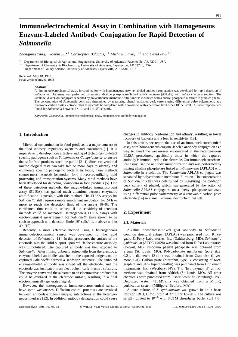
In the procedure presented here (Fig. 1),Salmonellawas directlyconjugated to APLAS in the solution. The effects of such bindingon enzyme activity have not been reported. In our first experiment,the effects of concentration and incubation time of APLAS withSalmonellaon the final oxidation peak current of phenol werestudied. As shown in Figure 3, incubating 105 cells/mL ofSalmonellawith APLAS at concentrations varying between 0 and1mg/mL, the maximum peak current of phenol generated bySalmonella-APLAS conjugates was above 0.2mg/mL of APLAS.Using 0.2mg/mL of APLAS, incubation times longer than 30minutes generated the maximum peak current of phenol. Since nochange in the alkaline phosphatase enzyme activity could bedetected after binding withSalmonella through the antibody-antigen interaction, the one-step separation of theSalmonella-APLAS conjugate from the excess of the APLAS was necessary in
914 Z. Yang et al.
Electroanalysis1998, 10, No. 13
Fig. 1. Procedure for the immunoelectrochemical assay. AP labeled anti-Salmonellais added to a sample tube containingSalmonella(I) and allowedto incubate for 30 min (II). AP labeledSalmonellaconjugates are retained byfiltration through a polycarbonate membrane (III) and then incubated with aphenyl phosphate substrate to generate phenol (IV), which is determined bydifferential pulse voltammetry. The resulting peak current is proportional tothe Salmonellain the sample.
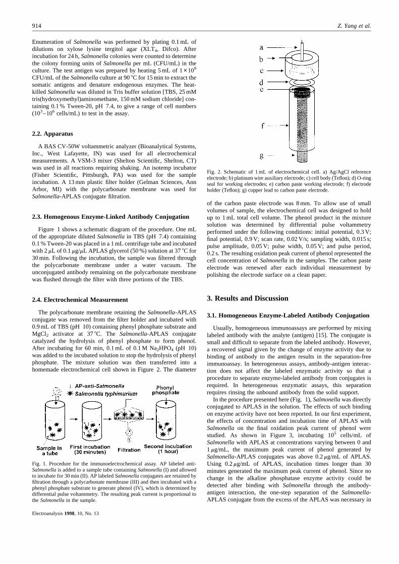
Fig. 2. Schematic of 1 mL of electrochemical cell. a) Ag/AgCl referenceelectrode; b) platinum wire auxiliary electrode; c) cell body (Teflon); d) O-ringseal for working electrodes; e) carbon paste working electrode; f) electrodeholder (Teflon); g) copper lead to carbon paste electrode.
the procedure. Fortunately, the excess of APLAS was easilyseparated fromSalmonella-APLAS conjugates (>0.4mm) byfiltration through the polycarbonate membrane (pore size0.22mm), due to the large size differential between the two.
3.2. Optimization of Enzyme Incubation with Substrate
Phenyl phosphate was chosen as the enzymatic substrate for theelectrochemical determination of alkaline phosphatase labeledanalyte [16]. Although its product, phenol, has a high oxidationpotential (ca. 0.6 V vs. Ag/AgCl at pH 10), which may result in ahigh background current at some solid electrodes, such as platinumor glassy carbon, use of carbon paste electrode as working electrodeeliminates some background current within a broad potential range[14]. Incubation times, MgCl2 activator concentrations, and pH ofthe enzymatic substrate solutions were varied to discover theoptimal conditions for phenol production catalyzed by theSalmonella-APLAS conjugate.
3.2.1. pHThe reaction rate at which alkaline phosphatase enzyme
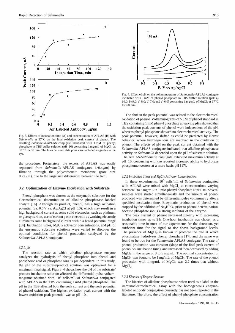
catalyzes the hydrolysis of phenyl phosphate into phenol andphosphoric acid or phosphate ions is pH dependent. In this study,the pH of the substrate/product solution was optimized for amaximum final signal. Figure 4 shows how the pH of the substrate/product incubation solution affected the differential pulse voltam-mograms obtained with 105 cells/mL of Salmonellaconjugatedwith APLAS in the TBS containing 1 mM phenyl phosphate. ThepH in the TBS affected both the peak current and the peak potentialof phenol oxidation. The highest oxidation peak current with thelowest oxidation peak potential was at pH 10.
The shift in the peak potential was related to the electrochemicaloxidation of phenol. Voltammograms of 5mM of phenol standard inTBS containing 1 mM phenyl phosphate at varying pHs showed thatthe oxidation peak currents of phenol were independent of the pH,whereas phenyl phosphate showed no electrochemical activity. Thepeak potential, however, shifted as could be predicted by Nernstbehavior, where hydrogen ions are involved in the oxidation ofphenol. The effects of pH on the peak current obtained with theSalmonella-APLAS conjugate indicated that alkaline phosphataseactivity onSalmonelladepended upon the pH of substrate solution.The APLAS-Salmonellaconjugate exhibited maximum activity atpH 10, concurring with the reported increased ability to hydrolyzephosphomonoesters at a more basic pH [17].
3.2.2 Incubation Times and MgCl2 Activator ConcentrationsIn these experiments, 105 cells/mL of Salmonellaconjugated
with APLAS were mixed with MgCl2 at concentrations varyingbetween 0 to 5 mg/mL in 1 mM phenyl phosphate at pH 10. Severalsamples were started simultaneously and the amount of phenolproduced was determined by differential pulse voltammetry after aspecified incubation time. Enzymatic production of phenol wasstopped by the addition of Na2HPO4 prior to phenol determination,because phosphate ion is a strong inhibitor of the enzyme.
The peak current of phenol increased linearly with increasingincubation times up to 2 h. One-hour incubation was chosen as areasonable time in most of our experiments for the detection, yetsufficient time for the signal to rise above background levels.The presence of MgCl2 is known to promote the rate at whichphosphatase hydrolyzes phenyl phosphate [17], and the same wasfound to be true for theSalmonella-APLAS conjugate. The rate ofphenol production was constant (slope of the final peak current ofphenol vs. incubation time), and increased then decreased by addingMgCl2 in the range of 0 to 5 mg/mL. The optimal concentration ofMgCl2 was found to be 1 mg/mL of MgCl2. The rate of the phenolproduction with 1 mg/mL of MgCl2 was 2.2 times that withoutMgCl2.
3.2.3 Kinetics of Enzyme ReactionThe kinetics of alkaline phosphatase when used as a label in the
immunoelectrochemical assay with the homogeneous enzyme-labeled antibody conjugation currently has not been reported in theliterature. Therefore, the effect of phenyl phosphate concentration
915Rapid Detection of Salmonella
Electroanalysis1998, 10, No. 13
Fig. 3. Effects of incubation time (A) and concentration of APLAS (B) withSalmonellaat 378C on the final oxidation peak current of phenol. Theresulting Salmonella-APLAS conjugate incubated with 1 mM of phenylphosphate in TBS buffer solution (pH 10) containing 1 mg/mL of MgCl2 at378C for 30 min. The lines between data points are included as guides to theeye.
Fig. 4. Effect of pH on the voltammograms ofSalmonella-APLAS conjugateincubated with 1 mM of phenyl phosphate in TBS buffer solution [pH: a)10.0; b) 9.0; c) 8.0; d) 7.0; and e) 6.0] containing 1 mg/mL of MgCl2 at 378Cfor 60 min.
on the enzymatic production of phenol was investigated using theimmunoelectrochemical assay of theSalmonella-APLAS conju-gate. The final peak current of phenol could represent the reactionrate of phenol production catalyzed by theSalmonella-APLASconjugate at the specified incubation time. A typical Michaelis-Menten curve of theSalmonella-APLAS conjugate was determinedby varying concentration of phenyl phosphate substrate in theincubated solution. According to the Michaelis-Menten equation,the enzymatic activity is proportional to the maximum reaction rateof the phenol production. The typical apparent Michaelis constant,Kapp
M , was found to be 1.6 mM in the presence of 105 cells/mL ofSalmonella-APLAS conjugate using the Lineweaver-Burk plot. Theconcentration of phenyl phosphate substrate in this assay wasselected to be 1 mM in the immunoelectrochemical assay.
3.3. Detection ofSalmonella
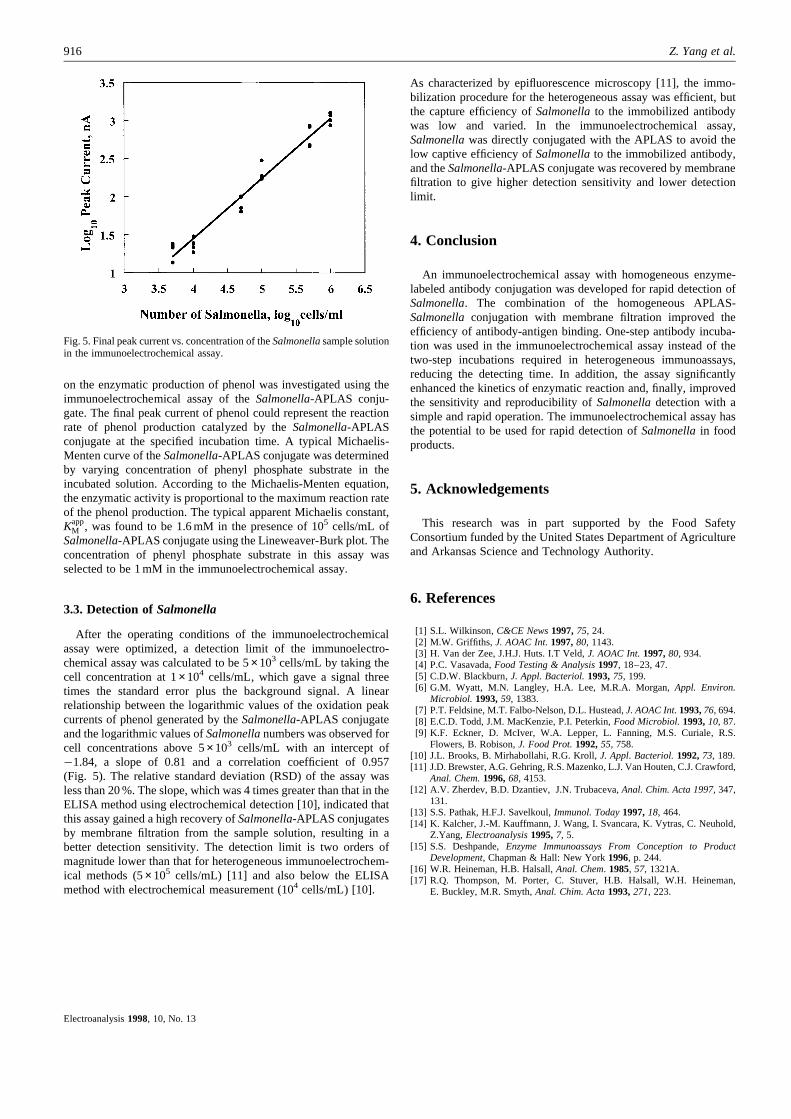
After the operating conditions of the immunoelectrochemicalassay were optimized, a detection limit of the immunoelectro-chemical assay was calculated to be 5×10³ cells/mL by taking thecell concentration at 1×10⁴ cells/mL, which gave a signal threetimes the standard error plus the background signal. A linearrelationship between the logarithmic values of the oxidation peakcurrents of phenol generated by theSalmonella-APLAS conjugateand the logarithmic values ofSalmonellanumbers was observed forcell concentrations above 5×10³ cells/mL with an intercept of¹1.84, a slope of 0.81 and a correlation coefficient of 0.957(Fig. 5). The relative standard deviation (RSD) of the assay wasless than 20 %. The slope, which was 4 times greater than that in theELISA method using electrochemical detection [10], indicated thatthis assay gained a high recovery ofSalmonella-APLAS conjugatesby membrane filtration from the sample solution, resulting in abetter detection sensitivity. The detection limit is two orders ofmagnitude lower than that for heterogeneous immunoelectrochem-ical methods (5× 10⁵ cells/mL) [11] and also below the ELISAmethod with electrochemical measurement (104 cells/mL) [10].
As characterized by epifluorescence microscopy [11], the immo-bilization procedure for the heterogeneous assay was efficient, butthe capture efficiency ofSalmonellato the immobilized antibodywas low and varied. In the immunoelectrochemical assay,Salmonellawas directly conjugated with the APLAS to avoid thelow captive efficiency ofSalmonellato the immobilized antibody,and theSalmonella-APLAS conjugate was recovered by membranefiltration to give higher detection sensitivity and lower detectionlimit.
4. Conclusion
An immunoelectrochemical assay with homogeneous enzyme-labeled antibody conjugation was developed for rapid detection ofSalmonella. The combination of the homogeneous APLAS-Salmonellaconjugation with membrane filtration improved theefficiency of antibody-antigen binding. One-step antibody incuba-tion was used in the immunoelectrochemical assay instead of thetwo-step incubations required in heterogeneous immunoassays,reducing the detecting time. In addition, the assay significantlyenhanced the kinetics of enzymatic reaction and, finally, improvedthe sensitivity and reproducibility ofSalmonelladetection with asimple and rapid operation. The immunoelectrochemical assay hasthe potential to be used for rapid detection ofSalmonellain foodproducts.
5. Acknowledgements
This research was in part supported by the Food SafetyConsortium funded by the United States Department of Agricultureand Arkansas Science and Technology Authority.
6. References
[1] S.L. Wilkinson,C&CE News1997,75, 24.[2] M.W. Griffiths, J. AOAC Int.1997,80, 1143.[3] H. Van der Zee, J.H.J. Huts. I.T Veld,J. AOAC Int.1997,80, 934.[4] P.C. Vasavada,Food Testing & Analysis1997, 18–23, 47.[5] C.D.W. Blackburn,J. Appl. Bacteriol.1993,75, 199.[6] G.M. Wyatt, M.N. Langley, H.A. Lee, M.R.A. Morgan,Appl. Environ.
Microbiol. 1993,59, 1383.[7] P.T. Feldsine, M.T. Falbo-Nelson, D.L. Hustead,J. AOAC Int.1993,76, 694.[8] E.C.D. Todd, J.M. MacKenzie, P.I. Peterkin,Food Microbiol.1993,10, 87.[9] K.F. Eckner, D. McIver, W.A. Lepper, L. Fanning, M.S. Curiale, R.S.
Flowers, B. Robison,J. Food Prot.1992,55, 758.[10] J.L. Brooks, B. Mirhabollahi, R.G. Kroll,J. Appl. Bacteriol.1992,73, 189.[11] J.D. Brewster, A.G. Gehring, R.S. Mazenko, L.J. Van Houten, C.J. Crawford,
Anal. Chem.1996,68, 4153.[12] A.V. Zherdev, B.D. Dzantiev, J.N. Trubaceva,Anal. Chim. Acta 1997, 347,
131.[13] S.S. Pathak, H.F.J. Savelkoul,Immunol. Today1997,18, 464.[14] K. Kalcher, J.-M. Kauffmann, J. Wang, I. Svancara, K. Vytras, C. Neuhold,
Z.Yang,Electroanalysis1995,7, 5.[15] S.S. Deshpande,Enzyme Immunoassays From Conception to Product
Development, Chapman & Hall: New York1996, p. 244.[16] W.R. Heineman, H.B. Halsall,Anal. Chem. 1985, 57, 1321A.[17] R.Q. Thompson, M. Porter, C. Stuver, H.B. Halsall, W.H. Heineman,
E. Buckley, M.R. Smyth,Anal. Chim. Acta1993,271, 223.
916 Z. Yang et al.
Electroanalysis1998, 10, No. 13
Fig. 5. Final peak current vs. concentration of theSalmonellasample solutionin the immunoelectrochemical assay.