immuniy, antigen and antibody, hypersensitivity reactions and ocular corelation by raju
TRANSCRIPT

ANTIGEN, ANTIBODY AND COMPLEMENTS.TYPES OF IMMUNE RESPONSES AND HYPERSENSITIVITY REACTIONS.
Raju Kaiti
Optometrist, Dhulikhel Hospital-Kathmandu University Hospital
REFERENCES
Short text book of medical microbiology, 6th edition, Satish Gupte
Lippincott’s microbiology Robbin’s pathology Immunopathology of the eye by A. H. S. Rahi and A.
Garner Ocular Pathology by Myron Yanoff and Ben S. Fine Internet Class notes

IMMUNITY
Greek word Immunis:- Free from burden Sequence of cellular and molecular events
designed to rid the host of an offending stimulus
Pathogenic organism\toxic substances\cellular debris\ neoplastic cells

Immunology Science which deals with the body’s response
to antigenic challenge.
Deals with the vital immune system.
Immune system is an interacting set of specialized cells and proteins designed to identify and destroy foreign invaders or abnormal substances before they damage the body.

Two arms of immune system
1.Innate (or natural) immune systema. Non specific Physical barrier: skin, mucus Proteins in serum and in tissues: Lysozyme, interferon,
complements. (eg. In tears…….)
b. Specific Antibody mediated Cell mediated
2.Adaptive(or acquired)specific immune system

Active Passive1.Produced actively by the immune system of host
1.Received passively by the host and the immune system doesn’t participate.
2.Induced by infection or by contact with immunogen.
2.Conferred by introducing ready made antibody.
3. Immune response-durable and effective
3. Immune response-short lived and less effective
4.Immunity develops only after a lag period.
4.Immunity effective immediately.
5.Immunological memory presumed.
5.No immunological memory.
6.Serves no purpose in immunodeficient host.
6.Applicable in immuno-deficient host.
7.No inheritance of immunity.
7.May be acquired from mother


Innate Immunity Components :
Macrophages Granulocytes Natural killer cells Complement Other chemicals eg. HCl, Lysozymes
Characteristics: Immediate action Non-specific response

Adaptive Immunity
Humoral
B cells Antibodies Complements
Cell-mediated
Antigen Presenting Cells T cells
Characteristics: Specific response
Late response

Cells of Immune system
T-Lymphocytes Thymus derived lymphocytes Role in cellular or cell-mediated immunity. Constitutes 60-70% of peripheral lymphocytes Differentiation of T-cells
Helper T cells Essential to the differentiation of B-cells into plasma
cells and their subsequent secretion of Antibodies. Each helper T-cell is capable of activating hundreds of
specific B-cells

Suppressor T-cells :
inhibit the development of B-cells in to plasma cells
regulate the activity of killer T-cells and suppress the production of Abs when they
become excessive. Also suppress auto-immune responses.

Killer T-cells
Have specific receptor for antigenic determinants.
Killer T-cell migrate from lymphoid tissue to the site of foreign cell invasion where they secrete small protein, lymphokines.
Prevent the reproduction of invading micro-organisms, infected host cells or viruses inside host cells.

Memory T-cells
The T-cells that remain potentially active and viable even after the antigen has been inactivated.
Upon 2nd encounter memory cells proliferate, differentiate into plasma cells and secrete Abs so rapidly that the symptoms of the disease may not even be observed.

Humoral immunity Cellular immunity
15
Types of Immunity

Antigens Basically Exogenous
Occasionally may be derived from body’s own tissues
Protein molecules or part which have specific AA sequence folded in tertiary shapes.
Substances that stimulate Ab production when they react.
Molecular wt. : 8000 or more

Properties of antigens
Foreignness Size
Types :
Complete antigen Partial antigen

Hapten e.g lipid,nucleic acid
Two types:Complex hapten andSimple hapten
Immunogen Vs antigen All immunogens are antigens but not all the
antigens are immunogens.

Antibody
o Specific glycoprotein molecules generated by ß –cells in response to antigens.
o Also called immunoglobulins.
o Humoral substance found in serum,lymphs and other body fluids.
o Highly specific in nature.

Organs producing Antibodies
Spleen, lymphnodes and bone marrow
Tissues like peyer’s patches, appendix, thymus
These structures contains lymphocytes macrophages and plasma cells

FUNCTIONS
Neutralization of toxins.
Activation of complement (results in improved opsonisation)
Lysis of invading microorganisms.

Immunoglobulin
Immunoglobulins are synthesized by plasma cells and also by lymphocytes.
All antibodies are Immunoglobulins but all Immunoglobulins may not be antibodies.
Immunoglobulin is the structural and chemical concept while antibody is biological and functional concept.

Classification:# Types based on Size, Carbohydrate
content and amino acid analysis:
IgG IgM IgA IgD IgE

Immunoglobulin
IgG: comprises 70% of total Ig. Shortest half life of 21 days Lowest mol. Wt. and found in highest concn in body. crosses placenta and provides much of maternal antibody. Responsible for late immune response. Bivalent in structure. Four sub classes: IgG1, IgG2, IgG3, IgG4.

IgA:
•In body secretion like milk, tears, saliva, urine etc.
•Also called secretory immunoglobulins
•Antibacterial and antiviral
•Present as either monomer or dimer
•Majorly generated in bone marrow

IgM: Highest mol.wt Present in serum as pentamer Constitute only 10% of serum immunoglobin Can’t cross transplacental barrier. Responsible for early immune response.

IgE:
Play role in parasitic and allergic disease. Shortest half life. Present in small quantities.

Contd…
IgD:
Present in the surface of the lymphocytes.
Least abundant of all. Mainly intravascular
distribution.


Chemistry of immunoglobulins
All immunoglobulins composed of same basic units.
Consists two light (L) and two heavy (H) chains.
L chains and H chains linked together by a disulphide bond.
Two H chains linked similarly.

Enzyme action breaks the structure into three. Two identical Fab fragments Third Fc fragment Two classes of L chains – Kappa (k) and Lamda (λ) Five classes of H chains µ- IgM α- IgA γ- IgG δ- IgD ε- IgE

Different regions
Constant region ‘C’
Variable region ‘V’
Adjuvants are substances which enhance the immune response
Weak antigens also evoke high order of antibody production


Complements
Protein substance involved in immune response
Synthesized by hepatocytes, blood monocytes, epithelial cell of GI tract and tissue macrophages
Functions include opsonisation, target cytolysis, inflamation and immune complex clearence with lysis of bacterial cells

2 pathway of complement activation. complement system
classical alternateBrought about by Ag-Ab complex.
Involves activation of nine major proteins C1 to C9
Brought about by certain bacterial polysaccharides, endo-toxin
Activated by agregated IgA

Contd…
Refers to series of factors occuring in normal serum activated by Ag-Ab interaction
Concentration is fairly constant for each species of animal
10% of human serum globulin. Concentration decreased in acute glomerulo-nephritis,
serum sickness Concentration increased in carcinomatosis, coronary
occlusion and rheumatic fever

Components of Complement
Known to have nine distinct components
One of which have three protein subunits making total 11 proteins
C1: 3 proteins held by calcium ions.

Biosynthesis of Complement
C1 – synthesized in interstitial epithelium C2 and C4 – macrophages C5 and C8 – in spleen C3, C6 and C9 – in liver C7 – not known

Features of antigen antibody reactions
Reactions highly specific Entire molecules react and not fragment No de-naturation of antigen or antibody during reaction Combination is formed but reversible Both Ag and Ab participate in formation of agglutinates
or precipitates Ag and Ab may combine in varying proportion

Cross reactions Particular antibody may react with other antigens
also
Heterophile antigens Antigens those are cross reacting with other
antibodies
Heterophile antibodies Antibodies those are cross reacting with or
antigens
Antibody title highest dilution of patient serum where visible
antigen antibody reaction takes place

Factors influencing antibody production
Age Nutritional status Root of adminstration Size and no. of doses
Critical dose and immunological paralysis
Multiple antigens Adjuvants Immunosuperssive agents

Figure of Ag-Ab Reation

Immune Response
Specific reactivity induced in host by antigenic stimulus
Primary response Secondary responseHumoral
Cell-mediated
Slow, sluggish, short lived with a long lag phage and low antibody production, Predominantly IgM
Prompt, powerful, prolonged with much higher level of Ab production predominantly IgG

Ocular Immune Responses
Conjunctiva
Tear film: Washes away debris and irritants lysosyme, betalysin, lactoferrin, IgA
Well vascularized, Langerhans cells, dendritic cells and macrophages

Conjunctiva
Papillary reaction in allergic conjunctivitis

CORNEA
No localized immune processing
Immune Privilege: Normal limbal physiology, avascularity, absence of APCs and lymphatics, intact immunoregulatory systems of anterior chamber

Cornea
Transplantation Immunology: type of rejection reactions:
High success rate of graft (90%): Immune privilege

Failure of Immune system
Immune system
Hypersensitivity
(Overactive immune response)
Immunodeficiency
(ineffective immune response)
Autoimmunity
(mistaken recognition of self antigens)

Hypersensitivity Term used to describe immune responses
that cause host tissue damage Detrimental effect on hosts
Fever shock Inflammatory nature Spasm of smooth muscle Gastrointestinal and pulmonary disorders Fatal circulatory collapse

Hypersensitivity
State in which the introduction of an antigen into the body elicits an unduly severe immunological reaction.
4 types: -1. Anaphylaxis, atopic or Type I reaction.2. Cytotoxic or Type II3. Immune complex, Arthus-Type III4. Delayed hypersensitivity Type IV

Hypersensitivity
Type I Exaggerated IgE response to relatively harmless
environmental antigens
Genetic predisposition
Results in release of several active substances including histamine, slow reacting substance and an eosinophil chemo tactic factor

Type I
Eg: Hay fever, atopic dermatitis, systemic anaphylaxis Atopic conjunctivitis
Diagnosis: In vivo skin testing with batteries of allergens In vitro RAST test (quantitate specific IgE levels

Allergic conjunctivitis

Type II
Antibody mediated hypersensitivity against self cells or receptors or membranes
Mediated by IgG or IgM antibodies against tissue antigens, resulting in organ-specific antibody production

Type II
Antibody binds to cells or tissues and causes local complement activation, influx of leukocytes, and tissue destruction by: ADCC Degranulation by phagocytes Production of oxygen radicals

Type II Diagnosis:
Detect immunoglobulins on affected cells or tissues
Detect complement in affected tissueDetect autoantibody or auto reactive T
cells

Type II
eg
Mooren’s ulcer Hemolytic disease of the newbornGoodpasture syndromeHyper acute graft rejection

Type III
Due to high levels of circulating, soluble immune complexes overwhelming the ability of the mononuclear phagocyte system to remove them
Damage is caused by antigen-antibody complex.

Type III
The excess complexes deposit in various tissues and activate complement
Subsequent attempt by neutrophils to remove them results in degranulation and tissue damage.

Type III
Can take one of two forms according to whether the immune complex develops in circulating blood or in tissues
Arthus reaction Local manifestation in tissue
Serum sickness Systemic form of type III hypersensitivity

Type III
Eg. Arthus reaction, serum sickness, Lupus, Rheumatoid
arthritis, etc. Immune ring formation in cornea in Herpes simplex
keratitis
Diagnosis: very low levels of complements in blood, esp. c3 and
c4

Immune ring formation in herpes simplex keratitis

Peripheral corneal thinning in rheumatoid arthritis

Type IV No role of antibody or complement One aspect of cell mediated immunity Antigen activates specifically macrophages
and sensitized T-lymphocytes leading to secretions of lymphokines
Due to activity of thymus dependent lymphocytes and clinically has a delayed onset
Two types: Classical or Tuberculin type Granulomatous reactions

Type IVExample:
Allergic Dermatoconjunctivitis, disciform keratitis

Example Corneal graft rejection Sympathetic ophthalamitis Vogt Koyonagi Harada’s syndrome Optic neuritis Recurrent herpetic keratitis Bacterial, fungal, viral, protozoal and parasytic
infection.

69
AIDS Ocular manifestations
Kaposi’s sarcoma of conjunctiva CMV retinitis Recurrent infections Viral infections

Auto-immune Diseases Myasthenia gravis Pemphigus vulgaris Ocular cicatricial Pemphigoid Sympathetic ophthalmitis Phacogenic uveitis Multiple sclerosis Auto immune hemolytic
anemia Idiopathic thrombocytopenic
purpurae
Organ-specific

Idiopathic leucopenia Primary billiary cirrhosis Active chronic hepatitis Cryptogenic cirrhosis Ulcerative colitis SjÖgren’s syndrome Rheumatoid arthritis Dermatomyositis Scleroderma Discoid lupus erythematosus
Non-organ specific


Antibody-mediated Diseases
Vernal conjunctivitis : Mostly affects children & adolescents
Occurs only in warm season of year.
Produces giant papillae (cobblestone) of tarsal conjunctiva.

Rheumatoid Diseases affecting the eye Juvenile rheumatoid arthritis
Females>males
C/F uveitis extensive synechia formation Cataract Secondary glaucoma

Reiter’s diseases males>females
C/F Self limited papillary conjunctivitis Acute iridocyclitis or both eyes occasionally with
hypopyn.

76
Other antibody mediated Diseases
Systemic lupus erythematosus Occlusive vasculitis of nerve fiber layer of retina. Infarcts results in cytoid bodies or cotton-wool spots in
retina.
Pemphigus vulgaris Intraepithelial bullae of conjunctiva

Lens induced Uveitis
Rare condition associated with circulating Ab to lens protein.
In individuals whose lens capsule is permeable to to these protein as a result of trauma or other Diseases.

Cell mediated Diseases
Ocular sarcoidosis Panuveitis with inflammatory involvement of
optic nerve and retinal blood vessels. Acute iridocyclitis Conjunctival erythema.
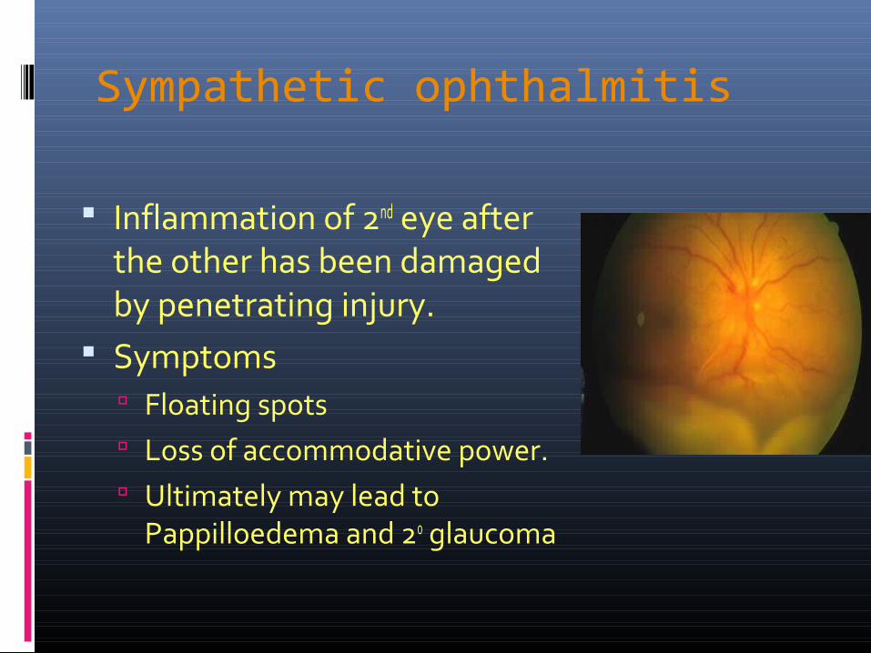
Sympathetic ophthalmitis
Inflammation of 2nd eye after the other has been damaged by penetrating injury.
Symptoms Floating spots Loss of accommodative power. Ultimately may lead to
Pappilloedema and 20 glaucoma

Thank You!Thank You! Suggestions!Suggestions!