immune reconstitution and hiv - south central aids ... filecase 1 51yo ethiopian female, immigrated...
TRANSCRIPT

IMMUNE RECONSTITUTION AND HIV
Jason Gillman, MD Prism Health North Texas November 28, 2017

Disclosures
Gilead Sciences ◦ Speakers bureau
Abbvie ◦ Speakers bureau
Overview Definition and Pathogenesis Risk factors Timing and Incidence IRIS syndromes and their management ◦ MAC ◦ TB ◦ PML ◦ CMV ◦ Cryptococcus ◦ KS
Prevention ◦ Timing of HAART initiation

What’s New
Timing of ART in setting of active TB ◦ Minor guideline update
PML-IRIS ◦ Minor guideline update

Case 1 51yo Ethiopian female, immigrated to the U.S. in August,
2013, admitted in October with cough, dyspnea, and weight loss, found to have a new diagnosis of HIV (CD4 21, VL 2 million c/mL) as well as pulmonary TB (culture-confirmed). HIV genotype was wild-type, and she was treatment naïve.
She was started on RIPE, and ~two weeks later on Atripla. Discharged in stable condition
She returned to the hospital 8 days later with fever to 38.6C, tachycardia, hypoxia, and worsening pleuritic chest pain and cough
CT chest showed increased cavitation of mediastinal lymph nodes and larger bilateral pleural effusions
HIV RNA had declined to 6684 c/mL, although she had to self-discontinue Atripla due to nausea. CD4 count unchanged.

IRIS/IRD
Immune Reconstitution Inflammatory Syndrome (IRIS) ◦ Also referred to as immune reconstitution disease (IRD)
“Paradoxical” IRIS - Worsening of a recognized OI “Unmasking” IRIS – Symptoms from an unrecognized
pre-existing OI that manifest in the setting of improving immunologic function.

Example: TB-IRIS
Patient on treatment for
TB
(Worsening disease)
Paradoxical IRIS
ART
Patient not known to have
TB
(Symptoms of TB)
Unmasking IRIS
ART

Pathogenesis
IRIS
Suppresion of viremia
Immune Recovery
Antigenic Burden
Genetic susceptibility
Cytokines
Tmem
Tnaive
Treg

Mayer K H , and French M A Clin Infect Dis. 2009;48:101-107

Incidence Study Type Incidence
Michelet, et al. AIDS. 1998 Retrospective 68%
French, et al. HIV Med. 2000 Retrospective 25%
Ratnam, et al. CID. 2006 Retrospective 23%
Murdoch, et al. AIDS. 2008 Prospective 10%
Grant, et al. PLoS One. 2010 Prospective 7.6%
Muller, et al. Lancet Infect Dis. 2010 Meta-analysis 13%
Previously diagnosed OI Incidence of IRIS
Kaposi’s Sarcoma 6.4%
Herpes Zoster 12.2%
TB 15.7%
PML 16.7%
Cryptococcal meningitis 19.5%
CMV retinitis 37.7%

Risk Factors: CD4 count < 100 Disseminated infection (high antigenic
burden) Shorter interval between initiating
treatment for OI and starting HAART Younger age Rapidity and magnitude of virological
response to ART Fungal infection (other than PJP)

Timing: Highly variable As little as 3 days after initiation of
HAART, up to 3 months or longer IRD can occur early due to the
immediate improvement in immune function that occurs with virologic suppression
For the ID board exams – remember 2-4 weeks
Diagnosis:
No strict diagnostic criteria, diagnosis based on degree of clinical suspicion
Case Definition (INSHI) 1. Response to anti-retroviral therapy by: ◦ a. receiving HIV anti-retroviral therapy and; ◦ b. virologic response with >1 log10 copies/mL
decrease in HIV RNA (if available). 2. Clinical deterioration of an infectious or
inflammatory condition temporally related to ART initiation.
3. Symptoms cannot be explained by: ◦ a. expected clinical course of a previously recognized
and successfully treated infection ◦ b. medication side effect or toxicity ◦ c. treatment failure ◦ d. complete non-adherence.

Challenges:
Diagnosis is clinically challenging, one must differentiate from: ◦ Progression of the initial OI antimicrobial resistance treatment failure for any reason, including poor
adherence
◦ Development of a new OI ◦ Unrelated organ dysfunction ◦ Drug toxicity Allergic Reaction – Abacavir Hypersensitivity

IRIS SYNDROMES

Clinical Manifestations: Symptoms are diverse and have not been
defined precisely Usually characterized by: ◦ Fever ◦ Worsening of the clinical manifestations of
underlying OI.

Causes of IRIS: Mycobacteria ◦ MAC ◦ TB
Viruses ◦ PML ◦ CMV ◦ Herpes virus VZV HSV
◦ HBV/HCV Fungal ◦ Cryptococcus ◦ Histoplasmosis ◦ PJP
KS Toxoplasmosis
Parvovirus B19
Strongyloides stercoralis and other parasites
Sinusitis
Folliculitis
Rheum/autoimmune RA, SLE
Graves disease
Sarcoid
Tattoo ink
AIDS-related lymphoma
Guillain-Barre Syndrome

Case 2 50 y/o man with HIV, CD4 initially 16, VL 2 million,
PPD neg Started on HAART Returned to clinic with tender lump in axilla one
month later ◦ 1 x 2 cm, firm mass, almost cystic ◦ No overlying cellulitis ◦ CD4 up to 192, VL down to 4K
Referred to FNA clinic, around which time it had started to drain
Sample was sent for culture, AFB stain negative 2 months later culture found to be growing MAC.

MAC-IRIS Incidence ~ 3.5% among patients with CD4 < 100 Median time to onset of *symptoms is 3 weeks after ART initiation Usually unmasking of subclinical disease (75% of cases)
Peripheral lymphadenitis
• Enlarged lymph node
• Draining sinus • Less than half with
fever
Pulmonary thoracic disease
• Fever • Night sweats • Cough • Dyspnea • CT findings
• Mediastinal lymphadenopathy
• Parenchymal infiltrates
• Cavitary lesions • Nodules
Intra-abdominal disease
• Fever • Night sweats • Abdominal pain • CT findings
• Intra-abdominal lymphadenopathy
Phillips P, et al. CID. 2005

Riddell J, et al. J Translational Med. 2007

MAC-IRIS: Management
Anti-mycobacterial therapy ◦ Azithromycin + ethambutol (+/- rifabutin)
For moderate-severe IRIS ◦ Initial treatment with NSAIDs (CIII) ◦ If no improvement, prednisone 20-40 mg daily
for 4-8 weeks (CII)
Relapse is common after stopping steroids
Symptoms may persist up to 6 months

TB, HIV, and IRIS
HIV and TB co-infection ◦ Pulmonary disease ◦ Lymphadenitis ◦ Intracranial tuberculomas/meningitis ◦ Cutaneous lesions ◦ Peritonitis ◦ Epididymitis/Prostatitis ◦ Cystitis ◦ Granulomatous nephritis/hepatitis ◦ Osteomyelitis

TB-IRIS Can occur with treatment of TB in the
absence of ART ◦ Restoration of TB-induced immunosuppression ◦ Release of new antigen targets during
mycobacterial killing
Incidence for patients started on ART is ~ 15%. Time to onset is typically 2-4 weeks ◦ Resolution of symptoms in ~ 3 months ◦ Generally paradoxical IRIS (except in developing
countries where the diagnosis may be missed)
Meintjes, et al. Clin Chest Med. 2009

Worsening radiographic
infiltrate (50% of patients
starting ART)
Enlarging serous effusions
Enlarging lymph node
Fever
A) Time of diagnosis of TB B) 3 weeks after ART initiation
Meintjes, et al. Clin Chest Med. 2009

TB-IRIS: Management
Continue HAART and continue RIPE Prednisone 1.5 mg/kg/day x 2 weeks,
followed by 0.75 mg/kg/day x 2 weeks ◦ This steroid regimen was evaluated in a
randomized, double-blind placebo-controlled trial ◦ Decreases hospital days (p=0.04), and
improves both symptoms and chest radiographs at 2 and 4 weeks
Meintjes, et al. AIDS. 2010
TB-IRIS: Management
For some patients, 4 weeks is insufficient, and DHHS guidelines suggest that tapering steroids over 3 months may be considered (BIII)
NSAIDs may be considered for mild TB-IRIS (CIII)
Needle aspiration of effusions for symptomatic relief
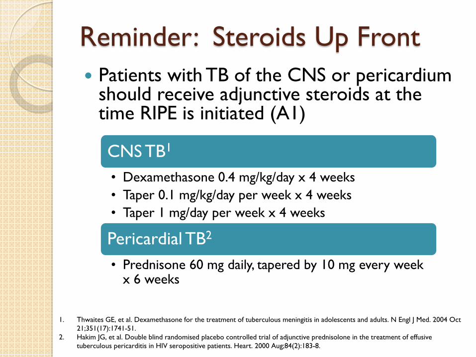
Reminder: Steroids Up Front Patients with TB of the CNS or pericardium
should receive adjunctive steroids at the time RIPE is initiated (A1)
1. Thwaites GE, et al. Dexamethasone for the treatment of tuberculous meningitis in adolescents and adults. N Engl J Med. 2004 Oct 21;351(17):1741-51.
2. Hakim JG, et al. Double blind randomised placebo controlled trial of adjunctive prednisolone in the treatment of effusive tuberculous pericarditis in HIV seropositive patients. Heart. 2000 Aug;84(2):183-8.
CNS TB1
• Dexamethasone 0.4 mg/kg/day x 4 weeks • Taper 0.1 mg/kg/day per week x 4 weeks • Taper 1 mg/day per week x 4 weeks
Pericardial TB2
• Prednisone 60 mg daily, tapered by 10 mg every week x 6 weeks

Case 3 40 y/o man with HIV p/w word
finding difficulty R hemiparesis and seizure. CD4 48.
LP + JC Virus on PCR Started on HAART 2.5 months later presented with
worsening aphasia and had repeat MRI. CD4 225, VL undetectable.
HAART held x 2 wks, pt treated with dexamethasone.
2.5 yrs later, only mild residual word-finding deficit.
Case and images from Tan and Koralnik. Lancet Neurol. 2010

PML-IRIS HIV+ patients co-infected
with JC virus with CD4 < 200 may develop Progressive multifocal leukoencephalopathy (PML)
10 – 20% of pts develop new or worsening neurologic symptoms following initiation of HAART.
Tan et al. Neurology. 2009

Classic PML
PML-IRIS
FLAIR sequence T1 post-Gadolinium Tan and Koralnik. Lancet Neurol. 2010

PML-IRIS Mediated by an influx of CD8+ T-cells into
existing lesions ◦ JCV actually less likely to be detected in CSF
IRIS with PML doesn’t seem to alter survival (1 yr survival rate 54% vs 49% in PML-IRIS vs PML without IRIS).
Marzocchetti, et al. Neurology. 2009

PML-IRIS: Management There are no effective anti-viral drugs for
JCV ◦ Continue HAART (AIII)
Steroids recommended for patients with
contrast-enhancing lesions on MRI, mass effect, or clinical deterioration (BIII). ◦ Methylprednisolone 1 gm IV daily x 3-5 days ◦ Prednisone 60 mg PO daily, tapered over 6 weeks
Consider maraviroc
Tan, et al. Neurology. 2009


CMV and IRIS Immune reconstitution disease occurs in patients with
inactive CMV retinitis who begin ART ◦ Immune recovery uveitis (IRU) Anterior uveitis Vitritis Cystoid macular edema Epiretinal membranes Retinal neovascularization
The most common OI to result in immune reconstitution disease following ART initiation (~38% incidence)
Symptoms: ◦ Anterior chamber: ocular pain and photophobia ◦ Posterior chamber: painless floaters and decreased visual acuity

Risk Factors for IRU
CMV involving >30% retinal surface area at the highest risk
Treatment of CMV retinitis with cidofovir compared to an alternative regimen
Almost exclusively occurs in patients with immune recovery to CD4 > 100 ◦ Does not occur in patients with a virologic
response but no immune recovery
Goldberg, et al. Retina. 2005

Goldberg, et al. Retina. 2005

Management of IRU/IRV Corticosteroids and
anti-CMV therapy (CIII)
Anterior chamber: topical corticosteroids
Posterior chamber: periocular or intravitreal corticosteroid injections Kosobucki, et al. Am J Ophthalmol. 2004

IRIS and Cryptococcus: ~20% of patients with cryptococcal meningitis and
HIV develop IRIS after initiation of ART.
Presentation ◦ Meningeal (73.7%) ◦ Other CNS complications (11%) ◦ Lymphadenopathy (11%) ◦ Pneumonitis (4.5%)
Timing: delayed compared to TB (1-10 months)
The key CSF findings are elevated opening pressure
and elevated CSF WBC, with sterile cultures
Haddow LJ, et al. Lancet Infect Dis. 2010

Risk factors for C-IRIS
Fungal burden (initial CrAg titer) Lack of initial CSF inflammation (CSF
WBC < 25 cells/µL and protein < 50 mg/dL)
Early HAART initiation* Baseline HIV viral load and baseline CD4
do not appear to be risk factors
Sungkanuparph S, et al. CID. 2009 Bicanic T, et al. JAIDS. 2009 Boulware DR, et al. JID. 2010

C-IRIS: Management Continue ART Continue anti-fungal therapy ◦ Re-induction with amphotericin B is unnecessary
if CSF cultures are negative Reduce ICP with serial LP (AII) For severe symptoms, a brief course of
corticosteroids may be considered (CIII)
IRIS has been shown to increase mortality from cryptococcal meningitis, mainly in Africa

Kaposi’s Sarcoma and IRIS
High-risk of IRIS with KS ◦ Characterized by enlarging lesions and worsening peripheral edema
Visceral KS-IRIS can have mortality up to 50%
Visceral KS
Mucocutaneous KS
TB
PJP
Achenbach CJ, et al. CID. 2012

KS-IRIS: Management
Continue HAART (AIII) Do not give steroids ◦ Causes growth of the tumor
Chemotherapy is indicated for visceral KS (AI) and can be considered for severe cutaneous KS (BIII)

TIMING OF ART INITIATION
Preventing IRIS
The strongest risk factor for developing IRIS is a baseline CD4 < 100
The optimal time to initiate ART is before the patient develops advanced AIDS

Initiation of ART in the setting of Acute OI: Advantages
For some OI’s, initiation of ART is the only effective treatment ◦ Cryptosporidiosis ◦ Microsporidiosis ◦ Progressive multifocal leukoencephalopathy (PML) ◦ KS
Starting ART as secondary prevention: a
second OI is less likely to occur if ART is started promptly rather than delaying initiation.
ART malabsorption subtherapeutic serum levels antiretroviral drug resistance
ART toxicities vs. disease manifestations vs. OI drug toxicities
Drug-drug interactions between ART and other antimicrobials
Renal or hepatic dysfunction from acute OIs complicated ART dosing
IRIS
Initiation of ART in the setting of Acute OI: Disadvantages

When to Start ART during Acute OI
Late Early

ACTG A5164 RCT of “early” ART
(within 14 days of OI treatment) vs “late” (after completion of OI treatment) (N=282)
Primary endpoint : AIDS progression or death at 48 weeks ◦ OI distribution PJP 63% Crypto meningitis 12% Serious bacterial
infection12%
Zolopa 2009
In a sub-study published the following year, early ART did not increase the risk of IRIS

What about TB and cryptococcal meningitis?

TB: Five Randomized Controlled Trials SAPiT ACTG 5221 CAMELIA OXTREC
023-04 TB-HAART
TB status Known Known/suspected Known TB meningitis Known
Mean Age 34 34 35 28.5 32
N 429 809 661 253 1675
CD4 cutoff <500 <250 < 200 < 200 > 220
Median CD4 150 77 25 41 367
Median VL 5.21 log10 5.43 log10 5.64 log10 5.4 log10 Not reported
Early ART Within 4 weeks Within 2 weeks Within 2 weeks
Within 7 days After 2 weeks
Late ART Within 8 weeks 8-12 weeks Within 8 weeks
After 8 weeks After 6 months
ART regimen DDI, 3TC, EFV TDF, FTC, EFV d4T, 3TC, EFV AZT, 3TC, EFV AZT, 3TC, EFV
Setting South Africa Africa, Asia, N/S America
Cambodia Vietnam South Africa, Tanzania, Uganda, Zambia
Median follow up (months)
17.7 25 25 12 24

TB: Conclusions Overall mortality benefit for early ART?
Mortality benefit for patients with CD4 < 50
Rates of IRIS increased for early ART?
SAPiT No Yes Yes (16% vs 7%)
ACTG 5221
No Yes Yes (11% vs 5%)
CAMELIA Yes (but median CD4 25)
Yes Yes (HR 2.5)
OXTREC No No More grade 4 adverse events (p=0.04)
TB-HAART No N/A (CD4 > 220 in all) No (10% vs 10%)
1. ART is recommended for all HIV-infected patients with TB (AI) 2. ART should be started within 2 weeks of TB treatment for patients
with a CD4 count < 50 (AI) 3. For all others ART may be delayed 8 weeks (AI) 4. Consider delaying ART in patients with TB meningitis

Extremely Early ART with TB?
TB-HAART (CD4 < 200) ◦ Randomized to start ART at week 1, 4, or 8 ◦ No survival benefit to starting ART 1 week
after TB therapy ◦ Among patients who died during the trial:
higher rate of TB treatment interruption due to hepatotoxicity in the week 1 group

Crypto: Timing of ART
Zimbabwe study (Makadzange, et al, 2010) • Early initiation of ART (within 72 hours) versus delayed (>10 weeks) resulted in higher
mortality (HR 2.85) • Cryptococcal meningitis was treated with fluconazole 800 mg daily
Botswana study (Bisson, et al, 2013) • Early ART (within 7 days) versus delayed (> 28 days) did not improve the rate of fungal
clearance from CSF • Early ART resulted in a higher rate of IRIS (54% versus 0%) • Cryptococcal meningitis was treated with amphotericin B 0.7 mg/kg x 14 days
COAT study (Boulware, et al, 2014) • Terminated early by the DSMB • The patients randomized to early ART (1-2 weeks) versus delayed ART (5 weeks) had
substantially higher mortality at 26 weeks (45% versus 30%, p=0.03) • Cryptococcal meningitis was treated with amphotericin B 0.7 mg/kg + fluconazole 800
mg daily x 14 days

When to Start ART During Acute OIs
Early Treatment (< 2 wks): Pneumocystis
pneumonia Toxoplasma gondii
encephalitis Mycobacterium avium
complex disease CMV disease Esophageal candidiasis Bacterial TB (CD4 < 50
cells/μL)
Delay Treatment:
• TB (CD4 > 50) • Delay 8 weeks
• TB meningitis • Clinical judgement
• Cryptococcal meningitis • Delay 2-10 weeks

Summary Incidence of IRIS ~10-15%. Know the risk factors for IRIS (CD4 < 100) Presentation varies depending on the OI Start HAART within 2 weeks of initiation of OI treatment. ◦ TB – start within 2 weeks if CD4 <50, otherwise delay 8 weeks ◦ TB meningitis – exercise clinical judgement ◦ Cryptococcal meningitis – delay 2-10 weeks
Management of IRIS ◦ NSAIDs and steroids ◦ Continue HAART
Prevention: initiate HAART before the development of advanced AIDS
Questions?