immobilization of thermophilic recombinant esterase enzyme by
TRANSCRIPT
IMMOBILIZATION OF THERMOPHILIC
RECOMBINANT ESTERASE ENZYME BY
MICROENCAPSULATION IN ALGINATE-
CHITOSAN/CaCl2 POLYELECTROLYTE
BEADS
A Thesis Submitted to
the Graduate School of Engineering and Sciences of
İzmir Institute of Technology
in Partial Fulfillment of the Requirements for the Degree of
MASTER OF SCIENCE
in Chemistry
by
Çisem TERCAN
November 2011
İZMİR
We approve the thesis of Çisem TERCAN
______________________________
Assist.Prof.Dr. Gülşah ŞANLI
Supervisor
______________________________
Assist.Prof. Çağlar KARAKAYA
Committee Member
______________________________
Prof.Dr. Hürriyet POLAT
Committee Member
24 November 2011
_________________________ _________________________
Prof.Dr. Serdar ÖZÇELİK Prof.Dr. R. Tuğrul SENGER
Head of the Department of Chemistry Dean of the Graduate School of
Engineering and Sciences

ACKNOWLEDGEMENTS
First of all, I would like to state my special thanks to my supervisor Assist. Prof.
Dr. Gülşah ŞANLI for her guidance, support, her smiling face, providing me all the
opportunities, patience, understanding not only for this study but also for all other
situations.
I would like to thank to Prof. Dr. Ahmet E. EROĞLU for his kind dedication of
his valuable time, his everytime open door, his professional supervision and beneficial
suggestions for this study. I am also thankful to Mustafa M. DEMİR for his helps and
valuable comments on this study.
I would like to thank to Dr. Hüseyin ÖZGENER for providing me his technical
help. I would like to thank sincerely to Ezel BOYACI and Nesrin H.POLAT for their
helps and scientific supports.
Also, I would like to share my special thanks to my lab mates Seden
GÜRACAR, Hüseyin İLGÜ, Melda GÜRAY, Taylan TURAN, Erhan BAL, Tülin
BURHANOĞLU, Yusuf SÜRMELİ, Ayça ZEYBEK and school mates, Deniz BÖLEK,
Işıl ESMER, Cenk DAĞLIOĞLU, Çağdaş GÖKTAŞ, Merve DEMİRKURT and Esen
DÖNERTAŞ. Thanks for their good friendship, sincere helps and technical supports
during my experiments. I also wish to express my thanks to all my other friends
working in the Chemistry Department and Molecular Genetic Laboratory.
Finally, I am grateful to my parents, Mercan, Hamdi, and to my lovely sister,
Yağmur, for their endless support, love and understanding throughout my thesis study
as in all stages of my life. And my special thanks for Tamer ÖZKAYNAK for his
endless love, limitless support and encouragement. Without these people, I was not able
to finish this thesis.
iv
ABSTRACT
IMMOBILIZATION OF THERMOPHILIC RECOMBINANT ESTERASE ENZYME BY MICROENCAPSULATION IN ALGINATE-
CHITOSAN/CaCl2 POLYELECTROLYTE BEADS
In recent years, enzyme immobilization has gained importance for design of
artificial organs, drug delivery systems, and several biosensors. Polysaccharide based
natural biopolymers used in enzyme or cell immobilization represent a major class of
biomaterials which includes agarose, alginate, dextran, and chitosan. Especially,
chitosan has used many biomedical applications, including tissue engineering, because
of its biodegradability and biocompatibility, non-toxicity and degradation in the body.
In this research, Recombinant esterase enzyme was purified from Thermophilic
Bacillus sp. That was isolated from Balçova (Agamemnon) Geothermal region in İzmir
by using one-step affinity purification chromatography.
In the second step, purified enzyme encapsulated in alginate-chitosan/CaCl2
polyelectrolyte beads that were prepared by adding dropwise a protein-containing
sodium alginate mixture into a chitosan-CaCl2 crosslinker solution. And then the
polyelectrolyte beads were stabilized in at the same crosslinker solution 30 minutes
more.
In the third step, the effect of different conditions were tested such as
temperature and pH, bead diameter, reuse of beads. Also the effects of inhibition of
CaCl2, ZnCl2, MgCl2, CuSO4, MgSO4, Sodium dodecyl sulfate (SDS) and Triton X-100
onto the immobilized and free enzyme activity were studied.
In the last step, analysis of surface morphologies of polyelectrolyte beads were
determined and examined by means of Scanning Electron Microscope.
v
ÖZET
TERMOFİLİK REKOMBİNANT ESTERAZ ENZİMİNİN
MİKROENKAPSULASYON YÖNTEMİ İLE ALJİNAT-
KİTOSAN/KALSİYUMKLORÜR POLİELEKTROLİT BONCUKLAR
İÇERİSİNDE HAREKETSİZLEŞTİRİLMESİ
Son yıllarda enzim immobilizasyonu protez organların, ilaç salınım sistemlerinin
ve çeşitli biyosensörlerin dizaynı açısından büyük önem kazanmıştır. Enzim ya da hücre
immobilizasyonunda kullanılan polisakkarit yapılı doğal polimerler agaroz, aljinat,
dekstran ve kitosanın da içerisine dahil olduğu biyomateryallerin temel bir sınıfını
oluştururlar. Özellikle kitosan biyolojik olarak uyumlu, zehirsiz ve vücut içerisinde
paraçalanabilmesinden dolayı, doku mühendisliğinin de içerisine dahil olduğu pek çok
biyomedikal uygulama alanına sahiptir. Bu araştırma sırasında, rekombinant esteraz
enzimi tek basamaklı afinite kromatografisi kullanılarak, İzmir Balçova jeotermal
tesislerinden izole edilen Termofilik Basilus sp. türünden izole edilmiştir.
İkinci basamak da saflaştırılmış enzim, proteinimizi ihtiva eden sodyumaljinat
karışımının kitosan/kalsiyumklorür çözeltisi içerisine damlatılması yolu ile hazırlanmış
aljinat-kitosan/kalsiyum klorür polielektrolit boncukları içerisine hapsedilmiştir. Ve
daha sonra bu polielektrolit küreler aynı bağlayıcı çözelti içerisinde 30 dakika daha
bekletilmek sureti ile daha dayanıklı hale getirilmiştir.
Üçüncü aşamada karakterizasyon çalışmaları gerçekleştirilmiştir. Sıcaklık ve
pH‘ın immobilize esteraz enzimi üzerine etkisi incelendi. Ve boncuk çapı ve
boncukların yeniden kullanılması ile ilgili deneyler gerçekleştirildi. Ayrıca kalsiyum
klorür, çinko klorür, magnezyum klorür, bakır sülfat, sodyum dodesil sülfat ve triton
gibi kimyasalların immobilize ve serbest enzim üzerine inhibisyon etkileri incelendi.
Son basamak da ise polielektrolit boncukların yüzey morfolojileri elektron mikroskobu
ile belirlenip incelendi.
vi
TABLE OF CONTENTS
LIST OF FIGURES ......................................................................................................... ix
LIST OF TABLES ........................................................................................................... xi
CHAPTER 1. PRE-INTRODUCTION ............................................................................ 1
1.1.Overview .................................................................................................. 1
1.2.Aim of the Study ...................................................................................... 1
CHAPTER 2. INTRODUCTION ..................................................................................... 3
2.1. Enzymes .................................................................................................. 3
2.2. Why Recombinant Enyzme? ................................................................... 8
2.3. Enzyme Stability and Immobilization .................................................... 8
2.3.1. Importance of Enzyme Stability ........................................................ 8
2.4. Thermophiles .......................................................................................... 8
2.4.1. Thermophilic Bacillus ..................................................................... 10
2.4.2. Thermophilic Enzymes .................................................................... 10
2.4.2.1. Applications of Enzymes from Thermophiles ........................... 12
2.5. Esterases ................................................................................................ 13
2.5.1. The Chemical Reactions of Esterases .............................................. 14
2.5.2. Applications of Esterases................................................................. 15
2.6. Immobilization ...................................................................................... 18
2.6.1. Immobilization of Enzymes ............................................................ 19
2.6.2. Advantages of Enzyme Immobilization .......................................... 19
2.6.2.1. The Major Components of an Immobilized Enzyme ................. 20
2.6.2.2. The Requirements of an Ideal Immobilization Support ............. 20
2.6.3. Methods for Enzyme Immobilization .............................................. 22
2.6.3.1. Carrier Binding ........................................................................... 23
2.6.3.1.1. Physical Adsorption .............................................................. 23
2.6.3.1.2. Ionic Binding ........................................................................ 24
2.6.3.1.3. Covalent Binding .................................................................. 24
2.6.3.2. Crosslinking ............................................................................... 25

vii
2.6.3.3. Entrapping Enzymes .................................................................. 26
2.6.3.4. Microencapsulation .................................................................... 27
2.6.4. Choosen of Suitable Immobilization Method .................................. 29
2.7. Natural Polymers .................................................................................. 30
2.7.1. Alginate ........................................................................................... 31
2.7.2. Chitin and Chitosan ......................................................................... 34
CHAPTER 3. MATERIALS AND METHODS ............................................................ 39
3.1. Materials ............................................................................................... 39
3.2. Methods ................................................................................................. 39
3.2.1. Preparation of Protein Sample ......................................................... 39
3.2.1.1. Escherichia Coli Growth ............................................................ 39
3.2.1.2. Expression of the Transformed Genes ....................................... 40
3.2.1.3. Total Protein Extraction ............................................................. 40
3.2.1.4. Protein Purification and Determination ..................................... 41
3.2.1.4.1. Affinity Chromatography ..................................................... 41
3.2.1.4.2. Nanodrop .............................................................................. 41
3.2.1.4.3. Size-exclusion Chromatography ........................................... 41
3.2.1.4.4. SDS-PAGE ........................................................................... 42
3.2.1.4.5. Protein Concentration Determination ................................... 44
3.2.2. Esterase Activity Determination ...................................................... 44
3.2.3. Preparation of Suitable Immobilization Polymer ............................ 48
3.2.3.1. Chitosan Synthesis ..................................................................... 48
3.2.4. Immobilization of Thermophilic Esterase Enzyme in to Alginate-
Chitosan/CaCl2 Polyelectrolyte Beads .......................................... 49
3.2.4.1. Preparation of Alginic acid-Esterase Enzyme Solution ............. 49
3.2.4.2. Preparation of Chitosan/CaCl2 Solution ..................................... 50
3.2.4.3. Microencapsulation of Esterase in to Alginate-Chitosan/CaCl2
Polyelectrolyte Beads ................................................................ 51
3.2.5. Characterization of Immobilized Thermophilic Esterase Enzyme .. 52
3.2.5.1. Effect of Different Temperatures on Immobilization ................ 52
3.2.5.2. Effect of Different pH Values on Immobilization ..................... 53
3.2.5.3. Effect of Chemicals on Immobilized Esterase ........................... 53
3.2.5.4. Effect of Reuse of Immobilized Esterase ................................... 53
viii
3.2.5.5. Effect of Bead Diameter ............................................................. 54
3.2.5.6. Scanning Electron Microscope (SEM) ....................................... 54
CHAPTER 4. RESULTS AND DISCUSSION .............................................................. 55
4.1. Expression and Purification of the Recombinant Esterases in E.coli ... 55
4.1.1. Expression ....................................................................................... 55
4.1.2. Purification of Esterase Protein by Affinity Chromatography ........ 55
4.2. Immobilization of Thermophilic Esterase Enzyme
in Alginate-Chitosan/CaCl2 Polyelectrolyte Beads .............................. 57
4.2.1. Immobilization Yield ....................................................................... 57
4.3. Characterization of Immobilized Thermophilic Esterase Enzyme ....... 57
4.3.1. Effect of Temperature ...................................................................... 58
4.3.1.1. Entrapment efficiency for 5 minutes .......................................... 60
4.3.2. Temperature Stability ...................................................................... 61
4.3.2.1. Entrapment Efficiency ............................................................... 62
4.3.3. Effect of pH on Thermophilic Esterase Activity ............................. 63
4.3.3.1. Entrapment Efficiency ............................................................... 65
4.3.4. pH Stability of Esterase Enzyme ..................................................... 66
4.3.4.1. Entrapment Efficiency ............................................................... 67
4.3.5. Effect of Different Chemicals on Enzyme Activity ........................ 68
4.3.6. Reuse of Immobilized Enzyme........................................................ 69
4.3.7. Effect of Bead Size on Immobilized Esterase Enzyme ................... 70
4.3.8. Scanning Electron Microscope (SEM) ............................................ 71
CHAPTER 5. CONCLUSION ....................................................................................... 72
REFERENCES ............................................................................................................... 73
APPENDICES
APPENDIX A. CHEMICALS, SOLUTIONS AND BUFFERS ................................... 79
APPENDIX B. REAGENTS AND GEL PREPARATION FOR SDS-PAGE .............. 80
APPENDIX C. PREPARATION OF BRADFORD REAGENT ................................... 83
ix
LIST OF FIGURES
Figure Page
Figure 2.1. Reaction coordinate diagram for a chemical reaction ............................... 4
Figure 2.2. Binding of a substrate to an enzyme on the active site .............................. 4
Figure 2.3. Classification of extreme thermophiles according to environments ......... 9
Figure 2.4. Hydrolase reaction of a typical esterase enzyme ..................................... 13
Figure 2.5. Different reactions catalysed by lipases/esterases in aqueous and non-
aqueous solution ...................................................................................... 14
Figure 2.6. Various immobilization methods ............................................................. 22
Figure 2.7. Immobilization by covalent binding ......................................................... 25
Figure 2.8. The crosslinking agent glutaraldeyhde .................................................... 26
Figure 2.9. The illustration of entrapment in a matrix and other is in droplets ......... 27
Figure 2.10. Illustration of beads that formed by microencapsulating ......................... 27
Figure 2.11. The structure of a microencapsulated bead .............................................. 28
Figure 2.12 The sub-classification of microencapsulation method ............................. 28
Figure 2.13. Chemical structures of mannuronic (M) and guluronic (G) acid
monomers and alginate chain conformation ............................................. 31
Figure 2.14. Enzyme immobilization with Ca-alginate beads ..................................... 32
Figure 2.15. The first structure is ‘’egg-box’’ model for binding of divalent cations
to homopolymeric blocks of α-L-guluronate residues ............................ 33
Figure 2.16. Basic mechanism for the formation Ca-alginate beads ........................... 33
Figure 2.17. Structure of repeated units of chitin ......................................................... 34
Figure 2.18. Structure of repeated units of chitosan ..................................................... 35
Figure 2.19. Protonation of chitosan ............................................................................. 35
Figure 3.1. The illustration of interaction SDS with a protein molecule .................... 42
Figure 3.2. The Scheme of the pNPA assay ............................................................... 44
Figure 3.3. Spectrophotometer Illustration ................................................................. 45
Figure 3.4. Immobilized Esterase Activity Determination ......................................... 46
Figure 3.5. The Scheme of the Reflux System ........................................................... 49
Figure 3.6. Chitosan flakes ......................................................................................... 50
Figure 3.7. Calcium chloride ...................................................................................... 50
Figure 3.8. The scheme of alginate-chitosan/CaCl2 polyelectrolyte beads formation 51
x
Figure 4.1. The growth colonies on LBkan
plate .......................................................... 55
Figure 4.2. Affinity Chromatography (Sigma) ............................................................ 56
Figure 4.3. 15% SDS-PAGE analysis of selected fractions. (M: molecular
mass markers from the top to bottom 200, 116, 68, 43, 29, 14.4
and 6.5 kDa. 19, 25, 30, 36, 42, 45 (Fractions after size exclusion
step), A.A (fraction after affinity) selected fractions.) .............................. 57
Figure 4.4. Effect of temperature for 5 minutes on relative activity of immobilized
esterase enzyme ........................................................................................ 59
Figure 4.5. Entrapment efficiency at different temperatures for 5 minutes ................ 60
Figure 4.6. Effect of temperature stability on immobilized esterase enzyme
relative activity for 1 hour ........................................................................ 62
Figure 4.7. Entrapment efficiency at different temperatures for 1 hour ..................... 63
Figure 4.8. Effect of different pH values on immobilized esterase enzyme relative
activity for 5 minutes ............................................................................... 64
Figure 4.9. Entrapment efficiency at different pH values for 5 minutes .................... 65
Figure 4.10. Effect of pH on immobilized esterase enzyme activity for 1 hour ........... 66
Figure 4.11. Entrapment efficiency at different pH values for 1 hour .......................... 67
Figure 4.12. The effect of different chemicals on immobilized enzyme relative
activity ...................................................................................................... 68
Figure 4.13. Effect of reuse of immobilized esterase enzyme on relative activity ....... 69
Figure 4.14. The effect of bead size on immobilized enzyme mean activity ............... 70
Figure 4.15. SEM photos of the surface morphology of the chitosan-alginate beads .. 71
xi
LIST OF TABLES
Table Page
Table 2.1. Classifications of Enzymes ........................................................................ 5
Table 2.2. Enzymes in industry ................................................................................... 6
Table 2.3. Advantages and disadvantages of enzymes as biocatalysts in
comparison with chemical catalysts ............................................................ 7
Table 2.4. Main advantages of thermostable enzymes .............................................. 11
Table 2.5. Main problems of the application of thermophilic enzymes in industry . 12
Table 2.6. Main applications of thermostable enzymes at present ............................ 12
Table 2.7. Applications of esterases ......................................................................... 16
Table 2.8. Examples of Carriers Used for Enzyme Immobilization ......................... 21
Table 2.9. Functional groups used in covalent binding ............................................ 24
Table 2.10 Comparison of the Immobilization Methods ........................................... 29
Table 2.11 Requirements for natural polymers ......................................................... 31
Table 2.12. Principal applications for chitosan .......................................................... 37
Table 3.1. Experiment steps of preparation of protein sample ................................. 39
Table 3.2. Esterase activity determination ................................................................ 45
Table 3.3. The Proportions for Activity Measurement of Immobilized
Polyelectrolyte Beads .............................................................................. 46
Table 4.1. Temperature effect on activity for 5 minutes .......................................... 59
Table 4.2. Percent amount of enzyme in beads during pH effect test for 5 minutes 60
Table 4.3. The data for temperature stability test for 1 hour .................................... 61
Table 4.4. Percent amount of enzyme in beads in temperature stability test for 1 h .. 62
Table 4.5. The data for pH effect test for 5 minutes .................................................. 64
Table 4.6. Percent amount of enzyme in beads during pH effect test for 5 minutes 65
Table 4.7. The data for pH stability test for 1 hour .................................................. 66
Table 4.8. Percent amount of enzyme in beads during pH stability test for 1 hour . 67
Table 4.9. The experiment data of different chemical effects on immobilized
enzyme. ...................................................................................................... 68
Table 4.10. The experiment data for reuse of beads .................................................... 69
Table 4.11. Activity values according to the radius of the beads ............................... 70
1
CHAPTER 1
PRE-INTRODUCTION
1.1. Overview
Biocatalysis has emerged as an important tool in the industrial and
biotechnological synthesis of bulk chemicals, pharmaceutical and agrochemical
intermediates, active pharmaceuticals, and food ingredients. Biocatalysts have excellent
properties such as stability and selectivity. Despite of these properties, they also have
some disadvantages. For example, generally many enzymes are soluble in reaction
media so it is difficult to recover them from the reaction effluents. As a result of this
problem, some properties of enyzmes should be improved before their implementation
in industry in order to reduce the cost of the chemical process. The operational stability
of enzymes applied in chemical processes has been improved over the years through the
use of genetic engineering, immobilization or process alterations.
Enzyme immobilization method is the most efficient and suitable way to impart
the desirable features of conventional biocatalysts.
1.2. Aim of the Study
Proton conducting biopolymers have potential use for enzyme immobilization.
The cost effective, non-toxic and environmentally safe biopolymers such as chitosan
and alginate have a great importance in development of new immobilization matrixes.
In our study, alginate-chitosan/CaCl2 polyelectrolyte beads were prepared in
order to develop a biocompatible matrix for enzyme immobilization where the protein is
retained in a solid core and the bead allows permeability control over substrates and
products.
Esterase from E.coli was microencapsulated by drop-wise addition of an
aqueous mixture of sodium alginate and the biocatalyst to a hardening (crosslinker)
solution of chitosan and CaCl2. Then, firstly the catalytic activity and the stability of
immobilized esterase was examined at different conditions. After this step, operational

2
stability of polyelectrolyte beads was tested. The effect of different beads and different
metal ions on relative activity of immobilized enzyme were measured. Lastly, the
surfaces of the polyelectrolyte beads were studied with Scanning Electron Microscope.
3
CHAPTER 2
INTRODUCTION
2.1. Enzymes
During the last three decades, enzymology and enzyme technology have make
progressed considerably. In fact, the enzyme molecules are known to have existed over
a century. Many applications such as the production of some foods and beverages,
leather and polished plates or polishing clothes in ancient times, although even at that
time it is unknown, are important applications of enzymes. In France, Anselme Payen
and Jean - François Persoz in 1833 announced the isolation of barley sprouts amylolytic
components. Shortly after that the Swedish chemist Jons Jacob Berzelius in 1835,
defined the components those accelerating chemical reactions as catalyst. In Germany,
physiologist Theodor Schwann in 1836 has defined the digestive enzyme pepsin. In
1877 Wilhelm Kühne has proposed the use of the term of enzyme. In 1897 Hans and
Eduard Buchner showed that the conversion of glucose into ethanol in the extract of the
yeast cell is executed by chemical catalysts (enzymes). In the 1870's the Danish chemist
Christian Hansen achieved to obtain the cheese yield in pure form which results
construction of cheese and improves the quality and quantity of the cheese (Polaina and
MacCabe 2007). The enzyme studies which began in 19th century have accelerated in
20th century and the first enzyme in ure form was obtained in Cornell University
successfully. Isolation and crystallization of the urease enzyme from a male rabbit has
succeeded by Sumner (Bilen 2009).
Enzymes are biomacromolecules or in other words complex protein molecules
with specific catalytic functions that are produced by all living cells to catalyse the
biochemical reactions required for life. Enzymes have some excellent properties (high
catalytic activity, selectivity, and specificity). Thanks to these behaviours, compared
with inorganic catalysts, enzymes do not require the extremes of temperature and
pressure. Because of their enormous catalytic power in aqueous solution at normal
temperatures and pressures, enzymes are of great commercial and industrial importance.
4
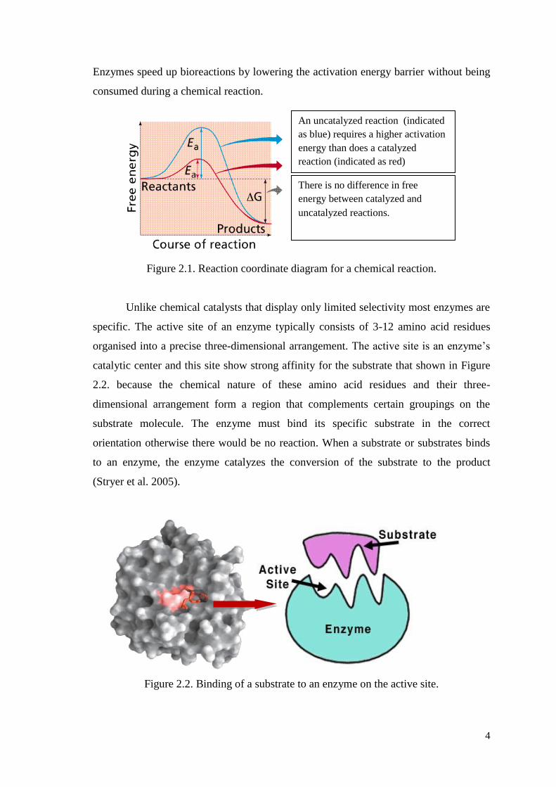
Enzymes speed up bioreactions by lowering the activation energy barrier without being
consumed during a chemical reaction.
Figure 2.1. Reaction coordinate diagram for a chemical reaction.
Unlike chemical catalysts that display only limited selectivity most enzymes are
specific. The active site of an enzyme typically consists of 3-12 amino acid residues
organised into a precise three-dimensional arrangement. The active site is an enzyme’s
catalytic center and this site show strong affinity for the substrate that shown in Figure
2.2. because the chemical nature of these amino acid residues and their three-
dimensional arrangement form a region that complements certain groupings on the
substrate molecule. The enzyme must bind its specific substrate in the correct
orientation otherwise there would be no reaction. When a substrate or substrates binds
to an enzyme, the enzyme catalyzes the conversion of the substrate to the product
(Stryer et al. 2005).
Figure 2.2. Binding of a substrate to an enzyme on the active site.
An uncatalyzed reaction (indicated
as blue) requires a higher activation
energy than does a catalyzed
reaction (indicated as red)
There is no difference in free
energy between catalyzed and
uncatalyzed reactions.
5
Active site is an enzyme’s catalytic center and typically a three-dimensional
pocket or groove on the surface of the enzyme into which the substrate fits.
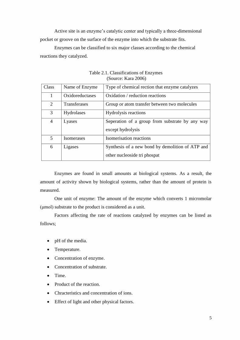
Enzymes can be classified to six major classes according to the chemical
reactions they catalyzed.
Table 2.1. Classifications of Enzymes
(Source: Kara 2006)
Class Name of Enzyme Type of chemical rection that enzyme catalyzes
1 Oxidoreductases Oxidation / reduction reactions
2 Transferases Group or atom transfer between two molecules
3 Hydrolases Hydrolysis reactions
4 Lyases Seperation of a group from substrate by any way
except hydrolysis
5 Isomerases Isomerisation reactions
6 Ligases Synthesis of a new bond by demolition of ATP and
other nucleoside tri phospat
Enzymes are found in small amounts at biological systems. As a result, the
amount of activity shown by biological systems, rather than the amount of protein is
measured.
One unit of enzyme: The amount of the enzyme which converts 1 micromolar
(μmol) substrate to the product is considered as a unit.
Factors affecting the rate of reactions catalyzed by enzymes can be listed as
follows;
pH of the media.
Temperature.
Concentration of enzyme.
Concentration of substrate.
Time.
Product of the reaction.
Chracteristics and concentration of ions.
Effect of light and other physical factors.
6
In order to measure the activity of enzyme, it is necessary to measure the amount
of substrate lost or the amount of product formed per unit time.
Advantages of the enzymes for use in industry:
They are of natural origin and nontoxic.
They have great specificity of action.
They work best under mild conditions of moderate temperature and near neutral
pH.
They have rapidly at relatively low concentrations and the rate of reaction can be
readily controlled by adjusting temperature, pH and amount of enzyme
employed.
They are easily inactivated when reaction has gone as far as desired.
They enable higher product quality, lower manufacturing cost, and less waste.
Enzymes are currently used in the following areas, fermenting of wine, the paper
industry, starch industry, leather industry, baking industry and beer brewing industry,
washing detergent industry, toxic wastes removal, diagnostic industry and production of
pharmaceuticals.
Table 2.2. Enzymes in industry
(Source: Brady and Jordaan 2009)
Laundry
Detergents
Proteinase (91%) Used in pre-soaks to remove protein-based stains
Lipase (6%) Now commonly included todigest oils and fats
Amylase (2%) Removes resistant starch residues
Cellulase (1%) Digests the cotton ‘fuzz’ which acumulates with
excessive washing
Starch
Industry
Amylases,
amyloglucosiadases
and glucoamylases
Converts starch to glucose and other sugar syrups
Glucose Isomerase
Converts glucose syrups into fructose syrups
Dairy
Industry
Rennin from the
stomachs of young
ruminant animals
Manufacture of cheese
Lipases Enhances ripening of blue-mold cheese
Lactases Break down lactose to glucose and galactose
(cont. on next page)
7
Table 2.2 (cont.)
Textile
Industry
Amylase Now widely usd to remove starch from woven
fabrics. Starch is used as an adhesive (or size)
on the threads of many fabrics to prevent
damage during weaving. Traditionally,
chemicals were favoured but now bacterial
amylases are commonly used.
Brewing
Industry
Amylases,
glucanases,
proteinases
Splits polysaccharides and proteins in the malt.
Proteinases Reduces clouding of beers
Amyloglucosidase Low Calorie beer production
β-glucanese Improves filtration characteristics
Baking
Industry
α-amylase Catalyses the breakdown of starch in flours.
Used in the manufaccture of bread.
β-xylanase Improves the characteristics and rising of
bread
Proteinases Reduces the protein in flour. Used in biscuits
manufacture
Leather
Industry
Proteinase (trypsin) The process known as ‘bating’ treats the
leather with proteinases to make it more
pliable. Trypsin isolated from both
slaughterhouses and micro-organisms replaces
the old method of using dog and pigeon faeces.
Pulp and
Paper
Industry
β-xylanases Emerging technology for enhancing pulp-
bleaching
Lipases Reduces ‘pitch’ which causes paper to stick to
rollers and tear.
Table 2.3. Advantages and disadvantages of enzymes as biocatalysts in comparison
with chemical catalysts.
ADVANTAGES DISADVANTAGES
Stereo-and regioselective Unstable at high temperatures
Low temperatures required Unstable at extreme pH values
Low energy consumption Unstable in aggressive solvents
Active at Ph 2-12 Inhibited by some metal ions
Non-toxic when correctly used Very expensive
Can be reused Require expensive cosubstrates
Can be biologically degraded
Can be produced in unlimited quantities
8
2.2. Why Recombinant Enyzme?
Today, enzymes are used in the industrial field generally is obtained from
micro-organisms. However, very little portion is provided as a part of the vegetable and
animal origin. Reasons for the choice of microorganisms as a source of enzyme,
formation of byproduct is less, activity is high, is more economical, to have stability and
producability at high levels of purity (Gümüşel 2002 and Wiseman 1987).
Recent developments in microbial genetics have created a new potential for
enzyme production. The enzyme industry is expected to expand as genetic engineering
(recombinant DNA technology) is applied to the microbial production of enzymes. The
techniques of genetic engineering can be used to manipulate DNA such that multiple
copies of a particular gene encoding an enzyme of commercial value can be made. Via
this technique large amounts of the desired protein that resistant to hard conditions can
be produced by using recombinant microorganisms.
2.3. Enzyme Stability and Immobilization
2.3.1. Importance of Enzyme Stability
Enzyme instability is an important factor that prevents their wider use in
industry. Enzymes may be exposed to unnatural non physiological environments.
Chemicals like organic solvents, elevated temperatures and pH values outside their
normal in vivo values can denature the enzyme with consequent loss of activity. In such
a case, thermophilic enzymes and enzyme immobilization draw attention to increase
enzyme durability. Thermophilic enzymes are more stable under hard reaction
conditions such as high temperatures and pressures when compared with other enzymes.
So they have great importance in industrial development (Kumar and Nussinov 2001,
Sterner and Liebl 2001).
2.4. Thermophiles
Microorganisms can be grouped into broad categories, according to their
temperature ranges for growth. The temperature in many natural environments changes
9
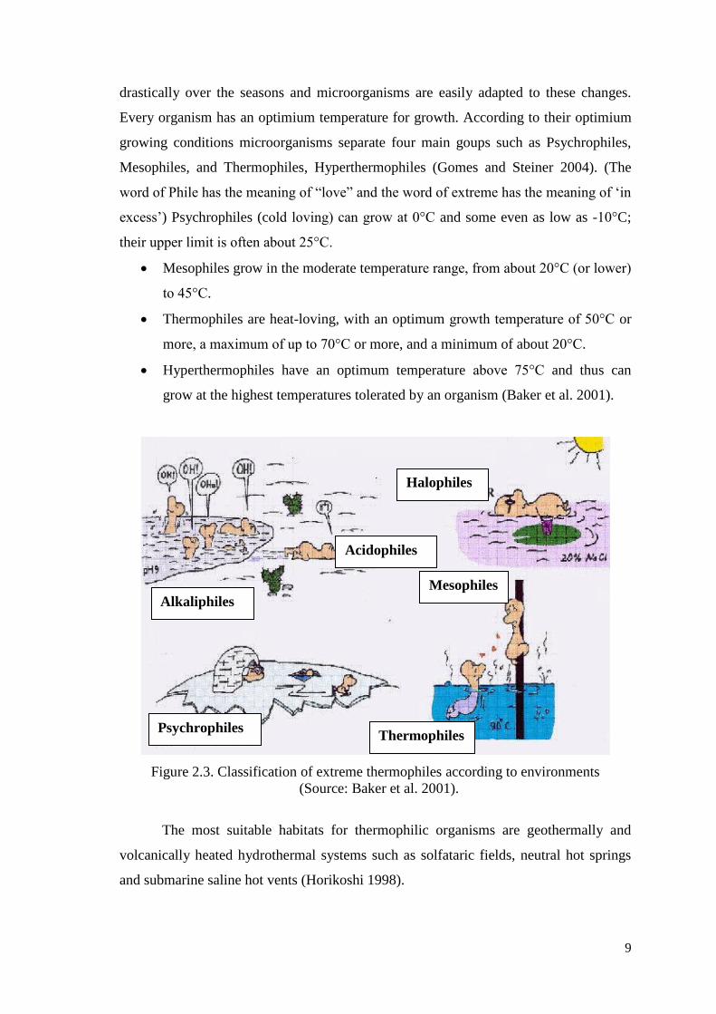
drastically over the seasons and microorganisms are easily adapted to these changes.
Every organism has an optimium temperature for growth. According to their optimium
growing conditions microorganisms separate four main goups such as Psychrophiles,
Mesophiles, and Thermophiles, Hyperthermophiles (Gomes and Steiner 2004). (The
word of Phile has the meaning of “love” and the word of extreme has the meaning of ‘in
excess’) Psychrophiles (cold loving) can grow at 0°C and some even as low as -10°C;
their upper limit is often about 25°C.
Mesophiles grow in the moderate temperature range, from about 20°C (or lower)
to 45°C.
Thermophiles are heat-loving, with an optimum growth temperature of 50°C or
more, a maximum of up to 70°C or more, and a minimum of about 20°C.
Hyperthermophiles have an optimum temperature above 75°C and thus can
grow at the highest temperatures tolerated by an organism (Baker et al. 2001).
Figure 2.3. Classification of extreme thermophiles according to environments
(Source: Baker et al. 2001).
The most suitable habitats for thermophilic organisms are geothermally and
volcanically heated hydrothermal systems such as solfataric fields, neutral hot springs
and submarine saline hot vents (Horikoshi 1998).
Halophiles
Acidophiles
Mesophiles
Thermophiles Psychrophiles
Alkaliphiles

10
Thermophilic microorganisms have some special characteristics when compared
with mesophiles. They have several advantages as high reproductive rates, easily
winning the final product, high process stability and yielding, they can directly ferment
natural polymers such as starch, cellulose. Thermophiles are found in various
geothermally heated regions of the earth. Also thermophiles can be subclassified as
moderately thermophiles (grow at 50-60°C) and extreme thermophiles (grow at 60-
80°C). Cellular components of thermophilic organisms (enzymes, proteins and nucleic
acids) are also thermostable. Thermostable enzymes are highly specific and thus have
considerable potential for many industrial applications (Kumar and Nussinov 2001).
2.4.1. Thermophilic Bacillus
Bacillus is an aerobic or facultatively anaerobic, gram positive, rod-shaped
bacteria that differentiate in to heat-resistant endospores under aerobic conditions are
placed in the genus Bacillus (Rainey and Oren 2006). Many kinds of species which
have thermophilic, psychrophilic, acidophilic, alkalophilic and halophilic properties are
included in the genus Bacillus (Nazina et al. 2001). Thermophilic bacteria belonging to
genus Bacillus show optimal growth temperatures in the range of 45-70ºC (Maugeri et
al. 2001, Rainey et al. 1993). The importance of thermophilic Bacillus increased
because of their biotechnological importance as sources of thermostable enzymes
(proteases, amylases, pullunases, glucose isomerases, lipases, xylanases, cellulases and
DNA restriction endonucleases) (Maugeri et al. 2001). In our experiment thermophilic
Bacillus are isolated from Balçova (Agamemnon) Geothermal region.
2.4.2. Thermophilic Enzymes
Thermostable enzymes, which have been isolated mainly from thermophilic
organisms and these enzymes which are thermostable, resist denaturation and
proteolysis (Kumar and Nussinov 2001). It has been demonstrated in studies that
thermophilic microorganisms make the cells durable in order to survive at high
temperature environments, and have high ratio of saturated fatty acids in the cell
membranes. And fatty acids created a hydrophobic environment for the cell (Herbert
and Sharp 1992). It has been determined that thermophiles with disulfide bonds and
11
hydrophobic interactions have become resistant to different temperature values.
Thermostable proteins contain many charged residues and hydrophobic residues
(Fujiwara 2002). In addition, it has been defined that their DNA's contain revese gyrase
that make up positive super-coils. That structure raises the melting point of DNA and
therefore makes the microorganisms more resistant to high temperatures (Robb et al.
2007). Also additional intermolecular interactions such as hydrophobic interactions,
disulfide bonds, electrostatic interactions metal binding and hydrogen bonds that are not
exist in mesophilic enzymes get thermophilic enzymes more stable (Steel and Walker
1991).
Table 2.4. Main advantages of thermostable enzymes
(Source: Haki and Rakshit 2003)
Property Advantages
Thermostability The half life of the enzymes increases. The purification of
the enzymes is easier.
Resistance against various
chemical agents
They can tolerate hard conditions including important
amounts of organic solvents, diverse pH level frequently
neccessaries during industrial process.
High optimal temperature Low activity at room temperature. It does not require
active cooling in fermentation. High diffusion rates of
substrates and products
Solubility At high temperatures the concentrations of substrates can
be increased, with the exception of gases.
Viscosity Decreases. Mixing and pumping can be also increased.
Microbial contamination The probability of contamination decreases as the
temperature rises. Contaminant enzymes are inactivated at
high temperature.
The ability of thermophilic enzymes to work at high temperatures implies many
advantages for their applications in industrial reactors or fermenters.
However, despite the many economically important advantages of thermophilic
enzymes, there are also disadvantages for specific applications.
12
Table 2.5. Main problems of the application of thermophilic enzymes in industry.
Property Main Problem Observed
Thermal sensivity There are many substrates, products or enzyme cofactors
unstable at high temperature
Solubility of gases Decrease. The diffusion of gases limits some reactions.
Enzyme stability The inactivation of the enzyme results extremely difficult.
Equipment Stress All the materials are damages in a short time, unless especially
designed
2.4.2.1. Applications of Enzymes from Thermophiles
As shown in Table 2.6 there are many applications of enzymes from
thermophiles. For each application the temperature range and microorganism type were
indicated.
Table 2.6. Main applications of thermostable enzymes at present.
Enzyme T(◦C)* Application Origin**
Enz. Acting on Carbohydrates
α-amylase 60-90 Starch hydrolysis Bacillus licheniformis
Pullulanase 50-60 Starch hydrolysis Klebsiella aerogenes
Xylose isomerase 50-55 Sweetening of corn syrups Actinoplanes
missouriensis
Cellulase
55-65
Hydrolysis of cellulose,
Ethanol production, paper
bleaching
Clostridium
thermocellum
Proteases
Neutral protease 40-80 Food prcessing Bacillus
stearothermophilus
Alkaline protease 40-80 Detergents Bacillus licheniformis
Molecular Biology
Taq polymerase 45-95 DNA amplification (PCR) Thermus spp.
Vent DNA polymerase 50-98 DNA amplification (PCR) Thermococcus litoralis
Pfu DNA polymerase 50-98 DNA amplification (PCR) Pyrococcus furiosus
Tth polymerase 45-95 Reverse transcription of RNA Thermus thermophilus
HB8
(cont. on next page)
13
Table 2.6 (cont.)
RNA polymerase 65-75 RNA synthesis Thermus spp.
Restriction Enzymes 65-75 DNA specific digestion Thermus spp. Bacillus
sulfolobus
Aneaerobic
treatment of residual
waters
50-60 Organic compounds elimination Methanogenic bacteria
Metanobacterium
Metanosarcina
*Range temperature at which the enzyme is used
**Microorganisms from which the enzym has been obtained
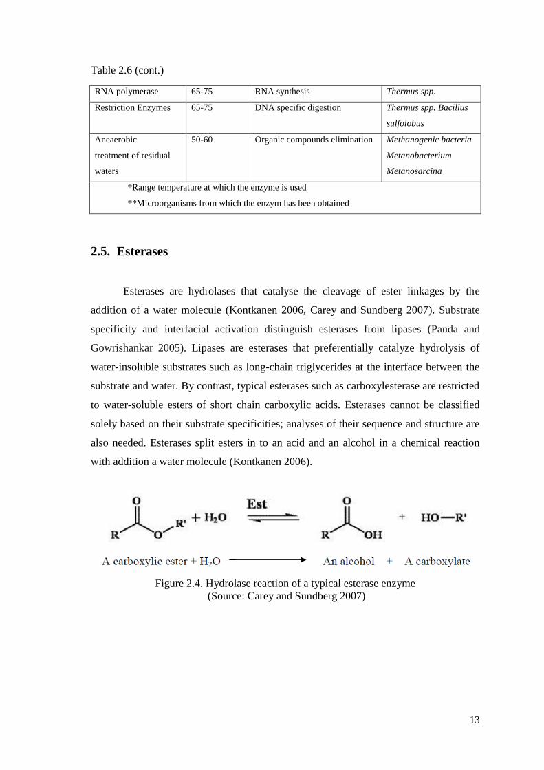
2.5. Esterases
Esterases are hydrolases that catalyse the cleavage of ester linkages by the
addition of a water molecule (Kontkanen 2006, Carey and Sundberg 2007). Substrate
specificity and interfacial activation distinguish esterases from lipases (Panda and
Gowrishankar 2005). Lipases are esterases that preferentially catalyze hydrolysis of
water-insoluble substrates such as long-chain triglycerides at the interface between the
substrate and water. By contrast, typical esterases such as carboxylesterase are restricted
to water-soluble esters of short chain carboxylic acids. Esterases cannot be classified
solely based on their substrate specificities; analyses of their sequence and structure are
also needed. Esterases split esters in to an acid and an alcohol in a chemical reaction
with addition a water molecule (Kontkanen 2006).
Figure 2.4. Hydrolase reaction of a typical esterase enzyme
(Source: Carey and Sundberg 2007)

14
2.5.1. The Chemical Reactions of Esterases
There are some different reactions catalysed by lipases and esterases. Those
reactions are listed in Figure 2.5.
Figure 2.5. Different reactions catalysed by lipases/esterases in aqueous and non-
aqueous solution (Source: Villeneuve et al. 2000)
Some properties of esterases are shown at below (Kim et al. 2008):
• They do not require cofactors.
• They have broad substrate specificity.
• They show enzymatic activity in both aqueous and nonaqueous solvents.
• They are enantioselective that catalyzes the reaction of only one of a pair of
enantiomer. As a result of this optically pure compounds are produced.
15
• They have the characteristic α/β hydrolase fold and a similar catalytic triad consisting
of the imidazole ring from a histidine.
There are several sources to produce esterases; from Streptomyces sp. (Nishmura
and Inovye 2000), Pseudomonas sp. (Kim et al. 2002), Bacillus sp. (Kim et al. 2004),
Lactobacillus sp. (Choi and Lee 2001), Pencillium sp. (Horne et al. 2002),
Saccharomyces sp. (Lomolino et al. 2003) etc., from animal sources (Finer et al. 2004),
from plants (Pringle and Dickstein 2004) for important applications in bioprocesses.
Different sources yield different esterases. Thus, a large and varied
nomenclature is reported in the literature, e.g., carboxylesterase, cholinesterase,
acetylxylan esterase, aryl esterase, phosphotriesterase, phenolic esterase, pig liver
esterase, tanin esterase.
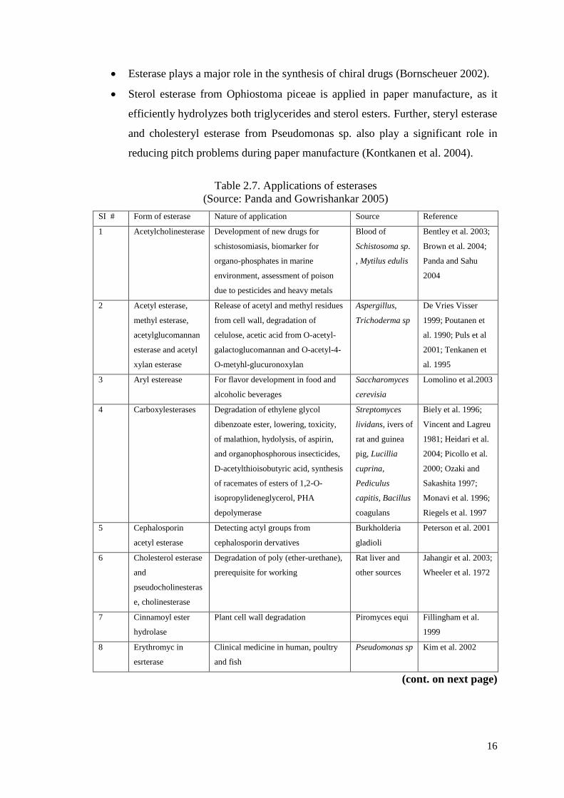
2.5.2. Applications of Esterases
Hydrolysis of some important methyl esters by esterase and product of this
reaction acid produced. Acid = Ferulic / Sinapic / Caffeic / p-Coumaric acid.
They are widely used in the food, beverage, and perfume industries (Chaabouni
et al. 1996).
An esterase from Fusarium oxysporum plays a significant role in producing
flavoring and fragrance compounds from geraniol and fatty acids
(Christakopoulos et al. 1998, Chaabouni et al. 1996).
Esterases are employed in dairies, and for the production of wine, fruit juices,
beer, and alcohol. In order to transform low value fats and oils in to more
valuable ones.
Esterases and lipases from Lactobacillus casei are used significantly for
hydrolysis of milk fat fort he purpose of flavor enhancement in the manufacture
of cheese-related products (Choi and Lee 2001).
An esterase from yeast plays a significant role in determining the final ester
level in products such as membrane filtered beer and bottle re-fermented beer
(Dufour and Bing 2001).
To degrade some man-made pollutants, such as plactics, polyurethane,
polyesters, polyethylene glycol adipate, etc., cholesterol esterase and
polurethanase are widely used (Jahangir et al. 2003).
16
Esterase plays a major role in the synthesis of chiral drugs (Bornscheuer 2002).
Sterol esterase from Ophiostoma piceae is applied in paper manufacture, as it
efficiently hydrolyzes both triglycerides and sterol esters. Further, steryl esterase
and cholesteryl esterase from Pseudomonas sp. also play a significant role in
reducing pitch problems during paper manufacture (Kontkanen et al. 2004).
Table 2.7. Applications of esterases
(Source: Panda and Gowrishankar 2005)
SI # Form of esterase Nature of application Source Reference
1 Acetylcholinesterase Development of new drugs for
schistosomiasis, biomarker for
organo-phosphates in marine
environment, assessment of poison
due to pesticides and heavy metals
Blood of
Schistosoma sp.
, Mytilus edulis
Bentley et al. 2003;
Brown et al. 2004;
Panda and Sahu
2004
2 Acetyl esterase,
methyl esterase,
acetylglucomannan
esterase and acetyl
xylan esterase
Release of acetyl and methyl residues
from cell wall, degradation of
celulose, acetic acid from O-acetyl-
galactoglucomannan and O-acetyl-4-
O-metyhl-glucuronoxylan
Aspergillus,
Trichoderma sp
De Vries Visser
1999; Poutanen et
al. 1990; Puls et al
2001; Tenkanen et
al. 1995
3 Aryl esterease For flavor development in food and
alcoholic beverages
Saccharomyces
cerevisia
Lomolino et al.2003
4 Carboxylesterases Degradation of ethylene glycol
dibenzoate ester, lowering, toxicity,
of malathion, hydolysis, of aspirin,
and organophosphorous insecticides,
D-acetylthioisobutyric acid, synthesis
of racemates of esters of 1,2-O-
isopropylideneglycerol, PHA
depolymerase
Streptomyces
lividans, ivers of
rat and guinea
pig, Lucillia
cuprina,
Pediculus
capitis, Bacillus
coagulans
Biely et al. 1996;
Vincent and Lagreu
1981; Heidari et al.
2004; Picollo et al.
2000; Ozaki and
Sakashita 1997;
Monavi et al. 1996;
Riegels et al. 1997
5 Cephalosporin
acetyl esterase
Detecting actyl groups from
cephalosporin dervatives
Burkholderia
gladioli
Peterson et al. 2001
6 Cholesterol esterase
and
pseudocholinesteras
e, cholinesterase
Degradation of poly (ether-urethane),
prerequisite for working
Rat liver and
other sources
Jahangir et al. 2003;
Wheeler et al. 1972
7 Cinnamoyl ester
hydrolase
Plant cell wall degradation Piromyces equi Fillingham et al.
1999
8 Erythromyc in
esrterase
Clinical medicine in human, poultry
and fish
Pseudomonas sp Kim et al. 2002
(cont. on next page)
17
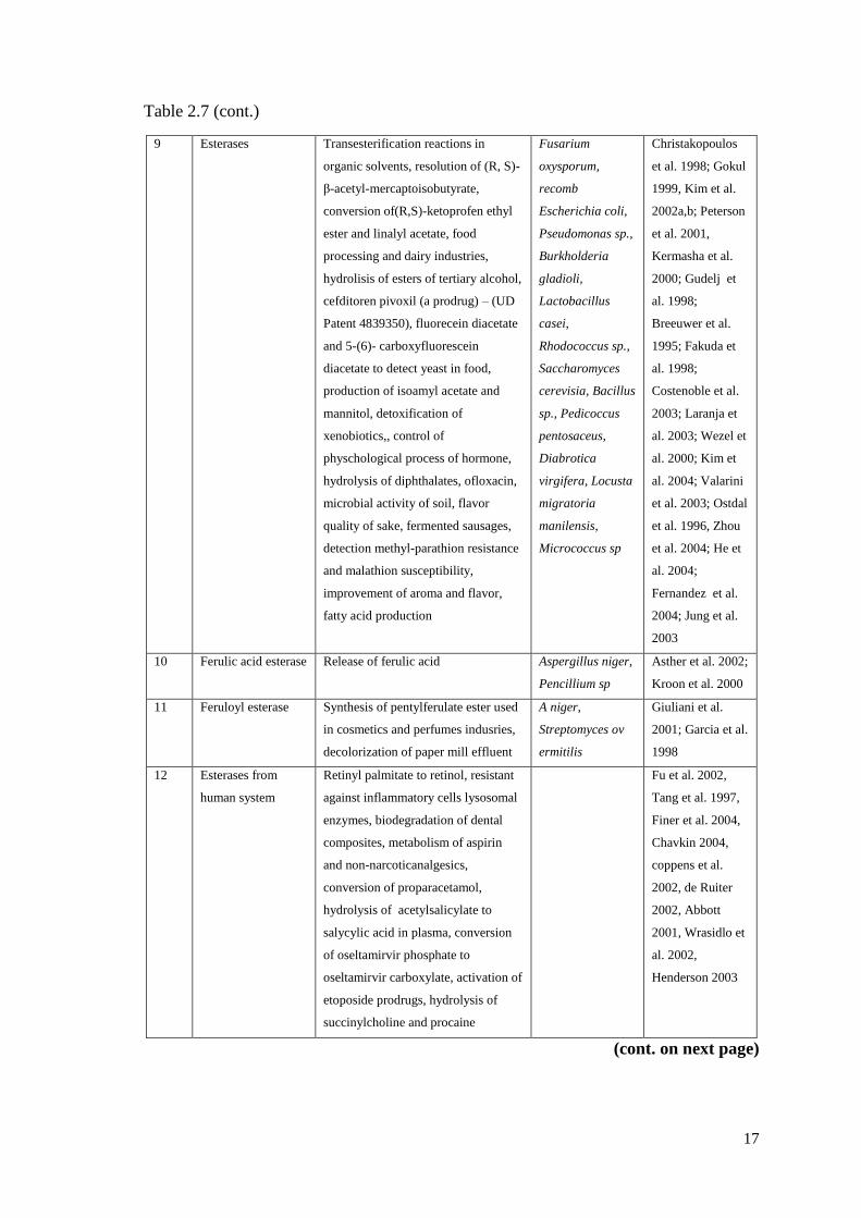
Table 2.7 (cont.)
9 Esterases Transesterification reactions in
organic solvents, resolution of (R, S)-
β-acetyl-mercaptoisobutyrate,
conversion of(R,S)-ketoprofen ethyl
ester and linalyl acetate, food
processing and dairy industries,
hydrolisis of esters of tertiary alcohol,
cefditoren pivoxil (a prodrug) – (UD
Patent 4839350), fluorecein diacetate
and 5-(6)- carboxyfluorescein
diacetate to detect yeast in food,
production of isoamyl acetate and
mannitol, detoxification of
xenobiotics,, control of
physchological process of hormone,
hydrolysis of diphthalates, ofloxacin,
microbial activity of soil, flavor
quality of sake, fermented sausages,
detection methyl-parathion resistance
and malathion susceptibility,
improvement of aroma and flavor,
fatty acid production
Fusarium
oxysporum,
recomb
Escherichia coli,
Pseudomonas sp.,
Burkholderia
gladioli,
Lactobacillus
casei,
Rhodococcus sp.,
Saccharomyces
cerevisia, Bacillus
sp., Pedicoccus
pentosaceus,
Diabrotica
virgifera, Locusta
migratoria
manilensis,
Micrococcus sp
Christakopoulos
et al. 1998; Gokul
1999, Kim et al.
2002a,b; Peterson
et al. 2001,
Kermasha et al.
2000; Gudelj et
al. 1998;
Breeuwer et al.
1995; Fakuda et
al. 1998;
Costenoble et al.
2003; Laranja et
al. 2003; Wezel et
al. 2000; Kim et
al. 2004; Valarini
et al. 2003; Ostdal
et al. 1996, Zhou
et al. 2004; He et
al. 2004;
Fernandez et al.
2004; Jung et al.
2003
10 Ferulic acid esterase Release of ferulic acid Aspergillus niger,
Pencillium sp
Asther et al. 2002;
Kroon et al. 2000
11 Feruloyl esterase Synthesis of pentylferulate ester used
in cosmetics and perfumes indusries,
decolorization of paper mill effluent
A niger,
Streptomyces ov
ermitilis
Giuliani et al.
2001; Garcia et al.
1998
12 Esterases from
human system
Retinyl palmitate to retinol, resistant
against inflammatory cells lysosomal
enzymes, biodegradation of dental
composites, metabolism of aspirin
and non-narcoticanalgesics,
conversion of proparacetamol,
hydrolysis of acetylsalicylate to
salycylic acid in plasma, conversion
of oseltamirvir phosphate to
oseltamirvir carboxylate, activation of
etoposide prodrugs, hydrolysis of
succinylcholine and procaine
Fu et al. 2002,
Tang et al. 1997,
Finer et al. 2004,
Chavkin 2004,
coppens et al.
2002, de Ruiter
2002, Abbott
2001, Wrasidlo et
al. 2002,
Henderson 2003
(cont. on next page)
18
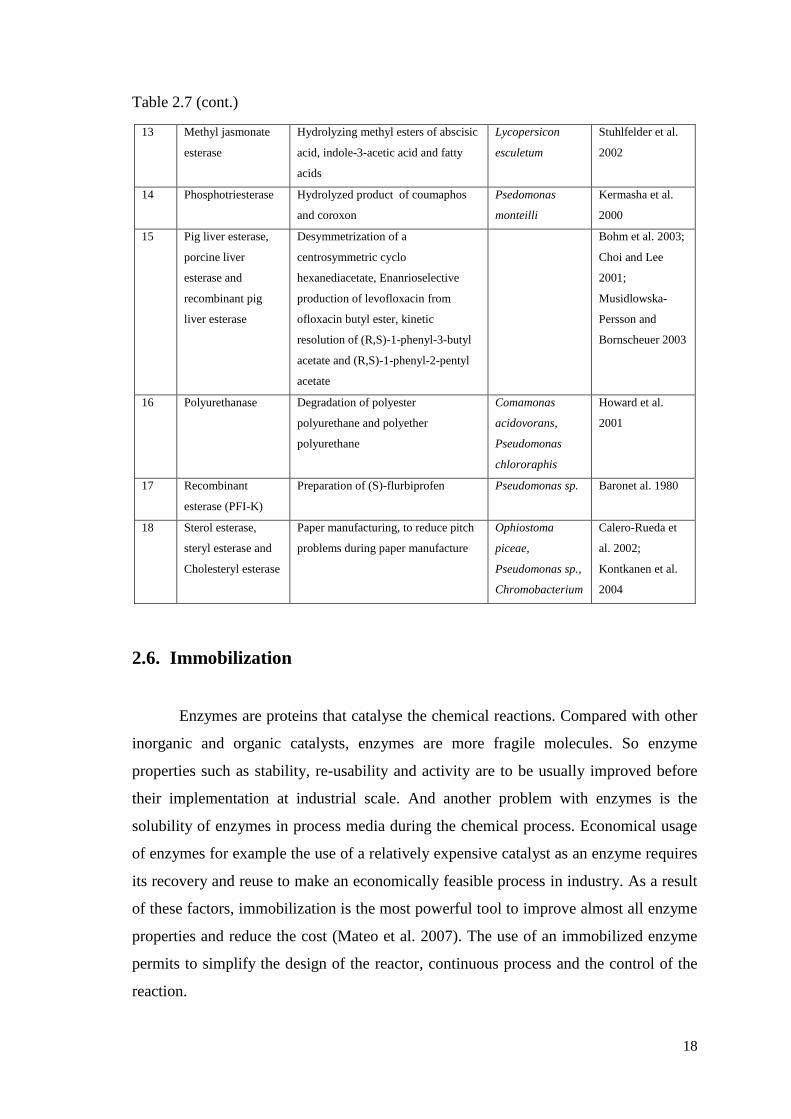
Table 2.7 (cont.)
13 Methyl jasmonate
esterase
Hydrolyzing methyl esters of abscisic
acid, indole-3-acetic acid and fatty
acids
Lycopersicon
esculetum
Stuhlfelder et al.
2002
14 Phosphotriesterase Hydrolyzed product of coumaphos
and coroxon
Psedomonas
monteilli
Kermasha et al.
2000
15 Pig liver esterase,
porcine liver
esterase and
recombinant pig
liver esterase
Desymmetrization of a
centrosymmetric cyclo
hexanediacetate, Enanrioselective
production of levofloxacin from
ofloxacin butyl ester, kinetic
resolution of (R,S)-1-phenyl-3-butyl
acetate and (R,S)-1-phenyl-2-pentyl
acetate
Bohm et al. 2003;
Choi and Lee
2001;
Musidlowska-
Persson and
Bornscheuer 2003
16 Polyurethanase Degradation of polyester
polyurethane and polyether
polyurethane
Comamonas
acidovorans,
Pseudomonas
chlororaphis
Howard et al.
2001
17 Recombinant
esterase (PFI-K)
Preparation of (S)-flurbiprofen Pseudomonas sp. Baronet al. 1980
18 Sterol esterase,
steryl esterase and
Cholesteryl esterase
Paper manufacturing, to reduce pitch
problems during paper manufacture
Ophiostoma
piceae,
Pseudomonas sp.,
Chromobacterium
Calero-Rueda et
al. 2002;
Kontkanen et al.
2004
2.6. Immobilization
Enzymes are proteins that catalyse the chemical reactions. Compared with other
inorganic and organic catalysts, enzymes are more fragile molecules. So enzyme
properties such as stability, re-usability and activity are to be usually improved before
their implementation at industrial scale. And another problem with enzymes is the
solubility of enzymes in process media during the chemical process. Economical usage
of enzymes for example the use of a relatively expensive catalyst as an enzyme requires
its recovery and reuse to make an economically feasible process in industry. As a result
of these factors, immobilization is the most powerful tool to improve almost all enzyme
properties and reduce the cost (Mateo et al. 2007). The use of an immobilized enzyme
permits to simplify the design of the reactor, continuous process and the control of the
reaction.
19
2.6.1. Immobilization of Enzymes
In general the term ‘immobilization’ refers to the act of the limiting movement
or making incapable of movement. The term ‘immobilized enzymes’ refers to enzymes
physically confined or localized in a certain defined region of space with retention of
their catalytic activities, and which can be used repeatedly and continuously.
Immobilization means associating the biocatalysts with an insoluble matrix or
immobilized proteins and cells to an insoluble support. Practically, the procedure
consists of mixing together the enzyme and the support material under appropriate
conditions and following a period of incubation, separating the insoluble material from
the soluble material by centrifugation or filtration. Today, a large number of
immobilized enzymes are used in industry (Karadağ 2001).
In general, immobilization applications are commonly used at appropriate
support materials, pharmaceutical, protein, microorganism, plant and animal cells,
biosensor and bioreactor applications and controlled drug delivery systems except
enzyme system (Aksoy 2003).
2.6.2. Advantages of Enzyme Immobilization
There are a number of advantages to immobilize enzymes from free solutions to
insoluble supports via immobilization technique. Some of them are listed below:
Immobilized enzyme is more robust and stable compared with soluble one.
Immobilized enzyme generally shows greater pH and thermal stability.
Thanks to immobilization enzymes can easily be added to or removed from
reaction media, it enables greater control of the reaction time and rate.
Problems of separating the catalyst from the products are practically eliminated.
Product is not contaminated with the enzyme (especially useful in food and
pharmaceuticals industries).
Low downstream processing cost.
Continuous processes using columns of immobilized enzyme become more
practical and automation is possible.
Enzymes may be stabilized against heat or solvent effects.
20
Immobilized enzyme easily reused multiple times for the same reaction with
longer half-lives (Brady and Jordaan 2009).
In spite of the advantages, the immobilization process has some disadvantages
that are shortly listed below (Guisan 2006):
Loss of enzymatic activity due to the immobilization process.
The cost of carriers and immobilization method.
Mass transfer limitations.
Changes in enzyme properties such as selectivity.
It is also possible to immobilize whole cells rather than individual enzymes or
some organelles.
2.6.2.1. The Major Components of an Immobilized Enzyme
An immobilized enzyme has some major components and those major
components are listed as follows:
The enzyme,
The carrier or support,
Mode of interaction of the enzyme with the carrier.
2.6.2.2. The Requirements of an Ideal Immobilization Support
There are a variety of insoluble materials to bind enzymes and several
techniques to achieve immobilization.
The support material can have a critical effect on the stability of the enzyme and
the efficiency of enzyme immobilization. It is difficult to predict in advance which
support will be most suitable. However, in general an enzyme carrier should have some
properties that listed as follows (Sandwick and Schray 1988):

21
It should present large surface to have good geometrical congruence with the
enzyme surface.
It should include physical resistance to compression.
It should be biocompatible and show inertness toward enzymes ease of
derivatization.
It should show resistance to microbial attack.
It should be available at low cost and biodegradable.
It should be simple non-toxic and sterile.
It should have a character like easy separation of carrier from reaction media.
Suitable shape and particle size for conventional reactor systems.
Should have mechanical strenght.
Should have high enzyme-mass loading capacity.
Carrier materials can be divided in to two group, first inorganic carriers and the
second is organic origin carriers. The advantage of inorganic materials, they are not
susceptible to microbial attack, and have a greater structural and operational stability.
Common organic supports are cellulose derivatives that have free hydroxyl or amino
groups. The goups can participate to link with covalent coupling the groups on the
enzyme molecule (Kara 2006).
Table 2.8. Examples of Carriers Used for Enzyme Immobilization
(Source: Kennedy and White 1985)(Source: Guisan 2006).
Organic
Natural polymers
Polysaccharides: Cellulose, agar, agarose, chitin, alginate dextrans.
Proteins: Collagen, albumin
Carbon
Synthetic polymers
Polystyrene
Other polymers: Polyacrylate polymethacrylates, polyacrylamide, polyamides,
vinyl, and allyl-polymers
Inorganic
Natural minerals: Bentonite, silica, sand.
Processed materials: Glass(nonporous and controlled pore), metals, controlled pore
Metal oxides(e.g. ZrO2, TiO2, Al2O3)
22
The physical characteristics of the matrices (such as mean particle diameter,
swelling behavior, mechanical strength, and compression behavior) will be of major
importance for the performance of the immobilized systems and will determine the type
of reactor used under technical conditions (i.e., stirred tank, fluidized, fixed beads). In
particular, pore parameters and particle size determine the total surface area and thus
critically affect the capacity for binding of enzymes. Nonporous supports show few
diffusional limitations but have a low loading capacity. Therefore, porous supports are
generally preferred because the high surface area allows for a higher enzyme loading
and the immobilized enzyme receieves greater protection from the environment. Porous
supports should have a controlled pore distribution in order to optimize capacity and
flow properties. In spite of the many advantages of inorganic carriers (e.g., high stability
against physical, chemical, and microbial degradation), most of the industrial
applications are performed with organic matrices.
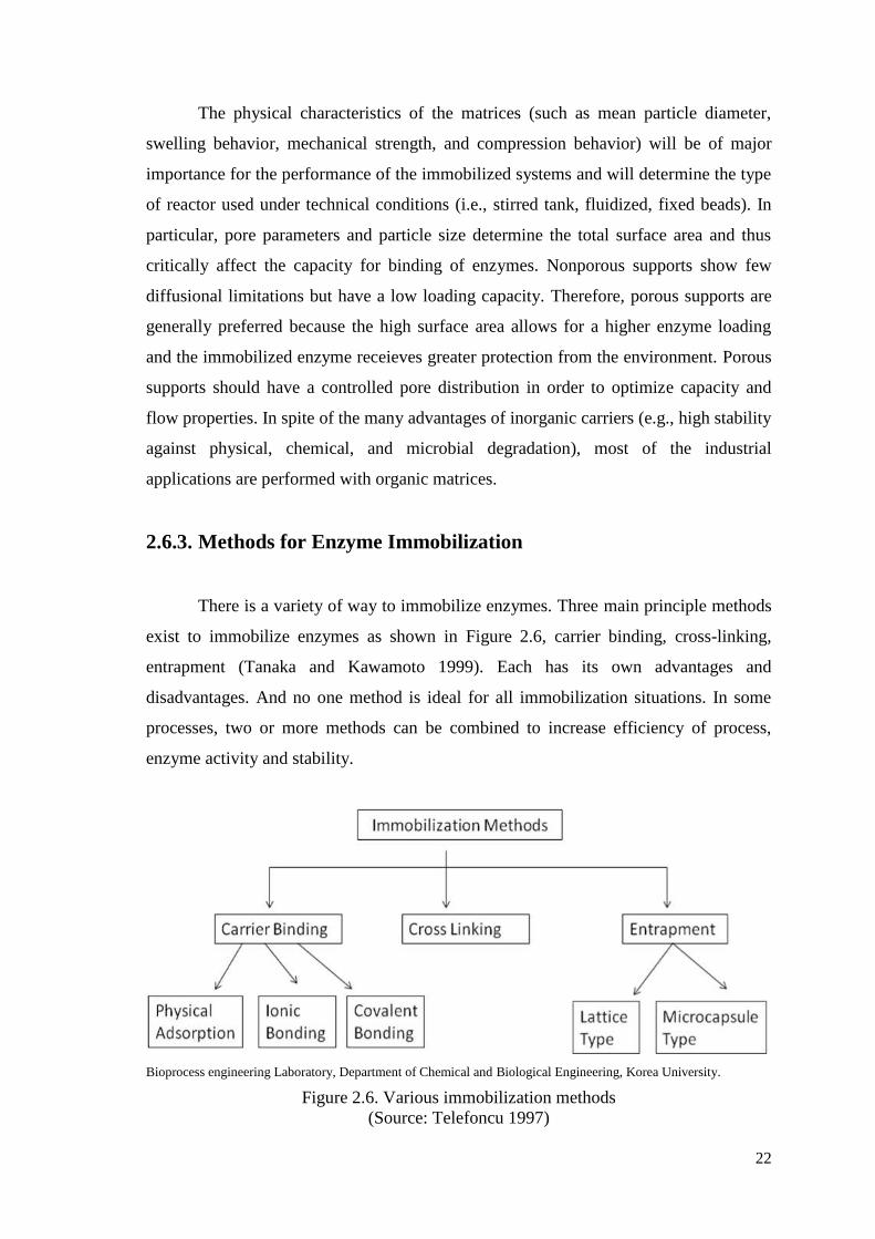
2.6.3. Methods for Enzyme Immobilization
There is a variety of way to immobilize enzymes. Three main principle methods
exist to immobilize enzymes as shown in Figure 2.6, carrier binding, cross-linking,
entrapment (Tanaka and Kawamoto 1999). Each has its own advantages and
disadvantages. And no one method is ideal for all immobilization situations. In some
processes, two or more methods can be combined to increase efficiency of process,
enzyme activity and stability.
Bioprocess engineering Laboratory, Department of Chemical and Biological Engineering, Korea University.
Figure 2.6. Various immobilization methods
(Source: Telefoncu 1997)
23
2.6.3.1. Carrier Binding
The carrier binding method is the oldest immobilization technique for enzymes.
The selection of the carrier depends on the nature of the enzyme itself, as well as the
following items:
Particle size
Surface area
Molar ratio of hydrophilic to hydrophobic groups
Chemical composition (Dumitriu et al 1988).
In general, an increase in the ratio of hydrophilic groups and in the concentration
of bound enzymes, results in a higher activity of the immobilized enzymes. Some of the
most commonly used carriers for enzyme immobilization are polysaccharide derivatives
such as cellulose, dextran, agarose, and polyacrylamide gel. According to the binding
mode of the enzyme, the carrier-binding method can be further sub-classified into (Cao
2006):
Physical adsorption
Ionic binding
Covalent binding.
2.6.3.1.1. Physical Adsorption
Adsorption method is the oldest and simplest method of immobilization (Glick
1979). This method for the immobilization of an enzyme is based on the physical
adsorption of enzyme protein on the surface of water-insoluble carriers. During physical
adsorption, the hyrogen bonds, van der Waals forces and hydrophobic interactions are
the responsible forces for immobilization (Chen et al. 1996).
Hence, the method causes little or no conformational change of the enzyme or
destruction of its active center. This method is reversible, and this provides reuse of
support material and enzymes again for different usages (Zaborsky 1973). If a suitable
carrier is found, this method can be both simple and cheap. However, it has the
disadvantage that the adsorbed enzyme may leak from the carrier during use due to a
weak binding force between the enzyme and the carrier.
24
2.6.3.1.2. Ionic Binding
The ionic binding method relies on the ionic binding of the enzyme protein to
water-insoluble carriers containing ion-exchange residues (Brena and Batista 2008).
Advantages of the ionic binding, first, the conditions are much milder than those needed
for the covalent binding method. Second is, little changes in the conformation and the
active site of the enzyme enable high activity in most cases. Addition to the advantages,
the disadvantage is leakage of enzymes from the carrier may ocur in substrate solution
of high ionic strength or upon variation of pH.
2.6.3.1.3. Covalent Binding
The most intensely studied of the immobilization techniques is the formation of
covalent bonds between the enzyme and the support matrix. This technique allows the
derivatives of enzyme to be stable and prevents enzymes penetration into solution (Carr
and Bowers 1980). Covalent binding is used generally when the structure of enzyme
and functional groups are known. When trying to select the type of reaction by which a
given protein should be immobilized, the choice is limited by two characteristics: (1)
the binding reaction must be performed under conditions that do not cause loss of
enzymatic activity, and (2) the active site of the enzyme must be unaffected by the
reagents used. Enzymes are covalently bound to the insoluble matrix through the
functional groups on the enzyme. The functional groups that may take part in this
binding are listed in Table 2.9.
Table 2.9. Functional groups used in covalent binding.
Amino group Carboxyl group Sulfhydryl group,
Hydroxyl group Imidazole group Phenolic group
Thiol group Threonine group Indole group
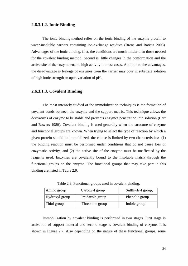
Immobilization by covalent binding is performed in two stages. First stage is
activation of support material and second stage is covalent binding of enzyme. It is
shown in Figure 2.7. Also depending on the nature of these functional groups, some
25
various activating materials such as cyanogen bromide, epichloridrin, glutaraldehyde,
carbodiimit, cyanuric chloride can be (Srere and Uyeda 1976) used.
Figure 2.7. Immobilization by covalent binding.
It is possible in some cases to increase the number of reactive residues of an enzyme
in order to increase the yield of the immobilized enzyme. This provides alternative
reaction sites to those essential for enzymatic activity.
2.6.3.2. Crosslinking
Immobilization of enzymes has been achieved by intermolecular cross-linking of
the protein, either to other protein molecules or to functional groups on an insoluble
matrix. Cross-linking an enzyme to itself is both expensive and insufficient, as some of
the protein material will inevitably be acting mainly as a support. This will result in
relatively low enzymatic activity. Generally, cross-linking is best used in conjunction
with one of the other methods. It is used mostly as a means of stabilizing adsorbed
enzymes and also for preventing leakage from polyacrylamide gels.
Enzyme activity depends on some factors such as reaction time, temperature,
ionic strength, pH, cross-linker material, enzyme concentration and balance between
those factor. The most important advantage of this method is using two or
multifunctional materials in order to immobilization of enzymes. The disadvantage of
this method is the difficulty in controlling intermolecular cross-linking reaction for
obtaining immobilized enzyme which shows high activity.
26
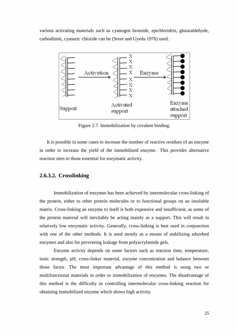
The most common reagent used in this method is glutaraldehyde which
establishes intermolecular cross-linking with amino groups of enzyme. The structure of
glutaraldehyde is shown in Figure 2.8.
Figure 2.8. The crosslinking agent glutaraldeyhde
(Source: Migneault et al. 2004).
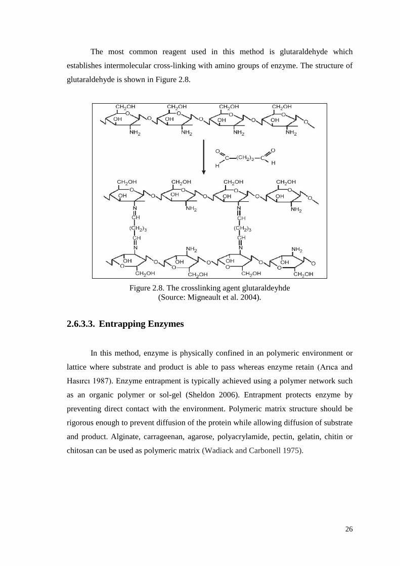
2.6.3.3. Entrapping Enzymes
In this method, enzyme is physically confined in an polymeric environment or
lattice where substrate and product is able to pass whereas enzyme retain (Arıca and
Hasırcı 1987). Enzyme entrapment is typically achieved using a polymer network such
as an organic polymer or sol-gel (Sheldon 2006). Entrapment protects enzyme by
preventing direct contact with the environment. Polymeric matrix structure should be
rigorous enough to prevent diffusion of the protein while allowing diffusion of substrate
and product. Alginate, carrageenan, agarose, polyacrylamide, pectin, gelatin, chitin or
chitosan can be used as polymeric matrix (Wadiack and Carbonell 1975).
27
Figure 2.9. The illustration of entrapment in a matrix and other is in droplets
(Source: Costa et al. 2004).
Also entrapment method can be separated into five major types as lattice,
microcapsule, liposome, membrane, and reverse micelle.
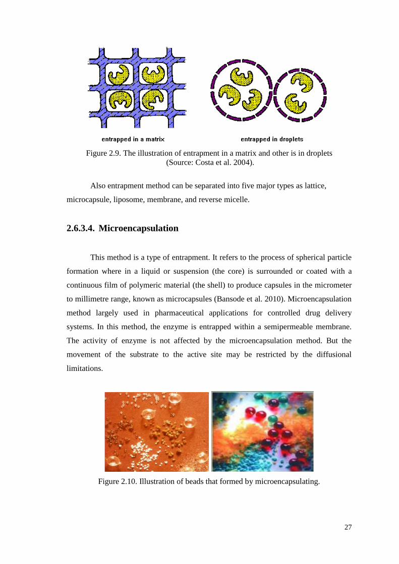
2.6.3.4. Microencapsulation
This method is a type of entrapment. It refers to the process of spherical particle
formation where in a liquid or suspension (the core) is surrounded or coated with a
continuous film of polymeric material (the shell) to produce capsules in the micrometer
to millimetre range, known as microcapsules (Bansode et al. 2010). Microencapsulation
method largely used in pharmaceutical applications for controlled drug delivery
systems. In this method, the enzyme is entrapped within a semipermeable membrane.
The activity of enzyme is not affected by the microencapsulation method. But the
movement of the substrate to the active site may be restricted by the diffusional
limitations.
Figure 2.10. Illustration of beads that formed by microencapsulating.
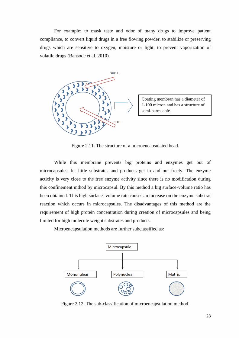
28
For example: to mask taste and odor of many drugs to improve patient
compliance, to convert liquid drugs in a free flowing powder, to stabilize or preserving
drugs which are sensitive to oxygen, moisture or light, to prevent vaporization of
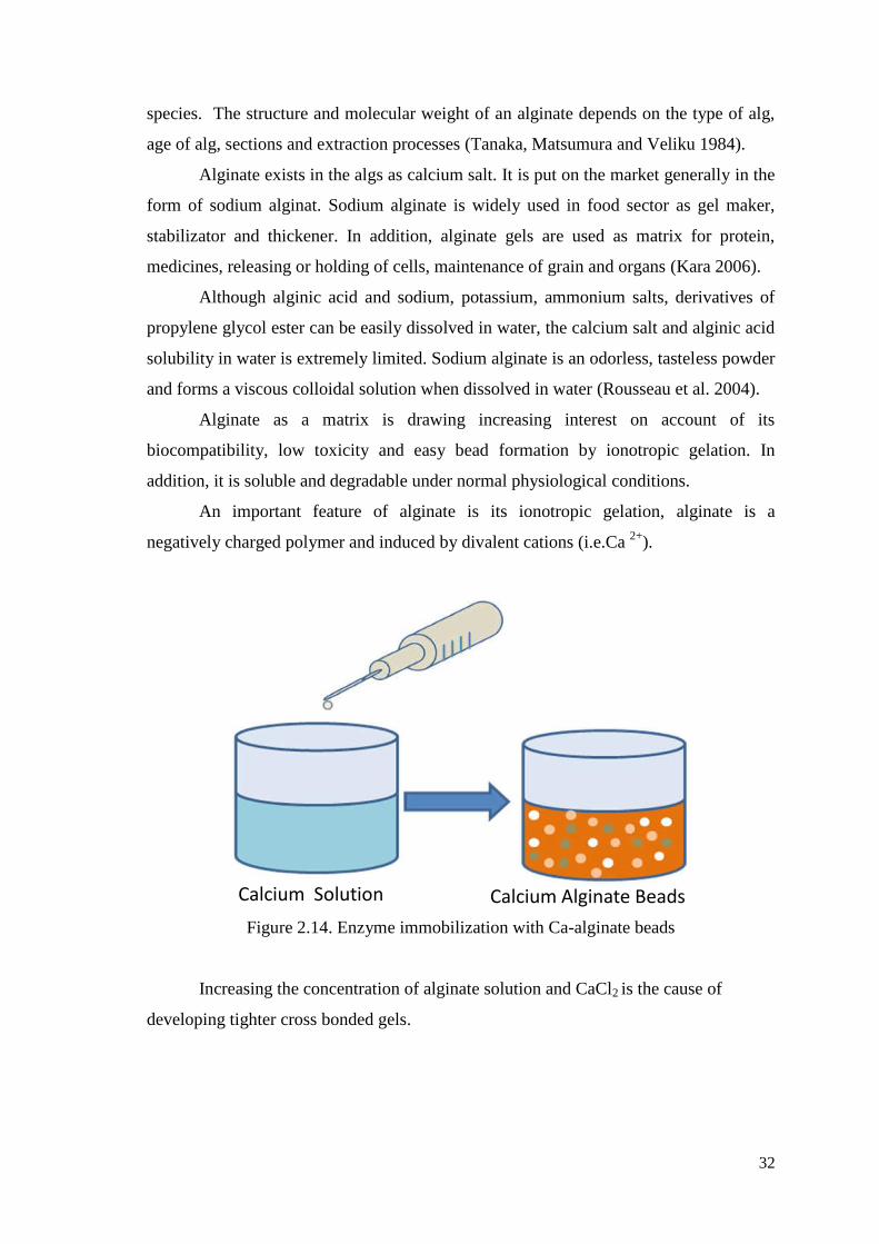
volatile drugs (Bansode et al. 2010).
Figure 2.11. The structure of a microencapsulated bead.
While this membrane prevents big proteins and enzymes get out of
microcapsules, let little substrates and products get in and out freely. The enzyme
acticity is very close to the free enzyme activity since there is no modification during
this confinement mthod by microcapsul. By this method a big surface-volume ratio has
been obtained. This high surface- volume rate causes an increase on the enzyme substrat
reaction which occurs in microcapsules. The disadvantages of this method are the
requirement of high protein concentration during creation of microcapsules and being
limited for high molecule weight substrates and products.
Microencapsulation methods are further subclassified as:
Figure 2.12. The sub-classification of microencapsulation method.
Coating membran has a diameter of
1-100 micron and has a structure of
semi-parmeable.
29
The most commonly used microencapsulation coating materials are Gums (gum
arabic, sodium alginate, and carrageenan), Carbohydrates (starch, dextran, sucrose),
Celluloses (carboxymethylcellulose, methycellulose), Lipids (bees wax, stearic acid,
phospholipids). Selection of the most convenient coating material for core is the
primary important factor for application of this method.
Suitable coating material should have the features listed below:
Capable of forming a film that is cohesive with the core material.
Chemically compatible and nonreactive with the core material.
Film-forming, pliable, tasteless, stable.
Controlled release under specific conditions.
Inert toward active ingredients.
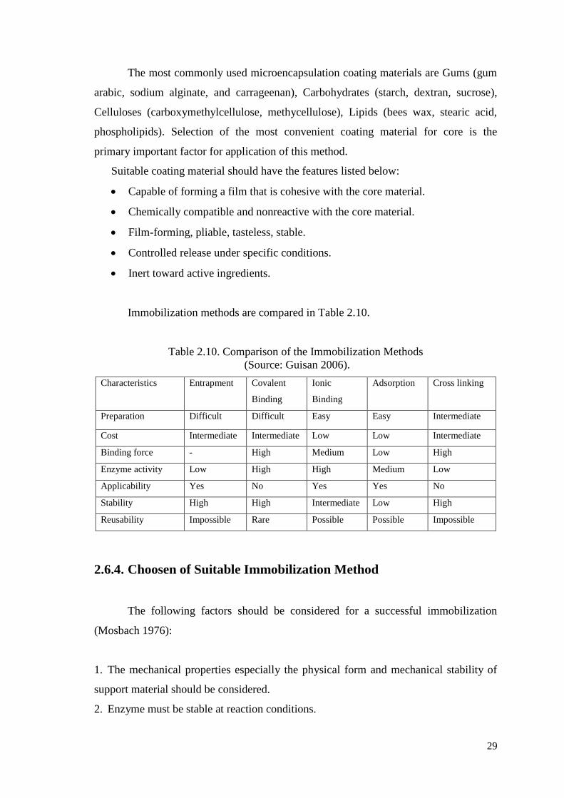
Immobilization methods are compared in Table 2.10.
Table 2.10. Comparison of the Immobilization Methods
(Source: Guisan 2006).
Characteristics Entrapment Covalent
Binding
Ionic
Binding
Adsorption Cross linking
Preparation Difficult Difficult Easy Easy Intermediate
Cost Intermediate Intermediate Low Low Intermediate
Binding force - High Medium Low High
Enzyme activity Low High High Medium Low
Applicability Yes No Yes Yes No
Stability High High Intermediate Low High
Reusability Impossible Rare Possible Possible Impossible
2.6.4. Choosen of Suitable Immobilization Method
The following factors should be considered for a successful immobilization
(Mosbach 1976):
1. The mechanical properties especially the physical form and mechanical stability of
support material should be considered.
2. Enzyme must be stable at reaction conditions.
30
3. Crosslinker reagents should not react with the enzyme’s active ends or they should
be big enough in order not to penetrate the active end of enzyme.
4. If possible, the active end of enzyme should be protected. For example, sulfhydryl
enzymes can be protected by reaction with glutathione or cysteine. Afterwards, enzyme
can be reactivated.
5. The washing process for removing the unbounded enzyme during immobilization
should not affect the enzyme.
6. If immobilized enzyme will be used as continuous catalyst in some chemical
reactions, teh nature of the reaction should be considered before choosing the method of
immobilization.
In our study, we tried to immobilize of esterase enzyme into Alginate-Chitosan /
CaCl 2 polymeric beads. Alginate and chitosan are both natural biopolymers and have
several advantages such as availability from replenishable agricultural or marine food
resources, biocompatibility, and biodegradability.
2.7. Natural Polymers
Natural polymers arepolymers those can be produced biologically and have
unique functional properties. Proteins such as collagen, gelatin, elastin, actin, etc.),
polysaccharides (cellulose, starch, dextran, chitin, etc.) and polynucleotide (DNA and
RNA) are the main natural polymers. Natural polymers have different fields of use due
to thier functional properties. They can be used as thickener, gel-maker, linker,
distributing agent, lubricant, adhesive and biomaterial. Natural polymers are
indispensable sources of field of biomaterials. They do not give adverse reactions such
as inflammation and toxic effect when in contact with a live body since they are similar
or identical to macromolecules in the biological environment. However, their main
disadvantages are having difficulty when being shaped and being immunogenic in order
to give rise to an immune response.
31
Table 2.11. Requirements for natural polymers
(Source: Park and Lakes 1992).
Property Description
Biocompatibility Noncarcinogenesis, nonpyrogenicity, nontoxicity, and
nonallergic response
Sterilizability Autoclave, dry heating, ethylenoxide gas, and radiation
Physical property Strength, elasticity, and durability
Manufacturability Machining, molding, extruding, and fiber forming
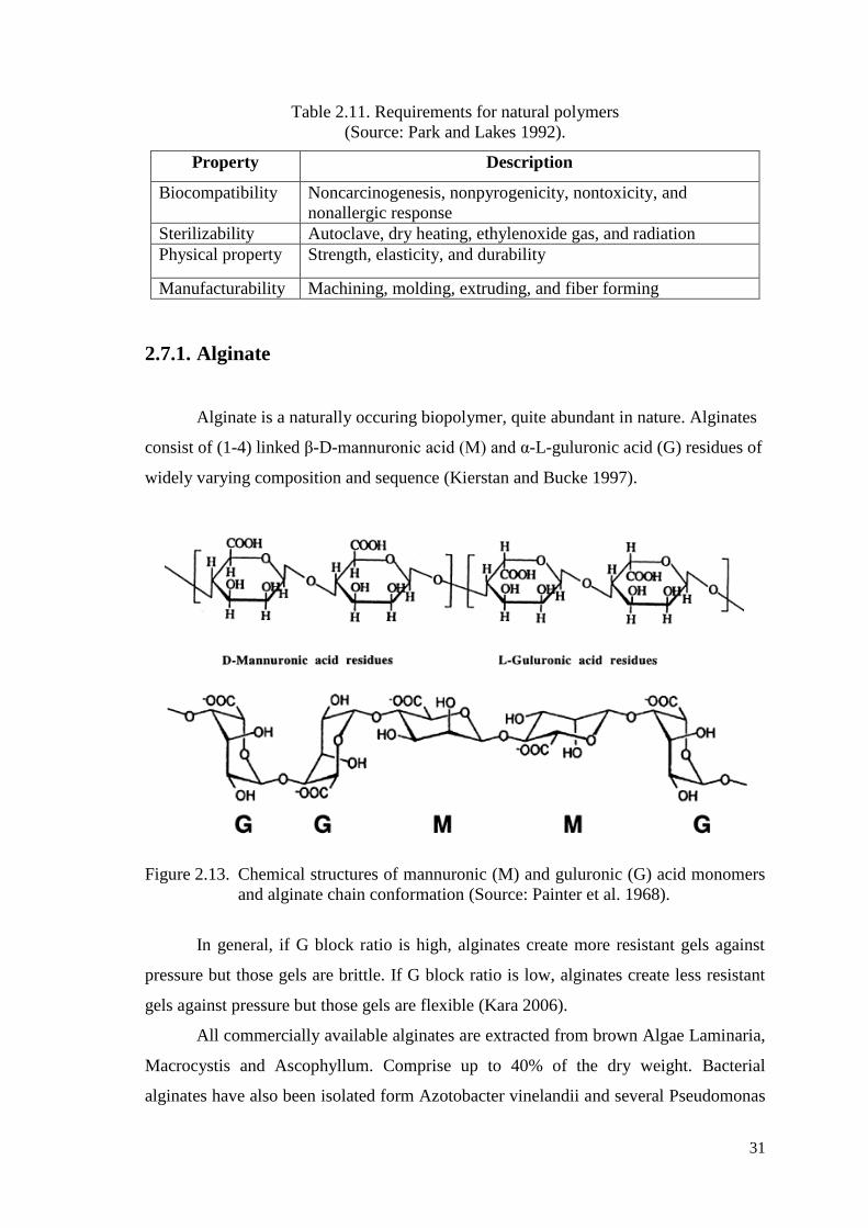
2.7.1. Alginate
Alginate is a naturally occuring biopolymer, quite abundant in nature. Alginates
consist of (1-4) linked β-D-mannuronic acid (M) and α-L-guluronic acid (G) residues of
widely varying composition and sequence (Kierstan and Bucke 1997).
Figure 2.13. Chemical structures of mannuronic (M) and guluronic (G) acid monomers
and alginate chain conformation (Source: Painter et al. 1968).
In general, if G block ratio is high, alginates create more resistant gels against
pressure but those gels are brittle. If G block ratio is low, alginates create less resistant
gels against pressure but those gels are flexible (Kara 2006).
All commercially available alginates are extracted from brown Algae Laminaria,
Macrocystis and Ascophyllum. Comprise up to 40% of the dry weight. Bacterial
alginates have also been isolated form Azotobacter vinelandii and several Pseudomonas
32
species. The structure and molecular weight of an alginate depends on the type of alg,
age of alg, sections and extraction processes (Tanaka, Matsumura and Veliku 1984).
Alginate exists in the algs as calcium salt. It is put on the market generally in the
form of sodium alginat. Sodium alginate is widely used in food sector as gel maker,
stabilizator and thickener. In addition, alginate gels are used as matrix for protein,
medicines, releasing or holding of cells, maintenance of grain and organs (Kara 2006).
Although alginic acid and sodium, potassium, ammonium salts, derivatives of
propylene glycol ester can be easily dissolved in water, the calcium salt and alginic acid
solubility in water is extremely limited. Sodium alginate is an odorless, tasteless powder
and forms a viscous colloidal solution when dissolved in water (Rousseau et al. 2004).
Alginate as a matrix is drawing increasing interest on account of its
biocompatibility, low toxicity and easy bead formation by ionotropic gelation. In
addition, it is soluble and degradable under normal physiological conditions.
An important feature of alginate is its ionotropic gelation, alginate is a
negatively charged polymer and induced by divalent cations (i.e.Ca 2+
).
Figure 2.14. Enzyme immobilization with Ca-alginate beads
Increasing the concentration of alginate solution and CaCl2 is the cause of
developing tighter cross bonded gels.
Calcium Alginate Beads Calcium Solution
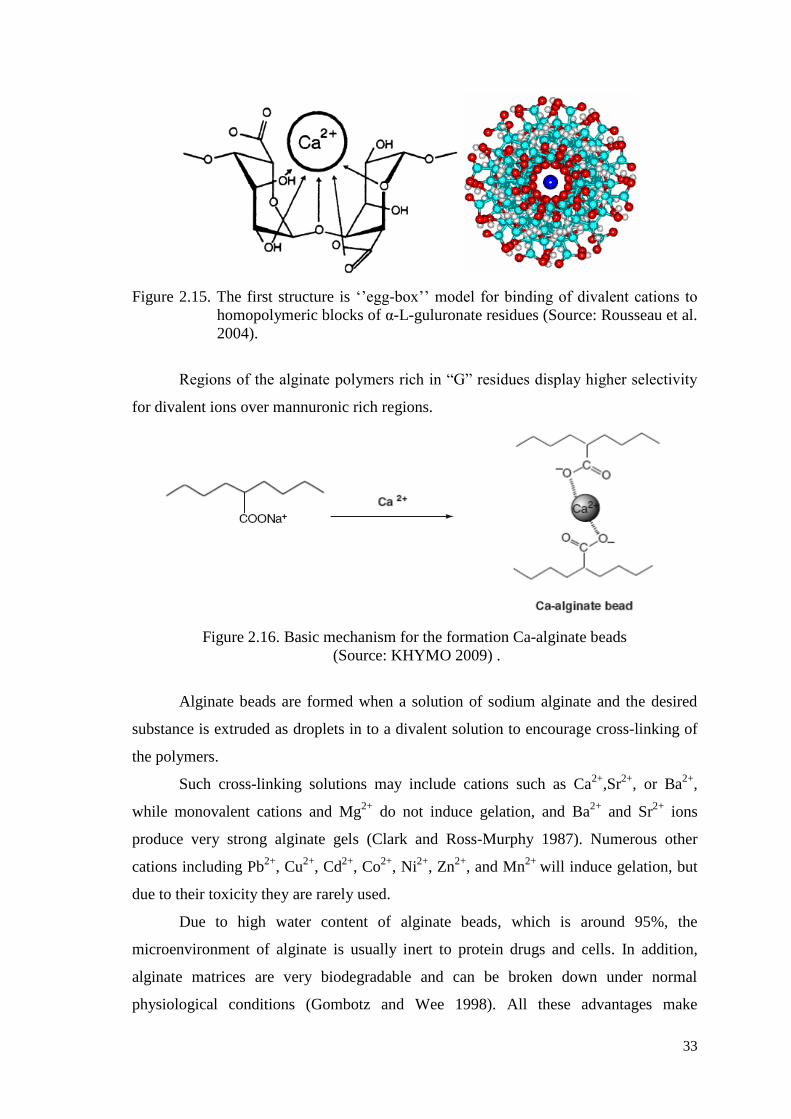
33
Figure 2.15. The first structure is ‘’egg-box’’ model for binding of divalent cations to
homopolymeric blocks of α-L-guluronate residues (Source: Rousseau et al.
2004).
Regions of the alginate polymers rich in “G” residues display higher selectivity
for divalent ions over mannuronic rich regions.
Figure 2.16. Basic mechanism for the formation Ca-alginate beads
(Source: KHYMO 2009) .
Alginate beads are formed when a solution of sodium alginate and the desired
substance is extruded as droplets in to a divalent solution to encourage cross-linking of
the polymers.
Such cross-linking solutions may include cations such as Ca2+
,Sr2+
, or Ba2+
,
while monovalent cations and Mg2+
do not induce gelation, and Ba2+
and Sr2+
ions
produce very strong alginate gels (Clark and Ross-Murphy 1987). Numerous other
cations including Pb2+
, Cu2+
, Cd2+
, Co2+
, Ni2+
, Zn2+
, and Mn2+
will induce gelation, but
due to their toxicity they are rarely used.
Due to high water content of alginate beads, which is around 95%, the
microenvironment of alginate is usually inert to protein drugs and cells. In addition,
alginate matrices are very biodegradable and can be broken down under normal
physiological conditions (Gombotz and Wee 1998). All these advantages make
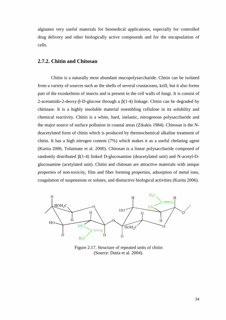
34
alginates very useful materials for biomedical applications, especially for controlled
drug delivery and other biologically active compounds and for the encapsulation of
cells.
2.7.2. Chitin and Chitosan
Chitin is a naturally most abundant mucopolysaccharide. Chitin can be isolated
from a variety of sources such as the shells of several crustaceans, krill, but it also forms
part of the exoskeleton of insects and is present in the cell walls of fungi. It is consist of
2-acetamido-2-deoxy-β-D-glucose through a β(1-4) linkage. Chitin can be degraded by
chitinase. It is a highly insoluble material resembling cellulose in its solubility and
chemical reactivity. Chitin is a white, hard, inelastic, nitrogenous polysaccharide and
the major source of surface pollution in coastal areas (Zikakis 1984). Chitosan is the N-
deacetylated form of chitin which is produced by thermochemical alkaline treatment of
chitin. It has a high nitrogen content (7%) which makes it as a useful chelating agent
(Kurita 2006, Tolaimate et al. 2000). Chitosan is a linear polysaccharide composed of
randomly distributed β(1-4) linked D-glucosamine (deacetylated unit) and N-acetyl-D-
glucosamine (acetylated unit). Chitin and chitosan are attractive materials with unique
properties of non-toxicity, film and fiber forming properties, adsorption of metal ions,
coagulation of suspensions or solutes, and distinctive biological activities (Kurita 2006).
Figure 2.17. Structure of repeated units of chitin
(Source: Dutta et al. 2004).
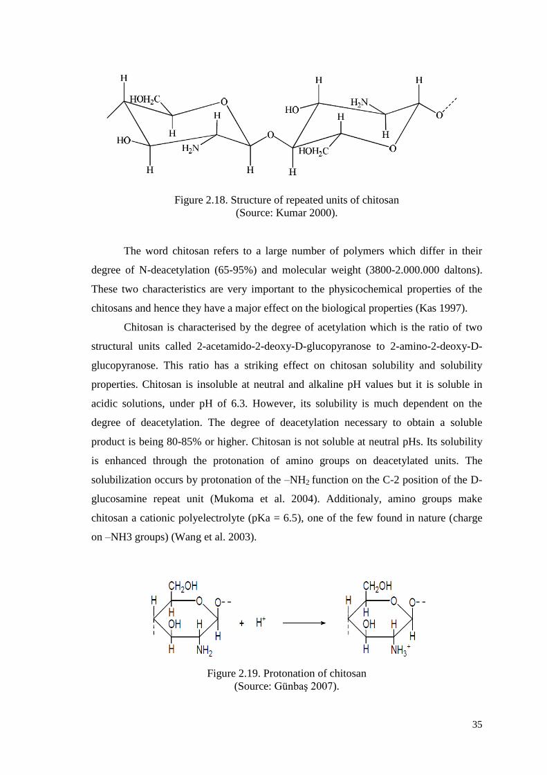
35
Figure 2.18. Structure of repeated units of chitosan
(Source: Kumar 2000).
The word chitosan refers to a large number of polymers which differ in their
degree of N-deacetylation (65-95%) and molecular weight (3800-2.000.000 daltons).
These two characteristics are very important to the physicochemical properties of the
chitosans and hence they have a major effect on the biological properties (Kas 1997).
Chitosan is characterised by the degree of acetylation which is the ratio of two
structural units called 2-acetamido-2-deoxy-D-glucopyranose to 2-amino-2-deoxy-D-
glucopyranose. This ratio has a striking effect on chitosan solubility and solubility
properties. Chitosan is insoluble at neutral and alkaline pH values but it is soluble in
acidic solutions, under pH of 6.3. However, its solubility is much dependent on the
degree of deacetylation. The degree of deacetylation necessary to obtain a soluble
product is being 80-85% or higher. Chitosan is not soluble at neutral pHs. Its solubility
is enhanced through the protonation of amino groups on deacetylated units. The
solubilization occurs by protonation of the –NH2 function on the C-2 position of the D-
glucosamine repeat unit (Mukoma et al. 2004). Additionaly, amino groups make
chitosan a cationic polyelectrolyte (pKa = 6.5), one of the few found in nature (charge
on –NH3 groups) (Wang et al. 2003).
Figure 2.19. Protonation of chitosan
(Source: Günbaş 2007).

36
Since chitosan is biodegradable, non-toxic, non-immunogenic and biocompatible
in animal tissues, much research has been directed toward its use in medical
applications such as drug delivery, artificial skin, and blood anticoagulants. Chitosan
has also been suggested for use as flocculant, food thickener, paper and textile adhesive,
membrane and chelating agent for metals. It is also has an antibacterial activity. Thanks
to it’s bioactivity, facilitates wound healing, reduce blood cholesterol levels, and
stimulate the immune system.
Cationic properties of chitosan:
Linear Polyelectrolyte
High charge density
Excellent flocculant
Adheres to negatively charged surfaces
Substantive to hair, skin
Chelates metal ions
Iron (Fe), Copper (Cu)
Toxic metals (Cd, Hg, Pb, Cr, Ni)
Radionucleids (Pu, U)
Chitosan is a linear polyelectrolyte at acidic pH’s. It has a high charge density,
one charge per glucosamine unit. Since many materials carry negative charges (e.g.
protein, aionic polysaccharides, nucleic acids, etc.), the positive charge of chitosan
interacts strongly with negative surfaces to give an electric neutrality. Chitosan adheres
easily to natural polymers such as hair and skin, which are composed of negatively
charged mucopolysaccharides and proteins. Also chitosan used for removing of toxic
heavy metal ions such as silver, cadmium.
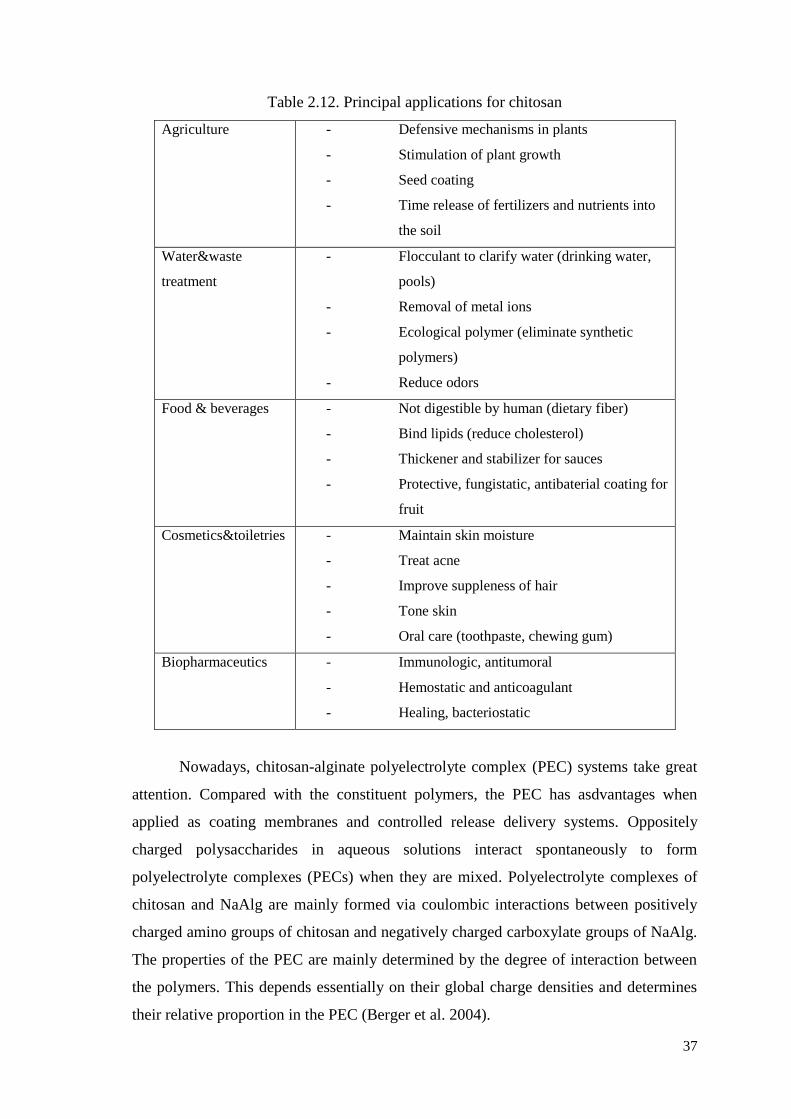
37
Table 2.12. Principal applications for chitosan
Agriculture - Defensive mechanisms in plants
- Stimulation of plant growth
- Seed coating
- Time release of fertilizers and nutrients into
the soil
Water&waste
treatment
- Flocculant to clarify water (drinking water,
pools)
- Removal of metal ions
- Ecological polymer (eliminate synthetic
polymers)
- Reduce odors
Food & beverages - Not digestible by human (dietary fiber)
- Bind lipids (reduce cholesterol)
- Thickener and stabilizer for sauces
- Protective, fungistatic, antibaterial coating for
fruit
Cosmetics&toiletries - Maintain skin moisture
- Treat acne
- Improve suppleness of hair
- Tone skin
- Oral care (toothpaste, chewing gum)
Biopharmaceutics - Immunologic, antitumoral
- Hemostatic and anticoagulant
- Healing, bacteriostatic
Nowadays, chitosan-alginate polyelectrolyte complex (PEC) systems take great
attention. Compared with the constituent polymers, the PEC has asdvantages when
applied as coating membranes and controlled release delivery systems. Oppositely
charged polysaccharides in aqueous solutions interact spontaneously to form
polyelectrolyte complexes (PECs) when they are mixed. Polyelectrolyte complexes of
chitosan and NaAlg are mainly formed via coulombic interactions between positively
charged amino groups of chitosan and negatively charged carboxylate groups of NaAlg.
The properties of the PEC are mainly determined by the degree of interaction between
the polymers. This depends essentially on their global charge densities and determines
their relative proportion in the PEC (Berger et al. 2004).
38
Since both chitosan and NaAlg are weak polyelectrolytes some factors have to
be controlled to determine the properties of PEC. The most important factor that has to
be controlled is the pH of the solution, but temperature, ionic strength (Chavasit et al.
1988, Argüelles-Monal et al. 1993, Park and Ha 1993, Lee et al. 1997) and the order of
mixing are also important.
39
CHAPTER 3
MATERIALS AND METHODS
3.1. Materials
Detailed list of used chemicals, buffers, solutions and their compositions are
presented in Appendix A, Appendix B and Appendix C.
3.2. Methods
3.2.1. Preparation of Protein Sample
The thermophilic esterase enzyme purified in six steps in this study. These steps
are shown at below:
Table 3.1. Experiment steps of preparation of protein sample.
3.2.1.1. Escherichia coli Growth
In this study, the source of protein was choosen a microorganism Thermophilic
Bacillus sp. that was isolated from Balçova (Agamemnon) Geothermal region in Izmir,
Number Steps
1 Cell Source
2 Cell disruption
3 Debris Removal
4 Initial Purification (Affinity Chromatography)
5 High Resolution Purification (Gel Filtration)
6 Drying under Vacuum
7 Concentrated Purified Enzyme
40
have used in our studies (Tekedar 2008). Also it belongs to prokaryotes as host because
of rapid growth and simple medium components.
The bacteria isolate that expression vector including esterase gene transformed
E.Coli was taken from -80°C. The isolate was named as Est33 in Pet28 BL21. The
isolate was spread on agar plates with Kanamycin and incubated overnight in an
incubator at 37°C. Our bacteria was inserted a gene that resistant to Kanamycin and
grown under these conditions.
Another day the agar plates were controlled if there was any bacterial growth on
it or not. And a single colony was choosen and inoculated in LB kan
media of 50 ml and
incubated one day more overnight at 37°C in a incubator shaker for 15-16 hours.
3.2.1.2. Expression of the Transformed Genes
After nearly 15-16 hours the 50 ml of bacteria culture was taken from shaker and
the volume was diluted into total 500 ml and grown at 37°C to an optical density of
A600=1,0 which is half-stationary phase. At that point expression of the esterase genes
was induced by the addition of 1 mM IPTG.
IPTG (isopropyl-β-D thiogalactopyranoside) induces the expression of the
esterase genes which is a lactose analog. It represses the lac operator and allows the
expression of T7 RNA polymerase, which in turn transcribes the target gene. The cells
were allowed to grow for an additional 4.0 h after addition of IPTG and were then
harvested by centrifugation (8,000 X g for 10 min). The harvested cell pastes were
stored frozen at -20 °C until ready for use of purification.
3.2.1.3. Total Protein Extraction
The cell pellets were taken from –20°C and dissolved nearly in 10 ml of 50 mM
sodium phosphate buffer, pH 7.0 and disrupted by a sonicator (Sim-Aminco, Spectronic
Instruments) for 10 min. Cells were agitated by ultrasound energy, cell membranes were
disrupted and cellular contents released. This sonication step was done at +4°C to
preserve protein from denaturation. After sonication cell content was harvested by high-
speed centrifugation at 10000 rpm, 10 min, +4°C. Then the cell debris was thrown away
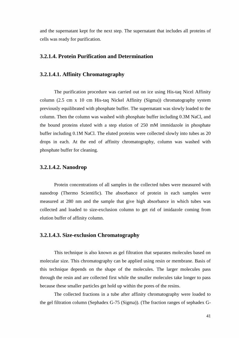
41
and the supernatant kept for the next step. The supernatant that includes all proteins of
cells was ready for purification.
3.2.1.4. Protein Purification and Determination
3.2.1.4.1. Affinity Chromatography
The purification procedure was carried out on ice using His-taq Nicel Affinity
column (2.5 cm x 10 cm His-taq Nickel Affinity (Sigma)) chromatography system
previously equilibrated with phosphate buffer. The supernatant was slowly loaded to the
column. Then the column was washed with phosphate buffer including 0.3M NaCl, and
the bound proteins eluted with a step elution of 250 mM immidazole in phosphate
buffer including 0.1M NaCl. The eluted proteins were collected slowly into tubes as 20
drops in each. At the end of affinity chromatography, column was washed with
phosphate buffer for cleaning.
3.2.1.4.2. Nanodrop
Protein concentrations of all samples in the collected tubes were measured with
nanodrop (Thermo Scientific). The absorbance of protein in each samples were
measured at 280 nm and the sample that give high absorbance in which tubes was
collected and loaded to size-exclusion column to get rid of imidazole coming from
elution buffer of affinity column.
3.2.1.4.3. Size-exclusion Chromatography
This technique is also known as gel filtration that separates molecules based on
molecular size. This chromatography can be applied using resin or membrane. Basis of
this technique depends on the shape of the molecules. The larger molecules pass
through the resin and are collected first while the smaller molecules take longer to pass
because these smaller particles get hold up within the pores of the resins.
The collected fractions in a tube after affinity chromatography were loaded to
the gel filtration column (Sephadex G-75 (Sigma)). (The fraction ranges of sephadex G-
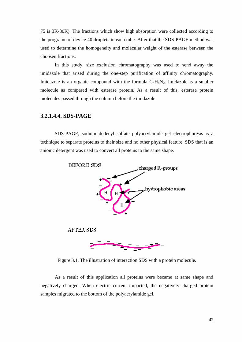
42
75 is 3K-80K). The fractions which show high absorption were collected according to
the programe of device 40 droplets in each tube. After that the SDS-PAGE method was
used to determine the homogeneity and molecular weight of the esterase between the
choosen fractions.
In this study, size exclusion chromatography was used to send away the
imidazole that arised during the one-step purification of affinity chromatography.
Imidazole is an organic compound with the formula C3H4N2. Imidazole is a smaller
molecule as compared with esterase protein. As a result of this, esterase protein
molecules passed through the column before the imidazole.
3.2.1.4.4. SDS-PAGE
SDS-PAGE, sodium dodecyl sulfate polyacrylamide gel electrophoresis is a
technique to separate proteins to their size and no other physical feature. SDS that is an
anionic detergent was used to convert all proteins to the same shape.
Figure 3.1. The illustration of interaction SDS with a protein molecule.
As a result of this application all proteins were became at same shape and
negatively charged. When electric current impacted, the negatively charged protein
samples migrated to the bottom of the polyacrylamide gel.
43
In this study, the molecular weight of esterase protein was determined by
following the Laemmli method (Laemmli 1970). Thermo Scientific Electrophoresis was
used as separating device. Firstly all equipments and glass plates were washed with
deionized water and after this all cleaned with ethanol. First, separating gel was
prepared according to the table in Appendix B, polymerization began as soon as the
TEMED had been added to mixture. Without delay, the mixture rapidly was poured into
the gap between the glass plates. The top of the gel was covered by deionized water to
avoid drying. After 30 minutes the polymerization finished. The distilled water was
poured off and the top of the gel was washed two times with deionized water to remove
any unpolymerized acrylamide. Secondly, the stacking gel was prepared according to
the table in appendix B. The components was mixed in the order shown. Polymerization
began as soon as the TEMED had been added. Without delay, the mixture was poured
slowly into gap over the separating gel. This process has been performed in a gentle
way so that the creation of foam could be prevented. Instantly a clean Teflon comb was
inserted into the stacking gel solution and more stacking gel was added to fill the spaces
of the comb completely. During the stacking gel polymerization, the protein samples
were mixed with sample buffer with the ratio of 1 : 4 (v/v). Then samples were heated
to 100°C for 5 minutes to denature proteins. After nearly 30 minutes the polymerization
of stacking gel was completed. The teflon comb was removed carefully and the 10
spaces were washed with deionized water two times to remove any unpolymerized
acrylamide. The broad range protein marker and protein samples were loaded to the gel
in buffer tank in the presence of running buffer. Electrophoresis was run at 65 volts for
30 minutes and followed by voltage at 100 volts for 2 hour until the blue dye reached at
the bottom of the gel.
Finally when the run completed, the gel was removed from between the glass
plates by using a spatula. And the gel was washed with deionized water, stained with
Coomassie Brilliant Blue for 24 hours with slow shaking. Another day, the gel was
washed first deionized water and then treated with destaining solution for 1 hour. When
destaining completed gel washed with deionized water very carefully and photographed
using the gel photo system.
44
3.2.1.4.5. Protein Concentration Determination
After purification and determination of esterase protein, it was concentrated
under vacuum to increase durability without loss activity at -20°C. Memmert Vacuum
Oven V0400 was used. Total esterase concentration was measured
spectrophotometrically according to the Bradford protein assay method (Bradford
1976). In this method, Coomassie Brilliant Blue G-250 dye was used. The dye bound to
our esterase protein and gave absorption at 595nm. Bradford method also known as
colorimetric protein assay. When the protein concentration increases, the color of the
sample becomes darker. Bovine Serum Albumin (BSA) was used as Standard protein.
After formed the immobilized beads, the protein concentration determination was
applied to the Chitosan/CaCl2 filtrate that crosslinking solution to measure
immobilization yield and enzyme leakage. All characterization studies were done also
with Bradford protein concentration determination method Appendix C.
3.2.2. Esterase Activity Determination
The esterase activity was assayed spectrophotometrically using p-nitrophenyl
acetate as a substrate having one of the best hydrolytic activity among the variety of
pnitrophenyl (p-NP) esters with different acyl chain lengths (C2-C16) in previous
studies (Tekadar 2008, Gülay 2009).
The enzyme hydrolyzes the acetate ester with the help of water. The products are
acetic acid and p-nitrophenol (pNP), the latter showing an absorption maximum at about
420 nm.
Figure 3.2. The Scheme of the pNPA assay
(Source: Gülay 2009)
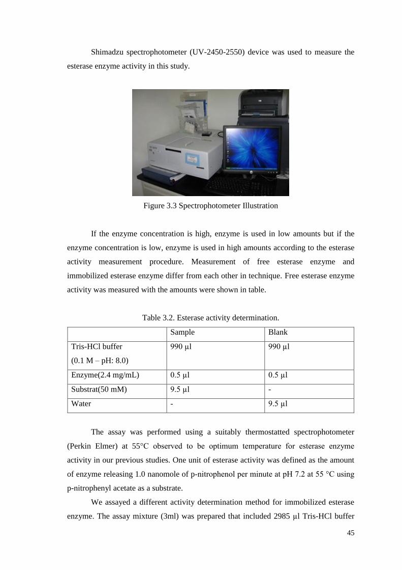
45
Shimadzu spectrophotometer (UV-2450-2550) device was used to measure the
esterase enzyme activity in this study.
Figure 3.3 Spectrophotometer Illustration
If the enzyme concentration is high, enzyme is used in low amounts but if the
enzyme concentration is low, enzyme is used in high amounts according to the esterase
activity measurement procedure. Measurement of free esterase enzyme and
immobilized esterase enzyme differ from each other in technique. Free esterase enzyme
activity was measured with the amounts were shown in table.
Table 3.2. Esterase activity determination.
Sample Blank
Tris-HCl buffer
(0.1 M – pH: 8.0)
990 µl 990 µl
Enzyme(2.4 mg/mL) 0.5 µl 0.5 µl
Substrat(50 mM) 9.5 µl -
Water - 9.5 µl
The assay was performed using a suitably thermostatted spectrophotometer
(Perkin Elmer) at 55°C observed to be optimum temperature for esterase enzyme
activity in our previous studies. One unit of esterase activity was defined as the amount
of enzyme releasing 1.0 nanomole of p-nitrophenol per minute at pH 7.2 at 55 °C using
p-nitrophenyl acetate as a substrate.
We assayed a different activity determination method for immobilized esterase
enzyme. The assay mixture (3ml) was prepared that included 2985 µl Tris-HCl buffer

46
(0.1M pH 8.0), 15 µl substrate (50 mM pNPA) and 10 silicate coated beads with
enzyme. After 5 minutes of incubation time at 55°C with slow shake, 1 ml solution
taken from the assay mixture to determine the catalytic activity and leakage of enzyme
if any. The absorbance was measured at 55°C at 420nm. with spectrophotometrically.
Figure 3.4. Immobilized Esterase Activity Determination.
The proportions of tris HCl buffer, solution, water and substrates for the
experiments of activity determination are given in Table 3.3.
Table 3.3. The Proportions for Activity Measurement of Immobilized Polyelectrolyte
Beads.
Tris HCl buffer
(0,1 M, pH:8,0)
15 µl solution
(protein)
Water Substrate PNPA
Blank 980 µl 10 µl 10 µl -
Sample 980 µl 10 µl - 10 µl
Activity calculation of free and immobilized enzyme is the same. Units were
calculated according to the equation (3.1):
Enzyme Activity = ⁄
(3.1)
Blank
3 ml reaction mixture
include;
- 2985 µl 0,1 M Tris-HCl
pH:8,0
- 10 beads
Sample 3ml reaction mixture include;
- 2985 µl 0,1 M Tris-
HCl pH:8,0
- 10 beads
- 15 µl PNPA substrate
47
∆A: Absorbance of the reaction sample
𝓔: Extinction coefficient (0.0148)
E: Volume of enzyme used in ten beads
t: Incubation time (minutes)
The volume of enzyme used in ten beads which was described as E; can be
found as follows: At the beginning 2,5 mg enzyme which is 1 ml used to crosslink with
4 ml of alginate solution. Therefore we obtain 5ml of solution which includes enzyme
of 0.5mg/ml concentration. However; 0.098 mg of enzyme was obtained as excess
amount without being crosslinked. This excess amount of 0.098 mg can be substracted
from the initial amount 0.5mg so that net used amount enzyme can be found as 0.402
mg. Thus, the volume of net used enzyme can be determined as 0.804 ml. Since from
0.804 ml enzyme approximately 500 beads can be formed; 10 beads can be formed from
0.016 ml enzyme. During all experiments for determination of enzyme activity 10 beads
were used as constant enzyme amount. Using the equation 3.1 the terms in the
denominator can be multiplied and in this study that denominator value used as constant
value for all calculations. Denominator value can be found by multiplication of 𝓔, E and
t. The nominator value which is absorbance can be read from the spectrophotometer at
420 nm. By putting these values in equation 3.1 the enzyme activity can be found.
The specific enzyme activity (Units/mg protein) was defined as the ratio of
enzyme activity (U/ml) to the mg of protein per ml (mg/ml). Therefore,
(3.2)
The calculated value from equation 3.1 can be put into nominator of equation of
equation 3.2. The denominator of this equation means that the amount enzyme in 10
beads. Since 10 beads makes 0.016 ml and we know that 1 ml of volume makes 0.5 mg
of enzyme; therefore it is calculated that there are 0.008 mg of enzyme in 10 beads.
However, during all experiments for determination of activity there are some portions of
enzyme leaking from bead to outside. For that reason, for each experiment that leaking
amount of enzyme must be calculated by Bradford protein assay. Net amount of
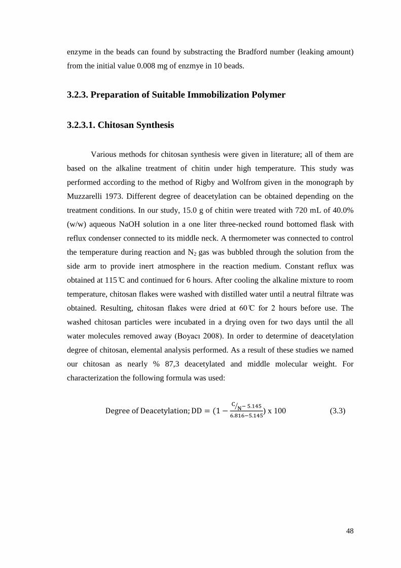
48
enzyme in the beads can found by substracting the Bradford number (leaking amount)
from the initial value 0.008 mg of enzmye in 10 beads.
3.2.3. Preparation of Suitable Immobilization Polymer
3.2.3.1. Chitosan Synthesis
Various methods for chitosan synthesis were given in literature; all of them are
based on the alkaline treatment of chitin under high temperature. This study was
performed according to the method of Rigby and Wolfrom given in the monograph by
Muzzarelli 1973. Different degree of deacetylation can be obtained depending on the
treatment conditions. In our study, 15.0 g of chitin were treated with 720 mL of 40.0%
(w/w) aqueous NaOH solution in a one liter three-necked round bottomed flask with
reflux condenser connected to its middle neck. A thermometer was connected to control
the temperature during reaction and N2 gas was bubbled through the solution from the
side arm to provide inert atmosphere in the reaction medium. Constant reflux was
obtained at 115 C and continued for 6 hours. After cooling the alkaline mixture to room
temperature, chitosan flakes were washed with distilled water until a neutral filtrate was
obtained. Resulting, chitosan flakes were dried at 60 C for 2 hours before use. The
washed chitosan particles were incubated in a drying oven for two days until the all
water molecules removed away (Boyacı 2008). In order to determine of deacetylation
degree of chitosan, elemental analysis performed. As a result of these studies we named
our chitosan as nearly % 87,3 deacetylated and middle molecular weight. For
characterization the following formula was used:
⁄
) x 100 (3.3)
49
Figure 3.5. The Scheme of the Reflux System
3.2.4. Immobilization of Thermophilic Esterase Enzyme in to Alginate-
Chitosan/CaCl2 Polyelectrolyte Beads
Firstly, the most suitable Alginate-Chitosan/CaCl2 ratio and reaction conditions
were determined with several optimization studies. In order to compare our results
suitably the same reaction conditions and same amounts were used as the previous
immobilization study of Gülay 2009.
3.2.4.1. Preparation of Alginic acid-Esterase Enzyme Solution
The purified thermophilic recombinant esterase enzyme and alginate is soluble
in water. Firstly, alginate (w/v, 2%) was dissolved in 10 ml Tris-HCl buffer (0.1M pH
8.0) by heating at 30°C for 30 minutes. When the dissolving of all alginate particles
completed, the liquid esterase enzyme (0.5 mg/ml) was slowly added by stirring into the
alginate solution. This mixture was stirred for 1 hour for complete homogenation of
enzyme and alginate.
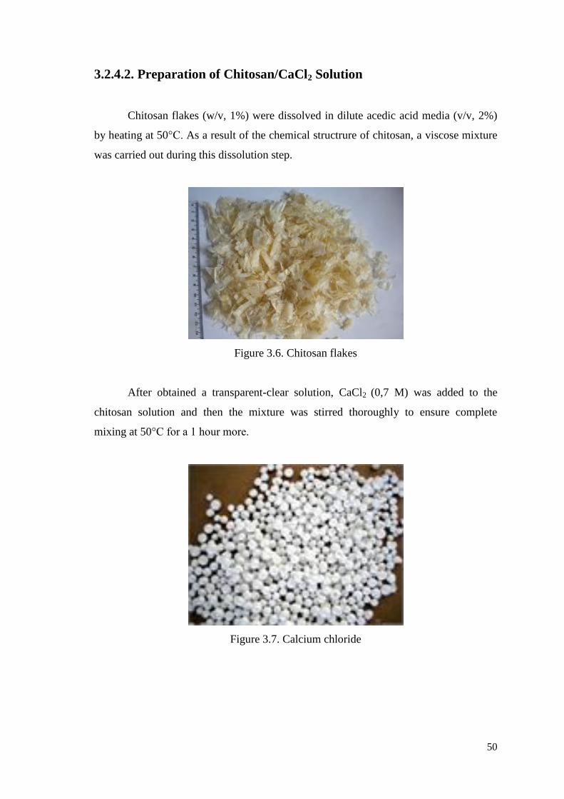
50
3.2.4.2. Preparation of Chitosan/CaCl2 Solution
Chitosan flakes (w/v, 1%) were dissolved in dilute acedic acid media (v/v, 2%)
by heating at 50°C. As a result of the chemical structrure of chitosan, a viscose mixture
was carried out during this dissolution step.
Figure 3.6. Chitosan flakes
After obtained a transparent-clear solution, CaCl2 (0,7 M) was added to the
chitosan solution and then the mixture was stirred thoroughly to ensure complete
mixing at 50°C for a 1 hour more.
Figure 3.7. Calcium chloride
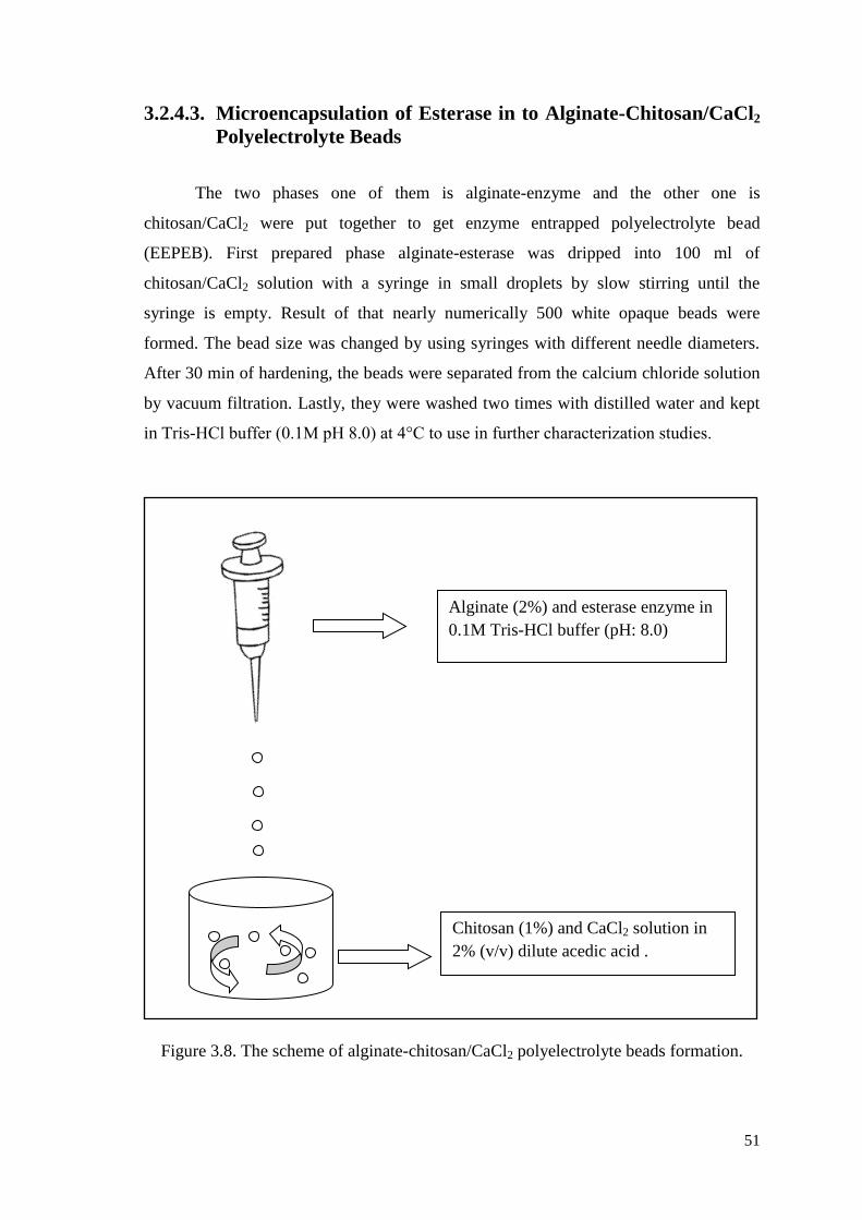
51
3.2.4.3. Microencapsulation of Esterase in to Alginate-Chitosan/CaCl2
Polyelectrolyte Beads
The two phases one of them is alginate-enzyme and the other one is
chitosan/CaCl2 were put together to get enzyme entrapped polyelectrolyte bead
(EEPEB). First prepared phase alginate-esterase was dripped into 100 ml of
chitosan/CaCl2 solution with a syringe in small droplets by slow stirring until the
syringe is empty. Result of that nearly numerically 500 white opaque beads were
formed. The bead size was changed by using syringes with different needle diameters.
After 30 min of hardening, the beads were separated from the calcium chloride solution
by vacuum filtration. Lastly, they were washed two times with distilled water and kept
in Tris-HCl buffer (0.1M pH 8.0) at 4°C to use in further characterization studies.
Figure 3.8. The scheme of alginate-chitosan/CaCl2 polyelectrolyte beads formation.
Alginate (2%) and esterase enzyme in
0.1M Tris-HCl buffer (pH: 8.0)
Chitosan (1%) and CaCl2 solution in
2% (v/v) dilute acedic acid .
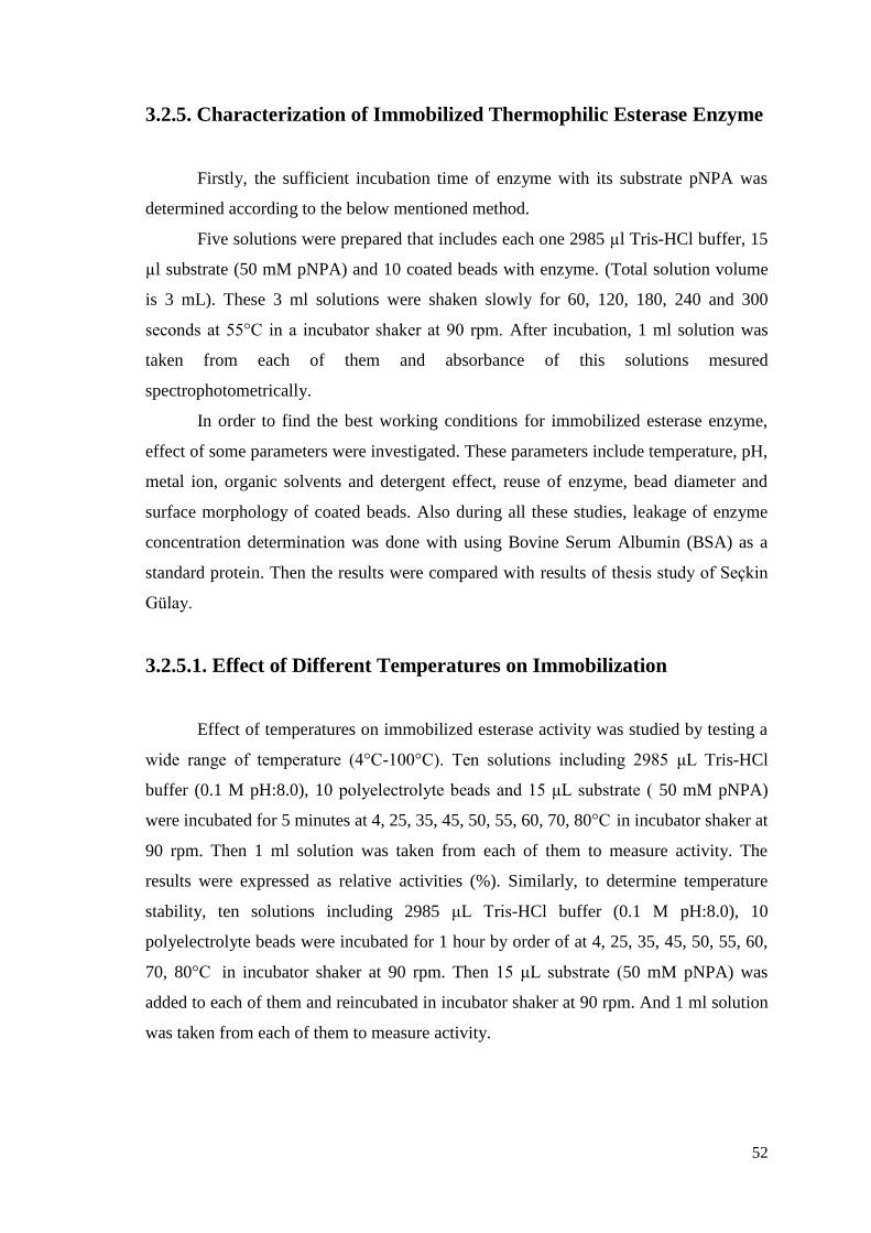
52
3.2.5. Characterization of Immobilized Thermophilic Esterase Enzyme
Firstly, the sufficient incubation time of enzyme with its substrate pNPA was
determined according to the below mentioned method.
Five solutions were prepared that includes each one 2985 µl Tris-HCl buffer, 15
µl substrate (50 mM pNPA) and 10 coated beads with enzyme. (Total solution volume
is 3 mL). These 3 ml solutions were shaken slowly for 60, 120, 180, 240 and 300
seconds at 55°C in a incubator shaker at 90 rpm. After incubation, 1 ml solution was
taken from each of them and absorbance of this solutions mesured
spectrophotometrically.
In order to find the best working conditions for immobilized esterase enzyme,
effect of some parameters were investigated. These parameters include temperature, pH,
metal ion, organic solvents and detergent effect, reuse of enzyme, bead diameter and
surface morphology of coated beads. Also during all these studies, leakage of enzyme
concentration determination was done with using Bovine Serum Albumin (BSA) as a
standard protein. Then the results were compared with results of thesis study of Seçkin
Gülay.
3.2.5.1. Effect of Different Temperatures on Immobilization
Effect of temperatures on immobilized esterase activity was studied by testing a
wide range of temperature (4°C-100°C). Ten solutions including 2985 μL Tris-HCl
buffer (0.1 M pH:8.0), 10 polyelectrolyte beads and 15 μL substrate ( 50 mM pNPA)
were incubated for 5 minutes at 4, 25, 35, 45, 50, 55, 60, 70, 80°C in incubator shaker at
90 rpm. Then 1 ml solution was taken from each of them to measure activity. The
results were expressed as relative activities (%). Similarly, to determine temperature
stability, ten solutions including 2985 μL Tris-HCl buffer (0.1 M pH:8.0), 10
polyelectrolyte beads were incubated for 1 hour by order of at 4, 25, 35, 45, 50, 55, 60,
70, 80°C in incubator shaker at 90 rpm. Then 15 μL substrate (50 mM pNPA) was
added to each of them and reincubated in incubator shaker at 90 rpm. And 1 ml solution
was taken from each of them to measure activity.
53
3.2.5.2. Effect of Different pH Values on Immobilization
Secondly to find effect of pH on immobilized esterase activity was studied by
testing a wide range of pH values (4, 5, 6, 7, 7.5, 8, 8.5, 9, 10 and 11). 10
polyelectrolyte beads and 15 μL substrate (50 mM pNPA) were incubated in 4, 5, 6, 7,
7.5, 8, 8.5, 9, 10 and 11 pH Tris-HCl buffer (0.1M) for 5 minutes in incubator shaker at
55°C at 90 rpm. Then 1 ml solution was taken from each of them to measure activity.
Determination of pH stability for 1 hour was investigated among pH values of 4, 5, 6, 7,
7.5, 8, 8.5, 9, 10 and 11. And after waiting 1 hour in different pH values, 15 µl substrate
(pNPA) was added and solution was shaken for 5 minutes more. Then 1 ml solution was
taken from each of them to measure activity.
3.2.5.3. Effect of Chemicals on Immobilized Esterase
To investigate effect of different chemicals on enzyme activity CaCl2, ZnCl2,
MgCl2, CuSO4, MgSO4, SDS, Triton X-100 were choosen. The experiment was done
with the incubation of 10 coated beads in 2000 µl pH: 8.0 Tris-HCl buffer (0.1M)
includes 985 µl chemical (1mM) at 55°C for 5 minutes with 15 µl substrate (50 mM
pNPA). Then 1 ml solution was taken from each of them to observe activity.
3.2.5.4. Effect of Reuse of Immobilized Esterase
Such as general enzyme activity method, firstly 10 polyelectrolyte beads were
incubated with 2985 µl Tris-HCl buffer and15 μL substrate ( 50 mM pNPA) for 5
minutes at 55°C in incubator shaker at 90 rpm. Then 1 ml solution was taken from each
of them to measure activity. After absorbance measurement, ten polyelectrolyte beads
washed three times with deionized water and lastly 2985 µl Tris-HCl buffer. And this
experiment was replicated 6 times more.
54
3.2.5.5. Effect of Bead Diameter
Alginate beads of three different sizes were generated by changing the size of a
needle. The diameter of beads was determined with the formula as shown below.
Increment in Volume = 4/3 π r3
Number of beads (3.4)
3.2.5.6. Scanning Electron Microscope (SEM)
The surface morphology and internal structure of the beads are examined using a
scanning electron microscope (Phillips XL-30S FEG). Polyelectrolyte beads were dried
for 1 hour at 50oC to get rid of wetness. Then, samples were mounted on carbon stubs,
and gold coated under vacuum, and then examined.
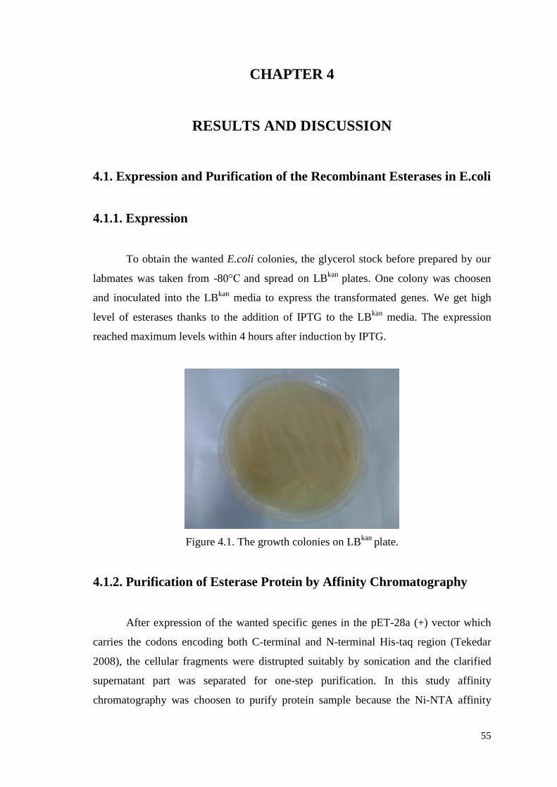
55
CHAPTER 4
RESULTS AND DISCUSSION
4.1. Expression and Purification of the Recombinant Esterases in E.coli
4.1.1. Expression
To obtain the wanted E.coli colonies, the glycerol stock before prepared by our
labmates was taken from -80°C and spread on LBkan
plates. One colony was choosen
and inoculated into the LBkan
media to express the transformated genes. We get high
level of esterases thanks to the addition of IPTG to the LBkan
media. The expression
reached maximum levels within 4 hours after induction by IPTG.
Figure 4.1. The growth colonies on LBkan
plate.
4.1.2. Purification of Esterase Protein by Affinity Chromatography
After expression of the wanted specific genes in the pET-28a (+) vector which
carries the codons encoding both C-terminal and N-terminal His-taq region (Tekedar
2008), the cellular fragments were distrupted suitably by sonication and the clarified
supernatant part was separated for one-step purification. In this study affinity
chromatography was choosen to purify protein sample because the Ni-NTA affinity
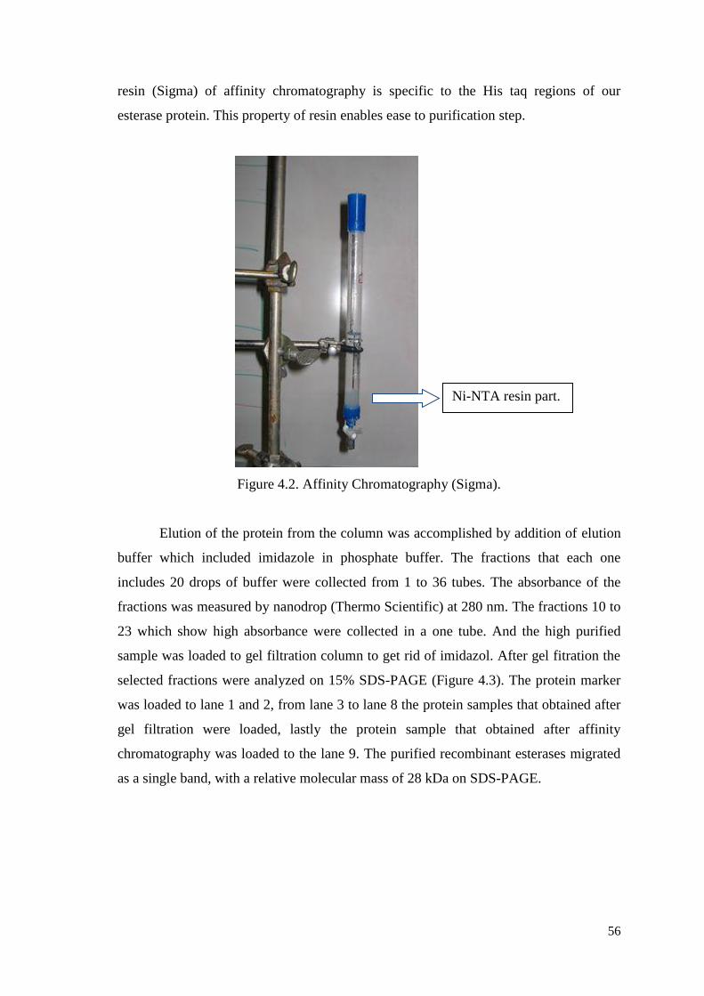
56
resin (Sigma) of affinity chromatography is specific to the His taq regions of our
esterase protein. This property of resin enables ease to purification step.
Figure 4.2. Affinity Chromatography (Sigma).
Elution of the protein from the column was accomplished by addition of elution
buffer which included imidazole in phosphate buffer. The fractions that each one
includes 20 drops of buffer were collected from 1 to 36 tubes. The absorbance of the
fractions was measured by nanodrop (Thermo Scientific) at 280 nm. The fractions 10 to
23 which show high absorbance were collected in a one tube. And the high purified
sample was loaded to gel filtration column to get rid of imidazol. After gel fitration the
selected fractions were analyzed on 15% SDS-PAGE (Figure 4.3). The protein marker
was loaded to lane 1 and 2, from lane 3 to lane 8 the protein samples that obtained after
gel filtration were loaded, lastly the protein sample that obtained after affinity
chromatography was loaded to the lane 9. The purified recombinant esterases migrated
as a single band, with a relative molecular mass of 28 kDa on SDS-PAGE.
Ni-NTA resin part.
57
Figure 4.3. 15% SDS-PAGE analysis of selected fractions. (M: molecular mass markers
from the top to bottom 200, 116, 68, 43, 29, 14.4 and 6.5 kDa. 19, 25, 30,
36, 42, 45 (Fractions after size exclusion step), A.A (fraction after affinity)
selected fractions.)
One-step purification of the esterase proteins using Ni-NTA affinity
chromatography has resulted in efficient purification to continue our studies with that
much pure enzyme. Size-exclusion chromatography was applied after affinity column
against phosphate buffer (pH 7.0) to get rid of imidazole from esterase enzyme. After
size-exclusion chromatography, enzyme was aliquoted in to eppendorf tubes as 1 ml
volume and kept it at -20oC until use.
4.2. Immobilization of Thermophilic Esterase Enzyme in Alginate-
Chitosan/CaCl2 Polyelectrolyte Beads
4.2.1. Immobilization Yield
In order to calculate immobilization yield, the total enzyme leakage was
measured in filtrate and the crosslinking solution that composed of Chitosan/CaCl2. The
initial concentration of esterase was 0.5 mg/ml and 0.098 mg/ml esterase enzyme has
been determined from washing steps. As a result of this, immobilization yield was
calculated as 80.40 %.
4.3. Characterization of Immobilized Thermophilic Esterase Enzyme
During characterization studies, we investigated effects of different parameters
such as pH, temperature, chemical agent, bead size on enzyme acvitiy, also we
M M 19 25 30 36 42 45 A.A.
28kDa
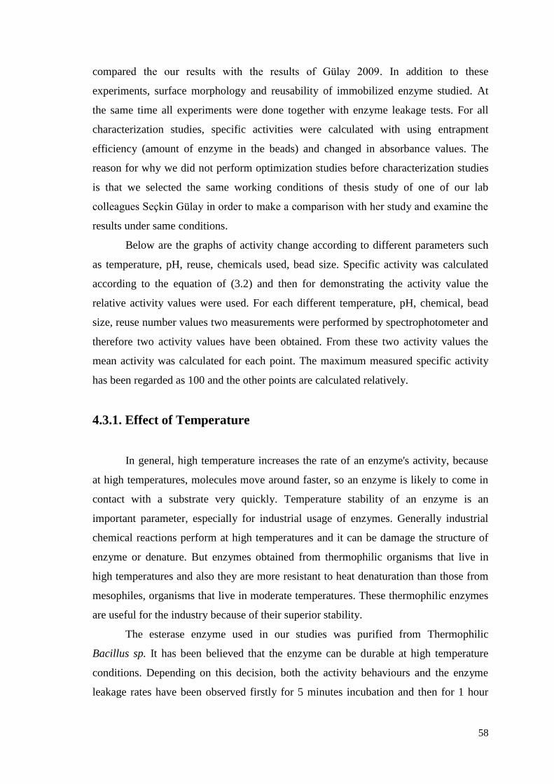
58
compared the our results with the results of Gülay 2009. In addition to these
experiments, surface morphology and reusability of immobilized enzyme studied. At
the same time all experiments were done together with enzyme leakage tests. For all
characterization studies, specific activities were calculated with using entrapment
efficiency (amount of enzyme in the beads) and changed in absorbance values. The
reason for why we did not perform optimization studies before characterization studies
is that we selected the same working conditions of thesis study of one of our lab
colleagues Seçkin Gülay in order to make a comparison with her study and examine the
results under same conditions.
Below are the graphs of activity change according to different parameters such
as temperature, pH, reuse, chemicals used, bead size. Specific activity was calculated
according to the equation of (3.2) and then for demonstrating the activity value the
relative activity values were used. For each different temperature, pH, chemical, bead
size, reuse number values two measurements were performed by spectrophotometer and
therefore two activity values have been obtained. From these two activity values the
mean activity was calculated for each point. The maximum measured specific activity
has been regarded as 100 and the other points are calculated relatively.
4.3.1. Effect of Temperature
In general, high temperature increases the rate of an enzyme's activity, because
at high temperatures, molecules move around faster, so an enzyme is likely to come in
contact with a substrate very quickly. Temperature stability of an enzyme is an
important parameter, especially for industrial usage of enzymes. Generally industrial
chemical reactions perform at high temperatures and it can be damage the structure of
enzyme or denature. But enzymes obtained from thermophilic organisms that live in
high temperatures and also they are more resistant to heat denaturation than those from
mesophiles, organisms that live in moderate temperatures. These thermophilic enzymes
are useful for the industry because of their superior stability.
The esterase enzyme used in our studies was purified from Thermophilic
Bacillus sp. It has been believed that the enzyme can be durable at high temperature
conditions. Depending on this decision, both the activity behaviours and the enzyme
leakage rates have been observed firstly for 5 minutes incubation and then for 1 hour
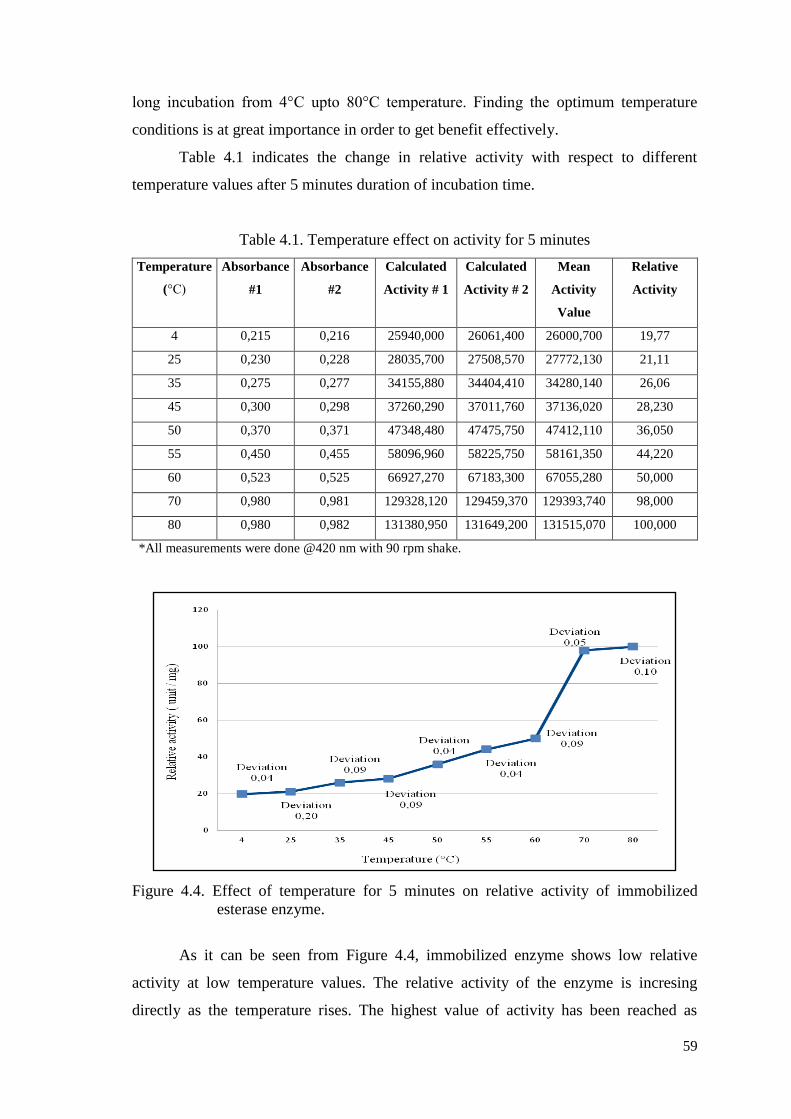
59
long incubation from 4°C upto 80°C temperature. Finding the optimum temperature
conditions is at great importance in order to get benefit effectively.
Table 4.1 indicates the change in relative activity with respect to different
temperature values after 5 minutes duration of incubation time.
Table 4.1. Temperature effect on activity for 5 minutes
Temperature
(°C)
Absorbance
#1
Absorbance
#2
Calculated
Activity # 1
Calculated
Activity # 2
Mean
Activity
Value
Relative
Activity
4 0,215 0,216 25940,000 26061,400 26000,700 19,77
25 0,230 0,228 28035,700 27508,570 27772,130 21,11
35 0,275 0,277 34155,880 34404,410 34280,140 26,06
45 0,300 0,298 37260,290 37011,760 37136,020 28,230
50 0,370 0,371 47348,480 47475,750 47412,110 36,050
55 0,450 0,455 58096,960 58225,750 58161,350 44,220
60 0,523 0,525 66927,270 67183,300 67055,280 50,000
70 0,980 0,981 129328,120 129459,370 129393,740 98,000
80 0,980 0,982 131380,950 131649,200 131515,070 100,000
*All measurements were done @420 nm with 90 rpm shake.
Figure 4.4. Effect of temperature for 5 minutes on relative activity of immobilized
esterase enzyme.
As it can be seen from Figure 4.4, immobilized enzyme shows low relative
activity at low temperature values. The relative activity of the enzyme is incresing
directly as the temperature rises. The highest value of activity has been reached as
60
temperature is 80°C. In Gülay’s study, for 5 minutes, maximum specific activity was
observed at 45°C. So we can easily say that our new formed alginate/chitosan
membrane is more stable from alginate/calcium chloride matrix.
4.3.1.1. Entrapment Efficiency for 5 Minutes
In addition, after 5 minutes of experiment the remaining enzyme amount in the
beads were calculated for each temperature values. Those amounts in per cent are given
in Table 4.2.
Table 4.2. Percent amount of enzyme in beads during pH effect test for 5 minutes
Temperature (°C) % amount of enzyme in beads
4 87,5
25 87,5
35 85
45 85
50 82,5
55 82,5
60 82,5
70 80
80 78,75
According to the values in Table 4.2 the graph in Figure 4.5 shows the amounts
of enzyme in beads as percent for each measure temperature values.
Figure 4.5. Entrapment efficiency at different temperatures for 5 minutes
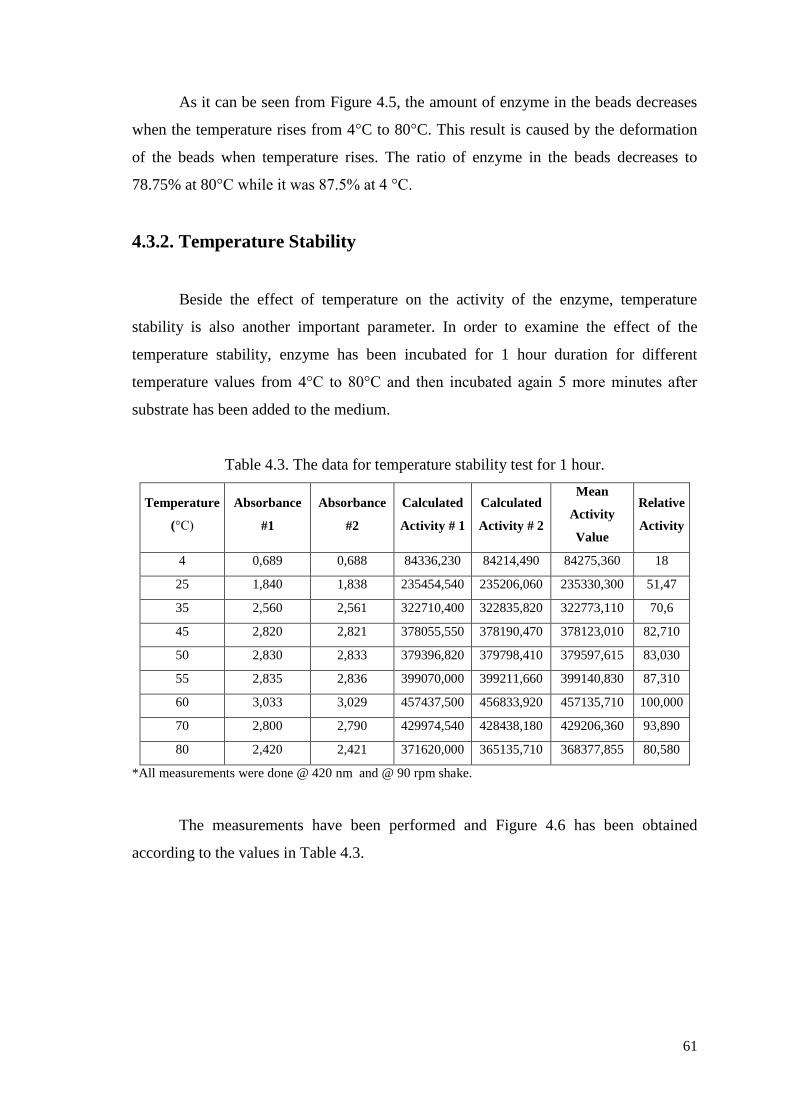
61
As it can be seen from Figure 4.5, the amount of enzyme in the beads decreases
when the temperature rises from 4°C to 80°C. This result is caused by the deformation
of the beads when temperature rises. The ratio of enzyme in the beads decreases to
78.75% at 80°C while it was 87.5% at 4 °C.
4.3.2. Temperature Stability
Beside the effect of temperature on the activity of the enzyme, temperature
stability is also another important parameter. In order to examine the effect of the
temperature stability, enzyme has been incubated for 1 hour duration for different
temperature values from 4°C to 80°C and then incubated again 5 more minutes after
substrate has been added to the medium.
Table 4.3. The data for temperature stability test for 1 hour.
Temperature
(°C)
Absorbance
#1
Absorbance
#2
Calculated
Activity # 1
Calculated
Activity # 2
Mean
Activity
Value
Relative
Activity
4 0,689 0,688 84336,230 84214,490 84275,360 18
25 1,840 1,838 235454,540 235206,060 235330,300 51,47
35 2,560 2,561 322710,400 322835,820 322773,110 70,6
45 2,820 2,821 378055,550 378190,470 378123,010 82,710
50 2,830 2,833 379396,820 379798,410 379597,615 83,030
55 2,835 2,836 399070,000 399211,660 399140,830 87,310
60 3,033 3,029 457437,500 456833,920 457135,710 100,000
70 2,800 2,790 429974,540 428438,180 429206,360 93,890
80 2,420 2,421 371620,000 365135,710 368377,855 80,580
*All measurements were done @ 420 nm and @ 90 rpm shake.
The measurements have been performed and Figure 4.6 has been obtained
according to the values in Table 4.3.
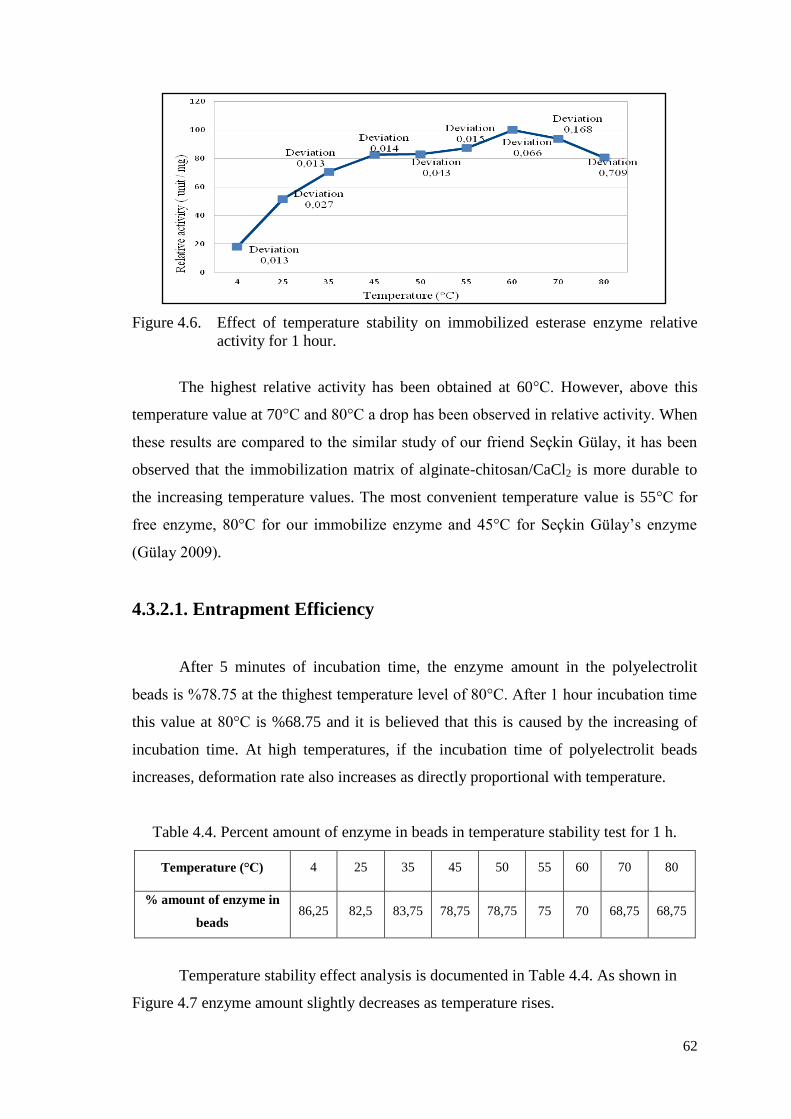
62
Figure 4.6. Effect of temperature stability on immobilized esterase enzyme relative
activity for 1 hour.
The highest relative activity has been obtained at 60°C. However, above this
temperature value at 70°C and 80°C a drop has been observed in relative activity. When
these results are compared to the similar study of our friend Seçkin Gülay, it has been
observed that the immobilization matrix of alginate-chitosan/CaCl2 is more durable to
the increasing temperature values. The most convenient temperature value is 55°C for
free enzyme, 80°C for our immobilize enzyme and 45°C for Seçkin Gülay’s enzyme
(Gülay 2009).
4.3.2.1. Entrapment Efficiency
After 5 minutes of incubation time, the enzyme amount in the polyelectrolit
beads is %78.75 at the thighest temperature level of 80°C. After 1 hour incubation time
this value at 80°C is %68.75 and it is believed that this is caused by the increasing of
incubation time. At high temperatures, if the incubation time of polyelectrolit beads
increases, deformation rate also increases as directly proportional with temperature.
Table 4.4. Percent amount of enzyme in beads in temperature stability test for 1 h.
Temperature (°C) 4 25 35 45 50 55 60 70 80
% amount of enzyme in
beads 86,25 82,5 83,75 78,75 78,75 75 70 68,75 68,75
Temperature stability effect analysis is documented in Table 4.4. As shown in
Figure 4.7 enzyme amount slightly decreases as temperature rises.
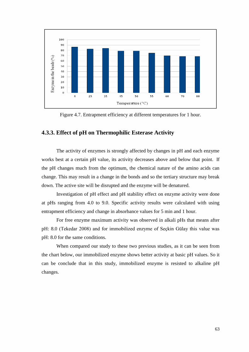
63
Figure 4.7. Entrapment efficiency at different temperatures for 1 hour.
4.3.3. Effect of pH on Thermophilic Esterase Activity
The activity of enzymes is strongly affected by changes in pH and each enzyme
works best at a certain pH value, its activity decreases above and below that point. If
the pH changes much from the optimum, the chemical nature of the amino acids can
change. This may result in a change in the bonds and so the tertiary structure may break
down. The active site will be disrupted and the enzyme will be denatured.
Investigation of pH effect and pH stability effect on enzyme activity were done
at pHs ranging from 4.0 to 9.0. Specific activity results were calculated with using
entrapment efficiency and change in absorbance values for 5 min and 1 hour.
For free enzyme maximum activity was observed in alkali pHs that means after
pH: 8.0 (Tekedar 2008) and for immobilized enzyme of Seçkin Gülay this value was
pH: 8.0 for the same conditions.
When compared our study to these two previous studies, as it can be seen from
the chart below, our immobilized enzyme shows better activity at basic pH values. So it
can be conclude that in this study, immobilized enzyme is resisted to alkaline pH
changes.
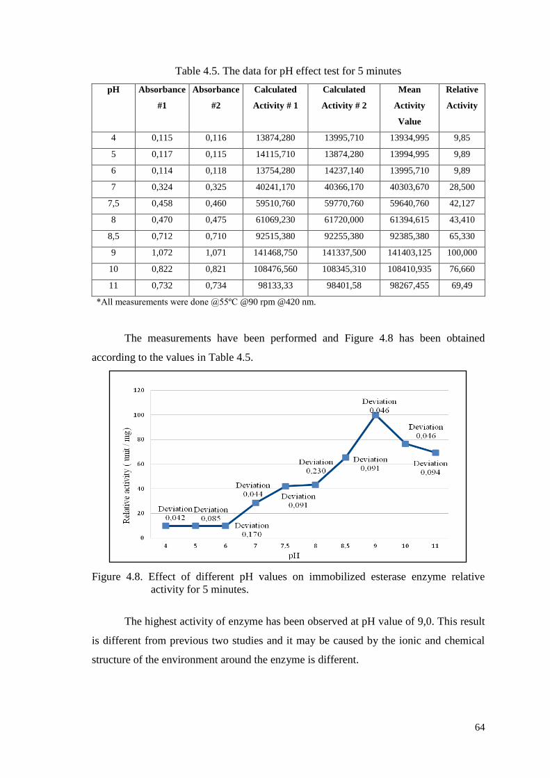
64
Table 4.5. The data for pH effect test for 5 minutes
pH Absorbance
#1
Absorbance
#2
Calculated
Activity # 1
Calculated
Activity # 2
Mean
Activity
Value
Relative
Activity
4 0,115 0,116 13874,280 13995,710 13934,995 9,85
5 0,117 0,115 14115,710 13874,280 13994,995 9,89
6 0,114 0,118 13754,280 14237,140 13995,710 9,89
7 0,324 0,325 40241,170 40366,170 40303,670 28,500
7,5 0,458 0,460 59510,760 59770,760 59640,760 42,127
8 0,470 0,475 61069,230 61720,000 61394,615 43,410
8,5 0,712 0,710 92515,380 92255,380 92385,380 65,330
9 1,072 1,071 141468,750 141337,500 141403,125 100,000
10 0,822 0,821 108476,560 108345,310 108410,935 76,660
11 0,732 0,734 98133,33 98401,58 98267,455 69,49
*All measurements were done @55ºC @90 rpm @420 nm.
The measurements have been performed and Figure 4.8 has been obtained
according to the values in Table 4.5.
Figure 4.8. Effect of different pH values on immobilized esterase enzyme relative
activity for 5 minutes.
The highest activity of enzyme has been observed at pH value of 9,0. This result
is different from previous two studies and it may be caused by the ionic and chemical
structure of the environment around the enzyme is different.
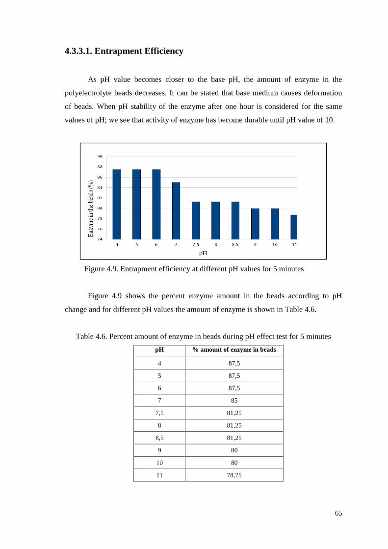
65
4.3.3.1. Entrapment Efficiency
As pH value becomes closer to the base pH, the amount of enzyme in the
polyelectrolyte beads decreases. It can be stated that base medium causes deformation
of beads. When pH stability of the enzyme after one hour is considered for the same
values of pH; we see that activity of enzyme has become durable until pH value of 10.
Figure 4.9. Entrapment efficiency at different pH values for 5 minutes
Figure 4.9 shows the percent enzyme amount in the beads according to pH
change and for different pH values the amount of enzyme is shown in Table 4.6.
Table 4.6. Percent amount of enzyme in beads during pH effect test for 5 minutes
pH % amount of enzyme in beads
4 87,5
5 87,5
6 87,5
7 85
7,5 81,25
8 81,25
8,5 81,25
9 80
10 80
11 78,75
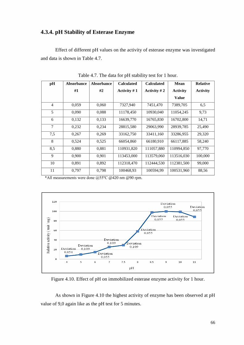
66
4.3.4. pH Stability of Esterase Enzyme
Effect of different pH values on the activity of esterase enzyme was investigated
and data is shown in Table 4.7.
Table 4.7. The data for pH stability test for 1 hour.
pH Absorbance
#1
Absorbance
#2
Calculated
Activity # 1
Calculated
Activity # 2
Mean
Activity
Value
Relative
Activity
4 0,059 0,060 7327,940 7451,470 7389,705 6,5
5 0,090 0,088 11178,450 10930,040 11054,245 9,73
6 0,132 0,133 16639,770 16765,830 16702,800 14,71
7 0,232 0,234 28815,580 29063,990 28939,785 25,490
7,5 0,267 0,269 33162,750 33411,160 33286,955 29,320
8 0,524 0,525 66054,860 66180,910 66117,885 58,240
8,5 0,880 0,881 110931,820 111057,880 110994,850 97,770
9 0,900 0,901 113453,000 113579,060 113516,030 100,000
10 0,891 0,892 112318,470 112444,530 112381,500 99,000
11 0,797 0,798 100468,93 100594,99 100531,960 88,56
*All measurements were done @55ºC @420 nm @90 rpm.
Figure 4.10. Effect of pH on immobilized esterase enzyme activity for 1 hour.
As shown in Figure 4.10 the highest activity of enzyme has been observed at pH
value of 9,0 again like as the pH test for 5 minutes.

67
4.3.4.1. Entrapment Efficiency
Determination of pH stability for 1 hour was investigated among the pH of: 4.0-
6.0-7.0-7.5-8.0-8.5-9.0-10.0 and 11.0. After incubating immobilized enzyme for one
hour in different pH values, half of specific activity was still protected at pH: 10.0.
Figure 4.11. Entrapment efficiency at different pH values for 1 hour.
As a result, immobilized esterase enzyme for one hour was still displaying
maximum activity at pH: 10.0. Specific activity of immobilized esterase enzyme versus
pH values were shown in Figure 4.10 and entrapment efficiency of enzyme were shown
in Figure 4.11 after one hour incubation time.
Table 4.8. Percent amount of enzyme in beads during pH stability test for 1 hour.
pH % amount of enzyme in beads
4 85
5 85
6 83,75
7 85
7,5 85
8 83,75
8,5 83,75
9 83,75
10 82,5
11 83,75
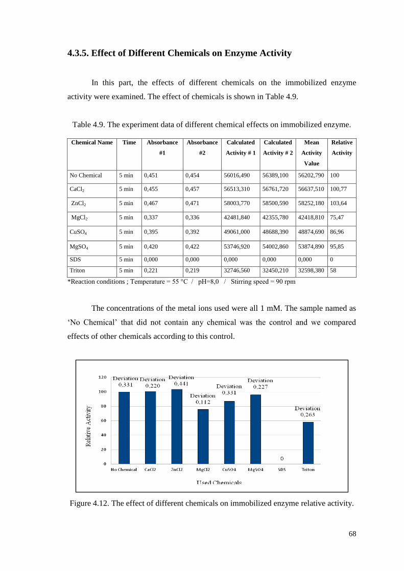
68
4.3.5. Effect of Different Chemicals on Enzyme Activity
In this part, the effects of different chemicals on the immobilized enzyme
activity were examined. The effect of chemicals is shown in Table 4.9.
Table 4.9. The experiment data of different chemical effects on immobilized enzyme.
*Reaction conditions ; Temperature = 55 °C / pH=8,0 / Stirring speed = 90 rpm
The concentrations of the metal ions used were all 1 mM. The sample named as
‘No Chemical’ that did not contain any chemical was the control and we compared
effects of other chemicals according to this control.
Figure 4.12. The effect of different chemicals on immobilized enzyme relative activity.
Chemical Name Time Absorbance
#1
Absorbance
#2
Calculated
Activity # 1
Calculated
Activity # 2
Mean
Activity
Value
Relative
Activity
No Chemical 5 min 0,451 0,454 56016,490 56389,100 56202,790 100
CaCl2 5 min 0,455 0,457 56513,310 56761,720 56637,510 100,77
ZnCl2 5 min 0,467 0,471 58003,770 58500,590 58252,180 103,64
MgCl2 5 min 0,337 0,336 42481,840 42355,780 42418,810 75,47
CuSO4 5 min 0,395 0,392 49061,000 48688,390 48874,690 86,96
MgSO4 5 min 0,420 0,422 53746,920 54002,860 53874,890 95,85
SDS 5 min 0,000 0,000 0,000 0,000 0,000 0
Triton 5 min 0,221 0,219 32746,560 32450,210 32598,380 58
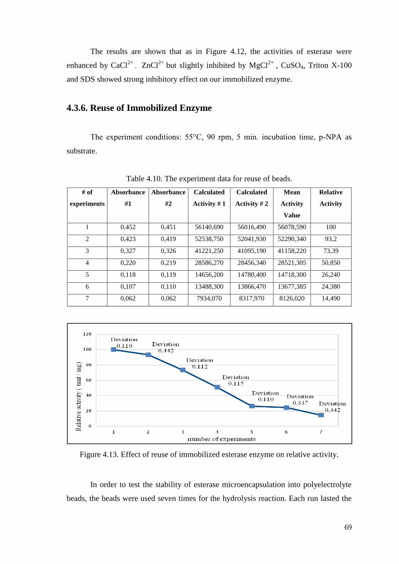
69
The results are shown that as in Figure 4.12, the activities of esterase were
enhanced by CaCl2+ , ZnCl
2+ but slightly inhibited by MgCl
2+ , CuSO4, Triton X-100
and SDS showed strong inhibitory effect on our immobilized enzyme.
4.3.6. Reuse of Immobilized Enzyme
The experiment conditions: 55°C, 90 rpm, 5 min. incubation time, p-NPA as
substrate.
Table 4.10. The experiment data for reuse of beads.
# of
experiments
Absorbance
#1
Absorbance
#2
Calculated
Activity # 1
Calculated
Activity # 2
Mean
Activity
Value
Relative
Activity
1 0,452 0,451 56140,690 56016,490 56078,590 100
2 0,423 0,419 52538,750 52041,930 52290,340 93,2
3 0,327 0,326 41221,250 41095,190 41158,220 73,39
4 0,220 0,219 28586,270 28456,340 28521,305 50,850
5 0,118 0,119 14656,200 14780,400 14718,300 26,240
6 0,107 0,110 13488,300 13866,470 13677,385 24,380
7 0,062 0,062 7934,070 8317,970 8126,020 14,490
Figure 4.13. Effect of reuse of immobilized esterase enzyme on relative activity.
In order to test the stability of esterase microencapsulation into polyelectrolyte
beads, the beads were used seven times for the hydrolysis reaction. Each run lasted the
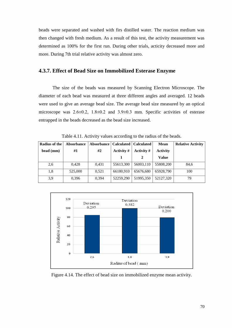
70
beads were separated and washed with firs distilled water. The reaction medium was
then changed with fresh medium. As a result of this test, the activity measurement was
determined as 100% for the first run. During other trials, acticity decreased more and
more. During 7th trial relative activity was almost zero.
4.3.7. Effect of Bead Size on Immobilized Esterase Enzyme
The size of the beads was measured by Scanning Electron Microscope. The
diameter of each bead was measured at three different angles and averaged. 12 beads
were used to give an average bead size. The average bead size measured by an optical
microscope was 2.6±0.2, 1.8±0.2 and 3.9±0.3 mm. Specific activities of esterase
entrapped in the beads decreased as the bead size increased.
Table 4.11. Activity values according to the radius of the beads.
Radius of the
bead (mm)
Absorbance
#1
Absorbance
#2
Calculated
Activity #
1
Calculated
Activity #
2
Mean
Activity
Value
Relative Activity
2,6 0,428 0,431 55613,300 56003,110 55808,200 84,6
1,8 525,000 0,521 66180,910 65676,680 65928,790 100
3,9 0,396 0,394 52259,290 51995,350 52127,320 79
Figure 4.14. The effect of bead size on immobilized enzyme mean activity.
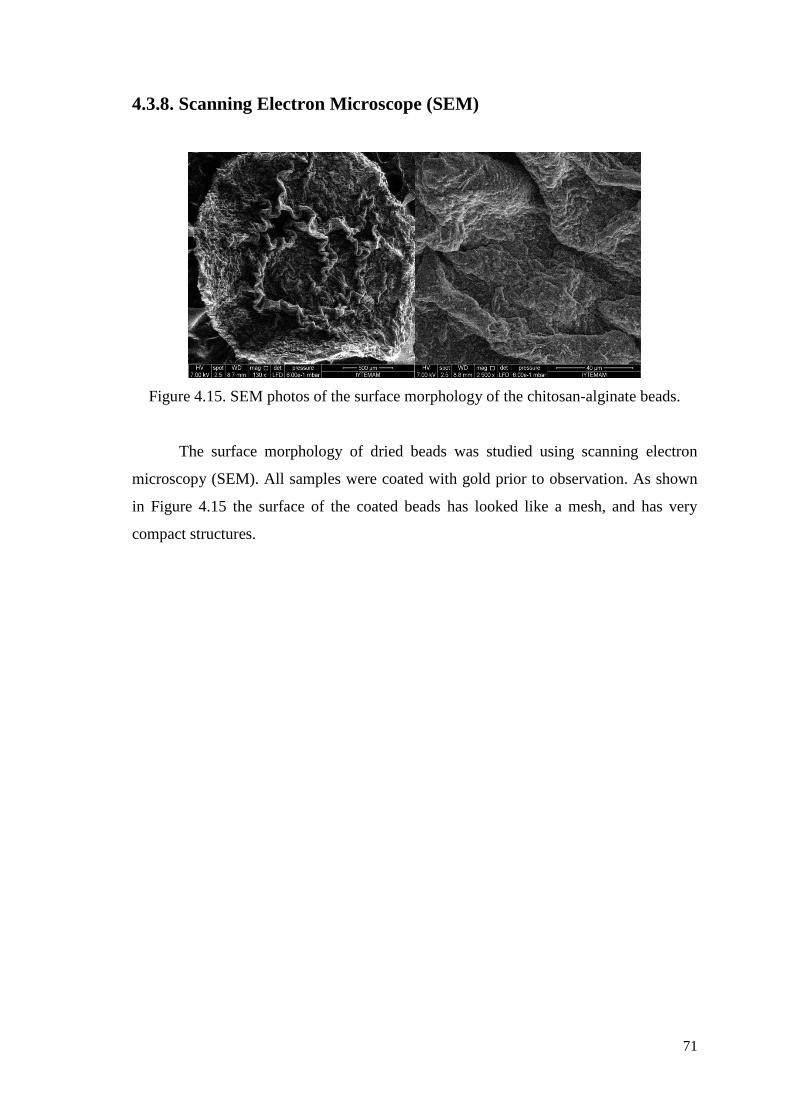
71
4.3.8. Scanning Electron Microscope (SEM)
Figure 4.15. SEM photos of the surface morphology of the chitosan-alginate beads.
The surface morphology of dried beads was studied using scanning electron
microscopy (SEM). All samples were coated with gold prior to observation. As shown
in Figure 4.15 the surface of the coated beads has looked like a mesh, and has very
compact structures.
72
CHAPTER 5
CONCLUSION
In this study, firstly the recombinant thermophilic esterase enzyme expressed
and purified from E.Coli strain. Purification of esterase protein was performed via one-
step affinity chromatography method. Then during immobilization procedure, purified
protein successfully microencapsulated into the Alginate-Chitosan/CaCl2
polyelectrolyte beads. Immobilization yield was 80.40%. After purification, the
characterization studies were done such as effect of temperature and pH, temperature
and pH stability, effect of bead size on immobilized enzyme, effect of chemical agents;
re-use of immobilized enzyme and lastly SEM (Scanning Electron Microscopy)
analyses of beads. Our experiments have shown that the esterase immobilized alginate-
chitosan/CaCl2 polyelectrolyte beads exhibits an improved resistance against thermal
and pH denaturation when compared with Gülay’s immobilized beads (Gülay 2009).
According to the pH stability results the optimum pH of esterase enyzme was
investigated that relatively alkaline pH values. Also especially we can say that SDS
shows strong inhibitory effect on activity of our immobilized esterase. And this
temparature and pH improvement enables applicability for use this enzyme in industrial
processes and other further laboratory studies.
73
REFERENCES
Aksoy, C. 2003. Lipaz ve Üreaz Enzimlerinin Çesitli Tasıyıcılara İmmobilizasyonu.
University of Istanbul, Msc. Thesis
Argüelles-Monal, W., Hechavarria, O. L., Rodriguez, L. and Peniche, C. 1993. Swelling
of membranes from the polyelectrolyte complex between chitosan and
carboxymethyl cellulose. Polymer Bulletin 31: 471-478.
Arıca, Y. M. and Hasırcı, V. N. 1987. Immobilization for the production of membranes.
Biomaterials 8: 489-495.
Baker, G. C., Gaffar, S., Cowan, D. A. and Suharto, A. R. 2001. Bacterial community
analysis of Indonesian hot springs. FEMS Microbiology Lett. 200:103-109
Bansode, S. S., Banarjee, S. K., Gaikwad, D. D., Jadhav, S. L. and Thorat, R. M. 2010.
Microencapsulation: a review. International Journal of Pharmaceutical Sciences
Review and Research 1, issue 2, p. 38.
Berger, J., Reist, M., Mayer, J. M., Felt, O. and Gurny, R. 2004. Structure and
interactions in chitosan hydrogels formed by complexation or aggregation for
biomedical applications. Europian Journal of Pharmaceutics and
Biopharmaceutics 57: 35-52.
Bilen, Ç. 2009. Immobilization with glutaraldehyde of paraoxonase enzyme and
investigation of some heavy metals effects onto the enzyme affinity. Balıkesir
University, Institute of Science, Department of Chemistry M.Sc.Thesis.
Bornscheuer, U. T. 2002. Microbial carboxylesterases: classification, properties and
application in biocatalyses. FEMS Microbial Rev. 26: 73-81.
Boyacı, E. 2008. Sorption of As(V) From Waters by Use of Novel Amine-Containing
Sorbents Prior to HGAAS and ICP-MS Determination. M.Sc.Thesis. İzmir
Institute of Technology.
Bready, D. and Jordaan, J. 2009. Advances in enzyme immobilization. Biotechnol. Lett.
31: 1639-1650
Brena, B. M. and Batista-Viera, F. 2008. Immobilization of Enzymes. In: Methods in
Biotechnology: Immobilization of Enzymes and Cells, edited by Gordon
Bickerstaff, Totowa, New Jersey: Humana Press.
Carey, F. A. and Sundberg, R. J. 2007. Advanced Organic Chemistry, Part: A Structure
and Mechanisms. Fifth Edition, Springer.
Carr, P. W. and Bowers, L. D. 1980. Support considerations in chemical analysis.
Enzymes, Academic Press, New York, 56: 167-170.
74
Chaabouni, M. K., Pulvin, S., Touraud, D. and Thomas, D. 1996. Enzymatic synthesis
of geraniol esters in a solvent free system by lipases. Biotechnol Lett. 18: 1083-
1088.
Chavasit, V., Kienzle-Sterzer, C. A. and Torres, J. A. 1988. Formation and
characterization of insoluble polyelectrolyte complex: chitosan-poly(acrylic acid).
Polymer Bulletin 19: 223-230.
Chen, S., Liu, Y. and Yu, P. 1996. Study on column reactor of chitosan immobilized.
Chem. Abstr. 127 (4): 127-129.
Choi, Y. J. and Lee, B. H. 2001. Culture conditions for the production of esterase from
Lactobacillus casei CL 96. Bioprocess Biosyst Eng. 24: 59-63.
Christakopoulos, P., Tzalas, B., Mamma, D., Stamatis, H., Liadakis, G. N., Tzia, C.,
Kekos, D., Kolisis, F. N. and Macris, B. J. 1998. Production of esterases from
Fusarium oxysporum catalyzing transesterification reactions in organic solvents.
Process Biochem. 33: 729-733.
Clark, A. H. and Ross-Murphy, S. B. 1987. Advances in Polymer Science 83: 57–192.
Costa, S. A., Azevedo, Helena S. and Reis, R. L. 2004. Enzyme immobilization in
biodegradable polymers for biomedical applications. Biodegradable Systems in
Tissue Engineering and Regenerative Medicine 1(4): 301 – 324.
Dufour, J. P. and Bing, Y. 2001. Influence of yeast strain and fermentation conditions
on yeast esterase activities. Brew Dig. 76: 44.
Dumitriu, S., Popa, M. and Dumitriu, M. 1998. Polymeric biomaterials as enzyme and
drug carriers. J. Bioactive and Compatible Polymers. 3: 243-312.
Dutta, P. K., Dutta, J. and Tripathi, V. S. 2004. Chitin and chitosan: Chemistry,
properties and applications. Journal of Scientific & Industrial Research 63: 20-31.
Finer, Y., Jaffer, F. and Santerre, J. P. 2004. Mutual influence of cholesterol esterase
and pseudocholinesterase on the biodegradation of dental composites.
Biomaterials 25: 1787-1793.
Fujiwara, S. 2002. Extremophiles: Developments of their special functions and potential
resource. Journal of Bioscience and Bioengineering 94, 6: 518-52.
Glick, D. 1979. Methods of Biochemical Analysis, Enzyme Immobilization. Academic
Press, New York, 25: 135-201.
Gombotz, W.R. and Wee, S. F. 1998. Protein release from alginate matrices. Adv. Drug
Deliv. Rev. 31: 267 –285.
Gomes, J. and Steiner, W. 2004. Extremophiles and extremozymes. Food Technol.
Biotechnol. 42 (4) 223–235.
Guisan, M. J. 2006. Immobilization of Enzymes and Cells, Second Edition. Institute of
Catalysis, CSIC
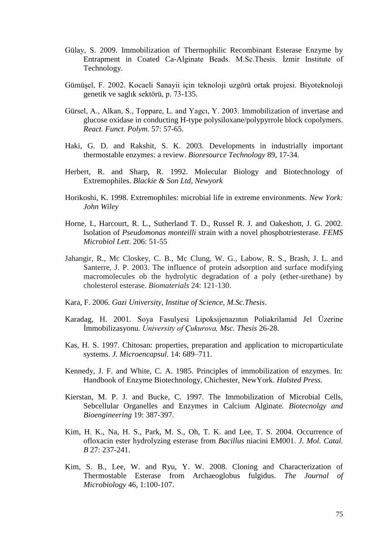
75
Gülay, S. 2009. Immobilization of Thermophilic Recombinant Esterase Enzyme by
Entrapment in Coated Ca-Alginate Beads. M.Sc.Thesis. İzmir Institute of
Technology.
Gümüşel, F. 2002. Kocaeli Sanayii için teknoloji uzgörü ortak projesi. Biyoteknoloji
genetik ve saglık sektörü, p. 73-135.
Gürsel, A., Alkan, S., Toppare, L. and Yagcı, Y. 2003. Immobilization of invertase and
glucose oxidase in conducting H-type polysiloxane/polypyrrole block copolymers.
React. Funct. Polym. 57: 57-65.
Haki, G. D. and Rakshit, S. K. 2003. Developments in industrially important
thermostable enzymes: a review. Bioresource Technology 89, 17-34.
Herbert, R. and Sharp, R. 1992. Molecular Biology and Biotechnology of
Extremophiles. Blackie & Son Ltd, Newyork
Horikoshi, K. 1998. Extremophiles: microbial life in extreme environments. New York:
John Wiley
Horne, I., Harcourt, R. L., Sutherland T. D., Russel R. J. and Oakeshott, J. G. 2002.
Isolation of Pseudomonas monteilli strain with a novel phosphotriesterase. FEMS
Microbiol Lett. 206: 51-55
Jahangir, R., Mc Closkey, C. B., Mc Clung, W. G., Labow, R. S., Brash, J. L. and
Santerre, J. P. 2003. The influence of protein adsorption and surface modifying
macromolecules ob the hydrolytic degradation of a poly (ether-urethane) by
cholesterol esterase. Biomaterials 24: 121-130.
Kara, F. 2006. Gazi University, Institue of Science, M.Sc.Thesis.
Karadag, H. 2001. Soya Fasulyesi Lipoksijenazının Poliakrilamid Jel Üzerine
İmmobilizasyonu. University of Çukurova, Msc. Thesis 26-28.
Kas, H. S. 1997. Chitosan: properties, preparation and application to microparticulate
systems. J. Microencapsul. 14: 689–711.
Kennedy, J. F. and White, C. A. 1985. Principles of immobilization of enzymes. In:
Handbook of Enzyme Biotechnology, Chichester, NewYork. Halsted Press.
Kierstan, M. P. J. and Bucke, C. 1997. The Immobilization of Microbial Cells,
Sebcellular Organelles and Enzymes in Calcium Alginate. Biotecnolgy and
Bioengineering 19: 387-397.
Kim, H. K., Na, H. S., Park, M. S., Oh, T. K. and Lee, T. S. 2004. Occurrence of
ofloxacin ester hydrolyzing esterase from Bacillus niacini EM001. J. Mol. Catal.
B 27: 237-241.
Kim, S. B., Lee, W. and Ryu, Y. W. 2008. Cloning and Characterization of
Thermostable Esterase from Archaeoglobus fulgidus. The Journal of
Microbiology 46, 1:100-107.
76
Kim, Y. H., Cha, C. J. and Cerniglia, C. E. 2002b. Purification and characterization of
an erythromycin resistant Pseudomonas sp. GD 100. Microbiol Lett. 210:239-244.
Kontkanen, H. 2006. Novel steryl esterases as biotechnological tools. VTT Publications
Kontkanen, H., Tenkanen, M., Fagerstrom, R. and Reinikainen, T. 2004.
Characterization of steryl esterase activities. J. Biotechnol. 108: 51-59.
Kumar, M. N. V. 2000. A review of chitin and chitosan applications. Reactive &
Functional Polymers 46: 1-27.
Kumar, S. and Nussinov R. 2001. How do thermophilic proteins deal with heat? . Cell
Mol. Life Sci. (58):1216-1233.
Kurita K. 2006. Chitin and chitosan: functional biopolymers from marine crustanceans.
Mar Biotechnol. 8: 203–226.
Lee, K. Y., Park, W. H. and Ha, W. S. 1997. Polyelectrolyte complexes of sodium
alginate with chitosan or its derivatives for microcapsules. Journal of Applied
Polymer Science 63: 425-432
Lomolino, G., Rizzi, C., Spettoli, P., Crioni, A. and Lante, A. 2003. Cell vitality and
esterase activity of Saccharomyces cerevisiae is affected by increasing calcium
concentration. Biotechnology (Nov/Dec): 32-35.
Mateo, C., Palomo, J. M., Fernandez-Lorente, G., Guisan, J. M. and Fernandez-
Laufente, R. 2007. Improvement of enzyme avtivity, stability and selectivity via
immobilization techniques. Enzyme and Microbial Technology 40: 1451-1463.
Maugeri, T. L., Gugliandolo, C., Caccamo, D. and Stackebrandt, E. 2001. A Polyphasic
Taxonomic Study of Thermophilic Bacilli from Shallow, Marine Vents. System.
Appl.
Microbiol. 24, 572-587.
Migneault, I., Dartiguenave, C., Bertrand, M. J. and Waldron K. C. 2004.
Glutaraldehyde: behavior in aqueous solution, reaction with proteins, and
application to enzyme crosslinking. BioTechniques. 37: 790-802.
Mosbach, K. 1976. Methods in enzymology. Enzmology, Academic Press, New York,
44-49.
Nazina, T. N., Tourova, T. P., Poltaraus, A. B., Novikova, E. V., Grigoryan, A. A.,
Ivanova A. E., Lysenko, A. M., Petrunyaka, V. V., Osipov, G. A., Belyaev, S. S.
and Ivanov, M. V. 2001. Taxonomic study of aerobic thermophilic bacilli:
descriptions of Geobacillus subterraneus gen. nov., sp. nov. and Geobacillus
uzenensis sp. nov. from petroleum reservoirs and transfer of Bacillus
stearothermophilus, Bacillus thermo-catenulatus, Bacillus thermoleovorans,
Bacillus kaustophilus, Bacillus thermoglucosidasius and Bacillus
thermodenitrificans to Geobacillus as the new combinations G.
stearothermophilus, G. thermocatenulatus, G. thermoleovorans, G. kaustophilus,
77
G. thermoglucosidasius and G. Thermodenitrificans. Int. J. Syst. Evol. Microbiol.
51: 433-446.
Painter, T. J., SmidsrØd, O. and Haug, A. 1968. A computer study of the changes in
composition-distribution occurring during random depolymerisation of a binary
linear
Heteropolysaccharide. Acta Chem Scand. 22, 1637-1648.
Panda, T. and Gowrishankar, B. S. 2005. Production and Applications of Esterases.
Appl Microbiol Biotechnol. 67: 160-169.
Park, J. B. and Lakes, R. S. 1992. Biomaterials, Plenum, New York
Park, W. H. and Ha, W. S. 1993. Formation of polyelectrolyte complex from chitosan
wool keratose, in: Kennedy, J.F., Phillips, G.O., Williams, P.A. (Eds.),
Cellulosics: Chemical, Biochemical and Material Aspects. Ellis Horwood,
London, pp. 375-380.
Polaina, J. and MacCabe, A.P. 2007. Industrial Enzymes. Springer, The Netherlands,
p. 9.
Pringle, D. and Dickstein, R. 2004. Purification of ENOD8 proteins from Medicago
sativa root nodules and their characterization as esterases. Plant Physiol Biochem
42: 73-79.
Rainey, F. A. and Oren, A. 2006. Extremophiles (Methods in Microbiology). Academic
Press 35: 821.
Robb, F., Antranikian, G., Grogan, D. and Driessen, A. 2007. Thermophiles Biology
and Technology at High Temperatures. CRC Press. 368.
Rousseau, I., Le Cerf, D., Picton, L., Argillier, J. F. and Muller, G. 2004. Entrapment
and release of sodium polystyrene sulfonate (SDS) from calcium alginate gel
beads. European Polymer Journal Vol: 40: 2709-2715.
Sandwick, R. K. and Schray, K. J. 1988. Conformational states of enzymes bound to
surfaces. J. Colloid Interface Sci. 121, 1–12.
Sheldon, R. A., Schoevaart, R. and Langen, L. M. V. 2006. Cross-Linked Enzyme
Aggregates. Methods in Biotechnology 22; 31-45.
Srere, P. A. and Uyeda, K. 1976. Functional Groups on Enzymes Suitable for Binding
to Matrices. Methods in Enzymology, Mosbach, K., Academic Press, Inc., New
York, 11-19.
Steel, D. M. and Walker, J. M. 1991. Thermostable Proteins. Life Chemistry Reports
Series 8, 49-96.
Sterner, R. and Liebl, W. 2001. Thermophilic adaptation of proteins. Crit. Rev. ,
Biochem Mol. (36): 39-106.
78
Stryer, L., Berg, J. M. and Tymocyzko, J. L. 2005. Biochemistry. New York, p. 190.
Tanaka, A. and Kawamoto, T. 1999. Cell and enzyme immobilization. In: Manual of
industrial microbiology and biotechnology, edited by Davis J. Washington, D. C
American Society for Microbiology Press.
Tanaka, H., Matsumura, M. and Veliku, I. A. 1984. Diffusion characteristics of
substrates in Ca-Aljinate gel beads. Biotecnolgy and Bioengineering 26: 53-58.
Telefoncu, A. 1997. Enzimoloji, Tübitak, Aydın, 193-243, 353-383
Terbojevich, M. and Muzzarelli, R. A. A. 2001. Part 21: Chitosan, Handbook of
Hydrocolloids 367-378.
Tolaimate A., Desbrieres J., Rhazi M., Alagui A., Vincendon M., Vottero P. 2000. On
the influence of deacetylation process on the physicochemical characteristics of
chitosan from squid chitin. Polymer (Guildf) 41: 2463–2469.
Villeneuve, P., Muderhwa, J. M., Graille, J. and Haas, M. J. 2000. Customizing lipases
for biocatalysis: a survey of chemical, physical and molecular biological
approaches. Journal of molecular catalysis B: enzymatic 9, issues 4-6, 113-148.
Wadiack, D. T. and Carbonell, R. G. 1975. Kinetic behavior of
microencapsulatedgalactosidase. Biotechnol. Bioeng. 17: 1157–1181.
Wang, G., Xu, J. J., Chen, H. Y., Lu, Z. H. 2003. Amperometric hydrogen peroxide
biosensor with sol-gel/chitosan network-like film as immobilization matrix.
Biosensor&Bioelectronics 18: 335-343.
Wiseman, A. 1987. Handbook of Enzymes Biotechnology. Second edition, Chapter 3,
The Application of Enzymes in Industry, p. 274-373.
Zaborsky, O. 1973. Adsorption ımmobilized enzyme, edited by Weast, R. C., CRC
Press, Ohio 75-78.
Zikakis J. P. 1984. Chitin, chitosan and related enzymes. Orlando: Academic Press, pp.
XVI I –XXIV
http://blog.khymos.org/wp-content/2006/09/calcium-alginate.jpg
79
APPENDIX A
CHEMICALS, SOLUTIONS AND BUFFERS
CHEMICALS:
Isopropyl-β-D thiogalactopyranoside (IPTG)
4-Nitrophenyl Acetate (pNPA)
Alginate
NaCl
Immidazole
Triton X-100 (C14H22O(C2H4O)n)
SOLUTIONS:
Luria Bertani (LB) broth, per liter
10 g tryptone, 5 g yeast extract, 5 g NaCl and dH2O up to 1 L.
Luria Bertani (LB) agar, per liter
10 g tryptone, 5 g yeast extract, 5 g NaCl, 15 g agar and dH2O up to 1 L.
BUFFERS:
Na-P phosphate buffer: for 1 L 0.1M stock buffer ph=7
1M Na2HPO4 --57.7ml
1M NaH2PO4--42.3ml diluet to 1L with 900ml distile water.
80
APPENDIX B
REAGENTS AND GEL PREPARATION FOR SDS-PAGE
Stock Solutions
A. 30% Acrylamide Mixture
• 29.2g acrylamide
• 0.8g N’N’-bis-methylene-acrylamide
Make up to 100 ml with distilled water. Filter and store at 4ºC in the dark for at least
one month.
B. 1.5M Tris-HCl, pH 8.8
• 18.15g Tris Base
• ~80ml distilled water
Dissolve Tris base in distilled water; adjust to pH 8.8 with HCl. Make up to 100ml with
distillled water and store at 4ºC.
C. 0.5M Tris-HCl, pH 6.8
• 6g Tris Base
• ~80ml distilled water
Dissolve Tris base in distilled water, adjust top H 6.8 with HCl. Make up to 100ml with
distilled water and store at 4ºC.
D. 10% SDS
Dissolve 10g SDS in 90ml water with gentle stirring and bring to 100 ml with distilled
water.
E. Sample Buffer
• 3.8 ml deionized water
• 1.0ml 0.5M Tris-HCl, pH 6.8
• 0.8ml Glycerol
• 1.6ml 10% (w/v) SDS
81
• 0.4ml 2-mercaptoethanol
• 0.4ml 1% (w/v) bromophenol blue
F. 5X Running Buffer
• 15g Tris Base
• 72g Glycine
• 5g SDS
Dissolve Tris base, glycine and SDS in ~800ml deionized water and make uo tp
1L with water. Store at 4ºC. For electrophoretic run, dilute 5X stock solution to 1X with
deionized water.
G. 10% Ammonium persulfate (APS)
Dissolve 0.1g APS in 1ml deionized water. This solution should be prepared fresh daily.
H. Colloidal Coomassie Staining Solution
Dissolve 40g ammonium sulfate in ~300ml water, add 8ml 85% o-phosphoric acid and
add 0.5g Coomassie Brilliant Blue G-250. Make up to 400ml with water, add 100ml
methanol to 500ml total volume. Store at 4ºC.
I. Neutralization Buffer
0.1M, pH 6.5 Tris-phosphate in deionized water.
J. Destaining Solution
25% (v/v) methanol solution.
K. Fixation Solution
20% (w/v) Ammonium sulfate in deionized water.
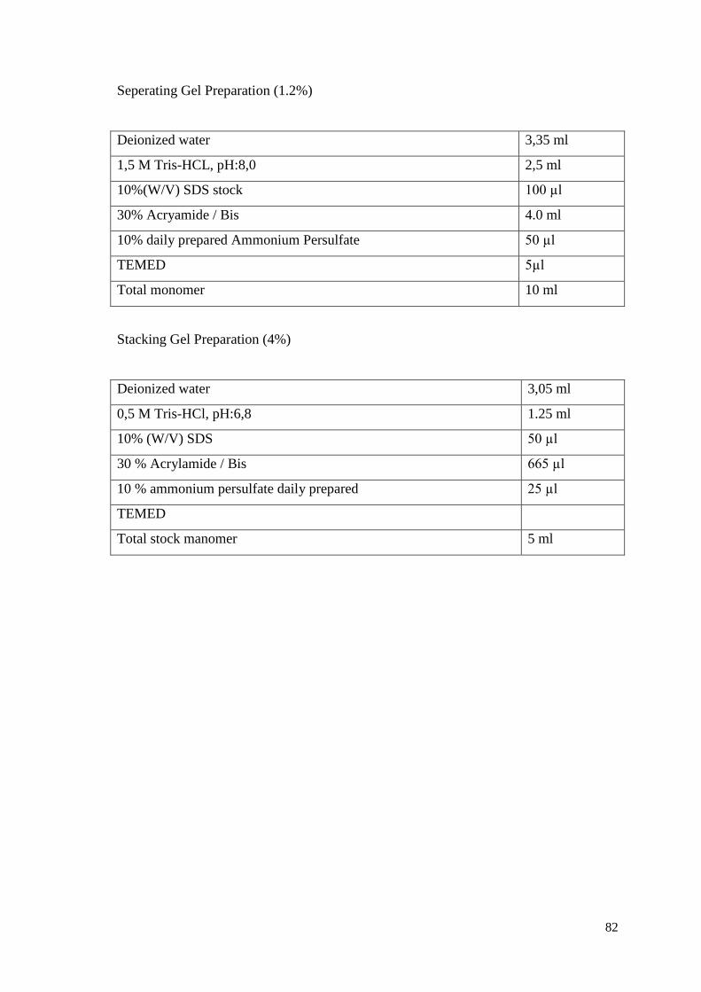
82
Seperating Gel Preparation (1.2%)
Deionized water 3,35 ml
1,5 M Tris-HCL, pH:8,0 2,5 ml
10%(W/V) SDS stock 100 µl
30% Acryamide / Bis 4.0 ml
10% daily prepared Ammonium Persulfate 50 µl
TEMED 5µl
Total monomer 10 ml
Stacking Gel Preparation (4%)
Deionized water 3,05 ml
0,5 M Tris-HCl, pH:6,8 1.25 ml
10% (W/V) SDS 50 µl
30 % Acrylamide / Bis 665 µl
10 % ammonium persulfate daily prepared 25 µl
TEMED
Total stock manomer 5 ml
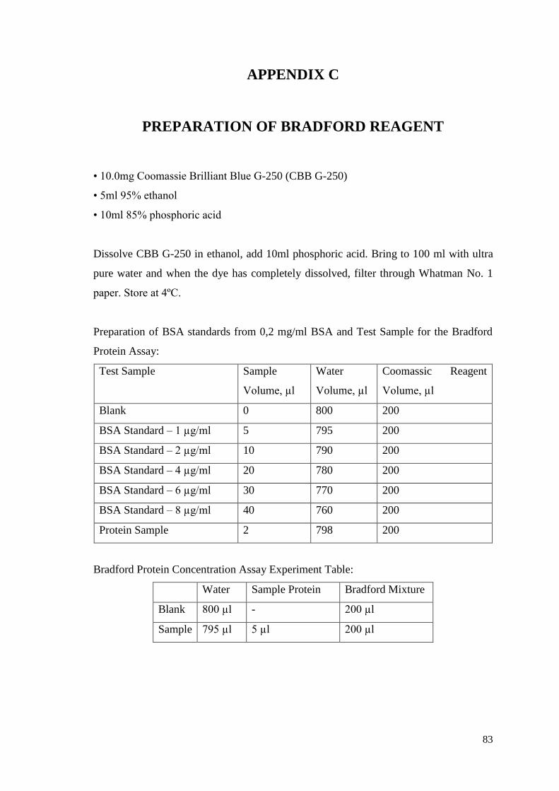
83
APPENDIX C
PREPARATION OF BRADFORD REAGENT
• 10.0mg Coomassie Brilliant Blue G-250 (CBB G-250)
• 5ml 95% ethanol
• 10ml 85% phosphoric acid
Dissolve CBB G-250 in ethanol, add 10ml phosphoric acid. Bring to 100 ml with ultra
pure water and when the dye has completely dissolved, filter through Whatman No. 1
paper. Store at 4ºC.
Preparation of BSA standards from 0,2 mg/ml BSA and Test Sample for the Bradford
Protein Assay:
Test Sample Sample
Volume, µl
Water
Volume, µl
Coomassic Reagent
Volume, µl
Blank 0 800 200
BSA Standard – 1 µg/ml 5 795 200
BSA Standard – 2 µg/ml 10 790 200
BSA Standard – 4 µg/ml 20 780 200
BSA Standard – 6 µg/ml 30 770 200
BSA Standard – 8 µg/ml 40 760 200
Protein Sample 2 798 200
Bradford Protein Concentration Assay Experiment Table:
Water Sample Protein Bradford Mixture
Blank 800 µl - 200 µl
Sample 795 µl 5 µl 200 µl