imaging the cytokine receptor cxcr4 in atherosclerotic ... of jnm march 2017 featured... · imaging...
TRANSCRIPT

Imaging the Cytokine Receptor CXCR4 in AtheroscleroticPlaques with the Radiotracer 68Ga-Pentixafor for PET
Fabien Hyafil1,2, Jaroslav Pelisek3, Iina Laitinen1, Margret Schottelius4, Miriam Mohring1, Yvonne Döring5,Emiel P.C. van der Vorst5, Michael Kallmayer3, Katja Steiger6, Andreas Poschenrieder4, Johannes Notni4,Johannes Fischer7, Christine Baumgartner7, Christoph Rischpler1–8, Stephan G. Nekolla1–8, Christian Weber5–9,Hans-Henning Eckstein3, Hans-Jürgen Wester4, and Markus Schwaiger1–8
1Department of Nuclear Medicine, Klinikum Rechts der Isar, Munich, Germany; 2Department of Nuclear Medicine, Bichat UniversityHospital, Assistance Publique–Hôpitaux de Paris, Inserm 1148, DHU FIRE, University Diderot, Paris, France; 3Department ofVascular and Endovascular Surgery, Klinikum Rechts der Isar, Munich, Germany; 4Pharmaceutical Radiochemistry, TechnischeUniversität München, Garching, Germany; 5Institute for Cardiovascular Prevention, Ludwig-Maximilians-Universität München,Munich, Germany; 6Institute of Pathology, Technische Universität München, Munich, Germany; 7Centre of Preclinical Research,Klinikum Rechts der Isar, Munich, Germany; 8DZHK (Deutsches Zentrum für Herz-Kreislauf-Forschung e.V.) partner site MunichHeart Alliance, Munich, Germany; and 9Cardiovascular Research Institute Maastricht (CARIM), Maastricht University, Maastricht,The Netherlands
68Ga-pentixafor is a radiotracer for PET that binds with nanomolar
affinity to CXCR4. The CXCR4 receptor is expressed at the surfaceof inflammatory cells. The objective of the study was to analyze the
ability of radiolabeled pentixafor to detect CXCR4 expression on
inflammatory cells present in atherosclerotic plaques of an experi-
mental rabbit model. Methods: Atherosclerotic plaques wereinduced by endothelial abrasion of the right carotid artery and ab-
dominal aorta of 7 rabbits fed an atherogenic diet. Five noninjured
rabbits fed a chow diet were used as controls. Rabbits were imagedon a PET/MR system after injection of 68Ga-pentixafor (15 MBq/kg).
Vascular signal was quantified as tissue-to-background ratio (TBR).
Biodistribution and autoradiographic studies were performed 1 h
after injection of 125I-pentixafor (7.5 MBq/kg). In addition, blockingstudies were performed in 2 atherosclerotic rabbits with preinjection
of the CXCR4 inhibitor AMD3100. Tracer uptake was quantified on
arterial cryosections using autoradiography and compared with
CXCR4 and RAM-11 (macrophage) expression on adjacent histo-logic sections. Results: One hour after injection of 68Ga-pentixafor,
strong signals were detected in vivo with PET/MR imaging in ath-
erosclerotic plaques of the abdominal aorta and right carotid arteryas compared with normal control arteries (mean TBR 5 1.95 6 0.51
vs. 1.22 6 0.25 and mean TBR 5 1.24 6 0.38 vs. 0.96 6 0.37,
respectively; P , 0.05 for both). Blocking studies with preinjection
of a CXCR4 inhibitor reduced 125I-pentixafor uptake in atheroscle-rotic plaques by approximately 40%. 125I-pentixafor uptake in the
vessel wall on autoradiographies was located in macrophage-rich
regions of atherosclerotic plaques and correlated with the intensity
of CXCR4 expression on corresponding cryosections (r2 5 0.61; P,0.05). Conclusion: 68Ga-pentixafor allows for the noninvasive de-
tection of CXCR4 expression in the vessel wall with PET and
emerges as a potential alternative to 18F-FDG for the assessment
of macrophage infiltration in atherosclerotic plaques.
Key Words: PET; atherosclerosis; radiotracer; inflammation;
CXCR4; macrophages
J Nucl Med 2017; 58:499–506DOI: 10.2967/jnumed.116.179663
In the past 10 y, 18F-FDG PET has proved a reliable noninvasiveimaging approach to assess the degree of inflammation present in
atherosclerotic plaques both in animal models of atherosclerosis
and in patients (1,2). Patients with the highest 18F-FDG uptake in
plaques present a higher rate of cardiovascular events and worse
prognosis than patients with low vascular 18F-FDG uptake (3).18F-FDG PET detects inflammation with high sensitivity but
has important limitations in the specific setting of cardiovascular
imaging: physiologic uptake in the heart and in the brain, back-
ground signal of blood, interaction with blood glucose, need for a
long fasting period, or dedicated diet before imaging (4). Conse-
quently, radiotracers that more specifically target inflammatory
cells might greatly facilitate the noninvasive detection of inflam-
matory activity in the vessel wall with PET.68Ga-pentixafor is the first PET agent that exhibits high affinity
and selectivity for C-X-C chemokine receptor type 4 (CXCR4)
(5). The CXCR4 is a constitutive cytokine receptor involved in
several biologic processes including the entry of HIV, the devel-
opment of metastasis, and the evolution of different autoimmune
diseases such as rheumatoid arthritis, systemic lupus erythemato-
sus, and multiple sclerosis (6). CXCR4 expression has been iden-
tified in more than 30 different types of cancers, and its activation
is a key trigger for enhanced tumor growth and progression, tumor
invasiveness, and metastasis. 68Ga-pentixafor was therefore first
evaluated in several tumors expressing CXCR4 in animal models
and demonstrated both high and specific uptake in CXCR4-positive
tumors and metastases (7). Proof-of-concept studies have recently
been performed with 68Ga-pentixafor in patients with lymphoma
(8) and multiple myeloma (9), both tumor entities expressing
high levels of CXCR4, and demonstrated intense uptake in tumor
Received Jun. 20, 2016; revision accepted Sep. 29, 2016.For correspondence or reprints contact: Fabien Hyafil, Department of
Nuclear Medicine, Klinikum Rechts der Isar, Ismaningerstrasse 22, 81675Munich, Germany.E-mail: [email protected] online Oct. 27, 2016.COPYRIGHT© 2017 by the Society of Nuclear Medicine and Molecular Imaging.
CXCR4 IMAGING IN ATHEROSCLEROSIS • Hyafil et al. 499
by SNMMI headquarters on March 1, 2017. For personal use only. jnm.snmjournals.org Downloaded from
cells, excellent pharmacokinetics, and favorable dosimetry in hu-mans (10), with a low residual signal in the blood. Furthermore,CXCR4 plays a pivotal role in the trafficking of inflammatorycells by mediating the homing of progenitor cells in the bonemarrow and regulating their mobilization into peripheral tissueson injury (6). In fact, the small molecular antagonist AMD3100(plerixafor) blocks the CXCR4 receptor and allows for mobili-zation of hematopoietic stem and progenitor cells in the bonemarrow for autografting on myeloablative treatment (11). Be-cause of the overexpression of CXCR4 at the surface of cellsinvolved in the inflammatory process, we hypothesized in thisstudy that 68Ga-pentixafor might find a role in the detection ofinflammatory cells with PET. Moreover, CXCR4 expression isstrongly increased under hypoxic conditions through the stimu-lation of the hypoxia-inducible factor (12). 68Ga-pentixafortherefore represents a promising candidate for the detection ofhigh-risk atherosclerotic plaques, which contain intense inflam-matory reaction and hypoxic conditions.The aim of this study was to test whether 68Ga-pentixafor spe-
cifically binds to CXCR4-expressing tissues and can be detected inatherosclerotic plaques induced in the abdominal aorta and rightcarotid artery of rabbits. In addition, we assessed the expression ofCXCR4 in carotid plaques in a small group of patients with 68Ga-pentixafor PET imaging and on histologic sections to evaluatewhether the results observed in a rabbit model with 68Ga-pentixaformay translate in the evaluation of human atherosclerotic plaques.
MATERIALS AND METHODS
Radiolabeling of Pentixafor
Radiosynthesis of 68Ga-Pentixafor. 68Ga was obtained by elution of
a 68Ge/68Ga generator with SnO2 matrix (iTHEMBA LABS) with 1 MHCl (5.5 mL) and immobilized on a strong cationic exchanger car-
tridge (SCX-Chromafix, size M; Macherey-Nagel).For animal studies, 68Ga-pentixafor was prepared on a Gallelut1
system in analogy to a previously published 68Ga-labeling procedure(SCINTOMICS GmbH) (13). Briefly, 68Ga generator eluate fractions
(1.25 mL, 600–800 MBq, buffered to pH 3.3 with 900 mL of a solutionof 14.4 g N-(2-hydroxyethyl)piperazine-N9-(2-ethanesulfonic acid) in
12 mL of water) were reacted with 3.5 nmol of pentixafor (SCINTOMICSGmbH) for 5 min. The radiochemical purity was always greater than
99% as confirmed by thin-layer chromatography and high-performanceliquid chromatography (HPLC). Addition of 1 mL of phosphate-
buffered saline and concentration in vacuo to 1 mL of total volumeyielded solvent-free formulations with specific activities ranging
from 100 to 150 MBq/nmol.For patient studies, the synthesis of 68Ga-pentixafor was performed
in a fully automated, good manufacturing practices–compliant pro-
cedure using a GRP module (SCINTOMICS GmbH) equipped witha disposable single-use cassette kit (ABX) including a standardized
labeling sequence (14). Upon being washed with water, 68Ga31was eluted into the reaction vessel (containing 20 mg of pentixafor in
3 mL of 1.5 M N-(2-hydroxyethyl)piperazine-N9-(2-ethanesulfonicacid)) using 1.7 mL of 5 M NaCl. The reaction mixture was heated
to 140�C for 10 min and, upon cooling, transferred onto a C18 lightcartridge (Waters). The cartridge was washed with water to remove
unreacted 68Ga activity, and 68Ga-pentixafor was eluted using 2 mL ofEtOH/water (50/50). The eluate was diluted with 12 mL of phosphate-
buffered saline and passed through a sterile filter. The radiochem-ical purity of the ready-to-inject formulation (14 mL) was always
greater than 99% as confirmed by radio-HPLC and thin-layer chro-matography, and the specific activity was in the range of 30–65
GBq/mmol.
Radiosynthesis of 125I-Pentixafor. For radioiodination, 100–200 mg
of pentixafor (cold gallium(III) complex) were dissolved in 0.5 mL ofTRIS iodination buffer (25 mM Tris�HCl, 0.4 M NaCl, pH 7.5) and
transferred to an Eppendorf reaction tube coated with 150 mg of IodoGen.Upon addition of 125I-Na (18–20 MBq; Hartmann Analytik), the re-
action vessel was briefly vortexed and the labeling reaction was allowed to
proceed for 15 min at room temperature. The peptide solution was thenremoved from the insoluble oxidizing agent. Separation of 125I-pentixafor
from unlabeled precursor was achieved using gradient reversed-phaseHPLC (column: Nucleosil 100 C18 [5 mm, 125 · 4.0 mm; CS GmbH];
gradient: 22%–42% ethanol [0.5% acetic acid] in water [0.5% acetic acid]within 20 min; flow: 1mL/min). The high-performance liquid chromatog-
raphy product fraction was used as such and diluted to the requiredconcentration using phosphate-buffered saline.
Imaging of Atherosclerotic Plaques in Rabbits
Animal Model. Atherosclerotic plaques were induced in male NewZealand White rabbits (n 5 7; mean age, 4 mo; mean weight 5 3.5 60.2 kg) by a combination of 4 mo of a high-cholesterol diet (0.3%cholesterol-enriched diet; Altromin Spezialfutter GmbH) and a double
balloon injury of the aorta and the right carotid artery (2 and 6 wk
after starting the high-cholesterol diet). Endothelial abrasion was per-formed in the aorta with a 4-French Fogarty embolectomy catheter
(Edward Lifescience GmbH) introduced through the femoral artery aspreviously described (2,15) and in the right carotid artery with a
3-French Fogarty embolectomy catheter (Edward Lifescience GmbH).All procedures were performed under general anesthesia by continu-
ous intravenous injection of propofol and analgesia with fentanyl.Nonoperated New Zealand White rabbits, fed a normal chow diet,
were used as controls (n 5 5). Experiments were approved by thelocal animal care committee and were in accordance with the German
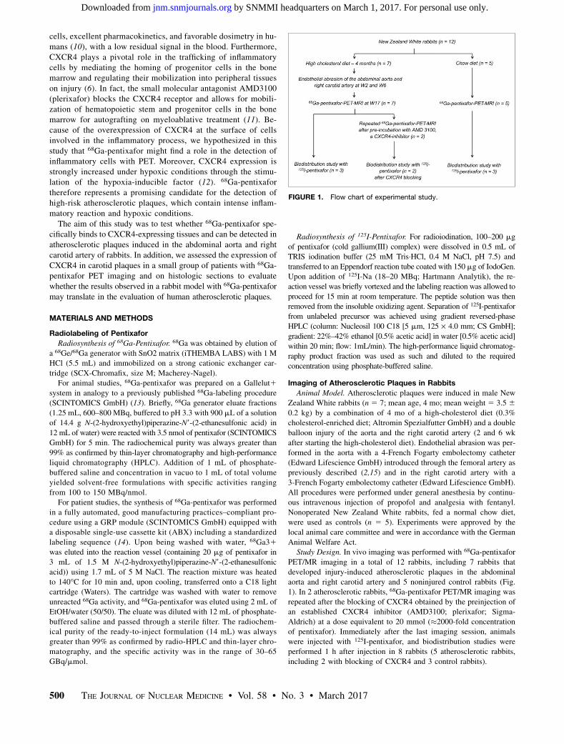
Animal Welfare Act.Study Design. In vivo imaging was performed with 68Ga-pentixafor
PET/MR imaging in a total of 12 rabbits, including 7 rabbits thatdeveloped injury-induced atherosclerotic plaques in the abdominal
aorta and right carotid artery and 5 noninjured control rabbits (Fig.1). In 2 atherosclerotic rabbits, 68Ga-pentixafor PET/MR imaging was
repeated after the blocking of CXCR4 obtained by the preinjection ofan established CXCR4 inhibitor (AMD3100; plerixafor; Sigma-
Aldrich) at a dose equivalent to 20 mmol (�2000-fold concentrationof pentixafor). Immediately after the last imaging session, animals
were injected with 125I-pentixafor, and biodistribution studies wereperformed 1 h after injection in 8 rabbits (5 atherosclerotic rabbits,
including 2 with blocking of CXCR4 and 3 control rabbits).
FIGURE 1. Flow chart of experimental study.
500 THE JOURNAL OF NUCLEAR MEDICINE • Vol. 58 • No. 3 • March 2017
by SNMMI headquarters on March 1, 2017. For personal use only. jnm.snmjournals.org Downloaded from
PET/MR Imaging Protocols. Imaging was performed on a human
whole-body simultaneous PET/MR (3T) scanner (Biograph mMR;Siemens). Rabbits were anesthetized with an intravenous infusion of
Propofol (2%, ;1.2–1.3 mg/kg/min; Propofol 2% MCT Fresenius,Fresenius Kabi Deutschland GmbH) and intubated, and catheters were
placed in ear veins. Rabbits positioned prone in the scanner wereventilated (Servo-I; Maquet Critical Care AB) and monitored (pulse
oxymeter and electrocardiogram) during the whole imaging session.PET and MR images were acquired simultaneously in all rabbits and
started 45 min after injection of 15 MBq/kg of 68Ga-pentixafor. De-tailed PET and MR acquisition protocols are provided in the section
“PET and MR Acquisition Protocols.”Image Analysis. Images were analyzed by an experienced nuclear
medicine physician masked to the rabbit group and to the results ofhistology. Image registration and fusion of the time-of-flight MR
acquisitions and attenuation-corrected PET images were performedwith an Osirix workstation (OsiriX version 3.5.1 64-bit; OsiriX Imaging
Software), an open-source DICOM viewer and image manipulator usingthe Horn algorithm, a manual rigid registration algorithm based on
multiple points (16,17). Matching of both datasets was considered as
correct when rabbit contours of PET and MR images were perfectlyaligned. If required (e.g., in the case of animal motion), alignment was
corrected manually using the Fusion tool of the OsiriX software. Forbiodistribution studies, SUVmean calculated as decay-corrected tissue
radioactivity divided by body weight and injected dose was measuredin circular regions of interest placed on each organ located using the
MR images.For the quantification of the vascular uptake, circular regions of
interest were placed on axial adjacent PET images of the abdominal
aorta and both carotid arteries (12 axial sections each) identified using
the time-of-flight images. SUVmax was recorded in each region ofinterest. In addition, the SUVmean of blood was measured by plac-
ing a circular region of interest in the right atria. The tissue-to-background ratio (TBR) was calculated as the ratio of SUVmax and
the background venous activity measured as SUVmean of blood (18).Mean TBRs were calculated for each arterial territory as the average
of TBR values throughout the entire arterial segment.
PET and MR Acquisition Protocols
MR Acquisition Protocol. MR imaging was performed using a
clinical knee coil for the aorta and a bilateral 4-channel phased array coilfor carotid arteries (4-channel, Machnet BV). MR acquisition first started
with a localizer scan to define the bed position and was followed by atime-of-flight 3-dimensional angiographic sequence to visualize the
abdominal aorta. Subsequently, a coronal 2-point Dixon 3-dimensionalvolumetric interpolated breath-hold T1-weighted (VIBE) MRI sequence
was acquired for the generation of attenuation maps (m-maps) and ana-tomic images in association with the PET acquisition. The Dixon MRI
sequence was set as follows: integrated parallel acquisition technique;
acceleration factor, 2; voxel size, 3.7 · 2.3 · 2.7 mm3 (in-plane resolu-tion · slice thickness); acquisition time, 19 s; repetition time, 3.6 ms; first
echo time, 1.23 ms; second echo time, 2.46 ms; matrix, 192 · 192;number of excitations, 1; field of view, 450 mm; phase field of view,
65.6%; 1 slab with 128 slices; slice thickness, 2.73 mm; flip angle, 10�;and bandwidth, 965 Hz/pixel. The software of the MRI scanner automat-
ically used the raw images to generate 4 different images : T1-weightedin-phase, T1-weighted out-of-phase, water-only, and fat-only. The same
MR acquisition protocol was repeated for the imaging of carotid arteries.PET Acquisition Protocol. Static emission
scans were acquired during 10 min in a singlebed position 50 min after the injection of68Ga-pentixafor in the abdominal aorta and60 min after the injection for carotid arteries.
Data were corrected for randoms, dead time,scatter, and attenuation based on the m-maps
generated from the Dixon images (19). Two-mm-thick axial PET images were recon-
structed with a 50 · 50 cm2 field of viewand a 344 · 344 matrix using a 3-dimensional
attenuation-weighted ordered-subsets expec-tation maximization iterative reconstruction
algorithm with 3 iterations and 21 subsets, azoom of 1, and no postreconstruction filter.
Spatial resolution of the reconstructed PETimages was previously determined to be 4.3 mm
at 1 cm from the scanner’s isocenter and 5.0mm at 10 cm in the transverse direction in
full width at half maximum (20).
Tracer Biodistribution and Histology
Tracer Biodistribution in Tissues. At theend of the imaging session, 8 rabbits (athero-
sclerotic rabbits, n5 5; control rabbits, n5 3)were injected intravenously with 7.5 MBq/kg
of 125I-pentixafor into the ear vein underisoflurane anaesthesia for biodistribution
studies. Because of the long half-life of125I (60 d), 125I-pentixafor allowed for more
accurate biodistribution studies and auto-radiographies in comparison to 68Ga-pentixa-
for (half-life, 68 min). The animals were sac-rificed 60 min after injection by intravenous
injection of 150 mg/kg of pentobarbiturate
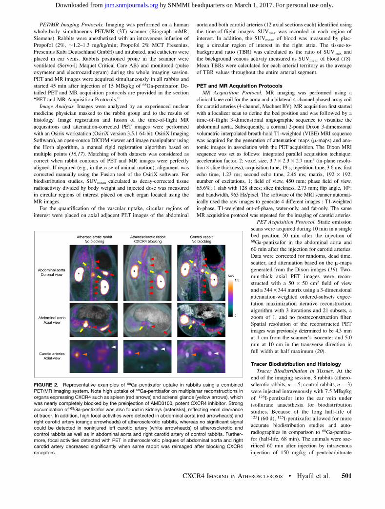
FIGURE 2. Representative examples of 68Ga-pentixafor uptake in rabbits using a combined
PET/MR imaging system. Note high uptake of 68Ga-pentixafor on multiplanar reconstructions in
organs expressing CXCR4 such as spleen (red arrows) and adrenal glands (yellow arrows), which
was nearly completely blocked by the preinjection of AMD3100, potent CXCR4 inhibitor. Strong
accumulation of 68Ga-pentixafor was also found in kidneys (asterisks), reflecting renal clearance
of tracer. In addition, high focal activities were detected in abdominal aorta (red arrowheads) and
right carotid artery (orange arrowheads) of atherosclerotic rabbits, whereas no significant signal
could be detected in noninjured left carotid artery (white arrowheads) of atherosclerotic and
control rabbits as well as in abdominal aorta and right carotid artery of control rabbits. Further-
more, focal activities detected with PET in atherosclerotic plaques of abdominal aorta and right
carotid artery decreased significantly when same rabbit was reimaged after blocking CXCR4
receptors.
CXCR4 IMAGING IN ATHEROSCLEROSIS • Hyafil et al. 501
by SNMMI headquarters on March 1, 2017. For personal use only. jnm.snmjournals.org Downloaded from
(Narcoren; Merial), and the organs of interest including the aorta and
carotid arteries were dissected. The radioactivity was measured inweighted tissue samples using a g-counter (1480 Wizard; PerkinElmer
Wallac). Data are expressed in %IA/g.Autoradiographies were performed on 20-m-thick cryosections. Af-
ter an overnight exposure to image plates (Fuji TR; Fuji Photo FilmCo.), the plates were scanned with an image plate reader (CR 35 BIO;
Dürr Medical, Raytest Isotopenmeßgeräte GmbH). The mean activitywas quantified in regions of interest encompassing tissues and cor-
rected for background signal with an image analysis program (AIDA
Image Analyser; Raytest Isotopenmeßgeräte). Radiotracer uptake insections was expressed as the mean activity corrected for background
and adjusted to the mean activity measured in the vascular wall of theleft carotid artery used as a reference for each plate.
Histology of Rabbit Tissues. Five-mm-thick cryosections were fixedfor 10 min in acetone. CXCR4 expression and macrophages were
detected by immunohistochemistry using mouse monoclonal biotiny-lated antibodies directed against CXCR4 (clone MAB170 [R&D
Systems]; dilution, 1:500) and RAM-11 (M0633 [Dako]; dilution,1:3,000), respectively. Primary antibodies were visualized using a
peroxidase–diaminobenzidine mouse detection kit (Vectastain;Vector Laboratories). Unspecific primary immunoglobulin G an-
tibodies were used as negative controls. Total CXCR4-positivearea was measured digitally on each section using an automated,
contrast-based, area analysis function of the Image Scope soft-ware (Aperio; Vista). The percentage of CXCR4-positive cells was
calculated as the ratio between pixels with high contrast and pixelswith low contrast using the automated analysis tool of the software
Image Scope.Histology of Human Carotid Plaques. Atherosclerotic plaque
samples (n 5 10) obtained from carotid endarterectomy were fixedin formalin, embedded in parrafin, and sectioned in 5-mm-thick sec-
tions. CXCR4 expression and macrophages were detected by immu-
nostaining with the use of the following pri-
mary antibodies: anti-CXCR4 (ab124824 [AbcamInc.]; dilution, 1:300); anti-CD68 (clone KP1
[Dako]; dilution, 1:2,000); anti-CD31 (cloneJC70A [Dako]; dilution, 1:40); anti-CD45 (clone
2B11 [Dako]; dilution, 1:100). After primary an-tibody incubation, visualization was performed
using a peroxidase–diaminobenzidine ChemMateDetection Kit (Dako). Biotinylated antibody
against CXCR4 was detected by a streptavidin–horseradish peroxidase complex (Jackson Im-
munoresearch Laboratories), visualized withdiaminobenzidine (Sigma-Aldrich), and coun-
terstained with Mayer hematoxylin. Unspecificprimary immunoglobulin G antibodies were
used as negative controls.Expression of CXCR4 on Macrophage Cell
Surface. Human peripheral blood mononu-clear cells were isolated using density-
gradient centrifugation with polymorphprep
(Axis Shield). Isolated cells were plated forattachment in monocyte attachment medium,
according to the manufacturer’s protocol(C28051; PromoCell). Subsequent differentia-
tion was performed using Macrophage Gener-ation Media DXF according to the manufac-
turer’s protocol (C28055; PromoCell). After 10d of differentiation, cells were detached and
replated as 200,000 cells/well (48-well plates).Cells were stimulated with statins (Fluvadin, 5
mM, 48 h; Selleckchem), TNF (25 ng/mL,24 h; Sigma), oxLDL (25 mg/mL, 48 h; KB Kalen), or a combination.
Afterward, CXCR4 expression was measured using FACS analysis(CXCR4-eF450 with CD45-FITC and CD14-APC as controls, all eBio-
science).
Imaging of Atherosclerotic Plaques in Patients68Ga-pentixafor was synthesized and administered according to the
requirements of the German Medicinal Products Act (Arzneimittelge-setz §13 2b) and with the approval of the responsible local regula-
tory authority (Regierung von Oberbayern). Before the investigationbegan, all patients gave written informed consent to participate. The
data analysis was approved by the responsible local ethics committees.Four patients with carotid stenosis greater than 50% and 4 patients
without any significant carotid stenosis (,30%) were identified amongpatients who underwent 68Ga-pentixafor PET/MR imaging with neck
acquisitions and after exclusion of patients with high 68Ga-pentixaforuptake in tumors adjacent to carotid arteries. Patients were imaged on
average 456 4 min after injection of 1706 15 MBq of 68Ga-pentixafor.Carotid arteries were located using the coronal 2-point Dixon 3-dimen-
sional volumetric interpolated breath-hold T1-weighted MRI sequenceused for attenuation correction. Mean TBR was quantified on 8 consec-
utive PET axial sections centered on each carotid bulb using the samemethodology as in the animal model. In addition, the most-diseased-seg-
ment TBR was calculated for each carotid artery as the average of 3consecutive axial sections centered on the section with the highest TBR.
Statistical Analysis
Numeric values are expressed as mean 6 SD. Statistical analysis
was performed using SPSS software (SPSS Inc.). Comparisons ofquantitative values measured in 2 groups were performed using a
2-tailed Student t test if the number of values was greater than 20 ineach group and using a Wilcoxon signed-rank test if the number of values
was less than 20 in each group. For fluorescence-activated cell sorting
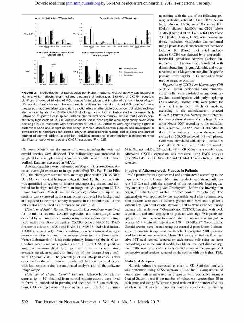
FIGURE 3. Biodistribution of radiolabeled pentixafor in rabbits. Highest activity was located in
kidneys, which reflects renal-mediated clearance of radiotracer. Blocking of CXCR4 receptors
significantly reduced binding of 68Ga-pentixafor in spleen and in adrenal glands in favor of spe-
cific uptake of radiotracer in these organs. In addition, increased uptake of 68Ga-pentixafor was
measured in abdominal aorta and right carotid artery of atherosclerotic vs. control rabbit and was
also reduced by about 40% after CXCR4 blocking. Ex vivo biodistribution studies confirmed high
uptake of 125I-pentixafor in spleen, adrenal glands, and bone marrow, organs that express con-
stitutively high levels of CXCR4. Activities measured in these organs were significantly lower when
blocking CXCR4 receptors with preinjection of AMD3100. Activities were significantly higher in
abdominal aorta and in right carotid artery, in which atherosclerotic plaques had developed, in
comparison to noninjured left carotid artery of atherosclerotic rabbits and to aorta and carotid
arteries of control rabbits. In addition, activities measured in atherosclerotic segments were
significantly lower when blocking CXCR4 receptor. *P , 0.05.
502 THE JOURNAL OF NUCLEAR MEDICINE • Vol. 58 • No. 3 • March 2017
by SNMMI headquarters on March 1, 2017. For personal use only. jnm.snmjournals.org Downloaded from

analysis, the levels of CXCR4 expression in the different groups werecompared with a Kruskal–Wallis test followed by a Dunn multiple com-
parison test. Pentixafor uptake on autoradiography and CXCR4 expres-sion on corresponding sections were compared using multiple regression
to take into account that 3 samples were analyzed in each animal.P values of less than 0.05 were considered as significant.
RESULTS
Detection of CXCR4 with 68Ga-Pentixafor in Rabbit Model of
Atherosclerotic Plaques
In Vivo Imaging of Atherosclerotic Plaques with 68Ga-PentixaforPET/MRI. Fifty minutes after intravenous injection of 68Ga-pentixafor, the radiotracer accumulated in the spleen (SUVmean 51.7 6 0.3) and in adrenal glands (SUVmean 5 1.9 6 0.7), organsknown to express physiologically CXCR4 receptors (Fig. 2). Thehighest activity was located in the kidneys (SUVmean 5 3.2 60.5), which reflects the renal-mediated clearance of the radio-tracer. The specificity of 68Ga-pentixafor uptake in the spleenand in adrenal glands was confirmed by blocking CXCR4 recep-
tors with a preinjection of AMD3100, which significantly reducedthe signal in these organs (SUVmean 5 1.7 6 0.3 vs. 0.7 6 0.2 forthe spleen and SUVmean 5 1.9 6 0.7 vs. 0.5 6 0.1 for adrenalglands, P , 0.05 for both; Fig. 3). In addition, focal areas of 68Ga-pentixafor uptake were detected in the abdominal aortas and rightcarotid artery of atherosclerotic rabbits by PET imaging, whereasno significant uptake was detected in the aortas and carotid arteriesof atherosclerotic rabbits as well as in control rabbits after block-ing the CXCR4 receptor. 68Ga-pentixafor uptake in atheroscleroticplaques was quantified in vivo as the average TBR over the dif-ferent arterial segments (Fig. 3). Higher TBRs were measured inthe abdominal aorta and right carotid artery as compared withnormal control arteries (mean TBR 5 1.95 6 0.51 vs. 1.22 60.25 and mean TBR 5 1.24 6 0.38 vs. 0.96 6 0.37; P , 0.05for both). TBRs measured in the abdominal aorta and in the rightcarotid artery were significantly reduced (;40%) after blockingCXCR4 receptors (mean TBR 5 1.95 6 0.51 vs. 1.13 6 0.52 andmean TBR 5 1.24 6 0.38 vs. 0.70 6 0.03; P , 0.05 for both).Biodistribution of Radiolabeled Pentixafor in Rabbits. The
percentage injected activity adjusted for weight (%IA/g) wasquantified 1 h after the injection of 125I-pentixafor in the differentorgans with and without blocking CXCR4 receptors (Fig. 3). Bio-distribution studies confirmed the binding of radiolabeled pentixafor
FIGURE 5. Accumulation of 125I-pentixafor uptake in CXCR4-expressing
macrophages of atherosclerotic plaques. Quantification of activities mea-
sured in arterial wall on autoradiography confirmed higher accumulation of125I-pentixafor in abdominal aorta and in right carotid artery in comparison
to noninjured thoracic aorta and left carotid artery of atherosclerotic rab-
bits and to aorta and carotid arteries of control rabbits. CXCR4 expression
in vascular wall was also higher in injured segments of abdominal aorta
and right carotid artery of atherosclerotic rabbits in comparison to corre-
sponding noninjured segments of control rabbits. Strong correlation was
found between mean activities measured in arterial segments on autora-
diography and percentage of CXCR4-positive areas quantified on adja-
cent histologic sections measured on immunohistology. On cell culture of
macrophages, addition of oxidized low-density lipoprotein induced strong
increase in expression of CXCR4 at cell surface detected with fluores-
cence-activated cell sorting analysis, whereas incubation with tumor ne-
crosis factor α did not change CXCR4 expression on macrophages.
Incubation with statins did not have any effect on CXCR4 expression at
surface of macrophages. *P , 0.05. Ath 5 atherosclerotic; C 5 control;
FACS5 fluorescence-activated cell sorting; IHC5 immunohistochemistry;
oxLDL 5 oxidized low-density lipoprotein; TNF 5 tumor necrosis factor.
FIGURE 4. Localization of 125I-pentixafor accumulation in plaques. In-
tense activities (red color) were detected on autoradiographies of ab-
dominal aorta and right carotid artery from atherosclerotic rabbits and
were associated with high expression of CXCR4 and high density of
macrophages (RAM-11 staining) in corresponding regions on immuno-
histochemistry of adjacent sections. In atherosclerotic rabbits prein-
jected with potent CXCR4 inhibitor AMD3100, low activities (blue color)
were detected in atherosclerotic plaques on autoradiography, whereas
intense expression of CXCR4 was present in adjacent sections on his-
tology. In noninjured thoracic aorta and left carotid artery of rabbits
fed high-fat diet and in arteries of control rabbits, only low activities
(blue color) were detected on autoradiography and associated with
low CXCR4 expression and rare macrophages in vascular wall by
immunohistochemistry. All autoradiographic images were acquired
using same color scale. Ath 5 atherosclerotic; HE 5 hematoxylin
and eosin.
CXCR4 IMAGING IN ATHEROSCLEROSIS • Hyafil et al. 503
by SNMMI headquarters on March 1, 2017. For personal use only. jnm.snmjournals.org Downloaded from

in adrenal glands, spleen, and bone marrow, which was significantlyreduced in rabbits blocked with a dose of 20 mmol of AMD3100(�2,000-fold molar excess over pentixafor), an established CXCR4inhibitor, before injection of 125I-pentixafor (0.33 6 0.14 vs.0.02 6 0.01 %IA/g for adrenal glands; 0.17 6 0.09 vs. 0.03 60.01 %IA/g for spleen; 0.14 6 0.04 vs. 0.03 6 0.01 %IA/g forbone marrow). In addition, significantly higher activities weremeasured in the right atherosclerotic carotid arteries and abdom-inal aortas in comparison to the noninjured left carotid arteries andabdominal aortas of control rabbits (0.08 6 0.02 %IA/g and 0.10 60.03 %IA/g vs. 0.05 6 0.01 %IA/g and 0.05% 6 0.01 %IA/g,respectively, P , 0.05 for both).
Correlation Between 125I-Pentixafor Accumulation and
Expression of CXCR4 in Plaques
CXCR4 expression was mostly located in macrophage-rich(RAM-11–positive) regions of atherosclerotic plaques (Fig. 4).The percentage of CXCR4-positive immunostaining was signifi-cantly higher in atherosclerotic plaques of the abdominal aorta andright carotid artery relative to the corresponding segments of con-trol rabbits (75% 6 25% vs. 26% 6 13% and 58% 6 15% vs.23% 6 16%; P , 0.05 for both; Fig. 5). In addition, atheroscle-rotic plaque sections that exhibited high-counting rates, as de-tected by autoradiography, contained high CXCR4 expression inthe corresponding adjacent histologic sections (Fig. 4). In contrast,atherosclerotic plaques with low autoradiographic activities aswell as normal aortic walls from control rabbits contained onlyfew cells expressing CXCR4. A strong correlation (r2 5 0.61; P,
0.05) was found between the mean activity quantified on autoradi-ography in arterial sections and the percentage of CXCR4-positiveareas measured in the arterial wall on adjacent histologic sections byimmunohistology (Fig. 5). Interestingly, the addition of oxidized low-density lipoprotein to macrophage cell culture induced a strong in-crease in the expression of CXCR4 at the cell surface detected withfluorescence-activated cell sorting analysis (Fig. 5; 17,892 6 5,964vs. 39,157 6 22,285; P , 0.05), whereas incubation with tumornecrosis factor a did not have any effect on CXCR4 expression onmacrophages (17,892 6 5,964 vs. 18,200 6 4,683; P . 0.05). In-cubation with statins did not have any effect on CXCR4 expression atthe surface of macrophages incubated with oxidized low-density li-poprotein (48,600 6 6,520 vs. 39,157 6 22,285; P . 0.05).
Detection of 68Ga-Pentixafor Uptake with PET in Human
Carotid Plaques68Ga-pentixafor uptake in carotid arteries was analyzed with
PET/MR imaging in 4 patients with carotid stenosis greater than50% and 4 patients with carotid stenosis less than 30%. Focal, intenseuptake of 68Ga-pentixafor (SUVmax5 3.4 and 2.1, and most-diseased-segment TBR 5 2.6 and 2.1) was identified in carotid atheroscleroticplaques of 2 of 4 patients with carotid stenosis greater than 50%(Supplemental Table 1; supplemental materials are available at
FIGURE 7. Representative examples of CXCR4 expression in human
carotid plaques on immunohistochemistry. In human carotid plaques,
CXCR4 expression by immunohistochemistry was mostly located in
macrophage-rich areas (CD-68–positive cells), whereas only weak expres-
sion of CXCR4 was present in lymphocyte-rich areas (CD-45–positive cells)
and in endothelial cells (CD-31–positive cells).
FIGURE 6. In vivo imaging of human carotid atherosclerotic plaques
with 68Ga-pentixafor PET/MRI. Representative example of focal accu-
mulation of 68Ga-pentixafor in carotid atherosclerotic plaque detected
with PET/MRI. Interestingly, this patient had presented for 2 wk ische-
mic stroke ipsilateral to carotid plaque showing high 68Ga-pentixafor
uptake (white arrows). Note also intense accumulation of 68Ga-pentixafor
in perivascular lymph node and in tonsils (white arrowheads). MIP 5maximum-intensity projection.
504 THE JOURNAL OF NUCLEAR MEDICINE • Vol. 58 • No. 3 • March 2017
by SNMMI headquarters on March 1, 2017. For personal use only. jnm.snmjournals.org Downloaded from
http://jnm.snmjournals.org). Interestingly, 1 patient had presented anischemic stroke for 2 wk and showed high uptake of 68Ga-pentixaforin the carotid plaques ipsilateral to the territory of the stroke (Fig. 6).Intense accumulation of 68Ga-pentixafor could also be detected inperivascular lymph nodes and tonsils. No significant 68Ga-pentixaforuptake was detected in the carotid arteries of the patient with asymp-tomatic carotid stenosis and in patients with carotid stenosis less than30%. On immunohistochemistry of human carotid plaques (Fig. 7),CXCR4 expression was mostly located in macrophage-rich areas(CD-68–positive cells), whereas only weak expression of CXCR4was present in lymphocyte-rich areas (CD-45–positive cells) and onendothelial cells (CD-31–positive cells).
DISCUSSION
In this study, using a rabbit model of atherosclerosis, wedemonstrate that the expression of CXCR4 in atherosclerotic plaquescan be detected in vivo with PET imaging after injection of 68Ga-pentixafor. A significantly higher uptake of 68Ga-pentixafor was de-tected both in vivo and ex vivo in atherosclerotic plaques of rabbitscompared with normal arterial wall and was significantly reducedafter a preinjection of the potent CXCR4 inhibitor AMD3100, ex-cluding a nonspecific retention of the tracer in the arterial wall. Inaddition, the intensity of 68Ga-pentixafor uptake in the aortic wallquantified on autoradiography strongly correlated with CXCR4-positive areas measured on adjacent histologic sections. Further-more, we confirmed in a small number of patients that focal68Ga-pentixafor uptake can be detected in carotid atheroscleroticplaques with PET. In human carotid atherosclerotic plaques, wefound that CXCR4 expression was localized in macrophage-richareas. Hence, 68Ga-pentixafor represents a promising PET radio-tracer for identification of macrophage infiltration present in high-risk atherosclerotic plaques.
Targeting CXCR4 in Atherosclerotic Plaques
The precise role of CXCR4 expression in atherosclerosis iscomplex and remains controversial (6). CXCR4 might play aproatherogenic role in attracting leukocytes in plaques but mayalso have a protective role through the recruitment of endothelialcells. In addition, CXCR4 expression is modulated by heterome-rization with CXCR7 (21) or by interaction with hetero-complexesformed of CXCL12 and high-mobility group box 1 proteins (22),underscoring the limitations of messenger RNA and protein ex-pression measurements of CXCR4 performed so far in atheroscle-rotic plaques. Radiolabeled pentixafor presents the advantage ofbinding exclusively to CXCR4 expressed on the cell membrane,and its uptake might therefore give a more precise estimation ofthe location and number of functional CXCR4 proteins in athero-sclerotic plaques. Interestingly, we evidenced in this study a strongcorrelation between the intensity of radiolabeled pentixafor uptakein plaques and the expression of CXCR4 on immunohistochemis-try. Radiolabeled pentixafor might thus provide an interesting op-portunity to understand more in depth the functional expression ofCXCR4 in plaques and its changes over time using in vivo PETimaging.
Imaging Macrophages in Atherosclerotic Plaques
with 68Ga-Pentixafor
To be relevant for the evaluation of atherosclerotic plaques, thebiologic target of a radiotracer needs to fulfill several conditions:to be sufficiently expressed in plaques so that the radiotracer canreach concentrations allowing for its detection with PET, to differ
significantly between high-risk and stable plaques, and to beassociated with the risk of subsequent plaque rupture and clinicalevents. On the basis of these criteria, macrophages appear as arelevant target for atherosclerosis imaging: high macrophagedensity can be found in atherosclerotic plaques (23), the numberof macrophages is significantly higher in complicated versusnoncomplicated plaques (24,25), and the identification of mac-rophages in atherosclerotic plaques has been associated with anincreased risk of cardiovascular events during follow-up (26).Interestingly, we found in this study that radiolabeled pentixaforaccumulated in macrophage-rich regions of rabbit and humanatherosclerotic plaques. By the means of fluorescence-activatedcell sorting analysis, we furthermore confirmed that a low-densitylipoprotein-rich milieu induced an increase in the expression ofCXCR4 at the surface of macrophages. Hence, 68Ga-pentixaforappears relevant for the evaluation of atherosclerotic plaques as anoninvasive imaging marker of macrophage infiltration in the vesselwall.
Advantages of 68Ga-Pentixafor Over Existing PET
Radiotracers in Atherosclerosis Imaging
Over the past 10 y, several PET radiotracers have been developedfor the imaging of atherosclerotic plaques, each of them targetingspecific biologic activities, cellular types, or protein expressionincreased in high-risk plaques (27). Nevertheless, 68Ga-pentixaformight hold several advantages over existing PET radiotracers inthis indication. First, the intensity of the signal in atheroscleroticplaques was high enough to be detectable with PET. Second,physiologic expression of CXCR4 is limited to the spleen, adrenalglands, and bone marrow. 68Ga-pentixafor uptake in atheroscle-rotic plaques can thus be precisely assessed thanks to a low back-ground signal in tissues adjacent to the arterial wall (myocardium,brain, muscle). Third, 68Ga-pentixafor-PET imaging does not re-quire the patient to fast as is the case for 18F-FDG. Fourth, pen-tixafor can readily be radiolabeled with the generator nuclide68Ga, which is obtained after elution from a 68Ge/68Ga generatorand does not require the use of an on-site cyclotron. The flexibilityof this synthesis could be particularly interesting for cardiovascularimaging. Taken together, 68Ga-pentixafor might greatly facilitate thenoninvasive detection of CXCR4 expression and macrophage infil-tration in atherosclerotic plaques with PET.This study shows minor limitations. First, additional studies are
required to unravel in more details the cellular processes in whichCXCR4 is involved and the underlying molecular mechanismsassociated with its expression in atherosclerotic plaques. In thiscontext, the understanding of the complex role of CXCR4 will besignificantly improved by the availability of an imaging methodallowing for the in vivo monitoring of CXCR4 receptor expressionin atherosclerotic plaques. Of note, radiolabeled pentixafor showsonly a low affinity for murine CXCR4 and can therefore not beused to follow CXCR4 expression over time in an atheroscleroticmice model. Second, the level of CXCR4 expression on macro-phages measured using fluorescence-activated cell sorting analysiswas not significantly affected by ex vivo incubation with statins.Additional experiments are, however, required to test the effects ofsystemic lipid-lowering on CXCR4 expression at the surface ofplaque macrophages. Third, definite conclusions cannot be drawnon the value of 68Ga-pentixafor PET imaging for plaque imagingbecause the number of patients evaluated in this study was small. Ina forthcoming study, we plan to evaluate in 45 additional patientswith high-grade carotid stenosis the interest of 68Ga-pentixafor
CXCR4 IMAGING IN ATHEROSCLEROSIS • Hyafil et al. 505
by SNMMI headquarters on March 1, 2017. For personal use only. jnm.snmjournals.org Downloaded from

PET imaging for the detection of CXCR4 expression in plaques andto compare the intensities of 68Ga-pentixafor and 18F-FDG uptakein human plaques. The results of this clinical study will give usmore hints on the value of 68Ga-pentixafor PET imaging for theevaluation of atherosclerotic plaques. Finally, whether the intensityof 68Ga-pentixafor uptake detected with PET in the vascular wallrepresents a relevant biomarker to predict the risk of plaque ruptureand subsequent clinical events will need to be validated in futurestudies.
CONCLUSION
In this study, we demonstrated that the intensity of CXCR4expression in atherosclerotic plaques could be assessed with 68Ga-pentixafor PET imaging in a rabbit model. In addition, we haveconfirmed in a small number of patients that 68Ga-pentixafor up-take can be detected in human carotid plaques. Taken together,these results support a potential role of 68Ga-pentixafor PET im-aging for the more specific identification of macrophages in ath-erosclerotic plaques by overcoming the current limitations of18F-FDG for the detection of inflammation in the cardiovascularfield. Future studies will need to be performed to understand morein depth the origin, meaning, and changes of CXCR4 expressionin tissues taking place during inflammatory processes. In fact, theinterest of 68Ga-pentixafor PET imaging is not restricted to ath-erosclerosis imaging but might also find a role for the more spe-cific detection of inflammatory cells in organs with intrinsic high18F-FDG uptake such as the heart.
DISCLOSURE
This work was supported by the Advanced Research Grant“Multimodal Molecular Imaging” (MUMI; grant 294582; EuropeanResearch Council Executive Agency), by the Deutsche For-schungsgemeinschaft (DFG; Grossgeräteinitiative and SFB 824;subprojects B5 to Prof. Hans-Jürgen Wester and Z2 to KatjaSteiger), by the High-Risk High-Volume grant from the DeutschesZentrum für Herz-Kreislauf Forschung (DZHK) to Prof. MarkusSchwaiger, and by the Deutsche Forschungsgemeinschaft (SFB1123-A1) to Yvonne Döring and Prof. Christian Weber. Hans-JürgenWester is shareholder of SCINTOMICS, Germany. No other poten-tial conflict of interest relevant to this article was reported.
ACKNOWLEDGMENTS
We thank Sylvia Schachoff, Claudia Meisinger, and AnnaWinter for their valuable help in acquiring PET/MR images; SybilleReder for her valuable support in animal experiments; and RenateHegenloh for her expertise in histologic analysis of atheroscleroticplaques.
REFERENCES
1. Rudd JH, Hyafil F, Fayad ZA. Inflammation imaging in atherosclerosis. Arte-
rioscler Thromb Vasc Biol. 2009;29:1009–1016.
2. Hyafil F, Cornily JC, Rudd JH, Machac J, Feldman LJ, Fayad ZA. Quantification
of inflammation within rabbit atherosclerotic plaques using the macrophage-
specific CT contrast agent N1177: a comparison with 18F-FDG PET/CT and
histology. J Nucl Med. 2009;50:959–965.
3. Figueroa AL, Abdelbaky A, Truong QA, et al. Measurement of arterial activity
on routine FDG PET/CT images improves prediction of risk of future CVevents.
JACC Cardiovasc Imaging. 2013;6:1250–1259.
4. Bucerius J, Hyafil F, Verberne HJ, et al. Position paper of the cardiovascular
Committee of the European Association of Nuclear Medicine (EANM) on
PET imaging of atherosclerosis. Eur J Nucl Med Mol Imaging. 2016;43:
780–792.
5. Demmer O, Gourni E, Schumacher U, Kessler H, Wester HJ. PET imaging of CXCR4
receptors in cancer by a new optimized ligand. ChemMedChem. 2011;6:1789–1791.
6. Döring Y, Pawig L, Weber C, Noels H. The CXCL12/CXCR4 chemokine ligand/
receptor axis in cardiovascular disease. Front Physiol. 2014;5:212.
7. Gourni E, Demmer O, Schottelius M, et al. PET of CXCR4 expression by a 68Ga-
labeled highly specific targeted contrast agent. J Nucl Med. 2011;52:1803–1810.
8. Wester HJ, Keller U, Schottelius M, et al. Disclosing the CXCR4 expression in
lymphoproliferative diseases by targeted molecular imaging. Theranostics. 2015;
5:618–630.
9. Philipp-Abbrederis K, Herrmann K, Knop S, et al. In vivo molecular imaging of
chemokine receptor CXCR4 expression in patients with advanced multiple my-
eloma. EMBO Mol Med. 2015;7:477–487.
10. Herrmann K, Lapa C, Wester HJ, et al. Biodistribution and radiation dosimetry
for the chemokine receptor CXCR4-targeting probe 68Ga-pentixafor. J Nucl
Med. 2015;56:410–416.
11. Debnath B, Xu S, Grande F, Garofalo A, Neamati N. Small molecule inhibitors
of CXCR4. Theranostics. 2013;3:47–75.
12. Schioppa T, Uranchimeg B, Saccani A, et al. Regulation of the chemokine re-
ceptor CXCR4 by hypoxia. J Exp Med. 2003;198:1391–1402.
13. Martin R, Juttler S, Muller M, Wester HJ. Cationic eluate pretreatment for
automated synthesis of [68Ga]CPCR4.2. Nucl Med Biol. 2014;41:84–89.
14. Mueller D, Klette I, Baum RP, Gottschaldt M, Schultz MK, Breeman WA. Sim-
plified NaCl based 68Ga concentration and labeling procedure for rapid synthesis
of 68Ga radiopharmaceuticals in high radiochemical purity. Bioconjug Chem.
2012;23:1712–1717.
15. Hyafil F, Cornily J, Feig J, et al. Noninvasive detection of macrophages using a
nanoparticulate contrast agent for computed tomography. Nat Med. 2007;13:
636–641.
16. Hutton BF, Braun M, Thurfjell L, Lau DY. Image registration: an essential tool
for nuclear medicine. Eur J Nucl Med Mol Imaging. 2002;29:559–577.
17. Rosset A, Spadola L, Ratib O. Osirix: an open-source software for navigating in
multidimensional Dicom images. J Digit Imaging. 2004;17:205–216.
18. Rudd JH, Myers KS, Bansilal S, et al. Atherosclerosis inflammation imaging
with 18F-FDG PET: carotid, iliac, and femoral uptake reproducibility, quantifi-
cation methods, and recommendations. J Nucl Med. 2008;49:871–878.
19. Martinez-Moller A, Souvatzoglou M, Delso G, et al. Tissue classification as a
potential approach for attenuation correction in whole-body PET/MRI: evalua-
tion with PET/CT data. J Nucl Med. 2009;50:520–526.
20. Delso G, Furst S, Jakoby B, et al. Performance measurements of the Siemens
mMR integrated whole-body PET/MR scanner. J Nucl Med. 2011;52:1914–
1922.
21. Decaillot FM, Kazmi MA, Lin Y, Ray-Saha S, Sakmar TP, Sachdev P. CXCR7/
CXCR4 heterodimer constitutively recruits Beta-arrestin to enhance cell migra-
tion. J Biol Chem. 2011;286:32188–32197.
22. Schiraldi M, Raucci A, Munoz LM, et al. HMGB1 promotes recruitment of
inflammatory cells to damaged tissues by forming a complex with CXCL12
and signaling via CXCR4. J Exp Med. 2012;209:551–563.
23. Kolodgie FD, Narula J, Burke AP, et al. Localization of apoptotic macrophages
at the site of plaque rupture in sudden coronary death. Am J Pathol. 2000;
157:1259–1268.
24. Narula J, Nakano M, Virmani R, et al. Histopathologic characteristics of athero-
sclerotic coronary disease and implications of the findings for the invasive and
noninvasive detection of vulnerable plaques. J Am Coll Cardiol. 2013;61:1041–
1051.
25. Figueroa AL, Subramanian SS, Cury RC, et al. Distribution of inflammation
within carotid atherosclerotic plaques with high-risk morphological features: a
comparison between positron emission tomography activity, plaque morphology,
and histopathology. Circ Cardiovasc Imaging. 2012;5:69–77.
26. Marnane M, Prendeville S, McDonnell C, et al. Plaque inflammation and un-
stable morphology are associated with early stroke recurrence in symptomatic
carotid stenosis. Stroke. 2014;45:801–806.
27. Tarkin JM, Dweck MR, Evans NR, et al. Imaging atherosclerosis. Circ Res.
2016;118:750–769.
506 THE JOURNAL OF NUCLEAR MEDICINE • Vol. 58 • No. 3 • March 2017
by SNMMI headquarters on March 1, 2017. For personal use only. jnm.snmjournals.org Downloaded from

Doi: 10.2967/jnumed.116.179663Published online: October 27, 2016.
2017;58:499-506.J Nucl Med. Wester and Markus SchwaigerBaumgartner, Christoph Rischpler, Stephan G. Nekolla, Christian Weber, Hans-Henning Eckstein, Hans-JürgenVorst, Michael Kallmayer, Katja Steiger, Andreas Poschenrieder, Johannes Notni, Johannes Fischer, Christine Fabien Hyafil, Jaroslav Pelisek, Iina Laitinen, Margret Schottelius, Miriam Mohring, Yvonne Döring, Emiel P.C. van der
Ga-Pentixafor for PET68Radiotracer Imaging the Cytokine Receptor CXCR4 in Atherosclerotic Plaques with the
http://jnm.snmjournals.org/content/58/3/499This article and updated information are available at:
http://jnm.snmjournals.org/site/subscriptions/online.xhtml
Information about subscriptions to JNM can be found at:
http://jnm.snmjournals.org/site/misc/permission.xhtmlInformation about reproducing figures, tables, or other portions of this article can be found online at:
(Print ISSN: 0161-5505, Online ISSN: 2159-662X)1850 Samuel Morse Drive, Reston, VA 20190.SNMMI | Society of Nuclear Medicine and Molecular Imaging
is published monthly.The Journal of Nuclear Medicine
© Copyright 2017 SNMMI; all rights reserved.
by SNMMI headquarters on March 1, 2017. For personal use only. jnm.snmjournals.org Downloaded from