imaging of hypoxic ischemic injury in a neonate · •us: echogenic sol in cerebellar hemisphere....
TRANSCRIPT

Dhanashree Rajderkar,MD
Assistant Professor
Department of Radiology
University of Florida in Gainesville, FL
IMAGING OF HYPOXIC ISCHEMIC INJURY IN A
NEONATENICU GRAND ROUNDS
02/02/17
Contact:[email protected]

PURPOSE:
To discuss the role of Imaging in the neonates
suspected top have Hypoxic Ischemic injury
To assess imaging patterns in neonates with hypoxic-
ischemic injury
To discuss the patterns of HI injury in term versus
premature infants


DEFINITIONS
Hypoxic-ischemic injury to designate any brain impairment caused by insufficient oxygenation and blood flow
Hypoxic-ischemic encephalopathy, a condition that is diagnosed on the basis of specific clinical findings of profound acidosis, a poor Apgar score (0–3) at birth, seizure, coma, hypotonia, and multiorgan dysfunction
Brain ischemia leads to a shift in metabolism from oxidative phosphorylation to anaerobic oxidation
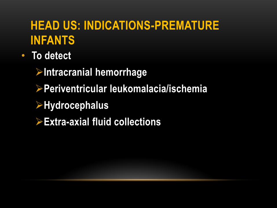
HEAD US: INDICATIONS-PREMATURE
INFANTS
• To detect
Intracranial hemorrhage
Periventricular leukomalacia/ischemia
Hydrocephalus
Extra-axial fluid collections

HEAD US: INDICATIONS-PREMATURE
INFANTS
• To follow
Intracranial hemorrhage, hydrocephalus, extra-axial fluid collections
Usually at day 7 ….
Day 1-PENUT, Seizures, decreased hematocrit, changes in neurologic status, bradycardia
< 32 weeks or < 1500 g

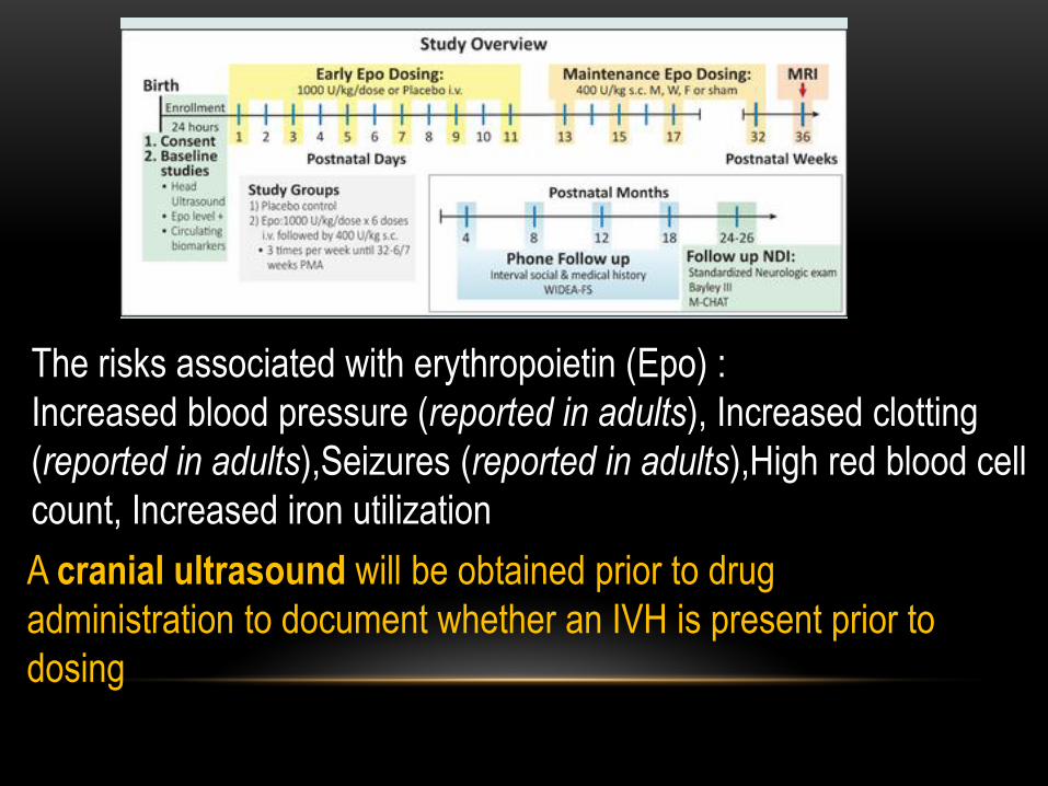
The risks associated with erythropoietin (Epo) :
Increased blood pressure (reported in adults), Increased clotting
(reported in adults),Seizures (reported in adults),High red blood cell
count, Increased iron utilization
A cranial ultrasound will be obtained prior to drug
administration to document whether an IVH is present prior to
dosing

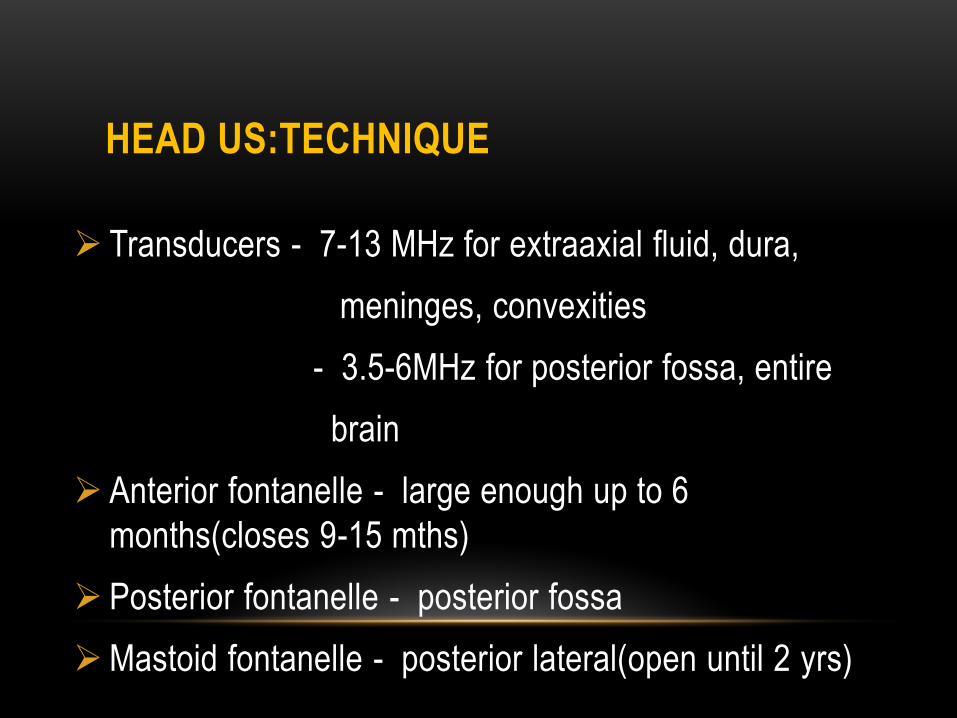
HEAD US:TECHNIQUE
Transducers - 7-13 MHz for extraaxial fluid, dura,
meninges, convexities
- 3.5-6MHz for posterior fossa, entire
brain
Anterior fontanelle - large enough up to 6
months(closes 9-15 mths)
Posterior fontanelle - posterior fossa
Mastoid fontanelle - posterior lateral(open until 2 yrs)


• < 32 weeks - smooth surface
• 36 weeks - reaches adult configuration
• Subarachnoid space should be < 5 mm in premature
infants; less in term
• Cavum septum pellucidum – usually closes by 2-6
months
• Normal cisterna magna height 3-8 mm
Premature brain-normal

Undersulcation
Venticular prominence,
prominent extraaxial
spaces,
open sylvian cistern
Cavum septum pellucidum
Premature brain-normal


INTRACRANIAL HEMORRHAGE
Premature Infants:
- Incidence : 20-25%
- Risks: < 30 wks / < 1500 g
Germinal matrix
67% of premature infant less than 32 weeks have ICH
versus 5% for term
25-50%-clinically silent, 50%-Day 1, 90% Day 3

INTRACRANIAL HEMORRHAGE
Predisposing factors
Increased systemic BP- Increased pCO2, increased IV
vol,decreased Hb
Increased CNS venous pressure-Tension
pneumothorax, asphxyia, CHF, mechanical ventilation
Decreased CNS perfusion-Hypotension, decreased
pO2,Hb

Germinal matrix
• Proliferating cells which give rise to neuroblast that
migrate peripherally to form neurons of the cerebral
cortex and basal ganglia
• Highly vascular-Ependyma of the lateral ventricle and
caudate nucleus, roof of the 3rd and the 4th ventricles
• Thin walled vessels are extremely fragile, single
endothelial lining

Germinal matrix
• Invoultion-3mth-9mths of gestation
• 28-32 weeks: only small amount left in caudothalamic
groove
• By 36 weeks: involution is complete
• Premature-Lack of autoregulation-High risk of bleed-
Capillary-Venous level hemorrhage

Burstein and Papile grading system
• Grade 1
Subependymal hemorrhage only
• Grade 2
Subependymal hemorrhage with blood in nondilated ventricles
• Grade 3 -35%
Subependymal hemorrhage with blood in dilated lateral ventricles
• Grade 4
Subependymal, blood in dilated ventricles, intraparenchymalblood

Grade 1 Hemorrhage
• Coronal image:
• Echogenic mass inferior and lateral to floor of frontal horns
• Parasagittal image:
• Echogenicity anterior to caudothalamic groove
• Clot liquefies over days to weeks, may form small 3-5 mm
subependymal cysts


Grade 2 Hemorrhage
• Most difficult to diagnose
• Germinal matrix hemorrhage ruptures through ependyma, entering lateral ventricle
• No choroid plexus in occipital horns or frontal horns, so echogenicity anterior to foramen of monroe is clot
• Clot avascular / choroid plexus is not
• Can develop hydrocephalus

Intraventricular extension

Normal Right side

Grade 3 Hemorrhage
• Expands the lateral ventricles, 3rd, 4th ventricle
• Resolves over 5-6 weeks
• Low level echoes, CSF/blood levels
• Hydrocephalus –Arrest/resolve-75%
• 10% require shunting

Grade 4 Hemorrhage
• Intraparenchymal hemorrhage
• Causes mass effect (vs PVL)
• Hemorrhagic venous infarct resulting from germinal matrix bleed compressing / thrombosis of periventricular veins
• Liquefies and retracts over several weeks
• Hypoechoic center
- Large porencephalic cysts (vs PVL) 2-3 months

Cystic encephalomalacia

PrognosisGrade Mortality Neuro
Sequelae
1 5 % 5 %
2 10 % 15 %
3 20 % 35 %
4 50 % 90 %
Neurologic Sequelae –
Mental retardation, visual impairment,spastic diplegia
or quadriplegia
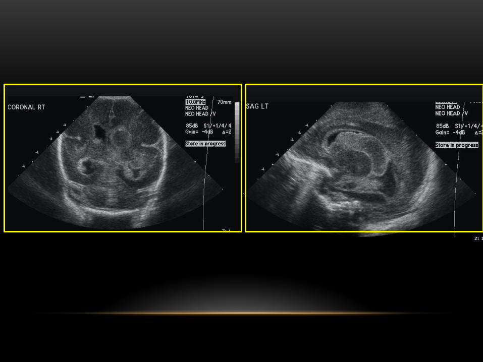
Cerebellar hemorrhage
• Cerebellar hemorrhages occur in approximately 25%
of preterm infants with very low birth weight
• External granular layer of cerebellum is also a
germinal zone
• Best imaged through post/post-lateral fontanelle
• Can result in brainstem compression, increased ICP,
cerebellar atrophy
• US: echogenic SOL in cerebellar hemisphere


WHITE MATTER INJURY /HIEOF
PREMATURITY
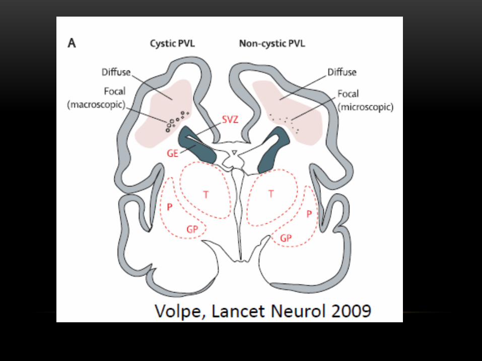
Old term “periventricular leukomalacia”
Lack of autoregulation
Periventricular white matter adjacent to trigones and
frontal horns; Deep or subcortical WM
Secondary gray matter-thalami,BG, cortex,cerebellum
US not sensitive to noncavitary white matter injury and
underestimates
Increased echogenicity of periventricular white matter
> choroid plexus
Definitive diagnosis: cystic necrosis


SUMMARY USG
Ultrasound fast and convenient for unstable infants
Better at detecting hemorrhage than ischemia/hypoxia
Initial evaluation in term infants-ischemia/hypoxia,
congenital malformations, infection


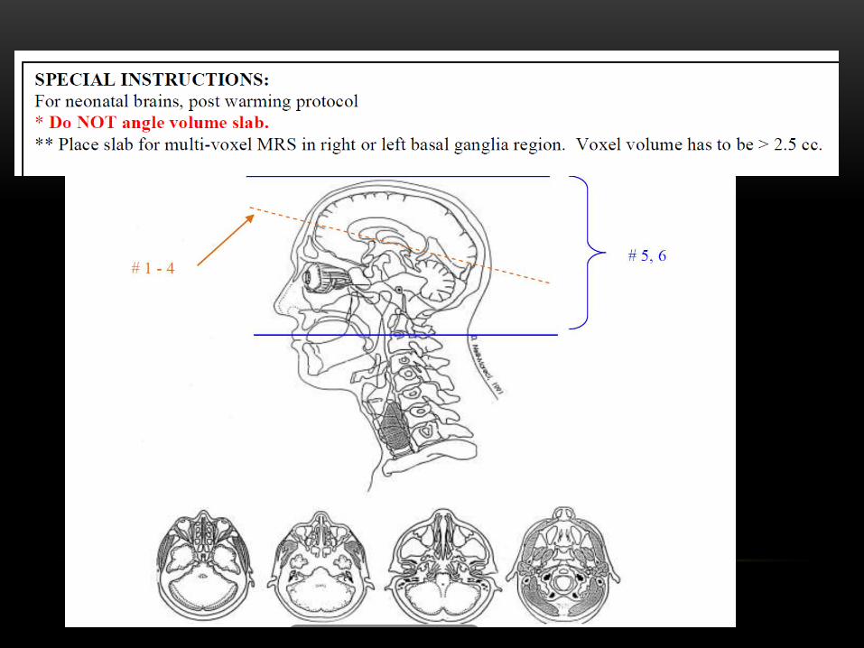
MRI

PATIENT IMAGING-MRI
Right preparation
Imaging parameters
Safety- Team, Suction pump,O2
supply,Laryngoscope,Monitoring devices
Examination on the day of the study
Swaddling
Scan on side
Adult knee coil
http://cfimedical.com/medvac/

MRI
Neonates’ vital signs are prone to fluctuate, and several
parameters must be closely monitored
STABLE- sugar, temperature, artificial breathing, blood
pressure, and laboratory test results
High-quality coronal diffusion-weighted images
also can be obtained-neonates lack pneumatized
paranasal sinuses
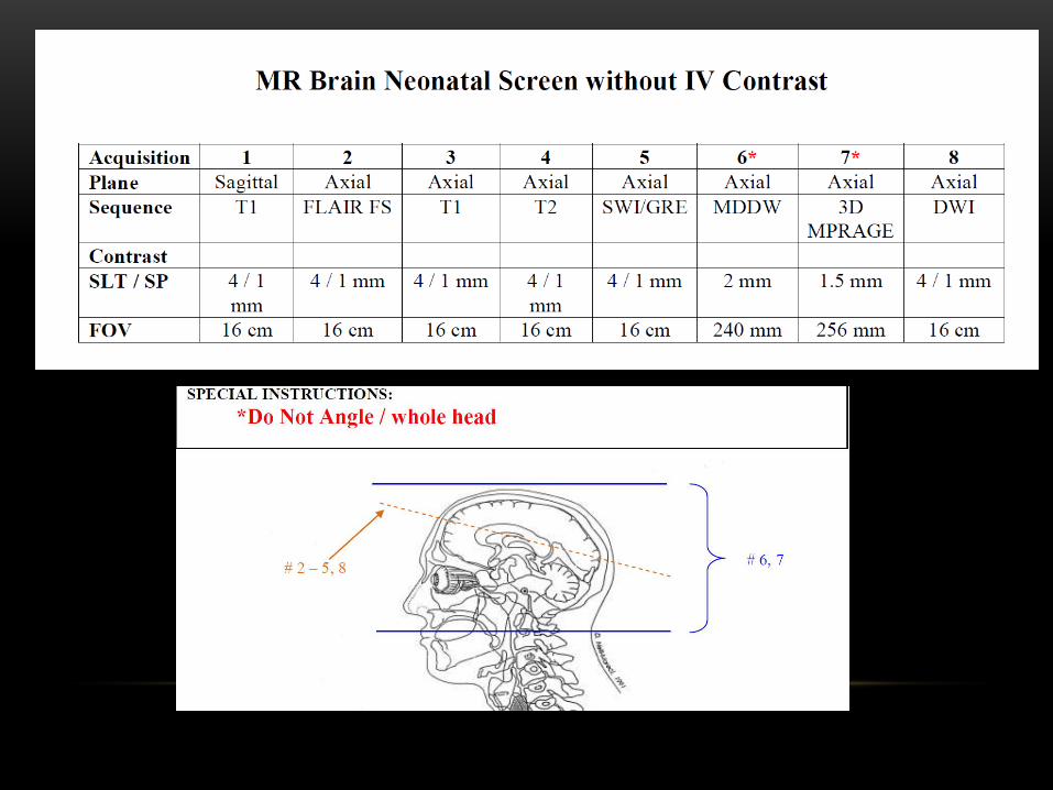


FLAIR-Poor due to high water content
Imaging best -1-2 week
Diffusion-False negative < 24 hrs
Pseudonormalize- 6 day
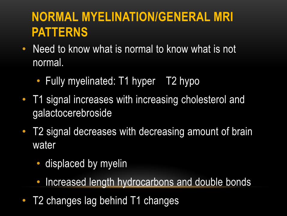
NORMAL MYELINATION/GENERAL MRI
PATTERNS
• Need to know what is normal to know what is not
normal.
• Fully myelinated: T1 hyper T2 hypo
• T1 signal increases with increasing cholesterol and
galactocerebroside
• T2 signal decreases with decreasing amount of brain
water
• displaced by myelin
• Increased length hydrocarbons and double bonds
• T2 changes lag behind T1 changes

PROGRESSION OF MYELINATION
Rostral to caudal; Posterior to anterior; Central to peripheral

Myelination• 20 weeks-Pons,Post medulla
• 29 weeks-Sup and Inf cerebellar peduncles
• 32 weeks-Midbrain
• 33 weeks-Inferior colliculi, lateral
putamen,ventrolateral thalami
• 35 weeks-Post limb of Internal capsule
• 35 weeks-2 mths- Optic tracts,medial temporal
lobes,perirolandic fissures,calcarine,central
white matter,rest of the basal ganglia

Courtesy: Dr. Robert McKinstry
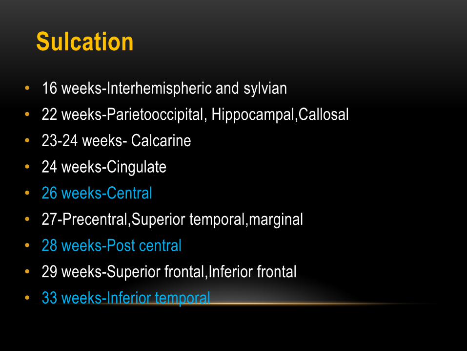
Sulcation
• 16 weeks-Interhemispheric and sylvian
• 22 weeks-Parietooccipital, Hippocampal,Callosal
• 23-24 weeks- Calcarine
• 24 weeks-Cingulate
• 26 weeks-Central
• 27-Precentral,Superior temporal,marginal
• 28 weeks-Post central
• 29 weeks-Superior frontal,Inferior frontal
• 33 weeks-Inferior temporal
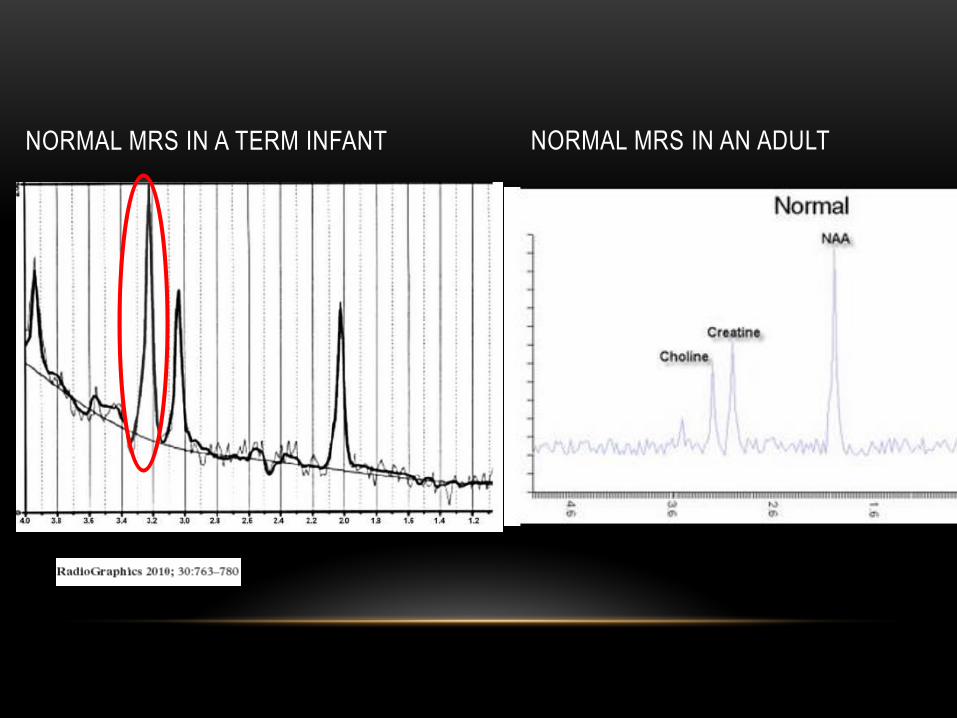
NORMAL MRS IN A TERM INFANT NORMAL MRS IN AN ADULT
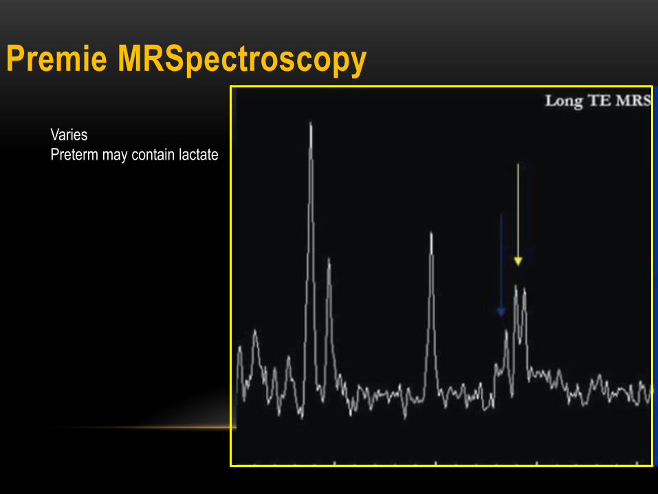
Premie MRSpectroscopy
Varies
Preterm may contain lactate


HIE IN PRETERM
• 50% of cases of cerebral palsy –Premature infants
• Up to 19% of infants born before 28 weeks of
gestation develop cerebral palsy
• Hypoperfusion –Watershed Ischemia-
Premyelinating neurons
• Lack of autoregulation

HIE IN PRETERM
• Severe hypoxic-ischemic insults to the premature
brain typically injure the thalamus, anterior part of
the vermis, and dorsal brainstem. Involvement of
the basal ganglia, hippocampus, cerebellum, and
corticospinal tracts also may be seen
• Mild to moderate hypoxic-ischemic injury may
result in a germinal matrix hemorrhage,
periventricular leukomalacia, or both
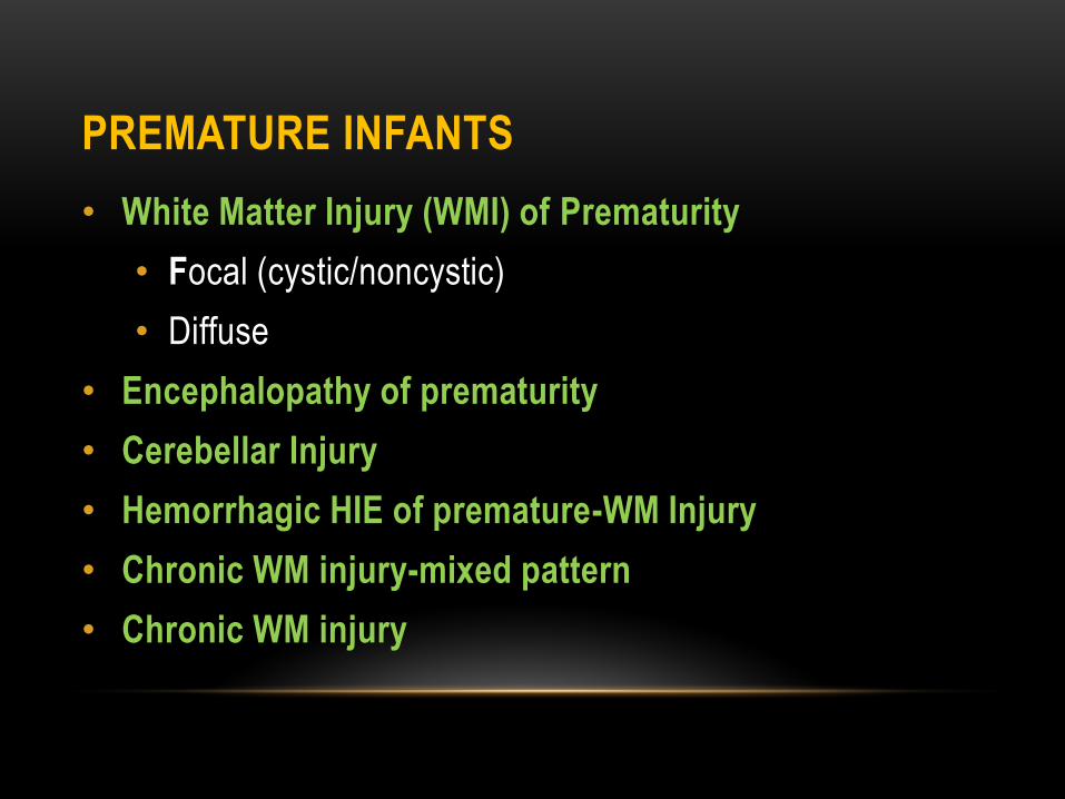
PREMATURE INFANTS
• White Matter Injury (WMI) of Prematurity
• Focal (cystic/noncystic)
• Diffuse
• Encephalopathy of prematurity
• Cerebellar Injury
• Hemorrhagic HIE of premature-WM Injury
• Chronic WM injury-mixed pattern
• Chronic WM injury

FOCAL NON CYSTIC EX 30 WEEK EGA

2 PATIENTS WITH CYSTIC TYPE INJURY
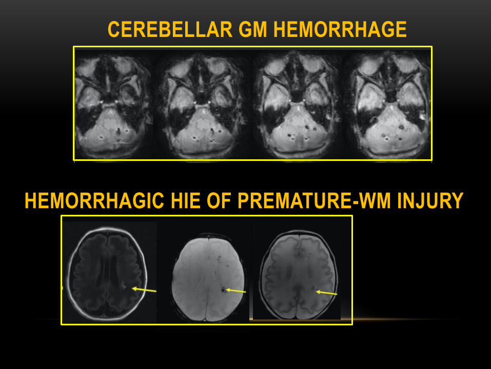
CEREBELLAR GM HEMORRHAGE
HEMORRHAGIC HIE OF PREMATURE-WM INJURY

CHRONIC WM INJURY-MIXED PATTERN
Thinning of the corpus callosum, particularly in the
posterior body and splenium, is a characteristic late
feature of periventricular leukomalacia
CHRONIC WM INJURY

PREMATURE- SEVERE INJURY
DAY 2
Died on day 16
DAY 15Day 7

Diffusion in the cortex is more restricted because of the higher
ratio of cells to extracellular space

DIFFUSE EXCESSIVE HIGH SIGNAL INTENSITY IN
WM (DEHSI)
• Controversial
• WM
• Increase diffusion
• Poor neurologic outcome
• Transient normal process
• No difference; No difference ADC values with controls


FULL TERM INFANTS
• Severe, basal ganglia pattern
• Severe, total hypoxia
• Mixed pattern
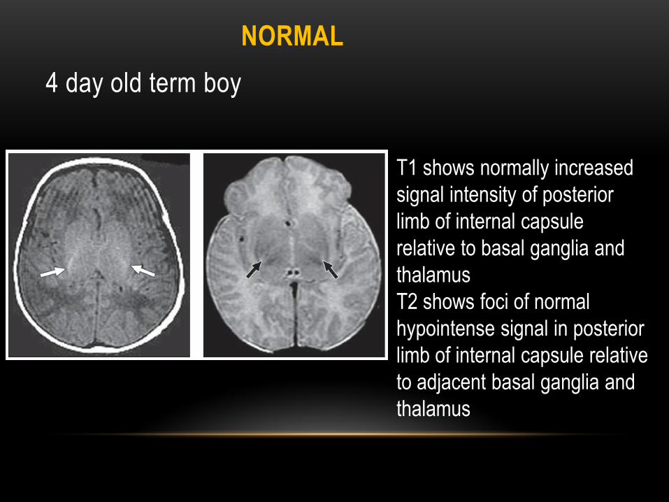
4 day old term boy
T1 shows normally increased
signal intensity of posterior
limb of internal capsule
relative to basal ganglia and
thalamus
T2 shows foci of normal
hypointense signal in posterior
limb of internal capsule relative
to adjacent basal ganglia and
thalamus
NORMAL

2 day old 36 week EGA boy
Hypointense T1 signal in post. Limb of
internal capsule. This is normal for age in
36 wk EGA
Range of variation in signal intensity that
can be seen in normal brain—basal ganglia
show moderately hyperintense signal,
although less than that typically seen in
hypoxia
NORMAL

ADC
DAY 4
DWI
DAY 47
High T1 signal in
basal ganglia and
thalamus from
intracellular
calcium shift and
necrosis
BASAL GANGLIA PATTERN
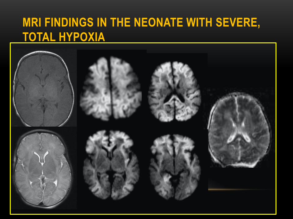
MRI FINDINGS IN THE NEONATE WITH SEVERE,
TOTAL HYPOXIA

ABNORMAL NORMAL
Abnormal high
signal throughout
the WM on T2
Blurring of GW
differentiation
more evident on
B=0 than
conventional T2-
weighted images

DAY 3

DAY 49

CURRENT THERAPIES
• Modest reductions in brain temperature-2º to 4º C, -neuroprotective
• STANDARD OF CARE
• The aim is to cool infants with moderate or severe HIE within 6 h of birth to a body temperature between 33.5°C and 34.5°C and maintain this degree of cooling without interruption for 72 h
• Slow re-warming over at least 4 h at a rate of 0.5°C per hour until their rectal temperature reaches the desired range (36.5-37°C)

Reduction in glutamate release
Reduction in cerebral metabolism
Decrease in intracellular acidosisand lactic acid accumulation
Preservation of endogenous antioxidants
Reduction of leukotriene production
Prevention of blood-brain barrier disruptionand brain edema
Inhibition of apoptosis

38 WEEK EGA GIRL INFANT BORN AFTER
INDUCTION FOR MATERNAL PRE-ECLAMPSIA
Hypoxic ischemic injury s/p cooling.
Infant is now 5 days old and is being
re-warmed

Inc T1 signal in corticospinal tracts, lentiform nuclei and thalami (subtle), and decreased T1 signal in
posterior limbs of internal capsule

Subtle decreased signal on ADC map in corticospinal tracts, lentiform nuclei and posterior limbs of
internal capsules. No DWI changes because they’ve already normalized.
KEY POINTS• HIE usually manifests within the first few hours after birth
• A few days after birth - without an obvious reason,
metabolic and infectious causes must be considered
• Normal Neonate MR Findings->37 weeks EGA
• ↑ T1 & ↓T2 signal in posterior half of posterior limb of
internal capsule
• At a minimum, 1/3 of the length should be T1
hyperintense
• Usually seen during first 24 hours of life
• If <36 weeks EGA: no ↑ T1 in this region = normal finding

TERM
Severe hypoxic-ischemic
insults to the premature brain
typically affects:
• Thalamus
• Anterior part of the
vermis
• Dorsal brainstem
• Injury to the basal
ganglia is usually less
severe and common
• Severe hypoxic-ischemic
injury in term baby involves:
• Ventral and lateral aspects
of the thalamus
• Posterior aspect of the
putamen
• Perirolandic regions
• Corticospinal tracts
PRETERM
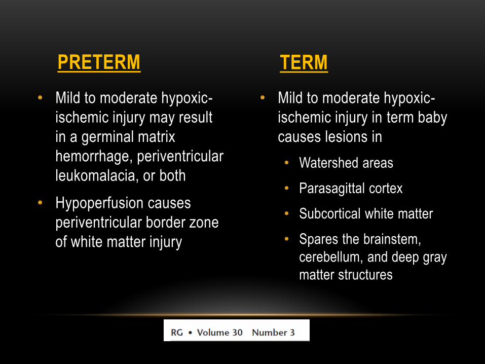
• Mild to moderate hypoxic-
ischemic injury may result
in a germinal matrix
hemorrhage, periventricular
leukomalacia, or both
• Hypoperfusion causes
periventricular border zone
of white matter injury
• Mild to moderate hypoxic-
ischemic injury in term baby
causes lesions in
• Watershed areas
• Parasagittal cortex
• Subcortical white matter
• Spares the brainstem,
cerebellum, and deep gray
matter structures
PRETERM TERM

FUTURE/ ADVANCES
Inder et al

FUTURE/ ADVANCES

IMPORTANT CLINICALCORRELATES
Long-term studies of the outcome of very
prematurely born infants -significant motor,
cognitive, and behavioral deficits
More prone to develop encephalopathies
In comparison to the term-born infants, the
premature infants at term demonstrated prominent
reductions in cerebral cortical and deep GM volume
The major predictors of altered cerebral volumes
were gestational age at birth and the presence of
cerebral WM injury
IMPORTANT CLINICALCORRELATES
Infants with significantly reduced cortical GM and
deep nuclear GM volumes and increased CSF
volume volumes exhibited moderate to severe
neurodevelopmental disability at 1 year of age
The nature of the cerebral abnormalities that
underlie these common and serious developmental
disabilities is not entirely understood
Postulated-WM injury and delayed WM and GM
gyral development

IMPORTANT CLINICALCORRELATES
No influence of immaturity on cerebral myelinated
or unmyelinated WM volumes
Deep nuclear GM volumes and the number of days
of ventilator support alteration of axonal fiber
development
High-dose postnatal dexamethasone therapy has
been shown to be associated with significantly
reduced cortical GM and cerebral tissue volumes

CONCLUSIONS:
Hypoxic ischemic injury manifests differently in a
full term than in a premature on MRI
USG of head serves as a baseline examination to
enroll a patient in the PENUT trial AND a routine
baseline scan on day 7 of a premature baby
Imaging of the patients who have undergone
cooling demonstrate lesser extent of brain injury
