imaging in aerosol medicine - respiratory...
TRANSCRIPT

Imaging in Aerosol Medicine
Timothy E Corcoran PhD
IntroductionTechniques to Assess Aerosol DeliveryAerosol-Based Physiology MeasurementsLimitations of Aerosol-Based TechniquesFuture Developments
Imaging techniques have been used extensively to study the delivery of inhaled medications. De-position scintigraphy involves the quantification of deposited aerosol dose and is performed using2-dimensional planar or 3-dimensional positron emission tomography (PET) or single-photon-emission computed tomography (SPECT) imaging techniques. Planar techniques have an extensivehistory of use, and quantification methods are well established. SPECT and PET techniques canprovide better dose localization, but quantification is more complex, and the techniques are in morelimited use. Aerosols have also been used to deliver radiopharmaceutical probes for the imaging oflung physiology. These studies include measurements of ventilation, mucus and cough clearance,and, more recently, liquid absorption in the airways. Clearance measurements have been used toassess therapeutic response in conditions such as cystic fibrosis. Future directions in aerosol-basedimaging are likely to include use of novel probes to measure new physiological processes in the lung,more thorough integration of anatomical imaging, and use of multiple probes to simultaneouslyimage drug and disease or interacting physiological processes. Key words: aerosol deposition; aerosolscintigraphy; mucus clearance; lung imaging; molecular imaging; nuclear imaging. [Respir Care2015;60(6):850–857. © 2015 Daedalus Enterprises]
Introduction
A key aspect in the development of inhaled medicationsis the measurement and optimization of deposited aerosoldose. The use of imaging techniques to measure aerosoldrug deposition has followed the development of morecomplex inhaled therapies and the invention of more ad-vanced aerosol delivery technologies. Deposition scintig-
raphy techniques provide quantitative information on aero-sol deposition using the radioactivity associated with anadded radiopharmaceutical as an analog for drug dose.Nuclear imaging cameras can depict the deposited aerosolwithin the body, allowing for regional dose quantification.Deposition scintigraphy techniques have evolved alongwith nuclear imaging technology, and a variety of 2-di-mensional and 3-dimensional techniques are now in use,some of which include anatomical imaging. These tech-niques have been applied with medical nebulizers, me-tered-dose inhalers, and powder inhalers and used in par-Dr Corcoran is affiliated with the Division of Pulmonary, Allergy,
and Critical Care Medicine, University of Pittsburgh, Pittsburgh,Pennsylvania.
Dr Corcoran has disclosed no conflicts of interest.
Dr Corcoran presented a version of this paper at the 53rd RRESPIRATORY
CARE Journal Conference “Aerosol Drug Delivery in Respiratory Care,”held June 6–7, 2014, in St Petersburg, Florida.
Correspondence: Timothy E Corcoran PhD, UPMC MUH NW628, 3459Fifth Avenue, Pittsburgh, PA 15213. E-mail: [email protected].
DOI: 10.4187/respcare.03537
850 RESPIRATORY CARE • JUNE 2015 VOL 60 NO 6

allel with pharmacokinetic measurements. Aerosols havealso been used to deliver radiopharmaceutical probes tothe lungs for physiological imaging purposes. These aero-sol-based imaging techniques allow measurements of ven-tilation, permeability, liquid absorption, and clearance inthe lungs. In some cases, these measurements have provedto be a valuable outcome measure for therapeutic devel-opment.
Techniques to Assess Aerosol Delivery
The importance of quantifying deposited aerosol dosecan be appreciated by considering medications with nar-row therapeutic indices, such as insulin, or with no imme-diate indication of clinical effect, such as an inhaled ste-roid. Dosing variability in aerosol drug delivery is welldocumented, especially in the setting of lung disease. De-position scintigraphy can be used to quantify and optimizedelivered aerosol dose and dosing variability ahead of theperformance of pivotal clinical studies.
Deposition scintigraphy involves the use of either director indirect radiopharmaceutical labels. Technetium-99m(99mTc) is often used for this labeling based on its strongimaging signal and its limited half-life (6 h).1 Indirect la-beling is performed by adding an approved 99mTc com-pound, such as 99mTc-diethylenetriamine penta-acetic or99mTc-sulfur colloid, to an aerosol medication and dem-onstrating that the radioactivity associated with the 99mTcdistributes proportionally to the active drug contentthroughout a range of different aerosol sizes.2 Direct la-beling can often be performed through stannous reductionof 99mTc before addition to the active drug.3 If performedsuccessfully, the 99mTc is then bound directly to the activedrug. Techniques for establishing and validating these la-bels are described in the literature.4
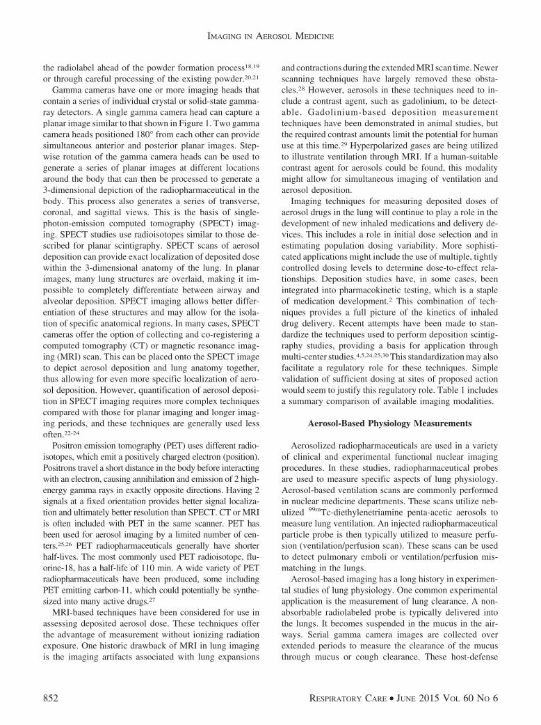
Once a labeling method is established, a combination ofthe inhaled medication and the radiopharmaceutical is de-livered using a clinically applicable delivery system. Theradioactivity associated with 99mTc acts as an analog fordrug dose. It can be measured in the delivery system be-fore and after delivery to determine the total delivereddose (a mass balance technique). When specific calibra-tions are in place, total deposited dose can also be deter-mined directly from gamma camera images. Figure 1 in-cludes a planar image of a lung transplant recipientperforming a deposition scintigraphy study. These imagesprovide both a qualitative picture of aerosol deposition inthe lungs and a means to quantify the dose in differentdeposition zones. Quantification of deposited dose fromgamma camera images involves a series of correctionstypically including decay, background, and attenuation cor-rection and the use of geometric mean averages of anteriorand posterior images to account for the different distancesbetween the organs and the camera. Regional dosing may
be determined by simply dividing up the total planar im-age counts by region. Reviews of these techniques havebeen published as part of attempts to standardize them formulti-center use.5
Typically �148 MBq (4 mCi) of 99mTc is added to anebulizer for a deposition scintigraphy scan. The associ-ated radiation exposure after inhalation depends on theefficiency of the delivery system, the physiological andradiological half-life of the radiopharmaceutical in use,and the size (age) of the patient. As an example, a 148-MBq dose of 99mTc-sulfur colloid delivered with 20% ef-ficiency would result in an internal dose of 29.6 MBq(0.8 mCi), which would be associated with an estimatedeffective dose equivalent exposure of 0.44 mSv (44 mrem)for an adult subject.6 This is �14% of yearly natural back-ground exposure.7 These exposure levels are low enoughto allow multiple studies and pediatric applications.
Nebulizer delivery systems are the simplest to assessusing deposition scintigraphy methods because the activedrug is directly accessible for labeling. Many examples ofdeposition studies with nebulizers are available in the lit-erature.2,8-12 These studies were instrumental in demon-strating the significant intersubject variability associatedwith nebulized drug delivery and contributed to the devel-opment of new and more precise delivery systems. Studieswith metered-dose inhalers require on-site canister charg-ing after drug labeling and thus pose more complica-tions.13-17 The labeling of powders presents even moresubstantial complications because the labeling techniquemust be applied without affecting the shape or size of thedrug particles. This can be accomplished by incorporating
Fig. 1. Planar image depicting aerosol deposition in a lung trans-plant recipient.
IMAGING IN AEROSOL MEDICINE
RESPIRATORY CARE • JUNE 2015 VOL 60 NO 6 851
the radiolabel ahead of the powder formation process18,19
or through careful processing of the existing powder.20,21
Gamma cameras have one or more imaging heads thatcontain a series of individual crystal or solid-state gamma-ray detectors. A single gamma camera head can capture aplanar image similar to that shown in Figure 1. Two gammacamera heads positioned 180° from each other can providesimultaneous anterior and posterior planar images. Step-wise rotation of the gamma camera heads can be used togenerate a series of planar images at different locationsaround the body that can then be processed to generate a3-dimensional depiction of the radiopharmaceutical in thebody. This process also generates a series of transverse,coronal, and sagittal views. This is the basis of single-photon-emission computed tomography (SPECT) imag-ing. SPECT studies use radioisotopes similar to those de-scribed for planar scintigraphy. SPECT scans of aerosoldeposition can provide exact localization of deposited dosewithin the 3-dimensional anatomy of the lung. In planarimages, many lung structures are overlaid, making it im-possible to completely differentiate between airway andalveolar deposition. SPECT imaging allows better differ-entiation of these structures and may allow for the isola-tion of specific anatomical regions. In many cases, SPECTcameras offer the option of collecting and co-registering acomputed tomography (CT) or magnetic resonance imag-ing (MRI) scan. This can be placed onto the SPECT imageto depict aerosol deposition and lung anatomy together,thus allowing for even more specific localization of aero-sol deposition. However, quantification of aerosol deposi-tion in SPECT imaging requires more complex techniquescompared with those for planar imaging and longer imag-ing periods, and these techniques are generally used lessoften.22-24
Positron emission tomography (PET) uses different radio-isotopes, which emit a positively charged electron (position).Positrons travel a short distance in the body before interactingwith an electron, causing annihilation and emission of 2 high-energy gamma rays in exactly opposite directions. Having 2signals at a fixed orientation provides better signal localiza-tion and ultimately better resolution than SPECT. CT or MRIis often included with PET in the same scanner. PET hasbeen used for aerosol imaging by a limited number of cen-ters.25,26 PET radiopharmaceuticals generally have shorterhalf-lives. The most commonly used PET radioisotope, flu-orine-18, has a half-life of 110 min. A wide variety of PETradiopharmaceuticals have been produced, some includingPET emitting carbon-11, which could potentially be synthe-sized into many active drugs.27
MRI-based techniques have been considered for use inassessing deposited aerosol dose. These techniques offerthe advantage of measurement without ionizing radiationexposure. One historic drawback of MRI in lung imagingis the imaging artifacts associated with lung expansions
and contractions during the extended MRI scan time. Newerscanning techniques have largely removed these obsta-cles.28 However, aerosols in these techniques need to in-clude a contrast agent, such as gadolinium, to be detect-able. Gadolinium-based deposition measurementtechniques have been demonstrated in animal studies, butthe required contrast amounts limit the potential for humanuse at this time.29 Hyperpolarized gases are being utilizedto illustrate ventilation through MRI. If a human-suitablecontrast agent for aerosols could be found, this modalitymight allow for simultaneous imaging of ventilation andaerosol deposition.
Imaging techniques for measuring deposited doses ofaerosol drugs in the lung will continue to play a role in thedevelopment of new inhaled medications and delivery de-vices. This includes a role in initial dose selection and inestimating population dosing variability. More sophisti-cated applications might include the use of multiple, tightlycontrolled dosing levels to determine dose-to-effect rela-tionships. Deposition studies have, in some cases, beenintegrated into pharmacokinetic testing, which is a stapleof medication development.2 This combination of tech-niques provides a full picture of the kinetics of inhaleddrug delivery. Recent attempts have been made to stan-dardize the techniques used to perform deposition scintig-raphy studies, providing a basis for application throughmulti-center studies.4,5,24,25,30 This standardization may alsofacilitate a regulatory role for these techniques. Simplevalidation of sufficient dosing at sites of proposed actionwould seem to justify this regulatory role. Table 1 includesa summary comparison of available imaging modalities.
Aerosol-Based Physiology Measurements
Aerosolized radiopharmaceuticals are used in a varietyof clinical and experimental functional nuclear imagingprocedures. In these studies, radiopharmaceutical probesare used to measure specific aspects of lung physiology.Aerosol-based ventilation scans are commonly performedin nuclear medicine departments. These scans utilize neb-ulized 99mTc-diethylenetriamine penta-acetic aerosols tomeasure lung ventilation. An injected radiopharmaceuticalparticle probe is then typically utilized to measure perfu-sion (ventilation/perfusion scan). These scans can be usedto detect pulmonary emboli or ventilation/perfusion mis-matching in the lungs.
Aerosol-based imaging has a long history in experimen-tal studies of lung physiology. One common experimentalapplication is the measurement of lung clearance. A non-absorbable radiolabeled probe is typically delivered intothe lungs. It becomes suspended in the mucus in the air-ways. Serial gamma camera images are collected overextended periods to measure the clearance of the mucusthrough mucus or cough clearance. These host-defense
IMAGING IN AEROSOL MEDICINE
852 RESPIRATORY CARE • JUNE 2015 VOL 60 NO 6

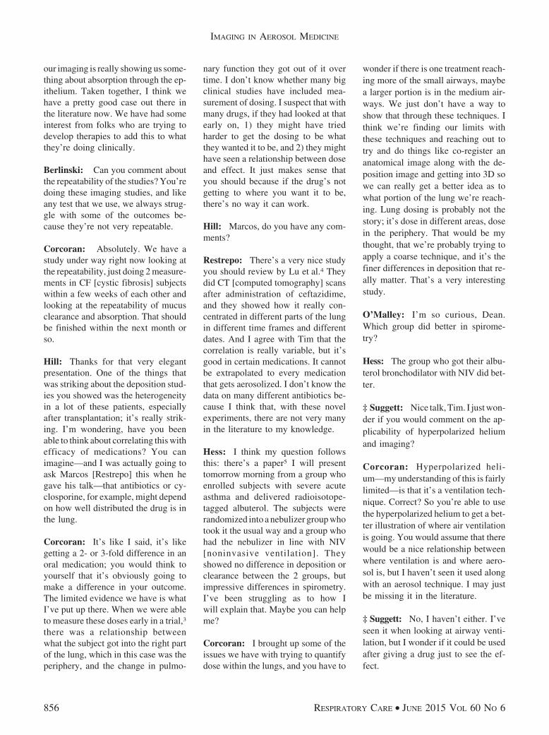
mechanisms are vital to protecting the lungs from inhaledpathogens and particulate. Their failure in diseases such ascystic fibrosis can lead to life-threatening opportunisticinfections. Mucus clearance scans have been applied totest new medications for cystic fibrosis. In some cases,these imaging methods may provide indications of thera-peutic efficacy ahead of other outcome measures.31-33 Fig-ure 2 shows example images depicting mucus clearance.
Radiolabeled small-molecule probes have been deliv-ered to the lungs by aerosol to assess lung permeability.These probes can be absorbed into the bloodstream, andtheir rate of clearance has been used as an indication oflung inflammation and injury. Increases in the absorption
rate of radiolabeled diethylenetriamine penta-acetic havebeen demonstrated in smokers34 and in subjects with in-flammatory diseases such as asthma.35 Small-moleculeprobes have also been developed to detect changes in air-way liquid absorption. In these studies, a multiprobe methodis utilized. A particle probe is delivered along with a small-molecule probe in the same aerosol. This small moleculeis removed from the lungs through both absorption andmucus clearance, whereas the particle is removed only bymucus clearance. Each probe has a different radiolabel, sothey can be independently tracked. The difference in theclearance rates of the probes provides an estimate of theabsorption rate of the small-molecule probe. These absorp-tive clearance rates have been shown to be related to liquidabsorption by in vitro studies and increased in the airwaysof patients with cystic fibrosis. Airway liquid hyperab-sorption is a key aspect of cystic fibrosis lung pathophys-iology that would be expected to change rapidly with asuccessful therapy, and this technique is being developedto rapidly screen new cystic fibrosis medications.36,37
Limitations of Aerosol-Based Techniques
Much of the experience in measuring aerosol depositionusing imaging techniques was obtained through studies ofuniform solutions delivered with jet nebulizers. As inhaleddrug development moves more toward the use of powders,suspensions, and complex large molecules, new labelingand validation techniques will be required. Techniquesthat could be applied more generally across different prep-arations with similar characteristics would be particularlyuseful.
Aerosol-based physiology techniques are ultimately lim-ited by the methods used to deliver aerosol probes. More
Table 1. Comparison of Various Imaging Modalities for Aerosol-Based Techniques (Deposition or Physiology)
Technique Advantages Disadvantages Citation
Planar scintigraphy Established quantification techniques, availablestandardized procedures
Poor representation of 3-dimensional lunganatomy
Newman et al5
3-Dimensional SPECTscintigraphy
3-Dimensional imaging, potential to localizesites of disease through co-registration withCT/MRI
Long imaging times possibly not suitable forphysiology, more complex quantification
Fleming et al24
3-Dimensional PETscintigraphy
3-Dimensional imaging, potential to localizesites of disease through co-registration withCT/MRI, potential for directly labeled drugsor sophisticated physiology probes
More complex radiopharmaceuticalproduction, often short-livedradiopharmaceuticals
Dolovich and Bailey25
MRI 3-Dimensional imaging, potential to localizesites of disease, potential for simultaneousimaging of ventilation
Human use limited by required large amountsof current contrast agents
Thompson andFinlay29
SPECT � single-photon-emission computed tomographyCT � computed tomographyMRI � magnetic resonance imagingPET � positron emission tomography
Fig. 2. Mucociliary clearance of radiolabeled particulate from thelarge airways depicted over �60 min.
IMAGING IN AEROSOL MEDICINE
RESPIRATORY CARE • JUNE 2015 VOL 60 NO 6 853

targeted delivery techniques would allow for more specificassessments, for example, better isolation of small airwaysfrom alveoli. In this regard, novel aerosol technologiesdesigned to target drug delivery may find a second appli-cation in the assessment of lung physiology. All of thesetechniques are limited by the spatial resolution providedby the imaging modality. No current modality is capableof imaging the smallest airways or alveoli. Physiologicalimaging methods are further limited by the temporal res-olution of the imaging modality.
Future Developments
PET imaging provides unique opportunities for physi-ological lung imaging. The increased resolution of thesetechniques can provide local assessments that may not bepossible with other modalities. A wide variety of PETprobes can be produced by cyclotron. These include mol-ecules such as carbon-11, nitrogen-13, fluorine-18, andoxygen-15. These basic molecules can be used to generatesimple probes, such as 13N-labeled ammonia or 15O-la-beled water, or more complex probes, such as active drugmolecules or antibodies. Many specific PET probes havebeen developed to quantify aspects of physiology such ascell metabolism, tumor growth, angiogenesis, and forma-tion of the extracellular matrix.38-41 PET probes are com-monly used to detect the increased metabolic rate associ-ated with tumor growth. Other injected PET probes havealready been used in studies of ventilation, perfusion, andinflammation.42 However, very little work has been donewith aerosol delivery. The attraction of this route is directaccess to the pulmonary epithelium. As described above,PET methods provide improved resolution and can be over-laid on matching CT or MRI scans collected in the samescanner, thus allowing for exact localization. Drawbacksto PET methods include the very limited half-life of someof the positron emitters and the extensive facilities re-quired to produce them.
Most new nuclear imaging scanners combine a func-tional imaging modality (SPECT or PET) with an anatom-ical imaging method (CT or MRI). This mixed-modalityimaging allows drug doses or physiological measurementsto be more exactly located. This is particularly advanta-geous given the complex 3-dimensional structure of thelung. Lung MRI, which was generally not considered use-ful in the past because of the motion artifacts associatedwith breathing, is becoming more common.28,43 This mo-dality could potentially provide finer resolution than haspreviously been available through CT scans and spare pa-tients the significant radiation burden associated with CT.
We had previously described the aerosol delivery ofmultiple radiolabeled probes to the lungs to measure ab-sorptive clearance (liquid absorption) in the airways.37 Inthose studies, the combination of the probes provided in-
formation that was not available with a single probe. Theseprobes were independently resolvable because they weretagged with different radioisotopes, specifically indium-111 and 99mTc. New imaging techniques could be de-signed based on a differential method such as this or aco-localization method. An intriguing example would bean imaging technique that independently depicts patho-physiology and therapeutic delivery in the lung. Consider,for example, an aerosol deposition study of an inhaledsteroid that also includes a depiction of lung inflammationor a chemotherapeutic that could be localized to the site ofa tumor. Delivery techniques could be tuned as part of thedevelopment process or customized to specific patients.As imaging technologies and probe developments advance,such measurements may become feasible and provide newinsights into disease treatments. More precise aerosol drugdelivery technologies would allow for easier and moreprecise application of these techniques to the lungs.
REFERENCES
1. Kuni C, duCret, RP. Manual of nuclear medicine imaging. NewYork: Thieme Medical Publishers; 1997.
2. Corcoran TE, Niven R, Verret W, Dilly S, Johnson BA. Lung de-position and pharmacokinetics of nebulized cyclosporine in lungtransplant patients. J Aerosol Med Pulm Drug Deliv 2014;27(3):178-184.
3. Saha G. Fundamentals of nuclear pharmacy, 5th edition. New York:Springer; 2004.
4. Devadason SG, Chan HK, Haeussermann S, Kietzig C, Kuehl PJ,Newman S, et al. Validation of radiolabeling of drug formulationsfor aerosol deposition assessment of orally inhaled products. J Aero-sol Med Pulm Drug Deliv 2012;25(Suppl 1):S6-S9.
5. Newman S, Bennett WD, Biddiscombe M, Devadason SG, DolovichMB, Fleming J, et al. Standardization of techniques for using planar(2D) imaging for aerosol deposition assessment of orally inhaledproducts. J Aerosol Med Pulm Drug Deliv 2012;25(Suppl 1):S10-S28.
6. International Commission on Radiological Protection. Radiation doseto patients from radiopharmaceuticals. Ann ICRP 1988;18(1-4):ICRPpublication 53.
7. United States Nuclear Regulatory Commission. Fact sheet on bio-logical effects of radiation. http://www.nrc.gov/reading-rm/doc-collections/fact-sheets/bio-effects-radiation.html. Accessed Septem-ber 26, 2014.
8. Amirav I, Balanov I, Gorenberg M, Luder AS, Newhouse MT, GrosharD. Beta-agonist aerosol distribution in respiratory syncytial virusbronchiolitis in infants. J Nucl Med 2002;43(4):487-491.
9. Nikander K, Prince I, Coughlin S, Warren S, Taylor G. Mode ofbreathing–tidal or slow and deep–through the I-neb adaptive aerosoldelivery (AAD) system affects lung deposition of 99mTc-DTPA. JAerosol Med Pulm Drug Deliv 2010;23(Suppl 1):S37-S43.
10. O’Riordan TG, Palmer LB, Smaldone GC. Aerosol deposition inmechanically ventilated patients. Optimizing nebulizer delivery. Am JRespir Crit Care Med 1994;149(1):214-219.
11. Behr J, Zimmermann G, Baumgartner R, Leuchte H, Neurohr C,Brand P, et al. Lung deposition of a liposomal cyclosporine A in-halation solution in patients after lung transplantation. J Aerosol MedPulm Drug Deliv 2009;22(2):121-130.
IMAGING IN AEROSOL MEDICINE
854 RESPIRATORY CARE • JUNE 2015 VOL 60 NO 6

12. Ilowite JS, Gorvoy JD, Smaldone GC. Quantitative deposition ofaerosolized gentamicin in cystic fibrosis. Am Rev Respir Dis 1987;136(6):1445-1449.
13. Newman SP, Pitcairn GR, Adkin DA, Vidgren MT, Silvasti M.Comparison of beclomethasone dipropionate delivery by Easyhalerdry powder inhaler and pMDI plus large volume spacer. J AerosolMed 2001;14(2):217-225.
14. Devadason SG, Huang T, Walker S, Troedson R, Le Souef PN.Distribution of technetium-99m-labelled QVAR delivered using anAutohaler device in children. Eur Respir J 2003;21(6):1007-1011.
15. Pitcairn G, Reader S, Pavia D, Newman S. Deposition of cortico-steroid aerosol in the human lung by Respimat Soft Mist inhalercompared to deposition by metered dose inhaler or by Turbuhalerdry powder inhaler. J Aerosol Med 2005;18(3):264-272.
16. Leach CL, Bethke TD, Boudreau RJ, Hasselquist BE, Drollmann A,Davidson P, Wurst W. Two-dimensional and three-dimensional im-aging show ciclesonide has high lung deposition and peripheral dis-tribution: a nonrandomized study in healthy volunteers. J AerosolMed 2006;19(2):117-126.
17. Brand P, Hederer B, Austen G, Dewberry H, Meyer T. Higher lungdeposition with Respimat Soft Mist inhaler than HFA-MDI in COPDpatients with poor technique. Int J Chron Obstruct Pulmon Dis 2008;3(4):763-770.
18. Glover W, Chan HK, Eberl S, Daviskas E, Verschuer J. Effect ofparticle size of dry powder mannitol on the lung deposition in healthyvolunteers. Int J Pharm 2008;349(1-2):314-322.
19. Newman S, Malik S, Hirst R, Pitcairn G, Heide A, Pabst J, et al.Lung deposition of salbutamol in healthy human subjects from theMAGhaler dry powder inhaler. Respir Med 2002;96(12):1026-1032.
20. Ball DJ, Hirst PH, Newman SP, Sonet B, Streel B, Vanderbist F. De-position and pharmacokinetics of budesonide from the Miat Monodoseinhaler, a simple dry powder device. Int J Pharm 2002;245(1-2):123-132.
21. Hirst PH, Bacon RE, Pitcairn GR, Silvasti M, Newman SP. A com-parison of the lung deposition of budesonide from Easyhaler, Tur-buhaler and pMDI plus spacer in asthmatic patients. Respir Med2001;95(9):720-727.
22. Majoral C, Fleming J, Conway J, Katz I, Tossici-Bolt L, Pichelin M,et al. Controlled, parametric, individualized, 2D and 3D imagingmeasurements of aerosol deposition in the respiratory tract of healthyhuman volunteers: in vivo data analysis. J Aerosol Med Pulm DrugDeliv 2014;27(5):349-362.
23. Conway J, Fleming J, Bennett M, Havelock T. The co-imaging ofgamma camera measurements of aerosol deposition and respiratoryanatomy. J Aerosol Med Pulm Drug Deliv 2013;26(3):123-130.
24. Fleming J, Bailey DL, Chan HK, Conway J, Kuehl PJ, Laube BL,Newman S. Standardization of techniques for using single-photonemission computed tomography (SPECT) for aerosol deposition as-sessment of orally inhaled products. J Aerosol Med Pulm Drug Deliv2012;25(Suppl 1):S29-S51.
25. Dolovich MB, Bailey DL. Positron emission tomography (PET) forassessing aerosol deposition of orally inhaled drug products. J Aero-sol Med Pulm Drug Deliv 2012;25(Suppl 1):S52-S71.
26. Dolovich MB. 18F-fluorodeoxyglucose positron emission tomo-graphic imaging of pulmonary functions, pathology, and drug deliv-ery. Proc Am Thorac Soc 2009;6(5):477-485.
27. van der Veldt AA, Smit EF, Lammertsma AA. Positron emissiontomography as a method for measuring drug delivery to tumorsin vivo: the example of [11C]docetaxel. Front Oncol 2013;3:208.
28. Gorkem SB, Coskun A, Yikilmaz A, Zurakowski D, Mulkern RV,Lee EY. Evaluation of pediatric thoracic disorders: comparison ofunenhanced fast-imaging-sequence 1.5-T MRI and contrast-enhancedMDCT. AJR 2013;200(6):1352-1357.
29. Thompson RB, Finlay WH. Using MRI to measure aerosol deposi-tion. J Aerosol Med Pulm Drug Deliv 2012;25(2):55-62.
30. Laube BL, Corcoran TE, Devadason SG, Dolovich MB, Fleming J,Newman S. Editorial: standards for lung imaging techniques. J Aero-sol Med Pulm Drug Deliv 2012;25(Suppl 1):S1-S2.
31. Donaldson SH, Bennett WD, Zeman KL, Knowles MR, Tarran R,Boucher RC. Mucus clearance and lung function in cystic fibrosiswith hypertonic saline. N Engl J Med 2006;354(3):241-250.
32. Donaldson SH, Corcoran TE, Laube BL, Bennett WD. Mucociliaryclearance as an outcome measure for cystic fibrosis clinical research.Proc Am Thorac Soc 2007;4(4):399-405.
33. Robinson M, Eberl S, Tomlinson C, Daviskas E, Regnis JA, BaileyDL, et al. Regional mucociliary clearance in patients with cysticfibrosis. J Aerosol Med 2000;13(2):73-86.
34. Nolop KB, Maxwell DL, Fleming JS, Braude S, Hughes JM, RoystonD. A comparison of 99mTc-DTPA and 113mIn-DTPA aerosol clear-ances in humans. Effects of smoking, hyperinflation, and in vitrooxidation. Am Rev Respir Dis 1987;136(5):1112-1116.
35. Ilowite JS, Bennett WD, Sheetz MS, Groth ML, Nierman DM. Per-meability of the bronchial mucosa to 99mTc-DTPA in asthma. AmRev Respir Dis 1989;139(5):1139-1143.
36. Corcoran TE, Thomas KM, Brown S, Myerburg MM, Locke LW,Pilewski JM. Liquid hyper-absorption as a cause of increased DTPAclearance in the cystic fibrosis airway. EJNMMI Res 2013;3(1):14.
37. Locke LW, Myerburg MM, Markovetz MR, Parker RS, Weber L,Czachowski MR, et al. Quantitative imaging of airway liquid ab-sorption in cystic fibrosis. Eur Respir J 2014;44(3):675-684.
38. Beer AJ, Grosu AL, Carlsen J, Kolk A, Sarbia M, Stangier I, et al.[18F]Galacto-RGD positron emission tomography for imaging of �5�3expression on the neovasculature in patients with squamous cell carci-noma of the head and neck. Clin Cancer Res 2007;13(22):6610-6616.
39. Gupta NC, Rogers JS, Graeber GM, Gregory JL, Waheed U, MulletD, Atkins M. Clinical role of F-18 fluorodeoxyglucose positron emis-sion tomography imaging in patients with lung cancer and suspectedmalignant pleural effusion. Chest 2002;122(6):1918-1924.
40. Jones HA, Donovan T, Goddard MJ, McNeil K, Atkinson C, ClarkJC, et al. Use of 18FDG-pet to discriminate between infection andrejection in lung transplant recipients. Transplantation 2004;77(9):1462-1464.
41. de Prost N, Tucci MR, Melo MF. Assessment of lung inflammationwith 18F-FDG PET during acute lung injury. AJR 2010;195(2):292-300.
42. Venegas J, Winkler T, Harris RS. Lung physiology and aerosoldeposition imaged with positron emission tomography. J AerosolMed Pulm Drug Deliv 2013;26(1):1-8.
43. Lee CU, White DB, Sykes AM. Establishing a chest MRI practiceand its clinical applications: our insight and protocols. J Clin Imag-ing Sci 2014;4:17.
Discussion
Rubin: Tim, that was great. I’m par-ticularly excited about the compari-son between absorption and mucusclearance. That is so powerful for our
understanding of the physiology.When is it going to be ready for primetime?
Corcoran: We published an imag-ing study,1 and we published a lot of
the background work as well.2 I thinkit’s important; it’s a tough case to makeunless you look at the cell work, andfrankly, I know people aren’t gener-ally as interested in that. But we did alot of mechanistic work to show that
IMAGING IN AEROSOL MEDICINE
RESPIRATORY CARE • JUNE 2015 VOL 60 NO 6 855
our imaging is really showing us some-thing about absorption through the ep-ithelium. Taken together, I think wehave a pretty good case out there inthe literature now. We have had someinterest from folks who are trying todevelop therapies to add this to whatthey’re doing clinically.
Berlinski: Can you comment aboutthe repeatability of the studies? You’redoing these imaging studies, and likeany test that we use, we always strug-gle with some of the outcomes be-cause they’re not very repeatable.
Corcoran: Absolutely. We have astudy under way right now looking atthe repeatability, just doing 2 measure-ments in CF [cystic fibrosis] subjectswithin a few weeks of each other andlooking at the repeatability of mucusclearance and absorption. That shouldbe finished within the next month orso.
Hill: Thanks for that very elegantpresentation. One of the things thatwas striking about the deposition stud-ies you showed was the heterogeneityin a lot of these patients, especiallyafter transplantation; it’s really strik-ing. I’m wondering, have you beenable to think about correlating this withefficacy of medications? You canimagine—and I was actually going toask Marcos [Restrepo] this when hegave his talk—that antibiotics or cy-closporine, for example, might dependon how well distributed the drug is inthe lung.
Corcoran: It’s like I said, it’s likegetting a 2- or 3-fold difference in anoral medication; you would think toyourself that it’s obviously going tomake a difference in your outcome.The limited evidence we have is whatI’ve put up there. When we were ableto measure these doses early in a trial,3
there was a relationship betweenwhat the subject got into the right partof the lung, which in this case was theperiphery, and the change in pulmo-
nary function they got out of it overtime. I don’t know whether many bigclinical studies have included mea-surement of dosing. I suspect that withmany drugs, if they had looked at thatearly on, 1) they might have triedharder to get the dosing to be whatthey wanted it to be, and 2) they mighthave seen a relationship between doseand effect. It just makes sense thatyou should because if the drug’s notgetting to where you want it to be,there’s no way it can work.
Hill: Marcos, do you have any com-ments?
Restrepo: There’s a very nice studyyou should review by Lu et al.4 Theydid CT [computed tomography] scansafter administration of ceftazidime,and they showed how it really con-centrated in different parts of the lungin different time frames and differentdates. And I agree with Tim that thecorrelation is really variable, but it’sgood in certain medications. It cannotbe extrapolated to every medicationthat gets aerosolized. I don’t know thedata on many different antibiotics be-cause I think that, with these novelexperiments, there are not very manyin the literature to my knowledge.
Hess: I think my question followsthis: there’s a paper5 I will presenttomorrow morning from a group whoenrolled subjects with severe acuteasthma and delivered radioisotope-tagged albuterol. The subjects wererandomized into a nebulizer group whotook it the usual way and a group whohad the nebulizer in line with NIV[noninvasive ventilation]. Theyshowed no difference in deposition orclearance between the 2 groups, butimpressive differences in spirometry.I’ve been struggling as to how Iwill explain that. Maybe you can helpme?
Corcoran: I brought up some of theissues we have with trying to quantifydose within the lungs, and you have to
wonder if there is one treatment reach-ing more of the small airways, maybea larger portion is in the medium air-ways. We just don’t have a way toshow that through these techniques. Ithink we’re finding our limits withthese techniques and reaching out totry and do things like co-register ananatomical image along with the de-position image and getting into 3D sowe can really get a better idea as towhat portion of the lung we’re reach-ing. Lung dosing is probably not thestory; it’s dose in different areas, dosein the periphery. That would be mythought, that we’re probably trying toapply a coarse technique, and it’s thefiner differences in deposition that re-ally matter. That’s a very interestingstudy.
O’Malley: I’m so curious, Dean.Which group did better in spirome-try?
Hess: The group who got their albu-terol bronchodilator with NIV did bet-ter.
‡ Suggett: Nice talk, Tim. I just won-der if you would comment on the ap-plicability of hyperpolarized heliumand imaging?
Corcoran: Hyperpolarized heli-um—my understanding of this is fairlylimited—is that it’s a ventilation tech-nique. Correct? So you’re able to usethe hyperpolarized helium to get a bet-ter illustration of where air ventilationis going. You would assume that therewould be a nice relationship betweenwhere ventilation is and where aero-sol is, but I haven’t seen it used alongwith an aerosol technique. I may justbe missing it in the literature.
‡ Suggett: No, I haven’t either. I’veseen it when looking at airway venti-lation, but I wonder if it could be usedafter giving a drug just to see the ef-fect.
IMAGING IN AEROSOL MEDICINE
856 RESPIRATORY CARE • JUNE 2015 VOL 60 NO 6

Corcoran: I’m a big fan of tryingto get a couple things out of a study;if you can image physiology and drugdeposition together, I think there’s alot more information in that than im-aging them separately. Imaging dis-ease using nuclear techniques is en-tirely possible now; we could thinkabout trying to image sites of diseasein the lung and then give the drug andsee how much of the drug is actuallyfinding its way to the sites of disease.For example, a marker of inflamma-tion you could see on an image likethis would be great. They do exist,and then maybe you come along afterthe inflammation and give the drugand see if the drug is going to thedisease. I think the future will be add-ing these techniques together.
‡ Suggett: Even looking to mucusclearance, if you’re trying to identifywhere clearance is needed in the lungs,it might become clearer if mucus plugswere linked to resultant impacts onlung ventilation.
Corcoran: Yes, and linking nuclearscans with anatomical scans is verydoable now.
‡ Jason A Suggett PhD MBA, Mon-aghan Medical.
REFERENCES
1. Locke LW, Myerburg MM, Markovetz MR,Parker RS, Weber L, Czachowski MR, etal. Quantitative imaging of airway liquidabsorption in cystic fibrosis. Eur Respir J2014;44(3):675-684.
2. Corcoran TE, Thomas KM, Brown S, My-erburg MM, Locke LW, Pilewski JM. Liq-uid hyper-absorption as a cause of increasedDTPA clearance in the cystic fibrosis air-way. EJNMMI Res 2013;3(1):14.
3. Corcoran TE, Smaldone GC, Dauber JH,Smith DA, McCurry KR, Burckart GJ, etal. Preservation of post-transplant lungfunction with aerosol cyclosporine. Eur Re-spir J 2004;23(3):378-383.
4. Lu Q, Yang J, Liu Z, Gutierrez C, AymardG, Rouby JJ; Nebulized Antibiotics StudyGroup. Nebulized ceftazidime and ami-kacin in ventilator-associated pneumoniacaused by Pseudomonas aeruginosa.Am J Respir Crit Care Med 2011;184(1):106-115.
5. Galindo-Filho VC, Brandao DC, FerreiraRde C, Menezes MJ, Almeida-Filho P, Par-reira VF, et al. Noninvasive ventilation cou-pled with nebulization during asthma cri-ses: a randomized controlled trial. RespirCare 2013;58(2):241-249.
IMAGING IN AEROSOL MEDICINE
RESPIRATORY CARE • JUNE 2015 VOL 60 NO 6 857