imaging hiv / tb co-infection hiv and the... · •aspiration •cardiac ... cd 4 trick: fever +...
TRANSCRIPT

Imaging HIV – and the problem of TB Co-infection in Children
Savvas Andronikou
University of the Witwatersrand

Why HIV is important for me
• 2/3 of all HIV infections = sub-Saharan Africa • 91% of newly HIV infected children = born in Africa • ..the result is …top 30 infant mortality rates = in Africa
• > 90% of children with TB live in developing world • Incidence TB sub-Saharan Africa = 2X S-E Asia
(350/100,000) • Cape Town South Africa has the second highest rate of TB in
the world (935/100,000)
• Of global 8, 6 million TB cases 13% are HIV +ve and of these 75% = in Africa (WHO 2013).

What I will show you today
CNS CHEST
Co-infection with TB
HIV

HIV and imaging the Chest

CXR differential in HIV is wide
• Bacterial / TB / MAC
• Fungi / Pneumocystis
• Viral
• LIP
• IRIS
• Interstitial
• Bronchiectasis
• Aspiration
• Cardiac
Infections Neoplastic Other
•Lymhoma •Kaposi

Please choose:
Milliary TB Pneumocystis Kaposi LIP
Strep TB Aspergilosis Varicella Kaposi

Cypriot Italian Portuguese
Greek
"Una Faccia Una Razza" (One Face, One Race)
Sav’s three tricks in HIV:
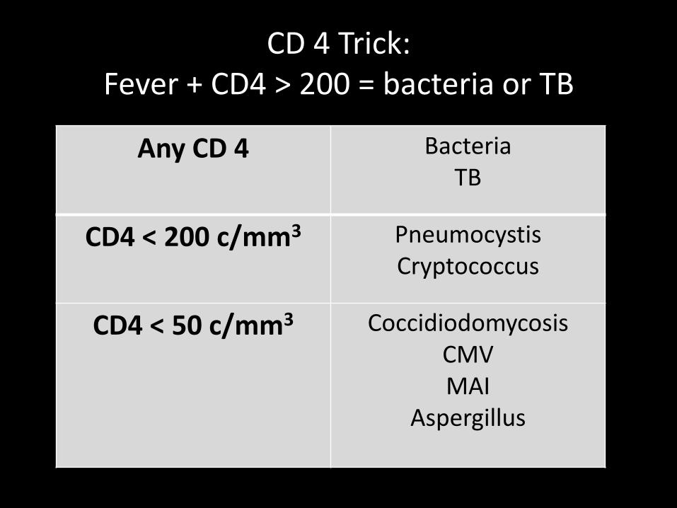
CD 4 Trick: Fever + CD4 > 200 = bacteria or TB
Any CD 4 Bacteria TB
CD4 < 200 c/mm3
Pneumocystis Cryptococcus
CD4 < 50 c/mm3
Coccidiodomycosis CMV MAI
Aspergillus

CXR lung parenchyma trick
Bacteria TB
Unilateral ---- (bilateral) Focal ------------(multifocal)
Segmental-----(lobar)
Pneumocystis CMV LIP
Kaposi Cardiac failure
Diffuse bilateral
Aspiration Dependent

CXR Exclusion trick
NOT in
Lymph-adenopathy
bacteria and aspiration
Effusion PJP / LIP
Cavities / cysts lymphoma / Kaposi

However, clinicians usually want to
know one thing: before starting HAARTreatment, can
we ‘exclude’ TB?

Progressive primary
Lymphnode TB
Milliary
TB / HIV co-infection

Diagnosis of TB in children =
identify lymphadenopathy

Lymphadenopathy on AP: Hilum should be a hippo’s open mouth Nodes = a cauliflower in the mouth
Right hilar lymphadenopathy Right hilar lymphadenopathy
Left hilar lymphadenopathy Calcified lymphadenopathy

Airways are a up-side-down tree Lymphadenopathy = ‘compressed air-way branches’

Lateral radiograph

Lymphadenopathy on Lateral
• Normal structures (=horseshoe)
• Diverging vessels (=tentacles)
• Lymphadenopathy (=‘doughnut’)
R main pulm art
L main pulm art Top Aortic arch
Lateral: doughnut replaces the horse-shoe and tentacles

Midsagital: subcarinal nodes
Far para-sagital: Left hilar nodes
What makes the doughnut?
Subcarinal and left hilar lymphadenopathy
And there is the doughnut………

Doughnuts and other foods
No mass behind bronchus intermedius

When child is HIV-infected: You’ve gottobe Sherlock Holmes and uncover
TB
Air space and airway Air space, cavity and airway
Air space expansile and airway Air space, effusion and airway Milliary nodules

If you don’t see the TB you may get IRIS
Before HAART After initiation of HAART

Is POC US the answer for Africa?

Point of Care US: for TB and Pneumonia
Tsung 2012

Some research going on Research projects using US at the Red
Cross Children’s Hospital in Cape Town,
South Africa for mediastinal TB lymphadenopathy and pneumonia
Others are using TCD for TB and HIV
Sagittal US: lymphadenopathy in Zone A
Sagittal US: Lymphadenopathy shwn in Zone D

HIV and the CNS

What can be seen with imaging?
‘HIV ENCEPHALOPATHY’
(HIVE)
- Atrophy
- White matter abnormality
‘CONSEQUENCES of HIV’
- Calcifications
- Infections
- Vascular events / lesions
- Maliganancy [uncommon]
Additional: monitoring disease progression and treatment response

What can I show you to look for?
• To look for atrophy as a marker of HIVE
• To identify HIVE and PML (progressive multifocal leukoencephalopathy) white matter signal patterns
• Understand that HIV is a major cause of BG calcification
• Look for vascular events
• Look for infections
• To understand the effects of HIV on TBM

HIV Encephalopathy (HIVE)
Atrophy and
White matter abnormality

What about atrophy? • It is the most common finding (90%) in HIV
imaging (Kauffman et al 1992, Safriel et al 2000, Kieck and Andronikou 2004)
• It’s the imaging representation of HIV encephalopathy
• It correlates with severity and viral load • It is reversible or can be halted on HAART (Di Carli
et al 1991)
• It is measurable on imaging • But it is a late finding - we need something
earlier in the disease

Volume loss
White matter: - Expanded ventricles (in presence of large SAS) - deep sulci (near ventricles) Doesn’t it remind you of chronic evolution of HIE???
This patient is HIV-infected

An objective, automated method of measuring volume loss: Matlab
T1 image CSF segmented Cortex segmented White matter segmented

Corpus Callosum: a surrogate marker of WM volume?
FOCAL LOSS GLOBAL LOSS

Results: Brain volume only showed a trend relationship with nadir CD4. Correlation degree of mental development and motor segmental CC thickness Correlation of the CC length with immunity and microcephaly

What about the white matter signal?

Encephalopathy (HIVE): T2 high signal
Bilateral ‘symmetric’ Spares sub-cortical U-fibres

Progressive multifocal leukoencephalopathy
(PML)
• Much less common
• Have the JC virus
• Confused with HIVE but…
• More focal
• Asymmetrical
• Common posterior parietal
• Involve U-fibres
• Advanced cases - ‘bar-bell’ sign

Summary HIVE vs. PML

More subtle WM abnormalities?

Prevalence of WMSA “HIV related brain disease” = 50%
Of the 22 patients with WMSA:
• 17 patients pinpoint lesions < 1cm
• 8 confluent lesions
• 2 patients lesions > 1cm
• Lesion size: 5 -12mm
(mean 7.2mm)

WMSA
• Half of children referred for HIV-related brain disease had WMSA on T2MRI
• Involved mainly frontal and parietal lobes.
• Positive correlation of ‘time on ART’ and presence of WMSA
• Trend correlating nadir CD4% and presence of WMSA.

HIVE with a normal signal?

Work in HIV infected adults: DTI and FA
• Normal looking subcortical WM and CC • BUT…areas where FA decreased • Patients with lowest FA had most advanced
HIV [Filippi CG et al 2001]
• Abnormalities corpus callosum in patients
with HIV, associated with dementia severity and motor speed losses [Wu Y et al 2006]
• Reasons: trafficking of virus from ventricular CSF

DTI: FA group
comparison of HIV
infected vs. Controls

Consequences

Calcification

Basal ganglia calcification
• Commonest cause of BG calcification in children is HIV
• Up to 1/3 of children with HIV have calcification
• Usually affect palidus and putamen
• Less often frontal white matter / cerebellum
• Not seen before 10 months age
palidus
Palidus and Putamen

Infections:

Infection:
TBM: best feature is basal enhancement Pyogenic meningitis:
Surface collections; Venous infarctions

Vascular: Infarction / aneurysms

Infarction

Other findings on the scan

Parotidomegally - painless bilateral Lymphadenopathy - cervical

HIV and TB together: Add petrol to the fire?

HIV and TB
• Because the body fails to contain TB in the lungs…..
• HIV predisposes to blood borne and extra-pulmonary TB……
• BUT
• HIV also affects imaging appearances of TB

TB and HIV co-infection: CNS

Concepts in imaging TBM
• It’s the immune response to bacilli in the meninges that results in pathological features of TBM…..

Diagnosis of TBM: MRI diagnosis of pediatric TBM = basal enhancement (93%)

Basal Enhancement in HIV
BE = infrequent, less prominent, atypically distributed, milliary nodules (100%)

Ventriculomegally in HIV CSF space = result of atrophy; hydrocephalus less frequent and
exclusively communicating

So now you know….
CNS CHEST
Co-infection with TB
HIV