image-processing technique for suppressing ribs in chest radiographs by means of massive
TRANSCRIPT
406 IEEE TRANSACTIONS ON MEDICAL IMAGING, VOL. 25, NO. 4, APRIL 2006
Image-Processing Technique for Suppressing Ribs inChest Radiographs by Means of Massive Training
Artificial Neural Network (MTANN)Kenji Suzuki*, Senior Member, IEEE, Hiroyuki Abe, Heber MacMahon, and Kunio Doi
Abstract—When lung nodules overlap with ribs or clavicles inchest radiographs, it can be difficult for radiologists as well ascomputer-aided diagnostic (CAD) schemes to detect these nodules.In this paper, we developed an image-processing technique forsuppressing the contrast of ribs and clavicles in chest radiographsby means of a multiresolution massive training artificial neuralnetwork (MTANN). An MTANN is a highly nonlinear filter that canbe trained by use of input chest radiographs and the corresponding“teaching” images. We employed “bone” images obtained by useof a dual-energy subtraction technique as the teaching images. Foreffective suppression of ribs having various spatial frequencies, wedeveloped a multiresolution MTANN consisting of multiresolutiondecomposition/composition techniques and three MTANNs forthree different-resolution images. After training with input chestradiographs and the corresponding dual-energy bone images, themultiresolution MTANN was able to provide “bone-image-like”images which were similar to the teaching bone images. By sub-tracting the bone-image-like images from the corresponding chestradiographs, we were able to produce “soft-tissue-image-like”images where ribs and clavicles were substantially suppressed.We used a validation test database consisting of 118 chest radio-graphs with pulmonary nodules and an independent test databaseconsisting of 136 digitized screen-film chest radiographs with 136solitary pulmonary nodules collected from 14 medical institutionsin this study. When our technique was applied to nontrainingchest radiographs, ribs and clavicles in the chest radiographs weresuppressed substantially, while the visibility of nodules and lungvessels was maintained. Thus, our image-processing technique forrib suppression by means of a multiresolution MTANN would bepotentially useful for radiologists as well as for CAD schemes indetection of lung nodules on chest radiographs.
Index Terms—Artificial neural network, chest radiography,computer-aided diagnosis (CAD), dual-energy subtraction, lungnodule, rib suppression.
I. INTRODUCTION
CHEST radiography is the most frequently used diagnosticimaging examination for chest diseases such as lung
cancer, tuberculosis, pneumonia, pneumoconioses, and pul-monary emphysema. More than 9 million people worldwide dieannually from chest diseases [1]. Lung cancer causes 945 000deaths [1], and is the leading cause of cancer deaths in the world
Manuscript received October 21, 2005; revised January 16, 2006. This workwas supported in part by the United States Public Health Service (USPHS) underGrant CA62625. Asterisk indicates corresponding author.
*K. Suzuki is with the Kurt Rossmann Laboratories for Radiologic ImageResearch, Department of Radiology, The University of Chicago, 5841 S. Mary-land Ave., Chicago, IL 60637 USA (e-mail: [email protected]).
H. Abe, H. MacMahon, and K. Doi are with the Department of Radiology,The University of Chicago, Chicago, IL 60637 USA.
Digital Object Identifier 10.1109/TMI.2006.871549
[1] and in countries [2] such as the United States, the UnitedKingdom, the Russian Federation, Canada, Poland, and Japan.In the United States alone, lung cancer is expected to cause160 440 deaths in 2004 [3]. Chest radiographs have been used fordetection of lung cancer [4]–[6] because some evidence suggeststhat early detection of lung cancer may allow a favorable prog-nosis [7]–[9]. Lung nodules (i.e., potential lung cancers) in chestradiographs, however, can be overlooked by radiologists in from12%–90% of cases in which nodules are visible in retrospect[10], [11]. Many, 82%–95%, of the missed lung cancers werepartly obscured by overlying bones such as ribs and/or a clav-icle [10], [11]. Therefore, a computer-aided diagnostic (CAD)scheme [12], [13] for nodule detection on chest radiographs hasbeen investigated, because the computer prompts indicating nod-ules could improve radiologists’ detection accuracy [14]–[16].
A major challenge in current CAD schemes [17]–[27] fornodule detection on chest radiographs is the detection of nod-ules overlapping with ribs, rib crossings, and clavicles, becausea majority of false positives are caused by these structures [18],[28]. This results in lowering the sensitivity as well as the speci-ficity of a CAD scheme. Because nodules overlapping with ribsand clavicles were reported to be difficult for radiologists to ob-serve [29], [30], detection of such nodules is important for CADschemes. Therefore, the suppression of ribs and clavicles inchest radiographs would be potentially useful for improving ra-diologists’ detection accuracy as well as the CAD performance.
Our purpose in this study was to develop an image-processingtechnique for suppressing the contrast of ribs in chest radio-graphs by means of a multiresolution massive training artificialneural network (MTANN).
II. MATERIALS AND METHODS
A. Massive Training Artificial Neural Network (MTANN)
In the field of image processing, supervised nonlinearimage-processing techniques [31]–[34] based on an artificialneural network (ANN), called a “neural filter” [32] and a“neural edge enhancer” [33], [34], have been investigated forreduction of the quantum mottle (specific noise observed inmedical x-ray images) in angiograms and radiographs [35] andfor supervised detection of left ventricular contours traced bycardiologists in angiography [36], respectively. By extendingthe neural filter and the neural edge enhancer, MTANNs [37]have been developed to accommodate the task of distinguishinga specific opacity from other opacities in medical images.MTANNs have been applied for reduction of false positives in
0278-0062/$20.00 © 2006 IEEE
SUZUKI et al.: IMAGE-PROCESSING TECHNIQUE FOR SUPPRESSING RIBS IN CHEST RADIOGRAPHS BY MEANS OF MTANN 407
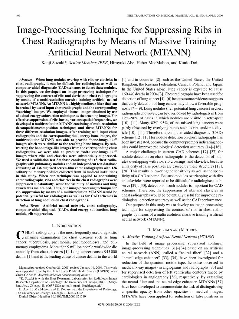
Fig. 1. Architecture and training of an MTANN consisting of a linear-outputmultilayer ANN model and a massive-subregions training scheme. The pixelvalues in the subregion extracted from a chest radiograph are entered as inputto the ANN. Single pixels extracted from teaching images are used as teachingvalues for the corresponding subregions.
computerized detection of lung nodules in low-dose computedtomography (CT) [37], [38] and chest radiography [39], fordistinction between benign and malignant lung nodules in CT[40]. In our previous studies [37]–[40], the MTANNs aimed atclassification of regions-of-interest into abnormal or normal;thus, these studies were in the field of pattern recognition,whereas this paper aims at suppression of ribs in chest radio-graphs, which would be in the field of image processing.
The architecture and the training method of an MTANN areshown in Fig. 1. The MTANN can be considered to be a highlynonlinear filter that can be trained with input images and thecorresponding “teaching” images. The MTANN consists of alinear-output multilayer ANN model [41], which is capable ofoperating on image data directly. The linear-output multilayerANN model employs a linear function instead of a sigmoid func-tion as the activation function of the unit in the output layer be-cause the characteristics of an ANN were improved significantlywith a linear function when applied to the continuous mappingof values in image processing [34], [41]. A conventional ANNhardly outputs values near zero and one because of the character-istics of a sigmoid function, whereas the linear-output multilayerANN outputs values linearly. The training for teaching valuesnear zero and one converges more slowly than do other valueswith the conventional ANN theoretically, whereas these valuesare trained evenly with the linear-output multilayer ANN model.This affects the convergence characteristics and the outputcharacteristics of ANN models. Therefore, the linear-outputmultilayer ANN would be suitable for image processing, wherethe teaching values may be continuous values ranging fromzero to one, whereas the conventional ANN is suitable for aclassification task where the teaching values are assigned toclasses (see [34], [41] for theoretical considerations). The pixelvalues of original chest radiographs are normalized first such thata pixel value of zero is zero and a pixel value of the maximumgray-scale level (1,023) is one. The inputs of the linear-outputmultilayer ANN are the pixel values in a subregion extractedfrom a chest radiograph. The output is a continuous value, whichcorresponds to the center pixel in the subregion, represented by
(1)
where
(2)
is the input vector to the MTANN, is an estimate fora teaching value, and are the coordinates of the image,
is the output of the linear-output multilayer ANN, andis a normalized pixel value in an input chest radiograph.
Note that only one unit is employed in the output layer. Theinput vector can be rewritten as
(3)
where is an input unit number, and is the number of inputunits. Because the activation functions of the units in the inputlayer are an identity function, the output of the th unit in theinput layer can be represented by . The output of the th unitin the hidden layer is represented by
(4)
where is a weight between the th unit in the input layerand the th unit in the hidden layer, is an offset of the thunit in the hidden layer, and is a sigmoid function
(5)
The output of the unit in the output layer is represented by
(6)
where is a weight between the th unit in the hidden layerand the unit in the output layer, is an offset of the unit inthe output layer, is a linear function
(7)
and is a slope parameter. The entire output image is obtainedby scanning of an input chest image with the MTANN.
The MTANN involves training with massive subregion-pixelpairs, where we call it a massive-subregions training scheme.Input chest radiographs are divided pixel by pixel into a largenumber of overlapping subregions. Single pixels correspondingto the input subregions are extracted from the teaching imagesas teaching values. The MTANN is massively trained by usingeach of a large number of the input subregions together witheach of the corresponding teaching single pixels. The trainingset of pairs of a subregion and a teaching pixel is represented by
(8)
where is a teaching image, is a training region whichcorresponds to the collection of the centers of subregions (orteaching pixels), is a pixel number in , and is the
408 IEEE TRANSACTIONS ON MEDICAL IMAGING, VOL. 25, NO. 4, APRIL 2006
number of pixels in . The error to be minimized by trainingis defined by
(9)
The MTANN is trained by a linear-output back-propaga-tion (BP) algorithm [34], [41] which was derived for thelinear-output multilayer ANN model in the same way as theBP algorithm [42], [43]. The correction of the weight betweenhidden units and output unit can be represented by
(10)
where is a learning rate. Please refer to [34], [41] for the de-tails and the property of the linear-output BP algorithm. Aftertraining, the MTANN is expected to produce images similar tothe teaching images.
We used a dual-energy subtraction technique [44] to obtainthe teaching images for MTANNs for suppression of ribs inchest radiographs. The dual-energy subtraction is a techniquefor separating bones from soft tissues in chest radiographs byuse of the energy dependence of the x-ray attenuation by dif-ferent materials; it can produce two tissue-selective images, i.e.,a “bone” image and a “soft-tissue” image. Chest radiographs areused as input images to MTANNs, and the corresponding dual-energy bone images are used as the teaching images. We didnot directly use dual-energy soft-tissue images as the teachingimages, because the MTANNs trained with dual-energy soft-tissue images produced results that were slightly inferior to theMTANNs trained with dual-energy bone images (see details inSection IV).
B. Multiesolution Decomposition and Composition
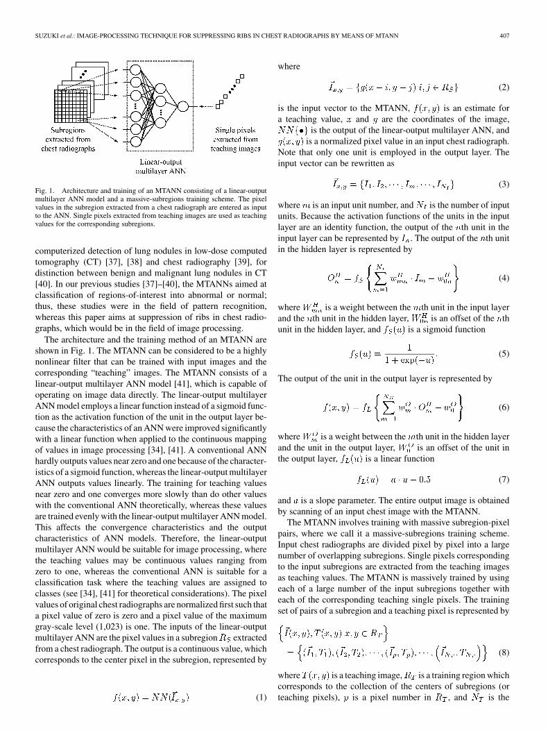
Ribs in chest radiographs include various spatial-frequencycomponents. For a single MTANN, suppression of ribs con-taining such various frequencies is difficult, because the capa-bility of a single MTANN is limited, i.e., the capability dependson the size of the subregion of the MTANN. Because the trainingof the MTANN takes a substantially long time, it is difficult inpractice to train the MTANN with a large subregion. In orderto overcome this issue, we employed multiresolution decom-position/composition techniques [45], [46]. The multiresolutiondecomposition, illustrated in Fig. 2(a), is a technique for de-composing an original high-resolution image into different-res-olution images. First, one obtains a medium-resolution image
from an original high-resolution image byperforming down-sampling with averaging, i.e., four pixels inthe original image are replaced by a pixel having the mean valuefor the four pixel values, represented by
(11)
where is a 2-by-2-pixel region. The medium-resolutionimage is enlarged by up-sampling with pixel substitution, i.e., a
Fig. 2. Illustrations of (a) a multiresolution decomposition technique and(b) a multiresolution composition technique. Lower-resolution images areproduced by repeatedly performing down-sampling and subtracting in amultiresolution decomposition technique. Exactly the same original resolutionimage can be obtained from the multiresolution images by performing amultiresolution composition technique.
pixel in the medium-resolution image is replaced by four pixelswith the same pixel value, as follows:
(12)
Then, a high-resolution difference image is obtainedby subtraction of the enlarged medium-resolution image fromthe high-resolution image, represented by
(13)
These procedures are performed repeatedly, producing furtherlower-resolution images. Thus, multiresolution images having
SUZUKI et al.: IMAGE-PROCESSING TECHNIQUE FOR SUPPRESSING RIBS IN CHEST RADIOGRAPHS BY MEANS OF MTANN 409
various frequencies are obtained by use of the multiresolutiondecomposition technique.
An important property of this technique is that exactlythe same original-resolution image can be obtainedfrom the multiresolution images, and , byperforming the inverse procedures, called a multiresolutioncomposition technique as shown in Fig. 2(b), as follows:
(14)
Therefore, we can process multiresolution images indepen-dently instead of processing original high-resolution imagesdirectly; i.e., with these techniques, the processed originalhigh-resolution image can be obtained by composing of theprocessed multiresolution images. An MTANN only needsto support a limited spatial frequency rage in each resolutionimage instead of the entire spatial frequencies in the originalimage.
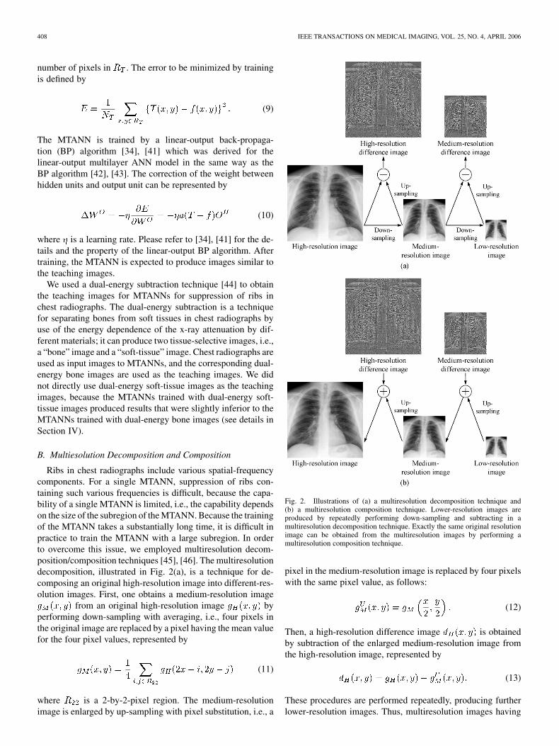
C. Multiresolution MTANN for Suppressing Ribs
Fig. 3 illustrates the architecture and training of a multireso-lution MTANN involving multiresolution decomposition/com-position techniques and MTANNs for different-resolutionimages. First, input chest radiographs and the correspondingteaching bone images are decomposed into sets of different-res-olution images, and then these sets of images are used fortraining three MTANNs in the multiresolution MTANN, asillustrated in Fig. 3(a). Each MTANN is an expert for a cer-tain resolution, i.e., a low-resolution MTANN is in chargeof low-frequency components of ribs, a medium-resolu-tion MTANN is for medium-frequency components, and ahigh-resolution MTANN for high-frequency components.Each resolution MTANN is trained independently with thecorresponding resolution images. After training, the MTANNsproduce different-resolution images, and then these images arecomposed to provide a complete high-resolution image by useof the multiresolution composition technique, as illustrated inFig. 3(b). The complete high-resolution image is expected tobe similar to the teaching bone image; therefore, the multires-olution MTANN would provide a “bone-image-like” image inwhich ribs are separated from soft tissues.
The multiple MTANN scheme in our previous studies[37]–[40] was developed for classification of candidates intomultiple categories; therefore, the output of the multipleMTANN scheme is a class, i.e., abnormal or normal, whereasthe output of the multiresolution MTANN is a pixel value. Inthe multiple-MTANN scheme, MTANNs were combined withscoring, thresholding, and the logical AND operation, whereasthe multiresolution MTANN does not use any of these. Inthe multiresolution MTANN, the input to each of MTANNsis certain frequency components obtained from the originalimages by use of the multiresolution decomposition, whereasthe input of the multiple MTANN scheme is pixel values of theoriginal images.
In this paper, we focused on the suppression of ribs andclavicles in lung regions, because most nodules overlappingwith these structures are in the lung regions. For processingonly in the lungs, lung regions are segmented by thresholding.
Fig. 3. Diagrams of (a) a training phase and (b) an execution phase ofa multiresolution MTANN consisting of MTANNs for different-resolutionimages. In the training phase, an input chest radiograph and a teaching boneimage are decomposed into multiresolution images by use of a multiresolutiondecomposition technique. Each of the multiresolution images is used for eachof the corresponding resolution MTANNs in the multiresolution MTANN.In the execution phase, the output multiresolution images of the trainedmultiresolution MTANN are composed to provide a “bone-image-like” imageby use of a multiresolution composition technique.
A threshold value is determined by use of a method [47] basedon linear discriminant analysis (LDA), which is a commonmethod in the fields of computer vision and pattern recog-nition (often referred as Otsu thresholding). It is expectedthat a reasonable threshold value can be determined by useof LDA, because threshold determination can be consideredas a two-class classification problem in the domain of thehistogram of gray levels, and the linear separation with LDAwould work well in this one dimensional space. This methodautomatically selects the lowest point between two classes inthe histogram of gray levels in a chest radiograph (i.e., thisis formulated as LDA). The method involves minimizing theratio of between-class variance to the total variance. After thesegmentation, a Gaussian filter is applied for smoothing theedges of the segmented lung regions to create an imagefor masking the outside of the lung regions. The masking imageis normalized to have values from 0 to 1. For suppression of
410 IEEE TRANSACTIONS ON MEDICAL IMAGING, VOL. 25, NO. 4, APRIL 2006
ribs in an original chest radiograph, the bone-image-like imageproduced by the multiresolution MTANN is subtracted
from the original chest radiograph with the maskingimage as follows:
(15)
where is a weighting parameter for determining the con-trast of ribs. By changing the weighting parameter , onecan obtain processed chest radiographs with different contrastof ribs. Thus, the multiresolution MTANN would be able toproduce a “soft-tissue-image-like” image where ribs are sup-pressed; therefore, this image processing may be considered asa “rib suppression” technique.
D. Database
The database used in our study consisted of 122 posterior-an-terior chest radiographs acquired with a computed radiographysystem with a dual-energy subtraction unit (FCR 9501 ES; Fu-jifilm Medical Systems, Stamford, CT) at The University ofChicago Hospitals. The dual-energy subtraction unit employeda single-shot dual-energy subtraction technique where image ac-quisition is performed with a single exposure that is detected bytwo receptor plates separated by a filter for obtaining imagesat two different energy levels [44], [48], [49]. The chest radio-graphs included 121 abnormal cases with pulmonary nodulesand a “normal” case (i.e., a nodule-free case). The matrix size ofthe chest images was 1,760 1,760 pixels (pixel size, 0.2 mm;gray scale, 10 bits). The absence and presence of nodules in thechest radiographs were confirmed by use of CT examinations.Most nodules overlapped with ribs and/or clavicles in chest ra-diographs. In order to train a multiresolution MTANN, we useda training set consisting of four chest radiographs and the corre-sponding dual-energy soft-tissue and bone images. Three of thefour chest radiographs were from nodule cases, and the otherwas the normal case. The registration error between the inputimages and the teaching images would be minimum becauseof the use of the single-shot dual-energy subtraction technique.We used a test set consisting of 118 nodule cases for testing ourtechnique. For computational efficiency, the size of the chest ra-diographs was reduced by a factor of four, i.e., 440 440 pixels.
We used another test set consisting of 136 digitizedscreen-film chest radiographs with 136 solitary pulmonarynodules, which was the Digital Image Database developedby the Japanese Society of Radiological Technology (JSRT)[50], a publicly available database. The chest radiographswere collected from 14 medical institutions. The absence andpresence of nodules in the chest radiographs were confirmedby CT. The locations of all nodules were confirmed by threechest radiologists. The chest radiographs were digitized witha 0.175-mm pixel size, a matrix size of 2,048 2,048, and a12-bit gray-scale level. The sizes of nodules ranged from 8.9to 29.1 mm, and the average size was 17.4 mm. The databasecontained 64 malignant and 27 benign nodules, which wereconfirmed by histologic or cytologic examinations or follow-upimaging. For computational efficiency, the size of the chestradiographs was reduced by a factor of four to 512 512 pixelswith a 10-bit gray-scale level by use of averaging.
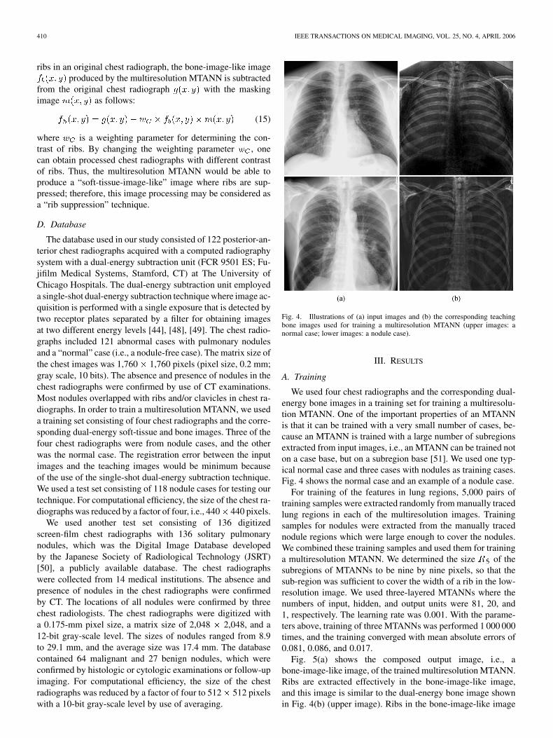
Fig. 4. Illustrations of (a) input images and (b) the corresponding teachingbone images used for training a multiresolution MTANN (upper images: anormal case; lower images: a nodule case).
III. RESULTS
A. Training
We used four chest radiographs and the corresponding dual-energy bone images in a training set for training a multiresolu-tion MTANN. One of the important properties of an MTANNis that it can be trained with a very small number of cases, be-cause an MTANN is trained with a large number of subregionsextracted from input images, i.e., an MTANN can be trained noton a case base, but on a subregion base [51]. We used one typ-ical normal case and three cases with nodules as training cases.Fig. 4 shows the normal case and an example of a nodule case.
For training of the features in lung regions, 5,000 pairs oftraining samples were extracted randomly from manually tracedlung regions in each of the multiresolution images. Trainingsamples for nodules were extracted from the manually tracednodule regions which were large enough to cover the nodules.We combined these training samples and used them for traininga multiresolution MTANN. We determined the size of thesubregions of MTANNs to be nine by nine pixels, so that thesub-region was sufficient to cover the width of a rib in the low-resolution image. We used three-layered MTANNs where thenumbers of input, hidden, and output units were 81, 20, and1, respectively. The learning rate was 0.001. With the parame-ters above, training of three MTANNs was performed 1 000 000times, and the training converged with mean absolute errors of0.081, 0.086, and 0.017.
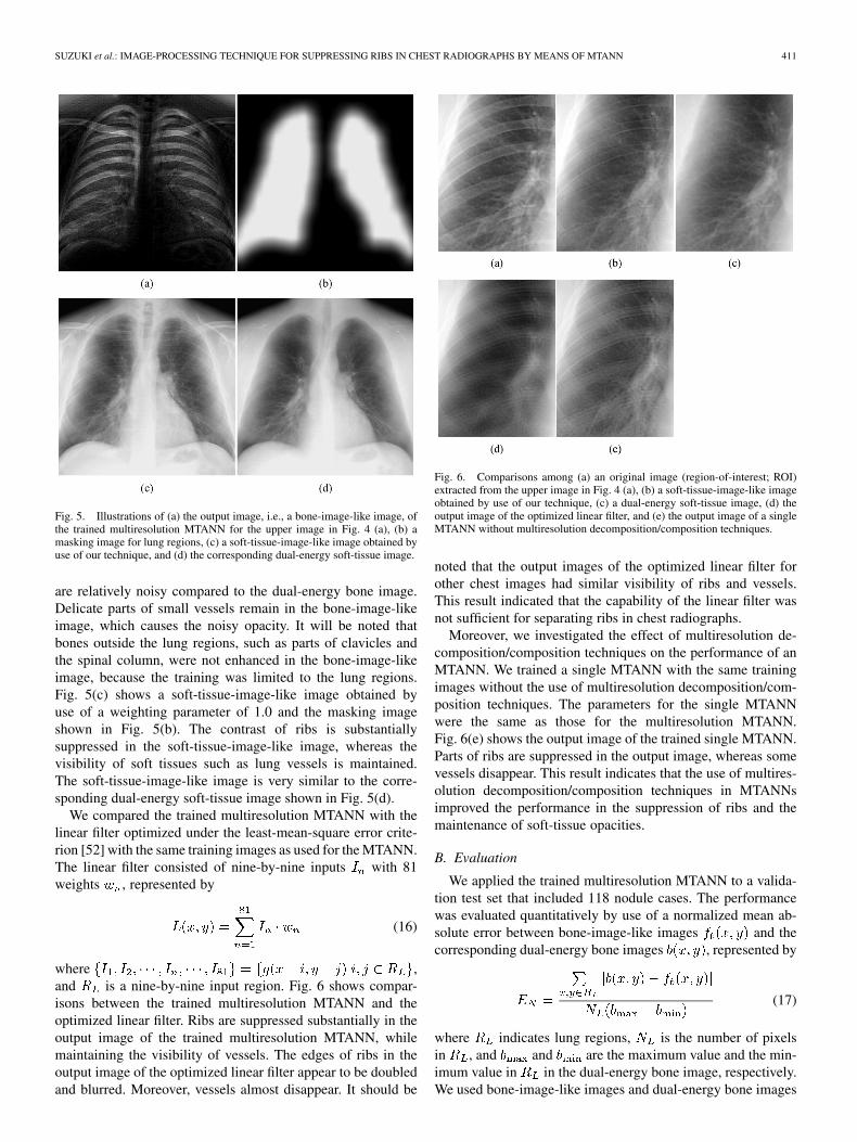
Fig. 5(a) shows the composed output image, i.e., abone-image-like image, of the trained multiresolution MTANN.Ribs are extracted effectively in the bone-image-like image,and this image is similar to the dual-energy bone image shownin Fig. 4(b) (upper image). Ribs in the bone-image-like image
SUZUKI et al.: IMAGE-PROCESSING TECHNIQUE FOR SUPPRESSING RIBS IN CHEST RADIOGRAPHS BY MEANS OF MTANN 411
Fig. 5. Illustrations of (a) the output image, i.e., a bone-image-like image, ofthe trained multiresolution MTANN for the upper image in Fig. 4 (a), (b) amasking image for lung regions, (c) a soft-tissue-image-like image obtained byuse of our technique, and (d) the corresponding dual-energy soft-tissue image.
are relatively noisy compared to the dual-energy bone image.Delicate parts of small vessels remain in the bone-image-likeimage, which causes the noisy opacity. It will be noted thatbones outside the lung regions, such as parts of clavicles andthe spinal column, were not enhanced in the bone-image-likeimage, because the training was limited to the lung regions.Fig. 5(c) shows a soft-tissue-image-like image obtained byuse of a weighting parameter of 1.0 and the masking imageshown in Fig. 5(b). The contrast of ribs is substantiallysuppressed in the soft-tissue-image-like image, whereas thevisibility of soft tissues such as lung vessels is maintained.The soft-tissue-image-like image is very similar to the corre-sponding dual-energy soft-tissue image shown in Fig. 5(d).
We compared the trained multiresolution MTANN with thelinear filter optimized under the least-mean-square error crite-rion [52] with the same training images as used for the MTANN.The linear filter consisted of nine-by-nine inputs with 81weights , represented by
(16)
where ,and is a nine-by-nine input region. Fig. 6 shows compar-isons between the trained multiresolution MTANN and theoptimized linear filter. Ribs are suppressed substantially in theoutput image of the trained multiresolution MTANN, whilemaintaining the visibility of vessels. The edges of ribs in theoutput image of the optimized linear filter appear to be doubledand blurred. Moreover, vessels almost disappear. It should be
Fig. 6. Comparisons among (a) an original image (region-of-interest; ROI)extracted from the upper image in Fig. 4 (a), (b) a soft-tissue-image-like imageobtained by use of our technique, (c) a dual-energy soft-tissue image, (d) theoutput image of the optimized linear filter, and (e) the output image of a singleMTANN without multiresolution decomposition/composition techniques.
noted that the output images of the optimized linear filter forother chest images had similar visibility of ribs and vessels.This result indicated that the capability of the linear filter wasnot sufficient for separating ribs in chest radiographs.
Moreover, we investigated the effect of multiresolution de-composition/composition techniques on the performance of anMTANN. We trained a single MTANN with the same trainingimages without the use of multiresolution decomposition/com-position techniques. The parameters for the single MTANNwere the same as those for the multiresolution MTANN.Fig. 6(e) shows the output image of the trained single MTANN.Parts of ribs are suppressed in the output image, whereas somevessels disappear. This result indicates that the use of multires-olution decomposition/composition techniques in MTANNsimproved the performance in the suppression of ribs and themaintenance of soft-tissue opacities.
B. Evaluation
We applied the trained multiresolution MTANN to a valida-tion test set that included 118 nodule cases. The performancewas evaluated quantitatively by use of a normalized mean ab-solute error between bone-image-like images and thecorresponding dual-energy bone images , represented by
(17)
where indicates lung regions, is the number of pixelsin , and and are the maximum value and the min-imum value in in the dual-energy bone image, respectively.We used bone-image-like images and dual-energy bone images
412 IEEE TRANSACTIONS ON MEDICAL IMAGING, VOL. 25, NO. 4, APRIL 2006
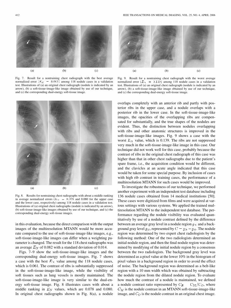
Fig. 7. Result for a nontraining chest radiograph with the best averagenormalized error (E = 0:061) among 118 nodule cases in a validationtest. Illustrations of (a) an original chest radiograph (nodule is indicated by anarrow), (b) a soft-tissue-image-like image obtained by use of our technique,and (c) the corresponding dual-energy soft-tissue image.
Fig. 8. Results for nontraining chest radiographs with about a middle rankingin average normalized errors (E = 0:078 and 0.080 for the upper caseand the lower case, respectively) among 118 nodule cases in a validation test.Illustrations of (a) original chest radiographs (nodule is indicated by an arrow),(b) soft-tissue-image-like images obtained by use of our technique, and (c) thecorresponding dual-energy soft-tissue images.
in this evaluation, because the direct comparison with the outputimages of the multiresolution MTANN would be more accu-rate compared to the use of soft-tissue-image-like images, e.g.,soft-tissue-image-like images can differ when a weighting pa-rameter is changed. The result for the 118 chest radiographs wasan average of 0.082 with a standard deviation of 0.014.
Figs. 7–9 show the soft-tissue-image-like images and thecorresponding dual-energy soft-tissue images. Fig. 7 showsa case with the best value among the 118 nodule cases,which is 0.061. The contrast of ribs is substantially suppressedin the soft-tissue-image-like image, while the visibility ofsoft tissues such as lung vessels is mostly maintained. Thesoft-tissue-image-like image is very similar to the dual-en-ergy soft-tissue image. Fig. 8 illustrates cases with about amiddle ranking in values, which are 0.078 and 0.080.In original chest radiographs shown in Fig. 8(a), a nodule
Fig. 9. Result for a nontraining chest radiograph with the worst averagenormalized error (E = 0:139) among 118 nodule cases in a validationtest. Illustrations of (a) an original chest radiograph (nodule is indicated by anarrow), (b) a soft-tissue-image-like image obtained by use of our technique,and (c) the corresponding dual-energy soft-tissue image.
overlaps completely with an anterior rib and partly with pos-terior ribs in the upper case, and a nodule overlaps with aposterior rib in the lower case. In the soft-tissue-image-likeimages, the opacities of the overlapping ribs are compen-sated for substantially, and the true shapes of the nodules areevident. Thus, the distinction between nodules overlappingwith ribs and other anatomic structures is improved in thesoft-tissue-image-like images. Fig. 9 shows a case with theworst value, which is 0.139. The ribs are not suppressedvery much in the soft-tissue-image-like image in this case. Ourtechnique did not work well for this case, probably because thecontrast of ribs in the original chest radiograph of this case washigher than that in other chest radiographs due to the patient’sspare frame, i.e., the acquisition condition would be different,and the clavicles at an acute angle indicated that this casewould be taken for some special purpose. By inclusion of caseswith high rib contrast in training cases, the performance of amultiresolution MTANN for such cases would be improved.
To investigate the robustness of our technique, we performedanother experiment with an independent test database including136 nodule cases obtained from 14 medical institutions [50].These cases were digitized from films and were acquired at var-ious settings with various systems. We applied the trained mul-tiresolution MTANN to the independent test database. The per-formance regarding the nodule visibility was evaluated quan-titatively by use of a nodule contrast defined by the differencebetween an average gray level in a nodule region and a back-ground gray level , represented by . The noduleregion was determined by two expert chest radiologists by thefollowing method: One of the two radiologists determined aninitial nodule region, and then the final nodule region was deter-mined by modifying of the initial nodule region by a consensusbetween the two radiologists. The background gray level wasdetermined as a pixel value at the lower 10% in the histogram ofpixel values in a background region in order to avoid the effectof noise. The background region was determined as a belt-likeregion with a 10 mm width which was obtained by subtractingthe nodule region from the dilated nodule region. To evaluatehow much the contrast of a nodule is maintained, we defineda nodule contrast ratio represented by , where
is the nodule contrast in an MTANN soft-tissue-image-likeimage, and is the nodule contrast in an original chest image.
SUZUKI et al.: IMAGE-PROCESSING TECHNIQUE FOR SUPPRESSING RIBS IN CHEST RADIOGRAPHS BY MEANS OF MTANN 413
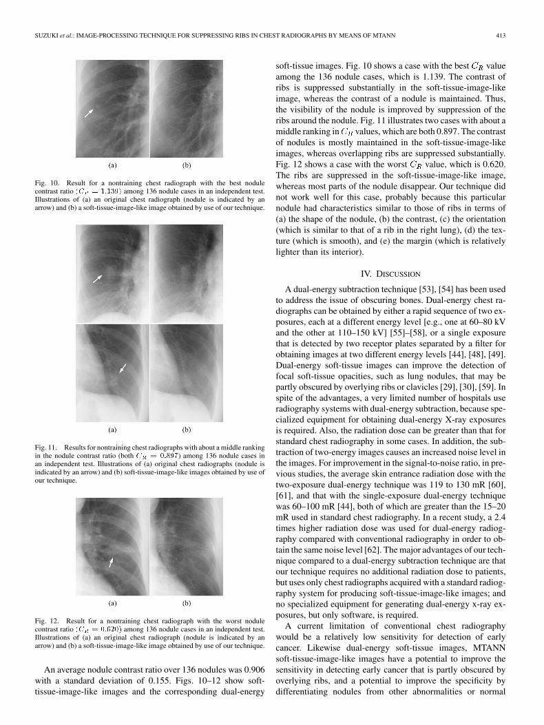
Fig. 10. Result for a nontraining chest radiograph with the best nodulecontrast ratio (C = 1:139) among 136 nodule cases in an independent test.Illustrations of (a) an original chest radiograph (nodule is indicated by anarrow) and (b) a soft-tissue-image-like image obtained by use of our technique.
Fig. 11. Results for nontraining chest radiographs with about a middle rankingin the nodule contrast ratio (both C = 0:897) among 136 nodule cases inan independent test. Illustrations of (a) original chest radiographs (nodule isindicated by an arrow) and (b) soft-tissue-image-like images obtained by use ofour technique.
Fig. 12. Result for a nontraining chest radiograph with the worst nodulecontrast ratio (C = 0:620) among 136 nodule cases in an independent test.Illustrations of (a) an original chest radiograph (nodule is indicated by anarrow) and (b) a soft-tissue-image-like image obtained by use of our technique.
An average nodule contrast ratio over 136 nodules was 0.906with a standard deviation of 0.155. Figs. 10–12 show soft-tissue-image-like images and the corresponding dual-energy
soft-tissue images. Fig. 10 shows a case with the best valueamong the 136 nodule cases, which is 1.139. The contrast ofribs is suppressed substantially in the soft-tissue-image-likeimage, whereas the contrast of a nodule is maintained. Thus,the visibility of the nodule is improved by suppression of theribs around the nodule. Fig. 11 illustrates two cases with about amiddle ranking in values, which are both 0.897. The contrastof nodules is mostly maintained in the soft-tissue-image-likeimages, whereas overlapping ribs are suppressed substantially.Fig. 12 shows a case with the worst value, which is 0.620.The ribs are suppressed in the soft-tissue-image-like image,whereas most parts of the nodule disappear. Our technique didnot work well for this case, probably because this particularnodule had characteristics similar to those of ribs in terms of(a) the shape of the nodule, (b) the contrast, (c) the orientation(which is similar to that of a rib in the right lung), (d) the tex-ture (which is smooth), and (e) the margin (which is relativelylighter than its interior).
IV. DISCUSSION
A dual-energy subtraction technique [53], [54] has been usedto address the issue of obscuring bones. Dual-energy chest ra-diographs can be obtained by either a rapid sequence of two ex-posures, each at a different energy level [e.g., one at 60–80 kVand the other at 110–150 kV] [55]–[58], or a single exposurethat is detected by two receptor plates separated by a filter forobtaining images at two different energy levels [44], [48], [49].Dual-energy soft-tissue images can improve the detection offocal soft-tissue opacities, such as lung nodules, that may bepartly obscured by overlying ribs or clavicles [29], [30], [59]. Inspite of the advantages, a very limited number of hospitals useradiography systems with dual-energy subtraction, because spe-cialized equipment for obtaining dual-energy X-ray exposuresis required. Also, the radiation dose can be greater than that forstandard chest radiography in some cases. In addition, the sub-traction of two-energy images causes an increased noise level inthe images. For improvement in the signal-to-noise ratio, in pre-vious studies, the average skin entrance radiation dose with thetwo-exposure dual-energy technique was 119 to 130 mR [60],[61], and that with the single-exposure dual-energy techniquewas 60–100 mR [44], both of which are greater than the 15–20mR used in standard chest radiography. In a recent study, a 2.4times higher radiation dose was used for dual-energy radiog-raphy compared with conventional radiography in order to ob-tain the same noise level [62]. The major advantages of our tech-nique compared to a dual-energy subtraction technique are thatour technique requires no additional radiation dose to patients,but uses only chest radiographs acquired with a standard radiog-raphy system for producing soft-tissue-image-like images; andno specialized equipment for generating dual-energy x-ray ex-posures, but only software, is required.
A current limitation of conventional chest radiographywould be a relatively low sensitivity for detection of earlycancer. Likewise dual-energy soft-tissue images, MTANNsoft-tissue-image-like images have a potential to improve thesensitivity in detecting early cancer that is partly obscured byoverlying ribs, and a potential to improve the specificity bydifferentiating nodules from other abnormalities or normal
414 IEEE TRANSACTIONS ON MEDICAL IMAGING, VOL. 25, NO. 4, APRIL 2006
anatomic structures better because of the suppression of ob-scuring ribs.
We obtained bone-image-like images and soft-tissue-image-like images by use of our technique that was trained with dual-energy bone images. We investigated the effect of the direct useof dual-energy soft-tissue images as the teaching images for amultiresolution MTANN. In the soft-tissue-image-like imagesobtained by use of the multiresolution MTANN trained withdual-energy soft-tissue images, the contrast of lung vessels wasrelatively low, and some details of the soft tissue disappeared.The use of dual-energy soft-tissue images as the teaching im-ages was not effective compared to the use of dual-energy boneimages, probably because the pattern variations of soft tissuesare greater than those of ribs. One advantage of training withdual-energy bone images is that a different contrast of ribs canbe obtained by changes in a weighting parameter in the subtrac-tion process used for our technique.
Our technique, likewise the dual-energy subtraction tech-nique, can be sensitive to noise due to subtraction. However,our technique uses standard-dose chest radiographs as input,whereas the dual-energy subtraction technique uses twohalf-dose chest images and subtracts them. Therefore, ourtechnique would be advantageous, in theory, in terms of thequantum noise level. One way of improving the quantum noiseproperty of both techniques would be to acquire chest imageswith a higher radiation dose.
We used a very small number of cases for training themultiresolution MTANN, and the multiresolution MTANNproduced reliable results for nontraining cases. However, amultiresolution MTANN would be more robust against vari-ations among cases if a larger number of cases were used fortraining.
A major challenge in current CAD schemes is the detection ofnodules overlapping with ribs and clavicles, because most falsepositives are caused by these structures [18], [28], [39]. Conse-quently, some researchers have investigated CAD schemes fordetection of nodules on dual-energy radiographs [63], [64]. Thedistinction between nodules and other anatomic structures suchas ribs and clavicles is improved in soft-tissue-image-like im-ages with our technique; therefore, these images have the po-tential to improve the performance of nodule-detection CADschemes.
Because the use of a multiresolution MTANN requiresonly software, this technique can be utilized on an existingviewing workstation. The processing time for creating asoft-tissue-image-like image and a bone-image-like imagefrom a chest radiograph is very short, i.e., 1.63 s on a PC-basedworkstation (CPU: Intel Pentium IV, 3.2 GHz); thus, the soft-ware can be applied prior to interpretation in every case withoutincurring any delay.
Because the fine structures of soft tissues such as small ves-sels are mostly maintained in soft-tissue-image-like images, theimages could potentially be used for quantitative assessment ofinterstitial lung diseases which are characterized by fine pat-terns. If our technique is applied to anatomic regions other thanthe lungs in x-ray images, training with dual-energy images ofthese anatomic regions would be required for accurate bonesuppression.
The multiresolution decomposition/composition techniqueswith two down/up-sampling steps allowed MTANNs to supporta 28.8-by-28.8 mm square region. The height of a posterior ribwould range roughly from 10 to 20 mm. We reduced the sizeof the original chest images by a factor of four before our tech-nique was applied. Because of this reduction, the pixel size waschanged from 0.2 to 0.8 mm. In order to process original-resolu-tion chest images with a pixel size of 0.2 mm, we need two moresteps of down/up-sampling to support the height of a rib suffi-ciently. By use of four steps of down/up-sampling, MTANNscan support a 28.8-by-28.8 region inthe original-resolution chest images. The number of MTANNsneeds to be increased to be five. We expect that the MTANNswith original-resolution chest images would produce better im-ages containing the details of soft tissue because of the use ofhigher-resolution images.
We employed a three-layer structure for the structure of theMTANNs, because it has been proved theoretically that anycontinuous mapping can be realized approximately by three-layer ANNs [65], [66]. The number of hidden units was de-termined by use of a method for determining the structure ofANNs [67]. The method is a sensitivity-based pruning method,i.e., the sensitivity to the training error was calculated when acertain unit was removed experimentally, and the unit with thesmallest training error was removed. Removing the redundanthidden units and retraining for recovering the potential loss dueto the removal were performed repeatedly, resulting in a reducedstructure where redundant units were removed. As a result, thenumber of hidden units was determined to be 20. Thus, the num-bers of units in the input, hidden, and output layers were 81, 20,and 1, respectively.
The training of an MTANN seeks to minimize errors be-tween output images and teaching bone images. The quality ofthe teaching bone images would affect the output image of thetrained MTANN, and thus, the final soft-tissue-image-like im-ages. A way to improve the quality of the teaching bone imageswould be to acquire teaching bone images with a higher radi-ation dose to reduce quantum noise. The relatively high-dosebone images should be used only for training. Once training iscompleted, the trained MTANN can be applied to standard dosechest radiographs.
An MTANN is a highly nonlinear complex model. A com-plex model usually tends to have a poor generalization ability.If a model (e.g., a standard ANN) is trained with only a smallnumber of samples, the generalization ability will be lower, i.e.,the model may fit only the training samples; this is known as“over-training” (or “over-fitting”) [68]. This issue often occurswhen the number of freedoms (parameters) in a model is greaterthan the number of training samples. A study showed that a stan-dard ANN with 100 parameters required 400–800 training sam-ples to achieve an adequate performance for nontraining cases[69]. The number of training samples was 4–8 times greater thanthe number of parameters in the ANN. On the other hand, theresults with the independent database in this paper showed thatMTANNs have a high generalization ability, which is consis-tent with what we experienced in other applications [37]–[40].In our previous study, we found that a key to the high general-ization ability of MTANNs is the massive training with a large
SUZUKI et al.: IMAGE-PROCESSING TECHNIQUE FOR SUPPRESSING RIBS IN CHEST RADIOGRAPHS BY MEANS OF MTANN 415
number of subregions extracted from images [70]. In this study,the number of training samples for each MTANN, which was7100 (5000 subregions from a normal case and 2100 subregionsfrom nodule cases), was 4.3 times greater than the number ofparameters of the MTANN, which was 1,661. This is withinthe above range (4–8 times) for obtaining an adequate perfor-mance for nontraining cases. Thus, the number of training sam-ples might have reached a necessary number for determining theparameters in the MTANNs adequately.
V. CONCLUSION
We developed an image-processing technique for suppres-sion of ribs in chest radiographs by means of a multiresolutionMTANN. With our technique, rib components in chest radio-graphs are suppressed substantially, while soft-tissues such aslung nodules and lung vessels are maintained. Therefore, ourtechnique would be potentially useful for radiologists as well asfor CAD schemes in the detection of lung nodules in chest ra-diographs.
ACKNOWLEDGMENT
The authors are grateful to F. Li for her clinical advice, to J.Shiraishi and Q. Li for their valuable suggestions, and to E. F.Lanzl for improving the manuscript.
REFERENCES
[1] C. J. Murray and A. D. Lopez, “Mortality by cause for eight regions ofthe world: global burden of disease study,” Lancet, vol. 349, no. 9061,pp. 1269–1276, 1997.
[2] G. E. Goodman, “Lung cancer. 1: prevention of lung cancer,” Thorax,vol. 57, no. 11, pp. 994–999, 2002.
[3] A. Jemal, R. C. Tiwari, T. Murray, A. Ghafoor, A. Samuels, E. Ward, E.J. Feuer, and M. J. Thun, “Cancer statistics, 2004,” CA. Cancer J. Clin.,vol. 54, no. 1, pp. 8–29, 2004.
[4] J. K. Frost, W. C. Ball Jr., M. L. Levin, M. S. Tockman, R. R. Baker, D.Carter, J. C. Eggleston, Y. S. Erozan, P. K. Gupta, and N. F. Khouri et al.,“Early lung cancer detection: results of the initial (prevalence) radiologicand cytologic screening in the Johns Hopkins study,” Am. Rev. Respir.Dis., vol. 130, no. 4, pp. 549–554, 1984.
[5] R. S. Fontana, D. R. Sanderson, W. F. Taylor, L. B. Woolner, W. E.Miller, J. R. Muhm, and M. A. Uhlenhopp, “Early lung cancer detec-tion: results of the initial (prevalence) radiologic and cytologic screeningin the Mayo Clinic study,” Am. Rev. Respir. Dis., vol. 130, no. 4, pp.561–565, 1984.
[6] C. I. Henschke, O. S. Miettinen, D. F. Yankelevitz, D. M. Libby, andJ. P. Smith, “Radiographic screening for cancer. Proposed paradigm forrequisite research,” Clin. Imag., vol. 18, no. 1, pp. 16–20, 1994.
[7] R. T. Heelan, B. J. Flehinger, M. R. Melamed, M. B. Zaman, W. B.Perchick, J. F. Caravelli, and N. Martini, “Non-small-cell lung cancer:results of the New York screening program,” Radiology, vol. 151, no. 2,pp. 289–293, 1984.
[8] T. Sobue, T. Suzuki, M. Matsuda, T. Kuroishi, S. Ikeda, and T. Naruke,“Survival for clinical stage I lung cancer not surgically treated. Com-parison between screen-detected and symptom-detected cases. TheJapanese Lung Cancer Screening Research Group,” Cancer, vol. 69,no. 3, pp. 685–692, 1992.
[9] B. J. Flehinger, M. Kimmel, and M. R. Melamed, “The effect ofsurgical treatment on survival from early lung cancer. Implications forscreening,” Chest, vol. 101, no. 4, pp. 1013–1018, 1992.
[10] J. H. Austin, B. M. Romney, and L. S. Goldsmith, “Missed bronchogeniccarcinoma: radiographic findings in 27 patients with a potentially re-sectable lesion evident in retrospect,” Radiology, vol. 182, no. 1, pp.115–122, 1992.
[11] P. K. Shah, J. H. Austin, C. S. White, P. Patel, L. B. Haramati, G. D.Pearson, M. C. Shiau, and Y. M. Berkmen, “Missed nonsmall cell lungcancer: radiographic findings of potentially resectable lesions evidentonly in retrospect,” Radiology, vol. 226, no. 1, pp. 235–241, 2003.
[12] M. L. Giger, K. Doi, and H. MacMahon, “Image feature analysis andcomputer-aided diagnosis in digital radiography. 3. Automated detec-tion of nodules in peripheral lung fields,” Med. Phys., vol. 15, no. 2, pp.158–166, 1988.
[13] B. van Ginneken, B. M. ter Haar Romeny, and M. A. Viergever, “Com-puter-aided diagnosis in chest radiography: a survey,” IEEE Trans. Med.Imag., vol. 20, no. 12, pp. 1228–1241, Dec. 2001.
[14] K. Abe, K. Doi, H. MacMahon, M. L. Giger, H. Jia, X. Chen, A. Kano,and T. Yanagisawa, “Computer-aided diagnosis in chest radiography.Preliminary experience,” Invest. Radiol., vol. 28, no. 11, pp. 987–993,1993.
[15] T. Kobayashi, X. W. Xu, H. MacMahon, C. E. Metz, and K. Doi, “Effectof a computer-aided diagnosis scheme on radiologists’ performance indetection of lung nodules on radiographs,” Radiology, vol. 199, no. 3,pp. 843–848, 1996.
[16] S. Kakeda, J. Moriya, H. Sato, T. Aoki, H. Watanabe, H. Nakata, N.Oda, S. Katsuragawa, K. Yamamoto, and K. Doi, “Improved detection oflung nodules on chest radiographs using a commercial computer-aideddiagnosis system,” AJR Am J Roentgenol, vol. 182, no. 2, pp. 505–10,2004.
[17] M. L. Giger, K. Doi, H. MacMahon, C. E. Metz, and F. F. Yin, “Pul-monary nodules: computer-aided detection in digital chest images,” Ra-diographics, vol. 10, no. 1, pp. 41–51, 1990.
[18] X. W. Xu, K. Doi, T. Kobayashi, H. MacMahon, and M. L. Giger,“Development of an improved CAD scheme for automated detectionof lung nodules in digital chest images,” Med. Phys., vol. 24, no. 9, pp.1395–1403, 1997.
[19] W. A. Lampeter and J. C. Wandtke, “Computerized search of chest radio-graphs for nodules,” Invest. Radiol., vol. 21, no. 5, pp. 384–390, 1986.
[20] S. C. Lo, S. L. Lou, J. S. Lin, M. T. Freedman, M. V. Chien, and S. K.Mun, “Artificial convolution neural network techniques and applicationsto lung nodule detection,” IEEE Trans. Med. Imag., vol. 14, no. 4, pp.711–718, Dec. 1995.
[21] J. S. Lin, S. C. Lo, A. Hasegawa, M. T. Freedman, and S. K. Mun,“Reduction of false positives in lung nodule detection using a two-levelneural classification,” IEEE Trans. Med. Imag., vol. 15, no. 2, pp.206–217, Apr. 1996.
[22] C. E. Floyd, Jr., E. F. Patz Jr., J. Y. Lo, N. F. Vittitoe, and L. E. Stam-baugh, “Diffuse nodular lung disease on chest radiographs: a pilot studyof characterization by fractal dimension,” AJR. Am. J. Roentgenol., vol.167, no. 5, pp. 1185–1187, 1996.
[23] M. G. Penedo, M. J. Carreira, A. Mosquera, and D. Cabello, “Com-puter-aided diagnosis: a neural-network-based approach to lung noduledetection,” IEEE Trans. Med. Imag., vol. 17, no. 6, pp. 872–880, Dec.1998.
[24] F. Mao, W. Qian, J. Gaviria, and L. P. Clarke, “Fragmentary windowfiltering for multiscale lung nodule detection: preliminary study,” Acad.Radiol., vol. 5, no. 4, pp. 306–11, 1998.
[25] M. Freedman, S. Lo, F. Lure, X. Xu, J. Lin, H. Zhao, T. Osicka, and R.Zhang, “Computer-aided detection of lung cancer on chest radiographs:algorithm performance vs. radiologists’ performance by size of cancer,”Proc SPIE (Medical Imaging: Image Processing), pp. 150–159, 2001.
[26] G. Coppini, S. Diciotti, M. Falchini, N. Villari, and G. Valli, “Neuralnetworks for computer-aided diagnosis: detection of lung nodules inchest radiograms,” IEEE Trans. Inf. Technol. Biomed., vol. 7, no. 4, pp.344–357, Dec. 2003.
[27] B. Keserci and H. Yoshida, “Computerized detection of pulmonary nod-ules in chest radiographs based on morphological features and waveletsnake model,” Med. Image Anal., vol. 6, no. 4, pp. 431–447, 2002.
[28] T. Matsumoto, H. Yoshimura, K. Doi, M. L. Giger, A. Kano, H.MacMahon, K. Abe, and S. M. Montner, “Image feature analysis offalse-positive diagnoses produced by automated detection of lungnodules,” Invest. Radiol., vol. 27, no. 8, pp. 587–597, 1992.
[29] D. L. Ergun, C. A. Mistretta, D. E. Brown, R. T. Bystrianyk, W. K. Sze,F. Kelcz, and D. P. Naidich, “Single-exposure dual-energy computedradiography: improved detection and processing,” Radiology, vol. 174,no. 1, pp. 243–249, 1990.
[30] F. Kelcz, F. E. Zink, W. W. Peppler, D. G. Kruger, D. L. Ergun, and C. A.Mistretta, “Conventional chest radiography vs dual-energy computed ra-diography in the detection and characterization of pulmonary nodules,”AJR. Am. J. Roentgenol., vol. 162, no. 2, pp. 271–278, 1994.
[31] K. Suzuki, I. Horiba, and N. Sugie, “A simple neural network pruningalgorithm with application to filter synthesis,” Neural Process. Lett., vol.13, no. 1, pp. 43–53, 2001.
[32] , “Efficient approximation of neural filters for removing quantumnoise from images,” IEEE Trans. Signal Process., vol. 50, no. 7, pp.1787–1799, Jul. 2002.
416 IEEE TRANSACTIONS ON MEDICAL IMAGING, VOL. 25, NO. 4, APRIL 2006
[33] , “Neural edge detector—a good mimic of conventional one yetrobuster against noise-,” in Lecture Notes in Computer Science. Berlin,Germany: Springer-Verlag, 2001, vol. 2085, pp. 303–310.
[34] , “Neural edge enhancer for supervised edge enhancement fromnoisy images,” IEEE Trans. Pattern Anal. Mach. Intell., vol. 25, no. 12,pp. 1582–1596, Dec. 2003.
[35] K. Suzuki, I. Horiba, N. Sugie, and M. Nanki, “Neural filter with selec-tion of input features and its application to image quality improvementof medical image sequences,” IEICE Trans. Inf. Syst., vol. E85-D, no.10, pp. 1710–1718, 2002.
[36] , “Extraction of left ventricular contours from left ventriculogramsby means of a neural edge detector,” IEEE Trans. Med. Imag., vol. 23,no. 3, pp. 330–339, Mar. 2004.
[37] K. Suzuki, S. G. Armato, F. Li, S. Sone, and K. Doi, “Massive trainingartificial neural network (MTANN) for reduction of false positives incomputerized detection of lung nodules in low-dose CT,” Med. Phys.,vol. 30, no. 7, pp. 1602–1617, 2003.
[38] H. Arimura, S. Katsuragawa, K. Suzuki, F. Li, J. Shiraishi, S. Sone, andK. Doi, “Computerized scheme for automated detection of lung nodulesin low-dose computed tomography images for lung cancer screening,”Acad. Radiol., vol. 11, no. 6, pp. 617–629, 2004.
[39] K. Suzuki, J. Shiraishi, H. Abe, H. MacMahon, and K. Doi, “False-pos-itive reduction in computer-aided diagnostic scheme for detecting nod-ules in chest radiographs by means of massive training artificial neuralnetwork,” Acad. Radiol., vol. 12, no. 2, pp. 191–201, 2005.
[40] K. Suzuki, F. Li, S. Sone, and K. Doi, “Computer-aided diagnosticscheme for distinction between benign and malignant nodules in tho-racic low-dose CT by use of massive training artificial neural network,”IEEE Trans. Med. Imag., vol. 24, no. 9, pp. 1138–1150, Sep. 2005.
[41] K. Suzuki, I. Horiba, K. Ikegaya, and M. Nanki, “Recognition ofcoronary arterial stenosis using neural network on DSA system,” Syst.Comput. Japan, vol. 26, no. 8, pp. 66–74, 1995.
[42] D. E. Rumelhart, G. E. Hinton, and R. J. Williams, “Learning representa-tions by back-propagating errors,” Nature, vol. 323, pp. 533–536, 1986.
[43] , “Learning internal representations by error propagation,” ParallelDistributed Process., vol. 1, pp. 318–362, 1986.
[44] T. Ishigaki, S. Sakuma, Y. Horikawa, M. Ikeda, and H. Yamaguchi,“One-shot dual-energy subtraction imaging,” Radiology, vol. 161, no.1, pp. 271–273, 1986.
[45] G. M. Stephane, “A theory for multiresolution signal decomposition: thewavelet representation,” IEEE Trans. Pattern Anal. Mach. Intell., vol. 11,no. 7, pp. 674–693, Jul. 1989.
[46] A. N. Akansu and R. A. Haddad, Multiresolution Signal Decomposi-tion. Boston, MA: Academic, 1992.
[47] N. Otsu, “A Threshold Selection Method from Gray Level Histograms,”IEEE Trans. Syst., Man, Cybern., vol. 9, no. 1, pp. 62–66, Jan. 1979.
[48] T. Ishigaki, S. Sakuma, and M. Ikeda, “One-shot dual-energy subtractionchest imaging with computed radiography: clinical evaluation of filmimages,” Radiology, vol. 168, no. 1, pp. 67–72, 1988.
[49] B. K. Stewart and H. K. Huang, “Single-exposure dual-energy computedradiography,” Med. Phys., vol. 17, no. 5, pp. 866–875, 1990.
[50] J. Shiraishi, S. Katsuragawa, J. Ikezoe, T. Matsumoto, T. Kobayashi, K.Komatsu, M. Matsui, H. Fujita, Y. Kodera, and K. Doi, “Developmentof a digital image database for chest radiographs with and without alung nodule: receiver operating characteristic analysis of radiologists’detection of pulmonary nodules,” AJR Am. J. Roentgenol., vol. 174, no.1, pp. 71–74, 2000.
[51] K. Suzuki, I. S. G. Armato, F. Li, S. Sone, and K. Doi, “Effect of asmall number of training cases on the performance of massive trainingartificial neural network (MTANN) for reduction of false positives incomputerized detection of lung nodules in low-dose CT,” Proc. SPIE(Medical Imaging), pp. 1355–1366, 2003.
[52] V. Madisetti and D. B. Williams, The Digital Signal Processing Hand-book. Boca Raton, FL: CRC/IEEE Press, 1998.
[53] R. Glocker and W. Frohnmayer, “Uber die rontgenspektroskopischeBestimmung des Gewichtsanteiles eines Elementes in Gememgen undVerbindungen,” Ann. Physik., vol. 76, pp. 369–395, 1925.
[54] B. Jacobson and R. S. Mackay, “Radiological contrast enhancingmethods,” Adv. Biol. Med. Phys., vol. 6, pp. 201–261, 1958.
[55] L. T. Niklason, N. M. Hickey, D. P. Chakraborty, E. A. Sabbagh, M. V.Yester, R. G. Fraser, and G. T. Barnes, “Simulated pulmonary nodules:detection with dual-energy digital versus conventional radiography,” Ra-diology, vol. 160, no. 3, pp. 589–593, 1986.
[56] R. G. Fraser, N. M. Hickey, L. T. Niklason, E. A. Sabbagh, R. F. Luna,C. B. Alexander, C. A. Robinson, A. L. Katzenstein, and G. T. Barnes,“Calcification in pulmonary nodules: detection with dual-energy digitalradiography,” Radiology, vol. 160, no. 3, pp. 595–601, 1986.
[57] N. M. Hickey, L. T. Niklason, E. Sabbagh, R. G. Fraser, and G. T. Barnes,“Dual-energy digital radiographic quantification of calcium in simulatedpulmonary nodules,” AJR. Am. J. Roentgenol., vol. 148, no. 1, pp. 19–24,1987.
[58] T. Freund, F. Fischbach, U. Teichgraeber, E. L. Haenninen, H. Eich-staedt, R. Felix, and J. Ricke, “Effect of dose on image quality in a de-tector-based dual-exposure, dual-energy system for chest radiography,”Acta Radiol., vol. 46, no. 1, pp. 41–47, 2005.
[59] J. T. Ho and R. A. Kruger, “Comparison of dual-energy and conventionalchest radiography for nodule detection,” Invest. Radiol., vol. 24, no. 11,pp. 861–868, 1989.
[60] W. R. Brody, D. M. Cassel, F. G. Sommer, L. A. Lehmann, A. Macovski,R. E. Alvarez, N. J. Pelc, S. J. Riederer, and A. L. Hall, “Dual-en-ergy projection radiography: initial clinical experience,” AJR. Am. J.Roentgenol., vol. 137, no. 2, pp. 201–205, 1981.
[61] H. Nishitani, Y. Umezu, K. Ogawa, H. Yuzuriha, H. Tanaka, and K. Mat-suura, “Dual-energy projection radiography using condenser x-ray gen-erator and digital radiography apparatus,” Radiology, vol. 161, no. 2, pp.533–535, 1986.
[62] G. J. Whitman, L. T. Niklason, M. Pandit, L. C. Oliver, E. H. Atkins, O.Kinnard, A. H. Alexander, M. K. Weiss, K. Sunku, E. S. Schulze, and R.E. Greene, “Dual-energy digital subtraction chest radiography: technicalconsiderations,” Curr. Probl. Diagn. Radiol., vol. 31, no. 2, pp. 48–62,2002.
[63] S. Kido, H. Nakamura, W. Ito, K. Shimura, and H. Kato, “Computerizeddetection of pulmonary nodules by single-exposure dual-energy com-puted radiography of the chest (part 1),” Eur. J. Radiol., vol. 44, no. 3,pp. 198–204, 2002.
[64] S. Kido, K. Kuriyama, C. Kuroda, H. Nakamura, W. Ito, K. Shimura,and H. Kato, “Detection of simulated pulmonary nodules by single-ex-posure dual-energy computed radiography of the chest: effect of a com-puter-aided diagnosis system (Part 2),” Eur. J. Radiol., vol. 44, no. 3, pp.205–209, 2002.
[65] K. Funahashi, “On the approximate realization of continuous mappingsby neural networks,” Neural Netw., vol. 2, pp. 183–192, 1989.
[66] A. R. Barron, “Universal approximation bounds for superpositionsof a sigmoidal function,” IEEE Trans. Inf. Theory, vol. 39, no. 3, pp.930–945, Mar. 1993.
[67] K. Suzuki, “Determining the receptive field of a neural filter,” Journalof Neural Engineering, vol. 1, no. 4, pp. 228–237, 2004.
[68] C. M. Bishop, Neural Networks for Pattern Recognition. New York:Oxford Univ. Press, 1995.
[69] H. P. Chan, B. Sahiner, R. F. Wagner, and N. Petrick, “Classifier designfor computer-aided diagnosis: effects of finite sample size on the meanperformance of classical and neural network classifiers,” Med. Phys.,vol. 26, no. 12, pp. 2654–2668, 1999.
[70] K. Suzuki and K. Doi, “How can a massive training artificial neural net-work (MTANN) be trained with a small number of cases in the distinc-tion between nodules and vessels in thoracic CT?,” Acad. Radiol., vol.12, no. 10, pp. 1333–1341, 2005.