image processing analysis of traditional gestalt vision ... · image processing analysis of...
TRANSCRIPT

Image Processing Analysis of Traditional Gestalt Vision Experiments
John J. McCann*, McCann Imaging
ABSTRACT
In the late 19th century, the Gestalt Psychology rebelled against the popular new science of Psychophysics. The Gestaltrevolution used many fascinating visual examples to illustrate that the whole is greater than the sum of all the parts. Colorconstancy was an important example. The physical interpretation of sensations and their quantification by JNDs and Weberfractions were met with innumerable examples in which two “identical” physical stimuli did not look the same. The fact thatlarge changes in the color of the illumination failed to change color appearance in real scenes demanded something more thanquantifying the psychophysical response of a single pixel. The debate continues today with proponents of both physical, pixel-based colorimetry and perceptual, image-basedcognitive interpretations. Modern instrumentation has made colorimetric pixel measurement universal. As well, newexamples of unconscious inference continue to be reported in the literature.1
Image processing provides a new way of analyzing familiar Gestalt displays. Since the pioneering experiments by FergusCampbell2 and Land3, we know that human vision has independent spatial and color channels. Color matching data fromcolor constancy experiments agrees with spatial comparison analysis.4 In this analysis, simple spatial processes can explainthe different appearances of “identical” stimuli by analyzing the multiresolution spatial properties of their surrounds.Benary's Cross5, White’s Effect5, the Checkerboard Illusion6 and the Dungeon Illusion can all be understood by the analysisof their low-spatial-frequency components. Just as with color constancy, these Gestalt images are most simply described bythe analysis of spatial components. Simple spatial mechanisms account for the appearance of “identical” stimuli in complexscenes. It does not require complex, cognitive processes to calculate appearances in familiar Gestalt experiments.
Keywords: Gestalt complex images, lightness, contrast, assimilation
1. INTRODUCTIONGestalt theorists protested against “the ‘new’ German psychology of the late nineteenth century, the psychology of Wundt, G.E. Muller and Titchner”8. One of their principle weapons was the direct comparison of identical stimuli that did not appearthe same. In Simultaneous Contrast a gray area surrounded by white has the same reflectance as the gray surrounded byblack. The gray in black looks lighter than the gray in white. Equal stimuli do not generate equal appearances. The Gestaltpsychologists argued that this was evidence that the whole (appearance) was not equal to the sum of the parts (reflectance ofthe gray pixels).
The analysis of these phenomena has been divided into contrast and assimilation. Contrast is the name of the mechanism thatmakes gray in white look darker. Assimilation is the name of the mechanism that makes grays in white look lighter.Contrast is often shown to be correlated with center-surround opponent cells. In other words, it is the result of simple retinalimage processing. Assimilation is often used as an example of the need for complex cognitive image processing capable ofdiscounting the illumination. Recent experiments studying White’s Effect, and Adelson’s and Logvinenko’s ‘Diamond
Walls”1 have suggested that coarse image sampling can explain these effects.
9 This paper analyzes four examples of
assimilation in terms of their coarse-sampling by large receptive fields with gaussian spatial responses. Further, this paperdiscusses contrast experiments testing the role of average surround mechanisms.
2. GAUSSIAN SPATIAL AVERAGESThe first set experiments analyses four assimilation experiments using coarse sampling of the input image with a gaussianspatial filter. The idea is to calculate the low-spatial-frequency component of the input image by receptor pooling. Theassumption is that each output pixel is the integrated response of a large receptive pool with a radially symmetric gaussianshape. All four assimilation targets were analyzed with a series of different size pools. For simplicity, only filters with size128 and sigma 16 are reported here. Figure 1 (top row) illustrates the calculation. It reads a 128 by 128 window of the inputimage, multiplies by the spatial filter, sums and normalizes to generate the output pixel value for the center of the window.The process is repeated for all pixels more than 63 pixels from the perimeter. Figure 1a (middle & bottom) and Figure 1b (topand bottom) show input images on the left. They show output images on the right along with the output for all gray pixelinputs. The output average values and standard deviations for these gray input pixels are shown below the output images onthe right.
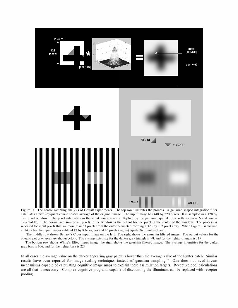
Figure 1a. The coarse sampling analysis of Gestalt experiments. The top row illustrates the process. A gaussian shaped integration filtercalculates a pixel-by-pixel coarse spatial average of the original image. The input image has 448 by 320 pixels. It is sampled in a 128 by128 pixel window. The pixel intensities in the input window are multiplied by the gaussian spatial filter with sigma =16 and size =128(middle). The normalized sum of all pixels in the window is the output for the pixel in the center of the window. The process isrepeated for input pixels that are more than 63 pixels from the outer perimeter, forming a 320 by 192 pixel array. When Figure 1 is viewedat 14 inches the input images subtend 12 by 8.6 degrees and 16 pixels (sigma) equals 26 minutes of arc. The middle row shows Benary’s Cross input image on the left. The right shows the gaussian filtered image. The output values for theequal-input gray areas are shown below. The average intensity for the darker gray triangle is 98, and for the lighter triangle is 119. The bottom row shows White’s Effect input image; the right shows the gaussian filtered image. The average intensities for the darkergray bars is 106, and for the lighter bars is 224.
In all cases the average value on the darker appearing gray patch is lower than the average value of the lighter patch. Similarresults have been reported for image scaling techniques instead of gaussian sampling.10 One does not need inventmechanisms capable of calculating cognitive image maps to explain these assimilation targets. Receptive pool calculationsare all that is necessary. Complex cognitive programs capable of discounting the illuminant can be replaced with receptorpooling.
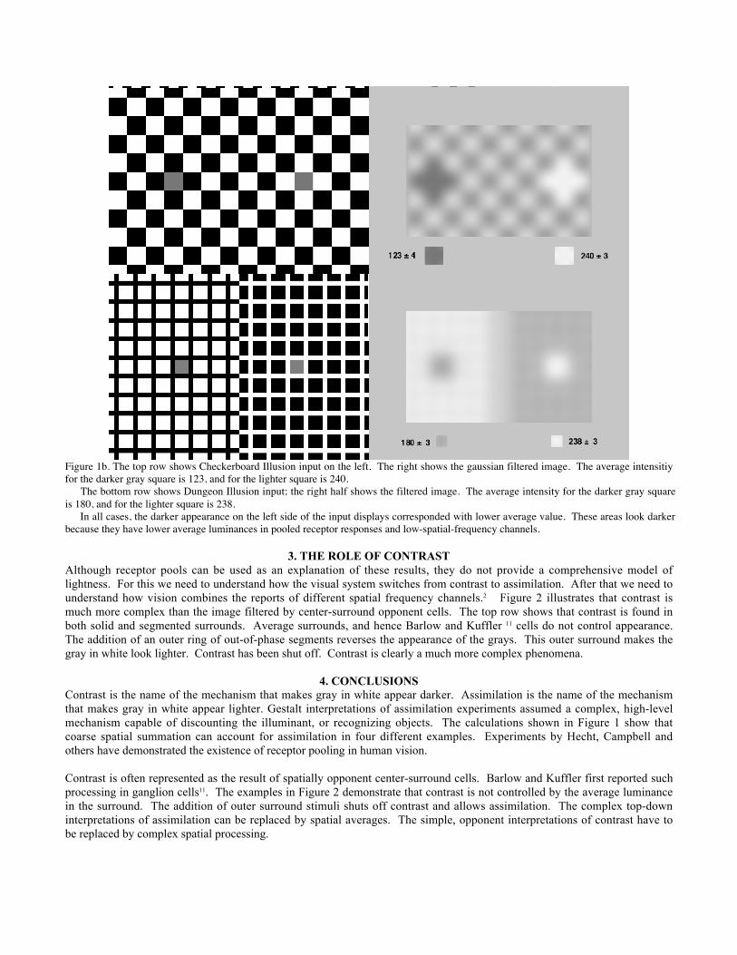
Figure 1b. The top row shows Checkerboard Illusion input on the left. The right shows the gaussian filtered image. The average intensitiyfor the darker gray square is 123, and for the lighter square is 240. The bottom row shows Dungeon Illusion input; the right half shows the filtered image. The average intensity for the darker gray squareis 180, and for the lighter square is 238. In all cases, the darker appearance on the left side of the input displays corresponded with lower average value. These areas look darkerbecause they have lower average luminances in pooled receptor responses and low-spatial-frequency channels.
3. THE ROLE OF CONTRASTAlthough receptor pools can be used as an explanation of these results, they do not provide a comprehensive model oflightness. For this we need to understand how the visual system switches from contrast to assimilation. After that we need tounderstand how vision combines the reports of different spatial frequency channels.2 Figure 2 illustrates that contrast ismuch more complex than the image filtered by center-surround opponent cells. The top row shows that contrast is found inboth solid and segmented surrounds. Average surrounds, and hence Barlow and Kuffler 11 cells do not control appearance.The addition of an outer ring of out-of-phase segments reverses the appearance of the grays. This outer surround makes thegray in white look lighter. Contrast has been shut off. Contrast is clearly a much more complex phenomena.
4. CONCLUSIONSContrast is the name of the mechanism that makes gray in white appear darker. Assimilation is the name of the mechanismthat makes gray in white appear lighter. Gestalt interpretations of assimilation experiments assumed a complex, high-levelmechanism capable of discounting the illuminant, or recognizing objects. The calculations shown in Figure 1 show thatcoarse spatial summation can account for assimilation in four different examples. Experiments by Hecht, Campbell andothers have demonstrated the existence of receptor pooling in human vision.
Contrast is often represented as the result of spatially opponent center-surround cells. Barlow and Kuffler first reported suchprocessing in ganglion cells11. The examples in Figure 2 demonstrate that contrast is not controlled by the average luminancein the surround. The addition of outer surround stimuli shuts off contrast and allows assimilation. The complex top-downinterpretations of assimilation can be replaced by spatial averages. The simple, opponent interpretations of contrast have tobe replaced by complex spatial processing.

Figure 2. Illustrations of contrast and assimilation using white, black and constant gray luminances. The top left pair of displays illustratesSimultaneous Contrast. The gray in white is darker than gray in black. Segmented surrounds (top right) demonstrate that averagesurround luminance does not cause contrast. Despite equal averages, contrast persists with half white and half black pixels in the surroundsegments. Radial segmented surrounds (bottom left) do not exhibit contrast. The addition of an outer surround (bottom right) cancelscontrast and introduces assimilation. Now the gray with adjacent white is lighter. In these complex periodic images assimilation isobserved because contrast in neutralized.
ACKNOWLEDGMENTSThe author wants to thank Alan Gilchrist, Jack Cowan, Steve Shevell and Mary McCann for very helpful discussions.
REFERENCES1. A. D. Logvinenko, “Lightness Induction Revisited”, Perception, 28, 803-816, (1999).2 O. Braddick, F. W. Campbell and J. Atkinson, “Channels in Vision: Basic Aspects”, in Handbook of Sensory Physiology VII, R. Held, H.W. Leibowitz and H. Teuber, Springer-Verlag, Berlin, 3-38, (1978).3. E. H. Land and J. J. McCann, “Lightness and Retinex Theory”, J. Opt. Soc. Am., 61, 1-11 (1971).4.J. J. McCann, S. McKee, and T. Taylor, “Quantitative studies in Retinex theory: A comparison between theoretical predictions andobserver responses to ‘Color Mondrian’ experiments,” Vision Res. 16, 445-458 (1976).5. W. Benary, “The influence of Form on Brightness”, in A Source Book of Gestalt Psychology, W, Ellis trans. Kegan, Paul, Trench, Truber
& Co. Ltd., London, 104-108, (1950).6. M. White, “A new effect of pattern lightness”, Perception, 8, 413-416, (1979).7. R. L. DeValois and K. K. DeValois, Spatial Vision, Oxford University Press, New York, (1988)..
8 E. Boring, “History of Experimental Psychology, p. 587, Appleton Century Croft, p. 229,(1957).9. J. J. McCann, “Calculating The Lightness Of Areas In A Single Plane”, J. Electron. Imaging, 10, pp. 110-122, (2001).10. J.J. McCann, “Gestalt Vision Experiments from an Image Processing Perspective”, Proc. IS&T PICS, Montreal, 4, pp. 9-14, (2001).11. H. Barlow, “Summation and inhibition in the frog’s retina”, J. Physiol. 119, 69-88, (1953); S. W, Kuffler, “Discharge patterns andfunctional organization of mammalian retina”, J. Neurophysiol. 16, 37-68, (1953)._____________*Correspondence: [email protected], phone 1-617-484-7865; McCann Imaging, 161 Claflin Street,. Belmont MA 02478, USA.