il-1α expression in pancreatic ductal...
TRANSCRIPT
IL-1α Expression in Pancreatic Ductal
Adenocarcinoma Affects the Tumor Cell
Migration and Is Regulated by the p38MAPK
Signaling Pathway
Vegard Tjomsland, Linda Bojmar, Per Sandström, Charlotte Bratthall, Davorka Messmer,
Anna Spångeus and Marie Larsson
Linköping University Post Print
N.B.: When citing this work, cite the original article.
Original Publication:
Vegard Tjomsland, Linda Bojmar, Per Sandström, Charlotte Bratthall, Davorka Messmer,
Anna Spångeus and Marie Larsson, IL-1α Expression in Pancreatic Ductal Adenocarcinoma
Affects the Tumor Cell Migration and Is Regulated by the p38MAPK Signaling Pathway,
2013, PLoS ONE, (8), 8.
http://dx.doi.org/10.1371/journal.pone.0070874
Licensee: Public Library of Science
http://www.plos.org/
Postprint available at: Linköping University Electronic Press
http://urn.kb.se/resolve?urn=urn:nbn:se:liu:diva-97445

IL-1a Expression in Pancreatic Ductal AdenocarcinomaAffects the Tumor Cell Migration and Is Regulated by thep38MAPK Signaling PathwayVegard Tjomsland1, Linda Bojmar2, Per Sandstrom2, Charlotte Bratthall3, Davorka Messmer4,
Anna Spangeus5, Marie Larsson1*
1 Molecular Virology, Department of Clinical and Experimental Medicine, Linkoping University, Linkoping, Sweden, 2 Division of Surgery, Department of Clinical and
Experimental Medicine, Linkoping University, Linkoping, Sweden, 3 Division of Oncology, Kalmar Hospital, Kalmar, Sweden, 4 Moores Cancer Center, University of
California San Diego, La Jolla, California, United States of America, 5 Division of Internal Medicine and Department of Endocrinology, Department of Medical and Health
Science, Linkoping University, Linkoping, Sweden
Abstract
The interplay between the tumor cells and the surrounding stroma creates inflammation, which promotes tumor growthand spread. The inflammation is a hallmark for pancreatic adenocarcinoma (PDAC) and is to high extent driven by IL-1a. IL-1a is expressed and secreted by the tumor cells and exerting its effect on the stroma, i.e. cancer associated fibroblasts (CAF),which in turn produce massive amount of inflammatory and immune regulatory factors. IL-1 induces activation oftranscription factors such as nuclear factor-kb (NF-kb), but also activator protein 1 (AP-1) via the small G-protein Ras.Dysregulation of Ras pathways are common in cancer as this oncogene is the most frequently mutated in many cancers. Incontrast, the signaling events leading up to the expression of IL-1a by tumor cells are not well elucidated. Our aim was toexamine the signaling cascade involved in the induction of IL-1a expression in PDAC. We found p38MAPK, activated by theK-Ras signaling pathway, to be involved in the expression of IL-1a by PDAC as blocking this pathway decreased both thegene and protein expression of IL-1a. Blockage of the P38MAPK signaling in PDAC also dampened the ability of the tumorcell to induce inflammation in CAFs. In addition, the IL-1a autocrine signaling regulated the migratory capacity of PDACcells. Taken together, the blockage of signaling pathways leading to IL-1a expression and/or neutralization of IL-1a in thePDAC microenvironment should be taken into consideration as possible treatment or complement to existing treatment ofthis cancer.
Citation: Tjomsland V, Bojmar L, Sandstrom P, Bratthall C, Messmer D, et al. (2013) IL-1a Expression in Pancreatic Ductal Adenocarcinoma Affects the Tumor CellMigration and Is Regulated by the p38MAPK Signaling Pathway. PLoS ONE 8(8): e70874. doi:10.1371/journal.pone.0070874
Editor: Guenter Schneider, Technische Universitat Munchen, Germany
Received January 18, 2013; Accepted June 25, 2013; Published August 12, 2013
Copyright: � 2013 Tjomsland et al. This is an open-access article distributed under the terms of the Creative Commons Attribution License, which permitsunrestricted use, distribution, and reproduction in any medium, provided the original author and source are credited.
Funding: This work has been supported by grants from: The Swedish Research Council (AI52731), VINNMER (Vinnova), the Medical Research Council of SoutheastSweden, and the Swedish Society of Medicine. The funders had no role in study design, data collection and analysis, decision to publish, or preparation of themanuscript.
Competing Interests: The authors have declared that no competing interests exist.
* E-mail: [email protected]
Introduction
A highly inflammatory environment is a hallmark for the
gastrointestinal malignancy pancreatic adenocarcinoma (PDAC)
including a rapid progression and a 5 year survival rate of less than
5% [1,2]. A massive fibrotic stroma encloses and infiltrates the
malignant cells [3] and the cellular composition of PDAC
microenvironment supports the recruitment of infiltrating immune
cells such as T cells, macrophages and dendritic cells (DCs) [4,5].
The CAFs play an important role in tumor progression and this is
supported by the fact that many tumors fail to develop unless the
stroma is modified [6] and these cellular modifications are induced
in a paracrine manner by adjacent tumor cells [7,8]. Proin-
flammatory factors such as IL-1, TNF-a, and COX-2 induce the
expression of inflammatory genes in CAFs and immune cells
present in the tumor [4,9].
Inflammation is strongly connected to most types of cancer and
involve activation of oncogenes and/or inactivation of tumor
suppressor genes that influence the proinflammatory transcrip-
tional programs by the malignant cells [10]. In the case for PDAC,
several factors have been shown to be involved in tumor and
stroma interactions including CXCL8, TGF-b and metallopro-
teases [11,12,13], all observed in our PDAC-CAF cross talk system
[9]. The inflammation in PDAC is to high extent driven by IL-1a,
expressed and secreted by the tumor cells and affecting the stroma
cells, i.e. CAFs, which produce massive amount of inflammatory
and immune regulatory factors both in vitro and in vivo [5,9].
The signaling event induced by IL-1 is well known and starts with
IL-1 binding to and signaling through the IL-1 receptor followed
by a subsequent activation of the p38 mitogen activated protein
kinase (MAPK) [14]. This occur via the small G protein Ras that
becomes associated with IRAK, TRAF6, and TAK-1, which
facilitate the p38MAPK activation by IL-1 [15]. In contrast, until
very recently the signaling events leading up to the expression of
IL-1a by the tumor cells had not been elucidated. Ling et al
showed for the first time involvement of the K-Ras mutation in
codon 12D in induction of IL-1a expression via the transcription
factor AP-1 [16]. Moreover, IL-1a activated NF-Kb and its target
genes IL-1a and p62 to initiate IL-1a/p62 feed forward loops,
PLOS ONE | www.plosone.org 1 August 2013 | Volume 8 | Issue 8 | e70874

which induced and sustained the NF-Kb activity [16]. Dysregu-
lation of Ras pathways is common in cancer as this oncogene is the
most frequently mutated in human cancers and contribute to
cancer cell survival [10]. Activating K-Ras mutations are present
in nearly all PDACs (up to 90%) and occur very early and are the
most frequent mutations in pancreatic cancer, followed by
mutation or silencing of p53, p1, and DPC4/smad4 [17,18]. For
pancreatic cancer, K-Ras mutations are a negative prognostic
factor after surgery and adjuvant chemoradiation [19]. The
mitogen activated protein kinases (extracellular signal-regulated
kinase (ERK), Jun N-terminal kinase (JNK), and p38MAPK) are
the best characterized signal pathways in transduction of Ras
activity and their oncogenic functions are mostly based on their
ability to activate AP-1 [20,21]. Ras/Raf/MAPK pathway is
involved in many cellular processes such as cell cycle regulation,
wound healing, cell migration, cell growth, division, and
differentiation [10].
So far, there is no selective specific inhibitor of K-Ras available
for routine clinical use and downstream targets such as MAPKs
are then interesting targets for inhibition of K-Ras signaling. ERK,
JNK, and p38MAPK are three major MAPKs and have key roles
in inflammation, tissue homeostasis, proliferation, differentiation,
migration and survival of cells. ERK is activated by mitogens,
whereas JNK and p38MAPKs are activated by cellular stress for
instant via the K-Ras signaling pathway. The p38MAPK signaling
has been shown to affect proliferation, differentiation, and
migration and is associated with cancers both in human and
mouse [20].
The aim of this study was to examine the signaling cascade
involved in the induction of IL-1a expression in PDAC. We
hypothesized that tumor cells creates an inflammatory microen-
vironment by inducing their expression of IL-1a through down-
stream targets of mutated K-Ras and deciphering this could be of
relevance to determine targets for treatment.
We found that the p38MAPK, activated by the K-Ras signaling
pathway, to be involved in the expression of IL-1a by PDAC cells,
as blocking this pathway decreased both the gene and protein
expression of IL-1a. Blockage of the p38MAPK signaling in
PDAC also dampened the ability of the tumor cell to induce
inflammation in CAFs and CAFs ability to enhance tumor cell
migration. Noteworthy, the IL1-a autocrine signaling regulated
the migratory capacity in PDAC cells. Taken together, the
blockage of signaling pathways leading to IL-1a expression and/or
neutralization of IL1-a in the PDAC microenvironment should be
taken into consideration as possible treatment or complement to
existing treatment of this cancer.
Results
p38MAPK/ERK is Involved in Tumor Cell Associated IL-1aExpression
The IL-1a positive primary PDAC cell line PC013 [9]
cultured in 1% FCS were exposed to 0–150 mM p38MAPK
(SB203580) inhibitor, 0–50 mM ERK (U0126) inhibitor, and 0–
75 mM JNK (SP600125) inhibitor for 24 h. We assessed if the
inhibitors asserted any negative effect on cell viability and found
no effects on the cell viability. The expression levels of IL-1amRNA decreased after exposure to the p38MAPK inhibitor,
while only a minor decrease was seen after ERK inhibitor,
while the JNK inhibitor slightly induce IL-1a expression
(Figure 1A–C). 100 mM of p38MAPK inhibitor SB203580
was found to be optimal to use for subsequent experiments. The
mRNA expression levels of IL-1b and IL-1RA decreased after
exposure to the p38MAPK inhibitor (Figure 1D–E) and had
similar expression curves as IL-1a. We have previously shown
that the IL-1a positive PDAC cell lines secrete IL-1a protein
and this is in accordance with findings from other tumor cell
lines [22]. In addition, our primary PDAC cell lines do not
express the IL-1b protein only the mRNA [9] and therefore did
we only examining the IL-1a protein expression and found that
it was significantly decreased after 24 h (p,0.005), 48 h
(p,0.005), and 72 h (p = 0.001) treatment with p38MAPK
inhibitor compared to vehicle treated cells (Figure 1F). We
had to use high concentration of SB303580 to block IL-1a and
as this inhibitor is known to also work on other MAPKs at high
concentration did we confirm the role of p38MAPK in the
regulation of tumor cell associated IL-1a by using SB220025,
which is considered to be a very specific inhibitor of p38
MAPK than SB203580. This inhibitor significantly reduced the
mRNA expression of IL-1a (P = 0.003) and IL-1b (P = 0.003),
but did not affect the expression levels of IL-1RA (Figure 1G–I). In addition, we found that p38 MAPK and ERK was
phosphorylated in our primary PDAC cell lines PC013, PC065
and that the inhibition of p38 MAPK with SB220025 did not
reduce the phosphorylation of this MAPK, whereas the
phosphorylation of ERK was reduced by the ERK inhibitor
(data not shown). The phosphorylation of p38 MAPK is in
accordance with previous findings for pancreatic cancer cell
lines [23,24], which indicate that this pathway is constitutively
activated in PDAC tumor cells.
The p38MAPK Inhibition had Different Effects on PDACsand CAFs Inflammatory Profiles
The p38MAPK inhibitors had in our study significantly
inhibitory effects on PDAC associated IL-1a expression and the
p38MAPK pathway have previously been found to be involved
in the regulation of chronic inflammation [20]. To examine the
regulatory role of p38MAPK on other inflammatory factors we
investigated the direct effects of SB203580 on PC013 cells and
CAFs. The results for the primary PDAC cell line PC013
showed significant reduced levels of IL-1a (P,0.001), IL-1b(P,0.001), IL-1RA (P = 0.001), and CXCL1 (P = 0.04), whereas
CCL20, and TNF-a levels were decreased but not significant
(Figure 2A). Levels of VEGFA, CXCL3 and COX-2 increased
but only VEGFA to a significant level (P = 0.01) (Figure 2A).Blockage of p38MAPK pathway in CAFs for 24 h resulted in
increased mRNA expression levels of CXCL2 (P = 0.02),
CXCL8 (P = 0.01) and CXCL3 (P = 0.05) in CAFs
(Figure 2B). Furthermore, the CAF gene expression levels of
CXCL5 (P = 0.024), CXCL6 (P,0.001), CCL20 (P = 0.03),
VEGFA (P = 0.02), COX-2 (P,0.001), IL-6 (P,0.001), and
IL-24 (P,0.001) decreased after blockage of p38MAPK
(Figure 2B). This clearly shows that the p38MAPK signaling
regulates the expression of inflammatory factors differently in
PDAC cells compared to CAFs.
Tumor Cell p38MAPK Signaling and IL-1 RegulationAffects the Tumor-CAF Crosstalk
IL-1a has previously been established as an important factor
involved in the communication between tumor cells and CAFs in
PDAC [9]. Our findings of an inhibiting role of p38MAPK
inhibitors on PDAC tumor cell IL-1a expression could have the
potential to affect the crosstalk between tumor cells and CAFs. We
examined the inflammatory profile of CAFs conditioned with
supernatants derived from PC013 first pretreated with p38MAPK
inhibitor for 72 h than recultured 48 h without inhibitor. The
conditioned CAFs showed significantly decreased gene levels of
Signaling Responsible for IL-1a Induction in PDAC
PLOS ONE | www.plosone.org 2 August 2013 | Volume 8 | Issue 8 | e70874

CXCL1 (P = 0.01), CXCL2 (P = 0.001), CXCL3 (P = 0.012),
CXCL5 (P = 0.023), CXCL8 (P = 0.004), CCL20 (P = 0.001),
COX-2 (P = 0.004), IL-6 (P,0.001), and IL-24 (P = 0.002)
(Figure 3A) compared to supernatants from vehicle treated
PC013 cells. To relate these findings in CAFs to tumor cell
associated IL-1a, as a downstream result of p38MAPK signaling,
we analyzed the IL-1a levels in the PC013 supernatants 2 days
after the exposure to p38MAPK inhibitor and found significantly
reduced levels of IL-1a (P = 0.038) (Figure 3B). Next was the
concentration of IL-1a in the p38MAPK pretreated PC013
derived supernatants returned to original levels by addition of
exogenous rhIL-1a and used to treat CAFs for 48 h. The CAF
gene profile showed increased levels of inflammatory factors
(Figure 3C). These results indicate an important role for
p38MAPK in regulating PDAC cell-CAF crosstalk through the
induction/upregulation of IL-1a expression in PDAC.
Figure 1. Blockage of p38MAPK signaling decreased the expression of IL-1a in primary PDAC cells. (A–E) The primary PDAC cell linePC013 was cultured for 24 h in 1% FBS medium supplemented with vehicle (DMSO) or different concentrations (0–150 mM) of the p38MAPK inhibitorSB203580, the ERK inhibitor U0126 (0–50 mM), or the JNK inhibitor SP600125 (0–75 mM). The mRNA expression levels of (A–C) IL-1a, (D) IL-1b, and (E)IL-1RA were analyzed using qRT-PCR. (F) PC013 cells were lysed after 24 h, 48 h, and 72 h after incubation with 100 mM SB203580 or vehicle and IL-1awas measured by ELISA and the result presented in pg/ml per 16105 cells. (G–I) To confirm the effect of SB203580, PC013 cells were treated for 24 hwith SB220025, another p38MAPK inhibitor, and the gene expression levels of (G) IL-1a, (H) IL-1b and (I) IL-1RA were analyzed and compared tovehicle. * = P,0.05, ** = P,0.005, and *** = P,0.001.doi:10.1371/journal.pone.0070874.g001
Signaling Responsible for IL-1a Induction in PDAC
PLOS ONE | www.plosone.org 3 August 2013 | Volume 8 | Issue 8 | e70874

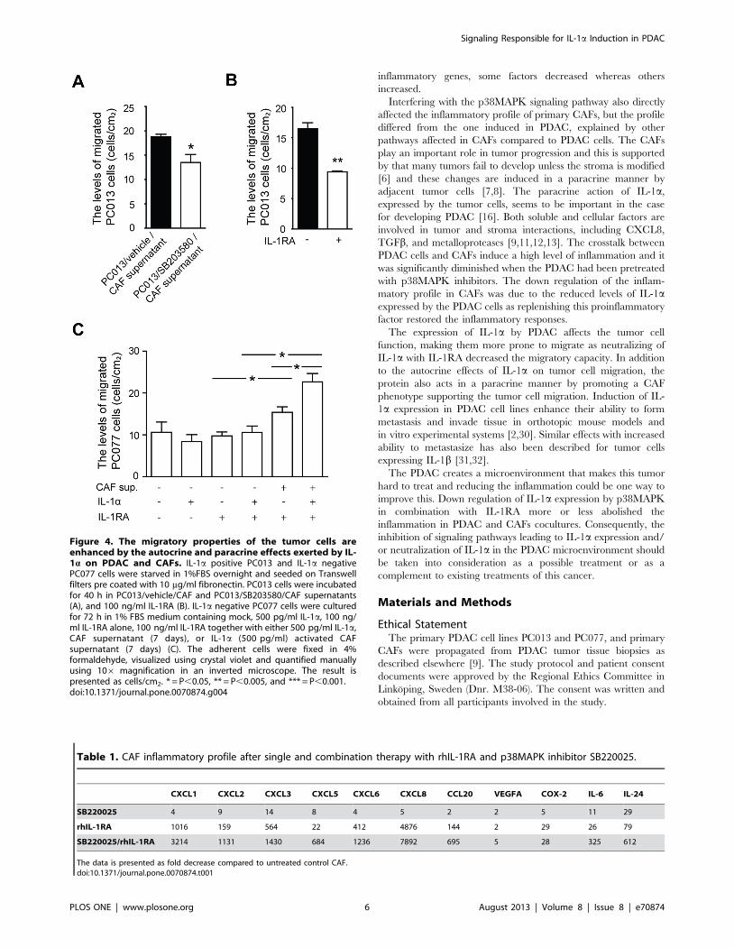
The Migratory Properties of the Tumor Cells is Enhancedby the Autocrine and Paracrine Effects Exerted by IL-1aon PDAC and CAFs
We further investigated if the decreased level of inflammatory
factors in CAFs, treated with supernatants derived from
p38MAPK signaling inhibited PC013 cells, affected the ability of
the tumor cells to migrate. PC013 cells exposed to supernatants
derived from CAFs cocultured with PC013 cells pretreated with
p38MAPK (IL-1a low) had significantly decreased migration
compared to cells exposed to supernatants derived from CAFs
cocultured with untreated PC013 cells (P = 0.038) (Figure 4A).The decreased levels of IL-1a in PC013 cells exposed to
p38MAPK inhibitor should reduce IL-1a autocrine feedback
and modulate the functions such as tumor cell mobility. We
investigated this by neutralized IL-1a with rhIL-1RA and found a
significantly reduced migration of PC013 cells (P = 0.002)
(Figure 4B). To in depth elucidate the effect of IL-1a on tumor
cell migration we used PC077, a primary PDAC cell line that is
IL-1a negative [9]. In addition, rhIL-1RA was added to all groups
in this experiment to eliminate any direct effects of IL-1a on the
tumor cell migration. Supernatants from untreated CAFs
(P = 0.025) significantly enhanced the migration of IL-1RA treated
PC077 cells, while supernatants from exogenous IL-1a activated
CAFs even further increased migration of PC077 cells compared
to both IL-1RA/IL-1a (P = 0.009) treated, IL-1a treated, and
untreated CAF supernatants (P = 0.038) (Figure 4C). Moreover,
no difference was found between rhIL-1RA and IL-1a/rhIL-1RA
treated PC077 cells. Taken together, this indicates that IL-1a not
only induce autocrine direct effects on cancer cell migration, but
also enhance migration through paracrine signaling and activation
of CAFs that obtain the ability to stimulate tumor cell migration by
altering the tumor cell phenotype, including increased expression
of metalloproteases and factors involved in epithelial mesenchymal
transition (work in progress).
rhIL-1RA in Combination with p38MAPK InhibitorEffectively Reduced the Levels of Inflammatory FactorsInduced in CAFs by PDAC Cells
IL-1RA has previously been shown to decrease the levels of
inflammatory factors in both single and CAFs cocultured with
PDAC cells [9]. Here we examined the effects treatment with IL-
1RA and p38MAPK inhibitor alone or as a combination therapy
had on PC013 and CAF cocultures. Blockage of p38MAPK
signaling significantly decreased the levels of CXCL1, CXCL5,
CXCL6, COX-2, IL-6, and IL-24 (P,0.001), CXCL2, and
CCL20 (P = 0.002), CXCL3 (P = 0.003), CXCL8 (P = 0.004), and
VEGFA (P = 0.009), (Figure 5) in CAFs cocultured with PDAC
cells compared to untreated controls (2–29 fold decease)
(Table 1). Neutralization of IL-1 by rhIL-1RA as a single agent
drastically reduced the levels of all the inflammatory factors (2–
4876 fold) compared to untreated cocultured CAFs (CXCL1.
CXCL5, CXCL6, CCL20, IL-6, IL-24 and COX-2 (P,0.001),
CXCL2, CXCL3, and CXCL8 (P = 0.002), and VEGFA
(P = 0.02)) (Figure 5 and Table 1). The combination of
p38MAPK inhibitor and rhIL-1RA decreased the inflammation
4.5–7895 folds compared to untreated controls (CXCL1, CXCL2,
CXCL5, CXCL6, CCL20, COX-2, IL-6, and IL-24 (P,0.001),
CXCL3, CXCL8, and VEGFA (P = 0.002)) (Figure 5 andTable 1). Moreover, the combination therapy decreased the
levels of the inflammatory factors compared to rhIL-1RA treated
cocultured CAFs (CXCL1, CXCL2, CXCL6 (P,0.001), CXCL3
(P = 0.06), CXCL5 (P = 0.003), CXCL8 (P = 0.04), CCL20
(P = 0.007), VEGFA (P = 0.02), IL-6 (P = 0.009) and IL-24
(P = 0.003)) (Figure 5).
Discussion
We have demonstrated that the p38MAPK signaling pathway is
involved in the expression of IL-1a by PDAC cells and that IL-1ainitiates a change in the PDAC phenotype and properties, making
the tumor cells more prone to migrate. The p38MAPK signaling
in tumor cells were shown to be involved in the upregulation of
inflammation in CAFs via the induction of IL-1a and to our
knowledge has this not been shown previously. Our previous
finding that IL-1a overexpression correlated with poor survival in
PDAC patients was confirmed in the study by Ling et al [16].
These findings suggest that inhibition of IL-1a expression and
activity in PDAC will drastically decrease the levels of inflamma-
tory factors in CAFs, and reducing and/or neutralizing the effects
of IL-1a could have the potential to reduce tumor spread and
improve the clinical outcome for the patients [5,9].
The signaling pathways/events giving rise to constitutive
expression of IL-1a have been elusive. A recent study by Ling
et al [16] using a mouse model where mutation in K-Ras G12D
was used to induce PDAC showed that this activating K-Ras
Figure 2. Inhibition of p38MAPK signaling affects the inflam-matory profile in PDAC cells and CAFs. The primary PDAC cell linePC013 (A) and primary CAFs (B) obtained from one PDAC patient werecultured for 24 h with 100 mM of the p38 inhibitor SB203580 in 1% FBSmedium. The gene expression levels for CXCL1, CXCL2, CXCL3, CXCL5,CXCL8, CCL20, VEGFA, IL6, COX-2, TNFa, TGFb, IL24, IL-1a, IL-1b, and IL-1RA were analyzed by qRT-PCR and normalized to vehicle. * = P,0.05,** = P,0.005, and *** = P,0.001.doi:10.1371/journal.pone.0070874.g002
Signaling Responsible for IL-1a Induction in PDAC
PLOS ONE | www.plosone.org 4 August 2013 | Volume 8 | Issue 8 | e70874

mutation induced activation of NF-kB, which was required for
PDAC development, and expression of IL-1a in the tumor cells
and that this create a intrinsic inflammatory response that promote
a pro-tumorigenic microenvironment through the expression of
inflammatory mediators, e.g. cytokines such as IL-1a [16]. The
proposed mechanism for NF-kB activation by K-Ras is through
AP-1 induced IL-1a overexpression [16]. The expression of IL-1aby tumor cells is detected in 90% of the PDAC patients [9] and
could correlate to mutations in the oncogene K-Ras as they also
are present in up to 90% of the PDAC cases [25,26,27]. These
findings clearly demonstrate the role of inflammation in the
development of PDAC. As all attempts of developing a drug that
directly blocks the mutated K-Ras oncogene has been unsuccessful
[28], did we target downstream factors activated by K-Ras. The
signaling cascades through K-Ras lead to many different events
inside the cell and several pathways, i.e. RAS/RAF/MAPK RAS/
P13K/AKT, are well characterized [29]. In response to cellular
stress and cytokine stimulation mediated through K-Ras do
p38MAPK kinases (MMK3 and MKK6) and JNK kinases
(MKK4 and MKK7) phosphorylate p38MAPK and JNK,
respectively [15].
PDAC have a very elevated expression of multiple inflammatory
genes and the main cell in the tumor stroma, i.e. CAFs, are the
major producers of these factors [5,9]. According to our own data
and the study by Ling et al [16], IL-1a is the pro-inflammatory
factor responsible for inducing the expression of the inflammation.
We found that the IL-1a gene and protein expression by primary
PDAC cell lines was down regulated only when the signaling
through p38MAPK was blocked. This clearly show that signaling
trough activated p38MAPK highly influence the IL-1a expression
and also eliminates ERK and JNK as contributors to the
regulation of IL-1a in PDAC cells. In addition, the inhibition of
p38MAPK in PDAC lead to a modulated gene profile for several
Figure 3. Down regulation of p38MAPK signaling decreased the levels of tumor cell associated IL-1a and the inflammatory profilein CAFs. The primary PDAC cell line PC013 was cultured in 1%FBS medium containing 100 mM SB203580 or vehicle for 72 h. The 1%FBS medium wasreplenished and the cells cultured for another 48 h before the supernatant was transferred to CAFs. The CAFs were cultured for 72 h. Theinflammatory gene profile for the CAFs (A) was analyzed by normalizing the effects of SB203580 to vehicle. The supernatant are referred to as (PC013/vehicle/CAF) and (PC013/SB203580/CAF). IL-1a was measured in the supernatants from SB203580 and vehicle treated PC013 cells by ELISA and theresult is presented in pg/ml per 16105 cells (B). The IL-1a levels in the supernatants from the SB203580 treated PC013 cells were equalized to thecontrol supernatants by adding rhIL-1a. The IL-1a normalized supernatants were added to CAFs and incubated for 72 h. The inflammatory geneprofile was analyzed by normalizing the IL-1a supernatants to vehicle supernatants (C).doi:10.1371/journal.pone.0070874.g003
Signaling Responsible for IL-1a Induction in PDAC
PLOS ONE | www.plosone.org 5 August 2013 | Volume 8 | Issue 8 | e70874
inflammatory genes, some factors decreased whereas others
increased.
Interfering with the p38MAPK signaling pathway also directly
affected the inflammatory profile of primary CAFs, but the profile
differed from the one induced in PDAC, explained by other
pathways affected in CAFs compared to PDAC cells. The CAFs
play an important role in tumor progression and this is supported
by that many tumors fail to develop unless the stroma is modified
[6] and these changes are induced in a paracrine manner by
adjacent tumor cells [7,8]. The paracrine action of IL-1a,
expressed by the tumor cells, seems to be important in the case
for developing PDAC [16]. Both soluble and cellular factors are
involved in tumor and stroma interactions, including CXCL8,
TGFb, and metalloproteases [9,11,12,13]. The crosstalk between
PDAC cells and CAFs induce a high level of inflammation and it
was significantly diminished when the PDAC had been pretreated
with p38MAPK inhibitors. The down regulation of the inflam-
matory profile in CAFs was due to the reduced levels of IL-1aexpressed by the PDAC cells as replenishing this proinflammatory
factor restored the inflammatory responses.
The expression of IL-1a by PDAC affects the tumor cell
function, making them more prone to migrate as neutralizing of
IL-1a with IL-1RA decreased the migratory capacity. In addition
to the autocrine effects of IL-1a on tumor cell migration, the
protein also acts in a paracrine manner by promoting a CAF
phenotype supporting the tumor cell migration. Induction of IL-
1a expression in PDAC cell lines enhance their ability to form
metastasis and invade tissue in orthotopic mouse models and
in vitro experimental systems [2,30]. Similar effects with increased
ability to metastasize has also been described for tumor cells
expressing IL-1b [31,32].
The PDAC creates a microenvironment that makes this tumor
hard to treat and reducing the inflammation could be one way to
improve this. Down regulation of IL-1a expression by p38MAPK
in combination with IL-1RA more or less abolished the
inflammation in PDAC and CAFs cocultures. Consequently, the
inhibition of signaling pathways leading to IL-1a expression and/
or neutralization of IL-1a in the PDAC microenvironment should
be taken into consideration as a possible treatment or as a
complement to existing treatments of this cancer.
Materials and Methods
Ethical StatementThe primary PDAC cell lines PC013 and PC077, and primary
CAFs were propagated from PDAC tumor tissue biopsies as
described elsewhere [9]. The study protocol and patient consent
documents were approved by the Regional Ethics Committee in
Linkoping, Sweden (Dnr. M38-06). The consent was written and
obtained from all participants involved in the study.
Figure 4. The migratory properties of the tumor cells areenhanced by the autocrine and paracrine effects exerted by IL-1a on PDAC and CAFs. IL-1a positive PC013 and IL-1a negativePC077 cells were starved in 1%FBS overnight and seeded on Transwellfilters pre coated with 10 mg/ml fibronectin. PC013 cells were incubatedfor 40 h in PC013/vehicle/CAF and PC013/SB203580/CAF supernatants(A), and 100 ng/ml IL-1RA (B). IL-1a negative PC077 cells were culturedfor 72 h in 1% FBS medium containing mock, 500 pg/ml IL-1a, 100 ng/ml IL-1RA alone, 100 ng/ml IL-1RA together with either 500 pg/ml IL-1a,CAF supernatant (7 days), or IL-1a (500 pg/ml) activated CAFsupernatant (7 days) (C). The adherent cells were fixed in 4%formaldehyde, visualized using crystal violet and quantified manuallyusing 106 magnification in an inverted microscope. The result ispresented as cells/cm2. * = P,0.05, ** = P,0.005, and *** = P,0.001.doi:10.1371/journal.pone.0070874.g004
Table 1. CAF inflammatory profile after single and combination therapy with rhIL-1RA and p38MAPK inhibitor SB220025.
CXCL1 CXCL2 CXCL3 CXCL5 CXCL6 CXCL8 CCL20 VEGFA COX-2 IL-6 IL-24
SB220025 4 9 14 8 4 5 2 2 5 11 29
rhIL-1RA 1016 159 564 22 412 4876 144 2 29 26 79
SB220025/rhIL-1RA 3214 1131 1430 684 1236 7892 695 5 28 325 612
The data is presented as fold decrease compared to untreated control CAF.doi:10.1371/journal.pone.0070874.t001
Signaling Responsible for IL-1a Induction in PDAC
PLOS ONE | www.plosone.org 6 August 2013 | Volume 8 | Issue 8 | e70874

PDAC and CAF Cell LinesThe primary PDAC cell lines PC013 and PC077, and
primary CAFs were cultured in RPMI 1640 (Fisher Scientific,
Pittsburgh, PA), supplemented with 1% FCS (Invitrogen), 2 mM
HEPES (Invitrogen), 30 mg/ml Gentamycin (Invitrogen) (R10).
For all assays, at 66105 PC013 and PC077 cells were seeded
per well and cultured for 24 h before adding the different
inhibitors.
Figure 5. Combination therapy with a p38MAPK inhibitor and IL-1RA was superior to single therapy in inhibiting the PDAC-CAFcrosstalk induced inflammation. The PDAC cell line PC013 was cocultured with primary CAFs using cell culture inserts for 5 days in 1% FBSmedium supplemented with p38MAPK inhibitor SB220025 (1 mM) or rhIL-1RA (10 mg/ml) or a combination of these agents. CAFs inflammatory geneexpression profile after SB220025 and rhIL-1RA single or combination treatment were compared to cocultured vehicle treated CAFs The combinationof the two agents were also compared to single agent treatment and the result is shown as relative gene expression. * = P,0.05, ** = P,0.005, and*** = P,0.001.doi:10.1371/journal.pone.0070874.g005
Signaling Responsible for IL-1a Induction in PDAC
PLOS ONE | www.plosone.org 7 August 2013 | Volume 8 | Issue 8 | e70874

Culturing PDAC Cells and CAFs with p38MAPK, ERK andJNK Inhibitors
PC013 cells were cultured in R10 containing vehicle (DMSO),
0.1–150 mM p38MAPK inhibitor SB203580 (Cayman Chemicals,
US), p38MAPK inhibitor SB220025, ERK inhibitor UO126, or
JNK inhibitor SP2600125 (SigmaAldrich, Sweden) for 24–72 h. In
other sets of experiments, PC013 cells were cultured with 100 mM
p38MAPK inhibitor SB203580 or vehicle (DMSO) for 72 h,
washed 3 times and recultured in 2 ml R10 for 48 h. The 48 h
supernatants were transferred and used to culture CAFs for 72 h.
The PC013-SB203580-CAF (C20) and PC013-vehicle-CAF (C30)
conditioned medium were normalized to number of cells/group
(16105 cells/ml medium) to adjust for differences in the total
amount of cells. To confirm the role of IL-1a in tumor cell-CAF
cross-talk, the levels of IL-1a was measured in the supernatants
from SB203580 and vehicle treated PC013 cells and equalized
with rhIL-1a (R&D Systems, UK). The IL-1a normalized medium
was added to CAFs for 72 h. The cells were lysed with RLT buffer
(Qiagen) and RNA prepared as described elsewhere [9].
Treatment of PDAC and CAFs with p38MAPK Inhibitorand IL-1RA
The effects of p38 inhibitor (SB220025) and IL-1RA on tumor
cell/CAF cross-talk were investigated and PC013 cells and CAFs
were cocultured in inserts (0.4 mm) (BD, USA) for 5 days in R10
medium supplemented with rhIL-1RA (10 mg/ml) (Kineret
100 mg, Biovitrum AB) and/or 1 mM SB220025.
RNA Extraction and QuantificationTotal RNA purification and, cDNA was prepared as previously
described [9]. Quantitative PCR was performed with Fast SYBR
Green Master Mix (Version 09/2007; Applied Biosystems, Foster
City, CA) on 7900 Fast Real-Time PCR system with 7900 System
SDS 2.3 Software (Applied Biosystems). In the negative controls
cDNA were replaced by distilled water. Specific primers for
CXCL8, CCL20, IL-1a, IL-1b, IL-6, IL-24, IL-1RA, TGFb,
VEGF-A, (CyberGene AB), and COX-2 (Invitrogen) were used.
Glyceraldehyde-3-phosphate dehydrogenase (GAPDH) (Cyber-
Gene AB) and actin were utilized as housekeeping genes. The
primers were designed using Primer Express (Applied Biosystems)
if not otherwise indicated. Real-time PCRs for the detection of
CXCL chemokines and TNFa were performed using TaqManHGene Expression Assays (Applied BioSystems) according to the
manufacturer’s protocol. All reactions were performed in tripli-
cates including none-template controls and endogenous control
probes. FAM conjugated, gene specific assays were
Hs00236937_m1 (CXCL1), Hs00236966_m1 (CXCL2)
Hs00171061_m1 (CXCL3), Hs00171085_m1 (CXCL5),
Hs00237017_m1 (CXCL6), and Hs99999043_m1 (TNFa). The
results were analyzed using the DDCt method [33] and presented
as either normalized data or as relative gene expression.
ELISASupernatants were collected and cells harvested and counted
before lysis with lysis buffer pH 7.5. The lysates and supernatants
were analyzed for the concentration of IL-1a by ELISA (Nordic
Biosite, Sweden) according to the manufacturers’ protocols. This
ELISA measures precursor, secreted, and membrane-associated
forms of IL-1a. The levels of IL-1a are presented as pg/16105
cells.
Migration AssayTranswell migration assay were conducted as described
elsewhere [34]. Briefly, PC077 and PC013 were starved in 1%
FBS overnight and seeded on 6-well plates Transwell filters
(Costar) (8 mm pore size) precoated with 10 mg/ml fibronectin
(Sigma, St. Louis, MO). PC077 cells were incubated for 40 h in
1% FBS medium containing 100 ng/ml IL-1RA and either
500 pg/ml IL-1a, CAF supernatant (7 days), or IL-1a (500 pg/ml)
activated CAF supernatant (7 days). PC013 cells were incubated
for 40 h in 1% FBS and/or 100 ng/ml IL-1RA, C20, and C30
conditioned medium. The upper compartment was removed and
the medium discarded. The adherent cells were washed in PBS,
fixed in 4% formaldehyde and visualized using crystal violet
(SigmaAldrich, Sweden). The sample identification was blinded
and all attached cells were manually counted using 106magnifications in an inverted microscope (Leica). The data was
obtained from 3 experiments (mean 6 standard error) and
presented as the mean cells/cm2.
Statistical AnalysisThe statistical analysis was performed with GraphPad Prism 5
(GraphPad Software), P,0.05 was considered statistically signif-
icant and error bars throughout indicate standard error of the
mean (SEM). The data were analyzed using the paired t-.test and
unpaired t-test was used for normalized data.
Author Contributions
Conceived and designed the experiments: VT ML. Performed the
experiments: VT. Analyzed the data: VT ML LB. Wrote the paper: ML
VT DM PS AS CB.
References
1. Li D, Xie K, Wolff R, Abbruzzese JL (2004) Pancreatic cancer. Lancet 363:
1049–1057.
2. Melisi D, Niu J, Chang Z, Xia Q, Peng B, et al. (2009) Secreted interleukin-
1alpha induces a metastatic phenotype in pancreatic cancer by sustaining a
constitutive activation of nuclear factor-kappaB. Mol Cancer Res 7: 624–633.
3. Korc M (2007) Pancreatic cancer-associated stroma production. Am J Surg 194:
S84–86.
4. Esposito I, Menicagli M, Funel N, Bergmann F, Boggi U, et al. (2004)
Inflammatory cells contribute to the generation of an angiogenic phenotype in
pancreatic ductal adenocarcinoma. J Clin Pathol 57: 630–636.
5. Tjomsland V, Niklasson L, Sandstrom P, Borch K, Druid H, et al. (2011) The
desmoplastic stroma plays an essential role in the accumulation and modulation
of infiltrated immune cells in pancreatic adenocarcinoma. Clin Dev Immunol
2011: 212810.
6. Franco OE, Shaw AK, Strand DW, Hayward SW (2011) Cancer associated
fibroblasts in cancer pathogenesis. Semin Cell Dev Biol 21: 33–39.
7. Sato N, Maehara N, Goggins M (2004) Gene expression profiling of tumor-
stromal interactions between pancreatic cancer cells and stromal fibroblasts.
Cancer Res 64: 6950–6956.
8. Somasundaram R, Herlyn D (2009) Chemokines and the microenvironment in
neuroectodermal tumor-host interaction. Semin Cancer Biol 19: 92–96.
9. Tjomsland V, Spangeus A, Valila J, Sandstrom P, Borch K, et al. (2011)
Interleukin 1alpha sustains the expression of inflammatory factors in human
pancreatic cancer microenvironment by targeting cancer-associated fibroblasts.
Neoplasia 13: 664–675.
10. Pylayeva-Gupta Y, Grabocka E, Bar-Sagi D (2011) RAS oncogenes: weaving a
tumorigenic web. Nat Rev Cancer 11: 761–774.
11. Sato N, Fukushima N, Maehara N, Matsubayashi H, Koopmann J, et al. (2003)
SPARC/osteonectin is a frequent target for aberrant methylation in pancreatic
adenocarcinoma and a mediator of tumor-stromal interactions. Oncogene 22:
5021–5030.
12. Saad S, Gottlieb DJ, Bradstock KF, Overall CM, Bendall LJ (2002) Cancer cell-
associated fibronectin induces release of matrix metalloproteinase-2 from normal
fibroblasts. Cancer Res 62: 283–289.
13. Dong Z, Nemeth JA, Cher ML, Palmer KC, Bright RC, et al. (2001) Differential
regulation of matrix metalloproteinase-9, tissue inhibitor of metalloproteinase-1
(TIMP-1) and TIMP-2 expression in co-cultures of prostate cancer and stromal
cells. Int J Cancer 93: 507–515.
Signaling Responsible for IL-1a Induction in PDAC
PLOS ONE | www.plosone.org 8 August 2013 | Volume 8 | Issue 8 | e70874

14. Freshney NW, Rawlinson L, Guesdon F, Jones E, Cowley S, et al. (1994)
Interleukin-1 activates a novel protein kinase cascade that results in thephosphorylation of Hsp27. Cell 78: 1039–1049.
15. McDermott EP, O’Neill LA (2002) Ras participates in the activation of p38
MAPK by interleukin-1 by associating with IRAK, IRAK2, TRAF6, and TAK-1. J Biol Chem 277: 7808–7815.
16. Ling J, Kang Y, Zhao R, Xia Q, Lee DF, et al. (2012) KrasG12D-inducedIKK2/beta/NF-kappaB activation by IL-1alpha and p62 feedforward loops is
required for development of pancreatic ductal adenocarcinoma. Cancer Cell 21:
105–120.17. Slebos RJ, Hoppin JA, Tolbert PE, Holly EA, Brock JW, et al. (2000) K-ras and
p53 in pancreatic cancer: association with medical history, histopathology, andenvironmental exposures in a population-based study. Cancer Epidemiol
Biomarkers Prev 9: 1223–1232.18. Hruban RH, Goggins M, Parsons J, Kern SE (2000) Progression model for
pancreatic cancer. Clin Cancer Res 6: 2969–2972.
19. Brunner TB, Cengel KA, Hahn SM, Wu J, Fraker DL, et al. (2005) Pancreaticcancer cell radiation survival and prenyltransferase inhibition: the role of K-Ras.
Cancer Res 65: 8433–8441.20. Wagner EF, Nebreda AR (2009) Signal integration by JNK and p38 MAPK
pathways in cancer development. Nat Rev Cancer 9: 537–549.
21. Chang L, Karin M (2001) Mammalian MAP kinase signalling cascades. Nature410: 37–40.
22. Ebrahimi B, Tucker SL, Li D, Abbruzzese JL, Kurzrock R (2004) Cytokines inpancreatic carcinoma: correlation with phenotypic characteristics and prognosis.
Cancer 101: 2727–2736.23. Takekawa M, Tatebayashi K, Itoh F, Adachi M, Imai K, et al. (2002) Smad-
dependent GADD45beta expression mediates delayed activation of p38 MAP
kinase by TGF-beta. Embo J 21: 6473–6482.24. Masui T, Doi R, Mori T, Toyoda E, Koizumi M, et al. (2004) Metastin and its
variant forms suppress migration of pancreatic cancer cells. Biochem BiophysRes Commun 315: 85–92.
25. Mu DQ, Peng YS, Xu QJ (2004) Values of mutations of K-ras oncogene at
codon 12 in detection of pancreatic cancer: 15-year experience.
World J Gastroenterol 10: 471–475.
26. Tabata T, Fujimori T, Maeda S, Yamamoto M, Saitoh Y (1993) The role of Ras
mutation in pancreatic cancer, precancerous lesions, and chronic pancreatitis.
Int J Pancreatol 14: 237–244.
27. Kim ST, Lim do H, Jang KT, Lim T, Lee J, et al. (2011) Impact of KRAS
mutations on clinical outcomes in pancreatic cancer patients treated with first-
line gemcitabine-based chemotherapy. Mol Cancer Ther 10: 1993–1999.
28. Gysin S, Salt M, Young A, McCormick F (2011) Therapeutic strategies for
targeting ras proteins. Genes Cancer 2: 359–372.
29. Meier F, Schittek B, Busch S, Garbe C, Smalley K, et al. (2005) The RAS/
RAF/MEK/ERK and PI3K/AKT signaling pathways present molecular
targets for the effective treatment of advanced melanoma. Front Biosci 10:
2986–3001.
30. Matsuo Y, Sawai H, Ochi N, Yasuda A, Takahashi H, et al. (2009) Interleukin-
1alpha secreted by pancreatic cancer cells promotes angiogenesis and its
therapeutic implications. J Surg Res 153: 274–281.
31. Chirivi RG, Garofalo A, Padura IM, Mantovani A, Giavazzi R (1993)
Interleukin 1 receptor antagonist inhibits the augmentation of metastasis
induced by interleukin 1 or lipopolysaccharide in a human melanoma/nude
mouse system. Cancer Res 53: 5051–5054.
32. Pantschenko AG, Pushkar I, Anderson KH, Wang Y, Miller LJ, et al. (2003) The
interleukin-1 family of cytokines and receptors in human breast cancer:
implications for tumor progression. Int J Oncol 23: 269–284.
33. Livak KJ, Schmittgen TD (2001) Analysis of relative gene expression data using
real-time quantitative PCR and the 2(-Delta Delta C(T)) Method. Methods 25:
402–408.
34. Manes T, Zheng DQ, Tognin S, Woodard AS, Marchisio PC, et al. (2003)
Alpha(v)beta3 integrin expression up-regulates cdc2, which modulates cell
migration. J Cell Biol 161: 817–826.
Signaling Responsible for IL-1a Induction in PDAC
PLOS ONE | www.plosone.org 9 August 2013 | Volume 8 | Issue 8 | e70874