iii.eye i: anatomy & optics of vision g&h ch 49 + ls +… g ... nerve retina direction of...
TRANSCRIPT
Annette Sims, MD, Ophthalmologistnext Tuesday! Hooray!!
BI 358 Lecture 18
I. Announcements Quiz 5 returned at end of lecture. Eye Dissection & Vision lab next Tuesday > Lecture by Dr. Sims! Final Quiz (6) next Thursday, then thoughts on grad schools in medicine & allied health.
II. Physiology in the News The sight-saving diet?UC Berkeley WellnessLetter, November 2011.
III. Eye I: Anatomy & Optics of Vision G&H ch 49 + LS +…IV.Eye II: Retinal Receptor & Neural Function G&H ch 50V.Eye III: Overview of Visual Pathways & Pathologies
G&H ch 51 + LS1 + Silverthorn +...
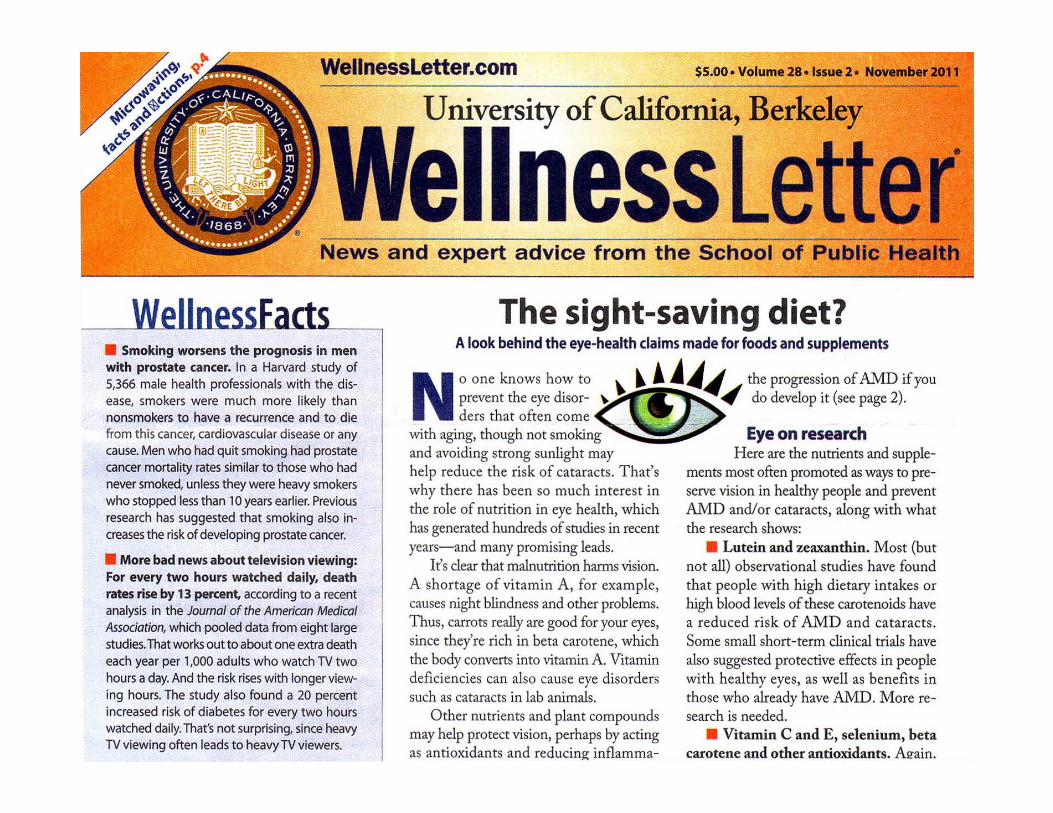

1. High intakes of lutein & zeaxanthin (carotenoids) may reduce risk of macular degeneration (AMD) & cataracts.
2. Consuming plant-foods rich in antioxidants including vitamins C & E, selenium & β-carotene also may reduce risk of macular degeneration & cataracts.
3. Older vegetarians are 30-40% less likely to develop cataracts compared to daily meat eaters.
4. The above holds for foods, but there is little evidence that anti-oxidant supplements have this effect.
5. Zinc is essential to good vision & is found in the retina & may protect eyes from light damage & inflammation. Get zinc from food (oysters, shrimp, whole grains, yogurt...)
6. High intakes of fish rich in Ω-3 fats also reduce AMD.
Sight-saving Diet?


Eye: Elaborate sensory receptor ≡ Camera
Aperture + Lens + Film!
≡
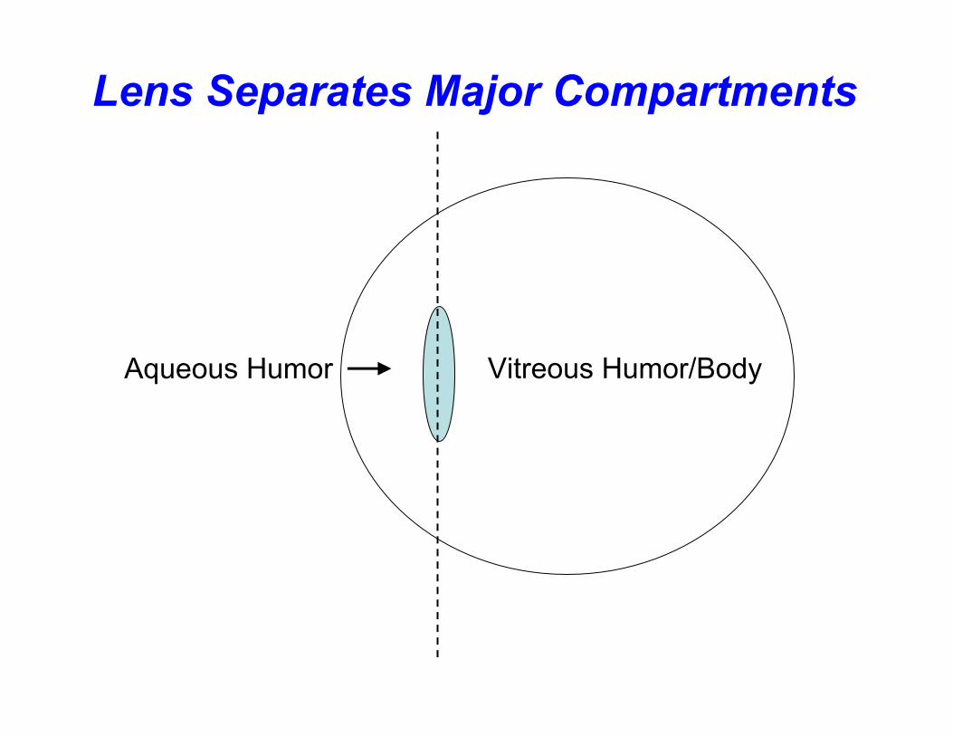
Lens Separates Major Compartments
Vitreous Humor/BodyAqueous Humor

Eye: Anterior View
L Sherwood 2012
Lacrimal Gland

L Sherwood 2012
Eye: Saggital View
1
23

Copyright © 2010 Pearson Education, Inc.
Fovea
Macula
Central retinalartery and vein(+ optic nerve)
(b)
Optic disk(blind spot)
The Blind Spot?
D. Silverthorn 2010

G&H 2011 fig 49-2
Convex lens convergence + focal length
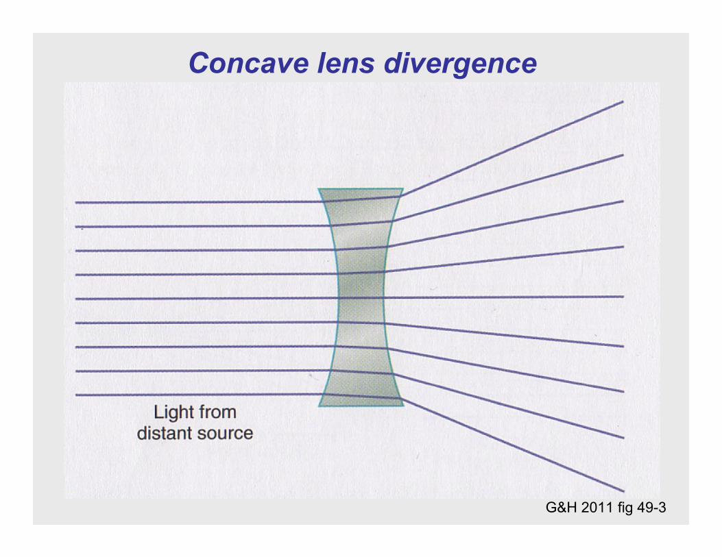
Concave lens divergence
G&H 2011 fig 49-3

G&H 2011 fig 49-7
Image formation by convex lens

What's a diopter? Refractive power measurement = f-1 or 1m divide by f
Focal length = f
G&H 2011 fig 49-8

Refractive index?
G&H 2011 fig 49-9

Mechanism of accommodation
G&H 2011 fig 49-10

Mini-tramp analogy
Lens
Suspensoryligaments Ciliary
muscle
http://trampolinefiend.com/

Accommodation ≡ Lens Thickens + Pupils Constrict + Eyes Adduct!
Radial/DilatorMuscles
Circular/ConstrictorMuscles
Radial Muscles Contract
Circular Muscles Contract
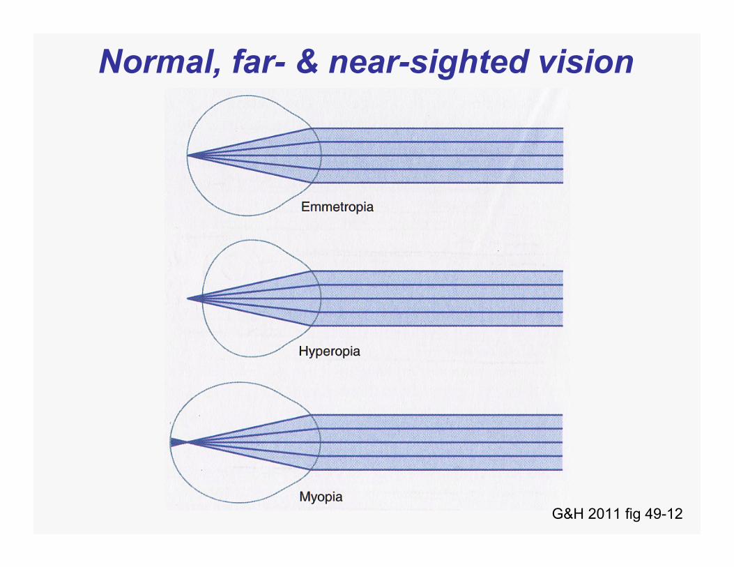
G&H 2011 fig 49-12
Normal, far- & near-sighted vision
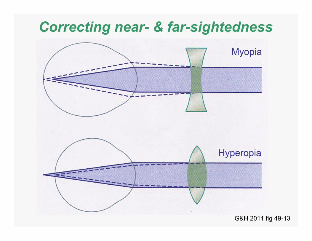
Correcting near- & far-sightednessMyopia
Hyperopia
G&H 2011 fig 49-13

Astigmatism?
G&H 2011 fig 49-15

G&H 2011 fig 49-19
Fluid formation & flow

Aqueous humor formation
G&H 2011 fig 49-20

Glaucoma & intraocular pressure (IOP)?
G&H 2011 fig 49-22
IOP Normal 12-20 mm Hg x = 15 ± 2 mm Hg_
Glaucoma ≥ 25-30 mm Hg up to 60-70 mm Hg!
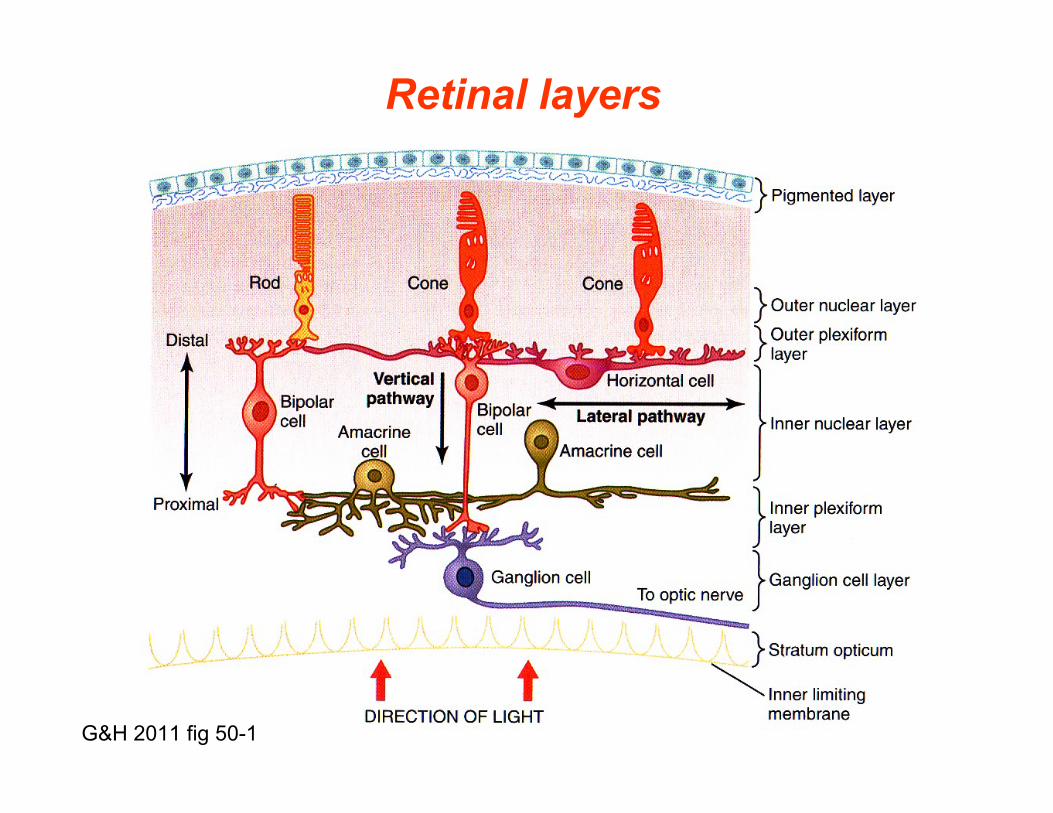
G&H 2011 fig 50-1
Retinal layers

fig 6-17 p 158 LS1 2006
Optic nerve
Retina
Direction of light
Direction of retinal visual processing
Frontofretina
Fibers ofthe opticnerve
Ganglioncell
Amacrinecell
Bipolarcell
Cone Rod
Photoreceptorcells
Horizontalcell
Retina
Pigment layer
Choroid layer
Sclera
Backofretina

Direction of light
Macula & fovea hot spot!
G&H 2011 fig 50-2

Exposed Cones @ Fovea/Macular RegionNormal Fovea
Photoreceptors Inner & Outer Segments!Nadia Al Kharousi, Upender K. Wali and Sitara Azeem Current Applications in Optical Coherence Tomography in Ophthalmology, March 2013.

Peripheral (L) vs. foveal (R) retina
G&H 2011 fig 50-12
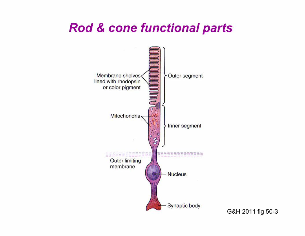
Rod & cone functional parts
G&H 2011 fig 50-3
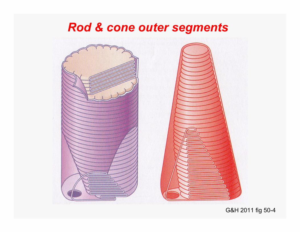
Rod & cone outer segments
G&H 2011 fig 50-4

In rods,light converts cis to trans retinal
L Sherwood 2012
Rhodopsin = Opsin + Retinal

Rhodopsin-retinal visual cycle
G&H 2011 fig 50-5
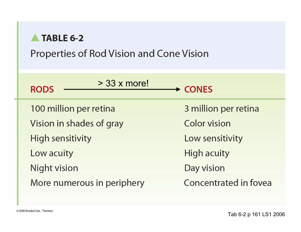
Tab 6-2 p 161 LS1 2006
> 33 x more!

Intermediate Colors Are Produced When 10 Colors Are Superimposed

Ratios of cone stimulation determine color interpretation: orange 99:42:0
G&H 2011 fig 50-10

http://www.color-blindness.com/coblis-color-blindness-simulator/
Color Deficiencies Can Impact Daily Activities, Pleasure & Work!
Red Cone Deficiency = ProtanopiaGreen Cone Deficiency = Deuteranopia
Blue Cone Deficiency = Tritanopia

Ishihara Chart for Normal (74) vs. Red-Green Color Blindness (21)
G&H 2011 fig 50-11a

Ishihara chart for red-blind protanope (2) vs. green-blind deuteranope (4)
G&H 2011 fig 50-11b
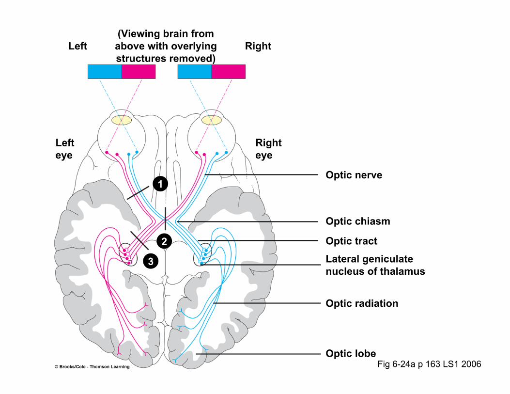
Fig 6-24a p 163 LS1 2006
(Viewing brain fromabove with overlyingstructures removed)
Left Right
Lefteye
Righteye
Optic nerve
Optic chiasm
Optic tract
Lateral geniculate nucleus of thalamus
Optic radiation
Optic lobe
1
2
3

Fig 6-24b p 163 LS1 2006
Visual deficits with specific lesions
1
2
3
Left optic nerve
Optic chiasm
Left optic tract (orradiation)
= Site of lesion = Visual deficit

Rods in Darkness Rhodopsin Not Active, cGMP High, CNG and K+ Channels Open
Rods – 3 Main Cation Channels1.CNG (Cyclic Nucleotide-Gated) Channel
Enable Na+ and Ca2+ entry into Rod
2.K+ Channel
Enables K+ to leak out of Rod
3.Ca2+-Voltage-Gate Channel
Enables Ca2+ Entry into Synaptic Terminal to
Regulate Glutamate Exocytosis
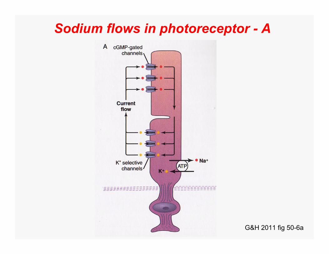
Sodium flows in photoreceptor - A
G&H 2011 fig 50-6a
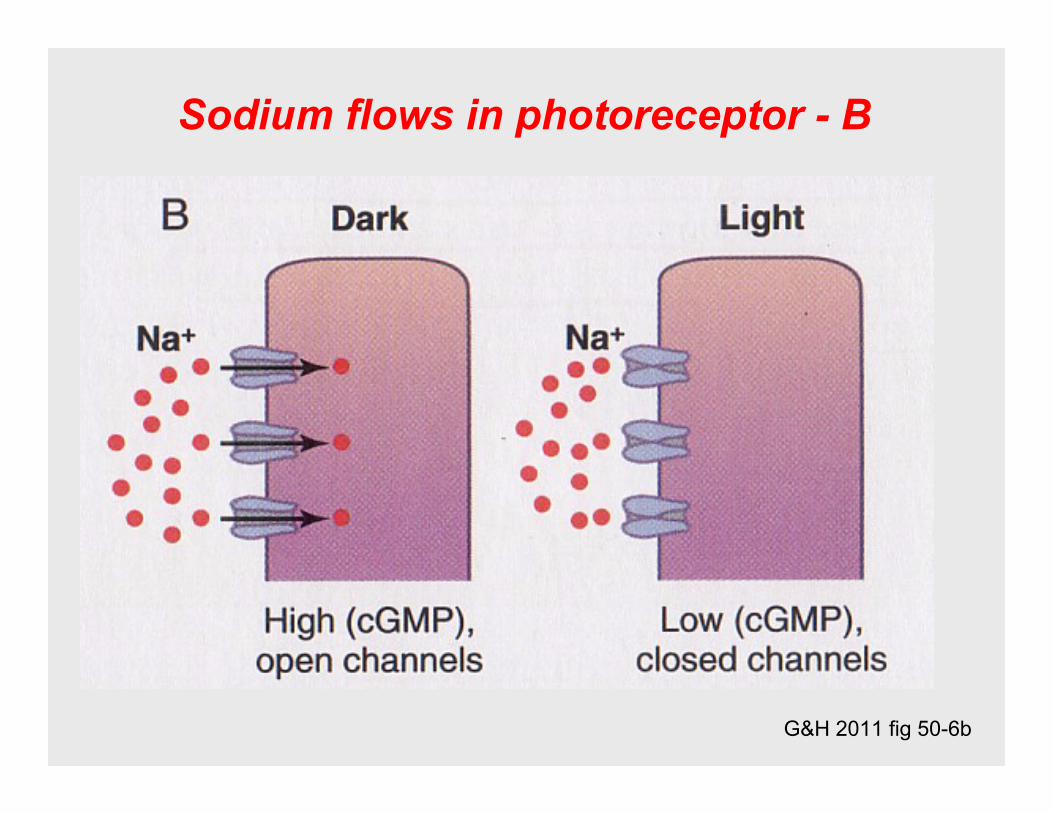
G&H 2011 fig 50-6b
Sodium flows in photoreceptor - B

Phototransduction (outer segment)
G&H 2011 fig 50-7

Light
Summary: Let There Be Light!
Bleaches Rhodopsin Opsin
cGMP
Closes CNG Channel(No more free inflow of Na+, Ca2+)
Hyperpolarizes Membrane(to -70 mV)
NT ReleaseLight
U