ifats san diego 2016 conference 14th annual ifats meeting · 1. ifats san diego 2016 conference....
TRANSCRIPT

1
IFATS SAN DIEGO 2016 CONFERENCE14th Annual IFATS Meeting
International Federation for Adipose Therapeutics and Science
November 17-20, 2016The Westin San Diego • Gaslamp Quarter
San Diego, Californiawww.ifats.org

MTF 1012015 Ad FINAL.indd 1 10/5/15 11:46 AM
IFATS thanksour platinum sponsor
for their continuing support

3
International Federation forAdipose Therapeutics and Science
IFATS SAn DIego 2016November 17-20, 2016
Westin Gaslamp • San Diego, California
Recording of any content presented at this educational program either by camera, video camera, cell phone, audio recorder, or any other device is strictly prohibited.
Endorsed by:
MTF 1012015 Ad FINAL.indd 1 10/5/15 11:46 AM

4
IFATS Executive Office45 Lyme Road - Suite 304 Hanover, NH 03755 USA
Tel: 1-603-643-2325 • Fax: 1-603-643-1444Email: [email protected] • Web: www.ifats.org
Catherine Foss - Executive Director • [email protected] Ambrose - Abstract Coordinator and Marketing Manager • [email protected] Carney - Membership Services Manager • [email protected] Nilsson, CMP - Education Specialist • [email protected] Rice - Accounting Manager • [email protected]
AbstrAct DeADline:Midnight EST, Wednesday, June 7, 2017
The Call for Abstracts will be sent this winter. All members of IFATS and all registered attendees of the 2016 IFATS Conference will be included in the mailing list. Any others who wish to be reminded to submit papers should contact the IFATS Executive Office.
MARK YOUR CALENDAR
International Federation forAdipose Therapeutics and Science
15th Annual MeetingIFATS MIAMI 2017
November 30 - December 3, 2017Loews Miami Beach Hotel
Miami, Florida

5
Table of Contents
Founders Board & Board of Directors. . . . . . . . . . . . . . . . . . . . . . . . . . . . . . . . . . . . . . . 6
Welcome from the President . . . . . . . . . . . . . . . . . . . . . . . . . . . . . . . . . . . . . . . . . . . . . . 7
Program Committee and Moderators . . . . . . . . . . . . . . . . . . . . . . . . . . . . . . . . . . . . . . . 8
Program in Brief . . . . . . . . . . . . . . . . . . . . . . . . . . . . . . . . . . . . . . . . . . . . . . . . . . . . . . . . 10 - 12
Program Schedule . . . . . . . . . . . . . . . . . . . . . . . . . . . . . . . . . . . . . . . . . . . . . . . . . . . . . . 15 - 37
Author Index . . . . . . . . . . . . . . . . . . . . . . . . . . . . . . . . . . . . . . . . . . . . . . . . . . . . . . . . . . . 39 - 42
Paper Presentations/Posters . . . . . . . . . . . . . . . . . . . . . . . . . . . . . . . . . . . . . . . . . . . . . . 43 - 134
Exhibitors . . . . . . . . . . . . . . . . . . . . . . . . . . . . . . . . . . . . . . . . . . . . . . . . . . . . . . . . . . . . . 137 - 142

6
FounDerS BoArD
Ramon Llull, MD, PhDPalma de Mallorca, Spain President 2003
J. Peter Rubin, MD, FACSChairman of the Founders Board Pittsburgh, Pennsylvania, USA President 2004
William Futrell, MDPittsburgh, Pennsylvania, USA
Adam J. Katz, MD, FACSGainesville, Florida, USA President 2005
Board of Directors
Stuart K. Williams, PhDPast President 2011Louisville, Kentucky, USA
Jeffrey M. Gimble, MD, PhDPast President 2006New Orleans, Louisiana, USA
Keith March, MD, PhDPast President 2007Indianapolis, Indiana, USA
Louis Casteilla, PhDPast President 2008Toulouse, France
Kacey Marra, PhDPast Co-President 2013Pittsburgh, Pennsylvania, USA
Marco Helder, PhDPast President 2014Amsterdam, Netherlands
Julie Fradette, PhDPast President 2012Québec, QC, Canada
Sydney Coleman, MDPast Co-President 2013New York, New York, USA
Bruce Bunnell, PhDPast President 2015New Orleans, Louisiana, USA
Ricardo Rodriguez, MDPresident 2016Baltimore, Maryland, USA
Spencer A. Brown, PhDPast President 2010Camden, New Jersey, USA
Kotaro Yoshimura, MDPresident 2017Shimotsuke, Japan

7
Founders BoardWilliam Futrell, MDUniversity of PittsburghUnited States
Adam J. Katz, MD, FACSUniversity of FloridaUnited States
Ramon Llull, MD, PhDUniversity of BarcelonaSpain
J. Peter Rubin, MD, FACSUniversity of PittsburghUnited States
Board of DirectorsRicardo Rodriguez, MD - PresidentCosmeticsurgUnited States
Spencer Brown, PhDCooper University HospitalUnited States
Bruce Bunnell, PhDTulane University School of MedicineUnited States
Louis Casteilla, PhDToulouse UniversityFrance
Sydney Coleman, MDNYU Medical CenterUnited States
Julie Fradette, PhDLOEX/Université LavalCanada
Marco Helder, PhDVU University Medical CenterNetherlands
Kacey Marra, PhDUniversity of PittsburghUnited States
Stuart K. Williams, PhDUniversity of LouisvilleUnited States
Members-at-LargeJeff Gimble, MD, PhDPennington BiomedicalUnited States
Keith March, MD, PhDIndiana UniversityUnited States
IFATS invites you to our 14th annual meeting in San Diego on November 17-20.
This year we kick off the meeting with an exciting morning dedicated to “The Process Engineering of the Fat Graft”. This is the first ever symposium of its kind, dedicated to all the aspects necessary to get the best fat graft possible. You will hear a panel of experts including clinicians, research scientists, and industry leaders discuss the “best practices” for every step involved in delivering “the ideal fat graft”. You will hear about the best cannulas, the best harvesting methods, whether pre-treatment of donor or recipient area works, get a heads-up on exciting state-of-the art products that will improve your results, and learn the best ways to deliver a fat graft.
On Saturday afternoon, a Legislative Issues panel will be really important, especially given the most recent FDA tissue guidances regarding Fat Grafting and what constitutes acceptable practices. Did you know that fat grafting to the breast is at risk according to these new FDA tissue guidances? Come to our meeting and find out more.
IFATS holds the premier annual meeting dedicated to the science of fat grafting and this year we bring together not only the leading clinical practitioners with whom we are all familiar, but also leading endocrinologists from the American Society of Bone and Mineral Research, experts in the field of genomics and big data as applied to medicine, and stem cell researchers from other specialties such as cardiology and orthopedics.
Don’t miss this rare opportunity to get “The Big Picture” about fat grafting, from the bench to clinical uses and beyond to the possibilities of applying “Big Data” techniques to our growing knowledge.
Ricardo Luis Rodriguez, MDIFATS President

8
Katarina Andjelkov, MD, PhDRobert Bowen, MD, FCCPSpencer Brown, PhDBruce Bunnell, PhDMary Ann Chirba, JD, DSc, MPHBryan Choi, MSWilliam Cimino, PhDSteven Cohen, MDSydney Coleman, MD
Sherry Collawn, MD, PhDAlexandra Conde-Green, MDVick Deka, BSJeffrey Gimble, MD, PhDGeoffrey Gurtner, MDMarie Francoise Harris, MBAJeffrey Hartog, MD, DMDCarlos M. Isales, MDNaynesh Kamani, MD
Adam Katz, MD, FACSLauren Kokai, PhDMike Longaker, MD, MBA, FACSKeith March, MD, PhDIan McNiece, PhDSusanna Miettinen, PhDBruno Péault, PhDIvona Percec, MD, PhDMarcille Pilkington
Ricardo Rodriquez, MDJ. Peter Rubin, MD, FACSNir Shani, PhDSammy Sliwin, MDDietrich Stephan, PhDShigeki Sugii, PhDFilip Stillaert, MDStuart Williams, PhDKevin Zwezdaryk, PhD
INVITED SPEAKERS AND SESSION MODERATORS
DISCLAIMERPapers are reprinted as they were submitted. IFATS takes no responsibility for typographical or other errors.All papers in this Program Book are listed in numerical order.
No one may present more than one paper at any IFATS Meeting, although an individual may be an author of more than one paper presented. The paper must be presented by one of the authors. If no alternate presenter is available, the paper will be replaced on the program.
Recording of any content presented at this educational program either by camera, video camera, cell phone, audio recorder, or any other device is strictly prohibited.
Katarina Andjelkov, MD, PhDJoel Aronowitz, MDPetra Bauer-Kreisel, PhDRoberto Blum, MDTorsten Blunk, PhDRobert Bowen, MD, FCCPBruce Bunnell, PhDLouis Casteilla, PhDEvangelia Chnari, PhDBryan Choi, MDSherry Collawn, MD, PhDAlexandra Conde-Green, MD, FICSVick Deka, BSPhilippe Foubert, PhDJulie Fradette, PhDDavid Genecov, MD, FACS
Jeffrey M. Gimble, MD, PhDVictoria Good, PhDRobert Harmanm, DVM, MPVMJeffrey Hartog, MD, DMDMarco Helder, PhDJoseph Itskovitz-Eldor, MD, DScBrian Johnstone, PhDAdam Katz, MD, FACSLauren Kokai, PhDMalgorzata Kolenda, PhDGorana Kuka, MDJanice F. Lalikos, MD, FACSHebert Lamblet, MDKeith March, MD, PhDKacey G. Marra, PhDRichard Martin, PhD
Susanna Miettinen, PhDAli Modarressi, MDIvona Percec, MD, PhDStephen Ray, MD, FACSRicardo Rodriguez, MDBrooke Seckel, MDShigeki Sugii, PhDNir Shani, PhDSammy Sliwin, MD, FRCSCFilip Stillaert, MDDeborah Sullivan, PhDDmitry Traktuev, PhDNing Yang, PhDKevin Zwezdaryk, PhD
SCIENTIFIC PROGRAM COMMITTEE

9
PROGRAM IN BRIEFThe program is correct at the time of printing; however, the Program Chairman
reserves the right to alter the schedule as necessary.

10
Thursday, November 17, 2016
8:00 - 8:30 am Opening AddressRicardo Rodriguez, MD - IFATS President
8:30 - 9:00 am Keynote Speaker The Process of Fat Grafting Sydney Coleman, MD
9:00 - 10:00 am Plenary Session 1 - The Fat Graft Process - Selected AbstractsModerators: Katarina Andjelkov, MD, PhD & Robert Bowen, MD
10:00 - 10:15 am Coffee Break and Exhibits
10:15 - 11:15 am Symposium - Fat Graft Process Engineering: Clinical, Research & Industry Perspectives Part IModerator: Ricardo Rodriguez, MDSpeakers: LifeCell Speaker - Marie Francoise Harris, MBA; Spencer Brown, PhD; Steven Cohen, MD
11:15 am - 12:15 pm Symposium - Fat Graft Process Engineering: Clinical, Research & Industry Perspectives Part IIModerator: Adam Katz, MD, FACSSpeakers: Geoffrey Gurtner, MD, Tulip Speaker - Marcille Pilkington & Bruno Péault, PhD
12:15 - 1:00 pm Panel Discussion - All SpeakersModerator: Ricardo Rodriguez, MD
1:00 - 2:00 pm Lunch
2:00 - 3:00 pm Guest Speaker Adipose Derived Stromal Cells: Progenitor Enrichment Strategies for Soft and Hard Tissue Clinical Needs Mike Longaker, MD, MBA, FACS - Deane P. & Louise Mitchell Professor; Vice Chair, Department of Surgery; Co-Director, Institute for Stem Cell Biology & Regenerative Medicine; Director, Program in Regenerative Medicine; Director, Children’s Surgical Research; Professor, by Courtesy, Department of Bioengineering; Professor, by Courtesy, Department of Materials Science and Engineering, Stanford University, Palo Alto, CA
Moderator: J. Peter Rubin, MD, FACS
3:00 - 5:00 pm Plenary Session 2 - Applied Research: Structure and MatrixModerators: Bryan Choi, MS & Lauren Kokai, PhD
5:00 - 6:30 pm Industry ShowcaseModerator: Ricardo Rodriguez, MDBiologica Technologies, CAREStream America, Millennium, SERVA, Worthington Biochemical, Andrew Tech, Kerastem, LifeCell
6:30 pm Adjourn for the day
Friday, November 18, 2016
8:00 - 8:15 am Introductory Remarks Ricardo Rodriguez, MD
8:15 - 9:00 am Keynote SpeakerBone, Fat and Aging: Therapeutic optionsCarlos M. Isales, MD - Professor, Department of Neuroscience and Regenerative Medicine, Department of Orthopaedic Surgery, Medicine and Cellular Biology and Anatomy; Augusta University, Augusta, GAModerator: Ricardo Rodriguez, MD
9:00 - 11:00 am Plenary Session 3 - Highest Scoring Abstracts (Mixed categories)
Moderators: Bruce Bunnell, PhD & Alexandra Conde-Green, MD
11:00 - 11:20 am Coffee Break and Exhibits
11:20 am - 1:00 pm Concurrent Free Paper Session 1 - Characterizing ASC, SVF and Regulatory IssuesModerators: Jeffrey Gimble, MD, PhD & Ivona Percec, MD, PhD
11
11:20 am - 1:00 pm Concurrent Free Paper Session 2 - Basic Research: Inflammation, FibrosisModerators: Shigeki Sugii, PhD & Susanna Miettinen, PhD
1:00 - 2:00 pm Lunch
2:00 - 3:30 pm Concurrent Free Paper Session 3 - Applied ResearchModerators: Ivona Percec, MD, PhD & Philippe Foubert, PhD
2:00 - 3:30 pm Concurrent Free Paper Session 4 - Basic Research: ASC and SVFModerators: Susanna Miettinen, PhD & Vick Deka, MS
3:30 - 4:00 pm Coffee Break and Exhibits
4:00 - 5:00 pm Poster PresentationsModerators: Kevin Zwezdaryk, PhD & Filip Stillaert, MD
5:00 - 6:30 pm Poster Session & Welcome Reception
6:30 pm Dinner on own
Saturday - November 19, 2016
8:00 - 9:00 am IFATS Members Meeting
9:00 - 10:30 am Concurrent Free Paper Session 5 - Clinical FaceModerators: Alexandra Conde-Green, MD & Sherry Collawn, MD, PhD
9:00 - 10:30 am Concurrent Free Paper Session 6 - Applied ResearchModerators: Filip Stillaert, MD & Jeffrey Gimble, MD, PhD
10:30 - 11:00 am Coffee Break and Exhibits
11:00 am - 1:00 pm Concurrent Free Paper Session 7 - Clinical TrunkModerators: Sammy Sliwin, MD & Jeffrey Hartog, MD, DMD
11:00 am - 1:00 pm Concurrent Free Paper Session 8 - Basic ResearchModerators: Nir Shani, PhD & Lauren Kokai, PhD
1:00 - 2:00 pm Lunch
1:00 - 2:00 pm Lunch Table Discussions (optional)
2:00 - 3:00 pm Guest Speaker Genomics Complementing Cell-based Therapies to Extend the Healthy Lifespan Dietrich Stephan, PhD - Professor and Chair of the Department of Human Genetics at the University of Pittsburgh Graduate School of Public Health
Moderator: Ricardo Rodriguez, MD
3:00 - 4:30 pm Regulatory Affairs PanelModerator: Adam Katz, MD, FACSFDA/USA Perspective - Mary Ann Chirba, JD, DSc, MPH & J. Peter Rubin, MD, FACSAABB Perspective - Naynesh Kamani, MD - Vice President, AABB Center for Cellular Therapies and ResearchAcademic Perspective - Keith March, MD, PhDIndustry Perspective - William Cimino, PhD (The GID Group)FACT Perspective - Ian McNiece, PhD - Professor of Medicine and Director, Cell Therapy Laboratories; The University of Texas; MD Anderson Cancer CenterPanel Discusssion - All Speakers
6:00 - 9:00 pm A Taste of San Diego - Wave House Beach Club Buses leave at 5:30 pm. Meet the buses outside the hotel lobby.
12
Sunday, November 20, 2016
8:00 - 8:10 am Introductory Remarks Ricardo Rodriguez, MD
8:10 - 9:00 am Plenary Session 4 - Clinical TrialsModerators: Stuart Williams, PhD & Keith March, MD, PhD
9:00 - 10:00 am Guest Speaker Machine Learning Research Applications Phil Nelson - Director, Software Engineering, Google
Moderator: Ricardo Rodriguez, MD
10:00 - 10:15 am Coffee Break and Exhibits
10:15 - 11:45 am Plenary Session 5 - Hot TopicsModerators: Bruce Bunnell, PhD & Ricardo Rodriguez, MD
11:45 am Concluding Remarks
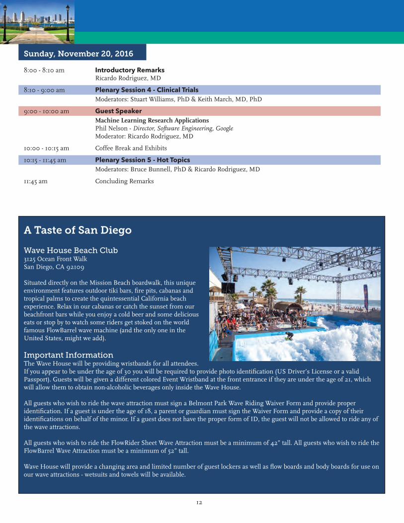
Wave House Beach Club3125 Ocean Front WalkSan Diego, CA 92109
Situated directly on the Mission Beach boardwalk, this unique environment features outdoor tiki bars, fire pits, cabanas and tropical palms to create the quintessential California beach experience. Relax in our cabanas or catch the sunset from our beachfront bars while you enjoy a cold beer and some delicious eats or stop by to watch some riders get stoked on the world famous FlowBarrel wave machine (and the only one in the United States, might we add).
Important InformationThe Wave House will be providing wristbands for all attendees. If you appear to be under the age of 30 you will be required to provide photo identification (US Driver’s License or a valid Passport). Guests will be given a different colored Event Wristband at the front entrance if they are under the age of 21, which will allow them to obtain non-alcoholic beverages only inside the Wave House.
All guests who wish to ride the wave attraction must sign a Belmont Park Wave Riding Waiver Form and provide proper identification. If a guest is under the age of 18, a parent or guardian must sign the Waiver Form and provide a copy of their identifications on behalf of the minor. If a guest does not have the proper form of ID, the guest will not be allowed to ride any of the wave attractions.
All guests who wish to ride the FlowRider Sheet Wave Attraction must be a minimum of 42” tall. All guests who wish to ride the FlowBarrel Wave Attraction must be a minimum of 52” tall.
Wave House will provide a changing area and limited number of guest lockers as well as flow boards and body boards for use on our wave attractions - wetsuits and towels will be available.
A Taste of San Diego

13
NOTES

14
NOTES

15
PROGRAM SCHEDULEThe program is correct at the time of printing; however, the Program Chairman reserves
the right to alter the schedule as necessary.
16
Thursday, November 17, 2016
8:00 - 8:30 am Opening AddressRicardo Rodriguez, MD - IFATS President
8:30 - 9:00 am Keynote Speaker The Process of Fat Grafting Sydney Coleman, MD
9:00 - 10:00 am Plenary Session 1 - The Fat Graft Process - Selected AbstractsModerators: Katarina Andjelkov, MD, PhD & Robert Bowen, MD
9:00 am 1 IMPROVING FAT GRAFT SURVIVAL THROUGH PRECONDITIONING OF THE RECIPIENT SITE WITH MICRONEEDLING Presenter: Billur Sezgin, MD (Turkey) Affiliation: Koc University School of Medicine Authors: Sezgin B, Ozmen SO, Bulam HB, Omeroglu SO, Yuksek SY, Cayci BC, Peker TP
9:10 am 2 EFFECT OF SUCTION PRESSURES ON CELL YIELD AND FUNCTIONALITY OF THE ADIPOSE- DERIVED STROMAL VASCULAR FRACTIONNOT PRESENTED Presenter: Hong-Wei Liu, MD, PhD (China) Affiliation: The First Affiliated Hospital of Jinan University Authors: Chen YW, Wang JR, Liao X, Li SH, Xiao LL, Cheng B, Xie GH, Song JX, Liu HW
9:20 am 3 CHARACTERIZATION AND DIFFERENTIATION OF HUMAN ADIPOSE DERIVED STEM CELLS ISOLATED NON ENZYMATICALLY FROM MICRO-FRACTURED FAT OBTAINED WITH A COMMERCIALLY AVAILABLE KIT(LIPOGEMS) Presenter: Ramon Coronado, PhD (USA) Affiliation: Lester Smith Medical Research Institute Authors: Coronado R, Krutchkoff B, Cormier M, Peault B
9:30 am 4 CANNULA SIZE AND VOLUME INJECTED PER PASS IMPACT FAT GRAFT ARCHITECTURE IN THE RECIPIENT TISSUE BED Presenter: Isaac B. James, MD (USA) Affiliation: University of Pittsburgh Authors: James IB, Bourne D, DiBernardo G, Wang S, Gusenoff J, Marra K, Rubin JP
9:40 am 5 THE ADIPOSE TISSUE IS NOT UNIFORM. THERE ARE DIFFERENCES INCLUDING THE SITES AND ALSO THE LAYERS. PECULIARITY OF THE SUPERFICIAL FAT Presenter: Angelo Trivisonno, MD (Italy) Affiliation: Sapienza University Author: Trivisonno A
9:50 am 6 AUTOLOGOUS FAT TRANSFER UTILIZING TISSUE LIQUEFACTION TECHNOLOGY: SAFETY, EFFICACY AND LONG-TERM RESULTS Presenter: Christopher P. Godek, MD (USA) Affiliation: Personal Enhancement Center Authors: Godek CP, Godek MA, Borab Z
10:00 - 10:15 am Coffee Break and Exhibits
10:15 - 11:15 am Symposium - Fat Graft Process Engineering: Clinical, Research & Industry Perspectives Part IModerator: Ricardo Rodriguez, MDSpeakers: LifeCell Speaker - Marie Francoise Harris, MBA; Spencer Brown, PhD; Steven Cohen, MD

17
11:15 am - 12:15 pm Symposium - Fat Graft Process Engineering: Clinical, Research & Industry Perspectives Part IIModerator: Adam Katz, MD, FACSSpeakers: Geoffrey Gurtner, MD, Tulip Speaker - Marcille Pilkington & Bruno Péault, PhD
12:15 - 1:00 pm Panel Discussion - All SpeakersModerator: Ricardo Rodriguez, MD
1:00 - 2:00 pm Lunch
2:00 - 3:00 pm Guest Speaker Adipose Derived Stromal Cells: Progenitor Enrichment Strategies for Soft and Hard Tissue Clinical Needs Mike Longaker, MD, MBA, FACS - Deane P. & Louise Mitchell Professor; Vice Chair, Department of Surgery; Co-Director, Institute for Stem Cell Biology & Regenerative Medicine; Director, Program in Regenerative Medicine; Director, Children’s Surgical Research; Professor, by Courtesy, Department of Bioengineering; Professor, by Courtesy, Department of Materials Science and Engineering, Stanford University, Palo Alto, CA
Moderator: J. Peter Rubin, MD, FACS
3:00 - 5:00 pm Plenary Session 2 - Applied Research: Structure and MatrixModerators: Bryan Choi, MS & Lauren Kokai, PhD
3:00 pm 7 AUTOMATED STROMAL VASCULAR FRACTION SPHEROID PRODUCTION USING 3D BIOPRINTING IN CONJUNCTION WITH A COMBINATION HYDROPHOBIC/HYDROPHILIC SURFACE TREATMENT Presenter: Brian C. Gettler, MEng (USA) Affiliation: University of Louisville Authors: Gettler BC, Gandhi PS, Zakhari JS, Williams SK
3:10 pm 8 BIOLOGIC LYMPH NODE SCAFFOLD FOR ALLOTRANSPLANTATIONNOT PRESENTED Presenter: Yujin Myung Sr., MD (Korea) Affiliation: Seoul National University Bundang Hospital Authors: Myung Y, Pak CS, Heo CY
3:20 pm 9 ADULT ADIPOSE-DERIVED MULTIPOTENT STROMAL CELL OSTEOGENESIS ON BIOCOMPATIBLE SCAFFOLDS WITH DISTINCT COMPOSITIONS Presenter: Mandi J. Lopez, DVM, MS, PhD (USA) Affiliation: Louisiana State University Authors: Lopez MJ, Duan W, Haque M, Kearney M, Gimble J
3:30 pm 10Presented by: ANTI-ADIPOGENIC EFFECT OF OXY133Reena Bakshi, MD Presenter: Akishige Hokugo, DDS, PHD (USA) Affiliation: University of California Los Angeles UCLA Authors: Hokugo A, Segovia LA, Rezzadeh K, Jarrahy R
3:40 pm 11 STROMAL CELL-HYDROGEL CONSTRUCT POSSIBLY GENERATES CLINICALLY RELEVANT NEO-TISSUE IN FACIAL HIV-LIPOATROPHY PHASE 2 PIVOTAL MULTICENTER CLINICAL TRIAL: EARLY ANALYSIS ON TESTING PATIENT SAMPLE Presenter: Ramon Llull, MD, PhD (Spain) Affiliation: Stem Europe Mallorca Center Authors: Llull R, Matas A, Bahr-Davidson J, Zarembinski T, Iglesias L, Soler A, Benito J, Paya M, Furr D

18
3:50 pm 12 PERIFOLLICULAR ADIPOSE TISSUE (PFAT) IS A METABOLIC STRUCTURE LIKE PERIVASCULAR ADIPOSE TISSUE (PVAT) AND MAY HAVE INFLUENCE IN THE TREATMENT OF BALDNESS Presenter: Marco A. Pellon, MD (Brazil) Affiliation: Clinica Sao Vicente Author: Pellon MA
4:00 pm 13 ADIPOSE-DERIVED EXTRACELLULAR MATRIX HYBRID HYDROGEL FOR MAINTENANCE OF PLURIPOTENCY IN HUMAN ADIPOSE-DERIVED STEM CELLSNOT PRESENTED Presenter: John N. Poche, BS (USA) Affiliation: Louisiana State University Authors: Poche JN, Hayes DJ
4:10 pm 14Presented by TAILORED ADIPOSE TISSUE 3D MICROENVIRONMENTS USING CELL SHEET TECHNOLOGYAlexandra Marques, MD Presenter: Manuela L. Lago, MSc (Portugal) Affiliation: 3Bs Research Group Authors: Lago ML, Cerqueira MT, Pirraco RP, Reis RL, Marques AP
4:20 pm 15 DELIVERY OF ADIPOSE DERIVED STEM CELLS WITHIN POLOXAMER 407 HYDROGEL FOR PERIPHERAL NERVE REPAIR Presenter: Deokyeol Kim, MD (USA) Affiliation: University of Pittsburgh Authors: Kim D, Allbright KO, Bliley JM, Havis E, DiBernardo G, Grybowski D, Sivak W, Rubin JP, Marra KG
4:30 pm 16 UTILIZATION OF ADIPOSE-DERIVED CELLS FOR BIO-ENGINEERING PRE-VASCULARIZED AND TRI-LAYERED SKIN SUBSTITUTES Presenter: Jakub Zimoch, MS (Switzerland) Affiliation: University of Zurich Authors: Zimoch J, Klar AS, Meuli-Simmen C, Meuli M, Scherberich A, Reichmann E
4:40 pm 17 INJECTABLE HUMAN ADIPOSE MATRIX FOR SOFT TISSUE FILLING: LONG-TERM ASSESSMENT IN THE IMMUNOCOMPETENT RAT MODEL Presenter: Lauren E. Kokai, PhD (USA) Affiliation: University of Pittsburgh Authors: Kokai LE, Schilling B, Chnari E, Mahoney C, Jacobs M, Marra KG, Rubin JP
4:50 pm 18 SOFT TISSUE RECONSTRUCTION BY STRUCTURAL FAT GRAFTING: RECIPIENT SITE OPTIMIZATION USING EXTERNAL VOLUME EXPANSION (EVE) COMBINED TO AN INJECTABLE ALLOGRAFT ADIPOSE MATRIX (AAM) Presenter: Giorgio Giatsidis, MD (USA) Affiliation: Brigham and Womens Hospital - Harvard Medical School Authors: Giatsidis G, Succar JS, Haddad AH, Lago GL, Schaffer CS, Wang XW, Matsumine HM, Orgill DO
5:00 - 6:30 pm Industry ShowcaseModerator: Ricardo Rodriguez, MDBiologica Technologies, CAREStream America, Millennium, SERVA, Worthington Biochemical, Andrew Tech, Kerastem, LifeCell
6:30 pm Adjourn for the day
19
Friday, November 18, 2016
8:00 - 8:15 am Introductory Remarks Ricardo Rodriguez, MD
8:15 - 9:00 am Keynote SpeakerBone, Fat and Aging: Therapeutic OptionsCarlos M. Isales, MD - Professor, Department of Neuroscience and Regenerative Medicine, Department of Orthopaedic Surgery, Medicine and Cellular Biology and Anatomy; Augusta University, Augusta, GAModerator: Ricardo Rodriguez, MD
9:00 - 11:00 am Plenary Session 3 - Highest Scoring Abstracts (Mixed categories)Moderators: Bruce Bunnell, PhD & Alexandra Conde-Green, MD
9:00 am 19 ADIPOSE STEM CELL SECRETOME ENHANCES FUNCTIONAL AND MOLECULAR MYOCARDIAL PRESERVATION DURING EX-VIVO COLD ISCHEMIA Presenter: Meijing Wang, MD (USA) Affiliation: Indiana University Authors: Wang M, Wang IW, Liu Y, Merfeld-Clauss S, Edenberg H, Traktuev DO, Prockop D, March KL
9:10 am 20 A PROSPECTIVE, RANDOMIZED, BLINDED AND PLACEBO-CONTROLLED EFFICACY STUDY OF INTRAARTICULAR ALLOGENEIC ADIPOSE STEM CELLS FOR THE TREATMENT OF OSTEOPresented by ARTHRITIS IN DOGSMark Hughes, MD Presenter: Robert Harman, DVM, MPVM (USA) Affiliation: VetStem Authors: Harman R, Carlson K, Gaynor J, Dhupa S, Clement K, McCarthy T, Hoelzler M Schwartz P, Adams C
9:20 am 21 LONG-TERM SAFETY AND EFFECT OF AUTOLOGOUS ADIPOSE-DERIVED STROMAL VASCULAR FRACTION INTO FINGERS FOR SYSTEMIC SCLEROSIS PATIENTS Presenter: Florence Sabatier, PhD (France) Affiliation: APHM Authors: Daumas A, Magalon J, Jouve E, Truillet R, Casanova D, Giraudo L, Veran J, Benyamine A, Dignat-George F, Magalon G, Sabatier F, Granel B
9:30 am 22 HUMAN ADIPOSE-DERIVED STEM CELLS LABELED WITH PLASMONIC GOLD NANOSTARS FOR CELLULAR TRACKING AND PHOTOTHERMAL CANCER CELL ABLATION Presenter: Ronnie L. Shammas Jr., BS (USA) Affiliation: Duke University Authors: Shammas RL, Fales AM, Crawford BM, Wisdom AJ, Devi GR, Vo-Dinh T, Hollenbeck ST
9:40 am 23 INTERIM ANALYSIS: SAFETY AND EFFECTIVENESS OF COMBINED CELLULAR THERAPY FOR THE TREATMENT OF PAIN AND FUNCTION ASSOCIATED WITH OSTEOARTHRITIS Presenter: Kevin Darr, MD (USA) Affiliation: Covington Orthopedic and Sports Medicine Institute Author: Darr K
9:50 am 24 CLINICAL EVIDENCE OF PIGMENT-REGULATING ACTIVITY OF NANOFAT ON HUMAN SKIN: 6 YEARS OF EXPERIENCE Presenter: Patrick Tonnard, MD, PhD (Belgium) Affiliation: University of Brussels Author: Tonnard PL, Verpaele AM
20
10:00 am 25 HUMAN ADIPOSE-DERIVED STEM CELLS ACTIVELY MAINTAIN HOMEOSTASIS DURING EARLY AGING Presenter: Ivona Percec, MD, PhD (USA) Affiliation: University of Pennsylvania Authors: Percec I, Roberts C, Brenner A, Grant G, Kim E, Shan X, Gersch R, Dierov R
10:10 am 26 A LINEAGE-TRACING MOUSE MODEL REVEALS MYH11 SMOOTH MUSCLE CELLS AND PERICYTES ARE MESENCHYMAL STEM CELLS Presenter: Howard C. Ray, BSE (USA) Affiliation: University of Virginia Authors: Ray HC, Dey P, Seaman SA, Mansour JD, Bruce AC, Peirce SM, Dey BK, Yates PA
10:20 am 27 CHANGING THE PARADIGM OF CRANIOFACIAL RECONSTRUCTION WITH AUTOLOGOUS FAT TRANSFER: A PROSPECTIVE CLINICAL TRIAL Presenter: Debra A. Bourne, MD (USA) Affiliation: University of Pittsburgh Medical Center Authors: Bourne DA, Bliley J, James IB, Haas GL, Meyer EM, Pfeifer M, Donnenberg AD, Donnenberg V, Branstetter B, Mitchell RT, Brown SA, Marra K, Coleman S, Rubin JP
10:30 am 28 EFFICACY OF AUTOLOGOUS MICROFAT GRAFT ON FACIAL HANDICAP IN SYSTEMIC Presented by SCLEROSIS PATIENTSGuy Magalon, MD Presenter: Jeremy Magalon, PharmD (France) Affiliation: APHM Authors: Granel B, Sautereau N, Daumas A, Magalon J, Jouve E, Truillet R, Casanova D, Dignat-George F, Veran J, Benyamine A, Magalon G, Sabatier F
10:40 am 29 FORCING A SQUARE PEG INTO A ROUND HOLE: THE CHALLENGE OF APPLYING PHARMA-BASED REGULATORY REQUIREMENTS FOR POTENCY TO ADIPOSE-DERIVED CELL THERAPIES Presenter: Kevin C. Hicok, MS (USA) Affiliation: VetStem Biopharma Author: Hicok KC
10:50 am 30 PERIVASCULAR SCAFFOLDS LOADED WITH ADIPOSE TISSUE-DERIVED STROMAL CELLS (ASC) ATTENUATE PROGRESSION OF EXPERIMENTAL ABDOMINAL AORTIC ANEURYSM (AAA) Presenter: Martin C. Harmsen, PhD (Netherlands) Affiliation: University Medical Center Groningen Authors: Harmsen MC, Parvizi M, Petersen AH, Van Spreuwel-Goosens CA, Kluijtmans SG
11:00 - 11:20 am Coffee Break and Exhibits
11:20 am - 1:00 pm Concurrent Free Paper Session 1 - Characterizing ASC, SVF and Regulatory IssuesModerators: Jeffrey Gimble, MD, PhD & Ivona Percec, MD, PhD
11:20 am 31 CLINICAL SAFETY OF POINT OF CARE STROMAL VASCULAR FRACTION CELL ISOLATION Presenter: Joel A. Aronowitz, MD (USA) Affiliation: Cedars Sinai Medical Center Authors: Aronowitz JA, Lockhart RA, Birnbaum ZE, Hakakian CS

21
11:28 am 32 SHIFT TOWARDS MECHANICAL ISOLATION OF HUMAN ADIPOSE-DERIVED STROMAL VASCULAR FRACTION: A REVIEW OF UPCOMING TECHNIQUES Presenter: Alexandra Conde-Green, MD (USA) Affiliation: Rutgers New Jersey Medical School - Graduate School of Biomedical Sciences Authors: Conde-Green A, Kotamarti VS, Sherman LS, Keith JD, Lee ES, Granick MS, Rameshwar P
11:36 am 33 MICROFAT GRAFTING USING LIPOGEMS (A 510K FDA APPROVED DEVICE FOR FAT TRANSFER) IN THE CONTEXT OF AESTHETIC, RECONSTRUCTIVE AND REGENERATIVE SURGERY Presenter: Allan Y. Wu, MD (USA) Affiliation: UC Riverside Authors: Wu AY, Krutchkoff B, Rogers C
11:44 am 34 THE FRACTIONATION OF ADIPOSE TISSUE (FAT) PROCEDURE FOR REGENERATIVE PURPOSES Presenter: Joris A. Van Dongen, BSc (Netherlands) Affiliation: University of Groningen and University Medical Center Groningen Authors: Van Dongen JA, Stevens HP, Parvizi M, Van Der Lei B, Harmsen MC
11:52 am 35 REDUCTION OF ACCUMULATED REACTIVE OXYGEN SPECIES CAN BE ACHIEVED BY BATHING STANDARD LIPOASPIRATE IN OXYGENATED MICRO/NANOBUBBLES Presenter: Derek A. Banyard, MD, MBA (USA) Affiliation: Univeristy of California Irvine Authors: Banyard DA, Chiang RS, Sarantopoulos NS, Borovikova AA, Klopfer MJ, Wirth GA, Paydar KZ, Bachman M, Evans GR, Widgerow AD
12:00 pm 36 A NEW CLOSED SYSTEM TO MIX FAT NANOGRAFT AND MICROGRAFT WITH PRP FOR THE NOT PRESENTED CORRECTION OF FACIAL WRINKLES AND AGE RELATED FACE VOLUME LOSS Presenter: Alessandro Di Petrillo, MS (Italy) Affiliation: Doctors Equipe Milano Authors: Di Petrillo A, Goisis M, Mele S, Rosset L
12:08 pm 37 MECHANICAL PROCESSING OF EMULSIFIED LIPOASPIRATE RESULTS IN A DOSE- DEPENDENT UPREGULATION OF STEM CELL MARKERS AND POPULATIONS Presenter: Derek A. Banyard, MD, MBA (USA) Affiliation: Univeristy of California Irvine Authors: Banyard DA, Sarantopoulos CN, Chiang RS, Borovikova AA, Qiu X, Wirth GA, Paydar KZ, Haun JB, Evans GR, Widgerow AD
12:16 pm 38 DOES EMULSIFICATION OF FAT IMPAIR THE QUALITY OF STROMAL VASCULAR FRACTION: COMPARISON OF TWO MEDICAL DEVICES FOR NANOFAT PRODUCTION Presenter: Jeremy Magalon, PharmD (France) Affiliation: Culture and Cell Therapy Unit INSERM CBT1409 Authors: Magalon J, Mesguich F, Abellan M, Arnaud L, Lyonnet L, Ghazouane A, Giraudo L, Aboudou H, Philandrianos C, Bertrand B, Casanova D, Paul P, Veran J, Sabatier F
12:24 pm 39 NON-ENZYMATIC ISOLATION OF STROMAL VASCULAR FRACTION FROM ADIPOSE TISSUE Presenter: Pamela Mok, PhD (Singapore) Affiliation: Celligenics Pte Ltd Authors: Mok P, Yeo A, Lau L, Sugii S, Wee K

22
12:32 pm 40 RISK MANAGEMENT OF ADVANCED THERAPY MEDICINAL PRODUCTS: THE MICROBIOLOGICAL RISK OF THE ADIPOSE-DERIVED STROMAL VASCULAR FRACTION Presenter: Julie Veran, PhD (France) Affiliation: Hospital Authors: Veran J, Chateau AC, Blanchet LB, Mendizabal HM, Bertrand BB, Giraudo LG, Philandrianos CP, Magalon JM, Sabatier FS
12:40 pm 41 FEASIBILITY OF GMP FACILITY DEVELOPMENT AND OPERATION FOR THE SMALL COMPANY Presenter: Carolyn Hoyal-Wrightson, MD (USA) Affiliation: VetStem Authors: Harman R, Hoyal C
12:48 pm 42 CGMP STANDARDS FOR FDA COMPLIANT POINT OF CARE SVF ISOLATION Presenter: Joel A. Aronowitz, MD (USA) Affiliation: Cedars Sinai Medical Center Authors: Aronowitz JA, Lockhart RA, Birnbaum ZE, Hakakian CS
11:20 am - 1:00 pm Concurrent Free Paper Session 2 - Basic Research: Inflammation, FibrosisModerators: Shigeki Sugii, PhD & Susanna Miettinen, PhD
11:20 am 43 HUMAN ADIPOSE STROMAL CELL THERAPY IMPROVES SURVIVAL AND REDUCES RENAL INFLAMMATION AND CAPILLARY RAREFACTION IN ACUTE KIDNEY INJURY Presenter: Keith L. March, MD, PhD (USA) Affiliation: Indiana University School of Medicine Authors: Collett JA, Traktuev DO, Mehrotra P, Crone A, Merfeld-Clauss S, March KL, Basile DP
11:28 am 44 COMPARISON OF OSTEOGENIC BEHAVIOR OF ADIPOSE DERIVED AND BONE MARROW NOT PRESENTED MESENCHYMAL STEM CELLS CHEMICALLY TRANSFECTED WITH MIR-148B Presenter: Lisa M. Kriegh, BS (USA) Affiliation: Louisiana State University Authors: Kriegh LM, Hayes DJ, Bunnell BA
11:36 am 45Presented by ANTIBACTERIAL EFFECT OF HUMAN ASCPaul Monsarrat, MD Presenter: Valerie Planat-Benard, PhD (France) Affiliation: STROMALab Authors: Planat-Bernard V, Monsarrat P, Taurand M, Kemoun P, Casteilla L
11:44 am 46 IMMUNOMODULATORY AND REGENERATIVE EFFECTS OF MURINE ADIPOSE STROMAL VASCULAR FRACTION CELLS IN A MODEL OF MULTIPLE SCLEROSIS Presenter: Annie C. Bowles, MS (USA) Affiliation: Tulane University Authors: Bowles AC, Wise RM, Thomas RC, Gerstein BY, Bunnell BA
11:52 am 47 PLATELET RICH PLASMA (PRP) INDUCES CHONDROPROTECTION VIA DECREASING AUTOPHAGY, APOPTOSIS AND INCREASING ANTI-INFLAMMATORY MARKERS IN HUMAN OSTEOARTHRITIC CARTILAGE Presenter: Nada M. Alaaeddine, PhD (Lebanon) Affiliation: University of St. Joseph Authors: Moussa M, El Atat O, Hilal G, Haykal G, Chalhoub A, Khalil C, Alaaeddine NM
23
12:00 pm 48 ANALYSIS OF GENE EXPRESSION PROFILES OF MICRORNAS IN SPHEROIDS FROM ADIPOSE-DERIVED STEM CELLS (S-ASCS) AND THEIR INVOLVEMENT IN MESENCHYMAL DIFFERENTIATION AND STEMNESS POTENTIAL Presenter: Anna Barbara Di Stefano, PhD (Italy) Affiliation: Medical Oncology Authors: Di Stefano AB, Fanale D, Montesano L, Perez A, Manahan MA, Sacks JM, Rosson GD, Russo A, Cordova A, Moschella F, Leto Barone AA
12:08 pm 49 USE OF PERIRENAL ADIPOSE TISSUE AS A NON INVASIVE SOURCE OF DONOR ENDOTHELIAL CELLS TO IMPROVE MONITORING OF ALLOIMMUNE RESPONSES ASSOCIATED TO TRANSPLANT VASCULOPATHY IN SOLID ORGAN TRANSPLANTATION Persenter: Pascale Paul, PhD (France) Affiliation: INSERM Assistance Publique Hopitaux de Marseille Authors: Paul P, Lyonnet L, Meunier M, Magalon J, Arnaud L, Giraudo L, Boissier R, Burtey S, Karsenty G, Veran J, Picard C, Sabatier F
12:16 pm 50 TIME DEPENDENT CHANGE IN THE SECRETION OF TROPHIC FACTORS AND IMMUNOMODULATORY CAPACITY OF ADIPOSE DERIVED MESENCHYMAL STEM CELLS (ADSCS) CULTURED ON A 3-D MATRIX Presenter: Meenakshi Gaur, PhD (USA) Affiliation: Aelan Cell Technologies Authors: Gaur M, Amaro-Ortiz AA, Wang LW, Dobke MD, Burgess RB, King Jordan IK, Lunyak VL
12:24 pm 51 TOWARD FULL THICKNESS SKIN GRAFTING WITHOUT DONOR SITE SCARS: COMBINATION OF DERMAL WOUND PASTE (DWP) AND MICRO SKIN TISSUE COLUMNS (MSTC) Presenter: Ning Yang, PhD (USA) Affiliation: University of Florida Authors: Yang N, Tam J, Shang H, Brown J, Anderson R, Katz A
12:32 pm 52 ANTIOXIDANTS IMPROVE CELLULAR DYSFUNCTIONS OF HUMAN ADIPOSE-DERIVED STEM CELLS Presenter: Shigeki Sugii, PhD (Singapore) Affiliation: Singapore Bioimaging Consortium and Duke NUS Graduate Medical School Author: Sugii S
12:40 pm 53Presented by ANTI-INFLAMMATORY EFFECTS OF ADIPOSE-DERIVED STEM CELL IN ACNE VULGARISBong-il Rho, MD Presenter: Leejin Park, MS (Korea) Affiliation: Glovi Plastic Surgery Clinic Author: Park L
12:48 pm 54 AUTOLOGOUS ADIPOSE DERIVED REGENERATIVE CELLS (ADRCS) THERAPY FOR THE PREVENTION AND TREATMENT OF HYPERTROPHIC SCARS USING A RED DUROC PORCINE MODEL Presenter: Philippe Foubert, PhD (USA) Affiliation: Cytori Therapeutics Authors: Foubert P, Liu M, Zafra D, Rajoria R, Gutierrez D, Tenenhaus M, Fraser JK
1:00 - 2:00 pm Lunch
24
2:00 - 3:30 pm Concurrent Free Paper Session 3 - Applied ResearchModerators: Ivona Percec, MD, PhD & Philippe Foubert, PhD
2:00 pm 55 FAS-L ENABLED CELL SELECTION FOR INCREASED YIELD OF ADIPOSE-DERIVED STEM CELLS Presenter: Nir Shani, PhD (Israel) Affiliation: Tel Aviv Sourasky Medical Center Authors: Shani N, Solodeev IS, Sela MS, Almog TA, Yarkoni SY, Gur EG
2:08 pm 56 CHARACTERIZATION AND COMPARISON OF STROMAL VASCULAR FRACTION OBTAINED FROM SYSTEMIC SCLEROSIS PATIENTS AND HUMAN HEALTHY DONORS FOR A THERAPEUTIC USE Presenter: Laurent Arnaud (France) Affiliation: Culture and Cell Therapy Unit INSERM CBT1409 Authors: Magalon J, Arnaud L, Lyonnet L, Giraudo L, Aboudou H, Casanova D, Philandrianos C, Bertrand B, Paul P, Veran J, Sabatier F
2:16 pm 57 CHARACTERIZATION OF BURN TISSUE DERIVED ADIPOSE STROMAL VASCULAR FRACTION: POTENTIAL FOR CLINICAL APPLICATIONS Presenter: Vasanth Kotamarti, BS (USA) Affiliation: Rutgers New Jersey Medical School - Graduate School of Biomedical Sciences Authors: Conde-Green, Kotamarti VS, Sherman LS, Marano MA, Rameshwar P
2:24 pm 58 ALTERATIONS OF ADIPOSE STROMAL-VASCULAR FRACTION CONTENT AND ADIPOSE STEM WITHDRAWN CELL BEHAVIOR IN MORBID OBESE AND POST BARIATRIC SURGERY EX-OBESE WOMEN Presenter: Karina R. Silva, PhD (Brazil) Affiliation: INMETRO Authors: Silva KR, Liechocki SL, Carneiro JR, Claudio-Da-Silva CS, Maya-Monteiro CM, Borojevic RB, Baptista LS2:32 pm 59 AUTOPHAGY MODULATES THE DIFFERENTIATION POTENTIAL OF ADIPOSE STEM CELL SHEETS UNDER HYPOXIA VS NORMOXIA Presenter: Rogerio P. Pirraco, PhD (Portugal) Affiliation: 3Bs Research Group Authors: Pirraco RP, Fernandes AM, Azevedo MM, Costa M, Sampaio-Marques B, Ludovico P, Reis RL
2:40 pm 60 FELINE ADIPOSE DERIVED MULTIPOTENT STROMAL CELLS EXPRESS MAJOR HISTOCOMPATIBILITY COMPLEX II AND HAVE ECTODERMAL TRANSDIFFERENTIATION CAPACITY Presenter: Mandi J. Lopez, DVM, MS, PhD (USA) Affiliation: Louisiana State University Authors: Lopez MJ, Duan W, Dietrich M
2:48 pm 61 ASC, SVF, AND ADIPOCYTE FRACTIONS FROM ADIPOSE: DOSE RELATIONSHIPS AND CLINICAL APPLICATION Presenter: William Cimino, PhD (USA) Affiliation: The GID Group Author: Cimino W
2:56 pm 62 HUMAN CYTOMEGALOVIRUS INFECTED HUMAN ADIPOSE-DERIVED STROMAL/STEM CELLS DISPLAY CHARACTERISTICS OF ADIPOSE BROWNING Presenter: Kevin Zwezdaryk, PhD (USA) Affiliation: Tulane University Authors: Zwezdaryk K, Ferris MB, Swan KF, Morris CM, Gimble JM, Bunnell BA, Lee SB, Sullivan DE
25
3:04 pm 63 INCREASED MANGANESE SUPEROXIDE DISMUTASE ACTIVITY PROMOTES SURVIVAL AND NOT PRESENTED ENGRAFTMENT OF TRANSPLANTED ADIPOSE TISSUE-DERIVED STROMAL AND VASCULAR CELLS Presenter: Angelo Trivisonno, MD (Italy) Affiliation: Sapienza University Author: Trivisonno A
3:12 pm 64 MIRNA BIOGENESIS ASSOCIATED GENES ARE ENHANCED DURING ADIPOSE DERIVED STROMAL/STEM CELL DIFFERENTIATION Presenter: Elizabeth Martin, PhD (USA) Affiliation: Tulane University Authors: Martin E, Llamas CB, Wu X, Gimble JM
3:20 pm 65 CLINICAL RESULTS OF ADIPOSE DERIVED STEM CELL INJECTION FOR FACET JOINT SYNDROME Presenter: Ralf Rothoerl, MD, PhD (Germany) Affiliation: Isarklinikum Authors: Rothoerl R, Alt C, Preuss A, Mueller C, Lackermeier P, Alt E
2:00 - 3:30 pm Concurrent Free Paper Session 4 - Basic Research: ASC and SVFModerators: Susanna Miettinen, PhD & Vick Deka, MS
2:00 pm 66 INHIBITION OF ENDOGENOUS OPIOIDS SIGNALISATION ALLOWS ADIPOSE TISSUE NOT PRESENTED REGENERATION VIA GENERATION OF REACTIVE OXYGEN SPECIES Presenter: Louis Casteilla, PhD (France) Affiliation: STROMALab Institute Authors: Casteilla L, Dromard C, Labit E, Lorsignol A, Rabiler L, Guissard C, Andre M, Mithieux G
2:08 pm 67 ADIPOSE-DERIVED STEM CELLS AND PLATELET-RICH PLASMA IMPROVE BURN WOUND HEALING IN YORKSHIRE PIGS Presenter: Mark Schusterman, MD (USA) Affiliation: University of Pittsburgh Authors: James IB, Bourne D, Wang S, Silva M, Albright K, Grybowski D, Schusterman MA, Zhang L, Satish L, Marra KG, Rubin JP
2:16 pm 68 NANO AND MICRO DIRECTIONAL TOPOGRAPHIES OPPOSITELY INFLUENCE ADIPOSE- DERIVED STEM CELLS DIFFERENTIATION TO SMOOTH MUSCLE CELLS Presenter: Gabriel R. Liguori, MD (Netherlands) Affiliation: University Medical Center Groningen - University of Groningen Authors: Liguori GR, Zhou Q, Barros GG, Kuhn PT, Moreira LF, Van Rijn P, Harmsen MC
2:24 pm 69 POTENTIAL OF CD271-SORTED HUMAN ADIPOSE-DERIVED STEM CELLS IN ADIPOSE TISSUE ENGINEERING Presenter: Richard P. Smith, MSc (United Kingdom) Affiliation: University of Manchester Authors: Smith RP, Lees VC, Hoyland J, Reid AJ
2:32 pm 70 AUTOLOGOUS FAT-DERIVED TISSUE MATRIX: BIOLOGIC CHARACTERISTICS AND RESULTS AFTER IMPLANTATION Presenter: Stephen A. Schendel, MD, DDS (USA) Affiliation: Stanford University Author: Schendel SA

26
2:40 pm 71 THE EFFECTS OF COLD STORAGE AND POLOXAMER 188 TREATMENT ON STROMAL VASCULAR FRACTION VIABILITY AND VOLUME RETENTION OF FAT GRAFTS Presenter: Gabriella A. DiBernardo, BS (USA) Affiliation: University of Pittsburgh Authors: DiBernardo GA, Bliley JM, Bourne D, Havis E, James IB, Schroth R, Grybowski D, Dees A, Wang S, Kokai L, Kelmendi-Doko A, Mahoney C, Sivak W, Marra K, Rubin JP
2:48 pm 72 ADIPOSE STEM/STROMAL CELLS FOR TENDON REGENERATION: DEVELOPMENT OF A NEW DIFFERENTIATION PROTOCOL AND COMPARISON WITH BONE MARROW STEM CELLS TENOGENIC ABILITY Presenter: Carlotta Perucca, PharmD (Italy) Affiliation: University of Pavia Authors: Perucca C, Vigana MV, Sants-Ruiz LS, Colombini AL, Pearson JP, De Girolamo LD
2:56 pm 73 DIFFERENTIATION OF HUMAN ADIPOSE-DERIVED STEM CELLS (ASC) TO ENDOTHELIUM FOR IMPROVEMENT OF FAT TRANSPLANTATION Presenter: William M. Harris, MD (USA) Affiliation: Cooper Univ Hospital Authors: Harris WM, Zhang PZ, Plastini MP, Kappy NK, Ortiz TO, Chang SC, Brown SB, Carpenter JC
3:04 pm 74 SPATIAL CONTROL OF ADIPOSE DERIVED STEM CELL DIFFERENTIATION IN A CELL SHEET NOT PRESENTED USING PHOTOCLEAVABLE NANOPARTICLES Presenter: Lisa M. Kriegh, BS (USA) Affiliation: Louisiana State University Authors: Kriegh LM, Forghani A, Chen C, Hayes DJ, Devireddy R
3:12 pm 75 A NOVEL BIOCOMPATIBLE MICROCARRIER WITH TUBULAR CONDUITS SUPPORTS HSVF SURVIVAL, MATRIX SECRETION, AND CORD STRUCTURE FORMATION Presenter: J. Christian Brown, MD (USA) Affiliation: University of Florida Authors: Brown JC, Willenberg BW, Shang HS, Yang NY, Katz AK
3:20 pm 76 USING HUMAN RECONSTRUCTED OSSEOUS TISSUES DERIVED FROM ADIPOSE STEM/ STROMAL CELLS AS A PLATFORM FOR STUDYING THE IMPACT OF MELATONIN ON OSTEOGENESIS UNDER PHYSIOLOGICAL AND INFLAMMATORY CONDITIONS Presenter: William P. Clafshenkel, PhD (Canada) Affiliation: LOEX CRCHUQ University Laval Authors: Clafshenkel WP, Galbraith T, Kawecki F, Eliopoulos N, Auger FA, Fradette J
3:30 - 4:00 pm Coffee Break and Exhibits
4:00 - 5:00 pm Poster PresentationsModerators: Kevin Zwezdaryk, PhD & Filip Stillaert, MD
4:00 pm 77P FAT GRAFTING FOR AUTOLOGOUS GLUTEAL AUGMENTATION: A META-ANALYSIS Presenter: Alexandra Conde-Green, MD (USA) Affiliation: Rutgers New Jersey Medical School - Graduate School of Biomedical Sciences Authors: Conde-Green A, Kotamarti VS, Nini KT, Wey PD, Ahuja NK, Granick MS, Lee ES

27
4:03 pm 78P SAFETY OF FAT GRAFTING IN PLEGIC PATIENTSWITHDRAWN Presenter: Reto Wettstein, MD (Switzerland) Affiliation: University Hospital Basel Author: Wettstein R
4:06 pm 79P AUTOLOGOUS FAT GRAFTING FOR IPG SITE COMPLICATIONS FOLLOWING SPINAL CORD STIMULATOR Presenter: Suresh M. Anandan, MBBS, MS, MCH, MRCS, FEBOPRAS (United Kingdom) Affiliation: Wexham Park Hospital Authors: Anandan SM, Pai AA, Desai P, Misra A
4:09 pm 80P CONTOUR PLASTIC OF THE FACE USING AUTOFAT WRAPPED IN AUTOPLASMA GEL Presenter: Ivan V. Krainik, MD (Russia) Affiliation: Medical Sugical centre by N I Pirogov Author: Krainik IV
4:12 pm 81P THREE DIMENSIONAL BIOPRINTING: THE FUTURE OF TISSUE ENGINEERING AND PLASTIC SURGERY. A SYSTEMATIC REVIEW OF THE LITERATURE Presenter: Vasanth Kotamarti, MD (USA) Affiliation: Rutgers New Jersey Medical School Authors: Kotamarti V, Conde-Green A, Ayyala H, Guiro K, Lee ES, Granick MS, Rameshwar P
4:15 pm 82P EFFICIENT TWO STEP PROCEDURE TO CORRECT SCALP AND FACIAL SCARS-FAT AND HAIR GRAFTING Presenter: Gorana Kuka-Epstein, MD (USA) Affiliation: Foundation for Hair Restoration Authors: Kuka-Epstein G, Epstein J
4:18 pm 83P NEW ALGORITHM AND AESTHETIC APPROACH FOR BREAST MULTILAYER FAT GRAFTING: NOT PRESENTED PRELIMINARY REPORT Presenter: Alfredo E. Hoyos, MD (Colombia) Affiliation: Elysium Authors: Hoyos AE, Guarin DE
4:21 pm 84P VALIDATION OF THE IMMUNODEFICIENT MOUSE ANIMAL MODEL FOR ASSESSING FAT GRAFTING OUTCOMES Presenter: Lauren E. Kokai, PhD (USA) Affiliation: University of Pittsburgh Authors: Kokai LE, Jones TL, Marra KG, Rubin JP
4:24 pm 85P DONOR AGE DEPENDENT FEATURES OF PEDIATRIC VERSUS ADULT ADIPOSE MESENCHYMAL STROMAL CELLS (ASC) Presenter: Valerie Planat-Benard, PhD (France) Affiliation: STROMALab Authors: Planat-Benard V, Abbo O, Tarand M, Monsarrat P, Raymond I, Galinier P, Casteilla L
4:27 pm 86P PORCINE ADIPOSE TISSUE HARVEST BY LIPOASPIRATION AND SVF ISOLATION USING A ‘POINT-OF-CARE’ DEVICE Presenter: Ning Yang, PhD (USA) Affiliation: University of Florida Authors: Yang N, Shang H, Brown J, Katz A
28
4:30 pm 87P CHARACTERIZATION OF RAT ADIPOSE-DERIVED STEM CELLS AND THEIR INDUCTION TOWARD A TENOGENIC LINEAGE FOR REGENERATION OF ACHILLES TENDON Presenter: Jolanta B. Norelli, BA (USA) Affiliation: Northwell Health System Authors: Norelli JB, Plaza DP, Liang H, Grande DA
4:33 pm 88P CHARACTERISATION OF ADIPOSE STEM CELLS ISOLATED AFTER MANUAL OR WATER JET-ASSISTED LIPOSUCTION Presenter: Rojda Gumuscu, MD (Sweden) Affiliation: Umea University Authors: Gumuscu R, Brohlin M, Wiberg M, Kingham PJ
4;36 pm 89P COLLAGENASE-FREE ADIPOSE-DERIVED STEM CELL ISOLATION: NOVEL PROTOCOLS FOR TRANSLATIONAL APPLICATIONS Presenter: Robert Gersch, PhD (USA) Affiliation: UPENN Authors: Gersch R, Flemming J, Percec I
4:39 pm 90P AN IN VITRO FUNCTIONAL ASSAY OF VASCULOGENESIS AND ANGIOGENESIS USING FRESHLY ISOLATED ADIPOSE STROMAL VASCULAR FRACTION CELLS Presenter: Joseph S. Zakhari, MA (USA) Affiliation: University of Louisville School of Medicine Authors: Zakhari JS, Zabonick JA, Gettler BC, Tweed B, Apakalai B, Williams SK
4:42 pm 91P AUTOLOGOUS GRANULAR FAT GRAFTING IN FACIAL REJUVENATION Presenter: Biao Wang, PhD (China) Affiliation: The First Affiliated Hospital of Fujian Medical University Authors: Wang B, Zheng H, Su C, Shan X, Chen R
4:45 pm 92P COMPARISON OF INTRAOPERATIVE PROCEDURES FOR ISOLATION OF CLINICAL GRADE STROMAL VASCULAR FRACTION: A SYSTEMATIC REVIEW Presenter: Aartje J. Tuin, MD (Netherlands) Affiliation: University Medical Center Groningen Authors: Tuin AJ, Van Dongen JA, Spiekman M, Van Der Lei B, Harmsen MC
4:48 pm 93P PATIENT SATISFACTION SCORES 3-18 MONTHS FOLLOWING AUTOLOGOUS FAT TRANSFER (AFT) OR STROMAL VASCULAR FRACTION-ENRICHED FAT TRANSFER (SVF+F) IN CONJUNCTION WITH FACIAL REJUVENATION SURGERY: A PROSPECTIVE, COMPARATIVE STUDY Presenter: Ahmad Saad, MD (USA) Affiliation: FACESplus/UCSDD Authors: Saad A, Hewett S, Lim S, Taylor K, Mailey B, Suliman A, Dobke M, Cohen S
5:00 - 6:30 pm Poster Session & Welcome Reception
6:30 pm Dinner on own
29
Saturday - November 19, 2016
8:00 - 9:00 am IFATS Members Meeting
9:00 - 10:30 am Concurrent Free Paper Session 5 - Clinical FaceModerators: Alexandra Conde-Green, MD & Sherry Collawn, MD, PhD
9:00 am 97 A RANDOMIZED PHASE II, DOUBLE-BLIND, DUAL ARM STUDY TO ASSESS THE EFFICACY OF ADIPOSE-DERIVED STROMAL VASCULAR FRACTION (SVF)-ENRICHED AUTOLOGOUS FAT GRAFTS, ISOLATED VIA THE ANTRIA CELL PREPARATION PROCESS (ACPP) Presenter: Leonard E. Maliver, MD (USA) Affiliation: Antria Inc. Authors: Maliver LE, Bizousky DB, Rahimian SR, Johns FJ, Boyer SB
9:08 am 98 THE USE OF PRP WITH FAT GRAFTS FOR FACIAL REJUVENATION. DOES IT MAKE ANY DIFFERENCE? Presenter: Elsayed M. Eldib, MD (Egypt) Affiliation: Tanta University Hospital Authors: Eldib EM, Esmail AM
9:16 am 99 TREATMENT OF PARRY-ROMBERG SYNDROME WITH FAT GRAFTING: IS IT BECOMING THE Presented by STANDARD PROCEDURE?Alexandra Conde-Green Presenter: Haripriya Ayyala, MD (USA) Affiliation: Rutgers New Jersey Medical School - Graduate School of Biomedical Sciences Authors: Conde-Green A, Ayyala H, Kotamarti VD, Dornelles R, Sherman LD, Rameshwar P
9:24 am 100 ADRCS IN THE TREATMENT OF ANDROGENETIC ALOPECIA - PRELIMINARY RESULTS Presenter: Katarina Andjelkov, MD, PhD (Serbia) Affiliation: BelPrime Clinic Authors: Andjelkov K, Sforza M
9:32 am 101 PERSONAL 20 YEAR EVOLUTION IN FACIAL FAT AESTHETIC SCULPTING Presenter: Andrew M. Wolin, MD (USA) Affiliation: Private Practice Author: Wolin AM
9:40 am 102 ADIPOCYTE-DERIVED STEM CELL USING IN HAIR FOLLICLE REGENERATION Presenter: Malgorzata Kolenda, MD PhD (Poland) Affiliation: Klinika Kolasinski Author: Kolenda M
9:48 am 103 A PHASE I OPEN-LABEL STUDY EVALUATING THE SAFETY OF ACELLULAR ADIPOSE TISSUE (AAT) Presenter: Amy E. Anderson, BS (USA) Affiliation: Johns Hopkins University Authors: Anderson AE, Wu I, Parrillo A, Sadtler K, Tam A, Cooney C, Cooney D, Aston J, Byrne P, Pardoll D, Elisseeff JH
9:56 am 104 A NOVEL ALLOGRAFT ADIPOSE-DERIVED INJECTABLE AS A PERMANENT, REGENERATIVE ALTERNATIVE TO HYALURONIC ACID FILLERS Presenter: Greg Grover, PhD (USA) Affiliation: Biologica Technologies Authors: Grover G, Choi B
30
10:04 am 105 THE EMERGING ROLE OF AUTOLOGOUS ADIPOSE TISSUE GRAFTING IN THE TREATMENT OF ALOPECIA AND SCARS OF THE SCALP Presenter: Gorana Kuka-Epstein, MD (USA) Affiliation: Foundation for Hair Restoration Authors: Kuka-Epstein G, Epstein J
10:12 am 106 BUCCAL FAT AUGMENTATION DURING FACELIFT USING A TRANSORAL APPROACH: PATIENT SELECTION AND SURGICAL TECHNIQUE Presenter: Steven R. Cohen, MD, FACS (USA) Affiliation: University of California San Diego Authors: Cohen SR, Hewett S, Saad A
10:20 am 107 UPDATE ON NANOFAT GRAFTING: WHAT WE’VE LEARNED, WHAT WE STILL DO AND WHAT WE’VE CHANGED Presenter: Patrick Tonnard, MD, PhD (Belgium) Affiliation: University of Brussels Authors: Verpaele AM, Tonnard PT
9:00 - 10:30 am Concurrent Free Paper Session 6 - Applied ResearchModerators: Filip Stillaert, MD & Jeffrey Gimble, MD, PhD
9:00 am 108 THE EFFECT OF LOCAL AND SYSTEMIC MINOCYCLINE ON FAT GRAFT SURVIVAL AND NOT PRESENTED APOPTHOTIC PATHWAY INHIBITION Presenter: Kirdar Guney, MD (Turkey) Affiliation: Reneclinic Authors: Guney K, Tuncer S, Ozel B, Elmas C, Seymen M, Cenetoglu S
9:08 am 109 DIRECT AND/OR INDIRECT EFFECT AND THE ROLE OF ADIPOSE-DERIVED STEM CELLS FOR TISSUE REPAIR AND REGENERATION Presenter: Doruk Orgun, MD (Japan) Affiliation: Juntendo University School of Medicine Authors: Orgun D, Tajima S, Horikoshi-Ishihara H, Tobita M, Oshita T, Tanaka R, Mizuno H
9:16 am 110 HUMAN AND AUTOLOGOUS ADIPOSE-DERIVED STROMAL CELLS IMPROVE FLAP SURVIVAL IN A RODENT MODEL Presenter: Navid M. Toyserkani, MD (USA) Affiliation: Odense University Hospital Authors: Toyserkani NM, Jensen CH, Sheikh SP, Sorensen JA
9:24 am 111 SPECIFIC TARGETING OF HASCS AND RF MEDIATED OSTEOGENESIS USING DUMBBELL SHAPED AUFE3O4 NANOPARTICLES CONJUGATED WITH ANTI-CD146 ANTIBODY AND WITHDRAWN MIR148B MIMIC Presenter: Jonathan S. Casey, MS (USA) Affiliation: Louisiana State University Authors: Casey JS, Forghani A, Hayes DJ
9:32 am 112 POTENTIAL REDUCTION OF BIOFILM FORMATION WITH REGENERATIVE FACIAL FILLER Presenter: Greg Grover, PhD (USA) Affiliation: Biologica Technologies Authors: Grover G, Choi B, Govil A

31
9:40 am 113 ANALYZING THE EFFECTS OF DESFERAL TREATMENT TO IRRADIATED TISSUE AND FAT GRAFT RETENTION Presenter: John S. Flacco, BS (USA) Affiliation: Stanford School of Medicine Authors: Flacco JS, Blackshear CP; Brett EA; Zielins ER; Hu M; Wan DC; Longaker MT
9:48 am 114Presented by HYPOTHERMIC PRESERVATION OF CELL SHEETS OF HUMAN ADIPOSE STEM CELLSRogerio Pirraco, MD Presenter: Sara Ribeiro, BSc (Portugal) Affiliation: 3Bs Research Group Authors: Ribeiro S, Costa M, Cerqueira MT, Marques AP, Pirraco RP, Reis RL
9:56 am 115 MECHANICAL ISOLATION OF ADIPOSE STROMAL VASCULAR CELLS: A SAFE AND LESS TIME-CONSUMING ALTERNATIVE TO ENZYMATIC DIGESTION Presenter: Tunc Tiryaki, MD (Turkey) Affiliation: Cellest Clinic Authors: Tiryaki T, Canikyan S, Conde-Green A
10:04 am 116NOT PRESENTED CLINICAL APLICATION OF POLOXAMER 188 ENHACED FAT GRAFTS Presenter: Alfredo E. Hoyos, MD (Colombia) Affiliation: Elysium Authors: Hoyos AE, Guarin DE
10:12 am 117 FACIAL INTRAMUSCULAR LIPOMA OCCURRENCE FOLLOWING TOPICAL COSMETIC NOT PRESENTED INJECTION WITH A MIXTURE OF BASIC FIBROBLAST GROWTH FACTOR: A REPORT OF TWO CASES Presenter: Xuan Liao, MD (China) Affiliation: The First Affiliated Hospital of Jinan University Authors: Liao X, Zhang ZD, Li SH, Xiao LL, Cheng B, Xie GH, Liu HW
10:20 am 118 THE APPLICATION OF AUTOLOGOUS FAT GRAFTING IN IMPLANT-BASED BREAST RECONSTRUCTION Presenter: Houbing Zheng, MD (China) Affiliation: The First Affiliated Hospital of Fujian Medical University Authors: Zheng H, Wang BW, Shan XS, Chen RC
10:30 - 11:00 am Coffee Break and Exhibits
11:00 am - 1:00 pm Concurrent Free Paper Session 7 - Clinical TrunkModerators: Sammy Sliwin, MD & Jeffrey Hartog, MD, DMD
11:00 am 119 INTRATISSULAR EXPANSION-MEDIATED, SERIAL FAT GRAFTING: A STEP-BY-STEP WORKING ALGORITHM TO ACHIEVE 3D BIOLOGICAL HARMONY IN AUTOLOGOUS BREAST RECONSTRUCTION Presenter: Filip B. Stillaert, MD (Belgium) Affiliation: University Hospital Gent Author: Stillaert FB
11:08 am 120 POST-MASTECTOMY FULL BREAST RECONSTRUCTION WITH FAT GRAFTING WITHOUT PRIOR INTERNAL OR EXTERNAL EXPANSION Presenter: Susanna C. Kauhanen, MD, PhD (Finland) Affiliation: Helsinki University Hospital Authors: Kauhanen SC, Hockerstedt AI
32
11:16 am 121Presented by LARGE VOLUME FAT GRAFTING IN BREAST RECONSTRUCTION: SIX YEARS EXPERIENCESaad Dibo, MD Presenter: Marwan H. Abboud, MD (Belgium) Affiliation: MA Clinic Authors: Abboud MH, Dibo SA
11:24 am 122 FAT PROCESSED BY SALINE-WASH, NEGATIVE-PRESSURE-FILTRATION, AND LARGE SCALE STERILE COTTON ABSORPTION FOR BREAST LIPOAUGMENTATION AFTER IMPLANT REMOVAL Presenter: Sarah A. Mess, MD (USA) Affiliation: Sarah Mess, MD LLC Author: Mess SA
11:32 am 123 A COMPARATIVE TRANSLATIONAL STUDY: THE COMBINED USE OF ENHANCED STROMAL WITHDRAWN VASCULAR FRACTION AND PLATELET-RICH PLASMA IMPROVES FAT GRAFTING MAINTENANCE IN BREAST SOFT TISSUE DEFECTS Presenter: Pietro Gentile, MD, PhD (Italy) Affiliation: University of Rome Tor Vergata Author: Gentile P
11:40 am 124 TREATMENT OF FEMALE URINARY INCONTINENCE WITH AUTOLOGOUS, MICRO-Presented by FRAGMENTED, AND MINIMALLY MANIPULATED ADIPOSE TISSUE (LIPOGEMS®)Todd Malan, MD Presenter: Heripsime Ohanian, PhD, MD (USA) Affiliation: Hackensack University Medical Center Author: Ohanian H
11:48 am 125 THE USE OF ADIPOSE TISSUE IN SKIN REGENERATION OF ACUTE DEEP BURNS Presenter: Marco A. Pellon, MD (Brazil) Affiliation: Clinica Sao Vicente Authors: Pellon MA, Conde-Green A
11:56 am 126 EFFICACY AND SAFETY OF GLUTEAL AUGMENTATION WITH AUTOLOGOUS FAT GRAFTING: A SYSTEMATIC REVIEW Presenter: Carlo M. Oranges, MD (Switzerland) Affiliation: Basel University Hospital Authors: Oranges CM, Harder Y, Haug M, Kalbermatten DF, Schaefer DJ
12:04 pm 127 POWER-ASSISTED GLUTEAL AUGMENTATION: A NEW TECHNIQUE FOR SCULPTING, HARVESTING, AND TRANSFERRING FAT Presenter: Saad Dibo, MD (Belgium) Affiliation: MA Clinic Authors: Dibo S, Abboud MH
12:12 pm 128 NON-RESPONSIVE MULTIFACTORIAL SHOULDER PAIN WITH OSTEOARTHRITIS AND ROTATOR CUFF TEARS TREATED WITH AUTOLOGOUS MICRO-FRAGMENTED AND MINIMALLY MANIPULATED ADIPOSE TISSUE UNDER CONTINUOUS ULTRASOUND GUIDANCE Presenter: Richard D. Striano, DC, RMSK (USA) Affiliation: OptimumJoint Integrated Joint Spine Authors: Striano RD, Bilbool N, Krutchkoff B

33
12:20 pm 129WITHDRAWN AUTOLOGOUS FAT GRAFTING TO TREAT PENILE LICHEN SCLEROSUS Presenter: Aurora Almadori, MD (Italy) Affiliation: Second University of Naples Authors: Almadori A, Nicoletti GF, D’Andrea F
12:28 pm 130 ADIPOSE-DERIVED STEM CELLS GIVEN INTRAVENOUSLY IMPROVES EPISTAXIS AND OBJECTIVE BRONCHOSCOPY SCORING IN HORSES WITH EXERCISE-INDUCED PULMONARY HEMORRHAGE Presenter: Mark Hughes, DVM (USA) Affiliation: VetStem Authors: Harman R, Rich R, Hughes M
12:36 pm 131 LYMPHATIC TISSUE REPAIR IN PATIENTS WITH ADIPOSE TISSUE DISORDERS USING LIPOGEMS Presenter: Todd K. Malan, MD (USA) Affiliation: Roxbury Regenerative Authors: Malan TK, Amron DA, Herbst KH
12:44 pm 132Presented by ULTRASONOGRAPHY IN PLASTIC SURGERYRicardo Rodriguez, MD Presenter: Tyler Safran, DEC (Canada) Affiliation: McGill University Authors: Safran T, Kanevsky J, Gorsky K, Luc M, Rodriguez R, Futrell W
12:52 pm 133 STROMAL VASCULAR FRACTION THERAPY FOR ALLEVIATION OF CHRONIC REFRACTORY MIGRAINES Presenter: Kenneth Rothaus, MD (USA) Affiliation: NY Presbyterian-Weill Cornell Authors: Rothaus K, Mauskop AM
11:00 am - 1:00 pm Concurrent Free Paper Session 8 - Basic ResearchModerators: Nir Shani, PhD & Lauren Kokai, PhD
11:00 am 134 ENHANCED ADIPOSE-TISSUE DERIVED SVF VASCULARIZATION POTENTIAL BY 3D NOT PRESENTED PERFUSION CULTURE: A POSSIBLE TREATMENT FOR ISCHEMIC TISSUE Presenter: Giulia Cerino, PhD (Switzerland) Affiliation: University and University Hospital of Basel Authors: Cerino G, Gaudiello E, Melly L, Muraro M, Martin I, Eckstein F, Scherberich A, Marsano A
11:08 am 135 IN VITRO AND IN VIVO INTERACTION OF ADIPOSE-DERIVED STEM CELLS AND BREAST CANCER CELLS Presenter: Hakan Orbay, MD, PhD (USA) Affiliation: University of California Davis Authors: Orbay H, Charvet HJ, Hinchcliff KM, Dehghani T, Kaur M, Sahar DE
11:16 am 136 INTRAMYOCARDIAL ADIPOSE-DERIVED STEM CELL TRANSPLANTATION INCREASES PERICARDIAL FAT WITH RECOVERY OF MYOCARDIAL FUNCTION AFTER ACUTE MYOCARDIAL INFARCTION Presenter: Jong-Ho Kim, PhD (Korea) Affiliation: Korea University College of Medicine Authors: Kim J, Park C, Park H, Lim I, Woo S, Choi S, Joo H, Hong S
34
11:24 am 137 ALTERNATIVELY ACTIVATED M2 MACROPHAGES IMPROVE AUTOLOGOUS FAT GRAFT WITHDRAWN SURVIVAL IN A MOUSE MODEL THROUGH INDUCTION OF ANGIOGENESIS Presenter: Michael Bezuhly, MD (Canada) Affiliation: Dalhousie University IWK Health Center Authors: Gebremeskel S, Phipps K, Gillis J, Johnston B, Hong P, Bezuhly M
11:32 am 138 NOTCH2 EXPRESSED ASC REGULATES PDGFR-BETA, MIGRATION AND ADHESION IN VITRO AND IN PATHOLOGICAL PROLIFERATIVE RETINOPATHY IN VIVO Presenter: Vincenzo Terlizzi, MS (Netherlands) Affiliation: University of Groningen Authors: Terlizzi V, Kolibabka M, Hammes HP, Harmsen MC
11:40 am 139 LIPOGRAFTING IMPROVES THERAPY RESISTANT DERMAL SCARS THROUGH ENHANCED REMODELING Presenter: Maroesjka Spiekman, BS (Netherlands) Affiliation: University Medical Center Groningen Authors: Spiekman M, Hoppe DL, Diercks GF, Ghods M, Harmsen MC
11:48 am 140 CIGARETTE SMOKING AS A FACTOR IN COMPROMISED FUNCTION AND THERAPEUTIC ACTIVITY OF ADIPOSE STEM CELLS, MANIFESTED AS DECLINE IN VASCULOGENIC POTENTIAL Presenter: Daria Barwinska, BA (USA) Affiliation: Indiana University Authors: Barwinska D, Traktuev D, Cook TG, Merfled-Clauss S, Petrache I, March KL
11:56 am 141 PERIODONTAL TISSUE REGENERATION BY TRANSPLANTATION OF ADIPOSE TISSUE-WITHDRAWN DERIVED MESENCHYMAL STEM CELLS. BASIC RESEARCH TOWARD THE CLINICAL APPLICATION Presenter: Chunman Lee, MD, PhD (Japan) Affiliation: Osaka University Authors: Lee C, Takedachi M, Sawada K, Ohkawara H, Matsuyama A, Kitamura M, Murakami S
12:04 pm 142 ADIPOSE DERIVED STEM CELLS IN A RAT POSTEROLATERAL SPINE FUSION MODEL Presenter: Ralf Rothoerl, MD, PhD (Germany) Affiliation: Isarklinikum Authors: Rothoerl R, Alt C, Coleman M, Martinez R, Karimi T, Alt E
12:12 pm 143 INTERFERON GAMMA INDUCTION OF TRAIL EXPRESSION IN ADIPOSE DERIVED STROMAL CELLS MAY REDUCE TUMORIGENIC POTENTIAL IN BREAST CANCER Presenter: Anne C. O’Neill, MD, PhD (Canada) Affiliation: University Health Network University of Toronto Authors: O’Neill AC, Aggarwal P, Keating A, Hofer So
12:20 pm 144 A PORCINE MODEL FOR THE STUDY OF AUTOLOGOUS CELL-ENRICHED LARGE VOLUME FAT GRAFTING Presenter: Bo S. Rasmussen, MD (Denmark) Affiliation: Rigshospitalet Authors: Rasmussen BS, Sorensen CL, Kubergovic S, Vester-Glowinski PV, Herly M, Trojahn-Kolle SF, Svalgaard J, Drzewiecki KT, Fischer-Nielsen A
35
12:28 pm 145 ADIPOSE-DERIVED REGENERATIVE CELLS PROMOTE PROLIFERATION OF CORNEAL EPITHELIAL CELL AND CORNEAL WOUND HEALING Presenter: Yoshi Nagakawa, PhD (USA) Affiliation: Cytori Therapeutics Authors: Foubert P, Nakagawa Y, Liu M, Zafra D, Fraser JK
12:36 pm 146 THREE DIMENSIONAL ULTRASOUND FOR THE ACCURATE IMAGING AND QUANTIFICATION OF ADIPOSE AND SYNTHETIC TISSUE GRAFTS Presenter: Charles P. Blackshear, MD (USA) Affiliation: Stanford University Authors: Blackshear CP, Flacco JS, Brett EA, Wan DC
12:44 pm 147 ADIPOSE-DERIVED STEM CELLS POSSESS HIV-1 RESERVOIR TROPISM AND LATENCY REACTIVATION POTENTIAL: IMPLICATIONS FOR DISEASE PROGRESSION AND THERAPEUTICS Presenter: Partha K. Chandra, PhD (USA) Affiliation: Tulane University School of Medicine Authors: Chandra PK, Gerlach SL, Swientoniewski LT, Wu C, Gimble JM, Japa S, Abdel-Mageed AB, Braun SE, Mondal D
12:52 pm 148 EXPLORATION OF THE FIELDS OF APPLICATION, SPATIAL AND TEMPORAL STRUCTURE OF THE CLINICAL RESEARCH BASED ON MESENCHYMAL STROMAL/STEM CELLS Presenter: Paul Monsarrat, DDS, PhD (France) Affiliation: Laboratoire STROMALab UPS-EFS-INSERM U1031-CNRS ERL5311-ENVT CHU Toulouse- Hopital De Rangueil Authors: Monsarrat P, Kemoun P, Vergnes JN, Ravaud P, Sensebe L, Casteilla L, Planat-Benard V
1:00 - 2:00 pm Lunch
1:00 - 2:00 pm Lunch Table Discussions (optional)
2:00 - 3:00 pm Guest Speaker Genomics Complementing Cell-based Therapies to Extend the Healthy Lifespan Dietrich Stephan, PhD - Professor and Chair of the Department of Human Genetics at the University of Pittsburgh Graduate School of Public Health
Moderator: Ricardo Rodriguez, MD
3:00 - 4:30 pm Regulatory Affairs PanelModerator: Adam Katz, MD, FACSFDA/USA Perspective - Mary Ann Chirba, JD, DSc, MPH & J. Peter Rubin, MD, FACSAABB Perspective - Naynesh Kamani, MD - Vice President, AABB Center for Cellular Therapies and ResearchAcademic Perspective - Keith March, MD, PhDIndustry Perspective - William Cimino, PhD - The GID GroupFACT Perspective - Ian McNiece, PhD - Professor of Medicine and Director, Cell Therapy Laboratories; The University of Texas; MD Anderson Cancer CenterPanel Discusssion - All Speakers
6:00 - 9:00 pm A Taste of San Diego - Wave House Beach Club Buses leave at 5:30 pm. Meet the buses outside the hotel lobby.

36
Sunday, November 20, 20168:00 - 8:10 am Introductory Remarks Ricardo Rodriguez, MD
8:10 - 9:00 am Plenary Session 4 - Clinical TrialsModerators: Stuart Williams, PhD & Keith March, MD, PhD
8:10 am 149 PLATELET RICH PLASMA AND ADIPOSE STEM CELLS: APPLICATION SPECIFIC TREATMENT OF WRIST ARTHRITIS Presenter: Randy B. Miller, MD (USA) Affiliation: University of Miami Author: Miller RB8:20 am 150 AN INNOVATIVE TREATMENT FOR ENTEROCUTANEOUS FISTULA IN CROHN DISEASE: LOCAL MICRO REINJECTION OF AUTOLOGOUS FAT AND ADIPOSE DERIVED STROMAL VASCULAR (ADSVF) FRACTION (CLINICALTRIALS.GOV NCT02520843, EUDRACT : 2013- 002602-31) Presenter: Cecile Philandrianos, MD (France) Affiliation: APHM Authors: Philandrianos C, Visee C, Orsoni P, Sabatier F, Veran J, Magalon J, Casanova D, Grimaud JC8:30 am 151 A PROSPECTIVE RANDOMIZED CONTROLLED TRIAL OF AUTOLOGOUS FAT GRAFTING FOR PEDAL FAT PAD ATROPHY Presenter: Sheri Wang, BS (USA) Affiliation: University of Pittsburgh Authors: James IB, Gusenoff BR, Wang S, Mitchell R, Wukich D, Gusenoff JA8:40 am 152 STROMAL VASCULAR FRACTION ENHANCED ADIPOSE TRANSPLANTATION IN HAIR LOSS: EARLY EXPERIENCE & ACTIVE PHASE II FDA INVESTIGATION Presenter: Joel A. Aronowitz, MD (USA) Affiliation: Cedars Sinai Medical Center Authors: Aronowitz JA, Daniels E, Washenik K, Lockhart RA, Birnbaum ZE, Hakakian CS8:50 am 153 INTRACAVERNOSAL INJECTION OF STROMAL VASCULAR FRACTION FOR TREATMENT OF VASCULOGENIC ERECTILE DYSFUNCTION - PRELIMINARY RESULTS OF PHASE I-II CLINICAL TRIAL Presenter: Ilya I. Eremin, MD (Russia) Affiliation: Central Clinical Hospital with Outpatient Health Center of Business Administration Authors: Eremin II, Pulin AA, Korsakov IN, Epifanova MV, Chalyi ME, Zorin VL, Gilmutdinova IR, Eremin PS, Kotenko KV9:00 - 10:00 am Guest Speaker Machine Learning Research Applications Phil Nelson - Director, Software Engineering, Google Philip Nelson leads a translational research team at Google, applying machine learning and advanced computational techniques to scientific challenges. He joined Google in 2008 and was previously responsible for a range of Google applications and geo services. He graduated from MIT in 1985 where he did award winning research on hip prosthetics at Harvard Medical School. Philip helped found and lead several Silicon Valley start ups in search, optimization, and genome sequencing and was also an Entrepreneur in Residence at Accel Partners.
Moderator: Ricardo Rodriguez, MD10:00 - 10:15 am Coffee Break and Exhibits10:15 - 11:45 am Plenary Session 5 - Hot Topics
Moderators: Bruce Bunnell, PhD & Ricardo Rodriguez, MD10:15 am 154 INVISIBLE FAT: SEEING FAT ANEW IN THE HISTORY OF ANATOMY Presenter: Nina V. Sellars, PhD (Australia) Affiliation: University of Western Australia and Monash University Author: Sellars NV
37
10:25 am 155 AUTOMATED CHARACTERIZATION OF FAT TRANSFER WITH ULTRASOUND: BUILDING A Presented by TRAINING LIBRARY Ricardo Rodriguez, MD Presenter: Jonathan Kanevsky, MD (Canada) Affiliation: McGill University Authors: Kanevsky J, Safran T, Rodriguez R, Futrell W
10:35 am 156 AUTOLOGOUS ADIPOSE TISSUE-DERIVED STROMAL VASCULAR FRACTION CELLS IN DOGS WITH OSTEOARTHRITIS – SAFETY, FEASIBILITY AND CLINICAL OUTCOME Presenter: Offer Zeira, DVM, PhD (Italy) Affiliation: San Michele Veterinary Hospital Authors: Zeira O, Scaccia S, Pettinari L, Ghezzi E, Asiag N, Martinelli L, Zahirpour D, Dumas M, Konar M, Fiette L, Aralla M
10:45 am 157 BIOENGINEERING OF INSULIN SECRETING CONSTRUCTS BY CO-ASSEMBLING SPHEROIDS OF ISLETS COATED WITH ENDOTHELIAL AND ADIPOSE STROMAL CELLS Presenter: Thomas J. Jones, PhD (USA) Affiliation: Indiana University School of Medicine Authors: Jones TJ, Feng D, Merfeld-Clauss S, March KL, Traktuev DO
10:55 am 158 ADIPOSE DERIVED STEM CELLS AND EXOSOMES AS THERAPEUTICS FOR NERVE REPAIR Presenter: Paul J. Kingham, PhD (Sweden) Affiliation: Umea University Authors: Kingham PJ, Ching RC, Wiberg M
11:05 am 159 THERAPEUTIC EFFECTS OF FAT, ASCS, AND OTHER FAT-DERIVED PRODUCTS ON EXPERIMENTAL RADIATION ULCERS Presenter: SzuHsien Wu, MD (Japan) Affiliation: University of Tokyo Hospital Authors: Wu SH, Mashiko T, Feng J, Yoshimura K
11:15 am 160 CHARACTERIZATION OF ADIPOSE-DERIVED CELLS FROM A NOVEL MAMMALIAN MODEL OF REGENERATION Presenter: Hulan Shang, MS (USA) Affiliation: University of Florida Authors: Shang H, Maden M, Yang N, Brown J, Katz A
11:25 am 161 ‘SYNAPSE-LIKE’ CONNECTIONS BETWEEN ADIPOSE TISSUE DERIVED PLURIPOTENT STEM CELLS AND ADIPOCYTES: MORPHOLOGICAL AND MOLECULAR FEATURES OF HUMAN ADIPOSE Presenter: Cristina Bertolotto, MD (Uruguay) Affiliation: Instituto de Investigaciones Biologicas Clemente Estable Authors: Fernandez AS, Rosillo JC, Heneidi S, Bertolotto C
11:35 am 162 EXAMINING THE ONCOLOGIC SAFETY OF ADIPOSE-DERIVED STEM CELL BASED WITHDRAWN RECONSTRUCTION ON BREAST CANCER PROGRESSION Persenter: Simon Gebremeskel, BScH (Canada) Affiliation: Dalhousie University IWK Health Center Authors: Gebremeskel S, Levatte T, Gencarelli J, Murphy A, Johnston B, Bezuhly M
11:45 am Concluding Remarks

Before use, physicians should review all risk information, which can be found in the Instructions for Use and User Manual for REVOLVE™ System.
*Correlation of these results to results in humans has not been established. † Ansorge H, Garza JR, McMormack MC, et al. Autologous fat processing via the revolve system: quality and quantity of fat retention evaluated in an animal model. Aesthet Surg J. 2014 Mar 1;34(3):438-47.
Copyright 2016 LifeCell Corporation. All rights reserved. All trademarks designated herein, unless are proprietary to LifeCell Corporation, its affiliates and/or licensors. MLC5371/6335/10-2016
Join the Fat RevolutionHigh-Volume Fat Processing. Engineered to yield rapid, reliable results.*,†
Does Fat Processing Matter to you? Learn how REVOLVE™ System makes a difference.
Contact your LifeCell Representative at 800-367-5737

39
AUTHOR INDEX

40
AAbbo O 85PAbboud MH 121, 127Abdel-Mageed AB 147Abellan M 38Aboudou H 38, 56Adams C 20Aggarwal P 143Ahuja NK 77PAlaaeddine NM 47Albright K 67Allbright KO 15Almadori A 129Almog TA 55Alt C 65, 142Alt E 65, 142Amaro-Ortiz AA 50Amron DA 131Anandan SM 79PAnderson AE 103Anderson R 51Andjelkov K 100Andre M 66Apakalai B 90PAralla M 156Arnaud L 38, 49, 56Aronowitz JA 42Asiag N 156Aston J 103Auger FA 76Ayyala H 81PAzevedo MM 59
BBachman M 35Bahr-Davidson J 11Banyard DA 35, 37Baptista LS 58Barros GG 68Barwinska D 140Basile DP 43Benito J 11Benyamine A 21, 28Bertolotto C 161Bertrand B 38, 40, 56Bezuhly M 137, 162Bilbool N 128Birnbaum ZE 31, 42Bizousky DB 97Blackshear CP 113, 146Blanchet LB 40Bliley J 27Bliley JM 15, 71Boissier R 49Borab Z 6Borojevic RB 58Borovikova AA 35, 37
Bourne D 4, 67, 71Bourne DA 27Bowles AC 46Boyer SB 97Branstetter B 27Braun SE 147Brenner A 25Brett EA 113, 146Brohlin M 88PBrown J 51, 86P, 160Brown JC 75Brown SA 27Brown SB 73Bruce AC 26Bulam HB 1Bunnell BA 44, 46, 62Burgess RB 50Burtey S 49Byrne P 103
CCanikyan S 115Carlson K 20Carneiro JR 58Carpenter JC 73Casanova D 21, 28, 38, 56, 150Casey JS 111Casteilla L 45, 66, 85P, 148Cayci BC 1Cenetoglu S 108Cerino G 134Cerqueira MT 14, 114Chalhoub A 47Chalyi ME 153Chandra PK 147Chang SC 73Charvet HJ 135Chateau AC 40Chen C 74Chen R 91PChen RC 118Chen YW 2Cheng B 2, 117Chiang RS 35, 37Ching RC 158Chnari E 17Choi B 104, 112Choi S 136Cimino W 61Clafshenkel WP 76Claudio-Da-Silva CS 58Clement K 20Cohen S 93PCohen SR 106Coleman M 142Coleman S 27
Collett JA 43Colombini AL 72Conde-Green A 32, 57, 77P, 81P, 99, 115, 125Cook TG 140Cooney C 103Cooney D 103Cordova A 48Cormier M 3Coronado R 3Costa M 59, 114Crawford BM 22Crone A 43
DD’Andrea F 129Darr K 23Daumas A 21, 28De Girolamo LD 72Dees A 71Dehghani T 135Desai P 79PDevi GR 22Devireddy R 74Dey BK 26Dey P 26Dhupa S 20Di Petrillo A 36Di Stefano AB 48DiBernardo G 4, 15, 71Dibo S 127Dibo SA 121Diercks GF 139Dierov R 25Dietrich M 60Dignat-George F 21, 28Dobke MD 50Dobke M 93PDonnenberg AD 27Donnenberg V 27Dornelles R 99Dromard C 66Drzewiecki KT 144Duan W 9, 60Dumas M 156
EEckstein F 134Edenberg H 19El Atat O 47Eldib EM 98Eliopoulos N 76Elisseeff JH 103Elmas C 108Epifanova MV 153Epstein J 82P, 105Eremin II 153Eremin PS 153
Esmail AM 98Evans GR 35, 37
FFales AM 22Fanale D 48Feng D 157Feng J 159Fernandes AM 59Fernandez AS 161Ferris MB 62Fiette L 156Fischer-Nielsen A 144Flacco JS 113, 146Flemming J 89PForghani A 74, 111Foubert P 54, 145Fradette J 76Fraser JK 54, 145Furr D 11Futrell W 132, 155
GGalbraith T 76Galinier P 85PGandhi PS 7Gaudiello E 134Gaur M 50Gaynor J 20Gebremeskel S 137, 162Gencarelli J 162Gentile P 123Gerlach SL 147Gersch R 25, 89PGerstein BY 46Gettler BC 7, 90PGhazouane A 38Ghezzi E 156Ghods M 139Giatsidis G 18Gillis J 137Gilmutdinova IR 153Gimble J 9Gimble JM 62, 64, 147Giraudo L 21, 38, 49, 56Giraudo LG 40Godek CP 6Godek MA 6Goisis M 36Gorsky K 132Govil A 112Grande DA 87PGranel B 21, 28Granick MS 32, 77P, 81PGrant G 25Grimaud JC 150Grover G 104, 112Grybowski D 15, 67, 71

41
Guarin DE 83P, 116Guiro K 81PGuissard C 66Gumuscu R 88PGuney K 108Gur EG 55Gusenoff BR 151Gusenoff JA 4, 151Gutierrez D 54
HHaas GL 27Haddad AH 18Hakakian CS 31, 42Hammes HP 138Haque M 9Harder Y 126Harman R 20, 41, 130Harmsen MC 30, 34, 68, 92P, 138, 139Harris WM 73Haug M 126Haun JB 37Havis E 15, 71Hayes DJ 13, 44, 74, 111Haykal G 47Heneidi S 161Heo CY 8Herbst KH 131Herly M 144Hewett S 93P, 106Hicok KC 29Hilal G 47Hinchcliff KM 135Hockerstedt AI 120Hoelzler M 20Hofer So 143Hokugo A 10Hollenbeck ST 22Hong P 137Hong S 136Hoppe DL 139Horikoshi-Ishihara H 109Hoyal C 41Hoyland J 69Hoyos AE 83P, 116Hu M 113Hughes M 130
IIglesias L 11
JJacobs M 17James IB 4, 27, 67, 71, 151Jarrahy R 10Japa S 147Jensen CH 110Johns FJ 97
Johnston B 137, 162Jones TJ 157Jones TL 84PJoo H 136Jouve E 21, 28
KKalbermatten DF 126Kanevsky J 132, 155Kappy NK 73Karimi T 142Karsenty G 49Katz A 51, 75, 86P, 160Kauhanen SC 120Kaur M 135Kawecki F 76Kearney M 9Keating A 143Keith JD 32Kelmendi-Doko A 71Kemoun P 45,148Khalil C 47Kim D 15Kim E 25Kim J 136Kingham PJ 88P, 158King Jordan IK 50Kitamura M 141Klar AS 16Klopfer MJ 35Kluijtmans SG 30Kokai LE 17, 71, 84PKolenda M 102Kolibabka M 138Konar M 156Korsakov IN 153Kotamarti VD 81P, 99Kotamarti VS 32, 57, 77PKotenko KV 153Krainik IV 80PKriegh LM 44, 74Krutchkoff B 3, 33, 128Kubergovic S 144Kuhn PT 68Kuka-Epstein G 82P, 105
LLabit E 66Lackermeier P 65Lago GL 18Lago ML 14Lau L 39Lee C 141Lee ES 32, 77P, 81PLee SB 62Lees VC 69Leto Barone AA 48Levatte T 162
Li SH 2, 117Liang H 87PLiao X 2, 117Liechocki SL 58Liguori GR 68Lim I 136Lim S 93PLiu HW 2, 117Liu M 54,145Liu Y 19Llamas CB 64Llull R 11Lockhart RA 31, 42Longaker MT 113Lopez MJ 9, 60Lorsignol A 66Luc M 132Ludovico P 59Lunyak VL 50Lyonnet L 38, 49, 56
MMaden M 160Magalon G 21, 28Magalon J 21, 28, 38, 49, 56, 150Magalon JM 40Mahoney C 17, 71Mailey B 93PMalan TK 131Maliver LE 97Manahan MA 48Mansour JD 26Marano MA 57March KL 19, 43, 140, 157Marques AP 14, 114Marra KG 4, 15, 17, 27, 67, 71, 84PMarsano A 134Martin E 64Martin I 134Martinelli L 156Martinez R 142Mashiko T 159Matas A 11Matsumine HM 18Matsuyama A 141Mauskop AM 133Maya-Monteiro CM 58McCarthy T 20Mehrotra P 43Mele S 36Melly L 134Mendizabal HM 40Merfeld-Clauss S 19Mesguich F 38Mess SA 122
Merfeld-Clauss S 43, 140, 157Meuli M 16Meuli-Simmen C 16Meunier M 49Meyer EM 27Miller RB 149Misra A 79PMitchell R 27, 151Mithieux G 66Mizuno H 109Mok P 39Mondal D 147Monsarrat P 45, 85P, 148Montesano L 48Moreira LF 68Morris CM 62Moschella F 48Moussa M 47Mueller C 65Murakami S 141Muraro M 134Murphy A 162Myung Y 8
NNakagawa Y 145Nicoletti GF 129Nini KT 77PNorelli JB 87P
OOhanian H 124Ohkawara H 141Omeroglu SO 1O’Neill AC 143Oranges CM 126Orbay H 135Orgill DO 18Orgun D 109Orsoni P 150Ortiz TO 73Oshita T 109Ozel B 108Ozmen SO 1
PPai AA 79PPak CS 8Pardoll D 103Park C 136Park H 136Park L 53Parrillo A 103Parvizi M 30, 34Paul P 38, 49, 56Paya M 11Paydar KZ 35, 37Pearson JP 72Peault B 3

42
Peirce SM 26Peker TP 1Pellon MA 12, 125Percec I 25, 89PPerez A 48Perucca C 72Petersen AH 30Petrache I 140Pettinari L 156Pfeifer M 27Philandrianos CP 38, 40, 56, 150Phipps K 137Picard C 49Pirraco RP 14, 59, 114Planat-Bernard V 45, 85P, 148Plastini MP 73Plaza DP 87PPoche JN 13Preuss A 65Prockop D 19Pulin AA 153
QQiu X 37
RRabiler L 66Rahimian SR 97Rajoria R 54Rameshwar P 32, 57, 81P, 99Rasmussen BS 144Ravaud P 148Ray HC 26Raymond I 85PRibeiro S 114Reichmann E 16Reid AJ 69Reis RL 14, 59, 114Rezzadeh K 10Rich R 130Roberts C 25Rodriguez R 132, 155Rogers C 33Rosillo JC 161Rosset L 36Rosson GD 48Rothaus K 133Rothoerl R 65, 142Rubin JP 4, 15, 17, 27, 67, 71, 84PRusso A 48
SSaad A 93P, 106Sabatier F 21, 28, 38, 49, 56, 150Sabatier FS 40
Sacks JM 48Sadtler K 103Safran T 132, 155Sahar DE 135Sampaio-Marques B 59Sants-Ruiz LS 72Sarantopoulos CN 37Sarantopoulos NS 35Satish L 67Sautereau N 28Sawada K 141Scaccia S 156Schaefer DJ 126Schaffer CS 18Schendel SA 70Scherberich A 16, 134Schilling B 17Schroth R 71Schusterman MA 67Schwartz P 20Seaman SA 26Segovia LA 10Sela MS 55Sellars NV 154Sensebe L 148Seymen M 108Sezgin B 1Sforza M 100Shammas RL 22Shan X 25, 91PShan XS 118Shang H 51, 86P, 160Shang HS 75Shani N 55Sheikh SP 110Sherman LS 32, 57Sherman LD 99Silva KR 58Silva M 67Sivak W 15, 71Smith RP 69Soler A 11Solodeev IS 55Song JX 2Sorensen CL 144Sorensen JA 110Spiekman M 92P, 139Stevens HP 34Stillaert FB 119Striano RD 128Su C 91PSuccar JS 18Sugii S 39, 52Suliman A 93PSullivan DE 62Svalgaard J 144
Swan KF 62Swientoniewski LT 147
TTajima S 109Takedachi M 141Tam A 103Tam J 51Tanaka R 109Tarand M 85PTaurand M 45Taylor K 93PTenenhaus M 54Terlizzi V 138Thomas RC 46Tiryaki T 115Tobita M 109Tonnard PT 107Toyserkani NM 110Traktuev DO 19, 43, 140, 157Trivisonno A 5, 63Trojahn-Kolle SF 144Truillet R 21, 28Tuin AJ 92PTuncer S 108Tweed B 90P
VVan Der Lei B 34, 92PVan Dongen JA 34, 92PVan Rijn P 68Van Spreuwel-Goosens CA 30Veran J 21, 28, 38, 40, 49, 56, 150Vergnes JN 148Verpaele AM 24, 107Vester-Glowinski PV 144Vigana MV 72Visee C 150Vo-Dinh T 22
WWan DC 113, 146Wang BW 118Wang JR 2Wang LW 50Wang M 19Wang IW 19Wang S 4, 67, 71, 151Wang XW 18Wee K 39Wettstein R 78PWey PD 77PWiberg M 88P, 158Widgerow AD 35, 37Willenberg BW 75Williams SK 7, 90PWirth GA 35, 37Wisdom AJ 22
Wise RM 46Wolin AM 101Woo S 136Wu AY 33Wu C 147Wu I 103Wu SH 159Wu X 64Wukich D 151
XXiao LL 2, 117Xie GH 2, 117
YYang N 51, 86P, 160Yang NY 75Yarkoni SY 55Yates PA 26Yeo A 39Yoshimura K 159Yuksek SY 1
ZZabonick JA 90PZafra D 54, 145Zahirpour D 156Zakhari JS 7, 90PZarembinski T 11Zeira O 156Zhang L 67Zhang PZ 73Zhang ZD 117Zheng H 91P, 118Zhou Q 68Zielins ER 113Zimoch J 16Zorin VL 153Zwezdaryk K 62

43
PAPER PRESENTATIONS

44
833 1IMPROVING FAT GRAFT SURVIVAL THROUGH PRECONDITIONING OF THE RECIPIENT SITE WITH MICRONEEDLINGPresenter: Billur Sezgin, MD (Turkey)Affiliation: Koc University School of MedicineAuthors: Sezgin B, Ozmen SO, Bulam HB, Omeroglu SO, Yuksek SY, Cayci BC, Peker TP
Introduction: Although fat tissue is considered as an ideal soft tissue filler, the main concern faced with fat grafts is their unpredictable long-term survival. Many techniques have been described to enhance the vascularity of the recipient area and thereby increase graft survival. Although these studies report substantial increase in graft take, most techniques are quite difficult to adapt to routine clinical applications. A study was undertaken to determine the impact of microneedling as a mechanical preconditioning model on the recipient site and to investigate its effects on fat graft survival.
Methods: The study consisted of a sham, control and study group. The source of fat was the wistar albino inguinal fat pad while the recipient area was a dorsal subcutaneous pouch. The study group consisted of six rats that underwent standard technique microneedling followed by fat grafting; the control group consisted of six rats that only underwent fat grafting, while the sham group only had a dorsal subcutaneous pouch lifted and sutured back into place. At the end of 15 weeks morphological, histological and immunohistochemical evaluation was carried out.
Results: Morphological evaluation demonstrated that the study group had maintained good structural integrity of the fat grafts and there was noteable neovascularization within the fat graft in close-up view while fat grafts of the control group had areas of visible resorption and necrosis. Volume analysis demonstrated higher graft survival in the study group in comparison to the control group.
Histological and immunohistochemical evaluation showed better adipocyte structure with intact nuclei, less fibrosis, vacuolization and inflammation and better vascularization in the study group.
Conclusions: Studies have shown that microneedling increases the vascularity of the treated area, but this study demonstrated that fat grafts transferred to an area preconditioned with microneedling results with a decrease in volume loss, an increase in graft integrity and stability, higher neovascularization and overall graft survival. These results have greatly influenced our practice as we now routinely perform microneedling prior to grafting both for cutaneous benefits and long-term graft survival.
889 2EFFECT OF SUCTION PRESSURES ON CELL YIELD AND FUNCTIONALITY OF THE ADIPOSE-DERIVED STROMAL VASCULAR FRACTIONPresenter: Hong-Wei Liu, MD, PhD (China)Affiliation: The First Affiliated Hospital of Jinan UniversityAuthors: Chen YW, Wang JR, Liao X, Li SH, Xiao LL, Cheng B, Xie GH, Song JX, Liu HW
NOT PRESENTED

45
830 3CHARACTERIZATION AND DIFFERENTIATION OF HUMAN ADIPOSE DERIVED STEM CELLS ISOLATED NON ENZYMATICALLY FROM MICRO-FRACTURED FAT OBTAINED WITH A COMMERCIALLY AVAILABLE KIT (LIPOGEMS)Presenter: Ramon Coronado, PhD (USA)Affiliation: LSMRIAuthors: Coronado R, Krutchkoff B, Cormier M, Peault B
Introduction: Enzymatic methods have been the choice for isolation of Mesenchymal stem cells (MSCs) from human adipose tissue. Although reports show successful isolation of Adipose-derived MSCs, enzymatic isolation has some fundamental disadvantages including cell injury, effect in cell metabolism, gene expression, and possible effect on subpopulations found in the native tissue. We have rigorously tested a non-enzymatic method for micro-fracturing lipoaspirates using a commercially available device (Lipogems) that yields small clusters of intact structural tissue, and compared to enzymatically digested adipose tissue derived cells.
Methods: Adipose tissues obtained from human liposuction were processed using the Lipogems system. The FDA cleared device consists of a transparent plastic cylinder with filters and stainless steel beads and uses only normal saline to wash rinse and resize the tissues. The Lipogems micro-fragmented adipose tissue was then explanted in tissue culture plasticware and incubated until cells migrated from the tissue clusters to the flask. Isolated cells from both enzymatic and non-enzymatic methods were cultured in the same culture conditions; MesenPro at 37C, 90% relative humidity (RH), 5% CO2, and 21% O2 in a GMP closed system. Flow cytometry analysis of surface markers showed expression of typical MSC surface markers including CD105(+), CD73(+), and CD90(+). Results of CD45(-), CD34(-). CD19 (-), CD11b(-) confirmed the absence of other cell types. Proliferation and cytokine profiles were compared between the cells isolated by various methods. Multipotency of Adipose-derived MSCs was confirmed by tri-lineage differentiation to adipocytes, osteocytes and chondrocytes.
Summary: This method is a rapid, robust and efficient way of further isolating cells from micro-fragmented adipose tissue without compromising some of the cellular properties. The non-enzymatic derived MSC isolates obtained from Lipogems showed pronounced proliferation, multipotency and unique genetic profiles compared to enzymatically digested isolates.
830 3CHARACTERIZATION AND DIFFERENTIATION OF HUMAN ADIPOSE DERIVED STEM CELLS ISOLATED NON ENZYMATICALLY FROM MICRO-FRACTURED FAT OBTAINED WITH A COMMERCIALLY AVAILABLE KIT (LIPOGEMS)Presenter: Ramon Coronado, PhD (USA)Affiliation: LSMRIAuthors: Coronado R, Krutchkoff B, Cormier M, Peault B
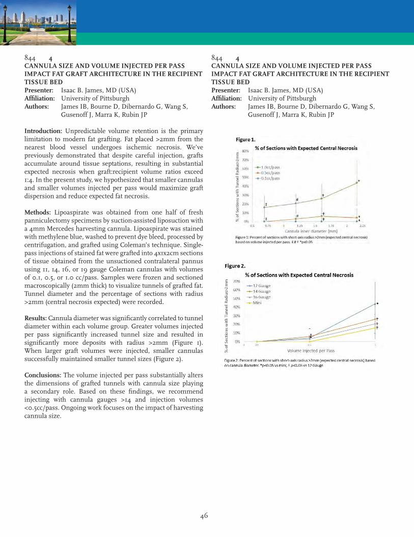
46
844 4CANNULA SIZE AND VOLUME INJECTED PER PASS IMPACT FAT GRAFT ARCHITECTURE IN THE RECIPIENT TISSUE BEDPresenter: Isaac B. James, MD (USA)Affiliation: University of PittsburghAuthors: James IB, Bourne D, Dibernardo G, Wang S, Gusenoff J, Marra K, Rubin JP
Introduction: Unpredictable volume retention is the primary limitation to modern fat grafting. Fat placed >2mm from the nearest blood vessel undergoes ischemic necrosis. We’ve previously demonstrated that despite careful injection, grafts accumulate around tissue septations, resulting in substantial expected necrosis when graft:recipient volume ratios exceed 1:4. In the present study, we hypothesized that smaller cannulas and smaller volumes injected per pass would maximize graft dispersion and reduce expected fat necrosis.
Methods: Lipoaspirate was obtained from one half of fresh panniculectomy specimens by suction-assisted liposuction with a 4mm Mercedes harvesting cannula. Lipoaspirate was stained with methylene blue, washed to prevent dye bleed, processed by centrifugation, and grafted using Coleman’s technique. Single-pass injections of stained fat were grafted into 4x1x2cm sections of tissue obtained from the unsuctioned contralateral pannus using 11, 14, 16, or 19 gauge Coleman cannulas with volumes of 0.1, 0.5, or 1.0 cc/pass. Samples were frozen and sectioned macroscopically (2mm thick) to visualize tunnels of grafted fat. Tunnel diameter and the percentage of sections with radius >2mm (central necrosis expected) were recorded.
Results: Cannula diameter was significantly correlated to tunnel diameter within each volume group. Greater volumes injected per pass significantly increased tunnel size and resulted in significantly more deposits with radius >2mm (Figure 1). When larger graft volumes were injected, smaller cannulas successfully maintained smaller tunnel sizes (Figure 2).
Conclusions: The volume injected per pass substantially alters the dimensions of grafted tunnels with cannula size playing a secondary role. Based on these findings, we recommend injecting with cannula gauges >14 and injection volumes <0.5cc/pass. Ongoing work focuses on the impact of harvesting cannula size.
844 4CANNULA SIZE AND VOLUME INJECTED PER PASS IMPACT FAT GRAFT ARCHITECTURE IN THE RECIPIENT TISSUE BEDPresenter: Isaac B. James, MD (USA)Affiliation: University of PittsburghAuthors: James IB, Bourne D, Dibernardo G, Wang S, Gusenoff J, Marra K, Rubin JP
47
850 5THE ADIPOSE TISSUE IS NOT UNIFORM. THERE ARE DIFFERENCES INCLUDING THE SITES AND ALSO THE LAYERS. PECULIARITY OF THE SUPERFICIAL FATPresenter: Angelo Trivisonno, MD (Italy)Affiliation: Sapienza UniversityAuthor: Trivisonno A
Historically the adipose tissue was considered only as an energy storage. Currently many others characteristics have been discovered as immunomodulatory and regenerative capacities. In this is important the role of the SVF cells. All research efforts are directed in trying to get more yield of SVFs with minimal manipulation. It has been considered the possibility to harvest fat with increased yield of the ADSCs, in relation to different body sites, But also in relation to its profondity. Starting from the evidence of some studies that show the origin from perivascular niches of the ADSCs, and that the number of these cells is directly related to the density of blood vessels, we assumed that to obtain higher yield ADSCs we had to do the harvest in the area most vascularized of adipose tissue. We performed a comparative study of the yield of SVF after harvest with a traditional technique, compared to the harvest with a 2 mm microcannula with very small holes of 1 mm at very superficial layer. We did the harvest in six caucasian women. In the laboratory we have isolated the SVFs by use of collagenase. The total number of cells collected with the micro cannula was approximately two times higher than that of the standard cannula no differences were observed in cell viability with different forces of aspiration. It was made culture in maintenance medium and in EBM-2 medium. Performed immunohistochemistry analysis, in vitro and in vivo analysis.
The number of plastic-adherent, fibroblast-like cells, resulted double with a microcannula than with standard cannula. In EBM-2 medium observed a greater number of clobestone-like. Greater capacity of angiogenesis is showed in vitro. Confirmed with CD31 markers. These observations were also confirmedin vivo after of cells labeled with firefly luciferase.
In conclusion the hypodermis level in contacts with dermis is more vascularized with greater number of ADSCs. The harvesting with microcannula allow us injection with 21-27 Gauge needles. Moreover several studies shown that this superficial layer of fat present most affinity with stem cells of skin. These superficial ADSCs are capable to proliferate significantly faster, and are more resistant to apoptosis, have a higher proportion of CD105 positive cells (a marker
891 6AUTOLOGOUS FAT TRANSFER UTILIZING TISSUE LIQUEFACTION TECHNOLOGY: SAFETY, EFFICACY AND LONG-TERM RESULTSPresenter: Christopher P. Godek, MD (USA)Affiliation: Personal Enhancement CenterAuthors: Godek CP, Godek MA, Borab Z
Background: Several key steps are required during autologous fat transfer to optimize long-term fat survival. Fat harvest must be performed in an atraumatic fashion to maintain adipocyte viability. There are numerous devices that are currently being used for fat harvest with little data regarding which device is best suited for this process. Tissue Liquefaction Technology (TLT) is a new and novel energy source for fat harvest. This technology was FDA cleared in 2012 for aesthetic body contouring and in 2013 received FDA clearance for autologous fat transfer.
Objective: To evaluate the efficacy, safety and long-term clinical outcomes of patients undergoing TLT harvest and fat transfer to multiple anatomic sites.
Methods: All procedures were performed on an outpatient basis with general anesthesia. Fat was harvested with TLT then purified with low speed centrifugation to remove excess fluid. Injection techniques were performed with a blunt cannula in multiple planes with needle band release when indicated.
Results: 104 consecutive patients were treated over 22 months. 103 were females and 1 male. 64 patients underwent fat transfer to the breast, 17 to the body and 25 to the face. 12 patients underwent more than one stage (range 2-5 stages). 42 of the patients also underwent concomitant aesthetic body-contouring. Lipoaspirate volumes ranged from 100cc to 3950cc (avg. 1127cc). Fat transfer volumes ranged from 10cc to 1420cc (avg. 310cc). Average volumes based on anatomical areas were as follows, facial 11cc, body 481cc and breast 376cc. Follow up time ranged from 6 to 44 months (avg. 25 months). All patients maintained significant volume of transferred fat (70% or greater). Complications were only seen in breast patients. There were two cases of cellulitis responding to oral antibiotics. Five patients developed mild to moderate palpable fat necrosis and one patient required return to the operating room for removal of an oil cyst. No revisions of donor sites were required. All patients were pleased with donor and recipient site contour improvement.
Conclusion: TLT is a new and novel method for fat harvest. Our review of 104 consecutive patients demonstrates safety, efficacy and long-term survival with favorable improvement in both the donor and recipient sites.

48
923 7AUTOMATED STROMAL VASCULAR FRACTION SPHEROID PRODUCTION USING 3D BIOPRINTING IN CONJUNCTION WITH A COMBINATION HYDROPHOBIC/HYDROPHILIC SURFACE TREATMENTPresenter: Brian C. Gettler, MEng (USA)Affiliation: University of LouisvilleAuthors: Gettler BC, Gandhi PS, Zakhari JS, Williams SK
Introduction: Hydrogel based spheroids that contain cells provide a three dimensional system for cellular localization, dosage control, and protection against mechanical forces. A limiting factor toward the formation of spheroids is their limited ability to maintain a spheroidal shape pre-polymerization or adjuvant gel forming materials must be added to accelerate polymerization. Adipose derived stromal vascular fraction (SVF) cells are a readily available source regenerative cells for cell based therapies and were selected as cellular material for encapsulation. This report describes a method to create spheroids composed of collagen, a polymer that neither rapidly polymerizes nor maintains its own shape pre-polymerization.
Methods: A multi-well plate was modified with a super-hydrophobic coating. 3.8% Pluronic F-127 was then loaded into a 3D Bioprinter (nScrypt, Inc) fitted with a 27G needle. Pluronic F-127 was dispensed into the center of each coated well in the plate creating a circle (250 micron diameter) of gel. SVF cells from human adipose tissue were suspended at 1.6x10⁶ cells/mL in 3mg/mL collagen in culture medium. The cell suspension was loaded into a 3D bioprinter fitted with an 18G needle. The spheroids were then dispensed automatically into each prepared well. The spheroids were then polymerized at 370C. After polymerization the spheroids were removed via Pluronic F-127 dissolution in media and transferred to desired culture conditions.
Results: The BAT bioprinter supported rapid production of 2mm collagen spheroids while the prepared plate allowed both shape-holding of the printed spheroids pre-polymerization and ease of spheroid removal post polymerization. Phase microscopic evaluation indicated the presence and homogeneous distribution of SVF cells throughout the spheroids and live/dead staining confirmed viability of the encapsulated cells.
Conclusions: 3D bioprinting can be used in conjunction with a hydrophobic/hydrophilic surface to create collagen spheroids encapsulating viable SVF cells. These spheroids can be used for a variety of therapeutic and research purposes. In addition, this production method allows for spheroid encapsulation of previously unusable polymers that were unable to hold their own shape pre-polymerization.
824 8BIOLOGIC LYMPH NODE SCAFFOLD FOR ALLOTRANSPLANTATIONPresenter: Yujin Myung Sr., MD (Korea)Affiliation: Seoul National University Bundang HospitalAuthors: Myung Y, Pak CS, Heo CY
NOT PRESENTED

49
881 9ADULT ADIPOSE-DERIVED MULTIPOTENT STROMAL CELL OSTEOGENESIS ON BIOCOMPATIBLE SCAFFOLDS WITH DISTINCT COMPOSITIONSPresenter: Mandi J. Lopez, DVM, MS, PhD (USA)Affiliation: Louisiana State UniversityAuthors: Lopez MJ, Duan W, Haque M, Kearney M, Gimble J
Though adipose-derived multipotent stromal cells (ASCs) have osteogenic capabilities, culture conditions for in vitro ASC-bioscaffold construct osteogenesis are undefined. The hypothesis that ASC osteogenesis on distinct bioscaffolds is similar with short term and continuous culture in osteogenic medium was tested in this study. Passage 2 ASCs were loaded onto three scaffolds, type I bovine collagen (BCI), hydroxyapatite, -tricalcium phosphate (CHT); BCI, -tricalcium phosphate (CT); and BCI (C). Constructs were cultured in stromal, osteogenic or in osteogenic for 48 hours followed by stromal medium (osteo-stromal) for up to 28 days. Cell viability, gene expression (alkaline phosphatase (ALP), osteoprotegerin (OPG), osteocalcin (OCN), cannabinoid receptors type I (CB1) and type II (CB2), receptor activator of nuclear factor kappa- ligand (RANKL)), DNA (dsDNA), total collagen, and sulfated glycosaminoglycans (sGAG) were quantified. Cell viability was lower in CT versus C constructs after 14 days in stromal medium. The CB1 expression in C constructs in osteogenic medium was highest at all times, and OCN expression was highest in C constructs after 14 and 28 days of culture in all media. Total sGAG was highest in CHT and C constructs in stromal and osteo-stromal media after 7 and 14 days, respectively. Total collagen was highest in C constructs after 28 days in all media. The dsDNA was highest in CHT constructs (osteogenic - 7, 14 days; osteo-stromal - 14 days; stromal - 28 days). Continuous culture of human ASC-type I bovine collagen bioscaffold constructs in osteogenic medium appears to support sustained osteogenic gene expression and robust extracellular matrix deposition.
828 10ANTI-ADIPOGENIC EFFECT OF OXY133Presenter: Akishige Hokugo, DDS, PHD (USA)Affiliation: University of California Los Angeles UCLAAuthors: Hokugo A, Segovia LA, Rezzadeh K, Jarrahy R
Introduction: Obesity and related diseases continue to escalate in morbidity and mortality incidence. Therapeutic options, excluding prevention by lifestyle modification, are limited by number and the negative consequences associated with them. While the process of adipogenesis remains to be entirely deciphered, key stages in stem cell commitment and maturation into functional mature Adipocyte are vastly studied, allowing for their manipulation. Oxysterol is a bioactive molecule, previously proven to shift phenotype differentiation of the multipotent stem cells into osteoblasts. To determine if this feature might function as an anti-adipogenic agent, a series of in vitro studies was designed to determine if one isoform, Oxy133, might influence the differentiation process of already committed premature adipose cells.
Methodology: Mouse preadipocyte, 3T3-L1 cell lines were cultured with basal growth medium (GM) containing DMEM, 10% fetal bovine serum (FBS), and penicillin-streptomycin. After cells reached confluency, they were detached and re-seeded on 24-well culture plates. Cells were treated with conventional adipogenic medium (AM) containing DMEM, 10% FBS, penicillin-streptomycin with isobutyl methylxanthine, dexamethasone, and insulin with or without Oxy133. A negative control using GM alone was also included. Measuring differentiation specific gene expression tested for active adipogenesis, and an Oil Red O stain established mature adipocyte functionality and morphology. A second experiment series was conducted to demonstrate a mechanism of action. Cells cultured with the incorporation of Hedgehog signaling inhibitor, Cyclopamine, where similarly tested for adipogenic differentiation.
Results: Expression of adipogenic differentiation specific genes was reduced in cells exposed to the Oxy133, in contrast to positive control groups. Functionality and morphology of mature adipocytes attenuated in the experimental group. The cells exposed to both Cyclopamine and Oxy133 yielded results similar to cells cultured in the AM.
Conclusion: Exposure to the Oxy133 inhibits the maturation process of committed premature adipocytes into functional mature adipocytes. This function is archived as a result of the stimulation of the Hedgehog signaling pathway.

50
865 11STROMAL CELL-HYDROGEL CONSTRUCT POSSIBLY GENERATES CLINICALLY RELEVANT NEO-TISSUE IN FACIAL HIV-LIPOATROPHY PHASE 2 PIVOTAL MULTICENTER CLINICAL TRIAL: EARLY ANALYSIS ON TESTING PATIENT SAMPLEPresenter: Ramon Llull, MD, PhD (Spain)Affiliation: Stem Europe Mallorca CenterAuthors: Llull R, Matas A, Bahr-Davidson J, Zarembinski T, Iglesias L, Soler A, Benito J, Paya M, Furr D
A novel hydrogel (Renevia (TM), Biotime, Inc) with controllable gelification behavior allows ex-vivo stromal cell suspension and injection, while providing in-vivo viable cell retention at the implantation site. Its safety was recently documented in a previously reported Phase I Trial. The present Phase 2 Trial explores the capacity for a cell-hydrogel construct to develop into a neo-tissue that histologically integrates with the recipient bed, and restores a subcutaneous volume deficit for HIV-related lipoatrophy patients.
A testing ‘dry-run’ patient sample was authorized in the trial’s design prior to statistical accruing: 9 treated (3 per site) subjects were screened according to protocol. Treated subjects underwent surgical liposuction to yield a minimum of 50 mL of subcutaneous tissue, which was intra-operatively processed and digested to generate autologous stromal-vascular fraction (SVF). SVF was suspended in Hydrogel in its ungelled (Sol) phase, and finally injected the subcutaneous space of facial volume deficits. Treated and Control (delayed treatment) subjects were subjected to clinical, photographic, ecographic, and 3D scan imaging recording during 1 year follow-up. Cell suspension aliquotes underwent quality, safety and potency assays.
No major Adverse Events, 1 persistent procedure related complication (hematoma) were recorded. Preliminary analysis on photographic evaluation disclosed discrete improvement in HIV-Associated Facial Lipoatrophy Grading Score, particularly for lower score subjects. Ecographic quantification of tissue thickness appeared inconsistent in both groups, but it documented reliable microvascularization within the construct. 3D facial volumetric suggests changes early in the postoperative period (1 month). Such changes remained steady at 3, 6 and 1 year follow-up. Histological examination of one patient biopsy at 1 year revealed presence of cell clusters, matrix remodeling, and peri- and intra-construct presence of basal lamina and endothelial lining.
In conclusion, the testing dry-run patient sample in the present ongoingtrial provided further data supporting feasibility, safety, in-vivo construct viability, and possible clinical relevance for subcutaneous implantation of stromal cell seeded Renevia(TM) implants.
827 12PERIFOLLICULAR ADIPOSE TISSUE (PFAT) IS A METABOLIC STRUCTURE LIKE PERIVASCULAR ADIPOSE TISSUE (PVAT) AND MAY HAVE INFLUENCE IN THE TREATMENT OF BALDNESSPresenter: Marco A. Pellon, MD (Brazil)Affiliation: Clinica Sao VicenteAuthor: Pellon MA
Introduction: The adipose tissue layer found lining blood vessels has long been interpreted as a mechanical protection to the vessels during contraction of neighboring tissues. However today it is know that the perivascular adipose tissue (PVAT) exerts various other influences on vascular function, by the secretion of metabolically active adipokines, chemokines and hormone-like factors (leptin, adiponectin, resistin), and vasoactive substances such as reactive oxygen species (superoxide, NO, and H2O2). In this study the author demonstrates, for the first time that, as well as vessels, hair follicles are coated with a layer of adipose tissue, accompanying the outer sheath (fig. 1) and forming an intradermal adipocyte niche. This structure, that we call perifollicular adipose tissue (PFAT) is metabolically active and participates in the mechanisms of hair growth and skin regeneration.
Method: The changes triggered after a skin trauma and the involvement of (PFAT) in the regeneration of epidermis and hair growth, were documented at all stages of these processes. The study was conducted in 36 patients with burns and other traumatic skin lesions and it was carried out to complete regeneration of the skin and hair. The lesions were accompanied during the stages of neovascularization, epithelial repopulation (fig. 2), the ascendance of melanin, and regeneration of pilosebaceous structure.
Results: After skin regeneration was observed, in all cases, hypertrophy of the hair around the lesions (fig.3), with accelerated growth and thickening. These changes persisted for a period of 2 years on average. Besides that, the patients who have suffered burns over 40% of body surface showed an increase in the length and thickness of the scalp hair.
Conclusions: In the regeneration of deep wounds, became evident the participation of factors expressed by PFAT, and the observed side effect around these lesions suggests that change in the metabolism of perifollicular adipose tissue contributes to the cause of some types of hair loss. The isolation and clinical use of these factors or the use of fat grafts to cause an artificial wound may be the answer for alopecia correction, associated or not with blocking the enzyme 5-alfa-reductase, as an alternative to hair transplants.

51
827 12PERIFOLLICULAR ADIPOSE TISSUE (PFAT) IS A METABOLIC STRUCTURE LIKE PERIVASCULAR ADIPOSE TISSUE (PVAT) AND MAY HAVE INFLUENCE IN THE TREATMENT OF BALDNESSPresenter: Marco A. Pellon, MD (Brazil)Affiliation: Clinica Sao VicenteAuthor: Pellon MA
935 13ADIPOSE-DERIVED EXTRACELLULAR MATRIX HYBRID HYDROGEL FOR MAINTENANCE OF PLURIPOTENCY IN HUMAN ADIPOSE-DERIVED STEM CELLSPresenter: John N. Poche, BS (USA)Affiliation: Louisiana State UniversityAuthors: Poche JN, Hayes DJ
NOT PRESENTED

52
930 14TAILORED ADIPOSE TISSUE 3D MICROENVIRONMENTS USING CELL SHEET TECHNOLOGYPresenter: Manuela E. Lago, MSc (Portugal)Affiliation: 3Bs Research GroupAuthors: Lago ME, Cerqueira MT, Pirraco RP, Reis RL, Marques AP
Cell sheet (CS) engineering has been widely explored in a range of applications because the self-produced extracellular matrix (ECM) of the created CS confers intrinsic features that are hardly achieved with other approaches. In this work we propose a methodology to tailor adipogenic features by stacking the CS taking advantage of this technology along with human adipose stem cells (hASCs), by inducing these cells to differentiate and/or maturate into distinct stages.
Upon confluence, hASCs isolated from lipoaspirates, were differentiated towards the adipogenic lineage using feeding regimes encompassing different combinations of differentiation and maintenance periods, in respective culture media. At different time points, to assess the degree of differentiation the expression of mesenchymal and adipogenic markers were analyzed by fluorescence microscopy, qPCR and flow cytometry to evaluate the degree of differentiation. The maturation degree was inferred by the lipid content. CS in different stages were stacked and maintained in culture for 7 days. Phenotypic analysis was carried out by ICC and the generated secretome characterized by ELISA.
Independently of the culture time/media CD73 and CD105 expression was down-regulated while CD90 mRNA expression was maintained at basal levels. Simultaneously, all the adipogenic characteristic markers (Leptin, PPARgamma and FABP4) were up-regulated confirming the triggering of the adipogenic differentiation. These results were corroborated at protein level by the increasing number of FABP4+ cells with the differentiation period. Maturation degree assessed by the lipidic content was modulated by the maintenance medium. The stacked CS with different adipogenic features formed a stable 3D construct in which the different layers were almost unnoticed, achieving particular microenvironments with specific organization and secretome.
This modulation of the CS allowed to recreate different 3D adipose tissue-like microenvironments, representing a step forward in the generation of accurate and reliable in vitro models that can be either used as in vitro platforms to study adipose tissue role in a wide range of settings or enable future developments in the TE field.
Acknowledgments: FCT for SFRH/BPD/96611/2013; SFRH/BPD/101886/2014
892 15DELIVERY OF ADIPOSE DERIVED STEM CELLS WITHIN POLOXAMER 407 HYDROGEL FOR PERIPHERAL NERVE REPAIRPresenter: Deokyeol Kim, MD (USA)Affiliation: University of PittsburghAuthors: Kim D, Allbright KO, Bliley JM, Havis E, Dibernardo G, Grybowski D, Sivak W, Rubin JP, Marra KG
Introduction: Peripheral nerve injuries are commonly encountered especially in trauma wounds and their regeneration is functionally important for the patients’ long-term quality of life. Our previous research showed that intra-luminal injection of adipose derived stem cells (ASCs) into polycaprolactone-based nerve conduit promoted overall nerve formation. However, effective cell delivery techniques were not established. Poloxamer 407 is an FDA approved additive and commercially available. The aim of this study is to investigate the capacity of poloxamer 407 hydrogel to enhance the delivery of human ASCs and peripheral nerve regeneration in a rat sciatic nerve defect model.
Methods: Human ASCs was isolated and cultured from surgical patients under IRB approval. A 15mm sciatic nerve defect in 36 rats was created (n=6/group). Treatment groups included ASCs delivered within a polycaprolactone conduit (1x10^6 cells/conduit) with or without poloxamer 407 hydrogel, and poloxamer 407 hydrogel alone. Reverse polarity autograft was used as a positive control and unrepaired defects and empty conduits were used as negative controls. Nerve conduits were explanted for histological evaluation at 6 weeks.
Results: After 6 weeks, the explanted grafts were assessed for ASC survival, nerve regeneration, and muscle regeneration. The survival of transplanted ASCs for up to 6 weeks was histologically confirmed using an anti-human mitochondria stain. Both sciatic nerve and distal muscle regeneration were analyzed with S100 (Schwann cells) and neurofilament (axons) staining. Data indicated treatment with ASCs and the poloxamer 407 hydrogel resulted in improvement in regeneration compared to poloxamer 407 hydrogel alone or ASCs alone.
Conclusions: Poloxamer 407 hydrogel may affect long term ASCs retention in a nerve conduit. Delivery of ASCs in combination with poloxamer 407 hydrogel can be a potential therapeutic method for peripheral nerve regeneration.

53
809 16UTILIZATION OF ADIPOSE-DERIVED CELLS FOR BIO-ENGINEERING PRE-VASCULARIZED AND TRI-LAYERED SKIN SUBSTITUTESPresenter: Jakub Zimoch, MS (Switzerland)Affiliation: University of ZurichAuthors: Zimoch J, Klar AS, Meuli-Simmen C, Meuli M, Scherberich A, Reichmann E
Introduction: As an outer covering of the human body, skin serves multiples functions. Its structure and biology make it a perfect boundary, sensory and thermoregulatory organ. The current state of regenerative medicine allows generation of personalised bio-engineered skin grafts that bring back biological functions to the damaged area of a patient’s body. In some cases, however, there might be a shortage of skin cells for a production of a substitute, which led to a quest for other suitable sources of cells. It appeared that adipose tissue is a ubiquitous and accessible source of both committed mesenchymal cells and mesenchymal stem cells.
Methods: Stromal Vascular Fraction (SVF) cells were isolated from adipose tissue of patients undergoing liposuction or excision surgery. Dermo-epidermal substitutes were prepared by seeding SVF cells into collagen hydrogels and cultured in conditions promoting formation of blood capillaries. Pre-vascularized grafts were transplanted onto immuno-compromised rats. In another approach, Adipose-derived Stem Cells (ASCs) obtained from the SVF, were embodied into a collagen hydrogel and differentiated into adipocytes. Finally, three layered constructs, composed of the epidermis, the dermis and the hypodermis, were created. These skin grafts were transplanted onto immuno-compromised rats.
Results: The ratio of endothelial and stromal cells in the SVF was intrinsically optimal, hence ideally supporting the development of capillary networks. Notably, after transplantation of pre-vascularized skin grafts the capillary plexus was rapidly connected to the rat’s microvascular system. We further present the successful creation of three-layered skin substitutes, both in vitro and in vivo. Sufficient numbers of adipocytes differentiated from ASCs were established. Moreover, they survived in the hypodermal compartment after transplantation of the substitutes.
Conclusions: Because of an uncomplicated isolation, adipose tissue represents an attractive cell source for the regenerative medicine. Our focus on bio-engineered skin substitutes showed that adipose-derived cells can be utilized not only for the efficient pre-vascularization but also for dermo-epidermal skin substitutes the third layer: the hypodermis.
901 17INJECTABLE HUMAN ADIPOSE MATRIX FOR SOFT TISSUE FILLING: LONG-TERM ASSESSMENT IN THE IMMUNOCOMPETENT RAT MODELPresenter: Lauren E. Kokai, PhD (USA)Affiliation: University of PittsburghAuthors: Kokai LE, Schilling B, Chnari E, Mahoney C, Jacobs M, Marra KG, Rubin JP
Introduction: There is a significant clinical need for an off-the-shelf soft-tissue replacement for reconstructive surgery. We have previously optimized a process to remove lipid and immunogenic elements from human subcutaneous adipose tissue while maintaining structural components and growth factors important for cellular ingrowth and adipogenesis. The goal of this study was to evaluate the efficacy of Allograft Adipose Matrix (AAM) in an immunocompetent Fisher 344 rat model.
Methods: Volume maintenance and de novo tissue formation were evaluated for AAM, Hyaluronic Acid (HA), INTEGRA® Flowable Wound Matrix, and human lipoaspirate on the bilateral dorsal flanks of 6 week female Fisher 344 rats, with up to 12 replicates per group. Volume retention for each material was measured at 72 hours, 3, 6, 12, 18 and 24 weeks with gas pycnometry. Histological analysis was performed with Masson’s trichrome, perilipin-A (adipocytes) and F4/80 (macrophages). qPCR was used to assess material adiposity, inflammation, and macrophage phenotype.
Results: No early adverse events were observed at 72 hours or 3 weeks. AAM showed significantly increased volume retention at 3 weeks, with 89 ± 16% of the original volume maintained, comparing favorably to HA controls with 25 ± 3%. Volume retention of Integra was 55% ± 47%. Genetic analysis of materials at 72 hrs showed that while AAM significantly increased Mac1 expression, an early macrophage gene, CD68, a mature macrophage gene was significantly decreased and Interleukin-10 expression, a known cytokine of M2a (anti-inflammatory) macrophages was significantly increased above both HA and Integra experimental groups. Longer study time points are currently underway.
Conclusions: Early analysis of a novel, off-the-shelf, adipose-derived injectable matrix showed superior volume retention at early time points in an immunocompetent rat model. qRT-PCR for inflammatory markers and macrophage phenotypes showed that AAM promoted anti-inflammatory M2a macrophages as early as 72 hours above HA controls and alternative ECM derived products.
54
875 18SOFT TISSUE RECONSTRUCTION BY STRUCTURAL FAT GRAFTING: RECIPIENT SITE OPTIMIZATION USING EXTERNAL VOLUME EXPANSION (EVE) COMBINED TO AN INJECTABLE ALLOGRAFT ADIPOSE MATRIX (AAM)Presenter: Giorgio Giatsidis, MD (USA)Affiliation: Brigham and Women’s Hospital - Harvard Medical SchoolAuthors: Giatsidis G, Succar JS, Haddad AH, Lago GL, Schaffer CS, Wang XW, Matsumine HM, Orgill DO
Introduction: Recipient site optimization is a novel area of research in structural fat grafting that aims to develop effective strategies to enhance graft take and long term graft volume retention. External Volume Expansion (EVE) has shown in murine models and clinical cases the capacity to induce angiogenesis and adipogenesis in recipient sites before grafting, improving long-term outcomes. Scaffolds, in particular allograft adipose matrices (AAMs), have also provided evidence of their structural and biochemical potential in inducing graft survival and adipocyte proliferation. In this preclinical study we combine for the first time the two strategies hypothesizing that their complementary role might further enhance recipient site optimization before structural fat grafting.
Method: 66 wild-type C57BL/6 mice were assigned to three experimental groups (n = 22 per group) undergoing continuous EVE, moderate-intensity intermittent EVE or no EVE (control) for five days. Five days after the last EVE stimulation mice underwent subcutaneous injection of 0.5cc of an AAM obtained from cadaveric tissue that was aseptically processed to remove lipid and cells remnants. On post-operative day (POD) 28 (n = 4 per group), 42 (n = 8 per group), and 84 (n = 10 per group) grafts were collected for macroscopic and histological analysis (skin and AAM graft structure, using H&E staining; angiogenesis, using immuno-histochemistry for the CD 31 marker; adipogenesis, using immuno-histochemistry for the Perilipin marker).
Results: At a long-term follow up (POD 84) density of blood vessels at the recipient site and surrounding the AAM grafts was significantly increased by EVE enhanced vascularity of AAM grafts (1.6- fold increase compared to controls). EVE also induced a peri-graft inflammatory reaction that gradually decreased over time. Clusters of proliferating adipocytes were observed along the external border of the AAM graft starting from POD 28.
Conclusions: EVE angiogenic and adipogenic potential can be used to increase vascularity of recipient sites and AAMs. AAM enhances and optimizes these outcomes.
942 19ADIPOSE STEM CELL SECRETOME ENHANCES FUNCTIONAL AND MOLECULAR MYOCARDIAL PRESERVATION DURING EX-VIVO COLD ISCHEMIAPresenter: Meijing Wang, MD (USA)Affiliation: Indiana UniversityAuthors: Wang M, Wang IW, Liu Y, Merfeld-Clauss S, Edenberg H, Traktuev DO, Prockop D, March KL
Introduction: Heart transplantation is a life-saving therapy for patients with end-stage heart disease, but its use is limited by extreme shortage in donor organ supply. The time “window” elapsing between procurement and transplantation sets the stage for myocardial ischemia/reperfusion injury, which limits the acceptable storage period and lowers utilization rate of donor organs. Human adipose stem cells (ASC) have gained attention for cardiac repair via their paracrine actions. However, the ability of ASC secretome to mitigate ischemic injury in donor hearts during storage in unknown. Here, we tested if ASC secretome would function as an adjunct to preservation solutions to ameliorate I/R-induced damage in donor hearts.
Methods: Isolated mouse hearts were perfused with 1 ml of cold University of Wisconsin (UW) solution either alone or with ASC-conditioned medium (CM), and stored at 4°C. After a six-hour period, ventricular function was analyzed using the Langendorff system. Myocardial TNF-?, IL-6, cleaved caspase-3, and cytosolic cytochrome c were detected by ELISA and Western Blot. Baseline controls were hearts without storage. Parallel hearts were subjected to deep RNA sequencing analysis in order to determine myocardial transcriptomes profiling.
Results: In hearts stored with UW solution alone, final cardiac function was 40% of control, whereas ASC CM-treated hearts showed 60% of baseline function (p<0.05).Hearts exposed to ASC-CM exhibited decreased production of TNF-? and IL-6, and reduced cleaved caspase-3 and cytosolic cytochrome c Six-hour cold ischemia resulted in defined changes of the myocardial transcriptome profile with 185 genes being differentially expressed compared to control. However, in hearts treated with ASC-CM, only 14 genes were differentially expressed vs. baseline, suggesting less abnormal changes in ASC CM-treated hearts.
Conclusions: Our results are the first evidence that ASC-CM during cold storage confers significant preservation of function, accompanied by decreased myocardial inflammation and apoptotic signaling, and remarkable preservation of a normal transcriptional “fingerprint”. This provides the foundation for clinical trials in which ASC-CM will optimize storage conditions for ex vivo organ preservation.

55
849 20A PROSPECTIVE, RANDOMIZED, BLINDED AND PLACEBO-CONTROLLED EFFICACY STUDY OF INTRAARTICULAR ALLOGENEIC ADIPOSE STEM CELLS FOR THE TREATMENT OF OSTEOARTHRITIS IN DOGSPresenter: Robert Harman, DVM, MPVM (USA)Affiliation: VetStem Harman R, Carlson K, Gaynor J, Dhupa S, Clement K, McCarthy T, Hoelzler M, Schwartz P, Adams C
Osteoarthritis (OA) is a high prevalence degenerative disease in canine joints. Autologous mesenchymal stem cells (MSC’s) are used for treatment of OA in humans, dogs and horses. This study was conducted to assess the efficacy and safety of an intraarticularly administered allogeneic adipose stem cell product in dogs with confirmed osteoarthritis. Dogs (93) were treated with either a saline placebo or a single dose of allogeneic adipose-derived MSC’s in either one or two joints. The MSC’s were characterized according to industry standard methods. The primary outcome variable was the owner Client-Specific Outcome Measurement (CSOM) and secondary measures included veterinary pain on manipulation, veterinary global score, and owner global score. Efficacy outcomes were statistically analyzed in 74 dogs. The primary outcome variable, CSOM, was statistically improved in the treated dogs compared to the placebo dogs (79.2% versus 55.4%, p=0.029). Veterinary pain on manipulation score (92.8% versus 50.2%, p=0.017) and veterinary global score (86.9% versus 30.8%, p= 0.009) were both statistically improved in treated dogs compared to placebo. No detected significant differences between the treated dogs and the placebo dogs in the incidence of adverse events or negative health findings. Allogeneic adipose-derived stem cell therapy was shown to be efficacious compared to placebo. This is the first reported large-scale prospective blinded efficacy study of allogeneic stem cells in canine osteoarthritis. This study was conducted under an FDA Investigational New Animal Drug Application. The use of stem cell therapy in canine orthopedics has proliferated in the veterinary profession with limited blinded, prospective, large-scale study evidence. This study contributes to both the safety and efficacy evidence and provides guidance for future randomized controlled testing of stem cell therapy as well as animal data in support of development of human stem cell therapies.
799 21LONG-TERM SAFETY AND EFFECT OF AUTOLOGOUS ADIPOSE-DERIVED STROMAL VASCULAR FRACTION INTO FINGERS FOR SYSTEMIC SCLEROSIS PATIENTSPresenter: Florence Sabatier, PhD (France)Affiliation: APHMAuthors: Daumas A, Magalon J, Jouve E, Truillet R, Casanova D, Giraudo L, Veran J, Benyamine A, Dignat-George F, Magalon G, Sabatier F, Granel B
Introduction: Hand involvement is frequent and confers a substantial handicap in work and daily activities in patients suffering from Systemic sclerosis (SSc). Autologous adipose-derived stromal vascular fraction (ADSVF) is recognized as an easily accessible source of cells with angiogenic, anti-inflammatory, immunomodulatory and regenerative effects. We previously performed a phase I open-label single center clinical trial, called SCLERADEC (NTC01813279) assessing the safety and efficacy of subcutaneous injection of autologous ADSVF in 12 SSc patients. We report the long term outcome of these patients.
Method: Twelve females, mean age 54.5±10.3 years, were assessed between 26 and 32 months after the ADSVF injection. Patients were eligible if they had a Cochin Hand Function Scale (CHFS) above 20/90. ADSVF was obtained from lipoaspirate using an automated processing system and subsequently injected into the subcutaneous tissue of each finger in a one-time procedure.
Results: From baseline, a 62.5% improvement of the CHFS score was observed with a 51.1% improvement of the Scleroderma Health Assessment Questionnaire, a 33.1% improvement of the hand pain and an 88.3% improvement of the Raynaud Condition Score (p<0.001). A decrease in total digital ulcers number was noted from 15 at entry to 6 at last visit. Other tested parameters of hand function (including objective data such as strength) and hand skin fibrosis also showed improvement. Global severity disease scale suggested stability in disease severity. Only 2 of the 12 patients showed a clinical worsening requiring additional medications. None of the 8 patients who had previously received iloprost infusion required new infusion.
Conclusion: Despite the limits of an open clinical study, the data are in favour of the long-term clinical benefits for at least two years. This innovative therapy is safe and can be considered cost-effective given the long duration of benefit following a single application and the autologous cells which can easily be harvested. Two randomized double blind, placebo-controlled clinical trials with ADSVF are ongoing in USA (NCT02396238) and in France (NCT02558543).
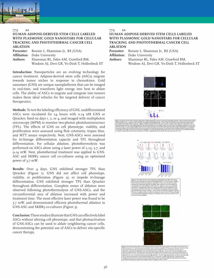
56
779 22HUMAN ADIPOSE-DERIVED STEM CELLS LABELED WITH PLASMONIC GOLD NANOSTARS FOR CELLULAR TRACKING AND PHOTOTHERMAL CANCER CELL ABLATIONPresenter: Ronnie L. Shammas Jr., BS (USA)Affiliation: Duke UniversityAuthors: Shammas RL, Fales AM, Crawford BM, Wisdom AJ, Devi GR, Vo-Dinh T, Hollenbeck ST
Introduction: Nanoparticles are an evolving technology for cancer treatment. Adipose-derived stem cells (ASCs) migrate towards tumor niches in response to chemokines. Gold nanostars (GNS) are unique nanoplatforms that can be imaged in real-time, and transform light energy into heat to ablate cells. The ability of ASCs to migrate and integrate into tumors makes them ideal vehicles for the targeted delivery of cancer therapeutics.
Methods: To test the labeling efficiency of GNS, undifferentiated ASCs were incubated for 24 hours with 0.14 nM GNS or Qtracker, fixed on days 1, 2, or 4, and imaged with multiphoton microscopy (MPM) to monitor two-photon photoluminescence (TPL). The effects of GNS on cell phenotype, viability, and proliferation were assessed using flow cytometry, trypan blue, and MTT assays respectively. Next, GNS-ASCs were assessed for tri-lineage differentiation capacity and TPL throughout differentiation. For cellular ablation, photothermolysis was performed on ASCs alone using a laser power of 2.19, 3.7, and 9.14 mW. Next, photothermal treatment was applied to GNS-ASC and SKBR3 cancer cell co-cultures using an optimized power of 3.7 mW.
Results: Over 4 days, GNS exhibited stronger TPL than Qtracker (Figure 1). GNS did not affect cell phenotype, viability, or proliferation (Figure 2); or impede tri-lineage differentiation. GNS exhibited stronger TPL than Qtracker throughout differentiation. Complete zones of ablation were observed following photothermolysis of GNS-ASCs, and the circumferential area of ablation increased with power and treatment time. The most effective laser power was found to be 3.7 mW, and demonstrated efficient photothermal ablation in GNS-ASC and SKBR3 co-cultures (Figure 3).
Conclusion: These studies illustrate that GNS can effectively label ASCs without altering cell phenotype, and that photoactivation of GNS-ASCs can be used to ablate neighboring cancer cells, demonstrating the potential use of ASCs to deliver site-specific cancer therapy.
779 22HUMAN ADIPOSE-DERIVED STEM CELLS LABELED WITH PLASMONIC GOLD NANOSTARS FOR CELLULAR TRACKING AND PHOTOTHERMAL CANCER CELL ABLATIONPresenter: Ronnie L. Shammas Jr., BS (USA)Affiliation: Duke UniversityAuthors: Shammas RL, Fales AM, Crawford BM, Wisdom AJ, Devi GR, Vo-Dinh T, Hollenbeck ST

57
823 23INTERIM ANALYSIS: SAFETY AND EFFECTIVENESS OF COMBINED CELLULAR THERAPY FOR THE TREATMENT OF PAIN AND FUNCTION ASSOCIATED WITH OSTEOARTHRITISPresenter: Kevin Darr, MD (USA)Affiliation: Covington Orthopedic and Sports Medicine InstituteAuthor: Darr K
Objectives: Successful treatment of degenerative osteoarthritis remains a difficult challenge. Recent research utilizing regenerative cellular therapies have shown promising results, exhibiting significant anti-inflammatory potential and subsequently providing pain relief. The objective of this study is to assess the clinical outcome of participants receiving a combination of adipose tissue (fat graft), bone marrow-aspirate concentrate (BMAC), and Platelet-Rich Plasma (PRP) for the treatment of pain and function associated with osteoarthritis.
Methods: This is a safety and efficacy study with a single treatment group. We prospectively plan to enroll 150 patients, ages 18-90 with a diagnosis of osteoarthritis. We followed a well established protocol for extracting, preparing, and administering autologous fat graft, BMAC, and PRP using ultrasound guidance. Participants completed the Western Ontario and McMaster Universities Arthritis Index (WOMAC) to assess function and a visual analog scale (VAS) to assess pain. MRI scans and x-rays will be obtained to determine the progression of osteoarthritis and potential increase joint space.
Results: A total of 93 patients were enrolled in the study for knee osteoarthritis prior to this interim report. 86 patients completed 6 week follow-up, including 123 knees. At 6 weeks, 80 (93%) participants reported at least some improvement in pain and function. On average, patients reported a 41% improvement (p<.001) in VAS score, and a 35% improvement (p<.001) in WOMAC. 69 patients completed 18 week follow-up (94 knees), with patients reporting an average 37% improvement (p<.001) in VAS scores and a 39% improvement (p<.001) in WOMAC. 27 patients completed 1 year follow-up (36 knees), with patients reporting an average 41% improvement (p<.001) in VAS and 47% improvement (p<.001) in WOMAC. At 1 year, MRI results show 56% of patients were stable or improving. 25% show less bone marrow edema, and 44% show an improvement in lesions. Radiographs show an average 0.5mm increase in joint space.
Conclusion: Cell based biologic treatment of osteoarthritis showed statistically significant improvement in both pain and function from baseline in this group of patients. Based on this report, further study into this treatment is warranted.
866 24CLINICAL EVIDENCE OF PIGMENT-REGULATING ACTIVITY OF NANOFAT ON HUMAN SKIN: 6 YEARS OF EXPERIENCEPresenter: Patrick L. Tonnard, MD, PhD (Belgium)Affiliation: University of BrusselsAuthor: Tonnard PL, Verpaele AM
Introduction: In an attempt to rejuvenate/regenerate the skin, we have been using fine needle intradermal nanofat grafting. The clinical effects were studied in different skin aging conditions and followed.
Methods: Adipose tissue was harvested with a superwet technique from the abdomen and thighs. A multiport 2.4-mm diameter cannula with 20 sharp 1 mm diameter holes attached to a 10 cc luer-lock syringe was used. The lipoaspirate was washed with normal saline and filtered through a sterile nylon cloth with approximately 500 micron pore size. Emulsification was achieved by shifting the lipoaspirate between two 10 cc syringes through a luer-lock connector (30 passes). The emulsified fat was again filtered using the nylon cloth and transferred into 1 cc luer-lock syringes. The nanofat obtained was injected intradermally with a 27 gauge needle, remaining superficial enough as to obtain a yellowish discoloration at the site of infiltration.
Results: From May 2010 to May 2016, we treated 360 areas of the face and body with nanofat grafting, in a total of 315 patients who presented with different skin aging conditions. The nanofat obtained consisted in a liquid fat emulsion with few viable adipocytes and a large number of viable stromal vascular cells including a high CD34+ to Stromal vascular fraction (SVF) ratio (5.1%). The treated areas were: perioral region (99), d’collet (60), lower eyelids (51), glabella (45), dorsal aspect of the hands (45), cervical region (30), full face (30). Temporary erythema at the injection sites lasted 1 to 4 weeks. The final results were observed after 6 to 8 months. The mean follow up period was 3.6 years. There were no major complications. Reinjection was performed in the lower eyelids in two patients. Based on a blind analysis of of pre and post operative photographs, the best scores were obtained for different hyperpigmentation disorders.
Conclusions: Adipose-derived stem cells were found to down-regulate melanin synthesis in laboratory settings. Our clinical experience with nanofat grafting seems to corroborate these findings as hyperpigmentation had significantly improved. Future in vitro and clinical studies need to be done to explain and confirm these findings.

58
866 24CLINICAL EVIDENCE OF PIGMENT-REGULATING ACTIVITY OF NANOFAT ON HUMAN SKIN: 6 YEARS OF EXPERIENCEPresenter: Patrick L. Tonnard, MD, PhD (Belgium)Affiliation: University of BrusselsAuthor: Tonnard PL, Verpaele AM
902 25HUMAN ADIPOSE-DERIVED STEM CELLS ACTIVELY MAINTAIN HOMEOSTASIS DURING EARLY AGINGPresenter: Ivona Percec, MD, PhD (USA)Affiliation: University of PennsylvaniaAuthors: Percec I, Roberts C, Brenner A, Grant G, Kim E, Shan X, Gersch R, Dierov R
Background: Adult stem cells play a critical role in human aging contributing to a progressive decline in homeostasis and regenerative capacity. Studies investigating aging largely focus on artificially induced endpoints or on terminal senescence, however, little is known about the early stages of normal stem cell aging. Here we examine genome-wide transcriptional networks and chromatin conformation in early human adipose-derived stem cell (ASC) aging.
Methods: Subcutaneous abdominal tissue was obtained from healthy patients of various ages (25-74) undergoing abdominoplasty. ASCs were isolated via standard collagenase protocols. Dermal fibroblasts were isolated from human skin specimens obtained from healthy patients of various ages (25-64). Transcriptional and chromatin accessibility profiles were examined via RNA- and ATAC-seq (Assay for Transposase-Accessible Chromatin) with high throughput sequencing analyses. Bioinformatics analyses were conducted by the Institute for Translational Medicine and Therapeutics. Nascent protein synthesis was measured by the incorporation of o-propargyl-puromycin.
Results: ASCs from older patients maintain highly stable transcriptional and chromatin accessibility profiles with advancing age, as demonstrated via RNA- and ATC-seq. This is in contrast to the transcriptional and chromatin accessibility profiles of age-matched fibroblasts that demonstrate significant differences with advancing age. The modest, but consistent, age-related alterations in gene expression in older ASCs specifically affect protein translation initiation pathways. Significantly, a 10% increase in nascent protein synthesis was observed in aging ASCs, as compared to a 8% decrease in aging fibroblasts.
Conclusions: In the fifth decade of life, ASCs begin to demonstrate subtle age-dependent functional and molecular changes. We demonstrate that despite advancing age, primary human ASCs are able to maintain stable transcription, chromatin conformation, gene accessibility profiles, and nascent protein synthesis functions, in contrast to age-matched fibroblasts. These observations suggest that ASCs are robust stem cells whose regenerative capacity is actively maintained during normal aging thereby reinforcing their therapeutic significance.

59
929 26A LINEAGE-TRACING MOUSE MODEL REVEALS MYH11 SMOOTH MUSCLE CELLS AND PERICYTES ARE MESENCHYMAL STEM CELLSPresenter: Howard C. Ray, BSE (USA)Affiliation: University of VirginiaAuthors: Ray HC, Dey P, Seaman SA, Mansour JD, Bruce AC, Peirce SM, Dey BK, Yates PA
Introduction: Previous studies solely relied on cell surface markers to define MSC characteristics of perivascular cells in adipose tissue. In this study, we address this issue using a tamoxifen-inducible Myosin heavy chain 11 (Myh11) lineage tracing mouse model. Myh11 classically marks smooth muscle cells and pericytes. However, Myh11-traced cells have not been isolated from adipose tissue and examined for their MSC characteristics. Our findings show that Myh11 specifically defines smooth muscle cells and pericytes, and these cells constitute a population of MSCs in the stromal vascular fraction of epididymal adipose tissue.
Methods: Confocal imaging and flow cytometry was used to characterize surface marker expression of Myh11 eYFP+ cells in a Myh11 lineage tracing mouse model. Fluorescence-activated cell sorting (FACS) was used to collect and then culture Myh11 eYFP+ cells from the stromal vascular fraction of epididymal adipose tissue. Cultured Myh11 eYFP+ cells were introduced to adipogenic, chondrogenic, and osteogenic media to determine their tri-differentiation potential. Gene expression and stem cell transcription factors were measured using quantitative RT-PCR. Myh11 eYFP+ cells were delivered intravitreally to determine their role in vascular remodeling in the Akimba diabetic retinopathy mouse model.
Results: Myh11 eYFP+ cells are marked in vivo by PDGFR? and CD146 in multiple tissues. Flow cytometry revealed that Myh11 eYFP+ cells in the stromal vascular fraction partially express MSC markers, CD146 (34%) and CD90 (35%). Myh11 eYFP+ cells are able to adhere to plastic and express CD90, CD73, CD105, and CD146. Myh11 eYFP+ cells were able to tri-differentiate and express stem cell transcription factors in vitro. We have injected Myh11 eYFP+ cells intravitreally in a diabetic mouse model (Akimba) and found that these cells integrate and remodel the retinal vasculature.
Conclusions: We demonstrate a detailed MSC characterization of lineage-traced smooth muscle cells and pericytes, and we establish a method of isolating perivascular cells based on a single marker. This work provides further evidence of a population of MSCs in the perivascular space of adipose tissue that can be a source for therapeutic use.
773 27CHANGING THE PARADIGM OF CRANIOFACIAL RECONSTRUCTION WITH AUTOLOGOUS FAT TRANSFER: A PROSPECTIVE CLINICAL TRIALPresenter: Debra A. Bourne, MD (USA)Affiliation: University of Pittsburgh Medical CenterAuthors: Bourne DA, Bliley J, James IB, Haas GL, Meyer EM, Pfeifer M, Donnenberg AD, Donnenberg V, Branstetter B, Mitchell RT, Brown SA, Marra K, Coleman S, Rubin JP
Purpose: Craniofacial deformities profoundly affect quality of life. Traditional reconstructive options include flap and alloplastic reconstructions. The aim of this clinical trial is to assess the efficacy of autologous fat transfer in the correction of craniofacial deformities.
Methods: This HRPO-approved prospective cohort study was funded by the Department of Defense. Twenty subjects with craniofacial deformities were enrolled and underwent fat grafting with an average volume of 23.9 ± 13.2mL. Volume retention was determined using CT scans at baseline, postoperatively and at 3 and 9 months after the procedure. Grafted fat was evaluated for stromal vascular fraction (SVF) viability and cellular sub-populations by flow cytometry. Quality of life (QOL) assessments were performed. After completion of 9 month follow-up, five subjects were enrolled in an arm of the study assessing a second autologous fat transfer procedure.
Results: There were no serious adverse events. Volume retention averaged 63 ± 17% at 9 months. The retention at 3 months was highly predictive of 9 month volume (r=.996, p<0.0001) (Figure 1). Patients who underwent a second procedure had similar retention rates for both grafts (p=0.05). There was no significant correlation between the percentage of cellular subpopulations and volume retention. Patients who had quit smoking at least 1 month prior to the operation had greater volume retention at 9 months compared to non-smokers (74.4% vs 56.2%, p=0.009) and a trend towards improved volume retention compared to current smokers (74.4% vs 57.7%). Satisfaction with both physical appearance (p=0.002) and social relationships (p=0.02) and social functioning quality of life (p=0.05) improved from baseline to 9 months.
Conclusions: Autologous fat transfer for the reconstruction of craniofacial defects is effective and safe, with a 63% volume retention. Improved satisfaction with physical appearance and social functioning QOL scores demonstrate the power of this procedure in changing patients’ lives. This study demonstrates that fat grafting is a safe, effective and predictable method of craniofacial reconstruction.
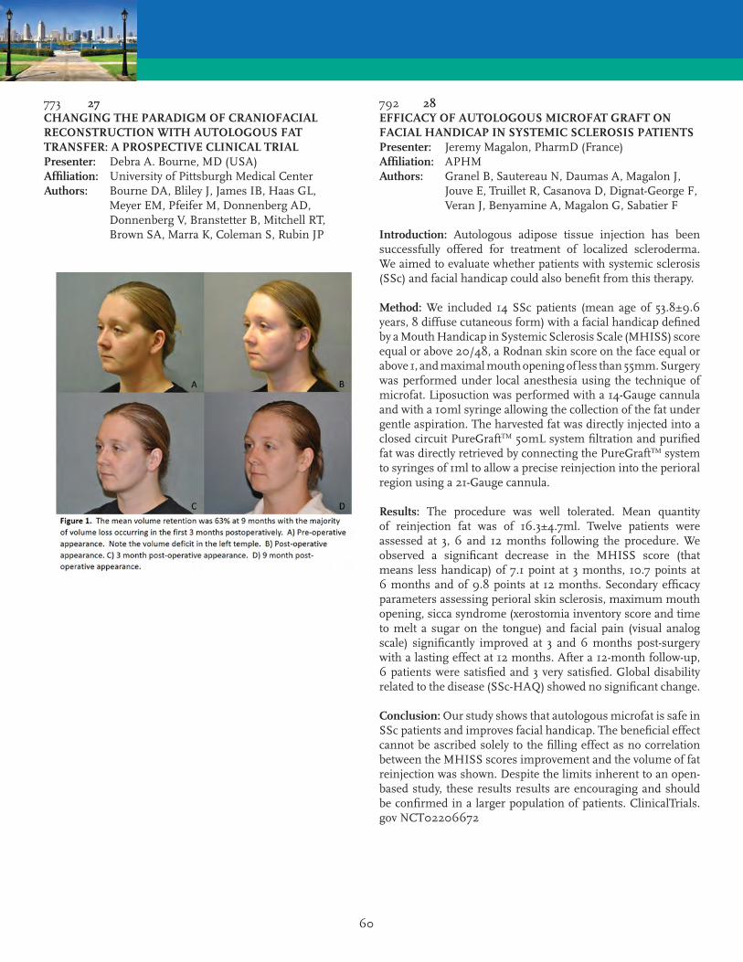
60
773 27CHANGING THE PARADIGM OF CRANIOFACIAL RECONSTRUCTION WITH AUTOLOGOUS FAT TRANSFER: A PROSPECTIVE CLINICAL TRIALPresenter: Debra A. Bourne, MD (USA)Affiliation: University of Pittsburgh Medical CenterAuthors: Bourne DA, Bliley J, James IB, Haas GL, Meyer EM, Pfeifer M, Donnenberg AD, Donnenberg V, Branstetter B, Mitchell RT, Brown SA, Marra K, Coleman S, Rubin JP
792 28EFFICACY OF AUTOLOGOUS MICROFAT GRAFT ON FACIAL HANDICAP IN SYSTEMIC SCLEROSIS PATIENTSPresenter: Jeremy Magalon, PharmD (France)Affiliation: APHMAuthors: Granel B, Sautereau N, Daumas A, Magalon J, Jouve E, Truillet R, Casanova D, Dignat-George F, Veran J, Benyamine A, Magalon G, Sabatier F
Introduction: Autologous adipose tissue injection has been successfully offered for treatment of localized scleroderma. We aimed to evaluate whether patients with systemic sclerosis (SSc) and facial handicap could also benefit from this therapy.
Method: We included 14 SSc patients (mean age of 53.8±9.6 years, 8 diffuse cutaneous form) with a facial handicap defined by a Mouth Handicap in Systemic Sclerosis Scale (MHISS) score equal or above 20/48, a Rodnan skin score on the face equal or above 1, and maximal mouth opening of less than 55mm. Surgery was performed under local anesthesia using the technique of microfat. Liposuction was performed with a 14-Gauge cannula and with a 10ml syringe allowing the collection of the fat under gentle aspiration. The harvested fat was directly injected into a closed circuit PureGraftTM 50mL system filtration and purified fat was directly retrieved by connecting the PureGraftTM system to syringes of 1ml to allow a precise reinjection into the perioral region using a 21-Gauge cannula.
Results: The procedure was well tolerated. Mean quantity of reinjection fat was of 16.3±4.7ml. Twelve patients were assessed at 3, 6 and 12 months following the procedure. We observed a significant decrease in the MHISS score (that means less handicap) of 7.1 point at 3 months, 10.7 points at 6 months and of 9.8 points at 12 months. Secondary efficacy parameters assessing perioral skin sclerosis, maximum mouth opening, sicca syndrome (xerostomia inventory score and time to melt a sugar on the tongue) and facial pain (visual analog scale) significantly improved at 3 and 6 months post-surgery with a lasting effect at 12 months. After a 12-month follow-up, 6 patients were satisfied and 3 very satisfied. Global disability related to the disease (SSc-HAQ) showed no significant change.
Conclusion: Our study shows that autologous microfat is safe in SSc patients and improves facial handicap. The beneficial effect cannot be ascribed solely to the filling effect as no correlation between the MHISS scores improvement and the volume of fat reinjection was shown. Despite the limits inherent to an open-based study, these results results are encouraging and should be confirmed in a larger population of patients. ClinicalTrials.gov NCT02206672

61
924 29FORCING A SQUARE PEG INTO A ROUND HOLE: THE CHALLENGE OF APPLYING PHARMA-BASED REGULATORY REQUIREMENTS FOR POTENCY TO ADIPOSE-DERIVED CELL THERAPIESPresenter: Kevin C. Hicok, MS (USA)Affiliation: VetStem BiopharmaAuthor: Hicok KC
Fifteen years after the initial characterization of adipose-derived stem/stromal cells (ASCs), their potential as a therapy is now well recognized. While several phase 2/3 clinical trials of adipose cell based therapies are underway and autologous cells are being used under the guise of transplantation; the FDA has yet to award final marketing approval for a GMP manufactured adipose cell therapy in the US. A major reason for this has been the challenges faced by the adipose cell therapy industry to adopt and apply a small drug/biologic based pharmacologic approach of product characterization, especially when trying to quantify cell potency during manufacturing that correlates to clinical efficacy. The path to achieving an acceptable potency assay can be fraught with pitfalls which cost both time and money. Guidance describing approaches to establish potency at first look seem to be excessively vague or not applicable to a multi-modal acting cell population, which in some cases is comprised of multiple cell types. Assay costs and the amount of cells required for testing appear unacceptably high. Determining how to approach clinical correlation of potency seems impossible without really knowing in advance which mode of action is in play or what outcome will be correlative. Recently, we have successfully validated what to our knowledge is the first ASC potency assay in veterinary regenerative medicine to receive FDA concurrence. The intent of this presentation is to share lessons learned and provide pragmatic approaches when undertaking development of potency assays that may satisfy regulatory bodies governing the SVF/ASC-therapeutic field.
839 30PERIVASCULAR SCAFFOLDS LOADED WITH ADIPOSE TISSUE-DERIVED STROMAL CELLS (ASC) ATTENUATE PROGRESSION OF EXPERIMENTAL ABDOMINAL AORTIC ANEURYSM (AAA)Presenter: Martin C. Harmsen, PhD (Netherlands)Affiliation: University Medical Center GroningenAuthors: Harmsen MC, Parvizi M, Petersen AH, Van Spreuwel-Goosens CA, Kluijtmans SG
Introduction: Abdominal aortic aneurysm (AAA) is the dilation of the abdominal aorta caused by pathological weakening of the media in particular. AAA features include inflammation, degradation of the extracellular matrix (ECM) and apoptosis of medial smooth muscle cells (SMC). We hypothesized that normalization of the media through biomaterial-delivered ASC would restore aortic homeostasis.
Materials & Methods: ASC were isolated from male Fischer rats. Patches from crosslinked Recombinant Collagen Protein (RCP) were generated and adhesion and function of ASC were assessed by SEM and a wound closing assay on rat aortic smooth muscle cells (RASMC). AAA was induced in male Fischer rats (6-8w), via intraluminal administration of elastase and periadventitial CaCl2. ASC-loaded RCP patches or bare patches were applied around the lesion after AAA induction and investigated after two weeks.
Results: ASC adhered and proliferated efficiently on RCP patches, while conditioned medium from these cultures promoted repair of damaged RASMC monolayers. Two weeks after AAA induction, the aortic diameter was significantly attenuated in both bare and ASC loaded patches in comparison to sham and aneurysm groups, likely due to the reinforcing nature of the patches. During the treatment, dye-labeled ASC had migrated into the aortic wall, in particular into the media. ASC-patch-treated walls contained more (intact) elastin fibers (p < 0.05) which were arranged in concentric layers. Moreover, in treated animals, the level of medial SMC was comparable to sham controls (p<0.05), indicating that ASC protected against apoptosis or had differentiated to SMC.
Conclusion: Local periadventitial biomaterial-guided delivery of ASC suppresses progression of AAA, via maintenance of the elastin turnover and suppression of SMC apoptosis. An additional value of the patches proved through their maintaining the structural integrity of aortic walls.as well as reduces degradation of elastin and apoptosis of medial smooth muscle cells. Local delivery of ASC in combination with biomaterial as an external scaffold is attractive to develop as clinical modality for treatment of aneurysms.

62
920 31CLINICAL SAFETY OF POINT OF CARE STROMAL VASCULAR FRACTION CELL ISOLATIONPresenter: Joel A. Aronowitz, MD (USA)Affiliation: Cedars Sinai Medical CenterAuthors: Aronowitz JA, Lockhart RA, Birnbaum ZE, Hakakian CS
Background: Pluripotential cells in adipose tissue are pivotal to long term volume retention and regenerative effects of fat grafting. Unfortunately, lipoaspiration significantly depletes the population of stromal cells, which includes adipose-derived stem cells, resulting in graft material which tends to be poor in these important cell populations. Stromal vascular fraction (SVF) cells may be isolated from excess lipoaspirate at the point of care and used to replenish fat grafts, a technique termed cell-assisted lipotransfer (CAL). Preclinical and clinical evidence supports the rationale of CAL but clinical adoption of the strategy requires additional evidence of clinical safety. This retrospective study reports on the clinical safety outcomes of SVF-enhanced fat grafting using a manual, collagenase based separation process to isolate autologous progenitor cells from lipoaspirate at the point of care.
Methods: 234 subjects underwent 244 SVF-enhanced autologous fat grafting procedures at University Stem Cell Center between August 2009 and December 2015 for a variety of cosmetic and reconstructive indications.
Results: CAL was performed for a variety of cosmetic and reconstructive indications. The mean time of the SVF isolation process was 71 min. Due to frequent concomitant procedures, the average operating room time increased by only 11 minutes. The average number of nucleated cells injected was 1.9 x 10⁸ with an average cellular viability of 85.3%. Mean follow-up was 21.3 months. There were no major and only 6 minor complications. No collagenase or neutral protease related complications were observed.
Conclusions: This series of 244 CAL cases demonstrates that SVF cell isolation using a standardized, manual, collagenase-based process at the POC is equivalent in safety compared to non-enhanced fat grafting. These results support expanded use of CAL in the clinical research setting.
803 32SHIFT TOWARDS MECHANICAL ISOLATION OF HUMAN ADIPOSE-DERIVED STROMAL VASCULAR FRACTION: A REVIEW OF UPCOMING TECHNIQUESPresenter: Alexandra Conde-Green, MD (USA)Affiliation: Rutgers New Jersey Medical School - Graduate School of Biomedical SciencesAuthors: Conde-Green A, Kotamarti VS, Sherman LS, Keith JD, Lee ES, Granick MS, Rameshwar P
Background: Adipose stromal vascular fraction (SVF) may be applied to promote regenerative healing in complex wounds. Standard isolation methods, relying on bacterial collagenase, are costly and time-intensive. Additionally, the type and concentration of collagenase is not standardized, and American regulators have regarded tissue exposed to the enzyme as more than minimally manipulated. Recent efforts to find alternatives have produced non-enzymatic isolation methods. The purpose of our study was to analyze the published literature reporting non-enzymatic isolation of adipose (SVF) to improve the understanding of current methods and to make their use more approachable.
Methods: A systematic review of the literature was performed with a search of six terms in the PubMed and Medline databases. One thousand sixty-six articles were evaluated using predetermined inclusion and exclusion criteria.
Results: Two articles of level II evidence and 7 basic science articles were selected. Stromal vascular fraction was isolated by subjecting lipoaspirate to shaking/vortexing followed by centrifugation or centrifugation only. While most articles isolated SVF in laboratory settings, three featured operating room-based protocols. Non-enzymatically isolated SVF contained CD44, CD73, CD90 and CD105-positive cells capable of adipogenic and osteogenic differentiation. Additionally, mechanical isolation methods required less time than enzymatic methods but yielded fewer cells. In two articles, improved fat graft take was observed after supplementation with mechanically isolated SVF.
Conclusions: Non-enzymatically isolated stromal SVF contains regenerative cells with laboratory and clinical applications. Due to lower cell yields, mechanical protocols may be suitable in cases featuring abundant, excess harvested adipose tissue that may be subjected to SVF isolation. Additional randomized comparative studies comparing these techniques are needed to optimize the number and quality of isolated cells and to identify the ideal clinical applications for these cells.
63
917 33MICROFAT GRAFTING USING LIPOGEMS (A 510K FDA APPROVED DEVICE FOR FAT TRANSFER) IN THE CONTEXT OF AESTHETIC, RECONSTRUCTIVE AND REGENERATIVE SURGERYPresenter: Allan Y. Wu, MD (USA)Affiliation: UC RiversideAuthors: Wu AY, Krutchkoff B, Rogers C
Microfat grafting is a process whereby routine lipoaspirate is processed by simple saline wash and “re-sizing” by passage through progressively smaller filters using the Lipogems® system, an FDA cleared 510-K device. This novel method allows for an extremely fine fat graft with proportionately higher regenerative properties in an intact stromal vascular macrostructure (niche) and immediate removal of cellular debris and oil contamination. The need for centrifugation or any additional costly laboratory equipment is obviated using this process. This unique and versatile graft may be used in aesthetic and/or reconstructive procedures in both the body and face and will be explained in a series of case studies detailing optimal use protocols. Micro fat, or micro-fractured fat, Lipogems®, is being promoted in the US for lipofilling for aesthetics and to cushion, repair, and restructure soft tissue defects. Lipogems has been shown to induce Yamanaka factors and promote neovascularization in a number of independent scientific literature, which is encouraging as we explore regenerative therapy options that can be performed in clinics within the U.S. in a regulatory compliant manner.
836 34THE FRACTIONATION OF ADIPOSE TISSUE (FAT) PROCEDURE FOR REGENERATIVE PURPOSESPresenter: Joris A. van Dongen, BSc (Netherlands)Affiliation: University of Groningen and University Medical Center GroningenAuthors: van Dongen JA, Stevens HP, Parvizi M, Van Der Lei B, Harmsen MC
Introduction: Autologous fat cell transplantation is frequently successfully used as treatment in regenerative medicine to reduce scarring and to restore volume loss. The therapeutic effect is partly due to the presence of (stromal) precursors of adipose stromal cells (ASC) located in the stromal vascular fraction (SVF) of fat tissue. It would be ideal to harvest this SVF fraction alone to be used in a more specific concentrated way. This can be achieved either by enzymatic or by mechanical fat dissociation. Enzymatic dissociation procedures are rather time-consuming, expensive and the obtained SVF loses its extracellular matrix (ECM). Therefore, we developed a new mechanical dissociation procedure to obtain the SVF from adipose tissue by mechanical dissociation suitable for direct therapeutic injection: this procedure is named the FAT procedure: (Fractionation of Adipose Tissue).
Material & Methods: The FAT procedure was performed in eleven patients to compare the ASC isolated from the SVF with non-dissociated adipose tissue (control). Phenotypically, ASC isolated from both samples were investigated for their expression of six CD-surface markers CD29, CD31, CD44, CD45, CD90, CD105. ASCs were cultured in adipogenic, osteogenic and smooth muscle cell medium to compare their function. A Masson’s Trichrome staining for ECM, -Smooth Muscle Actin staining for small vessels, von Willebrand Factor for endothelial cells and Perillipin A staining for adipocytes were performed. A live/dead staining was performed to visualize the cell viability.
Results: The FAT procedure resulted in a 7.5 times higher concentration of cells as compared to the control and culturable ASC. The samples were enriched with an ECM and contained a microvasculature. No dead cells were present in both samples. Phenotypically and functionally, ASC derived from both the SVF as well as from the control showed similar expression of CD-surface markers and differentiation capacity.
Conclusion: FAT procedure is a rapid effective mechanical dissociation procedure to generate SVF ready for injection with all its therapeutic components of adipose tissue: it contains a 7.5 times higher number of cells and culturable ASC embedded in their natural supportive ECM together with microvasculature.

64
853 35REDUCTION OF ACCUMULATED REACTIVE OXYGEN SPECIES CAN BE ACHIEVED BY BATHING STANDARD LIPOASPIRATE IN OXYGENATED MICRO/NANOBUBBLESPresenter: Derek A. Banyard, MD, MBA (USA)Affiliation: Univeristy of California IrvineAuthors: Banyard DA, Chiang RS, Sarantopoulos NS, Borovikova AA, Klopfer MJ, Wirth GA, Paydar KZ, Bachman M, Evans GR, Widgerow AD
Introduction: Micro/Nanobubbles (MNBs) are gaining attention in the field of medicine due to their stability in solution and potential as a therapeutic delivery system. Here, we tested whether oxygenated MNBs could be used to reduce the deleterious effects of hypoxia on standard liposuction aspirate.
Methods: Lipoaspirate from a routine liposuction case was maintained at room temperature for 24 hours. Oxygenated MNBs were infused into phosphate buffered saline (PBS) to a concentration >20 mg/L using our custom MNB generator. The lipoaspirate was bathed in either PBS or PBS + MNBs for 15, 30 or 60 minutes on a rocker. At each time point, samples were snap frozen in liquid nitrogen, cut to a thickness of 25 ?m and stained with dihydroethidium (DHE) and 4’,6-diamidino-2-phenylindole (DAPI). Images were obtained using a fluorescence microscope. Positive nuclear staining (DAPI) versus reactive oxygen species (ROS) staining (DHE) were quantified and represented as a ratio (DAPI/DHE).
Results: Standard lipoaspirate that was bathed in oxygenated MNBs expressed a significant reduction in accumulated ROS at 15 and 30 minutes when compared to the control group (1.01 ± 0.04 vs 0.85 ± 0.13, p = 0.05 and 1.27 ± 0.22 vs. 1.10 ± 0.14, p = 0.05 respectively). There was no comparable reduction in ROS for either group at 1 hour.
Conclusion: Phosphate buffered saline oxygenated with micro/nanobubbles can be used as a bathing solution for lipoaspirate to mitigate the deleterious effects of hypoxia represented by the generation of ROS. Further studies including a model that more closely mimics fat processing in the operative setting are needed to assess the translatable clinical relevance of these findings.
816 36A NEW CLOSED SYSTEM TO MIX FAT NANOGRAFT AND MICROGRAFT WITH PRP FOR THE CORRECTION OF FACIAL WRINKLES AND AGE RELATED FACE VOLUME LOSSPresenter: Alessandro Di Petrillo, MS (Italy)Affiliation: Doctors Equipe MilanoAuthors: Di Petrillo A, Goisis M, Mele S, Rosset L
NOT PRESENTED

65
852 37MECHANICAL PROCESSING OF EMULSIFIED LIPOASPIRATE RESULTS IN A DOSE-DEPENDENT UPREGULATION OF STEM CELL MARKERS AND POPULATIONSPresenter: Derek A. Banyard, MD, MBA (USA)Affiliation: Univeristy of California IrvineAuthors: Banyard DA, Sarantopoulos CN, Chiang RS, Borovikova AA, Qiu X, Wirth GA, Paydar KZ, Haun JB, Evans GR, Widgerow AD
Introduction: Mechanical processing of lipoaspirate (LA) is a commonly employed technique prior to reinjection for lipofilling and skin rejuvenation. Our group previously demonstrated that one form, “nanofat grafting”, results in a significant upregulation of multipotent mesenchymal stem cell (MSC) markers, adipose-derived stem (ADSCs) and endothelial progenitor cell populations (EPCs) [1]. Recently, a pluripotent population termed multilineage stress-enduring (Muse) cells was described after subjecting lipoaspirate to various extreme stress conditions [2]. Based on these findings, we hypothesized that modulation of shear-stress alone would result in a correlative induction of markers associated with multipotency and/or pluripotency.
Methods: Two microfluidic devices were created from acrylics and methacrylic ester using laser etching and 3D printing. Each multichannel construct consists of expansion and constriction regions with maximum widths 500 μm (v4) or 250 μm (v2) where the narrower the channel, the greater shear force generated. Standard LA (n = 7) was set aside as a control or processed as nanofat [3]. Subsequently, each nanofat sample was processed via the microfluidic devices regulated by a syringe pump (12.5 ml/min for 10 passes). Finally, each sample was exposed to collagenase digestion and the resulting stromal vascular fraction (SVF) pellets were subjected to automated cell count and multicolor flow cytometry panels.
Results: On average, nanofat processing ± microfluidic device yielded a four-fold decrease in nucleated cells when compared to control SVF. A dose-dependent pattern of stress-to-phenotype induction was observed for markers CD34 and CD13, as well as the subpopulations of MSCs, Muse cells, EPCs and ADSCs. The induction of MSCs (p < 0.003), Muse cells (p < 0.002), EPCs (p < 0.04) and ADSCs (p < 0.05) was much greater in all mechanically emulsified groups when compared to control, with v2 stress resulting in the largest populations.
Conclusion: Shear stress results in a dose-dependent induction of mesenchymal stem cell markers as well as multipotent/pluripotent populations. Further in vitro assays are needed to define the underlying mechanisms as are in vivo studies to determine the clinical significance of these findings.
862 38DOES EMULSIFICATION OF FAT IMPAIR THE QUALITY OF STROMAL VASCULAR FRACTION: COMPARISON OF TWO MEDICAL DEVICES FOR NANOFAT PRODUCTIONPresenter: Jeremy Magalon, PharmD (France)Affiliation: Culture and Cell Therapy Unit INSERM CBT1409Authors: Magalon J, Mesguich F, Abellan M, Arnaud L, Lyonnet L, Ghazouane A, Giraudo L, Aboudou H, Philandrianos C, Bertrand B, Casanova D, Paul P, Veran J, Sabatier F
Introduction: Nanofat grafting is a recent and innovative technique consisting to mechanically emulsify the harvested fat which is then filtered. The liquid suspension obtained can be injected superficially with finer sharp needles until 27 or 30 gauge. Medical devices are now available to produce nanofat in the operating room. In this study, we assess the impact of nanofat grafting compared the quality of the stromal vascular fraction (SVF) from nanofat obtained either with the Tulip® or Corios® devices
Method: Subcutaneous (SC) adipose tissue (AT) was obtained from two donors who underwent liposuction for plastic surgery. Dedicated cannulas furnished with the devices were used for the harvesting and SVF was obtained using an already described manual AT digestion process. Four conditions were compared: SVF obtained from AT harvested with Tulip® or Corios® Cannulas and with or without emulsification with the corresponding material. Viable nucleated cells were determined using the NC100 instrument. Combination of cell surface markers in flow cytometry were used to distinguish the leukocytes part and the regenerative compartment of SVF. Frequency of adipose-derived mesenchymal-like stem cells was estimated using a CFU-F clonogenic assay.
Results: The yield in total viable nucleated cells per g of AT emulsified and viability were not different regarding the device used.The quantity of viable SVF cells was 2,4 to 3,7 higher before the emulsification. Interestingly, the cellular loose mainly concern the leukocytes part of SVF (8,4 to 9,2% of recovery after emulsification given the device) whereas the regenerative compartment of SVF was preserved (48,7 to 72,8 % of recovery after emulsification given the device). Clonogenicity assay shows a similar proportion of CFU-F in SVF obtained from emulsified fat or not.
Conclusions: Mechanical emulsification of fat with Tulip® and Corios® devices does not impair the viability of SVF cells and present similar yield of viable SVF cells / g of emulsified AT. However, the quantity of available SVF cells is reduced by the emulsification but preserve the regenerative compartment of SVF. These findings confirm the therapeutic potential of nanofat and should be confirmed with a higher number of experiments.

66
825 39NON-ENZYMATIC ISOLATION OF STROMAL VASCULAR FRACTION FROM ADIPOSE TISSUEPresenter: Pamela Mok, PhD (Singapore)Affiliation: Celligenics Pte LtdAuthors: Mok P, Yeo A, Lau L, Sugii S, Wee K
Introduction: Fat tissue is becoming increasingly recognized as a rich source of mesenchymal stem cells (MSCs) known as adipose-derived stem cells. These cells comprise part of the stromal vascular fraction (SVF) from adipose tissue, and have the potential to treat a variety of diseases. As there are disadvantages to enzymatic methods of tissue dissociation, Adigenics has developed an alternative method of obtaining SVF from adipose tissue.
Methods: Human adipose tissue was minced and dissociated using Adigenics’ non-enzymatic method or collagenase enzyme. SVF obtained was quantified and cultured at specified cell densities. SVF and cell growth was quantified and expressed as the total number of live nucleated cells obtained relative to collagenase after dissociation and culture, respectively. Cells were quantified for four serial passages at specific cell densities. Cultured cells were analyzed by flow cytometry for CD73, CD90, CD105, CD14, CD31, CD45 expression, and subjected to differentiation into osteoblasts, chondrocytes, and adipocytes, and colony-forming assays.
Results: SVF yield with Adigenics’ non-enzymatic method relative to that of collagenase enzyme was higher (1.38±0.45 vs 0.96±0.3, P=0.040, Adigenics’ vs collagenase, respectively). Growth of isolated SVF after seven days for Adigenics’ method was lower relative to that of collagenase (0.75±0.2 vs 1.01±0.23, P=0.018, Adigenics’ vs collagenase, respectively). However, growth of cultured cells upon serial passaging was not significantly different between Adigenics’ method and collagenase. Cultured cells isolated with Adigenics’ method were MSC-like. The cells were adherent to plastic with fibroblastic morphology, were positive for CD73, CD90, and CD105, and negative for CD14, CD31, and CD45. Similar to MSCs, cultured cells could also undergo differentiation into osteoblasts, chondrocytes, and adipocytes, and demonstrated colony-forming capability.
Conclusion: SVF isolated from human adipose tissue using Adigenics’ non-enzymatic method was comparable to that of isolation using collagenase, and yielded cells with characteristics similar to MSCs. Therefore, Adigenics’ non-enzymatic method of obtaining SVF from human adipose tissue is a viable alternative to enzyme-based methods such as collagenase.
842 40RISK MANAGEMENT OF ADVANCED THERAPY MEDICINAL PRODUCTS: THE MICROBIOLOGICAL RISK OF THE ADIPOSE-DERIVED STROMAL VASCULAR FRACTIONPresenter: Julie Veran, PhD (France)Affiliation: HospitalAuthors: Veran J, Chateau AC, Blanchet LB, Mendizabal HM, Bertrand BB, Giraudo LG, Philandrianos CP, Magalon JM, Sabatier FS
Introduction: Increasing knowledge in stem cell biology and technological developments in cell engineering now offer promising innovative cell-based therapy for a wide range of clinical indications. Most of these new treatments have been regulated in Europe under the status of Advanced Therapy Medicinal Products (ATMP), thereby creating specific challenges for translation into clinics and commercialization. Among them, risk analysis is designed to address and evaluate the specific risks associated to their clinical use. Therefore, we designed a “generic” risk-based approach that provides a tool workable to any ATMP manufacturing process and presented its exploitation to the microbiological risks of the Stromal Vascular Fraction(SVF).
Method: Through collaboration between Cell Therapy Unit and Quality and Risk management Department, a decision-making tool complying with European Guidelines is developed: the selected methodology is the inductive and analytical Preliminary Risk Analysis. This tool could be applied to the entire drug development process or be focused to one risk.
Results: This new risk-based approach proposes a template based on specific ATMP manufacturing process risks and risk factors for which priority actions and measures were projected. During the global analysis, among the 20 major risks identified and analyzed, at the initial stage 29% are considered tolerable under control, 24% inacceptable and 47% acceptable. By 55 follow-up actions measures the global residual risk was minimize to 3% tolerable under control situations. Performing a selected microbiological analysis of the SVF manufacturing process, among 6 major risks identified, our experience has shown that risks could be specifically controlled after applying risk minimization procedures (statistical analysis in progress).
Discussion: This study highlights the major risks, the priority actions to control the risks and to implement areas for improvement of the analyzed ATMP process. It should be completed by a critical benefits/risks clinical assessment to evaluate the ATMP’s risk profile and may be scalable over time since the knowledge of the product increases. Results of this type of analysis may contribute to decrease patient risks and justify development plan to the health authorities.

67
894 41FEASIBILITY OF GMP FACILITY DEVELOPMENT AND OPERATION FOR THE SMALL COMPANYPresenter: Carolyn Hoyal, MD (USA)Affiliation: VetStemAuthors: Harman R, Hoyal C
Many small cell therapy companies are approaching the juncture where they must decide if production of GMP cell products will be done internally or externally through a CMO. As a company with less than 25 employees, it was a major commitment for VetStem Biopharma to decide to build out its own GMP cell production facility for its veterinary cell therapy product line. That facility is now producing cGMP cell product for final pivotal FDA studies and the company is preparing for final submission of the CMC technical section for its first product.
One advantage in the cell therapy arena is the true “platform” nature of cell therapy. Each new product builds upon the prior product’s CMC and uses many of the same resources in QC and often the exact same manufacturing equipment. GMP regulations are the same for human and veterinary product manufacturing and there are relatively instructive guidances from CBER and CVM for validation and operation of a cell production facility. Not to intimate that it is easy, but there are a number of resources that can help in the process.
The major areas of focus are: (1) facility design and specifications, (2) construction, (3) qualifications and validations of facility and equipment, (4) creation of quality system documents, (5) regulatory concurrence and guidance, and (6) operational management. Once the system is really up and running, final process validation is required. This lecture will review each of the above areas of focus and provide specific examples of challenges and solutions. Yes, a small company can succeed, but the appropriate time and resources must be applied in order to have a successful outcome.
912 42CGMP STANDARDS FOR FDA COMPLIANT POINT OF CARE SVF ISOLATIONPresenter: Joel A. Aronowitz, MD (USA)Affiliation: Cedars Sinai Medical CenterAuthors: Aronowitz JA, Lockhart RA, Birnbaum ZE, Hakakian CS
Summary: Progress in adult pluripotential cell research suggests a plethora of possible clinical applications. The drive toward translation of SVF based cellular therapies from bench to bedside has also attracted unprecedented FDA regulatory attention for many solo and small group clinical research programs unaccustomed to such rigorous regulatory scrutiny. FDA regulation will certainly dissuade many unscrupulous clinics from offering cellular therapies but compliance costs and uncertainty may also result in less public access to promising cellular therapies.
As cellular therapies based on the use of autogenous adipose derived pluripotential cells isolated at the point of care come under the FDA’s regulatory rubric it is important that centers that administer clinical research studies fully understand the FDA’s posture and participate in the establishment of standards for preparation of cellular therapies for use in clinical research. This paper outlines the basic requirements for GMP compliant point of care preparation of adipose derived pluripotential cell product.
A group of lot release tests should be carried out prior to administration of any therapy. These tests include determination of nucleated cell count and cellular viability, infection control testing, and determination of bacterial endotoxin levels. Additionally, an identity test should be developed for the therapeutic product, most easily conducted by flow cytometry to determine the cellular composition of the resulting SVF cell isolate and CFU-F assay to determine the growth characteristics and prevalence of the pluripotential cell population of interest. We discuss the current regulatory status of SVF based therapies, GMP compliance and review the necessary clinical research infrastructure necessary as well as the FDA’s rationale for these requirements based on the experience of University Stem Cell Center.

68
873 43HUMAN ADIPOSE STROMAL CELL THERAPY IMPROVES SURVIVAL AND REDUCES RENAL INFLAMMATION AND CAPILLARY RAREFACTION IN ACUTE KIDNEY INJURYPresenter: Keith L. March, MD, PhD (USA)Affiliation: Indiana University School of MedicineAuthors: Collett JA, Traktuev DO, Mehrotra P, Crone A, Merfeld-Clauss S, March KL, Basile DP
Damage to endothelial cells contributes to acute kidney injury (AKI) by causing impaired perfusion, while the permanent loss of the capillary network following AKI has been suggested to promote chronic kidney disease (CKD). Therefore, strategies to protect the renal vasculature may impact both short-term recovery and long-term functional preservation post-AKI. Human adipose stromal cells (hASCs) possess pro-angiogenic and anti-inflammatory properties and therefore have been tested as a therapeutic agent to treat ischemic conditions. This study evaluated hASC potential to facilitate recovery from AKI with specific attention to capillary preservation and inflammation. Male Sprague Dawley rats were subjected to bilateral Ischemia/Reperfusion (I/R) and allowed to recover for either two or seven days. At the time of reperfusion, hASCs or vehicle were injected into the suprarenal abdominal aorta. hASC-treated rats had significantly greater survival compared to the vehicle-treated group (88.7% vs. 69.3%). hASC-treated rats showed hastened recovery as evidenced by lower creatinine levels at 48 hours, while tubular damage was significantly reduced at 48 hours. By day seven, hASC-treated rats showed significantly attenuated capillary rarefaction in the cortex (15% vs. 5%) and outer medulla (36% vs. 18%) compared to vehicle-treated rats as well as reduced accumulation of interstitial alpha-smooth muscle actin-positive myofibroblasts. hASC-treatment resulted in a significant decrease in total T-cell and Th17 cell infiltration into injured kidneys at two days post-AKI, but an increase in accumulation of regulatory T-cells. These results suggest that hASCs improve recovery from I/R-induced injury by preserving peritubular capillaries and decreasing inflammation.
945 44COMPARISON OF OSTEOGENIC BEHAVIOR OF ADIPOSE DERIVED AND BONE MARROW MESENCHYMAL STEM CELLS CHEMICALLY TRANSFECTED WITH MIR-148BPresenter: Lisa M. Kriegh, BS (USA)Affiliation: Louisiana State UniversityAuthors: Kriegh LM, Hayes DJ, Bunnell BA
NOT PRESENTED

69
775 45ANTIBACTERIAL EFFECT OF HUMAN ASCPresenter: Valerie Planat-Benard, PhD (France)Affiliation: STROMALabAuthors: Planat-Benard V, Monsarrat P, Taurand M, Kemoun P, Casteilla L
Introduction: Polymicrobial infections are a major public health issue found in deep burns, chronic wounds or periodontal diseases. Such situations may benefit from cell therapy by mesenchymal stromal cells, particularly from adipose tissue (ASC). Their regenerative potential in skin ulcers and periodontitis is encouraging. Considering that ASC are so grafted in an environment with a remnant bacterial component, this study focused on the interaction between ASC and bacteria.
Materials & Methods: Carboxyfluorescein-labelled bacterial interaction with ASC was followed by confocal timelapse microscopy. 30 min after bacterial contact, immunostaining with LAMP-1 was analyzed by confocal microscopy. After 6 hours of contact with ASC, the viability of the eight bacterial strains tested was assessed by flow cytometry using SYTO-62/propidium iodide (PI) staining for the permeabilization and DiOC6 for depolarization of their membrane. The number of bacterial colonies was also counted on appropriate agar. A murine model of periodontitis induced by bacterial oral gavage was used to assess in vivo antibacterial capacities of ASC.
Results: After contact, bacteria were trapped by cells, and some of them were observed into phagolysosomes (LAMP-1 co-staining), confirming macrophage-like properties of ASC observed with Candida albicans. A significant increase of PI+ events for all bacterial strains, and an increase of the DiOC6 signal was obtained after contact with ASC. The number of CFU recovered on agar was also significantly decreased for Streptococcus sanguinis, Staphylococcus aureus or Escherichia coli. These effects were also positively correlated to the number of cells and negatively to the number of bacteria. 0.4µm transwell systems illustrated the necessary direct contact to induce maximal bacterial membrane damages. In vivo, the bacterial load was significantly lower in the grafted side compared to the control side.
Conclusion: This study demonstrated an antibacterial effect of ASC toward bacterial membranes that may be linked to the secretion of antibacterial peptides. Given the observed inter-individual variability, the relationship between medical criteria of patients (e.g. BMI, age, diabetes) and the antibacterial effect of ASC should be investigated.
787 46IMMUNOMODULATORY AND REGENERATIVE EFFECTS OF MURINE ADIPOSE STROMAL VASCULAR FRACTION CELLS IN A MODEL OF MULTIPLE SCLEROSISPresenter: Annie C. Bowles, MS (USA)Affiliation: Tulane UniversityAuthors: Bowles AC, Wise RM, Thomas RC, Gerstein BY, Bunnell BA
Adipose tissue is an abundant and accessible source of cells used for several applications including regenerative medicine. The heterogeneous combination of cells isolated from either human subcutaneous adipose tissue or murine inguinal white adipose tissue is known as the stromal vascular fraction (SVF) cells. The SVF contains multipotent adipose-derived stromal/stem cells (ASCs), adipocytes, lymphohematopoietic cells, endothelial cells, and numerous leukocytes. With the growing evidence from clinical and preclinical trials that support the safe and effective use of cultured ASCs, the SVF cells are increasingly implicated as an attractive alternative that eliminates the need for the culture process. We propose that the therapeutic use of SVF cells promotes regeneration in the murine experimental autoimmune encephalomyelitis (EAE) model of multiple sclerosis (MS). In both diseases, an aberrant immune response elicits an autoimmune attack against integral constituents of the central nervous system (CNS). This leads to immune cell infiltration into CNS tissues where inflammatory and demyelinating lesions are generated resulting mainly in progressive motor impairments. An intraperitoneal injection of SVF cells was administered to EAE mice at a time of established disease when demyelinating lesions in CNS were present and paralysis of the hind limbs was evident. We demonstrated that a single injection of one million SVF cells improved the pre-existing neuropathology and attenuated immune system responses. Moreover, our data suggest that SVF cells were more effective than a comparable injection with ASCs. Further investigation into the mechanistic effects of SVF cells that were attributed to the comprehensive improvements revealed immunomodulation, induction of regulatory and regenerative cell phenotypes, and stimulation of endogenous neural precursors. These results that demonstrate the multifaceted mechanistic processes following SVF therapy are the first reported in the EAE model to date. This evidence demonstrates in vivo mechanisms of SVF cells that repair and regenerate damaged tissue while modulating an inflammatory environment. These potent effects have high impact for other neurodegenerative diseases and easily translate for human indications.

70
838 47PLATELET RICH PLASMA (PRP) INDUCES CHONDROPROTECTION VIA DECREASING AUTOPHAGY, APOPTOSIS AND INCREASING ANTI-INFLAMMATORY MARKERS IN HUMAN OSTEOARTHRITIC CARTILAGEPresenter: Nada M. Alaaeddine, PhD (Lebanon)Affiliation: University of St JosephAuthors: Alaaeddine NM, Moussa M, El Atat O, Hilal G, Haykal G, Chalhoub A, Khalil C, Alaaeddine NM
Introduction: Osteoarthritis (OA) is a debilitating disease involving a cross talk between cartilage, bone and synovial tissue leading to the vicious circle of inflammation and cartilage degradation. Autophagy regulates the chondrocyte lifecycle and constitutes a defense mechanism to overcome aging and apoptosis in OA cartilage. Several cytokines and transcription factors are linked to autophagy and play an important role in the degradative cascade in OA. Most of the treatments for OA do not stop the progression of the disease nor replace the degrading cartilage. Cell therapy such as platelet rich plasma (PRP) has recently emerged as a promising therapeutic tool for many diseases including OA. However its mechanism of action on improving cartilage repair remains to be determined. The purpose of this study is to investigate the effect of PRP on osteoarthritic chondrocytes and to elucidate the mechanism by which PRP contributes to cartilage regeneration.
Methods: Osteoarthritic chondrocytes were cocultured with an increasing concentration of PRP obtained from 9 healthy donors. The effect of PRP on the proliferation of chondrocytes was performed using cell counting assay and WST8 proliferation assay. Autophagy, apoptosis and intracellular level of IL-4, IL-10, and IL-13 were determined using flow cytometry analysis. Quantitative polymerase chain reaction (qPCR) and ELISA were used to measure the changes in markers of inflammation in the tissues and cocultured media (a disintegrin and metalloproteinase with thrombospondin motifs-5 (ADAMDTS-5), Metalloproteinases (MMP-3, MMP13), tissue inhibitor of metalloproteinases (TIMP-1-2-3), Aggregan, Collagen, TGF-, Cox-2, Il-6, and the expression of FOXO1, FOXO3, HIF-1.
Results: In our study we found that PRP increased significantly the proliferation of chondrocytes, decreased apoptosis and increased autophagy along with its regulators FOXO1, FOXO3 and HIF-1 in human osteoarthritic chondrocytes with P<0.05. Furthermore, PRP caused a dose dependent significant decrease in MMP-3, MMP-13, and ADAMTS-5, IL-6 and COX-2 with P<0.05 while increasing TGF-1, Aggregan,collagen, TIMPs and intracellular IL-4, IL-10, IL-13.
Conclusion: These results suggest that PRP could be a potential therapeutic target for the treatment of OA.
876 48ANALYSIS OF GENE EXPRESSION PROFILES OF MICRORNAS IN SPHEROIDS FROM ADIPOSE-DERIVED STEM CELLS (S-ASCS) AND THEIR INVOLVEMENT IN MESENCHYMAL DIFFERENTIATION AND STEMNESS POTENTIALPresenter: Anna Barbara Di Stefano, PhD (Italy)Affiliation: Medical OncologyAuthors: Di Stefano AB, Fanale D, Montesano L, Perez A, Manahan MA, Sacks JM, Rosson GD, Russo A, Cordova A, Moschella F, Leto Barone AA
Background: For the past 5 years, we have characterized a stem cell population termed Spheroids from Adipose-derived Stem Cells (S-ASCs). These are in a quiescent state, express stem cell markers and show an enhanced ability to differentiate into mesenchymal lineages when compared to adherent ASCs in vitro. The miRNA can be released by stem cell populations and promote cell differentiation. The miRNA profiles of mesenchymal stem cells have been examined during cell differentiation suggesting miRNAs involvement in influencing stemness by negatively regulating gene expression. To further understand the full potential of SASCs, we investigated their miRNA expression in comparison to their adherent counterpart.
Methods: Lipoaspirate samples were processed for the extraction of SASCs. Adherent ASCs were plated as recommended by the manufacturer (Thermo S). The miRNAs profile was analyzed using Taqman Array Human MiRNA A Cards (Thermo S.) in SASCs and aASCs cells. Statistically significant changes are considered up- or down-regulation of miRNA expression higher than 2 folds compared to control (p<0.001).
Results: A screening analysis of 377 modulated miRNAs during cell differentiation was performed by qPCR, comparing SASCs with aASCs. The analysis displayed 46 up-regulated and 78 down-regulated miRNAs in the SASCs population. Among these, up-regulated miR223 is known to regulate osteogenic differentiation of MSCs through Fgfr2. In contrast, down-regulated miR100 promotes osteogenic differentiation of MSCs by BMP signaling. Of note, up-regulated miR-142-3p and miR-25 in SASCs, are expressed in induced pluripotent stem cells (iPS).
Conclusion: S-ASCs displayed significant up-regulation of mirRNAs that are specific of undifferentiated cells. Interestingly, up-regulated miR-142-3p and miR-25 are also expressed in iPS, suggesting that SASCs miRNA and functional profile is closer to iPS than to aASCs supporting the upstream nature of SASCs. Clinically, these findings could prompt the elective use of enhanced differentiation potential SASC (instead of ASCs) for tissue engineering purposes. Further studies will focus on correlating the function of specific miRNAs and their potential for affecting pluripotency, stem cell properties and different stages of differentiation.
71
916 49USE OF PERIRENAL ADIPOSE TISSUE AS A NON INVASIVE SOURCE OF DONOR ENDOTHELIAL CELLS TO IMPROVE MONITORING OF ALLOIMMUNE RESPONSES ASSOCIATED TO TRANSPLANT VASCULOPATHY IN SOLID ORGAN TRANSPLANTATIONPresenter: Pascale Paul, PhD (France)Affiliation: INSERM assistance Publique hopitaux de MarseilleAuthors: Paul P, Lyonnet L, Meunier M, Magalon J, Arnaud L, Giraudo L, Boissier R, Burtey S, Karsenty G, Veran J, Picard C, Sabatier F
Introduction: Organ transplantation is the best life saving option for patients with end stage organ failure. Use of expanded-criteria donors (ECDs) allows to overcome organ shortage, but in this transplant setting donor-related co-morbidy factors affect graft and patient survival. Limiting vascular damage is thus a challenge to preserve graft quality. Our working hypothesis was that the donor stromal vascular fraction (SVF) of perirenal adipose tissue (PR-AT), a surgical waste, can be a relevant source of donor endothelial cells to study vascular dysfunction and an innovative model to monitor alloreactivity in the context of kidney allograft.
Method: Viability of nucleated cells was determined. Multiparameter flow cytometry was used to characterize the endothelial cell compartment within other cell subsets found in PR-SVF samples. Level of stress-associated senescence transcripts were analyzed by qRT-PCR. Endothelial cells were purified from PR-SVF and expanded after ex vivo cell culture, and tested for their capacity to bind transplant recipient circulating alloantibodies. The immunogenic potential of these donor-derived cells was evaluated in a cross match assay of alloantibody activity resulting from FcR-driven antibody dependent NK cell cytotoxicity (ADCC).
Results: Total viable nucleated cells per g of AT obtained from ECD PR tends to be lower than that observed in SVF from non-ECD or living donors (p=0.08). The proportion of endothelial cells was markedly reduced in SVF issued from deceased donors (median 2.5% in ECD and 3.4% in non ECD) when compared to living donor SVF (11.45%). Membrane expression of the endothelial specific CD144 marker was significantly higher on PR-SVF endothelial cells derived from ECD donors SVF, as compared to non-ECDs or living donors (p= 0.022). Transcripts related to stress induced senescence, such as IL6, were also detected at higher levels in the SVF of ECD donors. In vitro NK-ADCC assay allowed to index the specific cytotoxic potential of donor anti HLA antibodies (DSA) towards endothelial cells derived from donor perirenal adipose tissue.
Conclusion: Endothelial cells can be isolated from PR-AT and serve as a relevant model to improve individualized assessment of donor endothelial dysfunction and immunogenicity.
946 50TIME DEPENDENT CHANGE IN THE SECRETION OF TROPHIC FACTORS AND IMMUNOMODULATORY CAPACITY OF ADIPOSE DERIVED MESENCHYMAL STEM CELLS (ADSCS) CULTURED ON A 3-D MATRIXPresenter: Meenakshi Gaur, PhD (USA)Affiliation: Aelan Cell TechnologiesAuthors: Gaur M, Amaro-Ortiz AA, Wang LW, Dobke MD, Burgess RB, King Jordan IK, Lunyak VL
Introduction: ADSCs are stem cells that are extensively used in clinical applications due to their abundance and ease of isolation from the stromal vascular fraction (SVF) of lipoaspirate. While numerous studies have sought to define the niche and secretory properties of ADSCs to increase their therapeutic effectiveness, the majority of such characterization studies have been performed on 2-D surfaces. In their native state, ADSCs reside in a niche of cells and interconnected dense fibrous tissue. This 3-D structure not only provides cushioning, but also supports the secretory properties of ADSCs and modulates their functional properties. While it is known that stem cells possess signaling capabilities and are able to produce and secrete factors, such production cannot be controlled with transplantation.
Method and Results: Here we report the secretory and immunomodulatory phenotype of the ADSCs placed into a Factor Production Unit (FPU) comprising of 3-D calprolactone, a biomimetic polymer scaffold that resembles soft tissue. We will report results of proteomic analysis of trophic factors secreted by ADSCs in a time dependent manner. In addition, we would present results for versatile applications of cytokine stimulated FPU in therapeutic application of this technology tailored for modulation of immune cells from the individual. In particular, we will discuss pre-clinical data of FPU-dependent induction of CD4+CD25+FoxP3+ T regulatory cells in the patient specific peripheral blood mononuclear cells (PBMCs) sample.
Conclusions: Our data support the intend that utilizing this technology in the clinical setting, it is feasible to impede oxidative insults, produce strong suppression of inflammation and autoimmune response, provide immunomodulation, guide angiogenesis and create microenvironment conducive to regeneration or organ and tissue repair.

72
899 51TOWARD FULL THICKNESS SKIN GRAFTING WITHOUT DONOR SITE SCARS: COMBINATION OF DERMAL WOUND PASTE (DWP) AND MICRO SKIN TISSUE COLUMNS (MSTC)Presenter: Ning Yang, PhD (USA)Affiliation: University of FloridaAuthors: Yang N, Tam J, Shang H, Brown J, Anderson R, Katz A
Introduction: The grafting of small (400-700 μm diameter) columns of full-thickness human skin (Micro Skin Tissue Columns, MSTCs), which can be harvested in large numbers without causing donor site scarring, can restore the epidermal and dermal components of normal human skin. However, challenges to expedite re-vascularization and reduce inflammation of the columns remain. This pilot study was designed to explore the co-culture of MSTCs with ‘wound paste’ containing adipose-derived cells.
Methods: Human MSTCs were randomly embedded in DWP with adipose-derived stromal vascular fraction (SVF) cells or acellular dermal wound paste (ADM) using organotypic culture for 6 weeks or submerged culture for 2 weeks followed by organotypic culture 4 weeks.
Results: Cell number was stable (organotypic culture) or increased (submerge culture followed by organotypic culture) for the first 4 weeks and then decreased in the last 2 weeks. H&E demonstrated that MSTC cells integrated into paste after 4 weeks of culture. Ki67 positive staining demonstrated the presence of proliferating cells in composite formulations, especially in the dermal region of MSTCs when maintained in organotypic culture. CD31 staining revealed the presence of lumen-like structures, primarily in the dermal layer of embedded MSTCs.
Conclusions: MSTCs can be embedded in DWP and co-cultured for up to 6 weeks with maintenance of cell survival and proliferation. MSTC cells can integrate into DWP after 4 weeks culture. These preliminary results demonstrate that DWP and MSTC co-culture is feasible. Future studies will explore the positioning of MSTCs on skin morphogenesis, the survival/presence of skin appendages, and ultimately in vivo engraftment.
841 52ANTIOXIDANTS IMPROVE CELLULAR DYSFUNCTIONS OF HUMAN ADIPOSE-DERIVED STEM CELLSPresenter: Shigeki Sugii, PhD (Singapore)Affiliation: Singapore Bioimaging Consortium and Duke NUS Graduate Medical SchoolAuthor: Sugii S
Adipose-derived stem cells (ASCs) hold great potential clinical values, but their qualities are often compromised due to various factors including age, passage number, disease conditions or location of original fat sources. We found that human ASCs isolated from visceral (VS) fat depot or subcutaneous (SC)-derived ASCs from aged subjects exhibit excessive oxidative stress characterized by high reactive oxygen species (ROS), compared to SC-derived ASCs from younger subjects. Gene expression and metabolomics analyses indicate that the VS-derived and aged SC-derived ASCs exhibit higher levels of genes and metabolites involved in pro-oxidant and pro-inflammatory pathways and lower levels of genes and metabolites in anti-oxidant and anti-inflammatory pathways. As a result, these ASCs have compromised cellular functions compared to young SC-ASCs, such as slower proliferation, earlier senescence, less migration capacity, less adipogenic and browning (beige) activities in vitro. Treatment with anti-oxidants, such as Vitamin C, decreased ROS levels drastically in compromised ASCs. Anti-oxidant treatment substantially improved proliferation, senescence, migration, adipogenic and browning capacities of compromised ASCs caused by higher ROS. These findings offer a potential strategy in improving ROS-mediated cellular dysfunctions of human ASCs and maximizing their potentials for clinical applications.

73
869 53ANTI-INFLAMMATORY EFFECTS OF ADIPOSE-DERIVED STEM CELL IN ACNE VULGARISPresenter: Leejin Park, MS (Korea)Affiliation: Glovi Plastic Surgery ClinicAuthor: Park L
Introduction: Acne vulgaris is common inflammatory skin disease. Acne vulgaris-involved skin induced the expression Toll-like receptor 2(TLR2). TLR are the main class of pattern recognition receptors in the skin and immune cells that recognize and bind to specific pathogenic antigens. TLR2 is known the main receptor stimulated by Propionibacterium acnes (P. acnes), the bacterium involved in acne vulgaris. Also, stimulation of P. acnes leads to causes expression of inflammatory cytokines. Recent studies suggested that Adipose-derived stem cells (ASCs) are able to suppression of the inflammation response. In this study, we investigated the ASCs acts as an immune suppression that is very useful in the acne vulgaris treatment.
Methods: ASCs for this study were isolated from healthy donor. Cells were tested for biological safety before in vitro and clinical trials experiments. Suppression by ASCs of P. acnes-induced inflammation was analyzed in vitro using cultured normal human skin cell (fibroblast or keratinocyte), and clinical trial using volunteer (acne patient). We examined TLR2 induced inflammation pathway in acne vulgaris by using Flow cytometry. And we evaluated the ASCs-injected area in volunteer’s (acne patient’s) face by means of photography.
Results: It was known that P. acnes induce the overexpression of TLR2 and inflammatory cytokines. But, in vitro, our data shows that the TLR2 overexpression was decreased in ASCs treated group. And the clinical trial shows that decrease in the acne induced inflammation and wound of volunteers (acne patients).
Conclusion: These observations suggest that ASCs might be a useful tool in therapy of skin inflammation induced by TLR2 overexpression in acne vulgaris.
819 54AUTOLOGOUS ADIPOSE DERIVED REGENERATIVE CELLS (ADRCS) THERAPY FOR THE PREVENTION AND TREATMENT OF HYPERTROPHIC SCARS USING A RED DUROC PORCINE MODELPresenter: Philippe Foubert, PhD (USA)Affiliation: Cytori TherapeuticsAuthors: Foubert P, Liu M, Zafra D, Rajoria R, Gutierrez D, Tenenhaus M, Fraser JK
The use of non-cultured autologous Stromal Vascular Fraction (SVF) or clinical grade Adipose Derived Regenerative Cells (ADRCs) cells represents a promising strategy to reduce hypertrophic scar (HTS) formation and progression, a common consequence of burn injury. The purpose of this study was to explore the influence of delivering ADRCs at time of injury (prophylactic approach) or in established HTS using the Red Duroc (RD) Porcine Model.
Bilateral pairs of deep partial thickness excisional wounds (2mm deep; 58cm2) were created using an electric dermatome on pigs (n=14). For the prophylactic approach, ADRCs or control vehicle were directly sprayed onto the wound following injury. For the treatment approach, ADRCs or control vehicle were injected into the established HTS six months following injury. Weekly and monthly assessments of wounds included digital imaging, hardness, pigmentation measurements and biopsy procurement at 6 months post-treatment.
For the prophylactic approach, results demonstrated that delivery of ADRC product immediately after injury led to a 15% lower skin hardness compared to the paired control wound (p=0.067). Preliminary histologic analyses collected at 6-months post-injury revealed changes in collagen organization and epithelium morphology. Control vehicle-treated wounds exhibited irregular arranged collagen bundles oriented parallel to epidermis. Conversely, ADRCs-treated wounds had quasi-randomly arranged collagen fibers. Quantitative analysis showed that delivery of ADRCs immediately after injury led to a 60% increase in the number of rete ridges within the epidermal-dermal junction (p<0.001) of treated wounds compared to the control. Data from the therapeutic strategy showed that ADRC-treated scars exhibited 20-40% lower skin hardness than paired control scars up to 4 months post-treatment (p=0.041). This study is ongoing and evaluations at 6-months post-treatment are pending. For both studies, ADRC delivery showed no effect on scar pigmentation or erythema.
These data indicate that autologous ADRCs delivered at the time of injury or within mature scars modulate the HTS response. ADRCs may represent an effective anti-scarring therapy as stand-alone therapeutic or in combination with other anti-scarring agents.
74
872 55FAS-L ENABLED CELL SELECTION FOR INCREASED YIELD OF ADIPOSE-DERIVED STEM CELLSPresenter: Nir Shani, PhD (Israel)Affiliation: Tel Aviv Sourasky Medical CenterAuthors: Shani N, Solodeev IS, Sela MS, Almog TA, Yarkoni SY, Gur EG, Solodeev IS, Sela MS, Almog TA, Yarkoni SY, Gur EG
Adipose-derived stem cells (ASCs) are multipotent progenitors, which can be isolated from routine lipoaspirates. ASCs possess both regenerative and immunosuppressive properties and their use is suggested for a variety of clinical indications. ASCs can be used in an autologous or allogeneic fashion and their clinical use often requires large cell quantities. Fas ligand (Fas-L or CD95-L) is a 40-kDa type II membrane protein belonging to the tumor necrosis factor (TNF)-1 family of proteins. Interaction of Fas-L with its receptor, termed Fas or CD95, triggers the formation of the death inducing signaling complex (DISC) eventually leading to caspase 8 activation and apoptosis. Importantly however, Fas-L signaling was also reported to induce non-apoptotic signaling pathways, such as regulation of peripheral tolerance and lymphoid homeostasis. Furthermore, Fas-L is one of the major effectors of cytotoxic T lymphocytes and natural killer cells.
Methods: Human ASCs were prepared from lipoaspirates by enzymatic isolation and cell culturing, followed by expansion either under standard conditions or in the presence of Fas-L. ASCs were then assessed for CFU-F yield, cell proliferation rate, cell surface marker expression, and apoptosis, at several time points.
Results: Cultured ASCs demonstrated ~90% surface expression of Fas (CD95). Fas-L treatment doubled ASC proliferation rate despite also inducing apoptosis in some of the treated cells. Treatment of cells with Fas-L resulted also in an increased yield of large CFU-F, representing early stem cell progenitor cells, compared to untreated control cells. Fas-L treated cells demonstrated a typical ASC surface marker expression.
Conclusions: We show here for the first time a novel Fas-L treatment protocol that doubles ASC proliferation and increases their proportion of early stem cell progenitor cells. This functional selection protocol can be used to increase ASC yield for clinical production and can be applied to both autologous and allogeneic ASCs.
834 56CHARACTERIZATION AND COMPARISON OF STROMAL VASCULAR FRACTION OBTAINED FROM SYSTEMIC SCLEROSIS PATIENTS AND HUMAN HEALTHY DONORS FOR A THERAPEUTIC USEPresenter: Laurent Arnaud (France)Affiliation: Culture and Cell Therapy Unit INSERM CBT1409Authors: Magalon J, Arnaud L, Lyonnet L, Giraudo L, Aboudou H, Casanova D, Philandrianos C, Bertrand B, Paul P, Veran J, Sabatier F
Introduction: Adipose cell based therapies are increasingly recognized as an easily accessible source of regenerative cells with therapeutic potential in the context of ischaemic or autoimmune diseases. However, the use of autologous Stromal Vascular Fraction (SVF) for systemic sclerosis (SSc) could be discussed as the systemic involvement of the disease could lead to an impaired SVF. In this study, we compare the quantitative and qualitative composition of SVF obtained from SSc patients and healthy donors.
Method: Subcutaneous (SC) adipose tissue (AT) from SSc patients (n=20) was obtained in the context of clinical trials conducted in Marseille’s University Hospital. SC AT from healthy donors (n=17) was obtained from patients who underwent liposuction for plastic surgery. SVF was produced using a semi automated medical device under GMP conditions. Viable nucleated cells (VNC) were determined using the NC100 instrument. Combination of CD90 CD34 CD45 CD146 markers in flow cytometry was used to identify six distinct subpopulations belonging to the following groups: leukocytes, vascular wall, subendothelial resident progenitors and mature cells. Frequency of adipose-derived mesenchymal stem cells was estimated using a CFU-F clonogenic assay.
Results: SVF obtain from SC AT of SSc and healthy patients present similar yield in total VNC per g of AT (p=0,98) and viability (p=0,83). No difference was observed in the repartition of the regenerative cells. Significant differences were reported in the leukocytes part of SVF: percentage of total leukocytes were significantly higher (p=0,04) in SSc patients compared to healthy donors whereas it was the opposite for macrophages (p=0,02). Clonogenicity assay shows a significantly higher proportion (p=0,002) of CFU-F in SVF of healthy donors.
Conclusion: The therapeutic interest of autologous SVF for SSc patients is confirmed by the absence of difference in this study regarding the regenerative cells present in the SVF compared to healthy donors. Clonogenicity level of stromal cells are lower in SSc patients but nevertheless within the acceptable limits described by the ISCT and IFATS. Regarding the differences related to the leukocytes, the nature of concerned subpopulations and their potential effect deserve to be investigated.

75
874 57CHARACTERIZATION OF BURN TISSUE DERIVED ADIPOSE STROMAL VASCULAR FRACTION: POTENTIAL FOR CLINICAL APPLICATIONSPresenter: Vasanth Kotamarti, BS (USA)Affiliation: Rutgers New Jersey Medical School - Graduate School of Biomedical SciencesAuthors: Conde-Green A, Kotamarti VS, Sherman LS, Marano MA, Rameshwar P
Background: Patients with extensive burns would not be ideal candidates for liposuction to harvest adipose stromal cells for regenerative purposes. Our previous studies demonstrated that such cells isolated from discarded burn tissues retain regenerative potential and may improve healing. Limited flow cytometry reported similar expression of mesenchymal stem cell (MSC) markers to abdominoplasty-derived cells. The goals of this study were: 1) Characterize by phenotype, the stromal vascular fraction (SVF) from burn tissue relative to lipoaspirate and abdominoplasty samples; 2) Determine if the MSCs from burn tissues have comparable functions as those from healthy subjects.
Methods: Lipoaspirate and abdominoplasty samples, each from five healthy adults, were compared with excised tissue from two patients who experienced deep burns. Intact adipose tissue was dissected and minced. SVF from all tissue samples was isolated enzymatically and incubated with fluorochrome-conjugated, monoclonal antibodies (CD14, CD31, CD34, CD45, CD73, CD90). Statistical analyses were carried out in IBM SPSS using independent-samples t-tests.
Results: Fat from abdominoplasty samples was easily dissected whereas fat from burn discards superficial to Scarpa’s fascia was fibrotic, making it difficult to separate. Approximately 5-fold fewer cells were isolated from burn tissues than from lipoaspirates or abdominoplasty samples. Burn SVF contained significantly fewer endothelial cells (CD45-/CD31+) and preadipocytes (CD45-/CD31-/CD34+/CD105-) as compared to lipoaspirate and abdominoplasty samples, respectively. There was no significant difference in the frequency of hematopoietic cells (CD45+), monocytes (CD45-/CD14+), or MSCs (CD45-/CD73+, CD45-/CD90+, CD45-/CD105+) was observed.
Conclusions: Burn tissue may not be the abundant source of regenerative cells that healthy adipose tissue has proven to be due to the reduced stromal vascular cell yield and frequency of CD45-/CD34+ cells. Due to low recovery of isolated cells, we cannot conclude about the function. Physicians treating patients with severe burns must evaluate the risks and benefits of performing fat harvest procedures on burn patients versus utilizing a smaller cell population from already-excised discarded burn tissues.
769 58ALTERATIONS OF ADIPOSE STROMAL-VASCULAR FRACTION CONTENT AND ADIPOSE STEM CELL BEHAVIOR IN MORBID OBESE AND POST BARIATRIC SURGERY EX-OBESE WOMENPresenter: Karina R. Silva, PhD (Brazil)Affiliation: INMETROAuthors: Silva KR, Liechocki SL, Carneiro JR, Claudio-Da-Silva CS, Maya-Monteiro CM, Borojevic RB, Baptista LS
WITHDRAWN

76
908 59AUTOPHAGY MODULATES THE DIFFERENTIATION POTENTIAL OF ADIPOSE STEM CELL SHEETS UNDER HYPOXIA VS NORMOXIAPresenter: Rogerio P. Pirraco, PhD (Portugal)Affiliation: 3Bs Research GroupAuthors: Pirraco RP, Fernandes AM, Azevedo MM, Costa M, Sampaio-Marques B, Ludovico P, Reis RL
Introduction: Hypoxia-induced autophagy has been shown to promote cell survival as a result of Hypoxia Inducible Factor-1alpha activation and downstream signalling. Additionally, other works have found that higher levels of autophagy resulted in preferential mesenchymal stem cell differentiation to certain lineages. In the present work, we intended to verify the existence of a correlation between the levels of hypoxia-induced autophagy and changes in the differentiation potential of cells sheets of human adipose derived stem cells.
Methods: Cell sheets (CS) of hASC were produced and cultured in normoxic and hypoxic conditions (5% O2), for 1 and 4 days, in the presence or absence of autophagy inhibitor bafilomycin. Controls with non-confluent conditions were also set up. At each time point, samples were collected for western blot and qPCR analysis or further cultured in osteogenic or adipogenic medium. Alizarin Red and Oil Red O were used to assess respectively osteogenic and adipogenic differentiation.
Results: Gene expression analysis of LC3B and western blot for LC3I/II revealed that hypoxic conditioning resulted in higher levels of autophagy. This increase was blocked in bafilomycin-cultured conditions. An increase in autophagic levels was also noticed when going from non-confluent cultures to cell sheets. Differentiation assessment allowed to verify that samples with higher levels of autophagy had increased osteogenic differentiation and decreased adipogenic differentiation. This effect was reversed in the presence of bafilomycin.
Conclusions: Hypoxic-conditioning of cell sheets resulted in increased levels of autophagy that led to enhanced osteogenic differentiation and decreased adipogenic differentiation. This suggests a link between autophagy levels and lineage commitment of hASC cultured in cell sheet form.
Acknowledgements: SFRH/BPD/101886/2014
882 60FELINE ADIPOSE DERIVED MULTIPOTENT STROMAL CELLS EXPRESS MAJOR HISTOCOMPATIBILITY COMPLEX II AND HAVE ECTODERMAL TRANSDIFFERENTIATION CAPACITYPresenter: Mandi J. Lopez, DVM, MS, PhD (USA)Affiliation: Louisiana State UniversityAuthors: Lopez MJ, Duan W, Dietrich M
Adult multipotent stromal cells (MSCs) express defined cell surface antigens and lack the class II major histocompatibility complex (MHCII). The in vitro behavior of feline MSC immunophenotypes harvested from reproductive organ adipose tissue during elective sterilization was evaluated. The three-part hypothesis was that cryopreservation increases MHCII expression in adipose-derived multipotent stromal cells (ASCs), MHCII- ASCs have significantly higher proliferation and plasticity than heterogeneous populations, and MHCII+ and MHCII- ASCS are capable of ectodermal transdifferentiation. Cell isolates (n=8 male, n=8 female) were divided into three cell treatments: 1) cryopreservation; 2) continuous culture (fresh, unsorted); 3) fluorescence activated sorted. The sorted group was divided into CD44+, CD90+, CD105+ and MHCII- or MHCII+, and half of each group was cryopreserved. Outcome assessments included P1 MHCII+ cell percentages, protein expression, neurogenic differentiation, and osteoblastic, adipocytic and fibroblastic colony forming unit frequencies. The majority of fresh cells were MHCII- and ~50% were CD44+, CD90+, CD105+, MHCII. All cells expressed MHCII following cryopreservation. Both fresh ASC immunophenotype groups had the ability to ‘transdifferentiate’ into neuron-like cells. Fresh MHC II- ASCs had significantly lower proliferation and higher colony forming unit frequencies than unsorted ASCs. Based on these findings, fresh ASCs with the MSC immunophenotype have superior plasticity, but may require support from heterogenous cells for efficient expansion. Cell isolates that are positive for MSC antigens and either MHCII+ or MHCII- are capable of ectodermal transdifferentiation. Increased MHCII expression following cryopreservation may impact both in vitro and in vivo cell plasticity and behavior of feline ASCs.

77
925 61ASC, SVF, AND ADIPOCYTE FRACTIONS FROM ADIPOSE: DOSE RELATIONSHIPS AND CLINICAL APPLICATIONPresenter: William Cimino, PhD (USA)Affiliation: The GID GroupAuthor: Cimino W
Introduction: Adipose tissue can be used for regenerative therapy as whole adipose tissue, as stromal vascular fraction (SVF) which is separated and concentrated from whole adipose tissue, or as adipose stem cells (ASC) which are isolated from the stromal vascular fraction. Clinical application of these cell fractions is a function of cell concentration and resulting volume of the therapeutic dose.
Methods: The cell populations in whole adipose, including adipocytes, SVF, and ASC subpopulations, are evaluated for total possible yield per volume of harvested tissue and for volume per dose for each fraction type. Enzymatic and mechanical methods of cell separation and concentration are compared for yield and required harvest volume. Total possible cell yield per volume of harvested tissue is used to evaluate processing methods.
Results: Total possible non-floating mononucleated cells isolated from adipose is shown to be approximately 4 million cells per gram of adipose tissue. Required tissue harvests to reach therapeutic dose levels for several clinical applications for the various cell fractions are presented.
Conclusions: Whole adipose tissue contains all of the cells and cell fractions used to develop cellular therapy doses. The various cell fractions that may be obtained from the whole adipose tissue vary widely with respect to volume and cell concentration in a therapeutic dose.
877 62HUMAN CYTOMEGALOVIRUS INFECTED HUMAN ADIPOSE-DERIVED STROMAL/STEM CELLS DISPLAY CHARACTERISTICS OF ADIPOSE BROWNINGPresenter: Kevin Zwezdaryk, PhD (USA)Affiliation: Tulane UniversityAuthors: Zwezdaryk K, Ferris MB, Swan KF, Morris CM, Gimble JM, Bunnell BA, Lee SB, Sullivan DE
Introduction: The identification of brown adipose tissue (BAT) depots in adult humans has renewed interest in adipose tissue due to the high metabolic capacity of BAT. The conversion of white adipose tissue into BAT, the process termed browning, is postulated as a mechanism to reduce obesity and improve metabolic health. Positive effects of browning on adiposity, insulin resistance, and hyperlipidemia have been reported. A common -herpesvirus, human cytomegalovirus (HCMV), significantly alters the metabolic state of infected cells. We have shown that adipose derived-stromal/stem cells (ASCs) are infected by HCMV. We hypothesize that HCMV infected ASCs have altered peroxisome proliferator activated receptor gamma (PPAR) transcriptional activity that promotes beige adipogenesis.
Methods: ASCs were infected with HCMV and qRT-PCR was performed to identify changes in genes associated with thermogenesis and beige adipogenesis. PPAR activity was measured by transfection with a PPRE-vector expressing luciferase. Upregulation of the stress sensor SIRT1 was determined using western blotting. Mitochondrial function was determined using Seahorse Mitostress test kits and Mitotracker dyes specific for mitochondrial membrane potential.
Results: HCMV infection of ASCs upregulates the expression of key genes in the process of beige/brown adipogenesis. Increased gene expression of the thermogenic genes, UCP1, and PGC1 was also observed in HCMV infected ASCs. The transcriptional activity of PPAR, considered to be the master regulator of brown and white adipogenesis, was significantly increased following HCMV infection. Expression of SIRT1, that has been shown to deacetylate and enhance PPAR function, was significantly upregulated. Mitochondrial density and membrane potential significantly increased in HCMV infected ASCs.
Conclusion: Our findings suggest that HCMV alters the transcriptional and metabolic profiles of ASCs to provide a favorable environment for viral replication. These changes to ASCs induce beige adipogenesis. Understanding the viral mechanisms that trigger beige adipogenesis could lead to novel therapies to combat obesity or treat metabolic disorders.
78
851 63INCREASED MANGANESE SUPEROXIDE DISMUTASE ACTIVITY PROMOTES SURVIVAL AND ENGRAFTMENT OF TRANSPLANTED ADIPOSE TISSUE-DERIVED STROMAL AND VASCULAR CELLSPresenter: Angelo Trivisonno, MD (Italy)Affiliation: Sapienza UniversityAuthor: Trivisonno A
NOT PRESENTED
795 64MIRNA BIOGENESIS ASSOCIATED GENES ARE ENHANCED DURING ADIPOSE DERIVED STROMAL/STEM CELL DIFFERENTIATIONPresenter: Elizabeth Martin, PhD (USA)Affiliation: Tulane UniversityAuthors: Martin E, Llamas CB, Wu X, Gimble JM
There are a vast array of regulatory mechanisms for the differentiation of stromal/stem cells. Included within these are methods of mRNA gene regulation which occurs at the level of epigenetics, transcription, or posttranscriptional modifications. Current studies evaluating posttranscriptional regulation of mRNA demonstrates that microRNAs (miRNAs) are key mediators of stem cell differentiation. To date many miRNAs are identified as regulators of both adipogenesis and osteogenesis. The changes which occur to the genes associated with the processing of miRNAs such as Drosha, Dicer, and the Argonautes (AGO) are less well understood. Here we demonstrate that adipose-derived stromal/stem cells (ASCs) induced to adipogenic or osteogenic lineages have altered expression of genes associated with miRNA biogenesis. We used quantitative RT-PCR to demonstrate changes in the AGOs, Drosha, and Exportin5 during different intervals of ASC adipogenic and osteogenic differentiation (4hr, 24hr, 48hr, 4day, 1wk, 2wk) compared to that of control media. Specifically we demonstrated altered expression of the AGOs occurs during both adipogenesis and osteogenesis. Furthermore, adipogenesis enhances expression of both Drosha and Exportin5. These data demonstrate changes to components of the miRNA biogenesis pathway during stromal/stem cell differentiation. Identifying regulatory mechanisms for miRNA processing during ASC differentiation may lead to novel mechanisms for the manipulation of lineage differentitation of the ASC through the global regulation of miRNA as opposed to singular regulatory mechanisms.
79
885 65CLINICAL RESULTS OF ADIPOSE DERIVED STEM CELL INJECTION FOR FACET JOINT SYNDROMEPresenter: Ralf Rothoerl, MD, PhD (Germany)Affiliation: IsarklinikumAuthors: Rothoerl R, Alt C, Preuss A, Mueller C, Lackermeier P, Alt E
Introduction: Facet joint syndrome is a common disabling condition. Degeneration of the motion segment leads to a loss of height of the segment resulting in an arthrosis of the facet joint. The degenerative changes in the joint result in a chronic inflammatory process with significant pain and disability. Advances in regenerative medicine have revealed that tissue resident stem cells generically referred to as MSC have immune-modulatory properties and may be utilized to treat chronic inflammation. Compared to bone marrow adipose tissue is more easily acquired and contains a much higher concentration of these cells in addition to other cell types that promote tissue regeneration. These adipose derived regenerative cells (ADRC) seem to be ideal for treating degenerative inflammatory processes in the musculoskeletal system and have been successfully employed as therapy to treat arthrosisin large joints such as the knee or hip in both human and veterinary patients.
Method: We report herein on a case series of 19 male patients (age range 31-78 years, mean 59) with facet joint syndrome treated with autologous ADRC. Facet joint syndrome was confirmed by fluoroscopic test injection of 1ml Ropivacaine. All patients signed consent forms. ADRC comprising the stromal vascular fraction were prepared from lipoaspirate (80-100 cc) at point-of-care using a commercially available system (InGeneron Transpose RT® System, InGeneron, Inc, Houston, TX, USA). The fresh, autologous ADRC were injected into the facet joint under fluoroscopic guidance without adding local anesthesia or cortisone.
Results: All patients reported a decrease in the overall pain level within 48 hours. VAS 7.2 mean preoperatively to 1.8 mean postoperatively. No worsening was observed in any of the patients in the post-treatment observation period (13.2 months mean). The longest observation period was 16 months without deterioration of the pain level.
Conclusions: Our results indicate that fresh, autologous ADRC may be used for regulating inflammatory responses and could offer therapeutic benefit in patients with chronic back pain and more generally in degenerative diseases of the musculoskeletal system characterized by chronic inflammation.
870 66INHIBITION OF ENDOGENOUS OPIOIDS SIGNALISATION ALLOWS ADIPOSE TISSUE REGENERATION VIA GENERATION OF REACTIVE OXYGEN SPECIESPresenter: Louis Casteilla, PhD (France)Affiliation: STROMALab InstituteAuthors: Casteilla L, Dromard C, Labit E, Lorsignol A, Rabiler L, Guissard C, Andre M, Mithieux G
NOT PRESENTED

80
846 67ADIPOSE-DERIVED STEM CELLS AND PLATELET-RICH PLASMA IMPROVE BURN WOUND HEALING IN YORKSHIRE PIGSPresenter: Mark Schusterman, MD (USA)Affiliation: University of PittsburghAuthors: James IB, Bourne D, Wang S, Silva M, Albright K, Grybowski D, Schusterman MA, Zhang L, Satish L, Marra KG, Rubin JP
Introduction: Burn injuries are the 4th most common type of trauma worldwide & require frequent surgeries to treat long-term sequelae. Burn contractures results in severe functional & aesthetic impairment, resulting in complications that account for up to 1/3rd of the operative burden. Adipose stem cells (ASCs) & platelet-rich plasma (PRP) enhance neovascularization & epithelialization in full-thickness excisional wounds, but studies on burn wounds remains limited. The present study determines the efficacy of ASCs & PRP in severe burn wounds treated with split-thickness-skin-grafts (STSG).
Methods: Forty 4x4cm burn wounds were created on the backs of two female Yorkshire pigs using a branding iron applied for 40s at 200°C. Wounds were debrided 48hrs after injury & treated with autologous meshed STSG. Saline, autologous PRP (1.2*10^9 platelets/ml), allogeneic ASCs (48*10^6 cells/wound), or PRP+ASCs were injected into the superficial wound bed & wound periphery (n=8 wounds/group) (Fig.1). Bolster dressings were applied for 7d, followed by standard wound care. Wounds were assessed for graft adherence & by surface area tracing. Following sacrifice at 2wks & 4wks post op, wounds were harvested for histology & protein quantification.
Results: STSG survival was near 100% for all groups, & no wound infection or delayed wound healing was observed. All wounds were fully epithelialized at 3wks post-grafting. Contraction trended lower in ASC (14.8+/-9.8%; p=0.19) & PRP+ASC groups (19.1+/-4.1%; p=0.23) vs saline controls (27.7+/-9.9%). When compared to saline control at 4wks post-burn, CD31, a measure of neovascularization, was significantly higher in the ASC group (p<0.01) & trended higher in PRP & PRP+ASC groups (p=0.08 & p=0.10 respectively).
Conclusions: ASCs & PRP are exciting new potential therapeutics for burns. They elevate markers of neovascularization at 4wks & may reduce early wound contraction after skin grafting. Long-term studies will be necessary to evaluate long-term wound remodeling.
846 67ADIPOSE-DERIVED STEM CELLS AND PLATELET-RICH PLASMA IMPROVE BURN WOUND HEALING IN YORKSHIRE PIGSPresenter: Mark Schusterman, MD (USA)Affiliation: University of PittsburghAuthors: James IB, Bourne D, Wang S, Silva M, Albright K, Grybowski D, Schusterman MA, Zhang L, Satish L, Marra KG, Rubin JP

81
791 68NANO AND MICRO DIRECTIONAL TOPOGRAPHIES OPPOSITELY INFLUENCE ADIPOSE-DERIVED STEM CELLS DIFFERENTIATION TO SMOOTH MUSCLE CELLSPresenter: Gabriel R. Liguori, MD (Netherlands)Affiliation: University Medical Center Groningen - University of GroningenAuthors: Liguori GR, Zhou Q, Barros GG, Kuhn PT, Moreira LF, Van Rijn P, Harmsen MC
Introduction: Directional topographies in biomaterials such as ‘wrinkles’ influence adhesion, alignment and differentiation of (stem) cells. However, to date, little is known how linear topographies could be exploited to direct cell behavior to optimize biomaterials to generate Tissue-Engineered Biological Vessels (TEBV). Thus, we investigated the response of adipose-derived stem cells (ASCs) to gradient wrinkle sizes, varying unidirectionally the wavelength and amplitude from nanometer to micrometer scale.
Methods: Polydimethylsiloxane (PDMS) samples with nanometer and micrometer topography linearly directional gradients (wavelength (w) range: 361nm to 10μm; amplitude (a) range: 16nm to 2μm) were fabricated. ASCs were cultured on the PDMS patterned samples, as well as in PDMS and plastic controls, and stimulated with 0, 0.1, 1 and 10ng/mL TGF-β1 to induce SMC differentiation. After 7 days, differentiation was assessed using SM22α immunofluorescence microscopy.
Results: Adhesion, proliferation and alignment of ASC preferentially occurred in the micrometer-sized wrinkles (w:1808nm/a:517nm or above). Differentiation to SMC required TGF-β1, but was reduced at the lowest concentration compared to the wrinkle-free controls at size range w:361nm/a:16nm to w:3838nm/a:1176nm (One-way ANOVA, p<0.001). In combination with both highest TGF-β1 concentrations, at size range w:361nm/a:16nm to w:4853nm/a:1506nm, SMC differentiation was lower compared to the wrinkle-free controls (One-way ANOVA, p<0.001).
Conclusions: ASCs’ differentiation to SMCs was inhibited in nanotopography directional patterns while adhesion, proliferation and alignment were preferably found in microtopography directional patterns. These findings could indicate ways to both avoid or induce ASCs differentiation into SMCs, respectively by using nano and microtopography directional patterns, as well as the best topography setups to achieve aligned cell layers to generate TEBV.
791 68NANO AND MICRO DIRECTIONAL TOPOGRAPHIES OPPOSITELY INFLUENCE ADIPOSE-DERIVED STEM CELLS DIFFERENTIATION TO SMOOTH MUSCLE CELLSPresenter: Gabriel R. Liguori, MD (Netherlands)Affiliation: University Medical Center Groningen - University of GroningenAuthors: Liguori GR, Zhou Q, Barros GG, Kuhn PT, Moreira LF, Van Rijn P, Harmsen MC
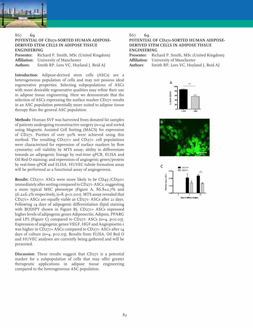
82
867 69POTENTIAL OF CD271-SORTED HUMAN ADIPOSE-DERIVED STEM CELLS IN ADIPOSE TISSUE ENGINEERINGPresenter: Richard P. Smith, MSc (United Kingdom)Affiliation: University of ManchesterAuthors: Smith RP, Lees VC, Hoyland J, Reid AJ
Introduction: Adipose-derived stem cells (ASCs) are a heterogeneous population of cells and may not possess ideal regenerative properties. Selecting subpopulations of ASCs with more desirable regenerative qualities may refine their use in adipose tissue engineering. Here we demonstrate that the selection of ASCs expressing the surface marker CD271 results in an ASC population potentially more suited to adipose tissue therapy than the general ASC population.
Methods: Human SVF was harvested from donated fat samples of patients undergoing reconstructive surgery (n=14) and sorted using Magnetic Assisted Cell Sorting (MACS) for expression of CD271. Purities of over 90% were achieved using this method. The resulting CD271+ and CD271- cell populations were characterised for expression of surface markers by flow cytometry; cell viability by MTS assay; ability to differentiate towards an adipogenic lineage by real-time qPCR, ELISA and Oil Red O staining; and expression of angiogenic genes/protein by real-time qPCR and ELISA. HUVEC tubule formation assay will be performed as a functional assay of angiogenesis.
Results: CD271+ ASCs were more likely to be CD45-/CD90+ immediately after sorting compared to CD271- ASCs, suggesting a more typical MSC phenotype (Figure A, 86.8±2.7% and 56.2±6.2% respectively, n=8, p<0.001). MTS assay revealed that CD271+ ASCs are equally viable as CD271- ASCs after 21 days. Following 14 days of adipogenic differentiation (lipid staining with BODIPY shown in Figure B), CD271+ ASCs expressed higher levels of adipogenic genes Adiponectin, Adipsin, PPARG and LPL (Figure C) compared to CD271- ASCs (n=4, p<0.05). Expression of angiogenic genes VEGF, HGF and Angiopioetin-1 was higher in CD271+ ASCs compared to CD271- ASCs after 14 days of culture (n=4, p<0.05). Results from ELISA, Oil Red O and HUVEC analyses are currently being gathered and will be presented.
Discussion: These results suggest that CD271 is a potential marker for a subpopulation of cells that may offer greater therapeutic applications in adipose tissue engineering compared to the heterogeneous ASC population.
867 69POTENTIAL OF CD271-SORTED HUMAN ADIPOSE-DERIVED STEM CELLS IN ADIPOSE TISSUE ENGINEERINGPresenter: Richard P. Smith, MSc (United Kingdom)Affiliation: University of ManchesterAuthors: Smith RP, Lees VC, Hoyland J, Reid AJ

83
780 70AUTOLOGOUS FAT-DERIVED TISSUE MATRIX: BIOLOGIC CHARACTERISTICS AND RESULTS AFTER IMPLANTATIONPresenter: Stephen A. Schendel, MD DDS (USA)Affiliation: Stanford UniversityAuthor: Schendel SA
Purpose: Autologous collagen is an ideal natural soft tissue filler and may serve as a matrix for stem cell implantation and growth. Procurement of autologous collagen has been limited secondary to a sufficient source. In addition, the tissue processing technology is complex and expensive. Liposuction is a widely performed with an average of 1,500 cc of fat removed in a typical procedure. This fat could be a source of autologous collagen. The amount and type of collagen composition in liposuctioned fat, however, remains unknown. The purpose of this study was to analyze the protein and cellular content of a collagen tissue matrix produced from fat and its subsequent implantation.
Methods and Materials: Fat was obtained from individuals undergoing routine liposuction was processed by a multi-step process to obtain the connective tissue as well as viable cells. The tissue was cryogenically stored after processing. The tissue was then thawed and evaluated by scanning electron microscope, (figure 1) western blot analysis and flow cytometry. In addition, the collagen matrix and a control, Juvederm®, was implanted in 43 nude mice and followed for one year.
Experimental Results: Liposuctioned fat was obtained from 10 individuals with an average volume processed of 298cc per subject. After processing an average of 1cc of collagen matrix was obtained from each 100cc of fat. Significant viable cell markers were present in descending order for dipocytes>CD90+>CD105+>CD45+>CD19+>CD144+>CD34+. Western Blot analysis showed collagen types I, III, IV and other proteins. SEM showed a regular pattern of cross-linked, helical collagen. The implanted collagen matrix compared favorably to Juvederm and was also found to be replaced by new collagen and fat.
Summary: Collagen and cells can be easily and quickly obtained from liposuctioned fat without alteration of the overall cellular composition of the tissue. Implantation results in new collagen and cellular growth.
Conclusion: Collagen matrix with viable cells for autologous use can be obtained from liposuctioned fat that has been cryogenically frozen and may provide long term results.
936 71THE EFFECTS OF COLD STORAGE AND POLOXAMER 188 TREATMENT ON STROMAL VASCULAR FRACTION VIABILITY AND VOLUME RETENTION OF FAT GRAFTSPresenter: Gabriella A. DiBernardo, BS (USA)Affiliation: University of PittsburghAuthors: DiBernardo GA, Bliley JM, Bourne D, Havis E, James IB, Schroth R, Grybowski D, Dees A, Wang S, Kokai L, Kelmendi-Doko A, Mahoney C, Sivak W, Marra K, Rubin JP
Two clinically relevant methods of reducing ischemic injury to fat grafts during the interval between harvest and injection are cold storage and use of a preservative agent. Poloxamer-188 (P188), an FDA approved pharmaceutical that can stabilize damaged cell membranes, prevent apoptosis of damaged cells and improve graft survival and enhance histological appearance of grafted fat. This study aimed to determine whether tissue cooling and/or P188 increases graft retention and viability of injected fat.
Fat harvested from surgical patients by liposuction was processed by filtration, and treated with the following experimental interventions: 1) storage at 4C, 2) storage at 4C and P188 admixed with graft, or 3) room temperature (RT) storage and P188 admixed with graft. Control grafts were stored at RT with no P188 added. To assess cell yield and viability, stromal vascular fraction (SVF) was isolated at 1.5, 3, 4.5, 6, 7.5, and 24 hours. Gene expression of apoptotic (Bax/Bcl2 ratio), angiogenic (VEGF, AGPT1) and senescent factors (p21) were determined in the 24-hour lipoaspirate grafts. Fat grafts were injected into flanks of athymic nude mice and explanted 6 weeks postoperatively to assess volume retention and tissue architecture.
Viability of the SVF decreased in RT and 4C groups at 4.5 hours. Cold storage had a negative effect on the fat. Adding P188 increased cell viability at both temperatures, with greatest viability seen in the RT+P188 group. 4C treated fat had downregulated p21 and PPAR gamma expression at 7.5 and 24 hours compared to RT groups suggesting increased apoptosis or senescence within the graft. Angiogenic factors were downregulated at 24 hours in 4C treatment compared to RT. While no significant difference between groups was observed in volume retention at 6 weeks postoperatively, 4C treatment resulted in greater inflammation and oil cysts on histology, compared with RT and P188 treated fat grafts.
Storing fat grafts at 4C after harvest negatively impacts SVF and adipocyte viability, gene expression, and histological appearance of transplanted grafts. Adding P188 improves these outcomes at both 4C and RT and may represent a clinically relevant strategy.

84
814 72ADIPOSE STEM/STROMAL CELLS FOR TENDON REGENERATION: DEVELOPMENT OF A NEW DIFFERENTIATION PROTOCOL AND COMPARISON WITH BONE MARROW STEM CELLS TENOGENIC ABILITYPresenter: Carlotta Perucca, PharmD (Italy)Affiliation: University of PaviaAuthors: Perucca C, Vigana MV, Sants-Ruiz LS, Colombini AL, Pearson JP, De Girolamo LD
Introduction: Tendinopathy is a common disorder, that can result in long period of inactivity. The healing process of an injured tendon is very slow and is often accompanied by scar tissue formation. The late stages of the pathology can lead to ruptures, with the surgical approach representing the only therapeutic option. In this scenario, the use of Mesenchymal Stem cells (MSCs) for the treatment of tendon disorders could represent a promising tool to improve the clinical outcome. Differently from adipogenic, chondrogenic and osteogenic differentiation, in vitro tenogenic differentiation of MSCs has been not well established yet. Thus, the purpose of this study is to improve the ability of MSCs to differentiate towards the tenogenic lineage and identify the most suitable source of MSCs for this application. The present work focused on the establishment of a well-performing inductive tenogenic treatment, and on the comparison of the tenogenic potential of Bone Marrow derived Stem Cells (BMSCs) and Adipose derived Stem Cells (ASCs).
Methods: In order to identify the most efficient inductive medium, the ability to up-regulate the expression of scleraxis and decorin by different combinations of growth factors and supplements, in both BMSCs and ASCs was tested using an immunofluorescence-based high-throughput system, Operetta® (Perkin Elmer). The gene expression analysis of several tendon specific markers, such as tenomodulin, tenascin and mohawk homeodomain protein, was also performed.
Results: Our results showed that BMP-12 is the main growth factor able to induce the production of tendon specific markers, even if elements such as bFGF, ascorbic acid, TGF?, CTGF, IGF-I significantly contributed to the differentiation towards the tenogenic lineage. Moreover, ASCs showed to be good responders to tenogenic induction, expressing higher levels of SCX with respect to BMSCs. Those results were confirmed by the gene expression analysis of a larger panel of tendon specific markers.
Conclusion: This study provides a large amount of data about the effect of different molecules in the tenogenic induction of MSCs, and in particular it supports the adoption of ASCs as the preferred cell source for regenerative medicine approach for the treatment of tendon disorders.
933 73DIFFERENTIATION OF HUMAN ADIPOSE-DERIVED STEM CELLS (ASC) TO ENDOTHELIUM FOR IMPROVEMENT OF FAT TRANSPLANTATIONPresenter: William M. Harris, MD (USA)Affiliation: Cooper Univ HospitalAuthors: Harris WM, Zhang PZ, Plastini MP, Kappy NK, Ortiz TO, Chang SC, Brown SB, Carpenter JC
Background: Adipose-derived stem cells (ASCs) are considered to facilitate the survival of fresh fat grafts, however, optimal vascularization of the graft site is significant for improving the outcome of fat grafting. This study was designed to test the hypothesis that ASCs that differentiate to a functional endothelial cell (EC) phenotype may improve the retention and angiogenesis of fat grafts using an autologous fat transplant rat model.
Methods and Results: Human ASCs were isolated from periumbilical fat tissue and cultured in Endothelial Growth Medium (EGM2) supplemented with FBS (2%) for 2wk. Human fat tissue (1.2g) was mixed with fresh stromal vascular fraction (SVF; 1.5x10⁵), EC differentiated ASCs (EC/ASCs; 1.5x10⁵) and no cells (control). These three fat mixtures were subcutaneously injected into the adult male Sprague Dawley rat dorsum at 3 locations (n=8). After 8 weeks, the grafted fat tissues were harvested and the extracted fat was evaluated using photographic analysis, volume measurements, and histological examination. Endothelial differentiated ASCs enhanced fat graft survival rates when compared to fat with SVF transplants and controls (56.8 ± 10.6% vs. 41.0 ± 10.1%, 41.2 ± 11%, p<.05). The transplants mixed with EC/ASCs exhibited the best survival, morphologic integrity, and the most uniform lipid droplets. They also revealed significantly less inflammation and fibrosis with richer blood vessels by histological analysis. Most of the CD31- positive endothelial cells were seen in the fat with EC/ASC transplant tissue. Immunofluorescence confirmed the presence of cells positive for both CD31 and human nuclear which indicates that the endothelial cells in the neovascular capillaries differentiated from the transplanted human ASCs. Finally, quantitative PCR demonstrated that the mRNA expression of EC-specific markers of CD31 (2-fold) and vWF (3-fold) were higher in the fat with EC/ASC transplants than in the fat with SVF transplants (p<.05).
Conclusions: These results indicate 1) differentiation of ASCs into an EC phenotype enhanced fat graft survival rates and improved the angiogenesis of grafted fat tissues, 2) co-implantation of EC differentiated ASCs with adipose tissues is a promising strategy in autologous fat transplantation.

85
944 74SPATIAL CONTROL OF ADIPOSE DERIVED STEM CELL DIFFERENTIATION IN A CELL SHEET USING PHOTOCLEAVABLE NANOPARTICLESPresenter: Lisa M. Kriegh, BS (USA)Affiliation: Louisiana State UniversityAuthors: Kriegh LM, Forghani A, Chen C, Hayes DJ, Devireddy R
WITHDRAWN
943 75A NOVEL BIOCOMPATIBLE MICROCARRIER WITH TUBULAR CONDUITS SUPPORTS HSVF SURVIVAL, MATRIX SECRETION, AND CORD STRUCTURE FORMATIONPresenter: J. Christian Brown, MD (USA)Affiliation: University of FloridaAuthors: Brown JC, Willenberg BW, Shang HS, Yang NY, Katz AK
Introduction: Diffusion supports a critical tissue volume of 2-3 mm3 in normal physiological settings1, and therefore, angiogenesis resulting in a neocapillary bed and inosculation with host vessels is pivotal in maintaining engineered tissue macro-constructs, especially when these constructs are created with the canonical, “bottom-up” approach.2 In this study, we aim to characterize hSVF seeding and supportive capabilities of a novel microcarrier CapgelTM an oligomeric gelatin and copper-capillary alginate micro-scaffold - possessing regular, tubular microstructures reminiscent of capillaries.
Methods: Cell Seeding: hSVF cells or hASCs or GFP-mASCs were statically and dynamically seeded onto gelatin and alginate-oligochitosan microcarriers, and cultured in DMEM+10%FBS:1%ABAM at 37C/5% CO2. The cells were harvested at 4-hour intervals for 24 hours. Eluate was mixed with trypan blue, counted using a TC10 automated cell counter, and confirmed with an hemocytometer. Seeded microcarriers were DAPI stained and fluorescently imaged. All statistical analyses were completed using Excel® and Prism®.
Static Culture: 1x10⁶ hSVF cells were statically cultured with 0.1g CultiSpher® and 0.3g Capgel, in the aforementioned medium. The constructs were harvested at 2/4/6/8 weeks, and examined with cryostat and paraffin-embedded sectioning at 10 m width. H&E staining and CD31 immunohistochemistry were performed.
Results: CapgelTM and Cultispher® microcarriers support mASC, hASC, and hSVF cell attachment. hSVF cells attached to CapgelTM and Cultispher® microcarriers at a maximum efficiency achieved at 4 and 8 hours, respectively. Static cultures demonstrated hSVF cellular survival and extracellular matrix secretion; probing for CD31-antigen revealed cord structures. Packed-bed bioreactor studies utilizing hSVF-seeded microcarriers are currently ongoing.
Conclusions: CapgelTM microcarriers reach a significantly higher maximal seeding efficiency faster than commercially-available CultiSpher® microcarriers (p = 0.0042). Seeded microcarriers can be maintained for a minimum of 8 weeks in static conditions, as demonstrated by cellular survival, matrix secretion, and CD31 cord formation supporting the concept of modular, “bottom-up” tissue engineering. Bioreactor studies are pending.

86
840 76USING HUMAN RECONSTRUCTED OSSEOUS TISSUES DERIVED FROM ADIPOSE STEM/STROMAL CELLS AS A PLATFORM FOR STUDYING THE IMPACT OF MELATONIN ON OSTEOGENESIS UNDER PHYSIOLOGICAL AND INFLAMMATORY CONDITIONSPresenter: William P. Clafshenkel, PhD (Canada)Affiliation: LOEX CRCHUQ University LavalAuthors: Clafshenkel WP, Galbraith T, Kawecki F, Eliopoulos N, Auger FA, Fradette J
Introduction: TNFα, IL-1, and IL-6 are critical mediators of bone resorption, suggesting inflammation is a risk factor for bone loss. TNFα promotes osteoclastogenesis and osteoblast apoptosis. IL-1β increases ROS production in osteoblasts and activates MMPs, promoting bone destruction. Our group has utilized adipose-derived stem/stromal cells (hASCs) to develop multilayered human reconstructed osseous tissues. We contend that these tissues could serve as microphysiological environments for the evaluation of osteogenic and/or immunomodulatory agents by replicating the functions of osseous tissues under normal and inflammatory conditions. Melatonin has been shown to exert anti-inflammatory effects and is a potent free-radical scavenger. The impact of melatonin on hASC osteogenic differentiation is understudied and the role melatonin plays in curtailing the detrimental effects of inflammation on osteogenically-induced hASCs is unknown. We hypothesize that melatonin administration will improve osteogenesis in hASC-derived osseous cell sheets or preserve hASC function under inflammatory conditions.
Methods: Using a self-assembly technique, hASC-derived cell sheets were produced under osteogenic conditions in the presence or absence of melatonin. 1 ng/mL TNFα+0.1 ng/mL IL-1β was used to simulate inflammatory conditions. Osteogenesis was evaluated by alkaline phosphatase (ALP) activity and calcium deposition. Supernatants collected under inflammatory conditions were analyzed for key pro-inflammatory molecules.
Results: Osteogenically-induced melatonin-treated tissues demonstrated enhanced ALP activity (2.3-fold) and mineralization (1.8-fold) after 21 days of differentiation. Moreover, melatonin treatment may prime hASC responsiveness via MT2 receptor signaling pathways during the early stages of differentiation. Melatonin treatment mitigated MCP-1 and IL-6 production in osseous cell sheets stimulated with the inflammatory cocktail.
Conclusions: By replicating native osseous tissues using self-assembly methods, we can study the impact of melatonin of melatonin on biological responses like osteogenesis and inflammation-driven bone remodeling. As a multi-functional molecule, melatonin may respond to microenvironmental changes to either promote or preserve osteogenesis.
805 77PFAT GRAFTING FOR AUTOLOGOUS GLUTEAL AUGMENTATION: A META-ANALYSISPresenter: Alexandra Conde-Green, MD (USA)Affiliation: Rutgers New Jersey Medical School - Graduate School of Biomedical SciencesAuthors: Conde-Green A, Kotamarti VS, Nini KT, Wey PD, Ahuja NK, Granick MS, Lee ES
Introduction: Throughout the years, many plastic surgeons have published their techniques for achieving a larger gluteal contour. Stil there’s no consensus on the best and safest way to perform fat grafting to the gluteal region. Due to the recent reported fatalities related to fat grafting to the gluteal region, we reviewed the techniques described in the literature in order to analyze and compare the different steps of the procedure, and identify those that could potentially be of concern.
Methods: We performed a systematic review of the literature with a search of 21 terms related to gluteal fat augmentation in 3 databases. Nineteen articles meeting our predetermined criteria were analyzed allowing evaluation and comparison of techniques. Independent-samples t-test and one-way ANOVA were used for statistical analysis.
Results: Seventeen case series and two retrospective studies were selected, mostly from Mexico, Columbia and Brazil. A total of 4,105 patients, composed of 98.2% women and 1.8% men with a mean age of 33.6 years and mean BMI of 24.3 were reported. Most patients received general anesthesia. The thighs and trochanteric regions were the most common donor sites. Harvesting was most often performed with vacuum and syringe-assisted liposuction, and processing was most commonly decantation or centrifugation. A mean of 400 ml of lipoaspirate was injected per gluteal region, in intramuscular and subcutaneous planes with 60 ml syringes. Results were evaluated mainly with pre and postop photographs. Most patients rated their results as excellent. The mean complication rate was 7%, consisting mainly of seroma (2.4%), erythema (1.3%) with no significant relation to the planes of injection. Note that one study, which reported 13 deaths, was not included in our data to reduce selection bias.
Conclusion: Fat grafting is an effective and predictable way to remodel the gluteal region, however the procedure is not without risks. Avoiding gluteal vessel damage may prevent most feared complications, such as fat embolism. Accurate analysis, systematization of the procedure and reporting cases in the fat grafting registry may provide the foundation for optimization of outcomes.

87
913 78PSAFETY OF FAT GRAFTING IN PLEGIC PATIENTSPresenter: Reto Wettstein, MD (Switzerland)Affiliation: University Hospital BaselAuthor: Wettstein R
WITHDRAWN
815 79PAUTOLOGOUS FAT GRAFTING FOR IPG SITE COMPLICATIONS FOLLOWING SPINAL CORD STIMULATORPresenter: Suresh M. Anandan, MBBS, MS, MCH, MRCS, FEBOPRAS (United Kingdom)Affiliation: Wexham Park HospitalAuthors: Anandan SM, Pai AA, Desai P, Misra A
Introduction: Spinal cord stimulation is an established treatment for chronic pain syndromes. Patients can benefit from a 50% pain reduction over prolonged periods, however it carries an overall complication rate of 35%. In this, the development of pain at the Implantable Pulse Generator (IPG) site can affect 4-12% of patients. Managing IPG site pain is challenging. Topical lignocaine patches work to some extent, but eventual IPG re-siting become necessary, with more chronic or IPG site pain issues or both, likely to return. We describe the successful use of autologous fat transfer to manage the pain at the IPG site.
Method: Three patients with IPG site pain were treated under general anesthetic, by a single surgeon. Autologous fat was harvested by liposuction and prepared for fat transfer using the Coleman technique. All patients required at least 2 sessions of 50 ml fat transfer in 4 to 6 months interval.
Results: All patients described improvement in their pain scores at 6 months. The implant edge was no longer palpable in one case.
Conclusions: Fat transfer is an effective modality to treat IPG site pain. The procedure is simple in comparison to IPG resiting, and does not require a cessation of cord stimulation therapy, when it is done. An additional benefit of tissue re-contouring at implant-skin adherence plane reduces the risk of explantation. As a small case series these results are encouraging. We intend to conduct a larger series with greater follow up to test the long-term efficacy of fat transfer, in this challenging situation.

88
863 80PCONTOUR PLASTIC OF THE FACE USING AUTOFAT WRAPPED IN AUTOPLASMA GELPresenter: Ivan V. Krainik, MD (Russia)Affiliation: Medical Sugical Centre by N I PirogovAuthor: Krainik IV
Lypofilling as a method of contour plastic of the face is not wide spread due to 30-50% resorption of the injected fat. Aspirated fat tissue consists of certain fat cells and fragments and has acidity within 5,5-5,8 pH. Using of sodium hydrocarbonate with aspirated fat leads to alkalization. Destroyed fat cells, acid or alkaline medium are course of phagocytosis induction and resorption of the injected fat. We offer a method of fat cells and grafts wrapping in own plasma gel. In this case every fat cell or graft is enveloped in its own plasma protein and doesn’t undergo leukocyte aggression and autolysis. Acidity of the fat mass wrapped in protein normalizes within 6,2-6,8.
Method of wrapping fat cells in protein: Blood is sampled in sterile tubes with solution of sodium citrate and centrifuged (2000 revolutions per minute). After centrifugation erythrocytic stratum is removed and plasma with platelets is placed in separate sterile glass. Aspirated fat mass is cleaned from the blood and placed in this glass too. 10% solution of calcium chloride is added to the plasma and shook up carefully during 5 minutes. The blend of plasma with fat cells is polymerized and turned into gel-type mass, inside of which are enveloped fat cells and grafts. After polymerization wrapped in protein fat mass has maize yellow color. We usually inject this mass by a syringe through 2mm needle and use it in the next cases: liposculpture of nasolabial sulcus, lips and cheeks, correction of retracted scars and defects of soft tissues. In our practice the volume of single injected fat mass was 2-70ml. Fat resorption in postoperative period doesn’t overcome 15%.
Comparative experimental studies on rabbits showed that the transplanted fat is resorbed completely in two weeks while the fat wrapped in plasmagel remains viable for two months. Correction of age-related changes of the face and posttraumatic soft tissue deformations, symptomatic treatment of lipodystrophy with using of this method leads to perfect results.
802 81PTHREE DIMENSIONAL BIOPRINTING: THE FUTURE OF TISSUE ENGINEERING AND PLASTIC SURGERY. A SYSTEMATIC REVIEW OF THE LITERATUREPresenter: Vasanth Kotamarti, MD (USA)Affiliation: Rutgers New Jersey Medical SchoolAuthors: Kotamarti V, Conde-Green A, Ayyala H, Guiro K, Lee ES, Granick MS, Rameshwar P
Background: Plastic surgeons often treat complex wounds and defects involving multiple tissue types for which current approaches may fail to meet surgeon and patient expectations. Strategies that limit procedure complexity, improve both customizability and outcomes, and promote regenerative healing are needed. Innovations in three-dimensional printing have allowed ‘bioprinting’ of biocompatible materials and cells enabling the creation of complex, living tissues. We aimed to compare the available biomaterials and printing mechanisms to create tissues relevant in plastic and reconstructive surgery.
Methods: We performed a systematic review of the literature by searching the PubMed and Medline databases with four terms. Five hundred thirteen articles were evaluated using predetermined inclusion and exclusion criteria.
Results: Fifty-eight articles were analyzed. The majority of studies created bone and cartilage models with others printing skin, adipose tissue, nerves, and blood vessels. Chondrocytes, mesenchymal stromal cells and adipose-derived stromal cells most commonly were deposited by extrusion, inkjet, or laser-assisted printers in alginate hydrogels. Printed cells remained viable and capable of differentiation. With increasing construct size, incorporation of hollow channels is necessary to maintain flow of oxygen and nutrients to cells deep inside the different structures. Also, when placed in vivo, printed constructs integrated well into the surrounding tissue.
Conclusions: Three-dimensional bioprinting is an infant technology with promising applications in regenerative healing. Multiple cell types can be printed with defined locations in a structure to customize the composition of artificial tissue replacements. Alginate hydrogels, while allowing uniform encapsulation of cells and smooth, predictable flow during printing, can be cross-linked after printing to maintain structural integrity. Like any tissue graft, constructs initially depend on diffusion of oxygen and nutrients, requiring incorporation of vessel-like channels. Further research is needed to better understand what hydrogel compositions, cell types, and culture conditions are needed to optimize the creation of different tissue types.
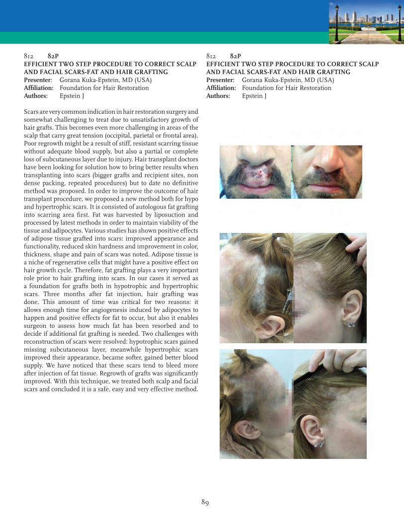
89
812 82PEFFICIENT TWO STEP PROCEDURE TO CORRECT SCALP AND FACIAL SCARS-FAT AND HAIR GRAFTINGPresenter: Gorana Kuka-Epstein, MD (USA)Affiliation: Foundation for Hair RestorationAuthors: Epstein J
Scars are very common indication in hair restoration surgery and somewhat challenging to treat due to unsatisfactory growth of hair grafts. This becomes even more challenging in areas of the scalp that carry great tension (occipital, parietal or frontal area). Poor regrowth might be a result of stiff, resistant scarring tissue without adequate blood supply, but also a partial or complete loss of subcutaneous layer due to injury. Hair transplant doctors have been looking for solution how to bring better results when transplanting into scars (bigger grafts and recipient sites, non dense packing, repeated procedures) but to date no definitive method was proposed. In order to improve the outcome of hair transplant procedure, we proposed a new method both for hypo and hypertrophic scars. It is consisted of autologous fat grafting into scarring area first. Fat was harvested by liposuction and processed by latest methods in order to maintain viability of the tissue and adipocytes. Various studies has shown positive effects of adipose tissue grafted into scars: improved appearance and functionality, reduced skin hardness and improvement in color, thickness, shape and pain of scars was noted. Adipose tissue is a niche of regenerative cells that might have a positive effect on hair growth cycle. Therefore, fat grafting plays a very important role prior to hair grafting into scars. In our cases it served as a foundation for grafts both in hypotrophic and hypertrophic scars. Three months after fat injection, hair grafting was done. This amount of time was critical for two reasons: it allows enough time for angiogenesis induced by adipocytes to happen and positive effects for fat to occur, but also it enables surgeon to assess how much fat has been resorbed and to decide if additional fat grafting is needed. Two challenges with reconstruction of scars were resolved: hypotrophic scars gained missing subcutaneous layer, meanwhile hypertrophic scars improved their appearance, became softer, gained better blood supply. We have noticed that these scars tend to bleed more after injection of fat tissue. Regrowth of grafts was significantly improved. With this technique, we treated both scalp and facial scars and concluded it is a safe, easy and very effective method.
812 82PEFFICIENT TWO STEP PROCEDURE TO CORRECT SCALP AND FACIAL SCARS-FAT AND HAIR GRAFTINGPresenter: Gorana Kuka-Epstein, MD (USA)Affiliation: Foundation for Hair RestorationAuthors: Epstein J

90
951 83PNEW ALGORITHM AND AESTHETIC APPROACH FOR BREAST MULTILAYER FAT GRAFTING: PRELIMINARY REPORTPresenter: Alfredo E. Hoyos, MD (Colombia)Affiliation: ElysiumAuthors: Hoyos AE, Guarin DE
Introduction: The desired breast shape is not always achieved by using silicone implants and in some cases the patients even refuse to have them. Instead of using local or distal flaps, the fat graft is a reasonable alternative. The needed volume is clearly different on each patient and requires a detailed examination and analysis. By using a multilayered approach, the graft is placed in the muscle and in the gland surround tissue. By these means the gland is protected and the graft is placed on a safe vascular layer.
Methods: Retrospective analysis from 2007 to 2015 patients. Presurgical markings were made indicating the breast limits and the areas to be grafted. Graft was harvested via ultrasonic assisted lipoaspiration. Only decantation was performed. Grafting was done by using a multi tunnel layered technique with a blunt curve 3mm cannula al 30° trough an anterior axillary incision starting in the deep muscular and 1,5mm cannula for the subglandular and subdermal layer until the desired shape was achieved. Pre and post surgical photograph and and a 3d scanning (crisalix’s.a. virtual aesthetics) were used.
Results: 36 patients were included; age was 32 years (21-47) 340ml (300-450ml) of fat was injected in each breast. In all the cases the desired pectoral shape and volume volume was achieved. The patients were followed up 14 months (10-27). No resorption was found during the study. No complications were found.
Conclusions: Multilayer fat grafting approach is a safe and reproducible way to achieve the breast sculpting. Further research and a longer follow up is needed to reassure the fat graft long term survival and the normal breast function.
903 84PVALIDATION OF THE IMMUNODEFICIENT MOUSE ANIMAL MODEL FOR ASSESSING FAT GRAFTING OUTCOMESPresenter: Lauren E. Kokai, PhD (USA)Affiliation: University of PittsburghAuthors: Kokai LE, Jones TL, Marra KG, Rubin JP
Introduction: Fat grafting has enormous potential to transform cosmetic and reconstructive surgery, however, unpredictability of graft retention is a major problem and increased utility of the procedure will be gained through experiments that define best practices. The immunodeficient mouse model has been published in over 50 original articles as a method for evaluating fat grafting variables. Typical end-study assessment includes residual graft weight or volume, with the unverified assumption that increased graft retention reflects superior fat grafting outcomes. In this study, we validate two important study parameters, injection volume and study length, for assessing fat grafting retention kinetics in the immunodeficient mouse.
Methods: Human lipoaspirate tissue was collected from 10 subjects, 4 male and 6 female, as medical waste following elective surgeries. To determine if volume retention of human fat grafted in the nude mouse reflects tissue quality, retention kinetics of viable lipoaspirate was compared with non-viable tissue for up to 18 weeks in vivo, with n=5 per time point. Injection volumes included 0.3mL and 1.0mL, with two injections per animal on the dorsal flank. Masson’s trichrome was used to visualize tissue morphology and adipocyte viability and presence of macrophages were assessed with immunohistochemistry using antibodies against perilipin and F4/80.
Results: At 3 and 6 weeks, non-viable 0.3mL grafts had several large oil cysts which were reduced in diameter by 12 weeks and were reabsorbed by 18 weeks. In contrast, oil cysts in 1.0mL grafts were not reabsorbed by 18 weeks and continued to comprise a significant portion of the graft volume. While viable 0.3mL fat grafts showed significantly higher retention than non-viable grafts at every study time point, no significant differences in volume retention were detected between viable and non-viable samples for 1.0mL samples at any time point.
Conclusion: Our results suggest that human fat graft retention in the immunodeficient mouse correlates with graft viability when small injection volumes are used (0.3mLs), however larger grafts may be significantly comprised of oil cysts or nonviable tissue and thus retention measurements are more difficult to interpret.

91
774 85PDONOR AGE DEPENDENT FEATURES OF PEDIATRIC VERSUS ADULT ADIPOSE MESENCHYMAL STROMAL CELLS (ASC)Presenter: Valerie Planat-Benard, PhD (France)Affiliation: STROMALabAuthors: Planat-Benard V, Abbo O, Tarand M, Monsarrat P, Raymond I, Galinier P, Casteilla L
Introduction: Adipose derived mesenchymal stromal cells (ASC) are currently tested in regenerative medicine to promote tissue reconstruction after injury. In autologous purpose the possible loss of therapeutic function and cell properties during aging have been questioned in adult. To date no reliable information is available concerning ASC from pediatric patients and a better knowledge is required to intend their use to clinical applications.
Materiel & Methods: Subcutaneous adipose tissue was collected from 27 donors (0-1 year old) and 50 donors (1-12 years old) and compared to adult ASC for in vitro characteristics. ASC were then tested in a mouse model of limb ischemia.
Results: Cells from the stromal vascular fraction (SVF) and subsequent cultured ASC were prepared. Only a higher amount in SVF cell number and ASC proliferative rate were found. Cell phenotype, CFU-F content, immunomodulation effect, adipogenic, osteoblastic and angiogenic potentials were not significantly different. In vivo, pediatric ASC induced an increase in microangiographic score in a mouse model of limb ischemia, even though improvement in vascular density was not significantly correlated to limb rescue. Finally mRNA analysis using microarray approach identified that only 305 genes were differentially expressed (217 down- and 88 up-regulated) in pediatric versus adult ASC, confirming that ASC from both groups of age shared very close intrinsic properties.
Conclusion: This is the first study reporting a comparative analysis of ASC from large number of donors and showing that their in vitro and in vivo properties were similar and maintained during aging.
904 86PPORCINE ADIPOSE TISSUE HARVEST BY LIPOASPIRATION AND SVF ISOLATION USING A ‘POINT-OF-CARE’ DEVICEPresenter: Ning Yang, PhD (USA)Affiliation: University of FloridaAuthors: Yang N, Shang H, Brown J, Katz A
Introduction: Stromal vascular fraction (SVF) cell therapy holds promise for various wound healing challenges. For pre-clinical testing and evaluation, many groups have reported on the use of SVF within rodent models of healing. Although cheaper and more feasible in many ways, porcine models of wound healing are considered the gold standard for the human condition. Porcine skin shares many functional, anatomical, and biochemical similarities with human skin. However, reports of SVF isolation procedures for porcine adipose tissue are scant, and most use excised adipose tissue which can be time-consuming and provide low cell yield. We explored the isolation of SVF cells from lipoaspirated adipose tissue from porcine tissues using a disposable, closed system point-of care device, so as to mimic proposed human studies as accurately as possible.
Methods: Animal protocol was approved by IACUC. Adipose tissues were liposuctioned using 3-hole ‘Mercedes’ cannula from farm-grade Yorkshire pigs at average weight 122 lbs (n=3). Cell isolation was performed using an SVF device (The GID Group, Inc.) following the manufacture’s instructions with a few modifications.
Results: The adipose tissue obtained by lipoaspiration ranged from 19 grams to 32 grams for each animal (average 25 grams per animal). The average cell yield was 1 million nucleated viable cells per gram fat. Average cell viability was 77.3%. Cells were attached to the plastic surface of the culture dish in DMEM/F12 medium supplemented with 10% fetal bovine serum and proliferated with average 47 hours of doubling from passage 1 to passage 2.
Conclusions: We show that the harvest of porcine adipose tissue by lipoaspiration and the subsequent isolation of SVF cells using a closed system device under development for human point-of-care therapies provide a new approach to rapidly yield abundant SVF cells. This will enable the evaluation and modeling of autologous point-of-care SVF wound therapies in porcine models, which are regarded as the gold standard for pre-clinical human wound healing objectives.

92
813 87PCHARACTERIZATION OF RAT ADIPOSE-DERIVED STEM CELLS AND THEIR INDUCTION TOWARD A TENOGENIC LINEAGE FOR REGENERATION OF ACHILLES TENDONPresenter: Jolanta B. Norelli, BA (USA)Affiliation: Northwell Health SystemAuthors: Norelli JB, Plaza DP, Liang H, Grande DA
Introduction: Recently, adipose-derived stem cells (ADSCs) have entered regenerative medicine and tissue engineering. Studies show promise in differentiating ADSCs toward mesodermal cell lineages to create bioactive tissue replacements. Musculoskeletal repair with ADSC adjuncts has shown improvement in tendon histology and biomechanics. No research to date has fully characterized ADSCs, the mechanism of tendon differentiation, or the effect of ADSCs on Achilles tendon repair in vivo. Our purpose was to characterize rat ADSCs, induce ADSC tenogenesis, and analyze ADSC influence on tendon repair. We hypothesize that differentiated ADSCs will yield superior tendon repair. Our mechanistic insights would expand treatment for acute and chronic tendon injuries, microscopic insults, and complete tendon tears.
Methods: ADSCs were harvested from Sprague-Dawley rats, isolated, grown in vitro, and characterized as stem cells with the following criteria: adherence to plastic confirmed by cell culture, spindle-shaped morphology confirmed by light microscopy, specific cell surface antigen expression confirmed by flow cytometry, multilineage differentiation potential in culture confirmed by specific gene expression and phenotype analysis. ADSC tenogenesis was induced with known tenogenic growth factors. We elucidated the optimal “growth factor cocktail” by analysis of cell morphology, immunostaining, and tenocytic gene expression. To test viability of treatment with ADSCs, surgically-induced tendon defects were injected with hydrogel solutions of ADSCs. We will use GFP tagging to track the fate of cells. Healing will be compared with unrepaired normal control to identify natural healing baseline via the rat Achilles tendon injury model and characterized by histology, biomechanics, and qPCR.
Results: We characterized rat ADSCs and achieved successful differentiation into multiple mesodermal lineages, including bone, fat, cartilage, and tendon, as evidenced by gene expression, histology, and immunostaining specific of each lineage.
Conclusions: Rat ADSCs were successfully characterized and differentiated into tendon-like cells. We plan to analyze tissue regeneration in surgically-created tendon defects injected with undifferentiated ADSC or with tenogenically differentiated ADSCs.
813 87PCHARACTERIZATION OF RAT ADIPOSE-DERIVED STEM CELLS AND THEIR INDUCTION TOWARD A TENOGENIC LINEAGE FOR REGENERATION OF ACHILLES TENDONPresenter: Jolanta B. Norelli, BA (USA)Affiliation: Northwell Health SystemAuthors: Norelli JB, Plaza DP, Liang H, Grande DA

93
880 88PCHARACTERISATION OF ADIPOSE STEM CELLS ISOLATED AFTER MANUAL OR WATER JET-ASSISTED LIPOSUCTIONPresenter: Rojda Gumuscu, MD (Sweden)Affiliation: Umea UniversityAuthors: Gumuscu R, Brohlin M, Wiberg M, Kingham PJ
Introduction: Adipose tissue can be harvested by various methods and the use of water jet-assisted liposuction has gained favour due its positive outcomes for fat grafting. The objective of this study was to compare stem cells obtained from fat isolated with manual or the water jet-assisted procedure.
Methods: Six women were included in this study and liposuction of subcutaneous abdominal fat was performed using the two methods on each patient. On one side, manual liposuction was performed using a 4mm cannula with a blunt tip and 50 mL Luer Lock syringe with vacuum (MER426L; Byron, MENTOR®, Santa Barbara, California). The Body-jet® (BJ) water assisted system (Human Med AG, Schwerin, Germany) was used on other side with 4mm cannulas and suction vacuum set to 0.5 bar. Minimal volumes of standard tumescent solution were infiltrated in a pulsatile fashion to the treated area. Thereafter continuous rinse with tumescent solution was used throughout the whole liposuction procedure and the fat aspirated into a lipocollector. Aspirate samples were collagenase digested followed by centrifugation to pellet the SVF which was then seeded in vitro. Proliferation and osteo-and adipo-genic differentiation assays, together with immunocytochemistry and qRT-PCR were used to characterize the plastic adherent cells.
Results: Both cell preparations proliferated at similar rates in early passage (population doubling time 57 ± 2 h for manual and 53 ± 3 h for BJ fat). The adherent cells were CD73, CD90 and CD105 positive and approximately 8% and 10% of the cells expressed CD31 and CD34 respectively (with no significant difference between preparation type). Oil Red O staining showed robust adipogenic differentiation in cultures expanded from both manual and BJ aspirated fat. Gene analysis at 2 weeks showed higher expression of the adipocyte markers GLUT4 and aP2 in the differentiating BJ preparations. All patient samples from the manual liposuction generated Alizarin red-positive calcium deposits under 4-week osteogenic differentiation conditions. In contrast, only 3 of the 6 BJ preparations mineralized.
Conclusions: Our data suggest that the procedure for fat liposuction might impact the characteristics of adipose stem cells necessary for different types of therapeutic application.
905 89PCOLLAGENASE-FREE ADIPOSE-DERIVED STEM CELL ISOLATION: NOVEL PROTOCOLS FOR TRANSLATIONAL APPLICATIONSPresenter: Robert Gersch, PhD (USA)Affiliation: UPENNAuthors: Gersch R, Flemming J, Percec I
Introduction: Adipose-derived stem cells (ASCs) are a powerful tool in plastic surgery and regenerative medicine research. The standard protocol for isolating ASCs involves collagenase digestion and red blood cell lysis. However, these enzymes and buffers may negatively impact ASC growth and differentiation capacity and are subject to increasingly severe FDA restrictions. We propose that collagenase-free methods can produce viable and robust ASCs and should replace enzyme-based approaches in both research and clinical applications. To determine the most robust enzyme-free protocols, we tested three novel collagenase-free ASC isolation methods from whole adipose tissue and three from lipoaspirate and compared them to standard collagenase protocols.
Methods: ASCs from five patients were isolated in triplicate via each of the eight methods, expanded, counted, and tested with proliferation assays and for stemness factor expression. ASCs from the collagenase-free groups were compared to ASCs isolated via standard collagenase protocols. Cells were analyzed throughout a 35 day time course and tested with proliferation assays on days 20 and 30. Stemness factor expression was examined via qRT-PCR for Nanog, Oct4, and Sox2 and compared to dermal fibroblasts.
Results: We demonstrate here that all isolation techniques tested successfully produce viable ASCs. While collagenase-based protocols yield significantly more cells at days 20 and 30, as compared to non-enzymatic protocols, collagenase-based protocols also demonstrated the highest variability among all protocols. ASC proliferation rates were similar between collagenase and non-collagenase isolated cells. Stemness factor expression was positive in ASCs isolated via both collagenase and non-collagenase protocols.
Conclusion: We conclude that human ASCs may be successfully isolated in a robust fashion using collagenase-free protocols from both en bloc adipose tissue and lipoaspirate. Our data suggest that collagenase-free human ASC isolation protocols, such as the ones presented here, should be employed to minimize the deleterious effects of enzymatic digestion for future ASC-based research and clinical applications. Future technical and molecular studies will be conducted for additional refinement of these

94
915 90PAN IN VITRO FUNCTIONAL ASSAY OF VASCULOGENESIS AND ANGIOGENESIS USING FRESHLY ISOLATED ADIPOSE STROMAL VASCULAR FRACTION CELLSPresenter: Joseph S. Zakhari, MA (USA)Affiliation: University of Louisville School of MedicineAuthors: Zakhari JS, Zabonick JA, Gettler BC, Tweed B; Apakalai B, Williams SK
Introduction: Adipose-derived stromal vascular fraction (SVF) is a heterogeneous cell source that contains endothelial cells, pericytes, smooth muscle cells, stem cells and other accessory immune and stromal cells. The SVF cell population has been shown to support vasculogenesis in vitro as well vascular maturation in vivo. Matrigel, an extracellular matrix (ECM) mixture has been utilized in vitro to evaluate tube formation of purified endothelial cell systems. We have developed an in vitro system that utilizes freshly isolated SVF and ECM molecules both in pure form (Fibrin, Laminin, Collagen) as well as premixed form (Matrigel) to evaluate vasculogenesis and angiogenesis including endothelial tip cell formation, endothelial stalk elongation, and early stages of inosculation and tubulogenesis.
Methods: SVF was isolated from human lipoaspirate as well as rat epididymal fat pads and seeded at 1.6x10⁵ cells/ well in 12 well plates coated with 3mg/mL collagen I, 5mg/mL fibrin, 2 g lamin 1, 2 g laminin 5, 10 g collagen IV, 1% gelatin, and matrigel (growth factor reduced or non-growth factor reduced) respectively. Cells were grown over a period of 6 days in a Cytation-5 tissue culture system with 4X phase images captured in 15 minute intervals. Images were assembled into stacks and movies using ImageJ software. The formation and persistence of cell clusters containing tip cells, stalk cells and branching were quantified over 6 days.
Results: Freshly isolated SVF from both rat and human demonstrate cell aggregation and clustering (presumptive vasculogenesis) on matrigel ECM within the first 36 hours of seeding followed by tip cell formation, stalk cell formation, branching and inosculation (presumptive angiogenesis) during the subsequent 4 days of culture. Purified ECM molecules (laminin, fibrin and collagen) promote cell proliferation but do not recapitulate vasculogenic and angiogenic events as seen on matrigel.
Conclusions: We have created an in vitro system that provides a functional assay to study the mechanisms of vasculogenesis and angiogenesis in freshly isolated SVF as a way to characterize SVF’s blood vessel forming potential prior to clinical implantation.
926 91PAUTOLOGOUS GRANULAR FAT GRAFTING IN FACIAL REJUVENATIONPresenter: Biao Wang, PhD (China)Affiliation: The First Affiliated Hospital of Fujian Medical UniversityAuthors: Wang B, Zheng H, Su C, Shan X, Chen R
Objective: Long term follow up and evaluate the effect of autologous fat transplantation in the Facial Rejuvenation.
Methods: The preferred inner thigh fat particles for extraction zone, followed by the lower abdomen, buttocks, upper arm and so on. Local tumescent anesthesia was performed. After 10-20min, Connecting the 20ml syringe with 2.0mm diameter needle liposuction. The harvest of adipose tissue via the multiple tunnels, multi-level pumping in Subcutaneous fat layer. Droping the fat suspension into the filtering net, after the liquid is encapsulated into the 1mL. The accepted area is for local anesthesia, and the guiding needle poked a puncture hole with 16 sharp needles, according to the injection site choosing different diameter of fat transplantation. Liposuction needle under the skin down to the most distal sag, inject fat to accepted area from far to near according to principle of multiple points, micro, multi layer and multi tunnel. The total injection volume can be more than 10% of the required filling amount or the affected area can be filled with full. The fat can be gently massage to make the fat particles evenly distributed.
Results: Between January 2013 to February 2016. Application of transplantation of autologous fat granules with facial rejuvenation in 279 cases, which also fill the frontotemporal (36 cases), cheeks (33 cases), eyes (15 cases), nose (7 cases), nasolabial fold (17 cases), chin (23 cases) or filling three or more parts in 148 cases. Followed up for 3 months to 2 year, assisted surgery before and after photos, evaluats according to whether the smooth facial contour, regional filling is full, facial wrinkles, texture, colour and lustre is improved compared with the previous etc. counting degree of satisfaction from the surgeon patient and third man.In this group of patients, 258 cases were operated by 1 times, and the results were satisfactory after surgery. 21 cases found obvious fat absorption was satisfied after second times of fat transplantation with the effect of facial rejuvenation.
Conclusion: A small amount of autologous granular fat grafting can effectively make facial rejuvenation and seem stable acceptable and satisfactory.
Key Words: fat grafting, autologous granular fat, Facial Rejuvenation, lineament

95
883 92PCOMPARISON OF INTRAOPERATIVE PROCEDURES FOR ISOLATION OF CLINICAL GRADE STROMAL VASCULAR FRACTION: A SYSTEMATIC REVIEWPresenter: Aartje J. Tuin, MD (Netherlands)Affiliation: University Medical Center GroningenAuthors: Tuin AJ, Van Dongen JA, Spiekman M, Van Der Lei B, Harmsen MC
Introduction: Adipose derived stem or stromal cells (ADSC) are a widely used cell source in regenerative medicine. However, enzymatic isolation for acquiring the stromal vascular fraction (SVF), containing ADSC, are time consuming and logistically challenging for being used during surgery. Therefore this systematic review was undertaken to compare all procedures for intraoperative isolation of SVF. Procedures were evaluated on cell yield, cell viability and composition of SVF.
Methods: Pubmed, EMBASE and The Cochrane Central Register of controlled trials databases were searched for studies comparing devices and procedures for intraoperative isolation of SVF. Outcomes of interest were cell yield, viability of the nucleated cells, composition of SVF, duration, cost and procedure characteristics.
Results: Sixteen studies with twenty-two procedures were included. Cell yield of nucleated cells varied between 0.2-13x10^5 cells/ml and the viability ranged from 57-98%. In general, all isolation procedures had comparable cell yield and cell viability as compared to their collagenase control group. The composition of SVF varied highly among the different procedures reviewed.
Conclusion: None of the intra-operative enzymatic or non-enzymatic isolation procedure can be designated as being the best isolation procedure for SVF when taken into account cell yield, viability of nucleated cells and composition of SVF. Most isolation procedures yield similar results regarding cell yield, viability of nucleated cells and composition of SVF as compared to collagenase isolation which is a rather time consuming procedure. Since most isolation procedures, described in this systematic review, are less time-consuming as compared to collagenase isolation, it seems that their use is more suitable for application during surgery.
883 92PCOMPARISON OF INTRAOPERATIVE PROCEDURES FOR ISOLATION OF CLINICAL GRADE STROMAL VASCULAR FRACTION: A SYSTEMATIC REVIEWPresenter: Aartje J. Tuin, MD (Netherlands)Affiliation: University Medical Center GroningenAuthors: Tuin AJ, Van Dongen JA, Spiekman M, Van Der Lei B, Harmsen MC
96
767 93PPATIENT SATISFACTION SCORES 3-18 MONTHS FOLLOWING AUTOLOGOUS FAT TRANSFER (AFT) OR STROMAL VASCULAR FRACTION-ENRICHED FAT TRANSFER (SVF+F) IN CONJUNCTION WITH FACIAL REJUVENATION SURGERY: A PROSPECTIVE, COMPARATIVE STUDYPresenter: Ahmad Saad, MD (USA)Affiliation: FACESplus/UCSDDAuthors: Saad A, Hewett S, Lim S, Taylor K, Mailey B, Suliman A, Dobke M, Cohen S
Introduction: With the use of stromal vascular fraction cells, efforts have been directed at improving graft survival and understanding the potential, anti-aging benefits of the biologic “drugstore” fat carries with it. Adipose-derived stromal vascular fraction (SVF) can be enzymatically isolated yielding a heterogeneous group of regenerative cells including adipose-derived stem cells. No comparative studies in patients undergoing facial rejuvenation have been reported to date. Herein, we present a prospective, 3-18 month patient satisfaction data comparing the two techniques.
Methods: Sufficient data was available in 24 female patients (mean age= 60, 46-85 years) having either AFT (n=17) or SVF+F (n=7). Patient satisfaction scores were rated using a Likert-type scale (0-6, extremely dissatisfied to extremely satisfied) at 3, 6, 12 and 18 months after treatment. Of the 24 patients receiving fat transfer to the face, 9 patients underwent full face and neck lifts (AFT=8; SVF+F=1); 5 patients underwent facelifts (AFT=3; SVF+F=2); 4 patients underwent mini-facelifts (AFT=3; SVF+F=1), and 6 patients underwent only fat transfer to the face (AFT=3; SVF+F=3).
Results: When subcategorizing patients undergoing only fat grafting to the face without the addition of other facial rejuvenation procedures an apparent increasing trend in SVF+F patients was observed. In this subcategory, patient satisfaction of overall facial appearance at 3 months was rated 4.7 for both AFT and SVF+F; at 6 months overall facial appearance was rated 5.0 for both AFT and SVF+F; at 12 months overall facial appearance was rated 5.3 for both AFT and SVF+F, and at 18 months overall facial appearance was rated 5.0 for AFT vs. 5.5 for SVF+F.
Conclusions: High overall satisfaction rates were present from 3-18 months in patients undergoing either AFT or SVF+F facial rejuvenation. In contrast to AFT, patients undergoing SVF+F rated their highest levels of satisfaction with overall appearance at 18 months after surgery. The progressive improvement of satisfaction among patients undergoing SVF enriched fat transfer may indicate a regenerative effect. Extension of the study to re-evaluate patient satisfaction scores at 36 months may help clarify these progressive improvements.
858 97A RANDOMIZED PHASE II, DOUBLE-BLIND, DUAL ARM STUDY TO ASSESS THE EFFICACY OF ADIPOSE-DERIVED STROMAL VASCULAR FRACTION (SVF)-ENRICHED AUTOLOGOUS FAT GRAFTS, ISOLATED VIA THE ANTRIA CELL PREPARATION PROCESS (ACPP)Presenter: Leonard E. Maliver, MD (USA)Affiliation: Antria Inc.Authors: Maliver LE, Bizousky DB, Rahimian SR, Johns FJ, Boyer SB
Facial lipoatrophy, a result of either pathology or aging, has been treated by various methods such as autologous lipografts and artificial dermal fillers. Specifically, autologous fat grafting has become associated with adverse events (i.e. lipograft necrosis, micro-calcification etc.) leading to inconsistent and, at times, ineffective results. These adversities may be overcome by the administration of SVF to autologous fat grafts. The Antria Phase 2 study aims to evaluate the efficacy of autologous facial fat grafts, which have been supplemented with autologous adipose-derived stromal vascular fraction (SVF).
The retention of autologous facial fat grafts, over a period of 12 months, have been assessed by placing subjects in two groups; 1) SVF-enriched facial fat graft, and 2) non-SVF-enriched facial fat graft. A total of thirty-four (34) subjects have been enrolled in this study, with 17 subjects in each group.
Quantification of the volume retention within both groups will be facilitated, primarily, by the Vectra 3-D imaging camera. Furthermore, efficacy will be assessed by physical assessment, graded on a standardized facial laxity scale. A secondary end-point, referencing safety of the procedure, will be assessed, primarily, through blood lab work-up, urinalysis, and EKG.
97
770 98THE USE OF PRP WITH FAT GRAFTS FOR FACIAL REJUVENATION. DOES IT MAKE ANY DIFFERENCE?Presenter: Elsayed M. Eldib, MD (Egypt)Affiliation: Tanta University HospitalAuthors: Eldib EM, Esmail AM
Introduction: Many authors advice adding PRP to the injected fat to the face assuming to improve fat survival but there is no quantitative measurement to prove this hypothesis.
Methods: In this study two groups of patients were randomly divided (20 patients each). Group A fat was injected after adding PRP, group B we used fat only. The amount of fat was calculated by using CT with special software pre operative and after 3 and 6 months postop.
Results: shoes no significant difference between the two groups.
Conclusion: The use of PRP with fat injection does not make any difference for fat survival at least in my hands.
804 99TREATMENT OF PARRY-ROMBERG SYNDROME WITH FAT GRAFTING: IS IT BECOMING THE STANDARD PROCEDURE?Presenter: Haripriya Ayyala, MD (USA)Affiliation: Rutgers New Jersey Medical School - Graduate School of Biomedical SciencesAuthors: Conde-Green A, Kotamarti VS, Dornelles R, Sherman LS, Rameshwar P
Background: Parry-Romberg syndrome presents with progressive soft tissue atrophy in a hemiface, possibly including the facial skeleton. Autologous fat grafting has been increasingly reported as an alternative to flap-based reconstruction, which suffers from potential donor site morbidity and increased cost. The purpose of our study was to assess whether or not autologous fat grafting is becoming the standard-of-care for Parry-Romberg syndrome.
Methods: A systematic review was performed in the PubMed, Medline, SciELO, and Revista brasileira de cirurgia plastica databases using 12 search terms. Case reports, articles lacking detailed documentation, and fat grafting for facial asymmetry due to scleroderma or an undefined etiology were excluded.
Results: Two articles of level II evidence, 3 of level III evidence, and 12 of level IV evidence were selected. In total, 301 patients with Parry-Romberg syndrome were treated with fat grafting exclusively or in combination with other techniques. Fat was most often harvested from the abdomen and thighs, centrifuged, and injected into multiple planes. Patients received, on average, 1.8 lipoinjections. Those with severe disease required significantly more injections than those with mild or moderate. Three-dimensional imaging for volumetric measurement was reported in 5 articles. Augmentation volume, peaking at 1-2 weeks postoperatively, declined until the third postoperative month before remaining stable. Stromal vascular fraction, adipose-derived stem cell or bone marrow-derived stem cell supplementation, reported in 5 studies, reduced graft resorption. Additionally, skin pigmentation improved in injected regions. The mean complication rate was 8.5%, and high patient satisfaction rates were reported.
Conclusions: Autologous fat transfer can restore facial symmetry in Parry-Romberg syndrome. Despite demonstrated success, more invasive procedures remain necessary to correct severe cases with bony involvement. Fat supplementation may improve volume retention, reducing the need for repeat procedures, and randomized comparative studies are needed to assess its impact on the predictability and durability of outcomes.

98
918 100ADRCS IN THE TREATMENT OF ANDROGENETIC ALOPECIA -PRELIMINARY RESULTSPresenter: Katarina Andjelkov, MD, PhD (Serbia)Affiliation: BelPrime ClinicAuthors: Andjelkov K, Sforza M
Introduction: It has been established that adipose tissue is an integral part of the normal hair cycle and it is hypothesized that telogen may be due to an absence of adipose tissue as it is reported that hair loss and decreased adipocytes occur together. Therefore, the transplantation of adipose tissue, or autologous fat transfer, into the subcutaneous layer of fat in the scalp for purposes of stimulating hair growth is consistent with the reported literature that indicates hair loss and adipose loss occur in tandem. We are presenting preliminary results of the androgenetic alopecia treatment using fat and autologous Adipose Derived Regenerative Cells (ADRCs).
Method: We had 15 patients (7 female and 8 male patients). Minimum follow up was 6 months and maximum 2 years. Age ranged between 27 and 58 years. Before the treatment global and micro pictures (20 and 70 times magnification) were taken using FotoFinder equipment and analyzed using Tricho Scale software for the hair couting, hair thickness, anagen/telogen ratio etc. Same procedure was done after 2, 6 and 12 months following the treatment and results were compared. Patients also filled the questionnaire containing questions regarding the hair growth, hair loss and overall satisfaction with the treatment.
While undergoing liposuction, lipoaspirate is processed in the Puregraft System to remove the lipoaspirate of impurities and in the Celution System (Cytori Therapeutics, San Diego, USA) to isolate and concentrate ADRCs. After the liposuction is completed, patients have, under a ring block local anesthesia, a subcutaneous scalp injection of Puregraft purified autologous fat followed by a separate second injection, of ADRCs.
Results: The improvement of hair density, hair thickness, anagen/telogen ratio and overall satisfaction with the treatment was achieved in all patients.
Conclusion: ADRCs treatment is a promising new treatment for the androgenetic alopecia.
783 101PERSONAL 20 YEAR EVOLUTION IN FACIAL FAT AESTHETIC SCULPTINGPresenter: Andrew M. Wolin, MD (USA)Affiliation: Private PracticeAuthor: Wolin AM
The routine use of autologous fat by Plastic Surgeons practicing in the U.S. has gained significant velocity over the last five years. However, the 15 to 20 year period leading up to this renaissance and embracing of this tool was a difficult time for all the surgeons who routinely used fat transfer in all of their face lifts and peri orbital surgeries. Anecdotal naysayers at the highest level of academics spread the word that the concept was fraught with unacceptable complications and unpredictable results. For those of us who embraced the concept and tooled the craft, we found that fat transfer was an indispensable tool in facial rejuvenation. Not only could we sculpt in a three dimensional fashion, but it appeared that the actual well being of the overlying integument also benefitted from the transfer.
Random subjective analysis of 5 view facial photos taken from the last 500 facial fat transfer cases over the past 10 years from the author’s private practice were evaluated at the one year post op time interval. Although the analysis is a non quantitative evaluation, it is clear that qualitatively there is longevity and more importantly an added positive effect to the facelift producing results that are universally greater than the sum of all the parts.
As in all surgical endeavors, the technique has and continues to evolve. This presentation highlights the present use of fat transfer globally as a 3D tool for facial sculpting. The peri orbital (including correction of the negative vector orbit), forehead, mid face and peri oral regions are all addressed with standard and micro parcel transfer. The majority of the transfers (standard and micro injection) were done concomitantly with a full smas facelift and sub mental plication. The periorbital region was frequently approached without associated invasive surgery. A novel approach to brow elevation facilitation via a 3mm lateral brow incision for orbital ligament release is also presented. Level 5
99
821 102ADIPOCYTE-DERIVED STEM CELL USING IN HAIR FOLLICLE REGENERATIONPresenter: Malgorzata Kolenda, MD, PhD (Poland)Affiliation: Klinika KolasinskiAuthor: Kolenda M
Introduction: Increasingly, surgical and conservative treatment of androgenetic alopecia, alopecia areata and scaring alopecia are used fat injections. Fat injections with growth factors in the scalp stimulate mitogenesis and angiogenesis which improve the hair regrowth.
Methods: Adipose-derived stem cells have various growth factors that improve hair regrowth. Since June 2014 retrospective study of fat injections treatment with various forms of alopecia obtained good and very good results in 25 patients (15 men and 10 women). Hair numbers were counted using trichoscan before and 6 months after treatment. Patients had one session and 10-15 ml of fat tissue was injected intradermally.
Results: Hair numbers were increased after treatment in both male and female patients. A very good and good results were noticed in 92% patients.
Conclusions: Adipose-derived stem cell appears a very good therapy for hair regenaration. The study is preliminary and further evaluation and analysis is required.
932 103A PHASE I OPEN-LABEL STUDY EVALUATING THE SAFETY OF ACELLULAR ADIPOSE TISSUE (AAT)Presenter: Amy E. Anderson, BS (USA)Affiliation: Johns Hopkins UniversityAuthors: Anderson AE, Wu I, Parrillo A, Sadtler K, Tam A Cooney C, Cooney D, Aston J, Byrne P, Pardoll D, Elisseeff JH
Adipose tissue is commonly used by surgeons for a variety of applications, including soft tissue reconstruction and wound healing. However, harvesting tissue from each patient presents challenges of donor site morbidity, outcome variability, and difficulty in achieving adequate volumes to correct large defects. To address this clinical challenge, we created an “off-the-shelf” adipose extracellular matrix material using mechanical and chemical processing techniques to remove lipids and living cells. Further development produced a particulate formulation with unique rheological properties in an injectable form preferred by patients and physicians. Preclinical testing of the soft tissue replacement technology, Acellular Adipose Tissue (AAT), was conducted in mouse, rat, and swine models. Overall, these preclinical studies highlight the biocompatibility of the AAT implants and their ability to provide soft tissue volume replacement while promoting the migration of adipose stem cells (ASCs) into the tissue matrix. More recently, preclinical studies investigated the local immune microenvironment created by AAT in both non-traumatic subcutaneous and injured environments. We evaluated immune profiles of AAT using flow cytometry (FACS) to quantify the presence of T cells, B cells, dendritic cells, M1 (inflammatory) macrophages, and M2 (wound-healing) macrophages. Our results indicate that AAT can promote pro-regenerative immune responses, including M2 macrophage polarization and increased IL-4 expression. A 12-week, prospective Phase I study in ten healthy volunteers is underway to assess the safety and tolerability of AAT by evaluation of subcutaneous injections administered in redundant tissues previously scheduled for removal. Participants are enrolled in five excision time points: 2, 4, 6, 8, and 12 weeks post-injection. Excised implants are assessed using histopathological analyses and FACS. The primary outcome of safety will be determined by implant biocompatibility and by the incidence of adverse/unanticipated events. Tolerability will be determined by patient- and physician-reported satisfaction with the intervention. Histological analysis will also evaluate cell migration into the matrix and tissue development, which are key components of long-term efficacy.

100
895 104A NOVEL ALLOGRAFT ADIPOSE-DERIVED INJECTABLE AS A PERMANENT, REGENERATIVE ALTERNATIVE TO HYALURONIC ACID FILLERSPresenter: Greg Grover, PhD (USA)Affiliation: Biologica TechnologiesAuthors: Grover G, Choi B
Introduction: Hyaluronic (HA) fillers have been used for years for volumization and resolution of soft tissue defects in the face. There are several challenges related to their use including their transient effect as well as their poor regenerative capacity. An allograft adipose-derived injectable filler containing native growth factors to encourage remodeling can be used as a permanent, regenerative alternative to HA fillers.
Methods: Adipose-derived injectable filler (ADIF) was manufactured from donated, cadaveric adipose tissue using proprietary methods yielding an acellular, lipid-free, flowable implant. Samples were characterized for extracellular matrix (ECM) proteins and structure using histology and scanning electron microscopy. Further, growth factors were quantified via ELISA. Mechanical characteristics were assessed compared to Juvederm Voluma® XC (Allergan – Irvine, CA) via parallel plate rheology. To determine in vivo response, ADIF and Voluma® were implanted subcutaneously into athymic mice. A novel structured light scanning method was used to obtain in life volume retention data over the course of 32 weeks. Histopathology was performed at the end of the in-life period.
Results: Structural analysis revealed that ADIF contained an open, porous structure. Collagen I, IV, and laminin were positively identified via immunohistochemistry. Verhoeff-van Gieson staining showed the presence of elastin fibers. Acidic and basic fibroblast growth factor (aFGF and bFGF) and vascular endothelial growth factor (VEGF) were identified via ELISA. Rheology measurements showed that ADIF had superior resistance to deformation as indicated by a higher G compared to Voluma®. In the animal study, after 32 weeks, ADIF demonstrated statistically significantly higher volume retention compared to Voluma®. Histopathology showed no signs of chronic inflammation and revealed angiogenesis and de novo adipogenesis within the implant space of ADIF implanted animals.
Conclusion: ADIF is a viable alternative to HA fillers for volume restoration. While HA fillers are present and provide an effect until they are degraded, ADIF is capable of providing a permanent, regenerative solution akin to fat grafting without the need for donor site morbidity and a second surgical procedure.
810 105THE EMERGING ROLE OF AUTOLOGOUS ADIPOSE TISSUE GRAFTING IN THE TREATMENT OF ALOPECIA AND SCARS OF THE SCALPPresenter: Gorana Kuka-Epstein, MD (USA)Affiliation: Foundation for Hair RestorationAuthors: Kuka-Epstein G, Epstein J
Autologous fat transplantation (or transfer) has been in the surgical armamentarium for over 100 years. While the technique has waxed and waned in interest during the past century, in the past ten years the procedure has enjoyed a considerable renaissance and is firmly and routinely used in the field of plastic and reconstructive surgery. While autologous fat is transplanted primarily for the restoration of diminished volume in the breast or mid-face, many authors have noted positive skin and hair changes post transplantation. More recently, investigators have reported initially promising results of hair growth in genetic alopecia following subcutaneous transplantation of adipose enriched with stromal vascular fraction (SVF). This early clinical work supports the emerging notion that adipocyte lineage cells may help drive the complex hair growth cycle. As our group has extensive experience in both aesthetic and reconstructive fat grafting as well as alopecia, we became interested in the potential overlap and began exploring the role of fat grafting in both non-inflammatory and inflammatory alopecias. In addition, we began to use subcutaneous fat grafting to reconstruct scarred sections of scalp prior to potentially improve the outcomes of hair transplantation.
Herein we present our work in and results with autologous fat transplantation. Indications have included alopecia areata, frontal fibrosinng alopecia, circumferential cicatricial alopecia, as well androgenic pattern hair loss, where fat was the only therapy utilized. For scalp scarring, fat transfer as a pretreatment prior to scalp and eyebrow transplantation has been utilized to enhance hair regrowth. While our work continues in this area, we are excited to share our experiences to date. Initial results from this varied group of patients appear promising and we will report on our techniques on fat preparation and transfer, and how we coordinate this therapy in the treatment of a variety of hair loss conditions.

101
810 105THE EMERGING ROLE OF AUTOLOGOUS ADIPOSE TISSUE GRAFTING IN THE TREATMENT OF ALOPECIA AND SCARS OF THE SCALPPresenter: Gorana Kuka-Epstein, MD (USA)Affiliation: Foundation for Hair RestorationAuthors: Kuka-Epstein G, Epstein J
768 106BUCCAL FAT AUGMENTATION DURING FACELIFT USING A TRANSORAL APPROACH: PATIENT SELECTION AND SURGICAL TECHNIQUEPresenter: Steven R. Cohen, MD, FACS (USA)Affiliation: University of California San DiegoAuthors: Cohen SR, Hewett S, Saad A
Introduction: In patients with significant loss of the buccal fat pad, especially in the presence of strong malar projection, aging takes on an almost ‘skeletal’ quality. In such patients, the loss of fat between the highlight areas of the malar and gonial regions may be better treated by injections directly into the buccal fat pad. Herein, we describe the methods and results of transoral buccal fat augmentation during facelift surgery.
Methods: Fat is harvested using disposable Caraway harvest cannula; processed with a bi-laminar filtration system and injected with disposable 18 gauge cannula. Intravenous clindamycin is given 1 hour before surgery and the intraoral mucosa at the site of injection is re-prepped with betadine. The upper lip is retracted and approximately 1-1.5 cm above and medial to the Stensen’s duct, the injection cannula is threaded carefully through the mucosa and guided by palpation/ultrasound into the buccal fat pad. The cannula is then moved radially and clockwise from 12:00 to 6:00 and counterclockwise from 12:00 to 6:00 filling the buccal space with 2-5 ml of fat, depending on physical findings.
Results: As part of a prospective IRB study on fat grafting, 10 female patients aged 52 to 78 (mean=65) underwent buccal fat injection using the transoral approach (total injection = 4ml to 10ml). No intraoperative or perioperative complications of nerve injury or infections occurred. Based on physical findings, patients had anywhere from a 20% to 80% improvement in buccal fat fullness at a mean follow up of at eighteen months. Aesthetic improvement was observed in all patients. A fresh cadaver dissection was performed to demonstrate the relationship between buccal fat pad to Stensen’s duct, the marginal mandibular nerve and to map out the anatomy defining the buccal fat pad.
Conclusions: Buccal fat injection is a powerful technique that restores the deep fat compartment between the zygoma and mandible, anterior to the masseter. Loss of subcutaneous fat is variable and in part based on the skeletal anatomy, patient age, and facial muscle activity. Restoration of the buccal fat pad volume can help reestablish harmony and balance to the face.
102
931 107UPDATE ON NANOFAT GRAFTING: WHAT WE’VE LEARNED, WHAT WE STILL DO AND WHAT WE’VE CHANGEDPresenter: Patrick L. Tonnard, MD, PhD (Belgium)Affiliation: University of BrusselsAuthors: Verpaele AM, Tonnard PT
Introduction: Nanofat grafting was introduced six years ago as a mechanical emulsification and filtration technique to reduce the particles of fat injected. The initial protocol included the use of 10 cc syringes linked to a luer-lock connector where the lipoaspirate was shifted 30 times and filtered through a nylon cloth. Due to the fibrotic nature of fat in many patients, shifting of the lipoaspirate was challenging at times and many surgeons had asked for a universal filtering device. Therefore, we have modified and systematized the technique, to make it easier and practical.
Methods: Fat is harvested with a multiport 2.4-mm cannula with 20 sharp 1 mm diameter holes to obtain the microfat, which is washed with normal saline and decanted. Emulsification is achieved by shifting the processed micrograft between two 10 cc syringes, 30 times through a 2.4 mm diameter connector followed by a 1.2 mm diameter connector. The emulsified fat is subsequently passed through a closed strainer once, containing a double 400-600 micron filter. The nanofat obtained is then transfered in 1 cc syringes and injected intradermally with a 27 gauge needle.
Results: The changes to our initial protocol started in May 2014. Since then we have treated 148 areas of the face and body with nanofat grafting, in 91 women and 14 men, aged 20-74 yo. The patients presented different skin aging conditions, scars and chronic wounds. The nanofat obtained (100 ml lipoaspirate for 10 ml nanofat) consisted in a liquid fat emulsion that was injected superficially at approximately 1cc/cm2. The areas treated were: perioral region (36), decollete (28), lower eyelids (23), dorsal aspect of the hands (21), the cervical region (15), scars (11), wounds (7), glabella (4), full face (3). The mean follow up period was 1 year. There were no major complications and no reinjections. The best satisfaction scores were seen on pigmentary disorders and skin quality/turgor.
Conclusions: The modifications proposed make this technique applicable for all patients and accessible to all surgeons. By squeezing the fat progressively through 2 different sized connectors, we have standardized the nanofat grafting technique and made it easier and practical.
789 108THE EFFECT OF LOCAL AND SYSTEMIC MINOCYCLINE ON FAT GRAFT SURVIVAL AND APOPTHOTIC PATHWAY INHIBITIONPresenter: Kirdar Guney, MD (Turkey)Affiliation: ReneclinicAuthors: Guney K, Tuncer S, Ozel B, Elmas C, Seymen M, Cenetoglu S
NOT PRESENTED
103
796 109DIRECT AND/OR INDIRECT EFFECT AND THE ROLE OF ADIPOSE-DERIVED STEM CELLS FOR TISSUE REPAIR AND REGENERATIONPresenter: Doruk Orgun, MD (Japan)Affiliation: Juntendo University School of MedicineAuthors: Orgun D, Tajima S, Horikoshi-Ishihara H, Tobita M, Oshita T, Tanaka R, Mizuno H
Introduction: Adipose-derived Stem Cells (ASCs) have proven to be effective for inducing tissue repair and regeneration when implanted into damaged tissues. There are mainly two types of mechanisms involved, one of which is that ASCs’ capability to directly differentiate into desired mature cells. However, ASCs are also known to have indirect effects on the surrounding microenvironment by secreting soluble factors in a paracrine manner. We investigated whether the direct or indirect effects of ASCs are playing more important roles in tissue regeneration.
Methods: We re-evaluated the data of our previous preclinical studies which demonstrated either cranial bone regeneration or angiogenesis in ischemic hind limb induced by ASCs in a murine model (Tajima et al. Tissue Eng 2015 and Horikoshi-Ishihara et al. J Vasc Surg in press, respectively). In each study, regeneration potential of bone and vasculatures with administrating not only ASCs but also ASCs stimulated by growth factors including Platelet Rich Plasma (PRP) and sustained-release of bFGF, respectively, were evaluated. Furthermore, in co-culture studies of ASCs with/without growth factors, several soluble factors release from ASCs were measured by ELISA.
Results: In both in vivo studies, cranial bone regeneration and vascular improvement significantly increased by using ASCs with PRP or control-released bFGF with compared to ASCs without growth factors. Similar to in vivo studies, in vitro studies showed that secretion of TGF-1, VEGF, HGF and IGF-1 were found in larger amounts in the group with ASCs/PRP in cranial defect study and TGF-1, VEGF, HGF were found in larger amounts in the group with ASCs/control-released bFGF in angiogenesis study.
Conclusions: Our two preclinical data have revealed that the indirect effect by growth factors released from ASCs might be contributing to repair and regeneration more than their direct differentiation capacity. Such findings suggest that co-administration of ASCs and growth factors facilitates tissue repair and regeneration by enhancing secretion of soluble factors from ASCs and may be more advanced therapeutic approaches in clinical situations.
829 110HUMAN AND AUTOLOGOUS ADIPOSE-DERIVED STROMAL CELLS IMPROVE FLAP SURVIVAL IN A RODENT MODELPresenter: Navid M. Toyserkani, MD (USA)Affiliation: Odense University HospitalAuthors: Toyserkani NM, Jensen CH, Sheikh SP, Sorensen JA
Introduction: Flap necrosis due to limited vascular supply is an ongoing challenge in plastic surgery. Adipose-derived stromal cells have emerged as a possible solution to improve flap survival. The cells can be used freshly isolated as the stromal vascular fraction (SVF) or after culture-expansion of the adipose-derived stromal cells (ASCs). The aim of this study was to compare the use of human SVF, SVF lysate, ASCs and autologous SVF for improving flap survival in a rat model.
Methods: Lipoaspirates from six human female donors were used for isolation of SVF. In total 96 rats were used for this project divided in three phases. In each rat a caudally based random flap measuring 2x7cm including a triangular area at the tip was elevated on the dorsum of each rat. The flaps were injected with human SVF cells, human SVF lysate, human ASCs, rat SVF cells or phosphate buffered saline. Seven days later flap survival was analyzed with standardized photography and flap skin was fixed for histologic analysis to examine vessel density with both alpha smooth muscle actin and CD31 staining and stem cell retention in the flap with human mitochondrial antigen.
Results: The mean survival rates for SVF treatment regardless of human or autologous origin were significantly increased compared with the control group. ASC and SVF lysate injection did not lead to significantly increased flap survival. CD31 staining showed an increased vessel density for SVF and ASC treatment but not SVF lysate. aSMA staining showed only an increased vessel density for ASC injection. No human cells were stained in the flaps after 7 days.
Conclusions: Flap survival was most improved with SVF treatment regardless of human or autologous origin. This result suggests that the increased flap survival is not related to rodent immune response. SVF and ASCs treatment increased vessel density, but it was most pronounced with ASCs. However, this was not reflected in flap survival. The clinical effect in general was modest and could represent the physiologic limits of either angiogenesis or vasodilation for improving flap survival. Further research should elaborate, what makes SVF treatment more efficacious.

104
878 111SPECIFIC TARGETING OF HASCS AND RF MEDIATED OSTEOGENESIS USING DUMBBELL SHAPED AUFE3O4 NANOPARTICLES CONJUGATED WITH ANTI-CD146 ANTIBODY AND MIR148B MIMICPresenter: Jonathan S. Casey, MS (USA)Affiliation: Louisiana State UniversityAuthors: Casey JS, Forghani A, Hayes DJ
WITHDRAWN
896 112POTENTIAL REDUCTION OF BIOFILM FORMATION WITH REGENERATIVE FACIAL FILLERPresenter: Greg Grover, PhD (USA)Affiliation: Biologica TechnologiesAuthors: Grover G, Choi B, Govil A
Introduction: Though usage of facial fillers is most often a safe procedure, when biofilms form the results can be catastrophic. Biofilms are a collection of bacteria that form an impenetrable membrane that is unsusceptible to topical or systemic antibiotics. Biofilms are often difficult to identify and are attributed to abscess, nodules, granulomas, or systemic infections. AllofillTM (Biologica Technologies, Carlsbad, CA) is a novel injectable filler derived from donated human adipose that contains an ultra low dose of antibiotics (~7 ug/cc) that serves as a preservative and storage agent. This study investigates the potential of ultra low dose antibiotics delivered in situ to help reduce biofilm occurrence in the clinic.
Methods: AllofillTM and a hyaluronic acid based filler, Juvederm Voluma® XC (Allergan, Irvine, CA) were tested for their influence on the growth of four of the most common bacteria strains responsible for biofilm formation using Kirby Bauer Zone of Inhibition Method (AATCC100). Implant components were exposed for 24 hours to each strain and then measured for inhibition of bacterial growth.
Results: AllofillTM inhibited growth for all bacterial strains and formed a zone of inhibition around the periphery of the sample. Voluma® samples did not inhibit growth of any of the bacterial strains tested.
Conclusion: By changing the approach to mitigate biofilm formation such to prevent bacterial colonization, biofilms may be prevented from the inside out using specialized implants rather than topical or systemic antibiotic treatment. These types of implants may help revolutionize the facial filler practice by offering a safer, more reliable outcome to the patient. Other aesthetic specialties may also benefit by utilization of antimicrobial products to reduce complications, liability to physicians, and improve patient satisfaction.
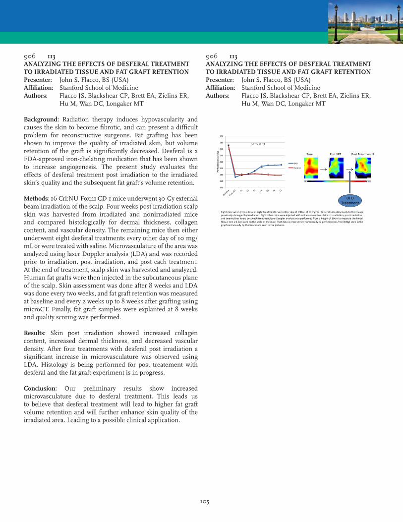
105
906 113ANALYZING THE EFFECTS OF DESFERAL TREATMENT TO IRRADIATED TISSUE AND FAT GRAFT RETENTIONPresenter: John S. Flacco, BS (USA)Affiliation: Stanford School of MedicineAuthors: Flacco JS, Blackshear CP, Brett EA, Zielins ER, Hu M, Wan DC, Longaker MT
Background: Radiation therapy induces hypovascularity and causes the skin to become fibrotic, and can present a difficult problem for reconstructive surgeons. Fat grafting has been shown to improve the quality of irradiated skin, but volume retention of the graft is significantly decreased. Desferal is a FDA-approved iron-chelating medication that has been shown to increase angiogenesis. The present study evaluates the effects of desferal treatment post irradiation to the irradiated skin’s quality and the subsequent fat graft’s volume retention.
Methods: 16 Crl:NU-Foxn1 CD-1 mice underwent 30-Gy external beam irradiation of the scalp. Four weeks post irradiation scalp skin was harvested from irradiated and nonirradiated mice and compared histologically for dermal thickness, collagen content, and vascular density. The remaining mice then either underwent eight desferal treatments every other day of 10 mg/mL or were treated with saline. Microvasculature of the area was analyzed using laser Doppler analysis (LDA) and was recorded prior to irradiation, post irradiation, and post each treatment. At the end of treatment, scalp skin was harvested and analyzed. Human fat grafts were then injected in the subcutaneous plane of the scalp. Skin assessment was done after 8 weeks and LDA was done every two weeks, and fat graft retention was measured at baseline and every 2 weeks up to 8 weeks after grafting using microCT. Finally, fat graft samples were explanted at 8 weeks and quality scoring was performed.
Results: Skin post irradiation showed increased collagen content, increased dermal thickness, and decreased vascular density. After four treatments with desferal post irradiation a significant increase in microvasculature was observed using LDA. Histology is being performed for post treatement with desferal and the fat graft experiment is in progress.
Conclusion: Our preliminary results show increased microvasculature due to desferal treatment. This leads us to believe that desferal treatment will lead to higher fat graft volume retention and will further enhance skin quality of the irradiated area. Leading to a possible clinical application.
906 113ANALYZING THE EFFECTS OF DESFERAL TREATMENT TO IRRADIATED TISSUE AND FAT GRAFT RETENTIONPresenter: John S. Flacco, BS (USA)Affiliation: Stanford School of MedicineAuthors: Flacco JS, Blackshear CP, Brett EA, Zielins ER, Hu M, Wan DC, Longaker MT
Eightmiceweregivenatotalofeighttreatmentseveryotherdayof100uLof10mg/mLdesferalsubcutaneouslytotheirscalppreviouslydamagedbyirradia<on.Eightothermicewereinjectedwithsalineasacontrol.Priortoirradia<on,postirradia<on,andtwentyfourhoursposteachtreatmentlaserDoppleranalysiswasperformedfromaheightof30cmtomeasurethebloodflowa1cmx0.5cmareaonthescalpofthemice.Thatdataisrepresentednumericallybyperfusion(mL/min/100g)seeninthegraphandvisuallybytheheatmapsseeninthepictures.
DFOTreatment
Base PostXRT PostTreatment8
p<.05atT4
140
160
180
200
220
240
260
280
300
Perfusion(m
L/min/100g)
DFO
Control

106
907 114HYPOTHERMIC PRESERVATION OF CELL SHEETS OF HUMAN ADIPOSE STEM CELLSPresenter: Sara Ribeiro, BSc (Portugal)Affiliation: 3Bs Research GroupAuthors: Ribeiro S, Costa M, Cerqueira MT, Marques AP, Pirraco RP, Reis RL
Introduction: Cell Sheet Engineering is based on the retrieval of cells from culture dishes as extracellular matrix (ECM)-rich sheets. These can then be used for the regeneration/engineering of several tissues and organs, as demonstrated in ongoing clinical trials. Cell sheet fabrication, while simple, requires cell culture facilities. Thus, ensuring adequate cell function from the fabrication site to the bedside is essential to its efficiency. We tested the ability of two commercially available compounds to preserve the viability of sheets of human adipose stem cells (hASC) at 4°C.
Methods: hASC were cultured in basal medium to hyperconfluence to produce cell sheets. Culture medium was then either replaced by a solution of Hypothermosol® (HTS) or supplemented with Rokepie® (RP) and cells were maintained at 4°C for 3 and 7 days. Controls with basal medium at 4°C and for all the conditions at 37°C were established. Non-confluent cultures mirroring all the conditions were also set up. At each time point, cells were imaged for morphology and viability was assessed using flow cytometry (7-AAD) and Alamar Blue.
Results: After 3 days, a sharp decrease in the viability at 4°C without preservation solutions was noticed when comparing with control at 37°C. This decrease was more evident in non-confluent cultures. The viability in the presence of HTS or RP was comparable but slightly lower than for the respective controls at 37°C. At day 7, few viable cells were detected in the 4°C conditions without supplementation, while none were found in the non-confluent counterparts. HTS- and RP-supplemented cultures presented 40% of the viability of the 37°C control. The relative viability in non-confluent conditions was much lower but RP accomplished better results than HTS conditions.
Conclusions: Both HTS and RP demonstrated an excellent capability of hypothermic preservation of cell sheets up to 3 days, but after 7 days the relative viability decreased to 40%. Furthermore, confluence apparently confers protection against hypothermic insult. Surface marker characterization, differentiation potential and caspase activity of cells after preservation are currently being assessed.
Acknowledgements: SFRH/BPD/101886/2014, SFRH/BPD/96611/2013
806 115MECHANICAL ISOLATION OF ADIPOSE STROMAL VASCULAR CELLS: A SAFE AND LESS TIME-CONSUMING ALTERNATIVE TO ENZYMATIC DIGESTIONPresenter: Tunc Tiryaki, MD (Turkey)Affiliation: Cellest ClinicAuthors: Tiryaki T, Canikyan S, Conde-Green A
Introduction: Adipose tissue derived stromal vascular fraction (SVF) is a reliable source of stromal vascular cells with regenerative potential in plastic surgery and other specialties. The isolation of stromal vascular cells (SVCs) using enzymatic digestion relies on fat tissue dissociation by collagenase. Although the gold standard, this isolation method is expensive, requires expert personnel, raises legal and administrative concerns on the alteration of cell characteristics. We aimed to isolate SVCs by mincing, filtrating, incubating and centrifuging the lipoaspirate and compare them with the population obtained by enzymatic digestion.
Methods: Adipose tissue was harvested with the syringe from 25 healthy females aged 26-52 years undergoing liposuction. Twenty milliliters of fat was submitted to collagenase digestion, and 20 ml was minced with blades followed by different incubation protocols in crystalloid buffer solutions, and centrifuged. The SVCs obtained were quantified, assessed for viability and submitted to flow cytometry.
Results: The SVCs yields obtained by mechanical isolation were (4.0 ± 0, 8 to 9,0 ± 1,3 x10 5 cells/ml lipoaspirate) representing 40 to 80% of the cell yield obtained by enzymatic isolation (11 ± 1,3x 10 5 cells/ml lipoaspirate) (Table 1). An incubation time of 30 minutes at room temperature (Method 4) showed higher levels of CD34 expression. The mechanical and enzymatic isolation yields showed similar levels of CD44 expression. Overall, mechanically isolated SVCs showed higher CD90 and CD105 expression.
Conclusion: Our preliminary results suggest that our technique of mechanical SVCs isolation is safe, less time-consuming than enzymatic digestion. The fact that we can isolate 40 to 80% of the cell yield isolated enzymatically shows that it’s a viable alternative. This technique does not require specialized personnel or environment, as it can be done in the operating room. As a minimally manipulated method, it may spare the surgeon from the regulatory issues encountered with enzymatic SVF isolation protocols. Further studies are warranted to determine whether these changes in cell yield or composition will affect our clinical outcomes.
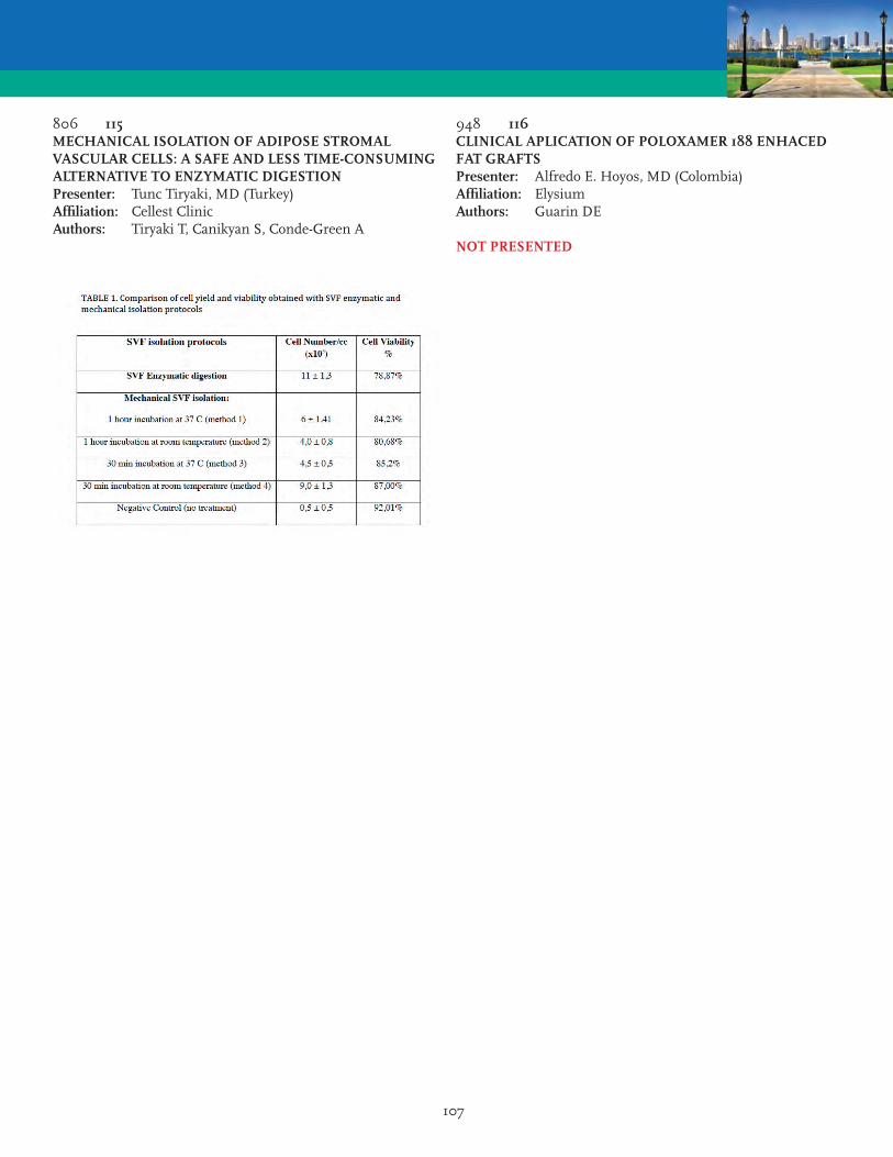
107
806 115MECHANICAL ISOLATION OF ADIPOSE STROMAL VASCULAR CELLS: A SAFE AND LESS TIME-CONSUMING ALTERNATIVE TO ENZYMATIC DIGESTIONPresenter: Tunc Tiryaki, MD (Turkey)Affiliation: Cellest ClinicAuthors: Tiryaki T, Canikyan S, Conde-Green A
948 116CLINICAL APLICATION OF POLOXAMER 188 ENHACED FAT GRAFTSPresenter: Alfredo E. Hoyos, MD (Colombia)Affiliation: ElysiumAuthors: Guarin DE
NOT PRESENTED

108
890 117FACIAL INTRAMUSCULAR LIPOMA OCCURRENCE FOLLOWING TOPICAL COSMETIC INJECTION WITH A MIXTURE OF BASIC FIBROBLAST GROWTH FACTOR: A REPORT OF TWO CASESPresenter: Xuan Liao, MD (China)Affiliation: The First Affiliated Hospital of Jinan UniversityAuthors: Liao X, Zhang ZD, Li SH, Xiao LL, Cheng B, Xie GH, Liu HW
Growth factors and cytokines control cell growth, proliferation, and differentiation via a network of inter and intracellular signaling pathways, and are involved in skin self-renewing and wound healing. In recent years, topical and injectable growth factors and cytokines have emerged as an intriguing therapeutic modality that can be harnessed for aesthetic purposes. However, very little data are available on their long-term safety and tolerability. In the present report, we describe two cases of two patients, who developed intramuscular lipoma of chin following topical injection with a mixture of basic fibroblast growth factor as main ingredients for chin augmentation. Biopsy in the two cases was done at our department, and showed intramuscular lipoma. Our report indicates that application of topical injection of growth factors can lead to tumorigenesis, and health care providers need to be aware of its potential consequences.
Keyword: Growth factor, Intramuscular lipoma, Tumorigenesis, Facial rejuventation, Cosmetic surgery.
922 118THE APPLICATION OF AUTOLOGOUS FAT GRAFTING IN IMPLANT-BASED BREAST RECONSTRUCTIONPresenter: Houbing Zheng, MD (China)Affiliation: The First Affiliated Hospital of Fujian Medical UniversityAuthors: Zheng H, Wang BW, Shan XS, Chen RC
Objective: To discuss the clinical application of autologous fat grafting in achieving symmetry in implant-based breast reconstruction.
Methods: To acquire symmetry of the breasts, in terms of volume and shape, we performed fat grafting on the reconstructed and/or the contralateral breast in 15 cases of implant-based breast reconstruction. 12 cases underwent simultaneous fat grafting with the post-expansion prosthesis implantation while 3 cases had the fat transplanted in a separate 2nd stage of operation. In simultaneous fat transplantation, fat granule is injected to the subcutaneous layer as well as the post- pectoralis major space, before the tissue expander is removed. By comparison, postponed autologous fat grafting usually involves mammoplasty on the contralateral breast and the fat granule is injected in the subcutaneous layer only. The therapeutic effects are evaluated through the comparison of pre- and post-operative photography, self-evaluation of the patients, and the post-operative adverse reaction of fat granule absorb, cystic fibrosis.
Results: Between 2012 and 2014, we have performed simultaneous fat grafting on 12 cases with implant-based breast reconstruction, while on 3 cases fat grafting is postponed to a 2nd stage of surgery. After a follow-up ranging from 1 to 12 months, all the 15 reconstructed breasts have achieved satisfactory appearances as well as symmetry concerning volume and location.
Conclusion: We recommend autologous fat grafting as an ideal solution to adjusting the appearance of the reconstructed breast, so that symmetry of the breasts, concerning shape, volume, location as well as flexibility, can be acquired. The breast reconstruction procedure involving simultaneous fat grafting is particularly appealing to the patients. Moreover, the surgical technique is simple and there is hardly any scar formed in the donor site.

109
898 119INTRATISSULAR EXPANSION-MEDIATED, SERIAL FAT GRAFTING: A STEP-BY-STEP WORKING ALGORITHM TO ACHIEVE 3D BIOLOGICAL HARMONY IN AUTOLOGOUS BREAST RECONSTRUCTIONPresenter: Filip B. Stillaert, MD (Belgium)Affiliation: University Hospital GentAuthor: Stillaert FB
Background: Breast reconstruction involves the use of autologous tissue or implants. Occasionally, microsurgical reconstruction is not an option due to insufficient donor tissues. Fat grafting has become increasingly popular in breast surgery. The challenge with this technique is how to reconstruct a stable and living “scaffold” that resembles a breast.
Methods: Breast reconstruction (n=7) was performed using intratissular expansion with serial deflation-lipofilling sessions. Mean age was 41 years old (22-53). The expander generated a vascularized capsule at 8 weeks, which demarcated a recipient site between the skin and the capsule itself and also functioned as a vascular source for angiogenesis. Serial sessions of deflation and lipofilling initiated at 8 weeks with removal of the expander at the completion of the treatment. An average of 644 ml (range, 415 ml to 950 ml) of lipoaspirate material was injected to reconstruct the breast mound. An average of 4 (range, 3 to 5) fat grafting sessions with a 3-month interval was needed to achieve symmetry with the contralateral breast. The average follow-up was 14 months (range, 9 to 29 months). MRI examination was performed at 8 months to analyze tissue survival and the residual volume.
Results: MRI examination retained tissue survival and the mean reconstructed breast volume was 386 ml (range, 231 ml to 557 ml). An aesthetically pleasant breast mound was created with a high satisfaction rate.
Conclusion: In a selected group of patients we have been able to reconstruct an aesthetically pleasant and stable breast mound using intratissular expansion and fat grafting.
855 120POST-MASTECTOMY FULL BREAST RECONSTRUCTION WITH FAT GRAFTING WITHOUT PRIOR INTERNAL OR EXTERNAL EXPANSIONPresenter: Susanna C. Kauhanen, MD, PhD (Finland)Affiliation: Helsinki University HospitalAuthors: Kauhanen SC, Hockerstedt AI
Introduction: Women seeking breast reconstruction are increasingly elderly and obese. Lipofilling in breast surgery is popularized as a tool for full breast reconstruction in combination with the BRAVA system. Mastectomy patients frequently suffer from excess tissue in the axillary fold. The purpose of this study is to evaluate the safety, efficacy and clinical outcome of delayed full breast reconstruction with Water Assisted Lipotransfer (WAL), without BRAVA or expanders in some cases combined with a lateral fasciocutaneous, flap.
Material & Methods: Fifty-six post-mastectomy patients were selected from June 2010 until to date. The mean age of the patients is 57,2 years (range 39-75). Twenty patients had undergone chest wall radiotherapy and five were smokers. Procedures done in local- or general anaesthesia lasted between 40-144 min (mean 62min). The abdomen, waist, and thighs served as donor sites. Time interval between procedures varied between 4-6 months.
Results: Thirty-one patients have been finalized with a mean of 3.6 (2-5) fat transfers. The mean follow-up time in finalized patients is 20.6 mo (range 2-48 months). The mean amount transferred/ session was 210 cc, (range 120-307). A fully sensate, B-C cup breast was achieved. Fat retention (50-70 %), was clinically evaluated at 3 months, thereafter remaining constant. In 38% of cases, disabling tissue in the axillary fold was treated by raising a local flap to reconstruct the inferior part of the breast and to gain control of the footprint. Contralateral reduction mammoplasties were performed in 48 % of cases. In 7 cases (12%) patients dropped out/changed method. In the 156 operations, no mortality, fat-, or air-embolism, or pneumothorax were reported. Three postoperative hematomas and one infection occurred. Breast cancer follow-up was done according to protocol. Sixe patients experienced a palpable tumour 12 months after lipofilling, amounting to a recall- and biopsy rate of 10.7%. No local cancer recurrencies were observed.
Conclusion: Internal or external pre-expansion is not needed to achieve a sustainable, sensate breast with full, without bra symmetry by 2-5 lipofilling procedures and an option to recycle axillary fullness into breast volume and lateral footprint control.

110
855 120POST-MASTECTOMY FULL BREAST RECONSTRUCTION WITH FAT GRAFTING WITHOUT PRIOR INTERNAL OR EXTERNAL EXPANSIONPresenter: Susanna C. Kauhanen, MD, PhD (Finland)Affiliation: Helsinki University HospitalAuthors: Kauhanen SC, Hockerstedt AI
817 121LARGE VOLUME FAT GRAFTING IN BREAST RECONSTRUCTION: SIX YEARS EXPERIENCEPresenter: Marwan H. Abboud, MD (Belgium)Affiliation: MA ClinicAuthors: Abboud MH, Dibo SA
Introduction: Lipofilling has widespread indications in patients with breast cancer because of the ability to improve the results of breast reconstruction and correct deformities after conservative surgery. In the context of continuous interest to develop, refine, and expand the indications of lipofilling in breast reconstruction, the authors adapted a power-assisted instrument (Lipomatic® Eva SP, Euromi SA, Verviers, Belgium) to facilitate fat harvesting and grafting technique, and share their five years experience with power assisted liposuction and lipofilling (PALL) in breast reconstruction.
Methods: Between 2009 and 2015 a total of 350 patients underwent breast reconstruction by lipomodeling using the PALL technique. Patient population included primary reconstruction by lipofilling, lipofilling coupled to or following autologous flap reconstruction, lipofilling to correct deformities following breast-conserving surgery or implant-based reconstruction, and lipofilling following removal of breast implants in failed implant reconstruction. Fat is harvested in a closed system using a 3-mm multiple-hole cannula attached to a hand-piece and set to 3 bars and 0.7 atm suction pressure. For grafting, the Lipomatic instrument is disconnected from its suction system and fat is injected in the superficial and deep planes through a customized v-shaped multi-hole cannula (3-mm diameter) that enables simultaneous vibration of the recipient site. Additional external vibration of the recipient site at the end of the procedure using the lipomatic hand piece improves diffusion of fat in the recipient site.
Results: Follow up ranged from 6 to 48 months, whereas the mean operative time for fat injection ranged between 45 and 60 min. The total complication rate was 7.33%. Oil cysts occurred in 6% of patients. The latter were closely observed and conservatively treated.
Conclusion: Power-assisted liposuction and lipofilling is a simple, time efficient and reproducible novel technique that offers a myriad of applications in breast reconstruction.

111
785 122FAT PROCESSED BY SALINE-WASH, NEGATIVE-PRESSURE-FILTRATION, AND LARGE SCALE STERILE COTTON ABSORPTION FOR BREAST LIPOAUGMENTATION AFTER IMPLANT REMOVALPresenter: Sarah A. Mess, MD (USA)Affiliation: Sarah Mess, MD LLCAuthor: Mess SA
Fat processed by saline-wash, negative-pressure-filtration, and large scale sterile cotton absorption was used for replacement of breast implant volume. A larger volume of fat was required than the implant since retention of fat has varied from 40-80% as measured by volumetric analysis of MRI and three-dimensional photography (1,2). The quantity of saline in the fat from the harvesting tumescent and processing wash may have affected published retention rates. In a study of fat harvested by water-assisted liposuction, the fat contained 50% saline as measured after centrifugation at 1200g for 3 minutes (3). The authors suggested that their retention rate would be near 100% if they accounted for the 50% saline.
Six patients underwent removal of breast implants, liposuction, and fat transfer to the breast. The fat was collected in a 3-liter lipofilter canister, washed of blood with saline, and filtrated with negative pressure. The fat was further consolidated by sterile ABD pad absorption, removing excess water and oil and decreasing volume 50%. The implants were removed via an inframammary incision and the capsule was kept intact. The fat was reinjected with 14-gauge cannula and 10cc syringe with low-pressure, retrograde, bimanual placement, less than 1 cc per pass. Infiltration sites were along the inframammary border, the areola, and the lateral border of the breast. Implant size removed ranged 200 - 475cc and fat injected 285-700cc per breast. Follow up ranged from 3-11 months. One of the 6 patients developed an oil cyst, otherwise there were no complications and patients reported satisfaction. Two patients increased their bra size, two patients maintained their bra size, and two patients decreased cup size (figures 1-3 show an example of each). The patients were assessed for satisfaction of appearance, feel, size, and symmetry at postoperative visits. Removal of implants alone can lead to ptotic, flat breasts with dents where the implant compressed tissue. Consolidation of saline-washed and negative-pressure -filtrated lipoaspirate with large scale sterile cotton absorption removes excess saline and oil and provides condensed fat for adequate lipoaugmentation after breast implant removal.
1) Spear ASJ 2014, 2)Khouri PRS 2012, 3)Jung ASJ 2016
785 122FAT PROCESSED BY SALINE-WASH, NEGATIVE-PRESSURE-FILTRATION, AND LARGE SCALE STERILE COTTON ABSORPTION FOR BREAST LIPOAUGMENTATION AFTER IMPLANT REMOVALPresenter: Sarah A. Mess, MD (USA)Affiliation: Sarah Mess, MD LLCAuthor: Mess SA

112
771 123A COMPARATIVE TRANSLATIONAL STUDY: THE COMBINED USE OF ENHANCED STROMAL VASCULAR FRACTION AND PLATELET-RICH PLASMA IMPROVES FAT GRAFTING MAINTENANCE IN BREAST SOFT TISSUE DEFECTSPresenter: Pietro Gentile, MD, PhD (Italy)Affiliation: University of Rome Tor VergataAuthor: Gentile P
NOT PRESENTED
807 124TREATMENT OF FEMALE URINARY INCONTINENCE WITH AUTOLOGOUS, MICRO-FRAGMENTED, AND MINIMALLY MANIPULATED ADIPOSE TISSUE (LIPOGEMS®)Presenter: Heripsime Ohanian, MD, PhD (USA)Affiliation: Hackensack University Medical CenterAuthor: Ohanian H
Background: Urinary incontinence (UI) is a very common disorder affecting women of various ages and impacts all aspects of life. Commonly seen among women with history of multiple vaginal deliveries, it is caused by the loss of urethral support. Later in life more frequently seen as mixed incontinence in menopausal women. These conditions have severe quality of life implications. Different surgical and injectable techniques have been used with limited success. New therapeutic approaches, such as the use of mesenchymal stem cells (MSCs), seem to have promising results. In this context, the regenerative capabilities of fat (adipose derived mesenchymal stem cells, ADSCs), with mesenchymal properties, are being widely explored. We present outcomes up to 16 month follow-up of three patients with mixed UI, treated with autologous, micro-fragmented, minimally manipulated adipose tissue (Lipogems®).
Cases Description: Three post-menopausal women presented with mixed UI: the first with overactive bladder and urge urinary incontinence (UUI), the second with urge urinary incontinence and intrinsic sphincter deficiency (ISD), the third with OAB and UUI.
Material and Methods: Micro-fragmented adipose tissue was obtained using a minimal manipulation technique in a closed system (Lipogems®), with no added enzymes or other additives. Under ultrasound guidance and direct visualization, Lipogems® was injected trans-vaginally into the periurethral space of the bladder neck in a clockwise pattern.
Results: Within three days, patients noted a major change in their urinary pattern and within two months, reported minimal, if any, leakage. Six month urodynamic testing demonstrated significant increases in bladder capacity as well as resting urethral pressures (UPP) with only negligible, if any, leakage. In this study we’ve demonstrated that improvement occurs until at least 12 months.
Conclusion: Although we report only 3 cases, part of an ongoing study approved by IRCM IRB, the results are encouraging and warrant further investigations. The long lasting improvement in symptoms of different UI demonstrate the safety, efficacy and potential benefits of using autologous, micro-fragmented, minimally manipulated adipose tissue for the treatment of female UI symptoms.
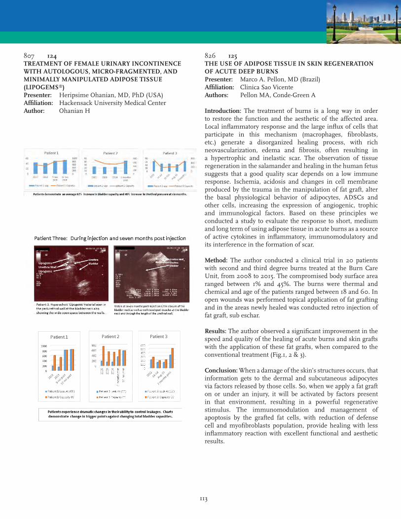
113
807 124TREATMENT OF FEMALE URINARY INCONTINENCE WITH AUTOLOGOUS, MICRO-FRAGMENTED, AND MINIMALLY MANIPULATED ADIPOSE TISSUE (LIPOGEMS®)Presenter: Heripsime Ohanian, MD, PhD (USA)Affiliation: Hackensack University Medical CenterAuthor: Ohanian H
826 125THE USE OF ADIPOSE TISSUE IN SKIN REGENERATION OF ACUTE DEEP BURNSPresenter: Marco A. Pellon, MD (Brazil)Affiliation: Clinica Sao VicenteAuthors: Pellon MA, Conde-Green A
Introduction: The treatment of burns is a long way in order to restore the function and the aesthetic of the affected area. Local inflammatory response and the large influx of cells that participate in this mechanism (macrophages, fibroblasts, etc.) generate a disorganized healing process, with rich neovascularization, edema and fibrosis, often resulting in a hypertrophic and inelastic scar. The observation of tissue regeneration in the salamander and healing in the human fetus suggests that a good quality scar depends on a low immune response. Ischemia, acidosis and changes in cell membrane produced by the trauma in the manipulation of fat graft, alter the basal physiological behavior of adipocytes, ADSCs and other cells, increasing the expression of angiogenic, trophic and immunological factors. Based on these principles we conducted a study to evaluate the response to short, medium and long term of using adipose tissue in acute burns as a source of active cytokines in inflammatory, immunomodulatory and its interference in the formation of scar.
Method: The author conducted a clinical trial in 20 patients with second and third degree burns treated at the Burn Care Unit, from 2008 to 2015. The compromised body surface area ranged between 1% and 45%. The burns were thermal and chemical and age of the patients ranged between 18 and 60. In open wounds was performed topical application of fat grafting and in the areas newly healed was conducted retro injection of fat graft, sub eschar.
Results: The author observed a significant improvement in the speed and quality of the healing of acute burns and skin grafts with the application of these fat grafts, when compared to the conventional treatment (Fig.1, 2 & 3).
Conclusion: When a damage of the skin’s structures occurs, that information gets to the dermal and subcutaneous adipocytes via factors released by those cells. So, when we apply a fat graft on or under an injury, it will be activated by factors present in that environment, resulting in a powerful regenerative stimulus. The immunomodulation and management of apoptosis by the grafted fat cells, with reduction of defense cell and myofibroblasts population, provide healing with less inflammatory reaction with excellent functional and aesthetic results.

114
826 125THE USE OF ADIPOSE TISSUE IN SKIN REGENERATION OF ACUTE DEEP BURNSPresenter: Marco A. Pellon, MD (Brazil)Affiliation: Clinica Sao VicenteAuthors: Pellon MA, Conde-Green A
879 126EFFICACY AND SAFETY OF GLUTEAL AUGMENTATION WITH AUTOLOGOUS FAT GRAFTING: A SYSTEMATIC REVIEWPresenter: Carlo M. Oranges, MD (Switzerland)Affiliation: Basel University HospitalAuthors: Oranges CM, Harder Y, Haug M, Kalbermatten DF, Schaefer DJ
Introduction: Recent surveys from the American Society for Aesthetic Plastic Surgery indicated gluteal augmentation as the single most growing cosmetic surgery performed in the United States during the last decade. This procedure is usually performed with silicone implant placement or gluteal fat grafting. The combination of liposuction and gluteal fat grafting is especially recommended to achieve the ideal female waist to hip ratio of approximately 0.7, and, unlike implant placement, allows to reshape the lateral third of the buttocks, respecting different ethnic beauty ideals related to buttock fullness, lateral buttock and lateral thigh shapes. However, efficacy and safety of gluteal fat grafting were never comprehensively analyzed. We performed a systematic review of the literature to determine outcomes and complications, including patient satisfaction, associated with this technique.
Methods: The MEDLINE database was searched for clinical studies on autologous fat grafting to the gluteal region. There were no restrictions on time or language of publication. Resulting articles were screened using a priori criteria.
Results: Seventeen articles (four prospective and ten retrospective case series, and three case reports), representing 2607 treated patients, were included. No randomized controlled studies met final inclusion criteria. The overall complication rate was 10.9 percent (n = 284). The most common complications included seroma of the donor site (3.8 percent), major or minor irregularities including asymmetry (2.1 percent), infection (1.7 percent), and sciatic pain (1.3 percent). Four cases of fat embolism, one of which leaded to death, were reported. Patient satisfaction after surgery was scored as high by the majority of the studies, although different methods of evaluation among the articles prevented quantitative analysis.
Conclusions: The prevalence of complications in gluteal fat grafting compared favorably to implant-based gluteal augmentation, yielding high patient satisfaction. However, the level of evidence of the studies identified, mainly representing single surgeon experiences, was low. Importantly, injections should be limited to the subcutaneous and superficial intramuscular planes of the gluteal region to prevent fat embolism.

115
818 127POWER-ASSISTED GLUTEAL AUGMENTATION: A NEW TECHNIQUE FOR SCULPTING, HARVESTING, AND TRANSFERRING FATPresenter: Saad Dibo, MD (Belgium)Affiliation: MA ClinicAuthors: Dibo S, Abboud MH
Introduction: A simple and reproducible surgical technique for gluteal shaping and augmentation with autologous fat is needed. The authors describe a novel approach to large-volume gluteal augmentation that combines power-assisted liposculpting and fat harvesting of the zones around the buttock with autologous fat transfer.
Methods: One hundred ten patients who underwent gluteal augmentation were evaluated in a prospective study. Liposculpting and fat harvesting were performed with power-assisted liposuction. Fat then was transferred to the gluteal region with simultaneous power-assisted vibration and tunnelization. A questionnaire to assess patient satisfaction was administered at 6 months postoperatively.
Results: The mean body mass index of the patients was 30 kg/m2 (range, 26-36 kg/m2). Liposuction volumes ranged from 1400 to 5000 mL, and injection volumes ranged from 300 to 900 mL per side for each session. Operating times ranged from 60 to 120 minutes. Patients were monitored for an average of 20 months (range 12-48 months). Complications included a burning sensation in 5 of 110 patients (4.5%), persistent swelling in the lower back in 3 patients (2.7%), and a mild infection in 1 patient (0.9%).
Conclusions: Power-assisted gluteal augmentation with autologous fat is an efficient, safe, and reproducible procedure that produces an aesthetically pleasing gluteal projection and contour.
786 128NON-RESPONSIVE MULTIFACTORIAL SHOULDER PAIN WITH OSTEOARTHRITIS AND ROTATOR CUFF TEARS TREATED WITH AUTOLOGOUS MICRO-FRAGMENTED AND MINIMALLY MANIPULATED ADIPOSE TISSUE UNDER CONTINUOUS ULTRASOUND GUIDANCEPresenter: Richard D. Striano, DC, RMSK (USA)Affiliation: OptimumJoint Integrated Joint SpineAuthors: Striano RD, Bilbool N, Krutchkoff B
Background: Chronic shoulder abnormalities affect a large portion of the population, thus affecting patient quality of life. Current treatment is challenging with few options if non-operative care fails. In this context, autologous adipose tissue has gained increasing interest. Fat is readily accessible and simple to harvest, to provide regenerative stimuli. Adipose tissue contains a structural matrix that naturally contains a plethoria of regenerative elements including perivascular and mesenchymal stem cells (hMSCs) that produce trophic bioactive elements.
Objective: We initiated our evaluations on the potential benefits of using autologous, and minimally manipulated adipose tissue as orthopedic lipofilling for structural support in cases of multifactorial shoulder pain having failed conventional care.
Study Description: These are the first 10 subjects reaching one-year follow-up, part of an ongoing IRB study approved by IRCM. Ages: 35 – 89, Kellgren-Lawrence OA average: 3.3. Tears (all confirmed on MRI and mapped for treatment with ultrasound imaging): eight supraspinatus tendon, four full thickness, four partial thickness, one partial subscapularis tendon, two partial bicep tendon, six labrum. Atrophy 4 and fatty atrophy 2.
Material and Methods: Micro-fragmented fat was obtained using a minimal manipulation technology in a closed system, Lipogems®, without the addition of enzymes or other additives. Lipogems® was injected in 1cc aliquots under ultrasound guidance, confirming the lipofilling of joint and soft tissue defects. Clinical outcomes are shown in Fig. 1-7.
Results: The improvement of the symptoms occurred few days after treatment and all measured scores show significant improvement until one-year follow-up (Figures 1-7).
Conclusion: Although more investigation is needed, the results are very promising. The injection of autologous, micro-fragmented, and minimally manipulated adipose tissue (Lipogems®) appears very effective in patients with shoulder disease that failed conventional treatments.
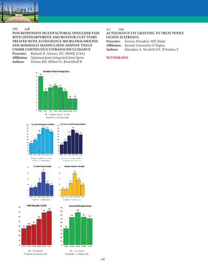
116
786 128NON-RESPONSIVE MULTIFACTORIAL SHOULDER PAIN WITH OSTEOARTHRITIS AND ROTATOR CUFF TEARS TREATED WITH AUTOLOGOUS MICRO-FRAGMENTED AND MINIMALLY MANIPULATED ADIPOSE TISSUE UNDER CONTINUOUS ULTRASOUND GUIDANCEPresenter: Richard D. Striano, DC, RMSK (USA)Affiliation: OptimumJoint Integrated Joint SpineAuthors: Striano RD, Bilbool N, Krutchkoff B
911 129AUTOLOGOUS FAT GRAFTING TO TREAT PENILE LICHEN SCLEROSUSPresenter: Aurora Almadori, MD (Italy)Affiliation: Second University of NaplesAuthors: Almadori A, Nicoletti GF, D’Andrea F
WITHDRAWN

117
848 130ADIPOSE-DERIVED STEM CELLS GIVEN INTRAVENOUSLY IMPROVES EPISTAXIS AND OBJECTIVE BRONCHOSCOPY SCORING IN HORSES WITH EXERCISE-INDUCED PULMONARY HEMORRHAGEPresenter: Mark Hughes, DVM (USA)Affiliation: VetStemAuthors: Harman R, Rich R, Hughes M
Exercise-induced pulmonary hemorrhage (EIPH) occurs in the majority of Thoroughbred and Standardbred race. EIPH is bleeding that occurs from the lungs of horses during exercise and has been shown to reduce athletic performance. Furosemide and other drugs have been used symptomatically to try to prevent EIPH and many equine oversight organizations are considering banning furosemide from performing horses. Pulmonary vascular damage and inflammation-induced perivascular fibrosis are seen in EIPH affected horses. There is substantial evidence in the literature that stem cells can reduce inflammation and fibrosis and repair damaged tissues. We hypothesized that intravenous therapy with stem cell may prevent EIPH and repair damage already present.
Materials and Methods: A prospective un-blinded study was started in 2015 to evaluate EIPH in performance horses. Inclusion criteria required pre-treatment reported epistaxis after racing and tracheobroncoscopy (TBS) confirmation of blood in the trachea. Surgically collected adipose tissue from each horse was submitted to the laboratory for enzymatic separation of stromal vascular cells containing adipose stem cells (ASC). Cell counts were approximately 8M cells per dose. Horses were treated twice with this intravenous ASC preparation, approximately 2 weeks apart. After enrollment in the program, no other therapeutic drugs were allowed for the duration of the study. A TBS was conducted post-race.
Results: Twelve horses were analyzed. All 12 raced without prophylactic medication. Following treatment with ASCs, only 1/12 showed epistaxis upon racing. TBS pre/post therapy was available on 8 horses. Pretreatment TBS outcomes: 7 grade 4 and 1 grade 3. Post-treatment TBS outcomes: 5 grade 0, 2 grade 1, and 1 grade 2.
Conclusions: All 12 horses showed a pretreatment post-race epistaxis while on prophylactic medications. Post-treatment 11/12 horses showed no epistaxis after racing and while on no prophylactic medications. TBS showed improvement in all 8 horses with 5/8 showing no evidence of EIPH upon post race TBS. This data demonstrates an association of ASC intravenous therapy and improvement in epistaxis and TBS evidence of EIPH within a relative short period of healing (4-6 weeks).
887 131LYMPHATIC TISSUE REPAIR IN PATIENTS WITH ADIPOSE TISSUE DISORDERS USING LIPOGEMSPresenter: Todd K. Malan, MD (USA)Affiliation: Roxbury RegenerativeAuthors: Malan TK, Amron DA, Herbst KH
Dercums Disease (DD or Adiposis Dolorosa) and Lipedema are rare adipose tissue disorders. DD is a syndrome of painful inflammatory growths in the subcutaneous fat. Lipedema is characterized by abnormal fatty tissue distribution and storage. Each of these disease share a presumed common pathophysiologic alternation of normal lymphatic tissue structure and function with a profound localized pro-inflammatory tissue changes. Long standing results with surgical debulking of DD lesion or Lipedema fat has been largely disappointing. It is believed that this is largely due to the further destruction of normal lymphatic tissue and function caused by surgical debulking. Previous studies have shown that mesenchymal stem cells (MSCs), can be utilized to enhance lymphatic regeneration and restore lymphatic fluid flow in the setting of lymphatic injury. These results were most readily pronounced when combined with a scaffold. Lipogems micro-fractured adipose tissue graft acts as a bio scaffold and contains a rich source of MSCs as well as prolymphangiogenic growth factors VEGFR3, FGF, and HGF and anti-inflammatory cytokines.
Method: 18 Pts with DD, and 4 Pts with Stage 4 Lipedema underwent syringe harvesting of an avg of 73gm of adipose tissue utilizing syringe and cannula included in the Lipogems disposable set. Harvested adipose tissue was then processed into Lipogems micro-fractured fat in accordance with manufacturers recommendations. Lipogems tissue graft was injected under ultrasound guidance along the lymphatic channels of the bilateral upper and lower extremities. Patients completed QOL and VAS Pain scores pre-procedure, 1, 3, and 6 months post procedure.
Results: Mean VAS Pain scores improved by -3 points (1-10) at 1 month, -3.8 points at 3 months, and -5.1 points at 6 months. 9 of the 18 DD reported complete or significant lesion reduction.
Conclusion: Lipogems micro-fractured adipose derived tissue grafts have the unique properties of serving as a bio scaffold that is rich in naturally occurring adipose MSCs, prolymphangiogenic growth factors, and anti-inflammatory cytokines. The majority of our patients with DD and Lipedema who were treated with only peri-lymphatic channel injections of Lipogems demonstrated continued improvement 6 mo post treatment.
118
940 132ULTRASONOGRAPHY IN PLASTIC SURGERYPresenter: Tyler Safran, DEC (Canada)Affiliation: McGill UniversityAuthors: Safran T, Kanevsky J, Gorsky K, Luc M, Rodriguez R, Futrell W
Introduction: The modern medical era is characterized by increasingly available portable imaging devices. High-resolution ultrasound devices are becoming a popular choice of non-invasive imaging among many specialists. Plastic and Reconstructive surgeons are in a unique position to implement and tailor the use of ultrasound to the clinical areas of need in their specialty. This article will introduce the modern plastic surgeon to established and novel uses of ultrasound in practice, with a specific focus on how it relates to adipose tissue harvest, procedural grafting and follow up.
Methods: A systematic electronic search was performed using the PubMed database. Search terms used were those for studies utilizing ultrasonography or ultrasound based techniques in plastic surgery. Studies were pooled and analyzed based on their procedure category (Breast, facial, microsurgery, skin procedures, aesthetic and innovative applications).
Results: 303 articles were included in the study spanning six procedure categories. Primarily, applications of ultrasound technology were found in adipose evaluation (post fat graft, liposuction guidance, facial lipo-atrophy and adipose distribution), pre-operative planning (anatomical location and variation), and focused ultrasound acoustic energy source procedures (microfocused skin tightening, adipose tissue removal, and facial rejuvenation). Within adipose evaluation, high-resolution ultrasound technology can allow the plastic surgeon to accurately quantify viable fat, aid with safe injection, and visualize fat survival post fat graft. Reported benefits of incorporating ultrasound in clinical activities included: Improved procedural outcomes, decreased complications and allowed for more comprehensive patient follow-up and quantitative evaluation.
Conclusion: Ultrasound offers a portable and non-invasive means to obtain real-time visualization of patients anatomy, while also providing an effective focused energy source for procedures. This review highlights novel applications of ultrasonography in the hands of a modern plastic surgeon, across the entire breadth of the specialty. Ultrasound technology has an emerging role in plastic surgery in the growing field of adipose tissue transfer.
797 133STROMAL VASCULAR FRACTION THERAPY FOR ALLEVIATION OF CHRONIC REFRACTORY MIGRAINESPresenter: Kenneth Rothaus, MD (USA)Affiliation: NY Presbyterian-Weill CornellAuthors: Rothaus K, Mauskop AM
Background: Autologous adipose-derived stromal vascular fraction (SVF) is rich in MSCs and has been reported to be effective for the treatment of trigeminal neuropathic pain. Autologous adipose-derived SVF cells given intravenously have also been reported to relieve chronic migraine. Stem cell activity possibly targets neurogenic inflammation, a well-documented aspect of migraine pathogenesis. Other reports indicate that an intramuscular injection of stem cells provides longer lasting effect than an intravenous infusion.
Methods: Patients 18 or older with severe disability on migraine disability assessment scale (MIDAS) who failed botulinum toxin injections and at least three prophylactic drugs were included in this IRB-approved study. Primary outcome measure was change in MIDAS score 3 months after treatment. Secondary measures included change in MIDAS score after 6 months, change in headache impact score (HIT-6), patient’s global impression of change, clinician’s global Impression of change, number of headache-free days, percentage of patients with 50% or greater reduction in headache-free days, change in number and type of abortive migraine medications taken. Power assisted liposuction was performed to obtain approximately 500 cc of aspirate. SVF was isolated using high-speed centrifugation and a proprietary collection device. 10-12 cc of SVF was collected for each patient. 2 cc of SVF was send to an independent laboratory for quantification of numbers of nucleated cells and their viability. The total SVF injected in each patient ranged from 8 to 10 ml with the cell count varying from 2.5 to 8.6 million viable cells. SVF was injected into the pericranial, neck and trapezius muscles.
Results: One male and 8 female patients were enrolled in the study. Mean patient demographics were: age (48 yr.), duration of headaches (16 yr.), number of prophylactic drugs (10), and MIDAS score at the baseline (147). 7 out of 9 patients showed improvement. Changes in the outcome measures will be presented.
Conclusion: The use of autologous adipose-derived SVF appears to be effective in the alleviation of chronic refractory migraines. Further investigation of this and other sources of biologically active MSCs seems warranted.

119
782 134ENHANCED ADIPOSE-TISSUE DERIVED SVF VASCULARIZATION POTENTIAL BY 3D PERFUSION CULTURE: A POSSIBLE TREATMENT FOR ISCHEMIC TISSUEPresenter: Giulia Cerino, PhD (Switzerland)Affiliation: University and University Hospital of BaselAuthors: Cerino G, Gaudiello E, Melly L, Muraro M, Martin I, Eckstein F, Scherberich A, Marsano A
NOT PRESENTED
784 135IN VITRO AND IN VIVO INTERACTION OF ADIPOSE-DERIVED STEM CELLS AND BREAST CANCER CELLSPresenter: Hakan Orbay, MD, PhD (USA)Affiliation: University of California DavisAuthors: Orbay H, Charvet HJ, Hinchcliff KM, Dehghani T, Kaur M, Sahar DE
Introduction: Fat grafting became increasingly popular for post-mastectomy breast reconstruction; however, it is still controversial whether or not fat grafting increases breast cancer recurrence rates. We investigate the in vitro and in vivo interaction of human breast cancer (BrCa) cells and adipose-derived stem cells (hASCs) to address this controversy.
Methods: For the in vitro arm of the study we harvested hASCs from human lipoaspirates (n=3) and examined the effect of hASCs on migration of MDA-MB-231 RFP (+) BrCa cells using a cell migration assay. For the in vivo arm of the study we divided lipoaspirate from a single subject into two halves: we used one half for hASCs harvest and prepared the other half as fat graft. We injected hASCs, BrCa cells, and fat grafts to the 4th mammary gland of female nude mice (n=20) bilaterally as shown in Figure 1A. We injected 8x10⁵ BrCa cells, 1.8x10⁵ hASCs, and 75 µl of fat graft per each site in corresponding groups. We followed the tumor growth with digital caliper measurements and examined the tumors, livers and lungs histologically after 2 weeks.
Results: In vitro migration potential of BrCa cells increased significantly when co-cultured with hASCs (p<0.05). Tumor growth rate in group IV (hASCs + BrCa cells + fat graft) was significantly higher than groups I and II (p<0.05) (Figure 1B). There was no significant difference between groups IV and III; also group III and groups I and II. The vascular density in the tumors in group IV was significantly higher than the tumors in other groups (p<0.01) in parallel with tumor growth rates. Histologically, injected fat grafts were largely replaced by BrCa cells. There was no liver or lung metastasis at 2 weeks.
Conclusion: hASCs increase the in vitro migration of BrCa cells. The injection of fat grafts or hASCs alone does not increase the in vivo growth rates of BrCa cells; however, when injected together fat grafts and hAS
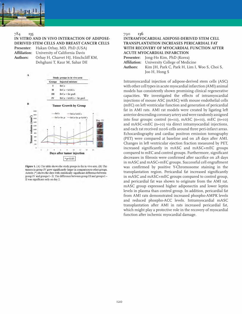
120
784 135IN VITRO AND IN VIVO INTERACTION OF ADIPOSE-DERIVED STEM CELLS AND BREAST CANCER CELLSPresenter: Hakan Orbay, MD, PhD (USA)Affiliation: University of California DavisAuthors: Orbay H, Charvet HJ, Hinchcliff KM, Dehghani T, Kaur M, Sahar DE
790 136INTRAMYOCARDIAL ADIPOSE-DERIVED STEM CELL TRANSPLANTATION INCREASES PERICARDIAL FAT WITH RECOVERY OF MYOCARDIAL FUNCTION AFTER ACUTE MYOCARDIAL INFARCTIONPresenter: Jong-Ho Kim, PhD (Korea)Affiliation: University College of MedicineAuthors: Kim JH, Park C, Park H, Lim I, Woo S, Choi S, Joo H, Hong S
Intramyocardial injection of adipose-derived stem cells (ASC) with other cell types in acute myocardial infarction (AMI) animal models has consistently shown promising clinical regenerative capacities. We investigated the effects of intramyocardial injections of mouse ASC (mASC) with mouse endothelial cells (mEC) on left ventricular function and generation of pericardial fat in AMI rats. AMI rat models were created by ligating left anterior descending coronary artery and were randomly assigned into four groups: control (n=10), mASC (n=10), mEC (n=10) and mASC+mEC (n=10) via direct intramyocardial injections, and each rat received 1x10⁶ cells around three peri-infarct areas. Echocardiography and cardiac positron emission tomography (PET) were compared at baseline and on 28 days after AMI. Changes in left ventricular ejection fraction measured by PET, increased significantly in mASC and mASC+mEC groups compared to mEC and control groups. Furthermore, significant decreases in fibrosis were confirmed after sacrifice on 28 days in mASC and mASC+mEC groups. Successful cell engraftment was confirmed by positive Y-Chromosome staining in the transplantation region. Pericardial fat increased significantly in mASC and mASC+mEC groups compared to control group, and pericardial fat was shown to originate from the AMI rat. mASC group expressed higher adiponectin and lower leptin levels in plasma than control group. In addition, pericardial fat from AMI rats demonstrated increased phospho-AMPK levels and reduced phospho-ACC levels. Intramyocardial mASC transplantation after AMI in rats increased pericardial fat, which might play a protective role in the recovery of myocardial function after ischemic myocardial damage.

121
801 137ALTERNATIVELY ACTIVATED M2 MACROPHAGES IMPROVE AUTOLOGOUS FAT GRAFT SURVIVAL IN A MOUSE MODEL THROUGH INDUCTION OF ANGIOGENESISPresenter: Michael Bezuhly, MD (Canada)Affiliation: Dalhousie University IWK Health CenterAuthors: Gebremeskel S, Phipps K, Gillis J, Johnston B, Hong P, Bezuhly M
WITHDRAWN
808 138NOTCH2 EXPRESSED ASC REGULATES PDGFR-BETA, MIGRATION AND ADHESION IN VITRO AND IN PATHOLOGICAL PROLIFERATIVE RETINOPATHY IN VIVOPresenter: Vincenzo Terlizzi, MS (Netherlands)Affiliation: University of GroningenAuthors: Terlizzi V, Kolibabka M, Hammes HP, Harmsen MC
Pericytes (PC) and endothelial cells (EC), in intimate contact, constitute the outer layer of capillaries which degenerate during diabetic retinopathy (DR). ASC (adipose stromal cells) phenotypically resemble PC and can replace lost PC in rodent DR models. Much of ASC action is achieved by paracrine signaling, yet PC and EC also act via juxtacrine signaling.
NOTCH is a contact-depended signaling, we thereby hypothesized that in ASC contributes to the homeostasis and stabilization of retinal microvascular endothelial cells in DR.
ASC key molecules of the NOTCH pathway showed a constitutively increased expression of NOTCH2. NOTCH2 expression was refractory to hyperglycemia. NOTCH2 loss of function (SH-NOTCH2) correlated with upregulation of mesenchymal markers such as CD29 and CD105. ASC supported the formation of vessel-like networks (VNF) by HUVEC in vitro. SH-NOTCH2 ASC abolished the VNF-supporting capacity of ASC. Moreover, SH-NOTCH2 ASC showed decreased migration towards EC-secreted chemoattractant PDGF-BB, as well as to conditioned media from HUVEC and BREC (bovine retinal endothelial cells). This associated with down regulation of PDGFR-beta at protein level. Finally, intraocular injection of SH-NOTCH2 ASC in OIR mice showed higher avascular areas in the retina compare to control. This indicates that NOTCH2 is involved both in homing and ‘docking’ of ASC onto retinal EC. In contrast, electroretinogram displayed higher response in b-wave on the neovascular zones when SH-NOTCH2 ASC were injected.
We show that NOTCH2 on ASC as important receptor for the juxtracrine interaction with EC. This is supported by the direct downregulation of PDGFR-beta, which is activated by PDGF-BB secreted by EC to attract PC in perivascular position. Finally, analysis on the retina showed integration of wild type ASC in perivascular position. Interestingly, the greater higher b-wave amplitude on the neovascular zone when ASC SH-NOTCH2 were present suggest that paracrine factors are still positively influencing the retina microenvironment independently from juxtacrine interaction.

122
837 139LIPOGRAFTING IMPROVES THERAPY RESISTANT DERMAL SCARS THROUGH ENHANCED REMODELINGPresenter: Maroesjka Spiekman, BS (Netherlands)Affiliation: University Medical Center GroningenAuthors: Spiekman M, Hoppe DL, Diercks GF, Ghods M, Harmsen MC
Introduction: Lipografting is emerging as an effective last resort for difficult-to-treat dermal scars. In lipografting, whole adipose tissue containing among others adipocytes, adipose-derived stromal cells (ASC), extracellular matrix (ECM) and vascular structures, is injected subdermally in the scar area. However, the mechanism behind the beneficial effect of lipografting is unknown. We hypothesized that the lipograft helps to remodel and reverse dermal scars by modulating fluxes and function of immune cells and subsequent beneficial changes in the deposited extracellular matrix.
Methods: Fifteen patients with therapy resistant scars were enrolled to be treated with two subsequent autologous lipografts. Photographs of the scar area were taken before and three months after each treatment. At the same time points, skin biopsies were collected. Tissue morphology, number of vessels, ECM deposition and elastin content were determined by means of histochemistry. Immunohistochemical stainings for macrophages, mast cells, B and T cells were performed. The number of positive cells per mm2 of tissue was counted.
Results: Clinically, macroscopic improvement of the scars was observed, in terms of colour, relief and pliability. Microscopically, there was a marked increase in thickness and amount of ECM fibres after lipografting. Perivascular cell infiltrates were visible. Preliminary results show an increase in the number of vessels and the amount of elastin fibres in the majority of patients. Also, the number of mast cells, macrophages and T cells increased after treatment.
Conclusion: Lipografting results in histological and clinical improvement of therapy resistant scars. Final results will be based on histological results of 15 patients currently enrolled in this study. In this immunohistochemical pilot study, the histological changes are accompanied by influx of macrophages, mast cells and T cells into the scar tissue. Moreover, the composition of the ECM changed from stiff (collagenous) to more flexible (increased elastin).
860 140CIGARETTE SMOKING AS A FACTOR IN COMPROMISED FUNCTION AND THERAPEUTIC ACTIVITY OF ADIPOSE STEM CELLS, MANIFESTED AS DECLINE IN VASCULOGENIC POTENTIALPresenter: Daria Barwinska, BA (USA)Affiliation: Indiana UniversityAuthors: Barwinska D, Traktuev D, Cook TG, Merfled-Clauss S, Petrache I, March KL
Objective: While it is known that cigarette smoking (CS) results in a dysfunctional angiogenic activity of endothelial cells (EC) and lowers the number of bone marrow hematopoietic stem cells, the effect of CS on adipose stem cell (ASC) therapeutic activity has not been fully studied. We previously demonstrated that human ASC obtained from smoking donors were inferior in their therapeutic activities compared to ASC derived from non-smoking donors. Here we begin to address mechanisms underlying the defect in the function of ASCs derived from smokers.
Methods: Human ASC were isolated from subcutaneous fat of smokers and non-smokers of both genders. Expanded cells as well as their conditioned media were tested for ability to support human EC vascular network formation (VNF). ASC-conditioned media (ASC-CM) from smokers and non-smokers was assessed functionally as well as by specific protein analyses. To assess the in vivo ability of human ASC from smokers and non-smokers to ameliorate mouse acute and chronic hindlimb ischemia, human-derived ASC were administered IV on Day 1 or Day 32 post ischemia induction surgery. Blood flow recovery was analyzed using Laser Doppler Imaging.
Results: While cultured ASC from non-smoking donors significantly improved blood flow in the ischemic limb when administered systemically, ASC from smokers were markedly impaired in their therapeutic activity. We recapitulated the same effect in in vitro studies using VNF assay. Evaluation of CM revealed significant alterations in several factors, and most notably a marked decline in SDF1 production and secretion. In addition, supplementation of ASC derived from smoking donors with normally functional CM as well as selected factors was unable to rescue the impaired functional phenotype, suggesting that ASC from smoking donors are compromised in addition to the diminished SDF-1 production.
Conclusion: Our data shows that cigarette smoking significantly compromises function, including secretome and therapeutic activity of human ASC. This information should be taken into account when considering using ASC for autologous applications. Studies are being conducted to test whether loss in therapeutic activity of ASC obtained from smokers can be compensated by increase of dose or treatment number.

123
864 141PERIODONTAL TISSUE REGENERATION BY TRANSPLANTATION OF ADIPOSE TISSUE-DERIVED MESENCHYMAL STEM CELLS. BASIC RESEARCH TOWARD THE CLINICAL APPLICATIONPresenter: Chunman Lee, MD, PhD (Japan)Affiliation: Osaka UniversityAuthors: Lee C, Takedachi M, Sawada K, Ohkawara H, Matsuyama A, Kitamura M, Murakami S
NOT PRESENTED
888 142ADIPOSE DERIVED STEM CELLS IN A RAT POSTEROLATERAL SPINE FUSION MODELPresenter: Ralf Rothoerl, MD PhD (Germany)Affiliation: IsarklinikumAuthors: Rothoerl R, Alt C, Coleman M, Martinez R, Karimi T, Alt E
Objective: The aim of our study was to compare the bone-formation potential of stem cells derived from autologous adipose tissue when used with either rhBMP2 or as a cell-based therapy alone in a rat posterolateral spine fusion model.
Methods: 30 rats (age 12-16 weeks, 300-350g), were randomized into 3 different groups for this study. In group I collagen matrix (MatryStypt®) was implanted, in group II fresh, autologous adipose derived regenerative cells (ADRC) comprising the stromal vascular fraction were implanted on the collagen matrix. In group III, fresh, autologous ADRCs were combined with 1 ?g/mL rhBMP-2 and matrix were implanted. Harvest of adipose tissue for processing and local administration of the matrix or cell/matrix combinations were performed as a single operative procedure to model point-of-care therapy in an OR setting. Briefly, rats were maintained under general anesthesia using isoflurane, and Approximately 0.5-1.0g adipose tissue was aspepticall excised from the inguinal fat and then processed using a point-of-care tissue processing system (Transpose® RT, InGeneron, Inc, Houston, TX, USA). A posterior midline incision was made and the transvea processes were then exposed. Graft test materials were implanted between the transverse processes bilaterally. 6 weeks after treatment radiographs were made. Animals were subsequently euthanized, and lumbar spine segments were harvested for manual testing. The harvested spines were evaluated for evidence of successful fusion by independent observers using a previously published scoring system. Furthermore histological analysis and micro CT were performed.
Results: All spines in Groups II and III (ADSCs, ADSCs+rhBMP2) were fused in the plain x-rays at six weeks postoperatively. In contrast, none of the spines in the group I (collagen alone) had fused according the plain X-rays. Furthermore, solid fusion according to the motion test, histology and micro CT occurred in Group II and III, but not in Group I.
Conclusion: The present study suggests that fresh, autologous ADRC prepared at point-of-care induce the formation of new bone and result in successful fusion in an experimental model of spinal fusion without the need for rhBMP-2.

124
927 143INTERFERON GAMMA INDUCTION OF TRAIL EXPRESSION IN ADIPOSE DERIVED STROMAL CELLS MAY REDUCE TUMORIGENIC POTENTIAL IN BREAST CANCERPresenter: Anne C. O’Neill, MD, PhD (Canada)Affiliation: University Health Network University of TorontoAuthors: O’Neill AC, Aggarwal P, Keating A, Hofer So
Purpose: Stem cell based reconstructive techniques offer minimally invasive options for autologous breast reconstruction but there are concerns regarding their oncological safety in cancer patients. In this study we investigate whether pre-treatment of human adipose derived stromal cells (ADSCs) could induce apoptosis in breast cancer cells and therefore confer oncological benefits in reconstruction.
Methods: ASCs were isolated from human abdominal fat and expanded in-vitro. ADSCs were exposed to interferon-gamma (IFN-γ) in culture for 72 hours. The effect of IFN-γ exposure on TRAIL expression was determined. The mesenchymal triple negative breast cancer cell line MDA-MB-231 was co-cultured with IFN-γ-treated ADSCs for 72hours. The effect on MDA-MB-231 cell survival was determined.
Results: Untreated ASCs did not express TRAIL. Exposure to IFN-γ induced TRAIL expression in ADSCs in a dose-dependent manner. Co-culture of IFN-γ-treated ADSCs with the MDA-MB-231 cell line resulted in significant apoptosis of cancer cells. Untreated ADSCs did not significantly alter MDA-MB-231 proliferation rates. The presence of TRAIL death receptors (DR1 / DR2) on MDA-MB-231 breast cancer cells was confirmed. Inhibition of TRAIL reversed the apoptotic effect of IFN-γ-treated ADSCs. Upregulation of caspase 3/7 confirmed apoptotic cell death.
Conclusion: Pre-treatment of ASCs with IFN-γ induces TRAIL expression resulting in apoptosis of the mesenchymal triple negative MDA-MB-231 breast cancer cell line. IFN-γ treatment of ADSCs may confer oncological benefits and improve the safety of stem cell based reconstructive strategies in patients with TRAIL sensitive tumor types.
928 144A PORCINE MODEL FOR THE STUDY OF AUTOLOGOUS CELL-ENRICHED LARGE VOLUME FAT GRAFTINGPresenter: Bo S. Rasmussen, MD (Denmark)Affiliation: RigshospitaletAuthors: Rasmussen BS, Sorensen CL, Kubergovic S, Vester-Glowinski PV, Herly M, Trojahn-Kolle SF, Svalgaard J, Drzewiecki KT, Fischer-Nielsen A
Introduction: Interest in fat graft enrichment with stromal vascular fraction (SVF) cells or adipose tissue-derived stem/stromal cells (ASCs) has burgeoned over the past decade. Translational animal models are of great importance in developing these techniques. Most animal studies to date have used small animals (rodents or rabbits). A large animal model, with the possibility of cell-enriched autologous large volume fat grafting, is important in the future research and development of cell-enriched fat grafting. We have investigated the Gottingen minipig in such a model and present the pearls and pitfalls of the model.
Method: Gottingen minipigs are a well proven animal in numerous translational studies and develops large subcutaneous fat depots when fed above average. The pigs were anaesthetized and after injection of tumescent solution, suction assisted liposuction was performed from the neck, back and abdomen. Additionally, fat tissue was excised from the same locations. ASCs were then isolated and cultured from both the lipoaspirate and the excised fat tissue. Porcine pooled platelet lysate (pPPL) was produced, and ASCs were cultured in four different media types for comparison of proliferation rate. pPPL, human pooled platelet lysate (hPPL), porcine serum (PS) and Fetal bovine serum (FBS) was used. ASCs from all four lines were differentiated and characterized by flow cytometri. Finally, fat grafting was performed and Magnetic Resonance Imaging was used as follow-up.
Results: Excised fat resulted in higher SVF/ASC yields compared to lipoaspirate. The highest SVF/ASC yield/ml fat was obtained from the neck, abdomen and back respectively. Porcine ASCs can be cultured in pPPL, hPPL, PS and FBS with comparable proliferation rates and retainment of identical differentiation potential between media types. Autologous fat grafting can be performed in the Gottingen minipig.
Conclusions: The Gottingen minipig can be used for translational studies in autologous cell-enriched large volume fat grafting. The strength of the model is that it is translational in all steps from harvest of fat by liposuction and culturing ASCs in allogeneic media to grafting of large volumes.

125
941 145ADIPOSE-DERIVED REGENERATIVE CELLS PROMOTE PROLIFERATION OF CORNEAL EPITHELIAL CELL AND CORNEAL WOUND HEALINGPresenter: Yoshi Nagakawa, PhD (USA)Affiliation: Cytori TherapeuticsAuthors: Foubert P, Nakagawa Y, Liu M, Zafra D, Fraser JK
Introduction: Diffuse corneal epithelial injury often causes poor outcomes. Immediate and appropriate management is related to visual prognosis. Adipose-derived regenerative cells (ADRCs) have gained increasing attention as a source of regenerative therapy because of their abundance, easy accessibility and capacity for tissue repair. In this study, we investigated the in vitro and ex vivo effects of ADRCs during corneal wound healing.
Methods: ADRCs were isolated from human adipose tissue by using the Celution® CRS/800. Following isolation, ADRCs were used to prepare conditioned media (CM) collected at day2 (fresh-CM) and day 5 (short-CM). Serum-starved human cornmeal epithelial cells (HCECs) were co-cultured indirectly with ADRCs or its CMs and, HCEC viability was determined using the Resazurin assay. In addition, the effects of ADRCs was investigated suing an ex vivo corneal wound healing. Porcine corneal injury was induced using 8mm biopsy punch and wounds were treated with freshly isolated ADRCs or control vehicle. Efficacy of treatment was monitored by fluorescein penetration test and wound size was determined by imaging 60hrs post-treatment.
Results: Using the Resazurin assay, ADRCs were able to significantly increase HCECs viability compared to control media in a dose-dependent manner (n=6, p<0.01). In addition, both fresh- and short ADRCs CM increased HCEC viability compared to control (p=0.003, p<0.001, n=6). Finally, topical application of freshly isolated porcine ADRCs significantly improved corneal epithelial wound healing ex vivo (p=0.028, n=4).
Conclusions: These preliminary data showed that ADRCs promote corneal epithelial wound healing. Our data suggest that ADRC improve corneal function through secreted factors. ADRC therapy may represent a novel promising cell therapy with high potential for translation in ocular diseases treatment.
953 146THREE DIMENSIONAL ULTRASOUND FOR THE ACCURATE IMAGING AND QUANTIFICATION OF ADIPOSE AND SYNTHETIC TISSUE GRAFTSPresenter: Charles P. Blackshear, MD (USA)Affiliation: Stanford UniversityAuthors: Blackshear CP, Flacco JS, Brett EA, Wan DC
Soft tissue grafting is a widely utilized reconstructive tool and the central topic of an exhaustive body of basic and clinical research into optimized technique and improved retention. Recently, grafting with synthetic materials such as hydrogel-based scaffolds has emerged in the literature as a potential alternative to human adipose tissue. Multiple reliable murine models exist for the study of grafts, and micro-computed tomography (micro-CT) is an oft used modality for visualization. However, the isodensity of aqueous scaffolds with surrounding soft tissue render precise discrimination difficult in CT images. In this work, we present the use of three-dimensional ultrasound to successfully image both a conventional adipose tissue graft, as well as a hydrogel-based scaffold. A novel, mouse model for studying grafting is employed by injecting subcutaneously in the scalp, as the absence of natural subcutaneous fat in this area allows for unambiguous delineation of grafted material. Twenty-four mice are grafted in this fashion, twelve with freshly harvested human adipose tissue and the remainder with a commercially available hydrogel scaffold. Evaluation of the grafts is performed by micro-CT and ultrasound immediately after grafting, and on post-injection days 1, 3, 7 and 14. Results demonstrate improved graft clarity and intra-graft detail (i.e. heterogeneity secondary to air bubbles, oil cysts) in the ultrasound group versus micro-CT, for the adipose tissue graft group, and most remarkably for the scaffold graft cohort. Moreover, three-dimensional reconstruction of the CT and ultrasound images reveal that ultrasound is not inferior to micro-CT with respect to accurate measurement of a known graft volume. These data illustrate the practicality of ultrasound as an adjunct or replacement for micro-CT, capable of delivering equivalent accuracy in three-dimensional reconstruction while enabling visualization of hydrogel scaffolds typically indiscernible on CT.

126
857 147ADIPOSE-DERIVED STEM CELLS POSSESS HIV-1 RESERVOIR TROPISM AND LATENCY REACTIVATION POTENTIAL: IMPLICATIONS FOR DISEASE PROGRESSION AND THERAPEUTICSPresenter: Partha K. Chandra, PhD (USA)Affiliation: Tulane University School of MedicineAuthors: Chandra PK, Gerlach SL, Swientoniewski LT, Wu C, Gimble JM, Japa S, Abdel-Mageed AB, Braun SE, Mondal D
Background: Persistence of latent HIV-1 reservoirs remains a formidable challenge despite potent anti-retrovirals (ARVs). Factors that reactivate viral reservoirs and facilitate AIDS progression are not delineated. Adipose stem cells (ASCs) migrate towards sites of inflammation and release factors that activate neighboring cells. We wanted to investigate whether ASCs are recruited by HIV-1 reservoirs in vitro and whether these reservoir-recruited ASCs enable viral reactivation.
Methods: Latently-infected cell lines, U1 (promonocytes), J-Lat 9.2, and ACH2 (T-cells) and primary human ASCs were used for these studies. Conditioned medium (CM) from HIV-infected cells were tested in ASC motility (scratch-wound assays) and migration (trans-well chamber), and CM from ASCs were tested in HIV-1 reactivation (HIV p24 ELISA). Ultracentrifugation of CM enabled the isolation of extracellular vesicles (EVs) and EV-free soluble factors. U937 cells transduced with a HIV-1/LTR-regulated GFP vector (U937-VRX494) were used to document HIV-1/LTR function (flow cytometry). Signal transduction inhibitors were utilized to delineate the mechanism(s) of HIV-1 reactivation.
Result: As compared to uninfected cells (U937 and PM1), factors secreted by latently-infected cells increased both motility and migration of ASCs, which was primarily associated with the EVs. Reactivation from latency, as evident by increased HIV p24 production and increased GFP expression, were observed following both direct ASC co-culture and upon exposure to ASC-CM. Heat-inactivation of ASC-CM decreased reactivation and implicated a role for soluble factors. Studies using inhibitors of PI3K/AKT, ROS, NF-кB and autophagy indicated that both ROS and PI3K/AKT pathways are responsible for HIV-1 reactivation by ASCs.
Conclusions: Latently-infected cells secrete EVs that increase the reservoir-recruitment of ASCs. These reservoir-recruited ASCs secrete factors that activate the latent provirus. Latency reactivation was due to the induction of PI3K/AKT signaling, and not NF-кB. Strategies to suppress the reservoir-tropism and latency-inductive effects of ASCs may increase the antiviral efficacy of ARVs. In addition, the novel properties of ASCs may be exploited towards a targeted reservoir eradication approach.
854 148EXPLORATION OF THE FIELDS OF APPLICATION, SPATIAL AND TEMPORAL STRUCTURE OF THE CLINICAL RESEARCH BASED ON MESENCHYMAL STROMAL/STEM CELLSPresenter: Paul Monsarrat, DDS, PhD (France)Affiliation: Laboratoire STROMALab UPS - EFS - INSERM U1031 - CNRS ERL5311 - ENVT CHU Toulouse - Hopital De RangueilAuthors: Monsarrat P, Kemoun P, Vergnes JN, Ravaud P, Sensebe L, Casteilla L, Planat-Benard V
Background: We aim to map the profusion of stem cell based clinical trials registered at ClinicalTrials.gov to 1) explore the diversity of the fields of application and the temporal complexity of the domain, using innovative visualizations 2) decipher the spatial and temporal structure of the research centers network dedicated to the therapeutic uses of Mesenchymal Stromal/Stem Cells (MSC).
Methods: Search strategy: ‘stromal OR stem OR mesenchymal OR progenitor’. MeSH thesaurus was used to classify diseases. From each trial using MSC, all research centers were extracted. Networks were generated using Cytoscape software, where each centre was assimilated to a node, and one trial to an edge connecting two nodes.
Results: On the 1075 included trials using stem cells, half were related to MSC. An independent segregation was obvious between continents. Isolated centres had less advanced phases (p<0.001), less completed studies (p=0.01) and less supported studies by the industry (p<0.001). Industrial funding was the highest for North America centres (90.5%). Over time, the uses of cultured cells have increased greatly, particularly from 2009. Adipose tissue derived cells were also increasingly used in trials compared to bone marrow cells. North America was mostly involved into bone-marrow-derived MSC research, while Europe and Asia dominated the use of adipose-derived MSC. More than half the MSC studies concerned allogeneic MSCs, and received more support from industry than autologous (p<0.001). Various thematic priorities among continents were identified: the cardiovascular, digestive and nervous system diseases were strongly studied by North America, Europe and Asia, respectively.
Conclusion: Mapping pointed out a lack of global strategy despite the regulations and related costs associated with good manufacturing practices. Strengthening of international standards and statements with institutional, federal and industrial partners, is necessary. More connections would facilitate transfer of knowledge, sharing of resources, mobility of researchers and trial advances. Developing partnership between industrial and academic partners seems beneficial to the advancement of trials across the different phases and would facilitate the translation of discoveries.

127
793 149PLATELET RICH PLASMA AND ADIPOSE STEM CELLS: APPLICATION SPECIFIC TREATMENT OF WRIST ARTHRITISPresenter: Randy B. Miller, MD (USA)Affiliation: University of MiamiAuthor: Miller RB
Purpose: To provide scientific rationale, literature review, application specific techniques, clinical examples, and outcome analysis of an autologous treatment modality that utilizes a combination of platelet rich plasma and adipose tissue containing mesenchymal stem cells.
Background: Platelet rich plasma (PRP) and adipose derived mesenchymal stem cells (AMSC) have been used alone and in combination to achieve regeneration of injured tissues. Growth factors, angiogenic factors, cytokines, lymphokines and plasma proteins are highly concentrated in PRP. The anti-inflammatory effects of PRP have been shown to greatly exceed the pro-inflammatory effects, particularly with the minimization of erythrocytes, leukocytes and granulocytes. The antimicrobial and hemostatic properties of PRP are well established. There are no known deleterious side effects associated with PRP. In vitro, PRP and platelet lysate have been shown to increase AMSC viability, proliferation, differentiation and minimize apoptosis. The industry standard for preparation of PRP from whole blood has yet to be defined and varies widely, leading to inconsistent product, outcomes and extensive controversy. While the combination of PRP, AMSC and adipose tissue is supported scientifically and clinically, the technique for combination and optimal ratios for specific applications have not been elucidated.
Methods: A prospective randomized double-blind trial was performed in 64 cases of moderate wrist arthritis which included 26 acute cases and 38 chronic cases. All patients were treated with PRP alone or in combination with minimally manipulated adipose tissue containing AMSC. The preparation of PRP and AMSC were standardized to minimize variability. Patients were followed from 10 days to 24 months and outcomes were measured in comparison with the pre-treatment Mayo Wrist Score and the Disabilities of the Arm Shoulder and Hand (DASH) Score.
Summary of Results: The results of treatment with PRP alone or in combination AMSC in cases of moderate acute wrist arthritis were similar with no significant variation. The results of treatment with PRP alone or in combination AMSC in cases of moderate chronic wrist arthritis varied significantly with the combination of PRP and AMSC being the most efficacious.
897 150AN INNOVATIVE TREATMENT FOR ENTEROCUTANEOUS FISTULA IN CROHN DISEASE: LOCAL MICRO REINJECTION OF AUTOLOGOUS FAT AND ADIPOSE DERIVED STROMAL VASCULAR (ADSVF) FRACTION (CLINICALTRIALS.GOV NCT02520843, EUDRACT : 2013-002602-31)Presenter: Cecile Philandrianos, MD (France)Affiliation: APHMAuthors: Philandrianos C, Visee C, Orsoni P, Sabatier F, Veran J, Magalon J, Casanova D, Grimaud JC
Introduction: Crohn’s disease is a chronic inflammation of all or part of the digestive tract, also called “from the mouth to the anus“. Very frequent anoperineal lesions represent a real clinical challenge and are currently difficult to treat despite a large therapeutic arsenal. Cell therapy based approach is promising for the treatment of inflammatory disease including Crohn’s disease and complexe fistulas. Autologous adipose-derived stromal vascular fraction (ADSVF) is recognized as an easily accessible source of cells with angiogenic, anti-inflammatory, immunomodulatory and regenerative properties. Therefore, we have designed a clinical trial called ADICROHN based of the local co-administration of Autologous microFat and ADSVF for the Crohn’s disease fistulas refractory to conventional medical and surgical treatment.
Patients and Methods: Eligible patients where those who presented a complex fistula objectively assessed by clinical examination and MRI associated to Crohn’s disease according to the recognized clinical, endoscopic and histological criteria and had a Crohn’s disease Activity Index 220 (non or slightly active luminal Crohn’s disease). ADICROHN is a prospective, open, non-comparative, single center, phase I-II clinical trial. It will enroll 10 patients who be followed for 6 months. The main objective is to assess tolerance, safety and security of the local co-administration of the 2 adipose derived-products. The secondary objective is to evaluate the effectiveness of this innovative treatment.
Results: Since October 2015, 5 patients were treated by this innovative local treatment. Among 10 cc of microfat and 15 to 45 millions of ADSVF viable cells were subsequently injected into the soft tissue around the fistulas. Any serious adverse events have been reported. The first treated patients seem to presented a local amelioration of their fistulas. The clinical monitoring data at weeks 1, 2, 8, 16 et 48 and paraclinical (blood sample and MRI of the perineum) evaluations at weeks 8 of the first patients treated will be presented.
Conclusion: Locally injected ADSVF in association with fat graft appears to be a simple and safe surgical regenerative therapy for perianal fistula in Crohn’s disease patients.

128
934 151A PROSPECTIVE RANDOMIZED CONTROLLED TRIAL OF AUTOLOGOUS FAT GRAFTING FOR PEDAL FAT PAD ATROPHYPresenter: Sheri Wang, BS (USA)Affiliation: University of PittsburghAuthors: James IB, Gusenoff BR, Wang S, Mitchell R, Wukich D, Gusenoff JA
Introduction: Pedal fat pad atrophy is associated with pain, decreased tissue thickness, & elevated foot pressures. To date, no objective studies have investigated the utility of injecting fat into the forefoot to treat this costly & debilitating problem. We hypothesize that pedal fat grafting can reduce pain, improve function, increase tissue thickness, & decrease pedal pressures.
Methods: This randomized, controlled trial assessed tissue thickness, pain, & foot pressures after fat grafting to the forefoot. Patients were randomized to either fat grafting or conservative management. Ultrasound-assessed tissue thickness, pedobarograph-assessed foot pressures, & the Manchester Foot Pain & Disability Index (MFPDI) were obtained at baseline, 6mo, & 12mo visits. 18 patients (4 Male, 14 Female) comprised the treatment group, & 12 patients (4 Male, 8 Female) comprised the control group.
Results: Average age was 60±8.7yrs for the treatment group & 65.3±8.5 for controls. Mean BMI was 26.8±4.7 & 25.6±6.1 in treatment & control groups respectively. 11 patients received bilateral injections with a mean volume of 4.8±0.8mL & 4.7±0.7mL in the right & left feet respectively. Mean follow-up time was 8.7±6.2mo for the treatment group & 13.8±4.2mo for controls (p=0.001). At 1yr, grafted subjects demonstrated improvements in foot function (p=0.022), pain (p=0.022), & work/leisure activities (p=0.021) with a increased tissue thickness over the metatarsal heads (p<0.04) at 6mo but not at 12mo. However, controls experienced decreases in metatarsal tissue thickness over the first 6mo (p<0.05), & in the thickness over the 3rd metatarsal at 12mo (p=0.036), with most of the worsening occurring between the 6mo & 12mo time point (p=0.023). Foot pressures did not decrease after grafting, but controls experienced increasing left foot pressure (P=0.011). At 1 yr, controls had significantly greater foot pressures & forces than patients receiving fat grafting (p<0.05).
Conclusions: Despite decreasing tissue thickness over time, fat grafting for forefoot fat pad atrophy significantly improves pain & disability outcomes & prevents worsening foot forces & pressures. Pedal fat grafting is a safe, minimally invasive approach to treat fat pad atrophy with minimal downtime.
919 152STROMAL VASCULAR FRACTION ENHANCED ADIPOSE TRANSPLANTATION IN HAIR LOSS: EARLY EXPERIENCE & ACTIVE PHASE II FDA INVESTIGATIONPresenter: Joel A. Aronowitz, MD (USA)Affiliation: Cedars Sinai Medical CenterAuthors: Aronowitz JA, Daniels E, Washenik K, Lockhart RA, Birnbaum ZE, Hakakian CS
Introduction: Evidence demonstrates the role of adipose tissue in support of the stem cell niche and driver of the complex hair growth cycle. Additional evidence supports that the growth factors from adipose-derived stem cells can promote hair growth. A number of investigators reported an increase in hair “growth” after subcutaneous fat grafting. These findings together have spurred the development of novel hair restoration therapies based on autologous fat grafting. This paper reports on a prospective, single blinded clinical trial of the effect of autologous fat grafting and SVF enhanced fat grafting on hair growth for alopecia androgenica.
Material and Methods: Nine healthy patients with male and female pattern hair loss were treated by autologous fat transplantation enriched with stromal vascular fraction (SVF) to the scalp. Harvested lipoaspirate was separated into two aliquots. One aliquot was purified using the Puregraft system (Puregraft®, Puregraft LLC). The remaining tissue was digested to obtain concentrated stromal vascular fraction cells (SVF, Kerastem Technologies, LLC). The SVF was mixed with the purified fat tissue and injected into the affected areas of the scalp. Patients were followed for safety, tolerability and differences in hair growth. To track progress, we employed global photography and macrophotography with trichoscan analysis. Trichoscan analysis was employed to quantitatively track hair count, hair density, anagen/telogen rates (48 hours later), and cumulative hair thickness. Follow-up was at 6, 12, and 24 weeks.
Results: 6 patients were analyzed at 6-month, 3 patients were lost to follow-up. 6-month trichoscan analysis revealed an average of 14% increase in hair count compared to baseline (p=0.01) along with a 34% average increase in the anagen percentage (p=0.09). An analysis of hair growth limited to individuals with Grade I-IV hair loss (n=5) showed an average of 17% (p=0.02) in hair count (mean difference of 30 hairs) at 6-months.
Conclusion: Initial data demonstrates that cell-enriched fat grafting to the scalp may represent a promising alternative to treating baldness in men and women. STYLE is an actively enrolling phase II study in the United States further investigating this promising therapeutic approach.

129
845 153INTRACAVERNOSAL INJECTION OF STROMAL VASCULAR FRACTION FOR TREATMENT OF VASCULOGENIC ERECTILE DYSFUNCTION - PRELIMINARY RESULTS OF PHASE I-II CLINICAL TRIALPresenter: Ilya I. Eremin, MD (Russia)Affiliation: Central Clinical Hospital with Outpatient Health Center of Business AdministrationAuthors: Eremin II, Pulin AA, Korsakov IN, Epifanova MV, Chalyi ME, Zorin VL, Gilmutdinova IR, Eremin PS, Kotenko KV
Introduction: Stromal vascular fraction (SVF) is considered as promising cell-based product for treatment of wide variety of diseases. Results of preclinical studies suggest that SVF might be effective in treatment of vasculogenic erectile dysfunction. SVF injection was proved to be safe in numerous clinical trials. The aim of the present study was to evaluate feasibility and safety of intracavernosal injection of SVF and to obtain preliminary efficiency data (NCT02472431).
Methods: Twelve male patients with vasculogenic erectile dysfunction (mean age 47 years) were undergone to liposuction under local anesthesia. Lipoaspirate was processed using Celution 800/CRS System (Cytori Therapeutics Inc). The injection performed on lateral surface of penis bilaterally proximally into the middle and distal parts of corpus cavernosum. Erectile function was evaluated with penile colour Doppler ultrasonography combined with prostaglandin-E1 injection, endothelial function and arterial stiffness were assessed by EndoPAT, quality of life was assessed by validated questionnaires: International Index of Erectile Function (IIEF-5), Sexual Encounter Profile (SEP), Erection Hardness Score (EHS).
Results: Twelve patients were enrolled. There were not observed any treatment emergent serious adverse events or reactions in all patients. To the moment 24 weeks follow up completed in six patients. Erectile function was significantly improved within six months in all patients. IIEF, SEP, and EHS increase was registered at 1, 3 and 6 months time points. Systolic speed increased and reached 30 cm/s in five of six patients 24 weeks after the treatment. Endothelial function was significantly improved 6 months after the procedure.
Conclusion: Our preliminary data confirms safety and feasibility of proposed method. Clinical trial is ongoing. When completed this study might provide important evidence of SVF intracavernosal injection efficiency for treatment of vasculogenic erectile dysfunction.
868 154INVISIBLE FAT: SEEING FAT ANEW IN THE HISTORY OF ANATOMYPresenter: Nina V. Sellars, PhD (Australia)Affiliation: University of Western Australia and Monash UniversityAuthor: Sellars NV
The recent turn to adipose tissue (aka fat) in stem cell research, regenerative medicine, and plastic surgery, has led regulatory authorities to question the anatomical classification of fat; in doing so, they have highlighted fat’s contested status. For example, the American Food and Drug Administration are currently considering whether to classify fat as a ‘drug, device, and/or biological product’ in its application in regenerative medicine, while scientific journal articles on stem cell research increasingly refer to fat as an organ. Locating a definitive answer to the question of what fat is and how fat operates is proving elusive at this time. In part, ambiguity arises because the materiality of fat itself seems to resist the traditional classifications of anatomy. Indeed, the apparent plasticity and adaptability of fat exceeds the anatomical convention that unifies organs into objects with a clearly discernible boundary, structure and function. In addressing these issues, this paper aims to redirect the contemporary challenge to fat, suggesting instead that it may in fact be beneficial to embrace fat’s resistance to anatomical classification. For it appears that to understand the complexities of fat we first need to question the history of anatomy and the limitations of its knowledge.
Invisible Fat presents the perspective of an anatomy historian and artist who has experience of working in a medical context. The paper examines three separate, but interrelated, approaches to anatomy that have influenced our perception of fat historically: Aristotle’s philosophy of hylomorphism; methods of dissection; and anatomy illustration of the Renaissance. In tracing these ideas, it becomes apparent that in many ways fat appears to have defined the practice of anatomy by being what anatomy is not. That is to say, traditionally fat is the matter that is to be removed in the practice of revealing anatomy. The question is, can fat be viewed differently. At the very least, it is imperative to have fat’s problematic history in anatomy opened up for discussion and to make clear that fat is a complex matter that will require time, and thoughtful examination, before any consideration is given to foreclosing on its classification.

130
938 155AUTOMATED CHARACTERIZATION OF FAT TRANSFER WITH ULTRASOUND: BUILDING A TRAINING LIBRARYPresenter: Jonathan Kanevsky, MD (Canada)Affiliation: McGillAuthors: Kanevsky J, Safran T, Rodriguez R, Futrell W
Introduction: Quantifying and characterizing fat graft viability is a challenging aspect of free adipose tissue transfer. Increasingly, ultrasound is becoming a popular portable non-invasive imaging modality, which allows for soft tissue evaluation. In addition, the application of artificial intelligence to traditional ultrasound is a powerful tool to assist in the diagnosis and characterization of tissue. Before automated ultrasound can become a prominent tool for the modern plastic surgeon, data libraries must be built to train computer algorithms. The purpose of this study is to build a library of labeled images of patient’s scans that have undergone fat grafting to the breast. These labeled images will be used to train a machine-learning algorithm to assess the effects and quality of the grafted fat.
Methods: Ten post-mastectomy patients underwent ultrasound evaluation of their breasts after fat grafting procedures. Ultrasound of the breast was performed using the Winprobe-RF system and Siemens Acuson Freestyle system in a standardized format. Images obtained were blinded and systematically labeled by tissue type and planes using the segmentation software ITK-SNAP.
Results: Ten post-mastectomy patients with different types reconstruction were scanned by ultrasound (2 autologous-flap based reconstruction, 6 implant based reconstruction, and 2 adipose grafting for lumpectomy defect). 5 of the patients underwent radiotherapy prior to fat injection. The scanned images were successfully segmented and labeled to produce an indexed library of images representing various tissue planes and types.
Conclusion: Training an intelligent automated system to identify and characterize grafted adipose tissue in real-time with ultrasound requires a well-labeled training data set. This study identifies a process to segment and label ultrasound image data using software (ITK-Snap). These images can be implemented in a machine-learning algorithm to quantify and accurately describe the appearance of fat graft on ultrasound.
847 156AUTOLOGOUS ADIPOSE TISSUE-DERIVED STROMAL VASCULAR FRACTION CELLS IN DOGS WITH OSTEOARTHRITIS – SAFETY, FEASIBILITY AND CLINICAL OUTCOMEPresenter: Offer Zeira, DVM, PhD (Italy)Affiliation: San Michele Veterinary HospitalAuthors: Zeira O, Scaccia S, Pettinari L, Ghezzi E, Asiag N, Martinelli L, Zahirpour D, Dumas M, Konar M, Fiette L, Aralla M
Introduction: Osteoarthritis (OA) is a painful musculoskeletal condition in dogs, often secondary to structural abnormalities or ligament injury. The outcome of OA is articular instability and structural modifications of the normal cartilage matrix. When changes associated to OA are severe enough to be recognized clinically, they are likely to be irreversible with current treatment. Stromal vascular fraction (SVF) contains large amount of stem cells and other regenerative cells and have the capacity to induce vascular stabilization and inhibit several macrophage functions involved in inflammation. SVF can be easily obtained from loose connective tissue that is associated with adipose tissue by both enzymatic and mechanical procedures. In this study we evaluated safety, feasibility and clinical efficacy of autologous SVF cells administrated in 109 dogs affected by spontaneous OA.
Method: 109 dogs with OA were included in the study. Orthopedic, radiographic, MRI and cytological exams evidenced a clinically manifested OA in one or more joints: hip, stifle, elbow, shoulder, carpus and tarsus. All dogs underwent liposuction from the lumbar flanks under general anesthesia. SVF cells were isolated and administrated with intra-articular injection. Results were analyzed by orthopedic, radiographic and owners scores. Eventual adverse effects and clinical outcomes were monitored up to 2 years.
Results: 236 joints were treated. Improvement in orthopedic status initiated after 3 to 23 days and continued gradually up to 4 months after the treatment. Controls at 1 month and every 3 months confirmed no local or systemic short- or long-term major adverse effects. Histology of treated joint evidenced microscopic changes characteristic of osteoarthrosis and remnants of the injected material (large eosinophilic cells with vacuoles). Minimal inflammatory changes were observed in the bone marrow and synovial membrane of the joint.
Conclusions: The results of this study suggest that adipose tissue-derived SVF treatment in dogs with OA is safe, feasible and with beneficial effects. Isolation and preparation of autologous SVF from lipoaspirate through a minimal manipulation, without the use of enzymatic procedures, is time sparing and cost-effective.

131
811 157BIOENGINEERING OF INSULIN SECRETING CONSTRUCTS BY CO-ASSEMBLING SPHEROIDS OF ISLETS COATED WITH ENDOTHELIAL AND ADIPOSE STROMAL CELLSPresenter: Thomas J. Jones, PhD (USA)Affiliation: Indiana University School of MedicineAuthors: Jones TJ, Feng D, Merfeld-Clauss S, March KL, Traktuev DO
Rationale: In type 1 diabetes, autoimmune destruction of the pancreatic beta cells leads to insulin deficiency and systemic increases in glucose that contribute to morbidity and mortality. Current treatments based on insulin injections which provide suboptimal glucose control. Liver transplantation of islets has been explored however, poor transplant survival provides only temporal relieve. We have shown that a vasculogenic cell mixture of endothelial (EC) and adipose stem cells (ASC), when subcutaneously implanted in semisolid matrix, spontaneously reorganize into functional vessels. Co-implantation of ASC and endothelial progenitors with islets can produces a vascularized graft with functional islets. Parallel studies have shown that ASC produced factors promote -cell survival in vitro and in mice with type 1 diabetes. Here we explore the feasibility of fabricating a pre-vascularized pancreatic islet constructs using a spheroid based printing approach and test its functionality.
Methods/Results: Insulin-secreting multicellular spheroids were generated by incubating human EC and ASC and mouse islets in a low binding plate that produced spheroids within 24 hours. Spheroidal organization did not affect insulin secretion and demonstrated vascular organization of EC. Obtained spheroids were used as building blocks in the Regenova Bio 3D fabrication system to generate a predesigned 3D structure by skewering the spheroids on “Kenzan” fine needle array. The production of macroscopic tissue did not rely on natural or synthetic scaffolding material. Further incubation of the constructs in the standard culture conditions led to cellular organization and spheroid fusion accompanied by natural matrix deposition. Comparative analysis of islet survival and function was performed in response to inflammatory factor exposure for purified islets, islet encapsulating vasculogenic spheroids, and fabricated 3D construct. The functional performance of 3D constructs were further evaluated after their subcutaneous implantation into STZ-induced hyperglycemic NOD-SCID.
Conclusions: The Regenova Bio 3D fabrication technique provides as alternative to “Ink jet” bio-printers methodology to fabricated exogenous matrix free islet-containing 3D constructs for transplantation applications
900 158ADIPOSE DERIVED STEM CELLS AND EXOSOMES AS THERAPEUTICS FOR NERVE REPAIRPresenter: Paul J. Kingham, PhD (Sweden)Affiliation: Umea UniversityAuthors: Kingham PJ, Ching RC, Wiberg M
Introduction: Outcomes following nerve injury are poor and it is clear that surgical reconstructions need to be combined with novel therapies which address inherent limitations to the recovery process. Adipose derived stem cells (ASC) can be stimulated to secrete many neurotrophic and angiogenic molecules and in vivo injury models indicate that when combined with nerve repair constructs, ASC boost axon regeneration and improve reinnervation of target muscles. To date however, there is very limited evidence showing that the stem cells directly form de-novo nerve tissue suggesting they modulate the injury microenvironment via their secretome. In this study we hypothesised that exosomes, secreted extracellular vesicles, play a role in the axon regeneration.
Methods: ASC were stimulated with a mix of factors (basic fibroblast growth factor, platelet derived growth factor-AA, neuregulin-1 and forskolin). Exosomes were precipitated and recovered by centrifugation from the medium of the ASC cultures and also Schwann cells (the peripheral nervous system glia). Conditioned media or exosomes were applied to neurons in vitro and computerised image analysis was used to assess neurite outgrowth. Total RNA and proteins were also purified from the exosomes. The exosomal cargo was investigated using RT-PCR to identify mRNA and microRNA involved in nerve regeneration.
Results: Neurons incubated with conditioned media or exosome preparations obtained from the ASC and Schwann cells produced significantly longer neurites than the control groups. RNA was identified in both cell type exosomes including mRNA for GAP43, Rac1, RhoA and Tau and a number of microRNAs all of which are known to play a role in nerve regeneration. Exosomal RNA transfer into neurons was shown using SYTO RNASelect Green Fluorescent Dye. The mechanism of exosome transfer to neurons is under evaluation using pharmacological modulators targeting receptor-mediated endocytosis, macropinocytosis and phagocytosis.
Conclusions: Exosomes from stimulated ASC increase neurite outgrowth to similar levels as Schwann cells. The mRNA and microRNA identified within these exosomes are likely to be instrumental in this response. These findings illustrate one possible mechanism how ASC could modulate nervous system repair.

132
861 159THERAPEUTIC EFFECTS OF FAT, ASCS, AND OTHER FAT-DERIVED PRODUCTS ON EXPERIMENTAL RADIATION ULCERSPresenter: SzuHsien Wu, MD (Japan)Affiliation: University of Tokyo HospitalAuthors: Wu S, Mashiko T, Feng J, Yoshimura K
Purpose: Irradiation damage clinically induces ischemia, fibrosis, tissue atrophy, and impaired wound healing. We tried to use fat, ASCs and other fat-derived products to revitalize/improve the irradiated tissue using radiation ulcer mice models.
Methods: Non-lethal irradiation with different-doses (5Gy, 10Gy, 15Gy) for different times (1, 2, 3 times in sequential days) was performed on targeted back skin in 7-week-old nude mice. The radiated tissue was evaluated macroscopically and microscopically up to 4 weeks. At 4 weeks, punch skin wounds were created and wound healing was evaluated compared with those of non-radiated animals. In addition, we treated the wound with local-injection of either DMEM, cultured ASCs, fat grafting, or micronized cellular adipose matrix (MCAM). We evaluated the therapeutic effects on wound healing up to 15 days.
Results: The radiated skin showed pale in color and atrophy since one week after radiation. Over 10Gy radiated skin showed spontaneous ulceration which was induced by direct radiation injury about two weeks after radiation. Azan and HE stainings demonstrated that more collagen deposits in the dermis and atrophy of the subcutaneous layer were induced. (Fig. 1) The dose-dependent (5-15 Gy) impairment of wound healing was also noted. Especially in the group of 15Gx3, there were still open wounds noted on day 15. (Fig. 2) About the experiment of treatment, the wounds treated by cultured ASCs have almost healed in day 12 and the wounds treated by fat grafting or fat-derived products also have healed faster than the non-treated group (p<0.05). There was no significance between the groups treated by centrifuged fat and emulsified fat. (Fig. 3)
Conclusions: This study established a reliable animal model for radiation tissue injury with delayed healing. By using this model, we proved that not only ASCs but also other fat-derived therapies are also effective to revitalize the radiated tissue and accelerate its wound healing.
861 159THERAPEUTIC EFFECTS OF FAT, ASCS, AND OTHER FAT-DERIVED PRODUCTS ON EXPERIMENTAL RADIATION ULCERSPresenter: SzuHsien Wu, MD (Japan)Affiliation: University of Tokyo HospitalAuthors: Wu S, Mashiko T, Feng J, Yoshimura K

133
788 160CHARACTERIZATION OF ADIPOSE-DERIVED CELLS FROM A NOVEL MAMMALIAN MODEL OF REGENERATIONPresenter: Hulan Shang, MS (USA)Affiliation: University of FloridaAuthors: Shang H, Maden M, Yang N, Brown J, Katz A
Introduction: The African Spiny Mouse (Acomys) has recently been described as having the ability to regenerate lost or damaged tissues, including full thickness skin and its associated appendages. This suggests the possibility of a unique stem cell niche compared to non-regenerating mammals. The purpose of this pilot study was to explore the phenotype of adipose-derived cells from Acomys relative to “normal” lab mice (Mus), and compare results between wounded and unwounded animals.
Methods: Adipose tissue was harvested from unwounded mice, and 48 hours after full thickness skin wounding. Cells were isolated using established methods and subjected to cell membrane immuno-phenotyping using flow cytometry.
Results: Freshly isolated stromal vascular fraction (SVF) cells from Acomys contained less than 4% of WBCs in the total viable nucleated cell population, compared to nearly 25% in control mice (Mus). In unwounded Acomys, ~13% of cells stained positively with a putative adipose stem/ progenitor cell marker (CD34), about twice the number found in Mus. These numbers decreased notably after wounding in both species, but even more in Mus. After culture and expansion, CD34+ cells were “lost” for both species. Interestingly, pericyte-related markers were notably higher in freshly isolated Acomys cells relative to Mus, but this relationship reversed after time in culture.
Conclusions: Our preliminary data suggest that there are distinct differences between adipose-derived cells from Acomys and Mus, and between wounded and unwounded donors. These findings may help explain differences in regenerative potential and provide a unique model with which to better understand mechanisms of tissue repair.
909 161‘SYNAPSE-LIKE CONNECTIONS BETWEEN ADIPOSE TISSUE DERIVED PLURIPOTENT STEM CELLS AND ADIPOCYTES: MORPHOLOGICAL AND MOLECULAR FEATURES OF HUMAN ADIPOSEPresenter: Cristina Bertolotto, MD (Uruguay)Affiliation: Instituto de Investigaciones Biologicas Clemente EstableAuthors: Fernandez AS, Rosillo JC, Heneidi S, Bertolotto C, Rosillo JC, Heneidi S, Bertolotto C
Adipose tissue (AT) isolated by human liposuction is derived from the embryonic mesenchyme and contains a complex mixture of cells composed of adipocytes, a stroma vascular fraction, and two types of adult adipose stem cells: adipose mesenchymal stem cells and multi-lineage differentiating stress enduring cells (MUSE Stem cells). Both adipose mesenchymal-derived stem cells and AT derived pluripotent stem cells have potential applications for the repair and regeneration of tissues that are acutely or chronically damaged. However, numerous aspects of biological characteristics of human AT derived pluripotent stem cells have yet to be elucidated.
Our study provides insight into unique morphological and molecular characteristics of these cells. In this study, human adipose tissue recently extracted, minced, and centrifuged for 10 minutes. We performed immunostaining to explore the role of stem cell markers, including as Sox 2, Pax 6 and Nestin, to characterize the tiny grape-like clusters of cells found among adipocytes. Similar fractions of tissues were fixed and processed for electron microscopy (EM). Interestingly, EM analysis found that the small cells have an intimate relationship, which we refer to here as “Synapses-like”, with both adipocyte progenitors and adipocytes. Our data demonstrates that AT derived pluripotent stem cells constitute a homogeneous cell population (in both, morphology and size) that needs to be in direct “Synapse-like” communication with more differentiated cells. This type of direct cell-to-cell communication, much like a traditional synapse, may be vital to cell development, and may play a role in cell dormancy, or activation upon induction of stress and alteration of the “synapse-like” communication. We observe that each type of cell has a unique molecular profile, which could allow the adipose tissue derived pluripotent stem cells to be resistant to severe cellular stress conditions, which is a key characteristic of the MUSE stem cell.
Additional pre-clinical, clinical safety, and efficacy studies are necessary to demonstrate the important value of the MUSE stem cells may hold for regenerative medicine and cell therapy.

134
800 162EXAMINING THE ONCOLOGIC SAFETY OF ADIPOSE-DERIVED STEM CELL BASED RECONSTRUCTION ON BREAST CANCER PROGRESSIONPresenter: Simon Gebremeskel, BScH (Canada)Affiliation: Dalhousie University IWK Health CenterAuthors: Gebremeskel S, Levatte T, Gencarelli J, Murphy A, Johnston B, Bezuhly M
WITHDRAWN

135
NOTES
136
Now Available –
NEW Celase® GMP
� For adipose stem cell isolation
� GMP quality AF enzyme blend
� Human and animal applications
� For adipose stem cell isolation
� GMP quality AF enzyme blend
� Human and animal applications
Unique animal free collagenase and neutral protease blends for stem and primary cell isolation applications.
STEMxyme ® AF Enzyme Blends
AF enzymes avoid BSE/TSE mammalian viral risks and regulatory issues associated with bovine and other animal sources.
AF Collagenase Types A, B and C
For regenerative medicine and stem cell, vaccine and bioprocessing applications.
AF DNases, RNases and Proteases
FREECollagenase Sampling Program and New 12th Edition Tissue Dissociation GuideAvailable Online!
See us at IFATS - Booth 6
Available Online!
Tel: 800.445.9603 • 732.942.1660 • Fax: 800.368.3108 • 732.942.9270
www.worthington-biochem.com • www.tissuedissociation.com
Worthington Biochemical Corporation
730 Vassar Avenue • Lakewood, New Jersey 08701
Tel: 800.445.9603 • 732.942.1660 • Fax: 800.368.3108 • 732.942.9270
www.worthington-biochem.com • www.tissuedissociation.com
Wo
rthingto
n Bio
chem
ica
l Co
rpo
ratio
n • Tissue D
issoc
iatio
n Guid
e
12th Ed
ition
TISSUETISSUETISSUEDISSOCIATIONDISSOCIATIONDISSOCIATION
GUIDEGUIDEGUIDE
WorthingtonAnimal Free Enzymes for Primary Stem Cell Applications
ISO9001 Worthington-Biochem.com 800.445.9603 • 732.942.1660
137
EXHIBITORS
Now Available –
NEW Celase® GMP
� For adipose stem cell isolation
� GMP quality AF enzyme blend
� Human and animal applications
� For adipose stem cell isolation
� GMP quality AF enzyme blend
� Human and animal applications
Unique animal free collagenase and neutral protease blends for stem and primary cell isolation applications.
STEMxyme ® AF Enzyme Blends
AF enzymes avoid BSE/TSE mammalian viral risks and regulatory issues associated with bovine and other animal sources.
AF Collagenase Types A, B and C
For regenerative medicine and stem cell, vaccine and bioprocessing applications.
AF DNases, RNases and Proteases
FREECollagenase Sampling Program and New 12th Edition Tissue Dissociation GuideAvailable Online!
See us at IFATS - Booth 6
Available Online!
Tel: 800.445.9603 • 732.942.1660 • Fax: 800.368.3108 • 732.942.9270
www.worthington-biochem.com • www.tissuedissociation.com
Worthington Biochemical Corporation
730 Vassar Avenue • Lakewood, New Jersey 08701
Tel: 800.445.9603 • 732.942.1660 • Fax: 800.368.3108 • 732.942.9270
www.worthington-biochem.com • www.tissuedissociation.com
Wo
rthingto
n Bio
chem
ica
l Co
rpo
ratio
n • Tissue D
issoc
iatio
n Guid
e
12th Ed
ition
TISSUETISSUETISSUEDISSOCIATIONDISSOCIATIONDISSOCIATION
GUIDEGUIDEGUIDE
WorthingtonAnimal Free Enzymes for Primary Stem Cell Applications
ISO9001 Worthington-Biochem.com 800.445.9603 • 732.942.1660
138
Amano Enzyme Inc.1415 Madeline LaneElgin, IL 60124Tel: 1-847-649-0101Fax: 1-847-649-0205Email: [email protected] Enzyme Inc. has been the largest producer of enzymes in Japan for over 60 years. Our specialty enzymes are used globally in a broad range of applications from pharmaceuticals, healthcare, diagnostics, food and industrial. Our mission is to contribute to society by pursuing the potential of enzymes and create new values.
Andrew Technologies1421 Edinger Avenue, Suite DTustin, CA 92780Tel: 1-888-959-7674Fax: 1-949-354-5567Email: [email protected] Technologies introduces HydraSolve®, which utilizes warmed, pressurized saline and a specially designed cannula to liquefy fat without damage to surrounding vital tissues. HydraSolve® is FDA cleared for fat transfer, for procedure including breast reconstruction. Andrew Technologies currently has an impressive research pipeline including minimally invasive surgery to reverse Type 2 Diabetes.
Biologica Technologies2800 Roosevelt StreetCarlsbad, CA 92008Tel: 1-800-677-6610Fax: 1-858-810-0318Email: [email protected] Technologies is fully dedicated to improving patients’ lives and the health care providers’ experience through innovative biologic solutions. Our first aesthetic product Allofill is an adipose-based filler containing naturally occurring growth factors. Allofill can be used to fill soft-tissue defects as a non-invasive alternative to autologous fat grafting.
CAREstream America774 S. North Lake Blvd., Suite 1016Altamonte Springs, FL 32701Tel: 1-407-960-3900Fax: 1-407-960-2758Email: [email protected] America is the premier distributor of Aesthetic products carrying human med, Christie Medical and CMI ~ ranging from water-jet assisted technology to vascular access imaging to nitrous oxide analgesia ~ help shape the body, highlight the veins and relieve the pain and anxiety of aesthetic procedures. CAREstream America introduces the evo and the eco ~ Safe, Effective and Effortless and offers the faster and easiest recovery on the market for your patients with the best results. The Water-Jet Assisted family of liposuction devices or WAL as they are known maximizes the viability of fat collected for fat grafting while being gentle on your patients. Stop by the CAREstream America Booth to learn more about all our products!
Cytori Therapeutics3020 Callan RoadSan Diego, CA 92121Tel: 1-858-458-0900Fax: 1-858-458-0994Email: [email protected]® is presenting Celase® GMP and Celbrush®. Celase GMP is for researchers who want to efficiently and reproducibly dissociate adipose tissue, maintaining continuity between studies. This proprietary enzyme blend of collagenase, neutral protease, and buffer salts is produced using avian and mammalian tissue-free raw materials, aseptically processed, sterile filtered, and highly purified under GMP guidelines. Celase has been included in multiple FDA-approved IDE applications. Celbrush is a medical-grade stainless steel instrument for the delivery of micro fat droplets for precision fat grafting.

139
The GID Group Inc.901 Front Street, #240Louisville, CO 80027Tel: 1-303-952-4901Fax: 1-303-952-4895Email: [email protected] GID Group, Inc. is the leading global provider of products and methods to process regenerative cells and adult stem cells from adipose and bone tissues. Our technology separates and concentrates the regenerative fraction of cells in real-time in the operating room, enabling therapeutic application in a single surgical treatment.
Harvest Technologies10811 W. Collins AvenueLakewood, CO 80215www.harvesttech.comHarvest Technologies is a global leader in point-of-care cellular therapy products that process and concentrate multiple biologics using Smart PReP®3 System; PRP, BMAC®, AdiPrep™.
HK Surgical1271 Puerta Del SolSan Clemente, CA 92673Tel: 1-855-457-2537Email: [email protected] Surgical is the recognized leader in the development of highly specialized Tumescent Local Anesthesia, Liposuction and Fat Harvesting and Grafting products. We are the original designers of the HK Klein Infiltration Pump and the Klein Aspiration Pump. We pride ourselves with offering quality products, and a fair price. Stop by our table to pick up our catalog.
InGeneron Inc. - Silver Sponsor8205 El Rio StreetHouston, TX 77054Tel: 49-160-9666-1407Email: [email protected] and its partners have been conducting research for over ten years on the use of autologous adult stem cells in regenerative medicine. During this time, InGeneron’s team of scientists and clinicians have published more than 40 peer-reviewed studies in collaboration with renowned academic facilities such as M.D. Anderson Cancer Center and Tulane University. Following extensive clinical research, we now provide a high standard therapy with maximal safety to patients. Our mission is to translate science to patients’ benefit by developing innovative therapies, providing clinical value and improving quality of life globally.
Kerastem Technologies420 Stevens Avenue, #220Solana Beach, CA 92075Tel: 1-858-348-8050FAx: 1-858-217-5134Email: [email protected] has a focused US FDA Clinical Trial Strategy to enable novel hair growth therapies to the marketplace. Kerastem’s solution for female and male pattern baldness is currently in a phase II clinical trial. Kerastem is in parallel actively engaged in market development activities in Europe and Japan.
140
LifeCell an Acelity Company - Silver Sponsor95 Corporate DriveBridgewater, NJ 08807Tel: 1-908-947-1667FAx: 1-908-947-1087Email: [email protected] Corporation, an Acelity Company, develops and markets innovative products for reconstructive, cosmetic and general surgery. Core LifeCell™ products include: AlloDerm® Regenerative Tissue Matrix and Strattice™ Reconstructive Tissue Matrix for breast reconstruction, revision and abdominal wall repair surgery; and REVOLVE™ System for high-volume fat processing.
Lipogems1880 Beaver Ridge CircleNorcross, GA 30071Tel: 1-404-320-6018Email: [email protected]® is an Italian-based company which operates globally in the biotechnology and regenerative medicine sectors. LIPOGEMS® next generation adipose tissue technology mechanically micro-fragments autologous adipose tissue to achieve a minimally manipulated, enzyme-free injectable and structural product. The LIPOGEMS® is intended for lipofilling soft tissue defects as a point-of-care procedure.
Logos Biosystems, Inc.770 Little River Turnpike, Suite 207Annandale, VA 22003Tel: 1-703-622-4660Fax: 1-571-266-3925Email: [email protected] LUNA-STEM™ is a dual fluorescence automated cell counter that measures cell number and viability. The most advanced fluorescence optics and analysis software enables the LUNA-STEM™ to accurately count nucleated and non-nucleated cells such as stem cells and cells in the stromal vascular fraction (SVF), setting itself apart from other cell counters.
MD Resource5981 Graham CourtLivermore, CA 94550Tel: 1-510-732-9950Fax: 1-510-785-8182Email: [email protected]
Mentor Worldwide LLC33 Teechnology DriveIrvine, CA 92618Tel: 1-888-233-9945Fax: 1-888-867-4830Email: [email protected] in 1969, Mentor Worldwide LLC is a leading supplier of medical products for the global aesthetic market. The company develops, manufactures, and markets innovative, science-based products for surgical medical procedures that allow patients to improve their quality of life. The company is focused on breast and body aesthetics with a portfolio of breast implants proudly made in the U.S.A. Mentor joined the Johnson & Johnson Family of Companies in 2009. For more information about Mentor visit: www.mentorwwllc.com or either of Mentor’s two additional websites: www.LoveYourLook.com or www.YourBreastOptions.com

141
Millennium Medical Technologies, Inc.6352 Corte del Abeto, Suite ACarlsbad, CA 92011Tel: 1-949-215-8560Fax: 1-949-215-8504Email: [email protected] Medical Technologies is a 16 year old FDA registered manufacturer committed to providing comprehensive solutions and support for harvesting, isolation, and deployment of 361 compliant tissue and cells. We manufacture and distribute devices in liposuction and body shaping, adipose tissue collection, fat grafting, autologous cell isolation and processing, amniotic fluid and membrane grafts, the AQ Skin Solutions growth factor product line, as well as provide device development and clinical support services.
MTF - Platinum Sponsor125 May StreetEdison, NJ 08837Tel: 1-732-661-3199Fax: 1-510-785-8182Email: [email protected] Musculoskeletal Transplant Foundation, the nation’s leading tissue bank, changes lives by connecting donors with surgeons and transplant recipients. As a non-profit service organization, MTF is dedicated to providing quality tissue through a commit-ment to excellence in education, research, recovery and care for recipients, donors and their families. Since its inception in 1987, MTF has recovered tissue from more than 115,000 donors and distributed more than 7 million grafts for transplantation.
SERVA Electrophoresis GmbHCarl-Benz-Str. 7Heidelberg, Germany D-69115Tel.: 49-4122-712-413Fax: 49-4122-712-286Email: [email protected]’s Collagenase NB 6 GMP Grade is especially suitable for the high-yield isolation of viable adipose-derived stem cells in clinical applications. The pharmaceutical manufacturing standard guarantees stringent quality control and reliable lot-to-lot consistency. Supporting documentation and access to the drug master file registered with the FDA are available for GMP grade users. Our product portfolio also includes the world’s first animal-free GMP grade collagenase, Collagenase AF-1 GMP Grade and Neutral Protease AF GMP Grade. SERVA’s US distributor is Crescent Chemical Company www.creschem.com. Contact details: (631) 348-0333 or [email protected]
Shippert Medical6248 S. Troy Circle, Suite ACentennial, CO 80138Tel: 1-303-754-0044Fax: 1-303-754-0318Email: [email protected] Medical’s Tissu-Trans(r) devices offer a solution for simple, affordable collection of adipose tissue. The devices work with all standard harvesting equipment and aspirators, and do not alter the surgeon’s technique. The sterile devices connect inline via tubing between the cannula and the aspirator. Tissue is harvested with minimal air exposure or handling. Multiple collection methods and volumes are available from 1cc to 2000cc.
142
Tissue Genesis810 Richards StreetHonolulu, HI 96813Tel: 1-808-295-7594Fax: 1-808-539-4345Email: [email protected] Genesis (TG) is at the forefront of the cellular therapeutics revolution and is an emerging industry leader in the field of human cell isolation and culturing. Its flagship product, the Icellator®, is a clinical grade, fully-automated instrument – the re-sult of a decade of invested research and development. As an investigational device currently undergoing FDA clinical validation in the US, while approved for commercial sale in Korea with over 800 clinical uses, the Icellator produces a best-in-class yield of isolated cells from fat tissue for point-of-care therapeutic use. TG is ISO 9001 and 13485 certified.
TOBI - PartnerPO Box 6263Woodland Hills, CA 91365Tel: 1-888-534-9970www.prpseminar.comThe Orthobiologic Institute (TOBI) provides a world-class annual conference to share the latest research and best practices in orthobiologic regenerative medicine – CME Live, online, videos. TOBI 2017 provides lipoaspirate research, expert panel Q&A, basic science and techniques, and hands-on cadaver lab.8th ANNUAL PRP & Regenerative Medicine Symposium, Workshops & Cadaver LabsJune 8-11, 2017 • Wynn Las Vegas • www.prpseminar.com
Tulip Medical Products4360 Moreno Boulevard, Suite 100San Diego, CA 92117Tel: 1-858-270-5900Fax: 1-858-270-5901Email: [email protected] adipose harvesting and injection instrumentation from the fat transfer experts at Tulip Medical Products. Each Tulip instrument is designed to preserve tissue viability and to make harvesting and injection easy, quick and safe. Tulip Medical – Delivering the Benefits of Fat to Millions™
Worthington Biochemical Corporation - Silver Sponsor730 Vassar AvenueLakewood, NJ 08701Tel: 1-732-942-1660Fax: 1-732-942-9270Email: [email protected] Celase® GMP now available! Worthington is an ISO9001 Certified primary source of Animal/Xeno FREE (AF) enzymes produced under GMP guidelines. Collagenases, STEMxymes®, Neutral Protease(Dispase®), RNases & DNases and proteases for primary & adipose stem cell isolation, regenerative medicine, biopharm, etc. AF-enzymes avoid mammalian viral and BSE/TSE risks and regulatory issues associated with bovine and other animal-sourced enzymes.

143

144
IFATS Executive Office45 Lyme Road - Suite 304 Hanover, NH 03755 USA
Tel: 1-603-643-2325 • Fax: 1-603-643-1444Email: [email protected] • Web: www.ifats.org