ieiicaio o ca eosy acius gow i mice - ila.ilsl.brila.ilsl.br/pdfs/v54n4a12.pdf · eases osaka...
TRANSCRIPT

INTERNATIONAL JOURNAL OF LEPROSY^ Volume 54, Number 4Printed in the U.S.A.
Identification of Cat Leprosy Bacillus Grown in Mice'Tatsuo Mori and Kenji Kohsaka2
Mycobacteriosis in cats was initially re-ported by Brown, et al. in New Zealand (').The disease was differentiated from thatcaused by Mycobacterium tuberculosis inexperiments which mainly involved animalinoculation in guinea pigs. Lawrence andWilkham (3) found the same mycobacter-iosis in cats in Australia. They reported thisas cat leprosy because similar pathologicalchanges occurred in rats inoculated with ba-cilli from these cats as occurred in rats in-oculated with murine leprosy bacilli. Wil-kinson (10) also reported cat leprosy inEngland, and Leiker and PoeIma (4) foundcat leprosy in The Netherlands. It was foundthat the bacilli from these cases of cat lep-rosy could be successfully transferred to rats,mice, and hamsters, and that pathologicchanges occurred similar to those of murineleprosy. On the other hand, cats, guinea pigs,rabbits, and birds were resistant to infectionwith these bacilli.
In this report, we have attempted to iden-tify differences between cat and murine lep-rosy bacilli based on the characteristics ofthe cultivated bacilli in vitro and on the be-havior of the bacilli after inoculation intocats.
MATERIALS AND METHODSCat leprosy bacillus. A strain of cat lep-
rosy bacilli isolated and maintained in miceby Leiker was kindly supplied to Portaelsin Belgium. The isolate originated from acat with the disease, and the strain had beenmaintained in mouse passage. The liver andspleen from a mouse infected with these ba-cilli were generously donated by Dr. F. Por-taels.
Inoculation of cat leprosy bacilli to mice.A bacterial suspension was made from thefrozen infected spleen (after thawing) of the
' Received for publication on 21 February 1986; ac-cepted for publication in revised form on 12 May 1986.
= T. Mori, M.D., Ph.D., Associate Professor; K.Kohsaka, Ph.D., Assistant Professor, Department ofLeprology, The Research Institute for Microbial Dis-eases, Osaka University, 3-1 Yamada Oka Suita 565,Japan.
mouse supplied by Dr. Portaels. A total of0.2 g of spleen was homogenized with 20ml of HAM-F12. The homogenate was cen-trifuged at 200 x g x 5 min to remove thecoarse tissue debris, and 0.1 ml of the su-pernatant was injected into the subcuta-neous chest region of ten CBA mice.
Cultivation. The leproma was minced ina porcelain mortar and pestle until dry, thenmixed with an approximately equal volumeof 2% w/v sodium hydroxide solution, andone platinum loop of alkali paste was in-oculated on 1% Ogawa egg yolk mediumaccording to Ogawa's isolation method (8).Hemin 1.6 mg was added to the 100 ml ofbasal 1% Ogawa egg yolk medium to pro-mote the growth of the bacillus (5).
Biochemical identification. The cultivat-ed cat leprosy bacilli were identified by themethod reported in Kekkaku of the Jap-anese Tuberculosis Association (2).
Cytochromes. Whole cell suspensions ofthe cultivated cat leprosy bacilli were usedto detect cytochromes. The oxido-reductivedifference spectrum of the cytochromes wasmeasured under reduction by comparingwhole cell suspensions treated with a fewgrains of sodium hydrosulfite with the sameuntreated cell suspension as a control usingan automatic scanning Union Giken spec-trophotometer.
Identification of coproporphyrin. We useda method similar to that used for determi-nations of the coproporphyrin produced in1% Ogawa egg yolk medium by murine lep-rosy bacilli in culture (7). After the cat lep-rosy bacilli were cultivated on 1% Ogawaegg yolk medium, omitting malachite green,the bacilli were harvested and the residualslant medium was washed with distilledwater overnight. The red pigment which wasproduced on the 1% Ogawa egg yolk me-dium was then extracted with 1 N HC1 so-lution. The extracted pigment was absorbedonto a talc column, and the coproporphyrinwas then extracted from the red-colored talcwith a 1:1 solution of water:acetone. Thecoproporphyrin solution was then devel-oped by ascending paper chromatography
584

54, 4^Mori and Kohsaka: Cat Leprosy Bacilli^585
TABLE 1. Biological and biochemicalidentification of cat and marine leprosy ba-cilli.
with 2,6-lutidine (2,6-dimethyl pyridine):water (6:4) for 16 hr at 20°C (7).
Cat inoculations. Bacillary suspensions ofcat leprosy bacilli and murine leprosy ba-cilli, Hawaiian strain, were prepared fromsubcutaneous lepromas from mice. Cat lep-rosy bacilli and murine leprosy bacilli, eachat a concentration of 2 x 10⁷ bacilli/0.1 ml,were inoculated into the right femoral sub-cutaneous regions of newborn cats.
RESULTSInoculation of cat leprosy to mice. A small
nodule formed on the injected site 9 monthsafter injection. This leproma had many acid-fast bacilli (AFB) upon examination similarto murine leprosy lepromas, but was notsuitable for cultivation because of being toosmall. A bacterial suspension was made fromthis leproma and was inoculated into fiveCBA mice. After 5 months, all five miceshowed large lepromas at the sites of inoc-ulation on the chest region. These lepromaswere somewhat more firm than the usualmurine leprosy leproma, and had the ap-pearance of lepromas produced in rats bythe injection of murine leprosy bacilli. Threeof the lepromas out of five mice were usedfor the in vitro isolation of cat leprosy ba-cilli.
400^ 500^ 600m^700
FIG. 1. Difference spectrum of cytochromes re-duced by sodium hydrosulfite in cultivated cat leprosybacilli. Amounts of bacilli were dry weights of 5 mg/ml.
Isolation of cat leprosy bacillus. Alkali-treated emulsions of lepromas nos. 1, 2, and3 were inoculated on 10 tubes ofOgawa yolkmedium each. Five tubes, 3 tubes, and 8tubes out of the 10 tubes inoculated fromlepromas nos. 1, 2, and 3 were positive atthe third successive cultivation. To inves-tigate whether or not the isolated bacillicould produce a leproma in mice, one iso-late in the fourth successive cultivation ofcat leprosy bacilli from leproma no. 1 wasinoculated to CBA mice in a dose of 107/0.1 ml. All five mice produced big lepromas
Mor.ne leprosy bocilli
0^CD^Cli)^tCot leprosy bacilli
Front
FIG. 2. Red pigments from murine and cat leprosybacilli by paper chromatography developed with 2,6-lutidine : water (6:4) at 20°C for 16 hr. Red fluorescencespots were marked under ultraviolet lamp.

FIG. 3. Cat leprosy leproma produced in newborncat (Cat no. 1. Table 2) 4 months after infection.
in the inoculated region at 6 months afterinoculation.
Biologic and biochemical identification.Many characteristics of the cultivated catleprosy bacilli were identical to that of mu-rine leprosy bacilli as seen in Table 1.
Cytochromes. Cat leprosy bacilli have cy-tochrome b, which has an absorption peakat 426 nm, a Soret band at 560 nm, and acytochrome a,-like peak at 630 nm, as seenin Figure 1. These results are identical tothose found with murine leprosy bacilli.
Red pigment. Cat leprosy bacilli producemuch red pigment on 1% Ogawa egg yolkmedium. This is coproporphyrin III, whichis identical to that found with murine lep-rosy bacilli. This is shown in the paper chro-matography in Figure 2.
Inoculation into cats. Since the cat is aresistant animal to cat leprosy bacilli (4), weutilized newborn cats as our experimentalanimals. A total of 15 newborn cats wereinoculated with cat leprosy bacilli and sevenwith murine leprosy bacilli. Due to an ep-
FIG. 4. Murine leprosy leproma produced in new-born cat (Cat no. I, Table 3) 4 months after infection.
idemic of a type of conjunctivitis, only sev-en animals which had been inoculated withcat leprosy bacilli and only four which hadbeen inoculated with murine leprosy bacillisurvived. Findings in individual cats areshow in Tables 2 and 3. Five of seven catsinoculated with cat leprosy bacilli devel-oped lepromas in the region of injection 4months after inoculation. One of these Icp-romas is shown in Figure 3. Three out offour cats inoculated with murine leprosy ba-cilli developed lepromas at the site of in-jection after 4 months. One of these lepro-mas is shown in Figure 4.
At 4 months, the lepromas were biopsicd.The biopsies were smeared on a glass slideand then the tissue was fixed with 20% For-maim forfor histopathological sections. Manyglobi of AFB were detected in smear prep-arations of all of the lepromas. Figure 5shows a tissue section of a leproma in amouse caused by cat leprosy bacilli. Thenumber of AFB in cats produced by cat lep-rosy bacilli (Fig. 6) are fewer than in mice

54, 4^ Mori and Kohsaka: Cat Leprosy Bacilli^ 587
u+^ +^+ .-
^
.,^ 0 ,_^0^ 0^0•04^to^E^'^E ±^E +^E ± ,c''
^
0^ +
^
—^ 0^0 =^0 =^0 •_0 t-100
^
—^ a [./. c -^c 0^c 0 E0 .‹^0 4,^0 cs^0^rz
^
4.^ 0^I ^L'5^6"^II^I^(:-.5 0^1
^
-. —^ ...:-]^...a,^...^.4.-..^:-..-.^:-..,^0 E^ u^uu^La
^
....^ 0^ta^0^ 0^ta^0az,^c .0^ .0^0,„ .0
^
,..1), c6^ cI^T^T^ .c.7:1^T . ‘5^T
^
C '''''^ (..,^,^.^ u^con^ce,
^'E,' b^ ,i^• 2^E^2^•^0^E^cp.^.....-^‘...^;...E^or)^C...,^e0..^C.)^
0^..Z^0 1 .^...;^10.E^of,^u^,.....^u
^
I^0.^rx^-.1^I^I^1^r---^..1^1
^
--..^.
^
0^,.,^t-•)„,
^
r.)^1.,^•-••••
^
a)^:,-^z^1^1^1^1^1^I^ I•-:^a •-•=: A
^
-,...^cn^u-...,•"•••
^
c.)^•---.•,....^o^1^1^1^1^1^1^ 1
71^ '
^
L-,^c
^al.^to^c,-).,,
^cr,^C^t.'",^,
^
CO^c)^cl^I^1^I^I^I^I^ 1-0
^
....,^.^o^-aop^Lu
^
c..)^.^.0
-"^c^E^F
^
.::.••^Q-1^:.-^—^7.;^I^1^1^1^1^1^I
^
7--^0^-a^0^clu =
^
....^.-
^
ca^r.;^0.^u'-a,)^0^tda
p..^‘,-,^+^+^I^+^1^+^+^7—...'^b^-a
^
—^Lu
=
^
c,^0^I^I^I^I^I^+^+m
^
4..^a
^
'Z^I.,^1
^
....Z^“^,...,^0
^
'0^6A . 2^1^1^1^1^1^+^ 1•C^I'^c) 170'
c.),.-
^N.F.^e.I^ 1^1^i^1^+^+
-.1 2laa-a
^<^=. o^1---^o^co^,r,^co^co^ooH E
— 6,
0.-^ —^—^ E
^...0-^.0^ .0^.o^ .0•E;^,)^o^o^1^0^1^,S2
^
m^E^'3^(..7^(..7^(_7
^
= .,^ 6 5v,c o0^,-.toE __•zr^•71-^ .^c.,^.1-^c r-, A^ - - r,
^
,.:'^.^■...-^0^6^.L.'^,.:.^0^ •^0^ctE^E .0
^
u^$_. •.-^0^,..,_.^•-.^0^ u^,.... • -^0•—•^0^CI
^
Z u .0 E^-5 8 2 E^,r^ct 0-1-^=^-5 8 .2^E^20ci '-'^..6-1---^c.4"-^.b. 00^4^ci^E^-6^‘A^'-ci-^to•., r•-•^2.. -0E^• '^c-,^-^E^•^u^-^E^E^0^c-,^0^E^;,;^u^';'^ta.
^
.^. . c.^„^2 0^oc a^„^2 tl.* le E^2 0 73 ,,,^04—
^
—^`a E .2^to^c. E^`',2.^to^"0.^c- c)^5, r-^c. E .2^to'^',,-;^L'
^
Q^c.,^t,^,..,I^.__1^I^._.)^-I^.m^• ir.:00‘....„,0.
^
-0*^0.^.0^.0^0f^0.^...^0.^u ,„
^E^.. e4^0-:-^r,:.^,ri^,r5^r---'^"c; 74^c ^6^6^6^6^6,^6^6^. ..
^
<^4^4^4 4 4 4^4

588
00
International
CO^ClE^E
.Journal of Leprosy
Cl^ClEE1^1
1986
a.-73
o^c—5 +^-5 +
0^0-5 = -5 0:1
a•- a c0^a 00cl^CIa c..^a u.-
LL I...^LL.,^1-^LL4CI < CI <
L<^L< '(.5 C'S I I
.c.^._.
0 E=^:=u^uci^u^ms
=^^.. .-.^. *u^ucs u m
.10^0 .0 a0^0.., an•..?.. 6 c*:',^›, 1^3-, .'F',^-.>.^",^.,-.
• 0E0 • 0E004 -- 0^•. . E^c .^-^1 a .E^u cz. ca.,,,:, 0 '.. E^a^- aE^u c-,-_,.^u.,,i)
r--^62^,--1 r---^04^.--1 I I
--...^.E t a I I I
....... •—•^CI••--C.C)'....$
......
—. ..zV)^L)
,n
'-=.
CI I ± I ICZ).".■
CI^=C
••■■•■of)^0.^a^t-•--c..a— u^—^;,-, 1 + I 1
-. -c0^-a"
:..-.^o.0 0^47_ .2 ..,
a0.^•_•=E^_•--0..-•^F. ,,...,^u i + I I
...... '-'^=ta.,:,-CS^75
.0.^70-a.^,..,
5,,ZI.-^• `L'
0^te0-^cl + ± + I
..1^2c..... -5.0.....' LLI
.—cn.......Z
=.—0ucs I + I I
C) 0^=. —Z.! c4
,._.—.•.---,' -0^c.1 .2 I I I I.0 4c•-•• -.)^(..)C.,
.___•J C.)C.--,^GI.^Z
C.)1 + I I
rn1,1
00 04 0 00 00 kr) oo< .'^E
c..;0H o
0-7^`,74•- uu•-ano
•-..co•-..co I
E,g ...-,^E= . CS C-..5 3 c 0E 3a 2
• :^o,,•;^8 u:o^.,0.0
o^,;,71-^o^•^0
-5 0 Eo^—.^0E a0
o 0,.. clca) a0o CI^0,r^Ccl
4E^o^ct ,c)
COO•71.^= 0 cn
aE-& ,A
E o u 000 2c.^•^0^ca Eouoan
22
..... 2 8. -En E t)^6'^1s.'^Et.0 ,^‘...^E,_^04^‘.
01^c.,a0 —
0 -^‘.0 ca, E^u^4 0.^u^-,.7, sc >-. -4'ca—1 = —1 I o uu.^...,,o. =u ,C)^C)CC
.7I CO 0 CO CO . C . i:.
C4 ri 7_,002 2
dZ
c;Z
6Z Z
o
54, 4^Mori and Kohsaka: Cat Leprosy Bacilli^589
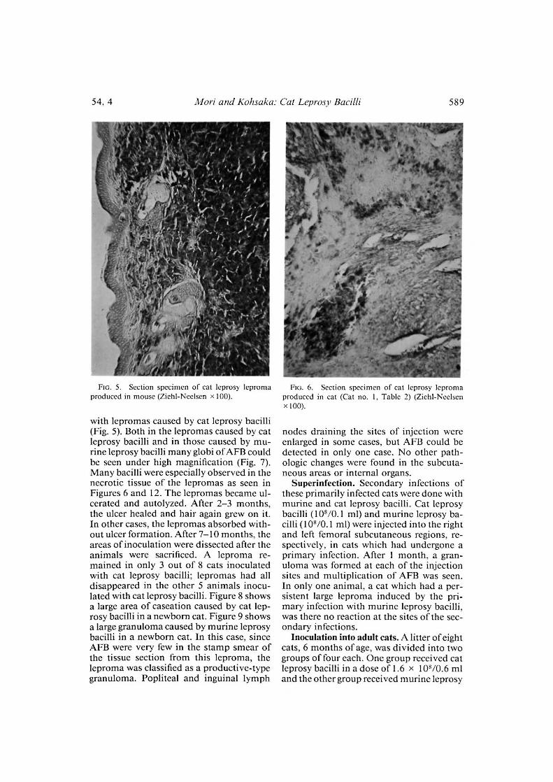
FIG. 5. Section specimen of cat leprosy lepromaproduced in mouse (Ziehl-Neelsen x 100).
with lcpromas caused by cat leprosy bacilli(Fig. 5). Both in the lepromas caused by catleprosy bacilli and in those caused by mu-rine leprosy bacilli many globi of AFB couldbe seen under high magnification (Fig. 7).Many bacilli were especially observed in thenecrotic tissue of the lepromas as seen inFigures 6 and 12. The lepromas became ul-cerated and autolyzed. After 2-3 months,the ulcer healed and hair again grew on it.In other cases, the lepromas absorbed with-out ulcer formation. After 7-10 months, theareas of inoculation were dissected after theanimals were sacrificed. A leproma re-mained in only 3 out of 8 cats inoculatedwith cat leprosy bacilli; lepromas had alldisappeared in the other 5 animals inocu-lated with cat leprosy bacilli. Figure 8 showsa large area of caseation caused by cat lep-rosy bacilli in a newborn cat. Figure 9 showsa large granuloma caused by murine leprosybacilli in a newborn cat. In this case, sinceAFB were very few in the stamp smear ofthe tissue section from this leproma, theleproma was classified as a productive-typegranuloma. Popliteal and inguinal lymph
FIG. 6. Section specimen of cat leprosy lepromaproduced in cat (Cat no. 1, Table 2) (Ziehl-Neelsenx 100).
nodes draining the sites of injection wereenlarged in some cases, but AFB could bedetected in only one case. No other path-ologic changes were found in the subcuta-neous areas or internal organs.
Superinfection. Secondary infections ofthese primarily infected cats were done withmurine and cat leprosy bacilli. Cat leprosybacilli (108/0.1 ml) and murine leprosy ba-cilli (108/0.1 ml) were injected into the rightand left femoral subcutaneous regions, re-spectively, in cats which had undergone aprimary infection. After 1 month, a gran-uloma was formed at each of the injectionsites and multiplication of AFB was seen.In only one animal, a cat which had a per-sistent large leproma induced by the pri-mary infection with murine leprosy bacilli,was there no reaction at the sites of the sec-ondary infections.
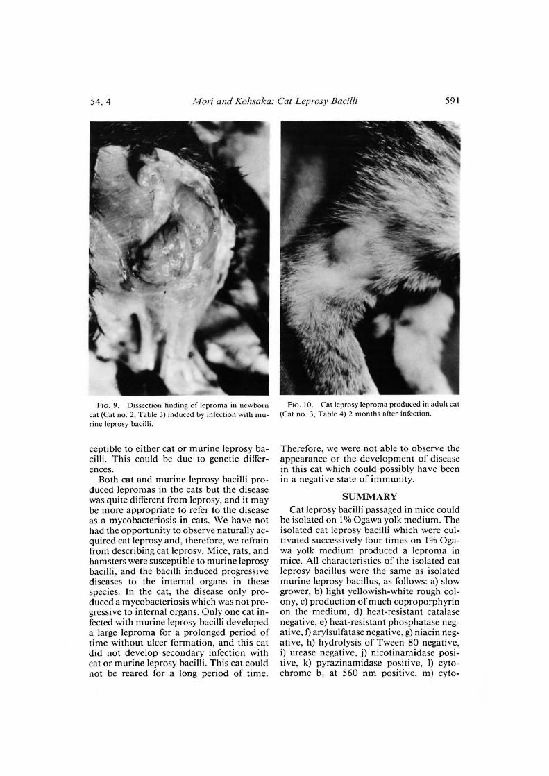
Inoculation into adult cats. A litter of eightcats, 6 months of age, was divided into twogroups of four each. One group received catleprosy bacilli in a dose of 1.6 x 10⁸/0.6 mland the other group received murine leprosy

590^ International Journal of Leprosy^ 1986
FIG. 7. Section specimen of cat leprosy lepromaproduced in cat (Cat no. 1, Table 2) (Ziehl-Neelsenx 1000).
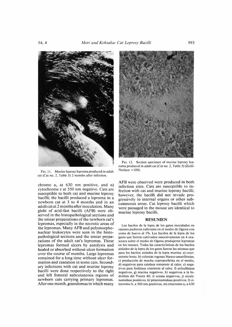
bacilli in a dose of 1.5 x 10⁸/0.5 ml injectedinto the femoral subcutaneous region. Re-sults of individual cats are shown in Tables4 and 5. All cats showed a reddish enlargedleproma at the injection region after 2months (Figs. 10 and 11). Biopsies were tak-en from the lepromas and a smear prepa-ration and tissue section prepared from each.In histopathologic findings, few AFB wereseen but neutrophilic infiltration was seen(Fig. 13). Many AFB were seen in the ne-crotic areas or the tissue section of the lep-roma, but there were no AFB detected inproductive areas of the granuloma withoutnecrosis (Fig. 12). After 3-5 months, thesites of the injections were dissected, andlepromas and AFB were found in all of thedissected cases. Figure 14 shows the lep-roma produced in an adult cat with murineleprosy bacilli. Popliteal and inguinal lymphnodes were enlarged in some cases, but AFBwere not detected in any cases. There wereno new lepromas or other pathologic changesfound in any of the subcutaneous areas orthe internal organs.
FIG. 8. Dissection finding of caseation in newborncat (Cat no. 6, Table 2) induced by infection with catleprosy bacilli.
DISCUSSIONIt was difficult to distinguish between cat
and murine leprosy bacilli based on the bio-chemical properties. We therefore initiatedanimal inoculations into cats with both catand murine leprosy bacilli. Our resultsshowed that the cat is a susceptible animalto murine leprosy bacilli. Leiker (4) reportedthat cats are resistant to cat leprosy bacilli.However, the present results show that new-born cats are fairly susceptible to the catleprosy bacillus, at least after having beenpassed for a number of years in mice. Pri-mary lepromas were produced in newborncats in 3-4 months, but only 2 months wererequired to produce lepromas in adult cats.This difference may be because adult catsmount a stronger tissue reaction, makingthe primary leproma more evident at anearlier stage. There were more AFB in thelepromas of newborn cats than in those ofadult cats. Some newborn cats were not sus-
54, 4^Mori and Kohsaka: Cat Leprosy Bacilli^ 591
FIG. 9. Dissection finding of leproma in newborncat (Cat no. 2, Table 3) induced by infection with mu-rine leprosy bacilli.
ceptible to either cat or murine leprosy ba-cilli. This could be due to genetic differ-ences.
Both cat and murine leprosy bacilli pro-duced lepromas in the cats but the diseasewas quite different from leprosy, and it maybe more appropriate to refer to the diseaseas a mycobacteriosis in cats. We have nothad the opportunity to observe naturally ac-quired cat leprosy and, therefore, we refrainfrom describing cat leprosy. Mice, rats, andhamsters were susceptible to murine leprosybacilli, and the bacilli induced progressivediseases to the internal organs in thesespecies. In the cat, the disease only pro-duced a mycobacteriosis which was not pro-gressive to internal organs. Only one cat in-fected with murine leprosy bacilli developeda large leproma for a prolonged period oftime without ulcer formation, and this catdid not develop secondary infection withcat or murine leprosy bacilli. This cat couldnot be reared for a long period of time.
FIG. 10. Cat leprosy leproma produced in adult cat(Cat no. 3, Table 4) 2 months after infection.
Therefore, we were not able to observe theappearance or the development of diseasein this cat which could possibly have beenin a negative state of immunity.
SUMMARYCat leprosy bacilli passaged in mice could
be isolated on 1% Ogawa yolk medium. Theisolated cat leprosy bacilli which were cul-tivated successively four times on 1% Oga-wa yolk medium produced a leproma inmice. All characteristics of the isolated catleprosy bacillus were the same as isolatedmurine leprosy bacillus, as follows: a) slowgrower, b) light yellowish-white rough col-ony, c) production of much coproporphyrinon the medium, d) heat-resistant catalasenegative, e) heat-resistant phosphatase neg-ative, f) arylsulfatase negative, g) niacin neg-ative, h) hydrolysis of Tween 80 negative,i) urease negative, j) nicotinamidase posi-tive, k) pyrazinamidase positive, 1) cyto-chrome b, at 560 nm positive, m) cyto-

592^ International Journal of Leprosy^ 1986
TABLE 4.^Adult cats infected with cat leprosy bacilli.
Dissection findings
Lymph nodesAni-^AFB^Killed^Infected region^ Spleen/
^
Infection^ Popliteal^Inguinalma!^smear (mos.) ^liverCase-^Leproma^BacilliEn-^En-^changesation^Bacilli^Bacillilarged^larged
No. I,^Leproma,^+^5^i^Id^reddish,
enlarged,2 mos.;polymor-phonuclearleukocyte++
No. 2,^Leproma,^+^3^ +d^enlarged,
2 mos.; notreddish
No. 3,^Leproma,^+^3^++ +^—^+^+^+2^reddish,
enlarged,2 mos.;polymor-phonuclearleukocyte++
No. 4,^Leproma,^+^5^+++^++^+^—^+2^enlarged,
2 mos.; notreddish
TABLE 5. Adult cats infected with murine leprosy bacilli.
Dissection findings
^
Infected region^Lymph nodesAni-^ AFB^Killed ^
^
Infection^ Spleen/ma!^ smear^(mos.) '
Lep-^Case-^Popliteal^Inguinal liver
Bacilli^En-
^
roma^ation^ Bacilli changes
Bacilli.^En-larged^larged
No. I,^Leproma,^+^5^+^+^+d^reddish,
enlarged, 2mos.; poly-morphonu-clear leuko-cyte ++
No. 2,^Leproma,^+ +^5^+^+^+^—^+d^reddish,
enlarged, 2mos.
No. 3,^Leproma,^++^3^+^+^+2^reddish,
enlarged, 2mos.
No. 4,^No leproma^3^+^+2
54, 4^Mori and Kohsaka: Cat Leprosy Bacilli^593
FIG. 11. Murine leprosy leproma produced in adult
cat (Cat no. 2, Table 5) 2 months after infection.
FIG. 12. Section specimen of murine leprosy lep-roma produced in adult cat (Cat no. 2, Table 5) (Zichl-
Ncelsen x 100).
chrome a, at 630 nm positive, and n)cytochrome c at 550 nm negative. Cats aresusceptible to both cat and murine leprosybacilli; the bacilli produced a leproma in anewborn cat at 3 to 4 months and in anadult cat at 2 months after inoculation. Manyglobi of acid-fast bacilli (AFB) were ob-served in the histopathological sections andthe smear preparations of the newborn cat'slepromas, especially in the necrotic areas ofthe lepromas. Many AFB and polymorpho-nuclear leukocytes were seen in the histo-pathological sections and the smear prepa-rations of the adult cat's lepromas. Theselepromas formed ulcers by autol■,,sis andhealed or absorbed without ulcer formationover the course of months. Large lepromasremained for a long time without ulcer for-mation and caseation in some cats. Second-ary infections with cat and murine leprosybacilli were done respectively to the rightand left femoral subcutaneous regions ofnewborn cats carrying primary lepromas.After one month, granulomas in which many
AFB were observed were produced in bothinfection sites. Cats are susceptible to in-fection with cat and murine leprosy bacilli;however, the bacilli did not invade pro-gressively to internal organs or other sub-cutaneous areas. Cat leprosy bacilli whichwere passaged in the mouse are identical tomurine leprosy bacilli.
RESUMENLos bacilos de la lepra de los gatos inoculados en
ratones pudieron cultivarse en el medio de Ogawa conyema de huevo al 1%. Los bacilos de la lepra de los
gatos que fueron cultivados suscesivamente en 4 oca-
siones sobre el mcdio de Ogawa produjeron lepromas
en los ratones. Todas las caracteristicas de los bacilosaislados de la lepra de los gatos fueron las mismas que
para los bacilos aislados de la lepra murina: a) creci-miento lento, b) colonias rugosas blanco-amarillentas,
c) producciOn de mucha coproporfirina en el medio,
d) negativos para catalasa resistente al calor, e) nega-tivos para fosfatasa resistente at calor, 0 arilsulfatasa
negativos, g) niacina negativos, h) negativos a la hi-drOlisis del Tween 80, i) ureasa negativos, j) nicoti-namidasa positivos, k) pirazinamidasa positivos, I) ci-
tocromo b, a 560 nm positivos, m) citocromo a, a 630

594^ International Journal of Leprosy^ 1986
FIG. 13. Section specimen of murine leprosy lep-roma produced in adult cat (Cat no. 2, Table 5) (Ziehl-Neelsen x 1000). FIG. 14. Dissection finding of leproma in adult cat
(Cat no. 2, Table 5) induced by infection of murineleprosy bacilli.
nm positivos, y n) citocromo c a 550 nm negativos.Los gatos son susceptibles a los bacilos de la lepra delos gatos y de los ratones; los bacilos producen unleproma en un gato recién nacido a los 3 o 4 meses yen un gato adulto a los 2 meses después de la inocu-lación. En las secciones histopatológicas y en los ex-tendidos de los lepromas de los gatos recién nacidosse observaron muchas globias de bacilos acid° resis-tentes (BAR), especialmente en las areas necróticas delos lepromas. En las secciones histopatolOgicas y en losextendidos de los lepromas de los gatos adultos, seobservaron muchos BAR y leucocitos PMN. Estos lep-romas formaron Ulceras por autolisis y sanaron o seabsorbieron sin formación de (ilcera a lo largo de variosmeses en algunos gatos. También se indujeron infec-ciones secundarias con los bacilos de la lepra murinay de los gatos en las regiones femorales derecha e iz-quierda, respectivamente, de gatos recién nacidos por-tadores de lepromas primarios. Después de 1 mes, seobservaron granulomas con muchos BAR en ambossitios de infección. Los gatos son susceptibles a la in-fección con los bacilos de la lepra de los gatos y de losratones, sin embargo, los bacilos no invaden ni losOrganos internos ni otras areas subcutaneas. Los ba-cilos de la lepra de los gatos que fueron pasados porratones, son idénticos a los bacilos de la lepra murina.
RESUMEDes bacilles de la lépre du chat ont pu 'etre isolés sur
un milieu au jaune d'oeuf d'Ogawa, aprés passage chezla souris. Les bacilles de la lépre du chat ainsilorsqu'ils avaient été cultivés avec succés quatre foissur un milieu A 1% au jaune d'oeuf d'Ogawa, ont en-trainé un léprome chez la souris. Toutes les caracté-ristiques relevées chez les bacilles de la lepre du chatainsi isolés, étaient semblables a celles observées chezle bacille de la lépre murine, A savoir: a) une croissancelente; b) des colonies rugueuses de couleur blanc jau-ndtre pale; c) la production d'une grande quantité decoproporphyrine dans le milieu; d) l'absence de pro-duction de catalase résistante A la chaleur; e) l'absencede production de phosphatase résistante A la chaleur;
l'absence de production d'arylsulfatase; g) l'absencede production de niacine; h) l'absence d'hydrolyse duTween 80; i) l'absence de production d'uréase; j) laproduction de nicotinamidase; k) la production de py-razinamidase; I) la mise en evidence de cytochrome b,A 560 nm; m) la mise en evidence de cytochrome a2 A630 nm; n) l'absence de cytochrome c a 550 nm. Leschats sont susceptibles A la fois aux bacilles de la lépredu chat et A ceux de la lépre murine. Les bacilles en-

54, 4^Mori and Kohsaka: Cat Leprosy Bacilli^595
trainent la production d'un léprome chez le chat nou-veau-ne trois A quatre mois apres l'inoculation, et deuxmois apres chez le chat adulte. On a observe dc nom-brcux globi de bacilles acido-résistants (AFB) dans lescoupes histopathologiques et dans les preparations defrottis obtenues A partir de lepromes de chats nouveau-nes, spécialement lorsque ces biopsies etaient prélevéesdans les zones nécrotiques des lépromes. On a &gale-ment pu observer de nombreux bacilles acido-résis-tants et des lcucocytes polymorphonucléaires dans lescoupes histopathologiques et dans les preparations defrottis recueillies au niveau de lépromes de chats adultes.Ces lépromes produisent des ulceres par autolyse; usguérissent ou sont résorbes au cours des mois sansformation d'ulcere. Chez certains chats, des lépromesétendus peuvent persister pendant longtemps, sans for-mation d'ulcere ni caseification. On a provoque desinfections secondaires avec des bacilles de la lepre duchat et des bacilles murins, respectivement dans lesregions sous-cutanées femorales droite et gauche, chezdes chats nouveau-nes porteurs de lepromes primaires.Apres un mois, on a observe aux deux endroits d'in-fection la formation de granulomes, avec nombreuxbacilles acido-résistants. Les chats sont done suscep-tibles A une infection par les bacilles de la lepre du chatet par ceux de la lepre murine; les bacilles, toutefois,n'envahissent pas progressivement les organes internesou d'autres regions cutanées. Apres passage chez lasouris, les bacilles de la lepre du chat sont identiquesaux bacilles de la lepre murine.
Acknowledgments. This work was partially support-ed by the Sasakawa Memorial Health Foundation andgrants from the U.S.—Japan Cooperative Medical Sci-
ence Program and the Osaka Dermatological Foun-dation.
REFERENCESI . BROWN, L. R., MAY, C. D. and WILLIAMS, S. E. A
non-tuberculous granuloma in cats. N.Z. Vet. J.10 (1962) 7-9.
2. Holt', M. Differential identification ofacid-fast ba-cillus isolated from clinical materials. Kekkaku 51(1976) 244-256.
3. LAWRENCE, W. E. and WICKHAM, N. Cat leprosy:infection by a bacillus resembling Mycobacteriumlepraemuriunz. Aust. Vet. J. 39 (1963) 390-393.
4. LEIKER, D. L. and POELMA, F. G. On the etiologyof cat leprosy. Int. J. Lepr. 42 (1974) 3 I 2-315.
5. MoRI, T. Successive cultivation of M. lepraelnu-'lion by bacterial mass inoculation. Lepro 44 (1975)49-54.
6. MORI, T. Biochemical properties ofcultivated My-cobacterium lepraemurium. Int. J. Lepr. 43 (1975)210-217.
7. NICHOLAS, R. E. H. and RIMINGTON, C. Qualita-tive analysis of porphyrins by partition chroma-tography. Scand. J. Clin. Lab. Invest. 1 (1949) 12—18.
8. OGAWA, T. and MOTOMURA, K. Studies on My-cobacterium lepraemurium. First report: attemptsto cultivate Al. lepraemurium. Lepro 38 (1969)246-254.
9. POELMA, F. G. and LEIKER, D. L. Cat leprosy inThe Netherlands. Int. J. Lepr. 42 (1974) 307-311.
10. WILKINSON, G. T. A nontubereulous granuloma ofthe cat associated with an acid-fast bacillus. Vet.Rec. 76 (1964) 777-778 and 833-834 (Letter).