identifiers: nct03664193 unique protocol id: 1802019010
TRANSCRIPT

Identifiers: NCT03664193 Unique Protocol ID: 1802019010
Brief Title: MRI-guided Stereotactic Body Radiotherapy (SBRT) With Simultaneous Integrated Boost for Prostate Cancer (SIBRT)
Version 3.1 Version date: 20FEB2019 IRB approval: 29MAY2019
Document History
Version Version date Amendment 4 3.1 02.20.2019
Amendment 3 (Protocol) 3.0 01.28.2019 Amendment 2 (Protocol) 2.1 10.30.2018 Amendment 1 Protocol 2.0 10.05.2018
Initial Protocol 1.0 06.13.2018

TITLE: MRI-guided Stereotactic Body Radiotherapy (SBRT) with Simultaneous Integrated Boost for Prostate Cancer
IRB Protocol #: 1802019010 Version 3.1 Version Date : 02/20/2019 Principal Investigator: Josephine Kang,M.D Co-Principal Investigator: Co- Investigators:
Protocol # 1802019010 Version Date 02/20/2019
2
Nurse Practitioner: Study Coordinator: Sharanya Chandrasekhar, M.S, CCRP [email protected]

Protocol # 1802019010 Version Date 02/20/2019
3
Data Coordinator: Pragya Yadav, PhD [email protected] Statistician:

Protocol # 1802019010 Version Date 02/20/2019
4
Confidentiality Statement
This document is confidential and is to be distributed for review only to investigators, potential investigators, consultants, study staff, and applicable independent ethics committees or institutional review boards. The contents of this document shall not be disclosed to others without written authorization from WCMC.
Protocol # 1802019010 Version Date 02/20/2019
5
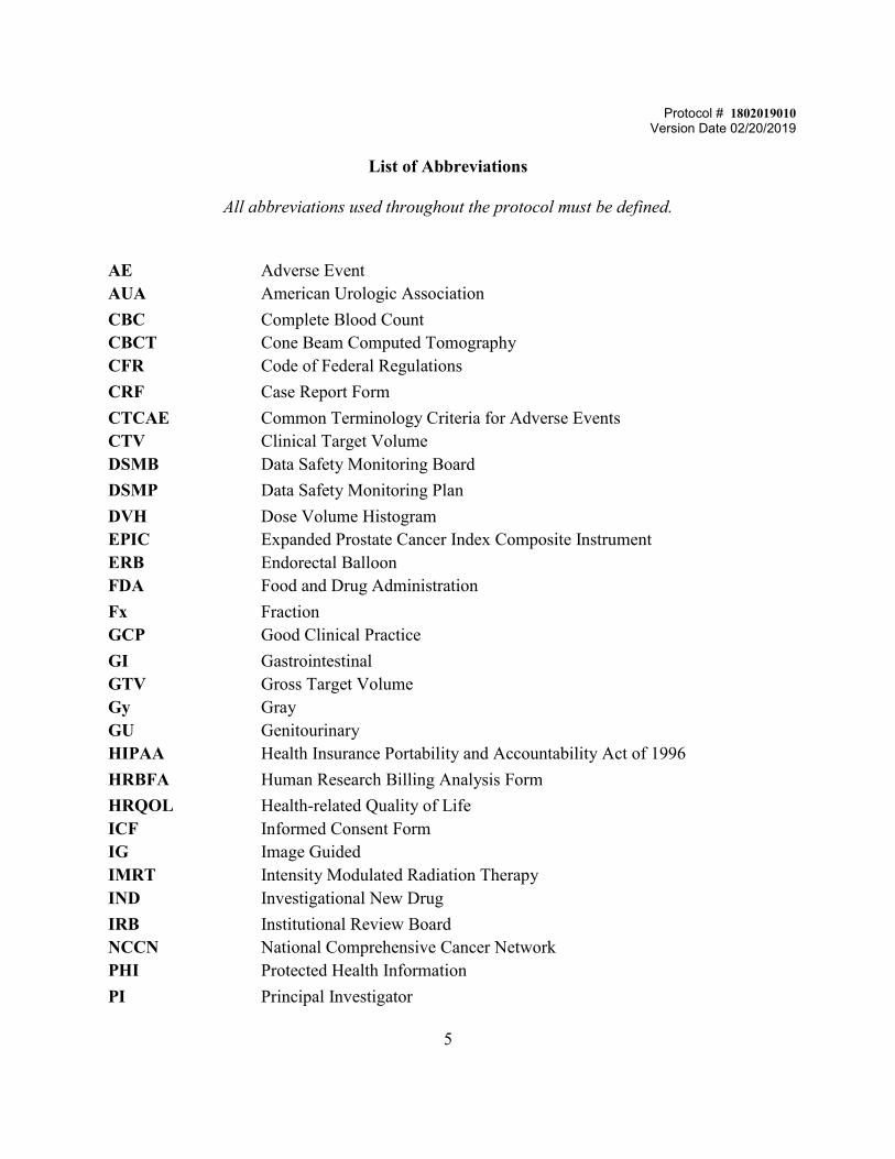
List of Abbreviations
All abbreviations used throughout the protocol must be defined.
AE AUA
Adverse Event American Urologic Association
CBC CBCT CFR
Complete Blood Count Cone Beam Computed Tomography Code of Federal Regulations
CRF Case Report Form CTCAE CTV DSMB
Common Terminology Criteria for Adverse Events Clinical Target Volume Data Safety Monitoring Board
DSMP Data Safety Monitoring Plan DVH EPIC ERB FDA
Dose Volume Histogram Expanded Prostate Cancer Index Composite Instrument Endorectal Balloon Food and Drug Administration
Fx GCP
Fraction Good Clinical Practice
GI GTV Gy GU HIPAA
Gastrointestinal Gross Target Volume Gray Genitourinary Health Insurance Portability and Accountability Act of 1996
HRBFA Human Research Billing Analysis Form HRQOL ICF IG IMRT IND
Health-related Quality of Life Informed Consent Form Image Guided Intensity Modulated Radiation Therapy Investigational New Drug
IRB NCCN PHI
Institutional Review Board National Comprehensive Cancer Network Protected Health Information
PI Principal Investigator

Protocol # 1802019010 Version Date 02/20/2019
6
PIRADS PSA PSMA PTV REDCap
Prostate Imaging Reporting and Data System Prostate Specific Antigen Prostate Specific Membrane Antigen Planning Target Volume Research Electronic Data Capture
RS RT SAE SBRT SHIM SUSAR TRUS UAP WCMC
Rectal Spacer Radiation Treatment Serious Adverse Event Stereotactic Body Radiation Therapy Sexual Health Inventory Men Suspected Unexpected Serious Adverse Reaction Transrectal Ultrasound Unanticipated Problem Weill Cornell Medical College

Protocol # 1802019010 Version Date 02/20/2019
7
Document History
Version Version date Amendment 4 3.1 02.20.2019 Amendment 3 (Protocol) 3.0 01.28.2019 Amendment 2 (Protocol) 2.1 10.30.2018 Amendment 1 Protocol 2.0 10.05.2018 Initial Protocol 1.0 06.13.2018
Summary of changes : Protocol version 3.1 02/20/2019
1. Adding Co-investigators Dr. Vishesh Agrawal and Dr. Reza Farjam. NO CHANGES TO THE INFORMED CONSENT. Summary of changes : Protocol version 3.0 01/28/2019 1. Clarification made in the protocol to clarify how the radiation dose is being prescribed and delivered, as follows: Page 22, section 5.3.2; also page 10 schema: We have clarified that the dose of 35 Gy in 5 fractions is the minimum mean dose to be achieved. The prescribed dose can be 7-7.25 Gy; this is reflected throughout the protocol. 2. On page 19, we have removed the exclusion criteria which previously excluded any patients who had a prior TURP (transurethral resection of the prostate), based on recent data suggesting that patients with TURP are not at increased risk of significant toxicitiy after radiation 3. We have removed the SHIM questionnaire as it is redundant with the EPIC 26 quality of life questionnaire 4. We are adding Jonathan Chen and Daniel Vanderbilt as co-investigators informed consent changes: SHIM questionnaire has been removed from the consent. Summary of changes : Protocol version 2.1 10.30.2018
1. Radiation treatment planning : Contour section 5.3.1 and 5.3.2 respectively.
Protocol # 1802019010 Version Date 02/20/2019
8
a. Select PIRADS 3-5 and/or PSMA avid (if PSMA PET/MR is available) and/or
biopsy positive nodules (chosen based on physician's clinical judgment and correlation with biopsy findings)
b. The CTV (prostate +/- SV) will be treated to dose of 35 Gy in 5 fractions. There will be a simultaneous integrated boost to PIRADS 3-5 or PSMA avid nodules, delivering at additional 0.5, 1.0, 1.5 or 2 Gy per fraction for total mean dose of 37.5, 40, 42.5 or 45 Gy to the contoured nodule.
2. Inclusion Criteria #4 :
a. Patient must have prostate MRI with a PIRADS 3-5 lesion or PSMA PET with an avid intraprostatic lesion
There is no changed to the informed consent. Summary of changes : Protocol version 2.0 10.05.2018
1. Changing the follow up from 1 month to 3-6 months 2. Radiation treatment planning – Contouring – Section 5.3.1
a. Select PIRADS 4-5 and/or PSMA avid (if PSMA PET/MR is available) and/or
biopsy positive nodules (chosen based on physician's clinical judgment and correlation with biopsy findings) will be contoured as gross tumor volume(s) (GTV).
ICF update 10.05.2018 : ICF was also updated to revise the follow up information from one month post RT to 3-6 months.

9
Protocol Summary MRI-guided Stereotactic Body Radiotherapy (SBRT) with Simultaneous Integrated Boost for Prostate Cancer IRB Protocol #: 1802019010 Short Title: MRI-guided prostate SBRT with Focal Boost Principal Investigator: Josephine Kang Sample Size: N = 30 Accrual Ceiling: This study plans to enroll a total of 30 patients Study Population: Patients with prostate cancer with visible focal lesion on MRI Accrual Period: 3 years Study Design: Single-arm study for feasibility Study Intervention Description: Prostate SBRT: Prostate SBRT is a standard of care treatment for prostate cancer that has not spread to distant metastatic sites. Radiation is delivered to the prostate and seminal vesicles in 5 treatment sessions (fractions). Doses ranging from 35-45 Gy in 5 fractions have demonstrated good outcomes with acceptable toxicity. In this initial study, MRI guided treatment planning and delivery will be used to deliver 7-7.25 Gy to the entire prostate and seminal vesicles, with a selective boost of additional 0.5, 1.0, 1.5 or 2 Gy per fraction for total dose of 37.5, 40, 42.5 or 45 Gy to biopsy proven lesions, defined using MRI. Hypothesis: MRI-guided treatment planning and delivery can selectively target high-risk prostate nodules and deliver a higher radiation dose, to achieve maximal local control without increasing treatment toxicity Primary objective: To assess feasibility and safety of delivering MRI-guided prostate SBRT with a simultaneous integrated boost to MRI-visible lesions.
Feasibility is defined as a plan meeting treatment planning objectives and normal tissue constraints. Specifically the dose distribution of the MRI-guided technique, will be assessed with regard to fulfilling protocol requirements of: a) coverage of the planning target volume (PTV) , b) constraints to organs at risk (OAR) by dose volume histogram (DVH) analysis and whether this can be done through standard set-up techniques or non-invasively through MRI based planning, c) coverage of high-risk prostate nodules. The total dose (either 37.5, 40, 42.5 or 45 Gy) will be determined by selecting the plan that delivers the highest dose to contoured visible nodules while maintaining normal tissue constraints to the urethra and rectum. We will track the number of treatment plans for each dose level (37.5, 40, 42.5 or 45 Gy) that can be delivered while meeting radiation planning objectives.

Protocol # 1802019010 Version Date 02/20/2019
10
Safety of this approach will be determined by assessing acute GI/GU toxicity (CTCAE 4.0 criteria). Treatment will be deemed safe of there is no acute >G3 GI/GU toxicity within the first 30 days of treatment (CTCAE 4.0 criteria). Given that the treatment plans will meet rigorous normal tissue constraints used for standard prostate SBRT, we do not anticipate any increased toxicity on this study compared to standard prostate SBRT.
Secondary objectives:
1. To obtain health-related quality of life (HRQOL) measured using: 1) the Expanded Prostate Cancer Index Composite (EPIC) short form questionnaire, 2) AUA. Assessment will occur at baseline, and at first follow up.

Protocol # 1802019010 Version Date 02/20/2019
11
SCHEMA
Blood samples for immune monitoring studies and stool samples for microbiome studies will be collected at baseline, end of Radiation at 3-6 months post follow up visit.
Localized Prostate Cancer with MRI (PIRADS 3-5) or PSMA PET-visible lesions
(Low, Intermediate and High Risk as per NCCN
Criteria)
Prostate SBRT (35-36.25 Gy in 5 fx)
w/SIB of 0.5-2 Gy per fx for total dose 37.5, 40, 42.5 or 45 Gy
Patients will have option of rectal spacer or balloon prior to treatment
Follow up visit at 1 month: Assess GI/GU toxicity, EPIC, AUA

Protocol # 1802019010 Version Date 02/20/2019
12
TABLE OF CONTENTS
PROTOCOL SUMMARY ............................................................................................................................................................. 9
SCHEMA ...................................................................................................................................................................................... 11
1 STUDY OBJECTIVES ..................................................................................................................................................... 14
1.1 PRIMARY OBJECTIVES ...................................................................................................................................................................... 14 1.2 SECONDARY OBJECTIVES ................................................................................................................................................................. 14
2 BACKGROUND ............................................................................................................................................................... 14
2.1 PROSTATE CANCER RADIOTHERAPY ............................................................................................................................................... 14 2.2 BIOLOGICAL RATIONALE FOR SBRT .............................................................................................................................................. 15 2.3 SBRT OUTCOMES ............................................................................................................................................................................. 16 2.4 RATIONALE FOR SIMULTANEOUS INTEGRATED BOOST .............................................................................................................. 17 2.5 NEED FOR IGRT IN PROSTATE RADIOTHERAPY ........................................................................................................................... 18 2.6 VIEWRAY: A UNIQUE RADIATION DELIVERY APPROACH ............................................................................................................. 18 2.8 RATIONALE FOR CORRELATIVE STUDIES BACKGROUND: MICROBIOME AND BLOOD SAMPLE COLLECTION .......................... 19
3 SUBJECT SELECTION ................................................................................................................................................... 20
3.1 STUDY POPULATION ......................................................................................................................................................................... 20 3.2 INCLUSION CRITERIA ........................................................................................................................................................................ 20 3.3 EXCLUSION CRITERIA ....................................................................................................................................................................... 20
4 REGISTRATION PROCEDURES ................................................................................................................................. 20
4.1 PATIENT REGISTRATION .................................................................................................................................................................. 20
5 STUDY PROCEDURES .................................................................................................................................................. 22
5.1 PRE-STUDY VISIT .............................................................................................................................................................................. 22 5.2 IMAGING STUDIES ............................................................................................................................................................................. 22 5.3 RADIATION TREATMENT PLANNING ............................................................................................................................................. 23
5.3.1 Contours ............................................................................................................................................................................... 23 5.3.2 Dose/Treatment Planning Parameters .................................................................................................................... 23
5.4 SUPPORTIVE CARE GUIDELINES ..................................................................................................................................................... 24 5.5 DURATION OF THERAPY AND CRITERIA FOR REMOVAL FROM STUDY ..................................................................................... 24 5.6 DURATION OF FOLLOW UP .............................................................................................................................................................. 24
6 DOSE MODIFICATIONS ............................................................................................................................................... 25
7 ADVERSE EVENT REPORTING REQUIREMENTS ................................................................................................ 25
7.1 ADVERSE EVENT DEFINITION ......................................................................................................................................................... 25 7.2 RECORDING OF ADVERSE EVENTS .................................................................................................................................................. 25
7.2.1 Reporting of AE to WCMC IRB ..................................................................................................................................... 25 7.3 DEFINITION OF SAE ......................................................................................................................................................................... 26
7.3.1 Reporting of SAE to IRB .................................................................................................................................................. 26 7.4 EXPEDITED ADVERSE EVENT REPORTING .................................................................................................................................... 26
7.4.1 AE/SAE Follow Up ............................................................................................................................................................ 27
8 PHARMACEUTICAL INFORMATION ....................................................................................................................... 27
9 CORRELATIVE/SPECIAL STUDIES .......................................................................................................................... 27
10 MEASUREMENT OF EFFECT ...................................................................................................................................... 28

Protocol # 1802019010 Version Date 02/20/2019
13
11 DATA REPORTING / REGULATORY CONSIDERATIONS .................................................................................. 29
11.1 DATA COLLECTION ...................................................................................................................................................................... 29 11.2 REDCAP ....................................................................................................................................................................................... 29 11.3 REGULATORY CONSIDERATIONS ............................................................................................................................................... 29 11.4 DATA MANAGEMENT .................................................................................................................................................................. 29
12 STATISTICAL CONSIDERATIONS ............................................................................................................................ 30
12.1 STUDY DESIGN/ENDPOINTS ..................................................................................................................................................... 30 12.1.1 Primary Objectives ..................................................................................................................................................... 30 12.1.2 Secondary Objectives ................................................................................................................................................. 30 12.1.3 Correlative Study Objectives ................................................................................................................................... 31
12.2 SAMPLE SIZE/ACCRUAL RATE .................................................................................................................................................. 31 12.3 STRATIFICATION FACTORS ........................................................................................................................................................ 31 12.4 ANALYSIS OF PRIMARY AND SECONDARY ENDPOINTS .......................................................................................................... 31
13 DATA AND SAFETY MONITORING PLAN (DSMP) .............................................................................................. 32
13.1 MONITORING PLAN ..................................................................................................................................................................... 32 13.2 STOPPING RULES ......................................................................................................................................................................... 32
14 REFERENCES .................................................................................................................................................................. 33

Protocol # 1802019010 Version Date 02/20/2019
14
1 STUDY OBJECTIVES HYPOTHESIS: MRI-guided treatment planning and delivery can selectively target high-risk prostate nodules and deliver a higher radiation dose, to achieve maximal local control without increasing treatment toxicity 1.1 Primary Objectives
1.11 Determine the feasibility and safety of delivering SBRT w/SIB without increased acute G3 GI/GU toxicity. Feasibility is defined as a plan meeting treatment planning objectives and normal tissue constraints. Specifically the dose distribution of the MRI-guided technique, will be assessed with regard to fulfilling protocol requirements of: a) coverage of the planning target volume (PTV) , b) constraints to organs at risk (OAR) by dose volume histogram (DVH) analysis and whether this can be done through standard set-up techniques or non-invasively through MRI based planning, c) coverage of high-risk prostate nodules. The total dose delivered (either 37.5, 40, 42.5 or 45 Gy mean dose to the contoured nodule) will be determined by selecting the plan with the highest dose to visible nodules while maintaining normal tissue constraints to the urethra and rectum. We will track the number of treatment plans for each dose level (37.5, 40, 42.5 or 45 Gy) that can be delivered while planning objectives. Safety of this approach will be determined by assessing acute GI/GU toxicity (CTCAE 4.0 criteria). Treatment will be deemed safe of there is no acute >G3 GI/GU toxicity within the first 30 days of treatment (CTCAE 4.0 criteria). Given that the treatment plans will meet standard normal tissue constraints used for prostate SBRT, we do not anticipate any increased toxicity on this study compared to standard prostate SBRT. 1.2 Secondary Objectives 1.21 To obtain health-related quality of life (HRQOL) measured using: 1)the Expanded Prostate Cancer Index Composite (EPIC) short form questionnaire, 2) AUA. Assessment will occur at baseline, and at first follow up which will occur 3-6 months after completion of radiation. 2 BACKGROUND 2.1 Prostate cancer radiotherapy
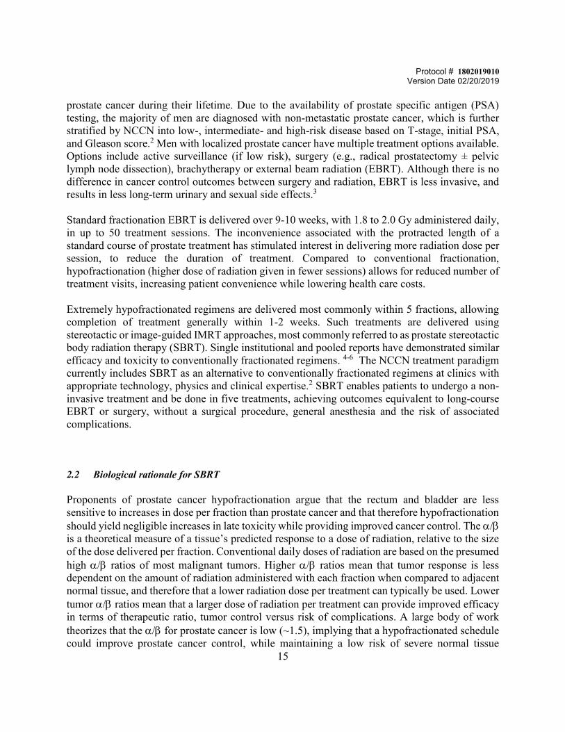
Next to skin cancer, prostate cancer is the most commonly diagnosed cancer in men, and second leading cause of cancer death.1 It is estimated that one out of seven men will be diagnosed with
Protocol # 1802019010 Version Date 02/20/2019
15
prostate cancer during their lifetime. Due to the availability of prostate specific antigen (PSA) testing, the majority of men are diagnosed with non-metastatic prostate cancer, which is further stratified by NCCN into low-, intermediate- and high-risk disease based on T-stage, initial PSA, and Gleason score.2 Men with localized prostate cancer have multiple treatment options available. Options include active surveillance (if low risk), surgery (e.g., radical prostatectomy ± pelvic lymph node dissection), brachytherapy or external beam radiation (EBRT). Although there is no difference in cancer control outcomes between surgery and radiation, EBRT is less invasive, and results in less long-term urinary and sexual side effects.3 Standard fractionation EBRT is delivered over 9-10 weeks, with 1.8 to 2.0 Gy administered daily, in up to 50 treatment sessions. The inconvenience associated with the protracted length of a standard course of prostate treatment has stimulated interest in delivering more radiation dose per session, to reduce the duration of treatment. Compared to conventional fractionation, hypofractionation (higher dose of radiation given in fewer sessions) allows for reduced number of treatment visits, increasing patient convenience while lowering health care costs. Extremely hypofractionated regimens are delivered most commonly within 5 fractions, allowing completion of treatment generally within 1-2 weeks. Such treatments are delivered using stereotactic or image-guided IMRT approaches, most commonly referred to as prostate stereotactic body radiation therapy (SBRT). Single institutional and pooled reports have demonstrated similar efficacy and toxicity to conventionally fractionated regimens. 4-6 The NCCN treatment paradigm currently includes SBRT as an alternative to conventionally fractionated regimens at clinics with appropriate technology, physics and clinical expertise.2 SBRT enables patients to undergo a non-invasive treatment and be done in five treatments, achieving outcomes equivalent to long-course EBRT or surgery, without a surgical procedure, general anesthesia and the risk of associated complications. 2.2 Biological rationale for SBRT Proponents of prostate cancer hypofractionation argue that the rectum and bladder are less sensitive to increases in dose per fraction than prostate cancer and that therefore hypofractionation should yield negligible increases in late toxicity while providing improved cancer control. The / is a theoretical measure of a tissue’s predicted response to a dose of radiation, relative to the size of the dose delivered per fraction. Conventional daily doses of radiation are based on the presumed high / ratios of most malignant tumors. Higher / ratios mean that tumor response is less dependent on the amount of radiation administered with each fraction when compared to adjacent normal tissue, and therefore that a lower radiation dose per treatment can typically be used. Lower tumor / ratios mean that a larger dose of radiation per treatment can provide improved efficacy in terms of therapeutic ratio, tumor control versus risk of complications. A large body of work theorizes that the / for prostate cancer is low (~1.5), implying that a hypofractionated schedule could improve prostate cancer control, while maintaining a low risk of severe normal tissue

Protocol # 1802019010 Version Date 02/20/2019
16
complications (see Table 1). 7 The radiobiological linear-quadratic cell survival model was used to calculate the biologically equivalent doses for tumor control and complications to normal organs, using standard 2 Gy fractions delivered five times a week.8-10 Table 1. Equivalent total doses in 2 Gy per fraction (EQD2) based on / Biologically Effective Doses (BED)
2.3 SBRT Outcomes To date, results from prospective and retrospective studies demonstrate good biochemical control and low toxicity for SBRT, with commonly used doses ranging from 35-45 Gy in 5 fractions (see Table 2). SBRT compares favorably to outcomes for standard fractionation EBRT to 81 Gy, which has 10-year biochemical RFS of 91%, 78% and 62%, respectively, for low-, intermediate- and high-risk prostate cancer, and late Grade 3 gastrointestinal (GI) and genitourinary toxicity of 1% and 5%, respectively.11 There is a lack of consensus regarding the appropriate dose to use for prostate SBRT. Extrapolating from dose escalation studies in conventionally fractionated EBRT,12,13 a higher dose in SBRT is hypothesized to result in improved local control. However, delivery of higher dose results in small but significant increase in GU toxicity. Katz and Kang showed significantly higher late grade 2-3 GU toxicity with 36.25 Gy compared to 35 Gy (13.2 vs 8.8%, P<0.05). With 35 Gy in five fractions, the majority of toxicity was grade 2, and overall toxicity was low without impacting GU quality of life. As shown in Table 2, late grade 3 GU/GI toxicity after SBRT appears to be comparable to that of conventionally fractionated EBRT, ranging from 0.6 – 3% 4,6 with longest median follow-up of 7 years. Table 2. Biochechemical control after prostate SBRT. *prospective Study SBRT Dose
(5 fx) FU # Pts Risk Categories bPFS Toxicity ≥3
Bolzicco14*
35 Gy 36m 71 Low (41%), Int (42%), High (17%)
3y 94.4% GI 0%, GU 1%
King15* 35-36.25 Gy 5y 41 Low, Favorable Int Risk
5y 92.7% GI 0%, GU 3.5%
/ (Gy) EQD2 Standard (1.8
Gy x 45 fx)
EQD2 SBRT (7 Gy x 5 fx)
EQD2 SBRT (7.5 Gy x 5 fx)
EQD2 SBRT (8 Gy x 5 fx)
EQD2 SBRT (8.5 Gy x 5 fx)
EQD2 SBRT (9 Gy x 5 fx)
Total Dose (Gy) 81 35 37.5 40 42.5 45Tumor 1.5 76.37 Gy 85 Gy 97.65 Gy 110 Gy 123.07 Gy 135 GyFibrosis/stricture 2 76.95 Gy 78.75 Gy 89.06 Gy 100 Gy 111.56 Gy 123.7 GyTelangiectasia 4 78.3 Gy 64.17 Gy 71.88 Gy 80 Gy 88.54 Gy 97.5 GyRectum 4 78.3 Gy 64.17 Gy 71.88 Gy 80 Gy 88.54 Gy 97.5 GyBladder 4 78.3 Gy 64.17 Gy 71.88 Gy 80 Gy 88.54 Gy 97.5 Gy

Protocol # 1802019010 Version Date 02/20/2019
17
McBride16*
36.25-37.5 Gy
44.5m
45 Low 3y 97.7% GI 4.4%, GU 2.2%
Madsen1
7* 33.5 Gy 41m 40 Low 4y 90% GI 0%, GU
0% Fuller18
* 38 Gy/ 4 fx 60m 259 Low (43%), Int
(57%) 5y Low 100%, Int 88.5%
GI 0%, GU 3.1%
Kim19 45-50 Gy 42m 47 Low (38%), Int (62%) 4y 98% GI 0%, GU 0%
Loblaw2
0* 35 Gy 55m 84 Low 5y 98% GI 1%, GU
1% Mantz21
* 40 Gy 60m 102 Low, Fav Int 5y 100% GI 0%, GU
0% Chen22 35-36.5 Gy 2.3y 100 Low (37%), Int
(55%), High (8%) 2y 99% GI 0%, GU
1% Katz and Kang5
35-36.25 Gy 72m 477 Low (68%), Int (32%) 7y Low 95.6%, Fav Int 93.5%, Unfav Int 79.3%
GI 0%, GU 2%
Katz and Kang23
35-36.25 Gy 84m 515 Low (63%), Int (30%), High (7%)
8y Low 93.6%, Int 84.3%, High 65.0%
GI 0%, GU 2%
Katz24 35-36.25 Gy 108m 230 Low 10y 93% GI 0%, GU 2%
Abbreviations: FU, follow up; Gy, Gray; Fx, fractions; m, months; y, years; IQR, interquartile range *SBRT monotherapy patients ** Heterogeneous SBRT planning such that at least 1% of PTV receives ≥ 150% of prescription dose. 2.4 Rationale for Simultaneous Integrated Boost Both EBRT and SBRT target the entire prostate gland with radiation. Studies on patterns of failure following conventional EBRT demonstrate that 85-100% of local failures occur in the region of macroscopic tumor.25,26 Modern treatment planning systems have the ability to selectively target visible lesions within the prostate to a higher dose, resulting in heterogeneous dose distributions that target high-risk nodules to increased dose and surrounding prostate to a lower dose. By selectively targeting high-risk nodules to a higher dose, local control could be maximized without increasing treatment toxicity. Recently, results of a phase 3 multicenter randomized

Protocol # 1802019010 Version Date 02/20/2019
18
controlled trial (Focal lesion ablative microboost in prostate cancer, “FLAME” NCT01168479) were reported, with patients randomized between standard treatment (conventionally fractionated EBRT to dose of 77 Gy in 35 fractions) versus dose-escalated treatment arm (77 Gy in 35 fractions to the entire prostate, with an integrated boost up to 95 Gy based on multi-parametric MRI-defined tumor within the prostate).27 All patients had target volumes delineated on a multi-parametric MRI (T2-weighted, diffusion-weighted and dynamic contrast-enhanced MRI) with margin of 5-8mm around the prostate, and no margin around the dose escalation target volume. There was no difference in late (2-year) GI and GU toxicity between the two groups, suggesting feasibility of a focal dose escalation approach. A phase II trial is currently ongoing in the Netherlands (Hypofractionated focal lesion ablative microboost in prostate cancer, “hypo-FLAME” NCT02853110), to look at feasibility of a focal ablative boost with SBRT, using dose of 35 Gy in 5 fractions with a boost to visible nodules up to 50 Gy. Similar to FLAME, hypo-FLAME uses a pre-treatment multi-parametric MRI to delineate target volumes. While MRI imaging informed the definition of the boosted target in these trials, the delivery was not MRI guided, and as a result larger treatment margins were required. MRI guided radiation, a property unique to ViewRay, will be exploited in the current trial to deliver precise treatment with resultant lower dose to surrounding normal structures. 2.5 Need for IGRT in prostate radiotherapy SBRT delivers high dose to the target with a rapid dose fall-off. Thus, it is important to account for prostate motion as much as possible during treatment delivery (intrafraction motion), and between each treatment fraction (interfraction motion). A small shift in the prostate, if unaccounted for, can result in significantly decreased dose to the target. Position changes are inevitable due to bowel gas/stool fluctuation, bladder filling and/or prostate edema from radiation.28 There are multiple techniques to account for prostate motion during delivery of SBRT. Such techniques include tracking via implanted radiofrequency transponders (e.g., Calypso), on-board kV imaging of prostate fiducials at 30-60 second intervals (e.g., Cyberknife), and use of multiple cone-beam CTs during treatment. Traditional image-guided radiation is unable to provide real-time feedback of target position. Treating multiple radiation targets (prostate, seminal vesicles, high-risk nodules) with steep dose gradients becomes particularly challenging with standard image guidance systems, because adjustments are made offline or at prolonged select intervals during treatment delivery. 2.6 ViewRay: a unique radiation delivery approach The ViewRay MRIdian Linac system is a radiation delivery machine that integrates a linear accelerator with real-time MRI imaging. The ViewRay machine allows fusion of MR imaging with treatment planning. The uniqueness of this approach is the image guided delivery component, based on precise MRI-based detection of target and normal tissue during treatment in real-time. The ViewRay will provide real-time MRI imaging to ensure that the high-dose region is precisely targeted. MRI guided radiation has the benefit of not requiring fiducial implantation or excess radiation exposure from multiple cone beam CT. Furthermore, motion monitoring can be

Protocol # 1802019010 Version Date 02/20/2019
19
performed with images acquired up to 4 frames a second, allowing live feedback of treatment position. ViewRay offers the ideal system for delivery of prostate SBRT, due to its ability to provide image guidance “live”, during treatment. Real-time MRI imaging allows superior soft-tissue differentiation with excellent visualization of the prostate. This ViewRay platform offers the ideal setting for this study, that aims at precisely delivering prostate SBRT with a simultaneous integrated boost to visible nodules. The goal of this study is to assess feasibility and safety of using the ViewRay to deliver prostate SBRT with SIB. Feasibility is defined as the ability to deliver treatment as planned (within the constraints defined in Section 5.3 Radiation Planning) and safety is defined as lack of acute G3 GI or GU toxicity within 30 days. The proposed SBRT dose (35-36.25 Gy in 5 fractions to the whole prostate) has been reported in the literature to be safe and well tolerated (Table 2), with no acute G3 toxicity. The proposed boost dose of an additional 0.5, 1.0, 1.5 or 2 Gy per fraction (total dose 37.5-45 Gy, depending on normal tissue constraints) is within the range of current standard of care SBRT doses, and is expected to be well-tolerated if normal tissue constraints are met. By applying rigorous normal tissue constraints and capitalizing on the imaging capability of the ViewRay we plan to deliver a treatment that is non-invasive and exceptionally precise. 2.8 Rationale for Correlative Studies Background: Microbiome and blood sample collection Patients accrued to the study will have the option to donate a stool specimen and a blood sample both before and after radiation, to study their changes in the microbiome and circulating immune correlates during and after radiotherapy. The host microbiome is an emerging topic of investigation, with growing evidence that commensal microbiota impacts anti-tumor immune response, and sensitivity to systemic therapies and immunotherapy. Commensal bacteria outnumber human cells by at least 10-fold, colonizing host mucosal surfaces and playing critical roles in metabolism, defense against pathogens, and crosstalk between the environment and immune system. Preclinical studies suggest that microbiota impact the immune system through a number of mechanisms, including possible modulation of myeloid-derived cells; stimulation of T helper cells, and enhancement of memory T-cell response. Dysbiosis, or shifts in microbial composition, may modulate response to cancer therapy. We hypothesize that the underlying microbial community composition may have an impact on the ability of radiation to generate an effective immune response, or be impacted by radiation. It is unknown how radiation impacts the complex interplay between the microbiome, immune system and tumor response. Through serial collection of gut microbiome samples, peripheral immune correlates and subsequent analyses, we hope to generate preliminary data on how the microbiome exposed to radiation is modulated during radiotherapy and explore associations of individual changes with immune correlates.
Protocol # 1802019010 Version Date 02/20/2019
20
3 SUBJECT SELECTION 3.1 Study Population
Men with a histologically confirmed diagnosis of non-metastatic prostate adenocarcinoma, meeting the inclusion and exclusion criteria below, and electing to undergo definitive radiation treatment with SBRT, will be eligible for participation in this study.
3.2 Inclusion Criteria
1. Biopsy-proven diagnosis of prostate adenocarcinoma 2. NCCN defined low-, intermediate- and high-risk prostate cancer 3. Age ≥ 18 4. Patient must have prostate MRI with a PIRADS 3-5 lesion or PSMA PET with an avid intraprostatic lesion
3.3 Exclusion Criteria
1. History of prior pelvic radiation (external beam or brachytherapy) 2. Inability to undergo MRI 3. Patients with metastatic disease (other than pelvic lymph nodes) are ineligible for this study 4. AUA score >17
4 REGISTRATION PROCEDURES 4.1 Patient Registration
Before any protocol specific procedures can be carried out, investigators/staff will fully explain the details of the protocol, the study procedures and the aspects of patient privacy regarding research information. Patients will be provided a comprehensive explanation of the proposed treatment including the type of therapy, the rationale for treatment on the protocol, alternative treatments that are available, any known adverse events, the investigational nature of the study and the potential risks and benefits of the treatment. The informed consent document will meet all requirements of the Institutional Review Board (IRB). All subjects/patients are informed in the consent that participation or refusal to participate in the research study will not affect any of the clinical treatment or services to which they would otherwise be entitled.

Protocol # 1802019010 Version Date 02/20/2019
21
The physicians who may obtain informed consent are listed on the title page of this protocol. The informed consent form will be signed by the participant and the registering physician. Once signed, a copy will be given to the patient and one will be maintained with the patient’s medical record. Once eligibility is confirmed and informed consent is documented, the patient will be registered by the study coordinator/data manager.
Patients will be centrally registered with the Office of Billing Compliance. To register a patient, submit the following documents via the JIRA Registration Process:
• Legible copy of the HRBAF • Signed informed consent
Registration must be completed within 24 hours of the signing of informed consent.

Protocol # 1802019010 Version Date 02/20/2019
22
5 STUDY PROCEDURES
Pre-Study Fx 1 Fx2 Fx 3
Fx 4
Fx 5*
Post-RT Visit (3-6 month s/p RT)
Informed consent X
Demographics
X
PSA
X
X
Acute/Late Toxicity Assessment (CTCAE)
X
X
X
EPIC-26, AUA
X
X
Imaging: Planning CT/MR;
X
Optional: blood draw and microbiome sample
X X X
*at physician’s discretion, lymph nodes may be targeted in the radiation field, either in 5 fractions or over 5-6 weeks using standard fractionation
5.1 Pre-Study Visit At the initial screening visit, patient will undergo:
o Informed consent o Medical history o Baseline EPIC-26 Questionnaire, AUA
5.2 Imaging Studies Patients enrolling on the protocol will be treated as follows. Placement of a rectal spacer is optional; patients who refuse or decline a rectal spacer will have the option to be treated with an endorectal balloon (ERB). Patients will be instructed to undergo a Fleet enema prior to planning CT and prior to each SBRT treatment. All patients will be sent for a pelvic MRI for treatment planning purposes, or have an MR on the Viewray machine. This should ideally be done within 1-2 weeks of the treatment planning CT. The ideal MRI, if not done on the Viewray, will be 3Tesla and in a position similar to treatment position, without use of an endorectal coil which can distort the prostate shape. If rectal spacer is

Protocol # 1802019010 Version Date 02/20/2019
23
placed, the treatment planning MRI should be performed after the spacer placement. Treatment planning CT will be performed with vac loc immobilization. Patients will be advised to drink 1-2 cups of water 1 hour prior to the CT simulation to allow for a comfortably full bladder, if tolerated. A rectal catheter will be utilized to dispel any excess bowel gas, if necessary. A urethral catheter to delineate the urethra at time of simulation can be considered if it is difficult to visualize the urethra on initial MRI. Oral contrast may be administered as per standard protocol if lymph nodes are being targeted. 5.3 Radiation Treatment Planning 5.3.1 Contours The treatment planning CT will be fused to the T2 sequence of the planning MRI. The prostate +/- seminal vesicles (SV) +/- pelvic lymph nodes will be contoured as the clinical target volume (CTV) as per usual practice. Select PIRADS 3-5 and/or PSMA avid (if PSMA PET/MR is available) and/or biopsy positive nodules (chosen based on physician's clinical judgment and correlation with biopsy findings) will be contoured as gross tumor volume(s) (GTV). There will be no planning target volume (PTV) expansion for the GTV. The PTV expansion for the CTV will be 0-5 mm on the prostat, depending on physician discretion as per standard care. The rectum will be drawn from the bottom of the ischial tuberosities to the sigmoid flexure. The urethra will be delineated on MRI from the prostatic apex, to entry of urethra into the penile bulb, using a 5 mm brush. The physician will have the option of placing a urethral catheter at time of sim as per standard care. The bladder, femoral heads, penile bulb will also be contoured as normal structures. 5.3.2 Dose/Treatment Planning Parameters The CTV (prostate +/- SV) will be treated to minimum mean dose of 35 Gy in 5 fractions. The prescribed dose to the CTV will be 7-7.25 Gy per fraction. There will be a simultaneous integrated boost to PIRADS 3-5 or PSMA avid nodules, delivering at additional 0.5, 1.0, 1.5 or 2 Gy per fraction for total mean dose of 37.5, 40, 42.5 or 45 Gy to the contoured nodule. The highest mean radiation dose possible (37.5, 40, 42.5 or 45 Gy) that can be delivered while respecting normal tissue constraints to rectum and urethra will be selected. Volume of the PTV receiving prescribed SBRT dose should be ≥ 95%; acceptable deviation is dose >90% Critical organ limits (SBRT monotherapy):

Protocol # 1802019010 Version Date 02/20/2019
24
1. Rectum: Maximum dose to 1 cc 38.5 Gy, Max dose to 3 cc 34.4 Gy,, Max point dose 40 Gy. Acceptable deviation is maximum dose to 1 cc 39 Gy, max dose to 3 cc 36 Gy and max point dose of 42 Gy.
2. Bladder: Maximum dose to 1 cc 38.5 Gy, Max point dose 40 Gy. Acceptable deviation is max point dose of 42 Gy.
3. Penile Bulb: No more than 105% of prescription dose; D3cc 25 Gy. This is a soft constraint.
4. Femoral heads: Maximum point dose 30 Gy 5. Small bowel: Maximum point dose 25 Gy 6. Urethra: Max dose 40 Gy. Will allow up to 42 Gy. 5.4 Supportive Care Guidelines
a. Urinary: A proportion of patients undergoing prostate SBRT can expect increase in urinary frequency or urgency. If this becomes bothersome to the patient, medication to alleviate symptoms can be prescribed at the discretion of the treating radiation oncologist and documented in patient chart.
b. Bowel: Bowel symptoms during time of prostate SBRT can occur. If patients develop rectal urgency, tenesmus or diarrhea, medication to alleviate symptoms can be prescribed at the discretion of the treating radiation oncologist, and documented in patient chart.
5.5 Duration of Therapy and Criteria for Removal from Study In the absence of treatment delays, the SBRT is anticipated to complete within 2-3 weeks time. Patients will undergo radiation 2-3 times a week, but treatment can be slowed down to once a week at the physician’s discretion; these differing fractionation patterns fall within standard of care for SBRT. Patients can be removed from the study at any point should they decide they no longer wish to participate. They will continue to receive routine medical care as necessary outside the confines of this study. Patients will be placed in a prospective database patients undergoing prostate SBRT in our department. As per our usual care, they will fill out AUA and EPIC-26 quality of life forms at each follow up visit. 5.6 Duration of Follow Up
Patients will be followed as per standard care. For purposes of this study, patients will be followed with CTCAE toxicity evaluation, EPIC-26 quality of life questionnaire, AUA scores up to first follow up visit at approximately 3-6 months after completion of radiation; afterwards they will be prospectively followed as per usual care in our department with quality of life questionnaires and toxicity evaluation, AUA scores.
Protocol # 1802019010 Version Date 02/20/2019
25
6 DOSE MODIFICATIONS
None. 7 ADVERSE EVENT REPORTING REQUIREMENTS Adverse event (AE) monitoring and reporting is a routine part of every clinical trial. The investigator will be required to provide appropriate information concerning any findings that suggest significant hazards, contraindications, side effects, or precautions pertinent to the safe use of the drug or device under investigation. Safety will be monitored by evaluation of adverse events reported by patients or observed by investigators or research staff, as well as by other investigations such as clinical laboratory tests, x-rays, electrocardiographs, etc. 7.1 Adverse Event Definition
An adverse event (also referred to as an adverse experience) can be any unfavorable and unintended sign (e.g., an abnormal laboratory finding), symptom, or disease temporally associated with treatment, and does not imply any judgment about causality.
Adverse Event Characteristics and Related Attributions CTCAE term (AE description) and grade: The descriptions and grading scales found in the revised NCI Common Terminology Criteria for Adverse Events (CTCAE) version 4.0 will be utilized for grade 3 or higher AE reporting. A copy of the CTCAE version 4.0 can be downloaded from the CTEP web site (http://ctep.cancer.gov).
• Attribution of the AE:
- Definite – The AE is clearly related to the study treatment. - Probable – The AE is likely related to the study treatment. - Possible – The AE may be related to the study treatment. - Unlikely – The AE is doubtfully related to the study treatment. - Unrelated – The AE is clearly NOT related to the study treatment.
7.2 Recording of Adverse Events
All adverse events will be recorded on a patient specific AE log. The AE log will be maintained by the research staff and kept in the patient’s research chart. 7.2.1 Reporting of AE to WCMC IRB
All AEs occurring on this study will be reported to the IRB according to the IRB policy, which can be accessed via the following link:

Protocol # 1802019010 Version Date 02/20/2019
26
http://researchintegrity.weill.cornell.edu/forms_and_policies/forms/Immediate_Reporting_Policy.pdf.
7.3 Definition of SAE
SAE’s include death, life threatening adverse experiences, hospitalization or prolongation of hospitalization, disability or incapacitation, overdose, congenital anomalies and any other serious events that may jeopardize the subject or require medical or surgical intervention to prevent one of the outcomes listed in this definition.
7.3.1 Reporting of SAE to IRB
All SAEs occurring on this study will be reported to the IRB according to the IRB policy, which can be accessed via the following link: http://researchintegrity.weill.cornell.edu/forms_and_policies/forms/Immediate_Reporting_Policy.pdf. 7.4 Expedited Adverse Event Reporting
The principal investigator is responsible for monitoring the safety of patients who enroll in the study. All AEs occurring after treatment will be followed until resolution. The descriptions and grading scales found in the revised NCI CTCAE version 4.0 will be used for adverse event reporting. A copy of the CTCAE version 4.0 can be downloaded from the CTEP web site (http://ctep.cancer.gov/reporting/ctc.html).
A serious adverse event (SAE) is any adverse experience that results in any of the following outcomes:
- Death.
- Life-threatening adverse experience.
- Requires inpatient hospitalization or prolongation of existing hospitalization.
- Persistent or significant disability/incapacity.
- A congenital anomaly/birth defect.
• Important medical events: Defined as AEs that, based upon appropriate medical judgment, may jeopardize the patient and may require medical or surgical intervention to prevent 1 of the outcomes listed above, even though these events may not be immediately life-threatening or result in death or hospitalization.

Protocol # 1802019010 Version Date 02/20/2019
27
All SAEs occurring on this study will be reported to the IRB according to the IRB policy, which can be accessed via the following link: http://researchintegrity.weill.cornell.edu/forms_and_policies/forms/Immediate_Reporting_Policy.pdf.
7.4.1 AE/SAE Follow Up
All SAEs and AEs reported during this study will be followed until resolution or until the investigator confirms that the AE/SAE has stabilized and no more follow-up is required. This requirement indicates that follow-up may be required for some events after the patient discontinues participation from the study.
8 PHARMACEUTICAL INFORMATION
There is no investigative agent used on this protocol. 9 CORRELATIVE/SPECIAL STUDIES
9.1 Correlative Study: Blood draws for immune correlates and microbiome collection Patients will have the option of having a blood draw prior to, at the completion of radiation and at 3-6 months post treatment follow up. They will also have the option of providing a stool sample for microbiome analysis at baseline, end of completion and 3-6 month follow up. This is an exploratory study where specimens will be stored for future analyses.
Blood Sample Collection and Procedure
Blood samples (40-ml) will be collected in heparinized “Green Top” tubes (for PBMC and plasma isolation) and processed within 4 h of sample receipt. The PBMC will be isolated using a 1.077 g/ml Ficoll layer to enrich the leukocytes and remove the dead cells and any red cells, and cryopreserved in 10% DMSO, 90% human AB serum at 10 x 106 cells/vial and stored in liquid nitrogen for batch analysis. Plasma will also be aliquoted and stored at -800C for batch analysis. We anticipate analyses of T cell subsets and neutrophil/lymphocyte ratios, as well as cytokine measurement and proliferation analyses (all markers of peripheral immune correlates to assess status over time) once funding is secured. Microbiome collection

Protocol # 1802019010 Version Date 02/20/2019
28
Collection of Specimen(s): Patients will be provided with a stool collection container and be instructed on providing a specimen appropriately, which can be done from home or in the clinic. Handling of Specimens(s): Samples received will be promptly frozen at 20 degrees C, and transferred within the next week to a -80 degree freezer or liquid nitrogen for long-term storage. DNA extractions and 16sRNA analysis will be performed once all samples are collected. The microbial DNA will be isolated and used to provide DNA sequence information. We will perform taxonomic characterization of bacteria (using 16S rRNA). Prokaryotic diversity will be screened using massively parallel DNA sequencing, exploiting a multiplexing technique to generate 16S rRNA sequence tags, followed by analyses (statistical, clustering, and phylogenetic) to estimate the distribution of phylotypes, differential abundance, and the relative contributions between phylotypes and community dissimilarities to the overall diversity in individuals. We will examine whether there are metagenome sequence content changes during radiation treatment, and study whether differences in gene content indicate differences in functional pathways between the normal and irradiated microbiome using pathway analysis. Samples will be collected by the research team and the samples will be stored in -80C freezer in Dr. Silvia Formenti’s lab until sample analysis. 10 MEASUREMENT OF EFFECT This is a feasibility study. Feasibility will be achieved if a treatment plan can be achieved with the parameters described below:
Specifically the treatment must fulfill all three aspects of a) coverage of the prostate PTV as defined in 5.3 to dose of 35 Gy in 5 fractions, b) no more than acceptable radiation dosage to organs at risk (OAR) by DVH analysis (refer to section 5.3), and c) full coverage of high-risk prostate nodules, as defined by mean dose to contoured nodules. The total mean dose to be delivered (either 37.5, 40, 42.5 or 45 Gy) will be determined by selecting the plan with the highest dose to visible nodules while maintaining normal tissue constraints. The first primary endpoint will be degree of feasibility, as measured by the percentage of treatments that are delivered according to plan.
Safety of this approach will be determined by assessing acute GI/GU toxicity (CTCAE 4.0 criteria). Treatment will be deemed safe of there are no acute greater than grade 3 GI/GU adverse events within the first 30 days of treatment (CTCAE 4.0 criteria). The second primary endpoint will be safety, as measured by the percentage of treatments that are delivered without GI/GU toxicity within the first 30 days from start of treatment. Given that the treatment plans will meet standard normal

Protocol # 1802019010 Version Date 02/20/2019
29
tissue constraints used for SBRT, and rigorous safety metrics need to be met before radiation is delivered, we do not anticipate any increased toxicity on this study compared to standard SBRT. 11 DATA REPORTING / REGULATORY CONSIDERATIONS 11.1 Data Collection
The data collection plan for this study is to utilize REDCap to capture all treatment, toxicity, efficacy, and adverse event data for all enrolled patients.
11.2 REDCap
REDCap (Research Electronic Data Capture) is a free data management software system that is fully supported by the Weill-Cornell Medical Center CTSC. It is a tool for the creation of customized, secure data management systems that include Web-based data-entry forms, reporting tools, and a full array of security features including user and group based privileges, authentication using institution LDAP system, with a full audit trail of data manipulation and export procedures. REDCap is maintained on CTSC-owned servers that are backed up nightly and support encrypted (SSL-based) connections. Nationally, the software is developed, enhanced and supported through a multi-institutional consortium led by the Vanderbilt University CTSA.
11.3 Regulatory Considerations All protocol amendments and consent form modifications will be made by the Principal Investigator.
11.4 Data Management
All patient data will be entered and maintained in REDCap. These data include clinical data and all patient safety data. The REDCap provides audit trails that track creation and modification of records that include user id and timestamp. Once entered, the data is subjected to validation procedures that are executed either immediately or upon saving the eCRF page or during the batch validation process. Validation failures that are identified before the page is saved can be corrected immediately. Validation failures during saving of the eCRF page and during batch validation processes will generate a discrepancy. Depending on the database account privileges, the data managers may be able to correct a discrepancy or if not, route it to the project data manager at WCMC who can take appropriate action to correct the problem. Data clarification forms can also be printed out when necessary to be sent to the project data manager at JCTO. Once the discrepancy is closed, by marking “resolved” or “irresolvable”, the data is marked clean and an audit trail is generated by the system.

Protocol # 1802019010 Version Date 02/20/2019
30
All key end points will be source verified by a second person at each site and errors will be corrected. Once the data is verified and all discrepancies are closed, the data can be locked/frozen. Locking and freezing can be done at different granular levels and will follow institutional SOPs and any specific requirements for the project.
Security measures that will be taken in order to protect patient data will include firewall technology and database level security which will be achieved by assigning roles and privileges to different levels of users and by requiring that the users authenticate themselves using user id and password. Additional security for data transfer between remote clients and servers will be achieved by using digital certificates/SSL. All data will be backed-up to tape periodically according to the Institutional SOPs. All data will be stored for at least 5 years following the termination of this study.
12 STATISTICAL CONSIDERATIONS 12.1 Study Design/Endpoints 12.1.1 Primary Objectives 1. Determine the feasibility and safety of delivering SBRT w/SIB without increased acute G3 GI/GU toxicity.
Feasibility is defined as a plan fulfilling the treatment plan defined before the start of treatment. Specifically the treatment must fulfill all three aspects of a) coverage of the prostate PTV as defined in 5.3 to dose of 35 Gy in 5 fractions, b) no more than acceptable radiation dosage to organs at risk (OAR) by DVH analysis (refer to section 5.3), and c) full coverage of high-risk prostate nodules, as defined by mean dose to contoured nodules. The total mean dose to be delivered (either 37.5, 40, 42.5 or 45 Gy) will be determined by selecting the plan with the highest dose to visible nodules while maintaining normal tissue constraints. The first primary endpoint will be feasibility, as measured by the percentage of treatments that are delivered according to plan.
Safety of this approach will be determined by assessing acute GI/GU toxicity (CTCAE 4). The second primary endpoint will be safety, as measured by the percentage of treatments that are delivered without greater than grade 3 GI/GU toxicity within the first 30 days from start of treatment. 12.1.2 Secondary Objectives

Protocol # 1802019010 Version Date 02/20/2019
31
Secondary objectives include health-related quality of life (HRQOL) measures including: 1) the Expanded Prostate Cancer Index Composite (EPIC) short form questionnaire, 2) AUA symptom survey. These will be assessed at baseline and first follow-up (approximately 1 month, or 30 days, status post end of treatment). Follow-up measures as well as changes from baseline to follow-up will be studied. 12.1.3 Correlative Study Objectives Microbiome and labs will be collected for exploratory analyses to see how radiation impacts the host microbiome and peripheral immune correlates found in blood. This is an exploratory analysis.
12.2 Sample Size/Accrual Rate
We plan to accrue 30 patients over 3 years, or about 5 patients every 6 months. Using an exact Clopper-Pearson binomial two-sided interval, we will be able to estimate the proportion of treatments that are feasible (comply with treatment plan) to within +/- 16%. This estimation conservatively assumes a 50% feasibility rate, leading to a 95% confidence interval from 31.3% to 68.7%.
12.3 Stratification Factors
Patients will not be risk-stratified.
12.4 Analysis of Primary and Secondary Endpoints Primary Endpoint:
The first primary endpoint of feasibility will be measured by the percentage of treatments that are delivered according to plan.The count and percent will be tabulated, and a 95% confidence interval estimated using an exact Clopper-Pearson binomial two-sided interval. The second primary endpoint of safety will be measured by the percentage of treatments that do not lead to a GI/GU adverse event (defined as >Grade 3) within 30 days of treatment.The count and percent will be similarly tabulated, and a 95% confidence interval estimated using an exact Clopper-Pearson binomial two-sided interval.

Protocol # 1802019010 Version Date 02/20/2019
32
Finally, the proportion of patients for whom the treatment was feasible and safe will be tabulated, and a 95% confidence interval estimated using an exact Clopper-Pearson binomial two-sided interval. Planned sub-group analyses include feasibility, safety, and combination of both within each of the four potential target dose levels (37.5, 40, 42.5 or 45 Gy).
Secondary Endpoints:
HRQOL measures at baseline and at follow up (3-6 months post treatment start) will be summarized numerically and graphically using mean (sd), median (interquartile range), or count (percent) as appropriate, and 95% confidence intervals estimated where possible. Changes in scoring from pre- to post-treatment will also be similarly studied. EPIC overall scores as well as domain summary scores (urinary incontinence, urinary irritative/obstructive, bowel, sexual, hormonal) and individual item responses will be studied. Similarly, overall score as well as individual item responses will be studied for the AUA symptom survey.
13 Data and Safety Monitoring Plan (DSMP)
The WCMC Data and Safety Monitoring Committee (DSMC) is the central monitoring board for this study.
13.1 Monitoring plan
This study will be conducted in accordance with the guidelines in the 2001 NCI approved data Safety and Monitoring plan for the WCMC Cancer Institute Monitoring will occur on a yearly basis from the date the first patient is enrolled. Reports to the Data Safety and Monitoring Committee will include the following information: accruals, targets, responses, adverse events and evidence of reporting to appropriate review committees. The WCMC Data and Safety Monitoring Board (DSMB) will review the IRB approved protocol, the data and safety monitoring plan and any stopping guidelines during protocol initiation. During the course of the study, the DSMB will review cumulative study data twice a year to evaluate safety, efficacy, study conduct, and scientific validity and integrity of the trial. The WCMC DSMB may also convene as needed if stopping criteria are met or other safety issues arise that the Principal Investigator and/or IRB would like the WCMC DSMB to address.
13.2 Stopping rules
Protocol # 1802019010 Version Date 02/20/2019
33
If patients experience more than one grade 4 or higher toxicity within one month of RT, the study will not proceed further.
14 REFERENCES 1. Siegel RL, Miller KD, Jemal A. Cancer statistics, 2016. CA Cancer J Clin. Jan-Feb
2016;66(1):7-30. 2. Network NCC. Prostate Cancer. 2016(01.2016). 3. Lane A, Metcalfe C, Young GJ, et al. Patient-reported outcomes in the ProtecT
randomized trial of clinically localized prostate cancer treatments: study design, and baseline urinary, bowel and sexual function and quality of life. BJU international. Dec 2016;118(6):869-879.
4. Katz A, Kang J. Stereotactic body radiotherapy with or without external beam radiation as treatment for organ confined high-risk prostate carcinoma: a six year study. Radiat Oncol. 2014;9:1.
5. Katz AJ, Kang J. Stereotactic body radiotherapy as treatment for organ confined low- and intermediate-risk prostate carcinoma, a 7-year study. Frontiers in oncology. 2014;4:240.
6. King CR, Freeman D, Kaplan I, et al. Stereotactic body radiotherapy for localized prostate cancer: pooled analysis from a multi-institutional consortium of prospective phase II trials. Radiother Oncol. Nov 2013;109(2):217-221.
7. Zaorsky NG, Palmer JD, Hurwitz MD, Keith SW, Dicker AP, Den RB. What is the ideal radiotherapy dose to treat prostate cancer? A meta-analysis of biologically equivalent dose escalation. Radiother Oncol. Jun 2015;115(3):295-300.
8. Withers HR, Thames HD. Dose fractionation and volume effects in normal tissues and tumors. Am J Clin Oncol. Jun 1988;11(3):313-329.
9. Withers HR, Thames HD, Jr., Peters LJ. A new isoeffect curve for change in dose per fraction. Radiother Oncol. Nov 1983;1(2):187-191.
10. Hegemann NS, Guckenberger M, Belka C, Ganswindt U, Manapov F, Li M. Hypofractionated radiotherapy for prostate cancer. Radiat Oncol. Dec 6 2014;9:275.
11. Alicikus ZA, Yamada Y, Zhang Z, et al. Ten-year outcomes of high-dose, intensity-modulated radiotherapy for localized prostate cancer. Cancer. Apr 1 2011;117(7):1429-1437.
12. Peeters ST, Heemsbergen WD, Koper PC, et al. Dose-response in radiotherapy for localized prostate cancer: results of the Dutch multicenter randomized phase III trial comparing 68 Gy of radiotherapy with 78 Gy. J Clin Oncol. May 1 2006;24(13):1990-1996.
13. Beckendorf V, Guerif S, Le Prise E, et al. 70 Gy versus 80 Gy in localized prostate cancer: 5-year results of GETUG 06 randomized trial. International journal of radiation oncology, biology, physics. Jul 15 2011;80(4):1056-1063.
14. Bolzicco G, Favretto MS, Satariano N, Scremin E, Tambone C, Tasca A. A single-center

Protocol # 1802019010 Version Date 02/20/2019
34
study of 100 consecutive patients with localized prostate cancer treated with stereotactic body radiotherapy. BMC urology. Oct 17 2013;13:49.
15. King CR, Brooks JD, Gill H, Pawlicki T, Cotrutz C, Presti JC, Jr. Stereotactic body radiotherapy for localized prostate cancer: interim results of a prospective phase II clinical trial. International journal of radiation oncology, biology, physics. Mar 15 2009;73(4):1043-1048.
16. McBride SM, Wong DS, Dombrowski JJ, et al. Hypofractionated stereotactic body radiotherapy in low-risk prostate adenocarcinoma: preliminary results of a multi-institutional phase 1 feasibility trial. Cancer. Aug 01 2012;118(15):3681-3690.
17. Madsen BL, Hsi RA, Pham HT, Fowler JF, Esagui L, Corman J. Stereotactic hypofractionated accurate radiotherapy of the prostate (SHARP), 33.5 Gy in five fractions for localized disease: first clinical trial results. International journal of radiation oncology, biology, physics. Mar 15 2007;67(4):1099-1105.
18. Fuller D, Kane BL, Medbery CA, et al. 5-year outcomes from a prospective multi-institutional trial of heterogeneous dosing stereotiactic body radiotherapy (SBRT) for low- and intermediate-risk prostate cancer. J Clin Oncol. 2017;35(suppl 6S):abstract 35.
19. Kim DN, Straka C, Cho LC, et al. Early and multiple PSA bounces can occur following high-dose prostate stereotactic body radiation therapy: Subset analysis of a phase 1/2 trial. Practical radiation oncology. Jan - Feb 2017;7(1):e43-e49.
20. Loblaw A, Cheung P, D'Alimonte L, et al. Prostate stereotactic ablative body radiotherapy using a standard linear accelerator: toxicity, biochemical, and pathological outcomes. Radiotherapy and oncology : journal of the European Society for Therapeutic Radiology and Oncology. May 2013;107(2):153-158.
21. Mantz C. A Phase II Trial of Stereotactic Ablative Body Radiotherapy for Low-Risk Prostate Cancer Using a Non-Robotic Linear Accelerator and Real-Time Target Tracking: Report of Toxicity, Quality of Life, and Disease Control Outcomes with 5-Year Minimum Follow-Up. Frontiers in oncology. 2014;4:279.
22. Chen LN, Suy S, Uhm S, et al. Stereotactic body radiation therapy (SBRT) for clinically localized prostate cancer: the Georgetown University experience. Radiat Oncol. Mar 13 2013;8:58.
23. Katz A, Formenti SC, Kang J. Predicting Biochemical Disease-Free Survival after Prostate Stereotactic Body Radiotherapy: Risk-Stratification and Patterns of Failure. Frontiers in oncology. 2016;6:168.
24. Katz A. Stereotactic Body Radiotherapy for Low-Risk Prostate Cancer: A Ten-Year Analysis. Cureus. 2017;9(9):e1668.
25. Arrayeh E, Westphalen AC, Kurhanewicz J, et al. Does local recurrence of prostate cancer after radiation therapy occur at the site of primary tumor? Results of a longitudinal MRI and MRSI study. International journal of radiation oncology, biology, physics. Apr 1 2012;82(5):e787-793.
26. Cellini N, Morganti AG, Mattiucci GC, et al. Analysis of intraprostatic failures in patients treated with hormonal therapy and radiotherapy: implications for conformal therapy planning. International journal of radiation oncology, biology, physics. Jul 1 2002;53(3):595-599.

Protocol # 1802019010 Version Date 02/20/2019
35
27. Monninkhof EM, van Loon JWL, van Vulpen M, et al. Standard whole prostate gland radiotherapy with and without lesion boost in prostate cancer: Toxicity in the FLAME randomized controlled trial. Radiotherapy and oncology : journal of the European Society for Therapeutic Radiology and Oncology. Jan 11 2018.
28. King BL, Butler WM, Merrick GS, et al. Electromagnetic transponders indicate prostate size increase followed by decrease during the course of external beam radiation therapy. Int J Radiat Oncol Biol Phys. Apr 1 2011;79(5):1350-1357.