identification of the dna binding sites of pera, the transcriptional
TRANSCRIPT
JOURNAL OF BACTERIOLOGY, May 2003, p. 2835–2847 Vol. 185, No. 90021-9193/03/$08.00�0 DOI: 10.1128/JB.185.9.2835–2847.2003Copyright © 2003, American Society for Microbiology. All Rights Reserved.
Identification of the DNA Binding Sites of PerA,the Transcriptional Activator of the bfp and per Operons
in Enteropathogenic Escherichia coliJ. Antonio Ibarra, Miryam I. Villalba, and Jose Luis Puente*
Departamento de Microbiología Molecular, Instituto de Biotecnología, Universidad NacionalAutonoma de Mexico, Cuernavaca, Morelos 62251, Mexico
Received 11 October 2002/Accepted 30 January 2003
The bundle-forming pilus (BFP) is an important virulence factor for enteropathogenic Escherichia coli(EPEC). Genes involved in its biogenesis and regulation are tightly regulated by PerA (BfpT), a member of theAraC/XylS family of transcriptional regulators. The aim of this work was to purify PerA and determine itsassociation with bfpA and perA (bfpT) regulatory regions by electrophoretic mobility shift and DNase Ifootprinting assays. PerA was purified as a maltose-binding protein (MBP) fusion, which was capable ofcomplementing bfpA expression and which was able to restore the localized adherence phenotype of an EPECperA mutant strain. Upstream of bfpA and perA, MBP-PerA recognized with similar affinity asymmetricnucleotide sequences in which a 29-bp-long AT-rich consensus motif was identified. These DNA motifs share66% identity and were previously shown, by deletion analysis, to be involved in the PerA-dependent expressionof both genes. Interestingly, in perA, this motif spans the sequence between positions �75 and �47, approx-imately one helix turn upstream of the �35 promoter sequence, while in bfpA, it spans the sequence betweenpositions �83 and �55, approximately two helix turns upstream from the promoter. An additional PerAbinding site was identified at the 5� end of the bfpA structural gene, which was not required for its activation.Experiments with LexA-PerA fusions suggested that PerA acts as a monomer to activate the transcription ofboth perA and bfpA, in contrast to what has been documented for other members of this family of transcrip-tional regulators.
Enteropathogenic Escherichia coli (EPEC) strains are a ma-jor cause of acute and persistent diarrhea in children and aleading cause of infant death in developing countries (28).EPEC infections are characterized by two distinctive pheno-types: localized adherence (LA) and formation of attaching-and-effacing lesions (A/E) (11, 53). The LA phenotype involvesthe initial adherence of EPEC to the intestinal brush border ina nonintimate fashion, forming discrete microcolonies on thesurface of epithelial cells. This phenotype is mediated by thebundle-forming pilus (BFP), a long flexible filament that formssurface organelles (bundles) that interconnect individual bac-teria to create tridimensional clusters. The genes required forthe biogenesis of the BFP are encoded by the EAF plasmid(42, 45, 48), while those responsible for the A/E phenotype arechromosomally encoded within the locus for enterocyte efface-ment (LEE) (10). The expression of BFP is under the positivecontrol of PerA (also known as BfpT), an AraC/XylS-like tran-scriptional activator encoded by the per operon, which containsthe genes perA, perB, and perC (14, 49). Expression of the bfpoperon is modulated by environmental and physiological cues,such as temperature, the presence of ammonium, and thegrowth medium (35). PerA also autoregulates its expression ina positive manner (23) and has been involved in the regulationof ler, a positive regulator of genes encoded within and outside
the LEE (9, 24), as well as that of trcA, a gene encoded withinthe locus for improving microcolony formation (LIM) (50).perA expression has also been shown to be modulated posi-tively by a quorum-sensing mechanism (44) and negatively byGadX, an activator of glutamate descarboxylase genes, underacidic conditions (41). The essential role of PerA in EPECpathogenesis was established in a study with volunteers inwhich a perA mutant strain showed significantly reduced viru-lence (3).
The nucleotide sequence alignment of the bfpA and perAregulatory regions revealed significant identity in a 40-bp-longAT-rich tract segment located upstream from the putative pro-moters, which was previously shown by deletion analysis to benecessary for the PerA-dependent activation of both promot-ers (5, 23). However, the precise binding site had not beendefined, mainly due to the difficulties in purifying PerA.
PerA contains two putative helix-turn-helix (HTH) motifs atthe C-terminal domain and is more related to those membersof the AraC family that regulate virulence factors (13). Rns,the transcriptional regulator of expression of CS1 and CS2 piliin enterotoxigenic E. coli, and ToxT, a transcriptional regulatorof cholera toxin and toxin-coregulated pilus in Vibrio cholerae,are two of the best-characterized members of this subfamily ofvirulence regulators (19, 27, 55). Rns has been shown to rec-ognize two DNA binding sites upstream of the transcriptionstart site of the coo operon and three sequence motifs—oneupstream and two downstream of the rns transcriptional startsite (27). It has also been shown that Rns can activate thecognate promoters of other AraC-like proteins, such as VirF,
* Corresponding author. Mailing address: Molecular MicrobiologyDepartment, Instituto de Biotecnología, UNAM, Avenida Universidad2001, Col. Chamilpa, Cuernavaca, Morelos 62210, Mexico. Phone:(52)(777) 3291621. Fax: (52)(777) 3138673. E-mail: [email protected].
2835
on April 10, 2018 by guest
http://jb.asm.org/
Dow
nloaded from

FapR, AggR, and CfaR, which, in turn, can replace Rns for theactivation of the coo and rns promoters. However, Rns was notinterchangeable with PerA (25).
The DNA sequences bound by AraC-like proteins involvedin the regulation of virulence factors have been characterizedfor only few members of the subfamily, such as Rns (26), VirFof Yersinia spp. (54), UreR (47), ToxT (55), and, more recently,HilC and HilD (31). The two HTH motifs at the C terminus ofthese proteins seem to make contact with two adjacent majorgroove regions along one face of the DNA helix (26, 36).However, it is still not clear whether these proteins act asmonomers or dimers.
Considering the important role of PerA in EPEC pathogen-esis, in this work, we have determined the PerA-binding se-quence of bfpA and perA by analyzing protein-DNA interac-tions and thus have proposed a consensus motif. An additionalbinding sequence was identified within the bfpA structuralgene, which was not required for bfpA activation. In addition,the analysis of protein-protein interactions generated evidencethat PerA acts as a monomer to activate the transcription ofthe bfpA and the perA promoters.
MATERIALS AND METHODS
Bacterial strains, plasmids, and growth conditions. The strains and plasmidsused in this study are listed in Table 1. Overnight cultures were grown at 37°C inLuria-Bertani (LB) broth medium (38). Dulbecco’s modified Eagle’s medium(DMEM) containing 0.45% (wt/vol) glucose and L-glutamine (584 mg/liter),without sodium pyruvate (Gibco Life Technologies), and supplemented withpyridoxal (4 �g/ml), was used for growth at 37°C. When necessary, antibioticswere added at the following concentrations: ampicillin, 100 �g/ml; kanamycin, 30�g/ml; gentamicin, 5 �g/ml; tetracycline, 15 �g/ml; chloramphenicol, 30 �g/ml;and streptomycin, 100 �g/ml.
Molecular biology techniques. DNA manipulations were performed accordingto standard protocols (38). Restriction and DNA-modifying enzymes were ob-tained from Roche, New England Biolabs, or Gibco BRL and used according to
the manufacturer’s instructions. Radiolabeled nucleotides ([�-32P]dATP at 3,000Ci mmol�1) were purchased from Amersham Corp. Oligonucleotides were pur-chased from BioSynthesis or provided by the Oligonucleotide Synthesis Facilityat Instituto de Biotecnologia, UNAM, Cuernavaca, Mexico. The sequences of alloligonucleotides not reported here are available upon request. PCRs were per-formed in volumes of 100 or 50 �l with Taq Gold polymerase (Perkin-Elmer)according to the manufacturer’s instructions. Double-stranded DNA sequencingof the plasmids generated in this work was carried out by the dideoxy-chaintermination procedure with a Thermo Sequenase cycle sequencing kit accordingto the manufacturer’s instructions (Amersham, Inc.).
Expression and purification of MBP-PerA. Expression and purification of themaltose-binding protein (MBP)-PerA fusion were conducted as described pre-viously (26). Briefly, the PerA expression plasmid pMAL-T2 was constructed byamplifying the perA gene from plasmid pCST with Taq Gold polymerase andcloned in frame at the 3� end of the malE gene in pMALC2xa (New EnglandBiolabs) by using the BamHI and PstI sites. Strain BL21/pLys7 transformed withpMAL-T2 was grown in LB medium with 0.2% glucose and 100 �g of ampicillinper ml at 30°C in an agitated water bath. The expression of MBP-PerA wasinduced by addition of isopropyl-�-D-thiogalactopyranoside (IPTG) to 0.3 mMwhen the culture reached an optical density at 600 nm (OD600) of 0.5 to 0.7, andthen the culture was incubated for another 3 h. The bacterial cells were collectedby centrifugation at 4°C, and the pellet was washed once with ice-cold columnbuffer (10 mM Tris-HCl [pH 7.4], 200 mM NaCl, 1 mM EDTA, 10 mM �-mer-captoethanol) and concentrated 100-fold with the same buffer. Cells were soni-cated at 4°C by five 30-s pulses with 30-s resting cycles. Crude extract wasobtained by centrifugation at 14,000 � g for 30 min at 4°C.
MBP-PerA was bound to an amylose column equilibrated with column bufferat room temperature and eluted with the same buffer supplemented with 10 mMmaltose. The concentration of the purified protein was determined by themethod of Bradford and analyzed by sodium dodecyl sulfate-polyacrylamide gelelectrophoresis (SDS-PAGE) (10% polyacrylamide).
Antibodies. Rabbit polyclonal antibodies against a synthetic peptide (PerA-COOH) with the sequence NH2-GCKKYNGVYSITQGTLP-COOH, corre-sponding to the last 15 amino acids of the carboxy terminus of PerA (positions260 to 274) plus a glycine and cysteine that were added at the N-termini toimprove the cross-linking reaction, were produced as follows. Peptide PerA-COOH was coupled to bovine serum albumin (BSA) by disulfide cross-linkingwith SPDP (N-succinimidyl 3-[2-pyridyldithio] propionate]) (Pierce), as de-scribed by Alagon and King (1). New Zealand rabbits were immunized weeklywith 200 �g of the BSA-peptide complex. After 13 weeks, the rabbits were bled,
TABLE 1. Strains and plasmids used in this study
Strain or plasmid Relevant characteristics Source or reference
Strains E2348/69 Wild-type EPEC O127:H26, Nalr J. B. KaperE2348/69perA::Km
E2348/69 with a nonpolar insertional mutation in perA Unpublished results
JPN15 EAF plasmid-cured derivative of E2348/69, Nalr J. B. KaperMC4100 F� araD139 (argF-lac)U169 rpsL150 relA1 flbB5301 deoC1 ptsF25 rbcR Laboratory strainBL21/pLys21 F� ompT (lon) hsdSB(rB mB) gal dcm (�DE3) InvitrogenKY2263 MC4100 clpPX-lon::Cm 51KY2266 MC4100 clpPX-lon::Cm hslVU::Tc 51BB2395 MC4100 lon146::mini-Tn10 Tcr 51SU101 E. coli JL 1434 lexA71::Tn5 (Def) sulA211 (lacIPOZYA)169/F� lacIq lacZM15::Tn9
(op�/op�)8
PlasmidspCST pACYC184 derivative carrying perA (bfpT) 23pMalC2xa Vector for constructing MBP fusions New England BiolabspMALT2 pMalC2xa derivative expressing MBP-PerA This workpKK232-8 pBR322 derivative containing a promoterless cat gene Pharmacia BiotechpCAT201 pKK232-8 derivative containing the bfpA-cat transcriptional fusion from nucleotides
�201 to �765
pCAT201 � 27 pKK232-8 derivative containing the bfpA-cat transcriptional fusion from nucleotides�201 to �27
Unpublished results
pSR660 Vector encoding the LexA DBD sequence used for homodimerization studies; Tcr oriV 7pMI660-PerA pSR660 derivative carrying the lexADBD-perA fusion This workpRS658-CAT pSR660 derivative carrying the lexADBD-cat fusion 7pRS658-HNS pSR660 derivative carrying the lexADBD-hns fusion Unpublished resultspMA660-AraC pSR660 derivative carrying the lexADBD-araC fusion This work
2836 IBARRA ET AL. J. BACTERIOL.
on April 10, 2018 by guest
http://jb.asm.org/
Dow
nloaded from

and the titers of antibody specific for the peptide were determined by enzyme-linked immunosorbent assay (ELISA). Western blot analysis demonstrated thatthe antipeptide antibodies were able to specifically recognize the full PerAprotein. Anti-MBP and anti-LexA antibodies were purchased from New EnglandBiolabs and Invitrogen, respectively. Anti-BfpA antibodies were kindly providedby Jorge Giron.
Adherence assay. Adherence assays were performed as described previously(35). Briefly, bacteria were grown overnight in LB medium at 37°C with agitation.The next day, 5 ml of fresh DMEM supplemented with 1% LB medium wasinoculated with a 1/50 dilution of the LB medium overnight and incubated at37°C for 2 to 3 h (to an OD600 of approximately 0.6 to 0.8). Cell monolayers ofHeLa cells, grown in DMEM on glass coverslips in a 24-well microplate (Costar),were infected with 100 �l of the bacterial culture and incubated at 37°C on a 5%CO2 atmosphere for 3 h. Unattached bacteria were removed by being washedthree times with phosphate-buffered saline (PBS) solution, fixed with cold meth-anol, stained with Giemsa, observed under an optical microscope (Nikon), andphotographed.
Western blotting. Overnight bacterial cultures in LB medium were subcul-tured into DMEM and incubated in an agitated water bath at 37°C to an A600 of1.0. One milliliter of each bacterial suspension was pelleted and resuspended inLaemmli buffer. Samples were boiled, subjected to SDS-PAGE (10% polyacryl-amide), and transferred to 0.22-�m-pore-size nitrocellulose membranes (Amer-sham, United Kingdom). Membranes were blocked with 5% nonfat milk andincubated with anti-BfpA, anti-PerA, anti-LexA, or anti-MBP antibodies. Mem-branes were washed with Tris-buffered saline (TBS)-0.05% Tween 20, immuno-stained with a 1:10,000 dilution of horseradish peroxidase-conjugated goat anti-rabbit antibody (Biomeda), and developed with Chemiglow chemiluminescencereagents according to the manufacturer (Alpha Innotech). Bands were detectedwith an Alpha-Imager imaging system. Native polyacrylamide gels were trans-ferred in a semidry electrophoresis unit (Bio-Rad) to 0.22-�m-pore-diameternitrocellulose membranes according to the manufacturer’s instructions and pro-cessed as described above.
EMSAs. Electrophoretic mobility shift assays (EMSAs) were carried out asfollows. PCR-amplified DNA fragments were incubated in binding buffer (25mM HEPES [pH 7.9], 40 mM KCl, 3 mM MgCl2, 1 mM dithiothreitol [DTT], 0.1mM EDTA, 5% glycerol) with different concentrations of MBP-PerA in a finalvolume of 20 to 25 �l at room temperature for 20 min. Samples were loaded onto0.25� Tris-borate-EDTA (TBE)–2.5% agarose or native 6% acrylamide gels andrun at 120 V at room temperature. Gels were stained with ethidium bromide andvisualized with an Alpha-Imager UV transilluminator (Alpha Innotech).
Double-stranded oligonucleotide shift assays were performed as previouslydescribed (39). Briefly, 100 ng of each primer was 5� end labeled with [�-32P]dATP and T4 polynucleotide kinase (Amersham). The same amount of thecorresponding complementary oligonucleotide was annealed to its radioactivepartner by boiling them together and slowly cooling to room temperature. Dou-ble-stranded molecules were purified with Centricon filters (Millipore), andradioactive counts were determined with a Beckman coulter counter. EMSAswere carried out as described above, except that samples were loaded onto a 6%native polyacrylamide gel and run at room temperature with TGE buffer (50 mMTris-HCl [pH 8.5], 384 nM glycine, 2 mM EDTA) at 300 V (37). Gels werevacuum dried and exposed in a PhosphorImager and/or exposed to X-ray films.
DNase I protection assay. DNase I protection assays were done as describedin references 26 and 32. PCR fragments carrying residues �201 to �26 of bfpAand �155 to �81 of perA were 32P labeled on the 5� end of the bottom strand.About 100,000 cpm of the probes was preincubated at room temperature withincreasing concentrations of MBP-PerA in the same binding buffer used forEMSAs. After 20 min, DNase I (0.003 U) (Roche) in dilution buffer (8 mMTris-HCl [pH 7.9], 40 mM MgSO4, 4 mM CaCl2, 40 mM KCl, 2 mM EDTA [pH8.0], 24% glycerol) was added to the mixture, which was incubated at roomtemperature for 2 min. The reaction was quenched by adding 300 �l of stopsolution (570 mM ammonium acetate, 80% ethanol, 50 �g of carrier tRNA perml). The DNA was precipitated, dried, and suspended in 8 �l of stop/loadingbuffer (45 mM Tris-borate [pH 8.0], 1 mM EDTA, 80% formamide). Sampleswere denatured at 85°C for 5 min and resolved by electrophoresis through an 8%polyacrylamide sequencing gel. Gels were vacuum dried and exposed to X-rayfilms or visualized with a PhosphorImager (Molecular Dynamics). Sequencingreactions performed with the primer corresponding to the 5� end of the foot-printing fragments were included as size markers.
Construction of the LexA-PerA fusion for protein-protein interaction assays.The LexA-based genetic system used here to monitor protein-protein interac-tions in an E. coli background was described previously (7, 8). The PerA gene wasPCR amplified with primers T-B2-190F and T-KpI-R by using Taq Gold poly-merase with plasmid pCST (Table 1) as a template. The resulting fragment was
cloned in-frame as a BglII-KpnI fragment into the LexA DNA binding domain(DBD)-coding sequence in pSR660, generating plasmid pMI660-PerA. PlasmidspSR658-HNS (V. H. Bustamante et al., unpublished results), pSR658-CAT (7),and pMA660-AraC (Table 1) were used as positive controls. These plasmidsrespectively encode LexADBD fusion proteins to H-NS, an oligomeric nucleoid-associated protein involved in both transcription regulation and DNA compac-tion (52); to chloramphenicol acetyltransferase (CAT), a homotrimeric enzyme(7); and to AraC, a dimeric transcriptional regulator involved in arabinosecatabolism that represents the prototype protein of the AraC family (13, 17, 43)These plasmids were introduced into E. coli SU101, which was used as thereporter strain to monitor the ability of the hybrid proteins to form functionalLexADBD dimers, with the capacity to repress the expression of a chromosomalsulA::lacZ fusion (8). Strains were grown overnight in LB medium plus 15 �g oftetracycline per ml at 37°C and subcultured the next day in LB medium supple-mented with 1 mM IPTG. Samples were collected at OD600s of 0.6, 0.8, 1.0, and1.2 and used for �-galactosidase assays as described before (32).
CAT assay. The CAT assay was performed as previously described (6, 35).
RESULTS
Purification of PerA. We have previously shown that expres-sion of both bfpA and perA requires the PerA protein and a40-bp-long AT-rich motif that is located upstream of theirrespective promoters (5, 23). To precisely define the PerAbinding sites in the bfpA and perA promoters, PerA was fusedto the MBP to facilitate its purification, by cloning the perAgene into pMALC2xa, generating pMAL-T2 (see Materialsand Methods). This strategy has been used successfully for thepurification of other AraC-like proteins, such as Rns andExsA, involved in the regulation of virulence determinants inother bacteria (18, 26). Upon IPTG induction in E. coli BL21/pMAL-T2, the protein was purified throughout an amyloseresin column, and two bands were observed in the elutedfractions (Fig. 1A). According to its apparent molecular mass,the first band, of approximately 71 kDa, corresponded to thefull fusion protein, while the second, of ca. 51 kDa, corre-sponded to a truncated product that contains MBP and aportion of the N-terminus domain of PerA. The integrity of theMBP-PerA fusion protein in the first band was corroborated byWestern blot analysis with rabbit polyclonal antibodies di-rected against the last 15 residues of the C-terminal sequenceof PerA (Fig. 1B). These antibodies only recognized the upperband, indicating that the entire PerA sequence is expressed atthe C terminus of the MBP. In contrast, the short band corre-sponded to a truncated version of the fusion, which was rec-ognized only by anti-MBP antibodies (data not shown). Thisproduct was also generated in other E. coli strains, includingEPEC and E. coli strains carrying mutations in major proteasessuch as Lon and ClpP (51), and in the presence of threeprotein inhibitors during the purification process (data notshown). Whether this processing is an artifact due to proteinoverexpression or to posttranscriptional events that controlPerA levels in the cell is under investigation.
Cleavage with protease factor Xa generated two bands: onecorresponding to MBP and the other corresponding to PerA,as tested by Western blotting with anti-MBP and anti-PerAantibodies (data not shown). However, the band correspondingto PerA was highly insoluble and unstable upon purification.Previous attempts to purify PerA by other strategies were un-successful, mainly due to its high insolubility, a characteristicshared by other AraC-like proteins (13). The purified MBP-PerA protein was soluble and stable upon storage at �70°C.
The ability of the recombinant MBP-PerA protein to cor-
VOL. 185, 2003 PerA BINDING SITES 2837
on April 10, 2018 by guest
http://jb.asm.org/
Dow
nloaded from

rectly activate bfpA expression was tested in EPEC strainE2348/69 perA::Km, a strain lacking a functional perA gene(Table 1). First, the expression of BfpA, the structural compo-nent of BFP, was tested by Western blotting with extracts ofEPEC strains E2348/69 wt, E2348/69 perA::Km, JPN15 (astrain lacking the EAF plasmid), and E2348/69 perA::Km/pMAL-T2. As shown in Fig. 1C, plasmid pMAL-T2 was able tocomplement the perA mutant for expression of BfpA. More-over, an adherence assay on Hep-2 cells demonstrated thatMBP-PerA was also able to restore the LA phenotype in theperA mutant strain (Fig. 1D).
Furthermore, the DNA binding activity of the purified MBP-PerA was evaluated by EMSA (Fig. 2) with PCR productscontaining the bfpA (positions �214 to �15) and perA (posi-tions �155 to �21) upstream regions that were previouslyshown to be essential for the PerA-dependent activation ofboth genes (5, 23). In both cases, DNA-protein complexeswere formed at MBP-PerA concentrations of 2.16 �g (1.5�M), indicating that the purified protein also maintained itsDNA binding activity in vitro (Fig. 2). In addition, Westernblot analysis of the shifted products with anti-PerA antibodiesdemonstrated that MBP-PerA was bound to the fragment.These complexes were further shifted when anti-PerA antibod-ies were included in the binding reaction (data not shown).These results demonstrated that MBP-PerA is functional andis appropriate for further in vitro studies.
Mapping of PerA binding sites. In order to delimit the PerAbinding motif on the bfpA regulatory region, EMSAs wereperformed with overlapping 200-bp-long PCR fragments thatspan upstream and downstream bfpA sequence from positions�374 to �262 with respect to the transcription start point (Fig.3). About 2.2 �g of purified MBP-PerA was mixed with ap-
proximately 50 ng of each PCR product, and the reactionmixtures were subjected to electrophoresis in 6% native poly-acrylamide gels. In these experiments, fragments 4 to 11formed one major shifted DNA-protein complex, with the in-teresting exception of fragment 7 (positions �94 to �106),
FIG. 1. Purification and characterization of the MBP-PerA fusion. (A) PAGE of samples corresponding to different steps of the purificationprocess. Lanes: 1, IPTG-induced cells; 2, crude extract after sonication and centrifugation; and 3, amylose column-purified protein. (B) Detectionof MBP-PerA fusion by Western blotting with anti-PerA polyclonal antibodies. Lanes: 1, noninduced cells; 2, crude extract of IPTG-induced cellsafter sonication and centrifugation; 3, amylose column-purified protein. (C) Complementation of an EPEC E2348/69 perA::Km mutant withplasmid pMALT2 encoding MBP-PerA. Shown are Western blotting results with anti-BfpA antibodies. Lanes: 1, EPEC E2348/69; 2, JPN15; 3,perA::Km; 4, perA::Km/pMALT2. (D) LA assay on HeLa cells.
FIG. 2. Binding of MBP-PerA to bfpA and perA. EMSAs with abfpA fragment from positions �214 to �15 (A) and a perA fragmentfrom positions �155 to �21 (B) were carried out with 0, 0.11, 0.21,0.43, 0.87, 2.16, and 4.3 �g (0, 74 nM, 148 nM, 298 nM, 596 nM, 1.5�M, and 3 �M, respectively) of purified MBP-PerA (lanes 1 to 7).Samples were resolved in 2.5% agarose gels, stained with ethidiumbromide, and photographed with an Alpha-Imager.
2838 IBARRA ET AL. J. BACTERIOL.
on April 10, 2018 by guest
http://jb.asm.org/
Dow
nloaded from

which formed two complexes showing a distinctive mobilitypattern (Fig. 3). Fragments 4 to 7 contained the previouslyproposed PerA binding sequence (5) and thus were expectedto be shifted by PerA. However, fragments 8 to 11 were unex-pectedly shifted, producing a complex similar to that observedfor fragments containing the PerA binding sequence (Fig. 3A)(see below). The pattern observed for fragment 7 and thecomplex formed by MBP-PerA with fragments 8 to 11, sug-gested that a second binding site was present between thetranscriptional start point and position �106 (Fig. 3B). Incontrast, fragments 1 to 3 and 12, which do not contain aputative or full-length PerA-binding sequence, were not boundby MBP-PerA, a result that also supported the specificity ofMBP-PerA binding to fragments containing a binding site.
Similar experiments with fragments spanning the perA se-quence from positions �161 to �425 allowed us to delimit theputative PerA binding sequence within the region previouslyproposed to contain this sequence motif (23) and to suggestthe presence of a second PerA binding site located in the perAstructural sequence downstream of position �180 (data notshown).
Analysis of PerA binding to mutant bfpA regulatory se-
quences. We have previously reported that a single nucleotidedeletion or insertion at different positions upstream from thepromoter, but within the region needed for PerA-dependentactivation, decreased bfpA expression to less than 2% (5). Inorder to define whether these mutations affected PerA affinityfor its binding site, EMSAs were performed with 200-bp PCRfragments carrying either a single deletion or insertion at the10-A tract between positions �65 and �74. Retardation ofthese fragments was equivalent to that observed for the wild-type sequence (data not shown), supporting the hypothesis thatsingle insertions or deletions do not affect binding, but ratherdisrupt the correct orientation and alignment between thePerA binding site and the promoter, probably affecting PerA-RNA polymerase interactions (5).
DNase I footprinting analysis. To determine more preciselythe sequence recognized by PerA on both promoters, DNase Iprotection assays (Fig. 4) were performed with radiolabeledfragments that included the putative PerA binding sequencesdescribed above and increasing amounts of MBP-PerA, in therange of 37 nM to 1.5 �M for perA and 158 nM to 2.2 �M forbfpA. MBP-PerA protected the sequence located between po-sitions �83 and �56 in bfpA (Fig. 4A), which is consistent with
FIG. 3. Mapping of the PerA binding site on the bfpA regulatory region by EMSA. (A) Overlapping 200-bp-long PCR fragments that span thebfpA upstream and downstream promoter region from positions �374 to �262 were incubated with (�) or without (�) 2.16 �g (1.5 �M) of purifiedMBP-PerA. Samples were resolved in 6% native polyacrylamide gels and visualized by ethidium bromide staining. The asterisk denotes the secondcomplex formed with fragment 7. (B) Schematic representation of the PCR fragments spanning the bfpA regulatory and structural regions. Thefragment number is indicated to the left, and the sequence range covered for each fragment is indicated to the right. The approximate locationsof the putative PerA binding sites, upstream and downstream of the bfpA promoter, are indicated by black and gray boxes, respectively. PBS,previously proposed PerA binding sequence
VOL. 185, 2003 PerA BINDING SITES 2839
on April 10, 2018 by guest
http://jb.asm.org/
Dow
nloaded from

the results described above and those described previously (5).For perA, the protected region, located approximately betweenpositions �81 and �47 (Fig. 4B), included a sequence motifthat was previously shown to be important for the PerA-de-pendent activation of perA (23). Part of this motif is locatedupstream from the homology region previously suggested toconstitute the PerA binding site (5).
PerA binds to double-stranded oligonucleotides containingthe putative PerA binding sites. To further characterize thePerA binding motifs, EMSAs were performed with double-stranded oligonucleotides of different lengths, which were de-signed according to the results described above (Fig. 5 andTables 2 and 3).
MBP-PerA binding to a 45-bp DNA probe (PBSA45) (po-sitions �90 to �46) (Fig. 5A) that contained the DNase I-pro-tected region of the bfpA promoter resulted in the formation ofa major retarded band. Most of the probe was retarded at anMBP-PerA concentration of 1.06 �M. This binding was spe-
cifically competed by an excess of cold probe (not shown). Inaddition, a nonspecific probe was not shifted by the highestconcentration of protein used in these experiments (Fig. 5A).An 11-bp extension, added to the 3� end to create the oligo-nucleotide PBSA56 (positions �90 to �35) (Table 2), did notsignificantly increase PerA binding for this site (Fig. 5A). Incontrast, elimination of 10 bp at the 3� end of PBSA45 tocreate PBSA35 (positions �90 to �56) reduced PerA bindingby about 81%, and a further 10-bp reduction at the 3� end tocreate PBSA25 (positions �90 to �66) abolished PerA binding(Table 2 and Fig. 5A). Similarly, a 27-bp probe, PBSA2/2(positions �65 to �39), which contains 20 bp of the 3� end ofthe original PBSA45 plus an extension of 7 bp, did not showany binding (Table 2) (data not shown).
Elimination of 5� bases with respect to PBSA45 down toposition �84 (probe PBSA39) did not affect binding signifi-cantly; however, a further 5� deletion down to position �80(probe PBSA35-2) reduced binding by about 53% (Table 2 and
FIG. 4. DNase I protection of the bfpA (A) and perA (B) regulatory regions by MBP-PerA. Increasing amounts of MBP-PerA were mixed with100,000 cpm of a 32P-end-labeled DNA fragment corresponding to positions �201 to �26 of bfpA and �155 to �81 of perA and treated with 0.003U of DNase I. Samples were subjected to electrophoresis on an 8% polyacrylamide sequencing gel. The �35 promoter sequence and otherupstream positions are indicated on the left; they were determined by running in parallel sequencing reactions with the same fragments (data notshown). The protected regions are indicated by vertical black bars. The black triangles above the gels represent increasing amounts of MBP-PerA.(A) Lanes: 1, DNA alone; 6 to 2, 159 nM, 316 nM, 634 nM, 1.26 �M, and 2.2 �M, respectively. (B) Lanes: 1, DNA alone; 7 to 2, 37 nM, 74 nM,148 nM, 295 nM, 753 nM, and 1.5 �M, respectively.
2840 IBARRA ET AL. J. BACTERIOL.
on April 10, 2018 by guest
http://jb.asm.org/
Dow
nloaded from

Fig. 5A). These data suggested that PerA binds efficiently to asequence located between positions �84 and �45 in the bfpApromoter and that around 39 to 45 bp is required for efficientbinding in vitro.
We then tested the role in PerA binding of particular nu-cleotide positions, by designing probes carrying different mod-ifications within the region that constitutes the binding site.Based on the sequence of PBSA45, positions �90 to �85, �84to �79, �76 to �71, and �60 to �56 were substituted withrandom sequences to create probes 5� Mut, Mut1, Mut2, andMut3, respectively (Table 2). These changes reduced PerAbinding by 25, 30, 18, and 57%, respectively (Table 2 and Fig.5A). For probes in which changes in Mut1 and -2 (Mut1-2) orMut1, -2, and -3 (Mut1-2-3) were combined, binding was re-duced by about 79% or almost abolished (Table 2 and Fig. 5A),while a single point mutation at position �84 (PBSAmG84) or�64 (PBSAmG64) did not affect PerA binding (Table 2) (datanot shown). Two unspecific probes (NE34 and NE47) wereused as negative controls for these assays, and for both cases,no shifted band was observed (Table 2 and Fig. 5A) (data notshown).
Changes at the 5� end of the sequence (5� Mut, Mut1, andMut2) only caused a moderate reduction in binding (Fig. 5A
and Table 2), suggesting that these motifs are not essential butmay contribute to create the appropriate structure for theefficient interaction between PerA and the DNA. However, thecombination of these changes had a more dramatic effect onbinding, especially when combined with the changes in thesequence motif modified in Mut3, which seems to play a morerelevant role in PerA binding (Table 2). In this regard, we havepreviously shown that the elimination of the sequence motifmodified in Mut1, in the context of a bfpA-cat transcriptionalfusion (pCAT77), caused an 84% reduction in CAT activitywith respect to a bfpA-cat fusion carrying a fragment up toposition �85 (5). Likewise, single insertions or deletions thatdisrupt the appropriate orientation of these motifs betweeneach other or between them and the promoter had dramaticeffects by almost abolishing bfpA expression (5). Overall, theseresults indicate that PerA binds to a sequence located betweenpositions �83 and �49 (Table 2), which shows some flexibilityfor in vitro binding. However, they also support the notion thatthe appropriate distribution of particular motifs allows theestablishment of functional protein-DNA interactions that per-mit the transcriptional activation of this promoter.
Similar experiments were performed with double-strandedoligonucleotides containing the putative PerA binding site
FIG. 5. PerA binds to double-stranded oligonucleotides containing the putative PerA binding site. EMSAs were performed by incubating32P-labeled double-stranded oligonucleotides of different lengths containing bfpA (A) or perA (B) sequences (as described in Tables 2 and 3,respectively), with 1.06 and 2.1 �M MBP-PerA. Control reactions with DNA alone were included as controls. A nonspecific 47-mer oligonucleotidecontaining an unrelated sequence was included as a negative control. In addition, control reactions with 50- and 100-fold molar excesses of theunlabeled 45-mer (PBSA45) were included to demonstrate the specificity of the interaction (data not shown). Samples were subjected toelectrophoresis on a 6% native polyacrylamide gel.
VOL. 185, 2003 PerA BINDING SITES 2841
on April 10, 2018 by guest
http://jb.asm.org/
Dow
nloaded from

identified for perA. A 45-bp probe (PBST45) (Table 3) thatspans positions �84 to �40 was retarded at an MBP-PerAconcentration of 1.06 �M, which was slightly more efficientthan the retardation shown at the same concentration forPBSA45 (data not shown). As for the binding site in bfpA, aperA probe with a 3� extension down to position �30 (PBST56)(Table 3), did not significantly increase PerA binding; in con-trast, binding of a 35-bp probe (PBST35), lacking 10 bp at the3� end with respect to the sequence of PBST45 (positions �84to �50), was severely reduced (Table 3 and Fig. 5B). Further-more, 40-bp-long probes carrying positions �79 to �40(PBST40-1) and �75 to �36 (PBST40-2), which still includedthe DNase I-protected region, were efficiently retarded byPerA (Table 3 and Fig. 5B), while binding to a 35-bp probe(PBST35-2) lacking 5� positions �84 to �75 was reduced by50% (Table 3 and Fig. 5B). Finally, a 35-bp probe (PBST35-3)containing slightly more than the 3� half of the site (positions�64 to �30) was not bound by the protein (Table 3 and Fig.
5B). These results demonstrated the relevance of the sequencelocated between positions �75 and �40 for PerA binding andare also in agreement with a deletion analysis showing that adeletion down to position �66 almost abolished the expressionof a perA-cat fusion (23).
As for other AraC-like proteins, here we have shown that, inorder to activate transcription, PerA binds to AT-rich regionsof about 40 bp located between positions �84 and �46 at thebfpA promoter and between �75 and �40 at the perA pro-moter. The comparison of these two motifs allows us to pro-pose a consensus PerA DNA binding sequence and a revisedsequence alignment between the bfpA and perA regulatoryregions (Fig. 6). Although they share an AT-rich composition,the consensus PerA DNA binding sequence does not havehomology with the consensus sequences proposed for otherAraC-like proteins that are involved in virulence gene regula-tion, such as Rns and ToxT (19, 26, 55).
Identification of the second PerA binding site in the struc-
TABLE 2. Binding to double-stranded oligonucleotides corresponding to the bfpA regulatory regiona
Probe
Sequence relative to bfpA promoter regionRelative shift
(%)b�98 �90 �85 �77 �64 �54 �46 �40 �35 GTACTGGGGGGGACGGAAATATATAAAAAAAAAAGAAAAAAAGATTATTTTTTTTCTTGGTGCTTGCGTGTCTT
PBSA56 GGGGACGGAAATATATAAAAAAAAAGAAAAAAAGATTATTTTTTTTCTTGGTGCT 100PBSA45 GGGGACGGAAATATATAAAAAAAAAAGAAAAAAAGATTATTTTTT 100PBSA35 GGGGACGGAAATATATAAAAAAAAAAGAAAAAAAG 19PBSA25 GGGGACGGAAATATATAAAAAAAAA �1PBSA2/2 AGAAAAAAAGATTATTTTTTTTCTTGG 0PBSA39 GGAAATATATAAAAAAAAAAGAAAAAAAGATTATTTTTT 100PBSA35-2 ATATATAAAAAAAAAAGAAAAAAAGATTATTTTTT 47Mut1 GGGGACAACCCGATATAAAAAAAAAAGAAAAAAAGATTATTTTTT 70Mut2 GGGGACGGAAATATCGCCCCAAAAAAGAAAAAAAGATTATTTTTT 82Mut3 GGGGACGGAAATATATAAAAAAAAAAGAAAGGCCAATTATTTTTT 43Mut1–2 GGGGACAACCCGATCGCCCCAAAAAAGAAAAAAAGATTATTTTTT 21Mut1-2-3 GGGGACAACCCGATCGCCCCAAAAAAGAAAGGCCAATTATTTTTT �15�Mut TCTTCTGGAAATATATAAAAAAAAAAGAAAAAAAGATTATTTTTT 75PBSAmG64 GGGGACGGAAATATATAAAAAAAAAAAAAAAAAAGATTATTTTTT 100PBSAmG84 GGGGACAGAAATATATAAAAAAAAAAGAAAAAAAGATTATTTTTT 100PBSA2-56c TTATGGTTTCTAAAATCATGAATAAGAAATACGAAAAAGGTCTGTCTTTGATTGAA 53d
NE34e TCGAGCTCTCTTCTGTTCTGCTTGACTTCCTAGG �1NE47e GATCAGTCCTTGAATGGTGTGAAGTAAAAGTGCCTTCAAAGAATCCC 4
a Modified sequences are underlined. The boldface letters indicate the location of the minimal sequence required for PerA binding.b Estimated by EMSA relative to PBSA45.c Sequence corresponding to positions �38 to �93 of bfpA.d Relative to PBSA56.e Random sequences.
TABLE 3. Relative binding to double-stranded oligonucleotides corresponding to the perA regulatory regiona
Probe
Sequence relative to perA promoter regionRelative shift
(%)b�81 �75 �66 �54 �40 �35
CAACACCTTGAAAAATATCAGTAAATTTTTAAAAAAAAAGCATAAAAGTATTGATTATTTACAGAG
PBST56 CAACACCTTGAAAAATATCAGTAAATTTTTAAAAAAAAAGCATAAAAGTATTGATT 100PBST45 CAACACCTTGAAAAATATCAGTAAATTTTTAAAAAAAAAGCATAA 100PBST35 CAACACCTTGAAAAATATCAGTAAATTTTTAAAAA 8PBST40-1 CCTTGAAAAATATCAGTAAATTTTTAAAAAAAAAGCATAA 100PBST35-2 AAAAATATCAGTAAATTTTTAAAAAAAAAGCATAA 50PBST40-2 GAAAAATATCAGTAAATTTTTAAAAAAAAAGCATAAAAGT 100PBST35-3 GTAAATTTTTAAAAAAAAAGCATAAAAGTATTGAT 0
a Modified sequences are underlined. The boldface letters indicate the location of the minimal sequence required for PerA binding.b Estimated by EMSA relative to PBST45.
2842 IBARRA ET AL. J. BACTERIOL.
on April 10, 2018 by guest
http://jb.asm.org/
Dow
nloaded from

tural sequence of bfpA. As described above, EMSAs with PCR-overlapping fragments spanning bfpA regulatory and structuralsequences allowed the identification of a second PerA bindingsite at the 5� end of the bfpA coding sequence, approximatelybetween positions �1 and �106 (Fig. 3). In order to map this
site, we performed footprinting analysis of this region withpurified MBP-PerA. In agreement with the EMSA shown inFig. 3, residues between positions �45 and �100 were pro-tected from DNase I digestion (Fig. 7A). To confirm thisfinding, we performed EMSAs with increasing amounts ofMBP-PerA and a 56-bp-long double-stranded oligonucleo-tide (PBSA2-56) (positions �38 to �93 of bfpA) that encom-passes most of the protected sequence (Table 2 and Fig. 7B).This region contains a sequence motif between positions �49and �77 that shares 59 and 41% identity with the PerA bindingsequences found in bfpA and perA, respectively (Fig. 6B). Forcomparison, PBSA56 carrying the PerA binding site identifiedupstream from the bfpA promoter (Table 2) was included inthe same experiment. PBSA2-56 was also shifted by MBP-PerA, but less efficiently than the retardation observed forPBSA56 (about 50%). In order to determine the role of thissecond binding site, we analyzed the transcriptional activity ofa bfpA-cat fusion containing a bfpA fragment from positions�201 to �27 (pCAT � 27), which lacks this sequence motif,and a fusion containing a fragment from positions �201 to�76 (pCAT201), which partially contains this motif. As shownin Fig. 7C, activation of both fusions was mainly PerA depen-dent, because they were highly active in the wild-type EPECstrain, but not in an EAF-negative strain, indicating that thissecond binding-site was not required for activation.
Although neither the role nor the mechanism for PerA bind-ing at a second site was elucidated in this study, we cannot ruleout the possibility that such a site has a function under specific
FIG. 6. PerA binding consensus sequence. (A) Nucleotide se-quence alignment of the bfpA (upper line) and perA (lower line)regulatory regions. The boxed sequence denotes the 29-bp-long regionthat shares 66% identity between bfpA and perA, is required for PerAbinding (this study), and has been previously shown to be required forthe PerA-dependent activation of both promoters (5, 23). The pro-posed consensus PerA binding sequence is shown below the alignment.The �35 promoter sequence of both bfpA and perA is underlined. (B).Comparison of the second PerA binding sequence identified at thebfpA structural gene. Positions relative to the transcriptional start siteare indicated to the left and right of each sequence.
FIG. 7. Characterization of the second binding site on bfpA. A) The DNase I protection assay of a fragment containing the sequence frompositions �54 to �166 was carried out as described in the legend to Fig. 4. The black triangle above the lanes indicates decreasing amounts ofMBP-PerA (0, 2.6 �M, 1.7 �M, 880 nM, 528 nM, 176 nM, and 88 nM). (B) EMSA of a 56-mer double-stranded oligonucleotide containing thebfpA sequence between positions �38 and �93 (PBSA2-56) (Table 2). The triangle above the lanes indicates increasing amounts of MBP-PerA(0, 133 nM, 238 nM, 475 nM, 958 nM, 1.85 �M, 5.04 �M, 6.6 �M, and 8.4 �M). Probe PBSA56 was included as a control in the absence or presenceof 1.85 �M MBP-PerA. (C) The second PerA binding sequence is not required for bfpA expression. EPEC strains E2348/69 (wild type) and JPN15(pEAF cured) were transformed with either pCAT201 or pCAT � 27, which carry transcriptional fusions between the reporter cat gene and bfpAregulatory fragments from positions �201 to �76 and �201 to �27, respectively. The CAT specific activity was determined from samples collectedfrom bacterial cultures grown on DMEM at an OD600 of 1.0. The results are the average of three different experiments.
VOL. 185, 2003 PerA BINDING SITES 2843
on April 10, 2018 by guest
http://jb.asm.org/
Dow
nloaded from
conditions. Additional binding sites, downstream of the tran-scriptional start site, have been described for other AraC-likeregulators involved in the regulation of virulence factors (25).For Rns, one of the additional sites located within the rnsstructural gene was essential for the Rns-dependent activationof its own promoter (27).
PerA does not form dimers as a functional LexA-PerA fu-sion protein. It is not known whether the active form of AraCfamily members that regulate virulence factors is a dimer or amonomer. Dimerization has been shown to be important forthe activity of AraC family members that regulate transcriptionin response to a chemical signal such as AraC, which is activein the presence of arabinose (20, 43), or UreR, which is activein the presence of urea (33). Dimerization has also been sug-gested based on genetic evidence for Rns-like proteins such asRns (26), VirF of Shigella flexneri (34), and ToxT of V. cholerae(19). The dimerization capacity of AraC and UreR correlateswith the presence of critical leucines that are part of predict-able coiled-coil domains at the amino-terminus domain. Acoiled-coil domain is a primary structural element in proteinsthat mediates homo- or hetero-oligomeric interactions (21).Analysis of the PerA sequence with different programs thatallow the prediction of potential coiled-coil domains did notreveal the presence of any predictable dimerization domain atits N terminus (data not shown), suggesting that PerA did nothave the capacity to form dimers. The apparent lack of sym-metry of the sequence motifs required for PerA binding (Fig.6) further suggested that PerA binds as a monomer instead ofas a dimer.
In order to assess PerA dimerization, a LexA-based geneticsystem was used to evaluate potential protein-protein interac-tions between monomers of PerA. As positive controls, theN-terminal DBD of LexA was fused to the full-length se-quences of various proteins, whose dimerization or oligomer-ization capacity had been tested before. H-NS and AraC aretwo transcriptional regulatory proteins in which the active formthat binds to DNA and regulates transcription is an oligomeror a dimer (22, 52), and CAT is a homotrimeric enzyme pre-viously used as a positive control in this assay (7). The full-length PerA protein was also fused in frame to the distal partof the LexA DBD, creating plasmid pMI660PerA (Table 1).This plasmid was first transformed into an EPEC E2348/69perA-negative strain to determine if the LexA-PerA hybridprotein still retained the transcriptional activator properties ofwild-type PerA. The hybrid protein restored the expression ofBfpA, as observed by Western blotting and the development ofthe LA phenotype (data not shown), indicating that PerA wasstill functional when fused to the LexA DBD.
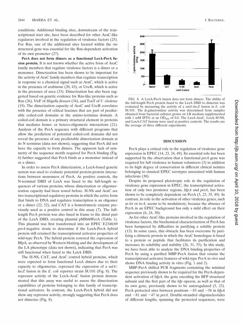
The H-NS, CAT, and AraC control hybrid proteins, whichwere expected to form functional LexA dimers due to theircapacity to oligomerize, repressed the expression of a sulA::lacZ fusion in the E. coli reporter strain SU101 (Fig. 8). Therepressor activity of the LexA-AraC fusion protein demon-strated that this assay was useful to assess the dimerizationcapabilities of proteins belonging to this family of transcrip-tional activators. In contrast, the LexA-PerA hybrid did notshow any repressor activity, strongly suggesting that PerA doesnot dimerize (Fig. 8).
DISCUSSION
PerA plays a critical role in the regulation of virulence geneexpression in EPEC (14, 23, 24, 49). Its essential role has beensupported by the observation that a functional perA gene wasrequired for full virulence in human volunteers (3) in additionto its high degree of conservation in different clinical isolatesbelonging to classical EPEC serotypes associated with humaninfections (30).
Despite its proposed pleiotropic role in the regulation ofvirulence gene expression in EPEC, the transcriptional activa-tion of only two promoter regions, bfpA and perA, has beenshown to be completely dependent on PerA (5, 23, 35, 49). Incontrast, its role in the activation of other virulence genes, suchas ler or trcA, seems to be modulatory, because the absence ofPerA in an EPEC background has only a mild effect on theirexpression (6, 24, 50).
As for other AraC-like proteins involved in the regulation ofvirulence factors, the biochemical characterization of PerA hasbeen hampered by difficulties in purifying a soluble protein(13). In some cases, this obstacle has been overcome by puri-fying a chimeric protein in which the AraC homologue is fusedto a protein or peptide that facilitates its purification andincreases its solubility and stability (26, 31, 55). In this study,we have been able to analyze the DNA binding properties ofPerA by using a purified MBP-PerA fusion that retains thetranscriptional activator features of wild-type PerA in vivo andshows DNA binding activity in vitro (Fig. 1 and 2).
MBP-PerA shifted PCR fragments containing the minimalsequence previously shown to be required for the PerA-depen-dent activation of bfpA, the gene encoding the BFP structuralsubunit and the first part of the bfp operon, as well as that ofits own gene, previously shown to be autoregulated (5, 23).PerA protected sites between positions �83 and �56 in bfpAand �81 and �47 in perA. Double-stranded oligonucleotidesof different lengths, spanning the protected sequences, were
FIG. 8. A LexA-PerA fusion does not form dimers. The ability ofthe full-length PerA protein fused to the LexA DBD to dimerize wasevaluated by measuring the activity of a sulA::lacZ fusion in E. coliSU101. The �-galactosidase activity was determined from samplesobtained from bacterial cultures grown on LB medium supplementedwith 1 mM IPTG at an OD600 of 0.8. The LexA-AraC, LexA–H-NS,and LexA-CAT fusions were used as positive controls. The results arethe average of three different experiments.
2844 IBARRA ET AL. J. BACTERIOL.
on April 10, 2018 by guest
http://jb.asm.org/
Dow
nloaded from

used in EMSAs to determine the minimal sequence requiredby PerA for efficient DNA binding in vitro. For both promot-ers, around 40 bp was required for efficient shifting. The nu-cleotide alignment of the defined minimal sequences allowedthe proposal of a consensus PerA binding motif, which con-sisted of an AT-rich 29-bp-long sequence in which 19 positionswere identical between the two promoters (Fig. 6). AraC-likeproteins recognize sequence motifs that range from 17 to 60bp. MarA, Rob, and SoxS, three AraC-like proteins involved instress response, interact as monomers with a 17- to 19-bp-longmotif in two adjacent segments of the major groove (16, 36).AraC binds two 17-bp-long inverted repeats that are separatedby 6 bp (17). Similarly, XylS, MelR, and RhaS have beenshown to bind sequences between 33 to 56 bp that generallyinclude two binding sites separated by a variable number ofbases (2, 4, 15, 46). The binding site for Rns and relatedvirulence gene regulators, such as VirFSf, CfaR, and AggR, iscontained in an AT-rich 30-bp-long sequence (25, 26). Despitethe sequence similarities at the HTH motifs between PerA andother virulence regulators, such as Rns, VirF, or ToxT, no clearsequence similarity was found between the PerA binding siteand the binding sites described for these proteins (19, 26, 27,54, 55). This offers an explanation for why PerA was not ableto substitute for Rns to activate the coo and rns promoters andwhy Rns could not substitute for PerA to activate the perApromoter (25).
In perA, the PerA binding motif is located about one helixturn upstream from the �35 promoter sequence, while in bfpAit is located about two helix turns upstream from the promoter(Fig. 6). The functional significance of this distribution for theactivation of both promoters is being further analyzed in thecontext of potential interactions between PerA and RNA poly-merase or additional regulatory proteins. In this regard, wehave recently observed that bfpA is not expressed in the ab-sence of integration host factor (IHF) (Y. Martínez-Laguna etal., unpublished observations), a global regulatory protein thathas also been involved in the positive regulation of ler expres-sion (12).
The lack of apparent symmetry in the sequence motif boundby PerA and the observation that it forms one major DNA-protein complex by EMSA suggested that it acts as a monomerand not as a dimer, as has been shown for other AraC-likeproteins (13). This possibility was tested with a LexA-basedhybrid bacterial system that allows the monitoring of protein-protein interactions (7, 8). In order to further corroborate thefunctionality of the assay, two DNA binding proteins, H-NSand AraC, and an enzyme, CAT, were used as positive controlsbecause their dimerization or multimerization properties havebeen well documented (7, 17, 52). In all three cases, the hybridproteins allowed the formation of functional LexA dimers thatinhibited the expression of the sulA::lacZ reporter fusion. Inparticular, the result obtained with the LexA-AraC fusiondemonstrated that the LexA-based system is useful to evaluatethe dimerization or multimerization properties of other mem-bers of the AraC family of transcriptional activators (Fig. 8). Incontrast, a LexA-PerA fusion that still conserved the ability toactivate bfpA transcription and BFP production in an EPECperA mutant strain (data not shown) was unable to repress thereporter fusion, strongly suggesting that PerA does not formdimers. Furthermore, expression of the N-terminal domain of
PerA from an inducible promoter did not exert a dominant-negative effect on the expression of the bfpA-cat fusion or onthe expression of the BfpA subunit in wild-type EPEC, asevaluated by CAT assays and Western blotting (data notshown). This observation supports the notion that PerA doesnot form dimers to activate transcription.
Based on these results, it is likely that PerA binds to itstarget sequences upstream of the bfpA and perA promoters asa monomer, probably following the interaction model based onthe crystal structure of MarA bound to its target DNA (36),which has been also proposed for Rns (26). In this model, bothHTH motifs form specific interactions with two adjacent seg-ments of the major groove along one face of the DNA helix, vianonidentical nucleotide contacts. Dimerization of AraC-likeproteins involved in virulence gene control has been proposed:for example, for VirF and ToxT, based on dominant-negativephenotypes observed with mutants that cannot bind DNA (19,34). At this point, we cannot rule out the possibility that PerAdimerizes upon binding to its target DNA sequence, a mech-anism that has been proposed for MelR (4).
The HTH2 motif is the region with the highest similaritybetween the AraC-like proteins and a consensus sequencederived from the alignment of more than 100 proteins, consti-tuting the AraC family signature (13). The high degree ofconservation of this motif has been used as an argument topropose that its function is common for all the AraC-likeproteins, which is likely to involve interactions with RNA poly-merase. In this regard, we have previously observed that theinsertion or deletion of a single nucleotide between the bfpApromoter and the putative PerA binding site abolished bfpAtranscription. This suggested that changes in the orientation ofthe binding site and the promoter affect the proper presenta-tion of a critical protein surface that allows PerA-RNA poly-merase interactions that are important to promote transcrip-tional activation (5). These insertions or deletions did notreduce the affinity of PerA for PCR fragments containing thesemodifications (data not shown), further supporting the notionthat they rather disrupt the appropriate contacts between PerAand RNA polymerase.
In the case of AraC, binding of a sugar effector molecule(arabinose) to the N-terminus domain of the protein induces astructural change that produces the activator form of the AraCdimer (29, 40, 43). For other members of the family, binding ofdifferent molecules, such as rhamnose for RhaS, urea forUreR, or alkylbenzoates for XylS, induces similar conforma-tional changes that allow transcriptional activation of theirtarget genes. Thus, the presence of these molecules constitutesthe regulatory signals (13). The PerA-mediated expression ofbfpA and perA in EPEC is negatively modulated by tempera-tures above or below 37°C, by the presence of ammonium, andby growth in rich medium (23, 35). Other AraC-like proteinsinvolved in the regulation of virulence factors have also beenshown to respond to environmental cues, such as temperature.However, no effector molecules controlling the activity of themembers of this subfamily of AraC-like proteins have beenidentified. It remains to be determined whether proteins suchas PerA also undergo conformational changes in response toenvironmental or physicochemical signals that shift the proteinfrom a constitutively active to inactive form.
VOL. 185, 2003 PerA BINDING SITES 2845
on April 10, 2018 by guest
http://jb.asm.org/
Dow
nloaded from

ACKNOWLEDGMENTS
We particularly thank A. Alagon, V. H. Bustamante, E. Calva, R. A.Edwards, G. Munson, N. Olivares, G. Pedraza, and Y. Rosenstein foradvice and helpful discussions. We are also grateful to M. G. Sosa forconstructing plasmid pMAL-T2, A. Vazquez for constructing theEPEC perA::Km strain, O. Rodríguez for providing HeLa cells for theadherence assays, J. A. Giron for the BfpA antibodies, R. P. Silver forthe LexA-based system, V. H. Bustamante for the plasmid encodingthe LexA-HNS fusion and pCAT � 27, E. Mata for animal care, andE. Lopez-Bustos for oligonucleotide synthesis.
This research was supported by grants from the Consejo Nacional deCiencia y Tecnología (CONACyT 33115-N) and the Universidad Na-cional Autonoma de Mexico (DGAPA IN-217201) and by a HowardHughes International Research Scholar Award to J.L.P. J.A.I. wassupported by a Ph.D. Fellowship from CONACYT (no. 86036) and bythe Direccion General de Estudios de Posgrado (DGEP), UNAM.
REFERENCES
1. Alagon, A. C., and T. P. King. 1980. Activation of polysaccharides with2-iminothiolane and its uses. Biochemistry 19:4341–4345.
2. Bhende, P. M., and S. M. Egan. 1999. Amino acid-DNA contacts by RhaS:an AraC family transcription activator. J. Bacteriol. 181:5185–5192.
3. Bieber, D., S. W. Ramer, C. Y. Wu, W. J. Murray, T. Tobe, R. Fernandez, andG. K. Schoolnik. 1998. Type IV pili, transient bacterial aggregates, andvirulence of enteropathogenic Escherichia coli. Science 280:2114–2118.
4. Bourgerie, S. J., C. M. Michan, M. S. Thomas, S. J. Busby, and E. I. Hyde.1997. DNA binding and DNA bending by the MelR transcription activatorprotein from Escherichia coli. Nucleic Acids Res. 25:1685–1693.
5. Bustamante, V. H., E. Calva, and J. L. Puente. 1998. Analysis of cis-actingelements required for bfpA expression in enteropathogenic Escherichia coli.J. Bacteriol. 180:3013–3016.
6. Bustamante, V. H., F. J. Santana, E. Calva, and J. L. Puente. 2001. Tran-scriptional regulation of type III secretion genes in enteropathogenic Esch-erichia coli: Ler antagonizes H-NS-dependent repression. Mol. Microbiol.39:664–678.
7. Daines, D. A., and R. P. Silver. 2000. Evidence for multimerization of Neuproteins involved in polysialic acid synthesis in Escherichia coli K1 usingimproved LexA-based vectors. J. Bacteriol. 182:5267–5270.
8. Dmitrova, M., G. Younes-Cauet, P. Oertel-Buchheit, D. Porte, M. Schnarr,and M. Granger-Schnarr. 1998. A new LexA-based genetic system for mon-itoring and analyzing protein heterodimerization in Escherichia coli. Mol.Gen. Genet. 257:205–212.
9. Elliott, S. J., V. Sperandio, J. A. Giron, S. Shin, J. L. Mellies, L. Wainwright,S. W. Hutcheson, T. K. McDaniel, and J. B. Kaper. 2000. The locus ofenterocyte effacement (LEE)-encoded regulator controls expression of bothLEE- and non-LEE-encoded virulence factors in enteropathogenic and en-terohemorrhagic Escherichia coli. Infect. Immun. 68:6115–6126.
10. Elliott, S. J., L. A. Wainwright, T. K. McDaniel, K. G. Jarvis, Y. K. Deng,L. C. Lai, B. P. McNamara, M. S. Donnenberg, and J. B. Kaper. 1998. Thecomplete sequence of the locus of enterocyte effacement (LEE) from en-teropathogenic Escherichia coli E2348/69. Mol. Microbiol. 28:1–4.
11. Frankel, G., A. D. Phillips, I. Rosenshine, G. Dougan, J. B. Kaper, and S.Knutton. 1998. Enteropathogenic and enterohaemorrhagic Escherichia coli:more subversive elements. Mol. Microbiol. 30:911–921.
12. Friedberg, D., T. Umanski, Y. Fang, and I. Rosenshine. 1999. Hierarchy inthe expression of the locus of enterocyte effacement genes of enteropatho-genic Escherichia coli. Mol. Microbiol. 34:941–952.
13. Gallegos, M.-T., R. Schleif, A. Bairoch, K. Hofmann, and J. L. Ramos. 1997.Arac/XylS family of transcriptional regulators. Microbiol. Mol. Biol. Rev.61:393–410.
14. Gomez-Duarte, O. G., and J. B. Kaper. 1995. A plasmid-encoded regulatoryregion activates chromosomal eaeA expression in enteropathogenic Esche-richia coli. Infect. Immun. 63:1767–1776.
15. Gonzalez-Perez, M. M., J. L. Ramos, M. T. Gallegos, and S. Marques. 1999.Critical nucleotides in the upstream region of the XylS-dependent TOLmeta-cleavage pathway operon promoter as deduced from analysis of mu-tants. J. Biol. Chem. 274:2286–2290.
16. Griffith, K. L., and R. E. Wolf, Jr. 2001. Systematic mutagenesis of the DNAbinding sites for SoxS in the Escherichia coli zwf and fpr promoters: identi-fying nucleotides required for DNA binding and transcription activation.Mol. Microbiol. 40:1141–1154.
17. Hendrickson, W., and R. Schleif. 1985. A dimer of AraC protein contactsthree adjacent major groove regions of the araI DNA site. Proc. Natl. Acad.Sci. USA 82:3129–3133.
18. Hovey, A. K., and D. W. Frank. 1995. Analyses of the DNA-binding andtranscriptional activation properties of ExsA, the transcriptional activator ofthe Pseudomonas aeruginosa exoenzyme S regulon. J. Bacteriol. 177:4427–4436.
19. Hulbert, R. R., and R. K. Taylor. 2002. Mechanism of ToxT-dependent
transcriptional activation at the Vibrio cholerae tcpA promoter. J. Bacteriol.184:5533–5544.
20. LaRonde-LeBlanc, N., and C. Wolberger. 2000. Characterization of the oli-gomeric states of wild type and mutant AraC. Biochemistry 39:11593–11601.
21. Lupas, A. 1996. Coiled coils: new structures and new functions. TrendsBiochem. Sci. 21:375–382.
22. Martin, R. G., and J. L. Rosner. 2001. The AraC transcriptional activators.Curr. Opin. Microbiol. 4:132–137.
23. Martinez-Laguna, Y., E. Calva, and J. L. Puente. 1999. Autoactivation andenvironmental regulation of bfpT expression, the gene coding for the tran-scriptional activator of bfpA in enteropathogenic Escherichia coli. Mol. Mi-crobiol. 33:153–166.
24. Mellies, J. L., S. J. Elliott, V. Sperandio, M. S. Donnenberg, and J. B. Kaper.1999. The Per regulon of enteropathogenic Escherichia coli: identification ofa regulatory cascade and a novel transcriptional activator, the locus of en-terocyte effacement (LEE)-encoded regulator (Ler). Mol. Microbiol. 33:296–306.
25. Munson, G. P., L. G. Holcomb, and J. R. Scott. 2001. Novel group ofvirulence activators within the AraC family that are not restricted to up-stream binding sites. Infect. Immun. 69:186–193.
26. Munson, G. P., and J. R. Scott. 1999. Binding site recognition by Rns, avirulence regulator in the AraC family. J. Bacteriol. 181:2110–2117.
27. Munson, G. P., and J. R. Scott. 2000. Rns, a virulence regulator within theAraC family, requires binding sites upstream and downstream of its ownpromoter to function as an activator. Mol. Microbiol. 36:1391–1402.
28. Nataro, J. P., and J. B. Kaper. 1998. Diarrheagenic Escherichia coli. Clin.Microbiol. Rev. 11:142–201.
29. Niland, P., R. Huhne, and B. Muller-Hill. 1996. How AraC interacts specif-ically with its target DNAs. J. Mol. Biol. 264:667–674.
30. Okeke, I. N., J. A. Borneman, S. Shin, J. L. Mellies, L. E. Quinn, and J. B.Kaper. 2001. Comparative sequence analysis of the plasmid-encoded regu-lator of enteropathogenic Escherichia coli strains. Infect. Immun. 69:5553–5564.
31. Olekhnovich, I. N., and R. J. Kadner. 2002. DNA-binding activities of theHilC and HilD virulence regulatory proteins of Salmonella enterica serovarTyphimurium. J. Bacteriol. 184:4148–4160.
32. Oropeza, R., C. L. Sampieri, J. L. Puente, and E. Calva. 1999. Negative andpositive regulation of the non-osmoregulated ompS1 porin gene in Salmo-nella typhi: a novel regulatory mechanism that involves OmpR. Mol. Micro-biol. 32:243–252.
33. Poore, C. A., C. Coker, J. D. Dattelbaum, and H. L. T. Mobley. 2001.Identification of the domains of UreR, an AraC-like transcriptional regula-tor of the urease gene cluster in Proteus mirabilis. J. Bacteriol. 183:4526–4535.
34. Porter, M. E., and C. J. Dorman. 2002. In vivo DNA-binding and oligomer-ization properties of the Shigella flexneri AraC-like transcriptional regulatorVirF as identified by random and site-specific mutagenesis. J. Bacteriol.184:531–539.
35. Puente, J. L., D. Bieber, S. W. Ramer, W. Murray, and G. K. Schoolnik.1996. The bundle-forming pili of enteropathogenic Escherichia coli: tran-scriptional regulation by environmental signals. Mol. Microbiol. 20:87–100.
36. Rhee, S., R. G. Martin, J. L. Rosner, and D. R. Davies. 1998. A novelDNA-binding motif in MarA: the first structure for an AraC family tran-scriptional activator. Proc. Natl. Acad. Sci. USA 95:10413–10418.
37. Roder, K., and M. Schweizer. 2001. Running-buffer composition influencesDNA-protein and protein-protein complexes detected by electrophoreticmobility-shift assay (EMSA). Biotechnol. Appl. Biochem. 33:209–214.
38. Sambrook, J., E. F. Fritsch, and T. Maniatis. 1989. Molecular cloning: alaboratory manual, 2nd ed. Cold Spring Harbor Laboratory Press, ColdSpring Harbor, N.Y.
39. Santana, M. A., G. Pedraza-Alva, N. Olivares-Zavaleta, V. Madrid-Marina,V. Horejsi, S. J. Burakoff, and Y. Rosenstein. 2000. CD43-mediated signalsinduce DNA binding activity of AP-1, NF-AT, and NFkappa B transcriptionfactors in human T lymphocytes. J. Biol. Chem. 275:31460–31468.
40. Saviola, B., R. Seabold, and R. F. Schleif. 1998. Arm-domain interactions inAraC. J. Mol. Biol. 278:539–548.
41. Shin, S., M. P. Castanie-Cornet, J. W. Foster, J. A. Crawford, C. Brinkley,and J. B. Kaper. 2001. An activator of glutamate decarboxylase genes reg-ulates the expression of enteropathogenic Escherichia coli virulence genesthrough control of the plasmid-encoded regulator, Per. Mol. Microbiol.41:1133–1150.
42. Sohel, I., J. L. Puente, S. W. Ramer, D. Bieber, C.-Y. Wu, and G. K.Schoolnik. 1996. Enteropathogenic Escherichia coli: identification of a genecluster coding for bundle-forming pilus morphogenesis. J. Bacteriol. 178:2613–2628.
43. Soisson, S. M., B. MacDougall-Shackleton, R. Schleif, and C. Wolberger.1997. Structural basis for ligand-regulated oligomerization of AraC. Science276:421–425.
44. Sperandio, V., J. L. Mellies, W. Nguyen, S. Shin, and J. B. Kaper. 1999.Quorum sensing controls expression of the type III secretion gene transcrip-tion and protein secretion in enterohemorrhagic and enteropathogenic Esch-erichia coli. Proc. Natl. Acad. Sci. USA 96:15196–15201.
2846 IBARRA ET AL. J. BACTERIOL.
on April 10, 2018 by guest
http://jb.asm.org/
Dow
nloaded from

45. Stone, K. D., H. Z. Zhang, L. K. Carlson, and M. S. Donnenberg. 1996. Acluster of fourteen genes from enteropathogenic Escherichia coli is sufficientfor the biogenesis of a type IV pilus. Mol. Microbiol. 20:325–337.
46. Tamai, E., T. A. Belyaeva, S. J. Busby, and T. Tsuchiya. 2000. Mutations thatincrease the activity of the promoter of the Escherichia coli melibiose operonimprove the binding of MelR, a transcription activator triggered by melibi-ose. J. Biol. Chem. 275:17058–17063.
47. Thomas, V. J., and C. M. Collins. 1999. Identification of UreR binding sitesin the Enterobacteriaceae plasmid-encoded and Proteus mirabilis urease geneoperons. Mol. Microbiol. 31:1417–1428.
48. Tobe, T., T. Hayashi, C.-G. Han, G. K. Schoolnik, E. Ohtsubo, and C.Sasakawa. 1999. Complete DNA sequence and structural analysis of theenteropathogenic Escherichia coli adherence factor plasmid. Infect. Immun.67:5455–5462.
49. Tobe, T., G. K. Schoolnik, I. Sohel, V. H. Bustamante, and J. L. Puente. 1996.Cloning and characterization of bfpTVW, genes required for the transcrip-tional activation of bfpA in enteropathogenic Escherichia coli. Mol. Micro-biol. 21:963–975.
50. Tobe, T., I. Tatsuno, E. Katayama, C. Y. Wu, G. K. Schoolnik, and C.
Sasakawa. 1999. A novel chromosomal locus of enteropathogenic Escherichiacoli (EPEC), which encodes a BfpT-regulated chaperone-like protein, TrcA,involved in microcolony formation by EPEC. Mol. Microbiol. 33:741–752.
51. Tomoyasu, T., A. Mogk, H. Langen, P. Goloubinoff, and B. Bukau. 2001.Genetic dissection of the roles of chaperones and proteases in proteinfolding and degradation in the Escherichia coli cytosol. Mol. Microbiol.40:397–413.
52. Ueguchi, C., C. Seto, T. Suzuki, and T. Mizuno. 1997. Clarification of thedimerization domain and its functional significance for the Escherichia colinucleoid protein H-NS. J. Mol. Biol. 274:145–151.
53. Vallance, B. A., and B. B. Finlay. 2000. Exploitation of host cells by entero-pathogenic Escherichia coli. Proc. Natl. Acad. Sci. USA 97:8799–8806.
54. Wattiau, P., and G. R. Cornelis. 1994. Identification of DNA sequencesrecognized by VirF, the transcriptional activator of the Yersinia yop regulon.J. Bacteriol. 176:3878–3884.
55. Yu, R. R., and V. J. DiRita. 2002. Regulation of gene expression in Vibriocholerae by ToxT involves both antirepression and RNA polymerase stimu-lation. Mol. Microbiol. 43:119–134.
VOL. 185, 2003 PerA BINDING SITES 2847
on April 10, 2018 by guest
http://jb.asm.org/
Dow
nloaded from