identification of the agent: flaviviridae pestivirus€¦ · cattle of all ages are susceptible to...
TRANSCRIPT
Cattle of all ages are susceptible to infection with bovine viral diarrhoea viruses (BVDV).
Distribution is world-wide although some countries have recently eradicated the virus. BVDV
infection results in a wide variety of clinical manifestations, including enteric and respiratory disease
in any class of cattle or reproductive and fetal disease following infection of a susceptible breeding
female. Infection may be subclinical or extend to severe fatal disease. Animals that survive in-utero
infection in the first trimester of gestation are almost always persistently infected (PI). PI animals
provide the main reservoir of the virus in a population and excrete large amounts of virus in urine,
faeces, discharges, milk and semen. Identification of such PI cattle is a key element in controlling
the infection. It is important to avoid the trade of such animals. They may appear clinically healthy,
or weak and unthrifty. Many PI animals die before reaching maturity. They may infrequently develop
mucosal disease with anorexia, gastrointestinal erosions, and profuse diarrhoea, invariably leading
to death. Mucosal disease can arise only in PI animals. Latent infections generally do not occur
following recovery from acute infection. However bulls may rarely have a persistent testicular
infection and excrete virus in semen for prolonged periods.
Identification of the agent: BVDV is a pestivirus in the family Flaviviridae and is closely related to
classical swine fever and ovine border disease viruses. The two genotypes (types 1 and 2) are
classified as separate species in the genus Pestivirus. A third putative genotype, BVDV type 3, has
also recently been proposed. Although both cytopathic and non-cytopathic biotypes of BVDV type 1
and type 2 exist, non-cytopathic strains are usually encountered in field infections and are the main
focus of diagnostic virus isolation in cell cultures. PI animals can be readily identified by a variety of
methods aimed to detect viral antigens or viral RNA directly in blood and tissues. Virus can also be
isolated by inoculation of specimens onto susceptible cell cultures followed by immune-labelling
methods to detect the replication of the virus in the cultures. Persistence of virus infection should be
confirmed by resampling after an interval of at least 3 weeks, when virus will again be detected. PI
animals are usually seronegative. Viraemia in acute cases is transient and difficult to detect. Virus
isolation in semen from bulls that are acutely or persistently infected requires special attention to
specimen transport and testing. RNA detection assays are particularly useful because they are
rapid, have very high sensitivity and do not depend on the use of cell cultures.
Serological tests: Acute infection with BVDV is best confirmed by demonstrating seroconversion
using sequential paired samples, ideally from several animals in the group. The testing of paired
(acute and convalescent samples) should be done a minimum of 21 days apart and samples should
be tested concurrently in the same assay. Enzyme-linked immunosorbent assays and the virus
neutralisation test are the most widely used.
Requirements for vaccines: There is no standard vaccine for BVD, but a number of commercial
preparations are available. An ideal vaccine should be able to prevent transplacental infection in
pregnant cows. Modified live virus vaccine should not be administered to pregnant cattle (or to their
sucking calves) due to the risk of transplacental infection. Live vaccines that contain cytopathic
strains of BVDV present a risk of inducing mucosal disease in PI animals. Inactivated viral vaccines
are safe and can be given to any class of animal but generally require booster vaccinations. BVDV
is a particularly important hazard to the manufacture of vaccines and biological products for other
diseases due to the high frequency of contamination of batches of fetal calf serum used as a culture
medium supplement.

Cattle of all ages are susceptible to infection with bovine viral diarrhoea viruses (BVDV). Distribution of the virus is world-wide although some countries have recently eradicated the virus. BVDV infection results in a wide variety of clinical manifestations, including enteric and respiratory disease in any class of cattle or reproductive and fetal disease following infection of a susceptible breeding female. Infection may be subclinical or extend to severe fatal disease. Clinical presentations and severity of disease may vary with different strains of virus. BVDV viruses also cause immune suppression which can render infected animals more susceptible to infection with other viruses and bacteria. The clinical impact may be more apparent in intensively managed livestock. Animals that survive in-utero infection in the first trimester of gestation are almost always persistently infected (PI). PI animals provide the main reservoir of the virus in a population and excrete large amounts of virus in urine, faeces, discharges, milk and semen. The virus spreads mainly by close contact between PI animals and other cattle. Virus shedding by acutely infected animals is usually less important. This virus may also persist in the environment for short periods or be transmitted with contaminated reproductive materials. Vertical transmission plays an important role in its epidemiology and pathogenesis.
Infections of the breeding female may result in conception failure or embryonic and fetal infection which results in abortions, stillbirths, teratogenic abnormalities or the birth of PI calves. Persistently viraemic animals may be born as weak, unthrifty calves or may appear as normal healthy calves and be unrecognised clinically for a long time. However, PI animals have a markedly reduced life expectancy, with a high proportion dying before reaching maturity. Infrequently, some of these animals may later develop mucosal disease with anorexia, gastrointestinal erosions, and profuse diarrhoea, invariably leading to death. Mucosal disease can arise only in PI animals. It is important to avoid the trade of viraemic animals. It is generally considered that serologically positive, non-viraemic cattle are „safe‟, providing that they are not pregnant. However, a small proportion of persistently viraemic animals may produce antibodies to some of the viral proteins if they are exposed to another strain of BVDV that is antigenically different to the persisting virus. Consequently, seropositivity cannot be completely equated with „safety‟. Detection of PI animals must be specifically directed at detection of the virus or its components (RNA or antigens). Latent infections generally do not occur following recovery from acute infection. However, semen collected from bulls during an acute infection is likely to contain virus during the viraemic period and often for a short time afterwards. Although extremely rare, some recovered bulls may have a persistent testicular infection and excrete virus in semen, perhaps indefinitely.
While BVDV strains are predominantly pathogens of cattle, interspecies transmission can occur following close contact with sheep, goats or pigs. Infection of pregnant small ruminants or pigs with BVDV can result in reproductive loss and the birth of PI animals. BVDV infections have been reported in both New World and Old World camelids. Additionally, strains of border disease virus (BDV) have infected cattle, resulting in clinical presentations indistinguishable from BVDV infection. The birth of cattle PI with BDV and the subsequent development of mucosal disease have also been described. Whilst BVDV and BDV have been reported as natural infections in pigs, the related virus of classical swine fever does not naturally infect ruminants.
Although ubiquitous, control of BVDV can be achieved at the herd level, and even at the national level, as evidenced by the progress towards eradication made in many European countries (Moennig et al., 2005).
Bovine viral diarrhoea virus (BVDV) is a single linear positive-stranded RNA virus in the genus Pestivirus of the family Flaviviridae. The genus contains a number of species including the two genotypes of bovine viral diarrhoea
virus (BVDV) (types 1 and 2) and the closely related classical swine fever and ovine border disease viruses. Viruses in these genotypes show considerable antigenic difference from each other and, within the type 1 and type 2 species, BVDV isolates exhibit considerable biological and antigenic diversity. Within the two BVDV genotypes, further subdivisions are discernible by genetic analysis (Vilcek et al., 2001). The two genotypes may
be differentiated from each other, and from other pestiviruses, by monoclonal antibodies (MAbs) directed against the major glycoproteins E2 and ERNS or by genetic analysis. Reverse-transcription polymerase chain reaction (RT-PCR) assays enable virus typing direct from blood samples (Letellier & Kerhofs, 2003; McGoldrick et al., 1999). Type 1 viruses are generally more common although the prevalence of type 2 strains can be high in North America. BVDV of both genotypes may occur in non-cytopathic and cytopathic forms (biotypes), classified according to whether or not microscopically apparent cytopathology is induced during infection of cell cultures. Usually, it is the non-cytopathic biotype that circulates freely in cattle populations. Non-cytopathic strains are most frequently responsible for disease in cattle and are associated with enteric and respiratory disease in any class of cattle or reproductive and fetal disease following infection of a susceptible breeding female. Infection may be subclinical or extend to severe fatal disease (Brownlie, 1985). Cytopathic viruses are encountered in cases of mucosal disease, a clinical syndrome that is relatively uncommon and involves the „super-infection‟ of an animal that is PI with a non-cytopathic virus by a closely related cytopathic strain. The two virus biotypes found in a mucosal disease case are usually antigenically closely related if not identical. Type 2 viruses are usually non-

cytopathic and have been associated with outbreaks of severe acute infection and a haemorrhagic syndrome. However some type 2 viruses have also been associated with a disease indistinguishable from that seen with the more frequently isolated type 1 viruses. Further, some type 1 isolates have been associated with particularly severe and fatal disease outbreaks in adult cattle. Clinically mild and inapparent infections are common following infection of non-pregnant animals with either genotype.
There is an increasing awareness of an “atypical” or “HoBi-like” pestivirus – a putative BVDV type 3, in cattle, also associated with clinical disease (Bauermann et al., 2013), but its distribution is presently unclear. These viruses are readily detected by proven pan-reactive assays such as real-time RT-PCR. Some commercial antigen ELISAs (enzyme-linked immunosorbent assays) have been shown to detect these strains (Bauermann et al., 2012); generally virus isolation, etc., follows the same principles as for BVDV 1 and 2. It should be noted however, that antibody ELISAs vary in their ability to detect antibody to BVDV 3 and vaccines designed to protect against BVDV 1 and 2 may not confer full protection against infection with these novel pestiviruses (Bauermann et al., 2012; 2013).
Acute infections with BVDV are encountered more frequently in young animals, and may be clinically inapparent or associated with fever, diarrhoea (Baker 1995), respiratory disease and sometimes sudden death. The severity of disease may vary with virus strain and the involvement of other pathogens (Brownlie, 1990). In particular, outbreaks of a severe form of acute disease with haemorrhagic lesions, thrombocytopenia and high mortality have been reported sporadically from some countries (Baker, 1995; Bolin & Ridpath, 1992). Infection with type 2 viruses in particular has been demonstrated to cause altered platelet function. During acute infections there is a brief viraemia for 7–10 days and shedding of virus can be detected in nasal and ocular discharges. There may also be a transient leukopenia, thrombocytopenia or temperature response, but these can vary greatly among animals. Affected animals may be predisposed to secondary infections with other viruses and bacteria. Although BVDV may cause a primary respiratory disease on its own, the immunosuppressive effects of the virus exacerbate the impact of this virus. BVDV is one of the major pathogens of the bovine respiratory disease complex in feedlot cattle and in other intensive management systems such as calf raising units.
Infection of breeding females immediately prior to ovulation and in the first few days after insemination can result in conception failure and early embryonic loss (McGowan & Kirkland, 1995). Cows may also suffer from infertility, associated with changes in ovarian function and secretions of gonadotropin and progesterone (Fray et al., 2002). Bulls may excrete virus in semen for a short period during and immediately after infection and may suffer a temporary reduction of fertility. Although the virus level in this semen is generally low it can result in reduced conception rates and be a potential source of introduction of virus into a naive herd (McGowan & Kirkland, 1995).
Infection of a breeding female can result in a range of different outcomes, depending on the stage of gestation at which infection occurred. Before about 25 days of gestation, infection of the developing conceptus will usually result in embryo-fetal death, although abortion may be delayed for a considerable time (McGowan & Kirkland, 1995). Surviving fetuses are normal and uninfected. However, infection of the female between about 30–90 days will invariably result in fetal infection, with all surviving progeny PI and sero-negative. Infection at later stages and up to about day 150 can result in a range of congenital defects including hydranencephaly, cerebellar hypoplasia, optic defects, skeletal defects such as arthrogryposis and hypotrichosis. Growth retardation may also occur, perhaps as a result of pituitary dysfunction. Fetal infection can result in abortion, stillbirth or the delivery of weak calves that may die soon after birth (Baker, 1995; Brownlie, 1990; Duffell & Harkness, 1985; Moennig & Liess, 1995). Some PI calves may appear to be normal at birth but fail to grow normally. They remain PI for life and are usually sero-negative. The onset of the fetal immune response and production of antibodies occurs between approximately day 90–120, with an increasing proportion of infected calves having detectable antibodies while the proportion in which virus may be detected declines rapidly. Infection of the bovine fetus after day 180 usually results in the birth of a normal seropositive calf.
Persistently viraemic animals are a continual source of infective virus to other cattle and are the main reservoir of BVDV in a population. In a population without a rigorous BVDV control programme, approximately 1–2% of cattle are PI. During outbreaks in a naive herd or breeding group, if exposure has occurred in the first trimester of pregnancy, a very high proportion of surviving calves will be PI. If a PI animal dies, there are no pathognomonic lesions due to BVDV and the pathology is often complicated by secondary infections with other agents. Some PI animals will survive to sexual maturity

and may breed successfully but their progeny will also always be PI. Animals being traded or used for artificial breeding should first be screened to ensure that they are not PI.
Persistently viraemic animals may later succumb to mucosal disease (Brownlie, 1985). However, cases are rare. This syndrome has been shown to be the outcome of the infection of a PI animal with an antigenically similar cytopathic virus, which can arise either through superinfection, recombination between non-cytopathic biotypes, or mutation of the persistent biotype (Brownlie, 1990). There is usually little need to specifically confirm that a PI animal has succumbed to mucosal disease as this is largely a scientific curiosity and of little practical significance, other than that the animal is PI with BVDV. However, cases of mucosal disease may be the first indication in a herd that BVDV infection is present, and should lead to more in depth investigation and intervention.
Bulls that are PI usually have poor quality, highly infective semen and reduced fertility (McGowan & Kirkland, 1995). All bulls used for natural or artificial insemination should be screened for both acute and persistent BVDV infection. A rare event, possibly brought about by acute infection during pubescence, can result in persistent infection of the testes and thus strongly seropositive bulls that intermittently excrete virus in semen (Voges et al., 1998). This phenomenon has also been observed following vaccination with an attenuated virus (Givens et al., 2007). Embryo donor cows that are PI with BVDV also represent a potential source of infection, particularly as there are extremely high concentrations of BVDV in uterine and vaginal fluids. While oocysts without an intact zona pellucida have been shown to be susceptible to infection in vitro, the majority of oocysts remain uninfected with BVDV. Normal uninfected progeny have also been „rescued‟ from PI animals by the use of extensive washing of embryos and in vitro fertilisation. Female cattle used as embryo recipients should always be screened to confirm that they are not PI, and ideally, are sero-positive or were vaccinated at least 4 weeks before first use.
Biological materials used for in-vitro fertilisation techniques (bovine serum, bovine cell cultures) have a high risk of contamination and should be screened for BVDV. Incidents of apparent introduction of virus via such techniques have highlighted this risk. It is considered essential that serum supplements used in media should be free of contaminants as detailed in Chapter 1.1.9 Tests of biological materials for sterility and freedom from contamination, using techniques described in Section B.3.1 of this chapter.
The diagnosis of BVDV infection can sometimes be complex because of the delay between infection and clinical expression. While detection of PI animals should be readily accomplished using current diagnostic methods, the recognition of acute infections and detection of BVDV in reproductive materials can be more difficult.
Unlike PI animals, acutely infected animals excrete relatively low levels of virus and for a short period of time (usually about 7–10 days) but the clinical signs may occur during the later stages of viraemia, reducing the time to detect the virus even further. In cases of respiratory or enteric disease, samples should be collected from a number of affected animals, preferentially selecting the most recently affected. Swabs should be collected from the nares and conjunctiva of animals with respiratory disease or from rectum and faeces if there are enteric signs. Lung and spleen are preferred from dead animals. Viral RNA may be detected by real-time RT-PCR assays and have the advantages of high sensitivity and being able to detect genome from non-infectious virus. As the virus levels are very low, it is not usually practical to undertake virus isolation unless there is a need to characterise the strain of BVDV involved. Serology undertaken on paired acute and convalescent sera (collected at least 21 days after the acute sample and from 8–10 animals) is worthwhile and gives a high probability of incriminating or excluding BVDV infection.
Confirmation that an abortion, stillbirth or perinatal death is caused by BVDV is often difficult to establish because there can be a long delay between initial infection and death or expulsion of the fetus. Sampling should take into consideration the need to detect either viral components or antibodies. Spleen and lung are preferred samples for virus detection while pericardial or pleural fluids are ideal samples for serology. The stomach of newborn calves should be checked to confirm that sucking has not occurred. While virus may be isolated from fetal tissue in some cases, emphasis should be placed on the detection of viral antigen by ELISA or RNA by real-time RT-PCR. For serology, both ELISAs and virus neutralisation test (VNT) are suitable though sample quality and bacterial contamination may compromise the ability to detect antibodies by VNT. Maternal serology, especially on a group of animals, can be of value, with the aim of determining whether there has been recent infection in the

group. A high antibody titre (>1/1000) to BVDV in maternal serum is suggestive of fetal infection and is probably due to the fetus providing the dam with an extended exposure to virus.
In the past, identification of PI animals relied heavily on the use of virus isolation in cell cultures. However, antigen detection ELISAs and real-time RT-PCR assays, each with relatively high sensitivity, are widely used for the detection of viral antigens or RNA in both live and dead animals. Virus isolation aimed at the detection of non-cytopathic BVDV in blood is also used, while in some countries, the virus has been identified by immunohistochemistry (IHC). Skin samples have been collected from live animals while a wide range of tissues from dead animals are suitable. Both virus isolation and IHC are labour intensive and costly and can be technically demanding. Virus isolation from blood can be confounded by the presence of maternal antibody to BVDV in calves less than 4–5 months of age. In older animals with persistent viraemia infection, low levels of antibody may be present due to their ability to seroconvert to strains of BVDV (including vaccines) antigenically different to the persisting virus (Brownlie, 1990). Bulk (tank) or individual milk samples have been used to monitor dairy herds for the presence of a PI animal. Antigen ELISA, real-time PCR and virus isolation have all been used. To confirm a diagnosis of persistent infection, animals should be retested after an interval of at least 3 weeks by testing of blood samples for the presence of the virus and for evidence of seroconversion. Care should be taken with retesting of skin samples as it has been shown that, in some acute cases, viral antigen may persist for many weeks in skin (Cornish et al., 2005).
Although not undertaken for routine diagnostic purposes, for laboratory confirmation of a diagnosis of mucosal disease it is necessary to isolate the cytopathic virus. This biotype may sometimes be isolated from blood, but it can be recovered more consistently from a variety of other tissues, in particular spleen, intestine and Peyer‟s patch tissue. Virus isolation is readily accomplished from spleen which is easy to collect and is seldom toxic for cell culture.
Semen donor bulls should be sampled for testing for freedom from BVDV infection prior to collection of semen, in accordance with the Terrestrial Animal Health Code. It is necessary to confirm that these bulls are not PI, are not undergoing an acute infection and to establish their serological status. This initial testing should be carried out on whole blood or serum samples. To establish that a seropositive bull does not have a persistent testicular infection (PTI), samples of semen should be collected on at least three separate occasions at intervals of not less than 7 days due to the possibility of intermittent low level virus excretion, especially during the early stages of infection. There is also a need to submit a number of straws from each collection, or an appropriate volume of raw semen. Particular care should be taken to ensure that sample transport recommendations are adhered to and that the laboratory documents the condition of the samples on arrival at the laboratory. Further details of collection, transport and test requirements are provided in sections that follow.
Method
Purpose
Population freedom
from infection
Individual animal freedom from
infection prior to movement
Contribution to eradication
policies
Confirmation of clinical
cases
Prevalence of infection –
surveillance
Immune status in individual animals or
populations post-vaccination
Agent identification1
Virus isolation + +++ ++ +++ – –
Antigen detection by
ELISA ++ +++ +++ +++ +++ –
1 A combination of agent identification methods applied on the same clinical sample is recommended.

Method
Purpose
Population freedom
from infection
Individual animal freedom from
infection prior to movement
Contribution to eradication
policies
Confirmation of clinical
cases
Prevalence of infection –
surveillance
Immune status in individual animals or
populations post-vaccination
Antigen detection by
IHC – – – ++ – –
NA detection by real-time
RT-PCR +++ +++ +++ +++ +++ –
Detection of immune response
ELISA +++ ++ +++ – +++ +++
VN + +++ ++ – + +++
Key: +++ = recommended method; ++ = suitable method; + = may be used in some situations, but cost, reliability, or other factors severely limits its application; – = not appropriate for this purpose; n/a = not applicable.
Although not all of the tests listed as category +++ or ++ have undergone formal validation, their routine nature and the fact that they have been used widely without dubious results, makes them acceptable.
ELISA = enzyme-linked immunosorbent assay; IHC = immunohistochemistry method; NA = nucleic acid; RT-PCR = reverse-transcription polymerase chain reaction; ISH = in-situ hybridisation; VN = virus neutralisation.
To prevent the shipment of either animals or animal derivatives (especially semen and embryos) that are infected with BVDV, it is necessary to test for the presence of the infectious virus (virus isolation), viral antigens (antigen detection ELISA) or RNA (real-time RT-PCR) in the blood of the animal being shipped, or the donor of the germplasm (semen or embryos). The exception is for seropositive bulls where semen must be tested rather than the donor bull. Serology only plays a role for establishing that sero-negative animals are not undergoing an acute infection or, to establish the serological status of donor bulls. Due to their variable sensitivity without prior virus amplification, procedures such as IHC or in-situ hybridisation (ISH) directly on tissues are not considered to be suitable for certification for freedom from BVDV for international trade purposes. In contrast, immune-staining is an essential component of virus isolation in cell culture to detect the presence of non-cytopathic strains of BVDV which predominate in field infections.
All test methods must be extensively validated by testing on known uninfected and infected populations of cattle, including animals with low- and high-titre viraemias. Methods based on polyclonal or MAb-binding assays (ELISA or IHC), immune labelling (VI) or on nucleic acid recognition (PCR) must be shown to detect the full range of antigenic and genetic diversity found among BVD viruses. There are three designated OIE Reference Laboratories for BVDV that can assist with relevant information (see list in Part 4 of this Terrestrial Manual); the reference laboratories for classical swine fever could also be approached to offer some advice.
When performed to a high standard, BVDV isolation is very reliable. However, it does have very exacting requirements to ensure that the cell cultures and medium components give a system that is very sensitive and are not compromised by the presence of either low levels of BVDV specific antibody or virus. Virus isolation only has the capacity to detect infectious virus which imposes certain limits on sample quality. Further, to detect low levels of virus that may be present in some samples, particularly semen, it may be necessary to examine larger volumes of specimen than is usual. Some of these limitations can be overcome by the use of antigen detection ELISAs with proven high analytical sensitivity, or the use of real-time RT-PCR.
The virus may be isolated in a number of bovine monolayer cell cultures (e.g. kidney, lung, testis or turbinate). In some instances, ovine cells are also suitable. Primary or secondary cultures can be frozen as cell suspensions in liquid nitrogen. These can then be tested over a series of passages, or seeded to other susceptible cells and checked for freedom from contaminants and to evaluate their sensitivity compared to an approved batch of cells before routine use. Such problems may be reduced by the use of continuous cell lines, which can be obtained BVD-free, however, their BVDV-free status

and susceptibility must be monitored regularly. Continuous cells should be used under a „seed lot‟ system where they are only used over a limited passage range, within which they have been shown to have acceptable sensitivity to BVDV infection. Although particular continuous cell lines are considered to be appropriate for use for BVDV isolation, there can be significant variation in batches of cells from different sources due to differing passage histories so their suitability must still be confirmed before routine use.
Non-cytopathic BVDV is a common contaminant of bovine tissues, and cell cultures must be checked for freedom from adventitious virus by regular testing. Cells must be grown in proven cell culture medium components and a large area of cells must be examined. It is not appropriate to screen a few wells of a 96 well plate – examining all wells of a 96 well plate will be more convincing evidence of freedom. The fetal bovine serum that is selected for use in cell culture must also be free not only from virus, but also and of equal or perhaps even greater importance, from BVDV neutralising antibody. Heat treatment (56°C for 30–45 minutes) is inadequate for the destruction of BVDV in contaminated serum; irradiation with a dose of at least 25 kiloGrays (2.5 Mrad) is more certain. Commercial batches of fetal bovine serum mostly test positive by real-time RT-PCR even after the virus has been inactivated by irradiation. Further, most commercially collected batches of fetal bovine serum contain antibodies to pestiviruses, sometimes at levels that are barely detectable but sufficient to inhibit virus isolation. To overcome this, serum can be obtained from BVD virus and antibody free donor animals and used with confidence. Testing of donors for both virus and antibody occurs on an individual animal basis. Although horse serum has been substituted for bovine fetal serum, it is often found to have poorer cell-growth-promoting characteristics. Further there has sometimes been cross contamination with fetal bovine serum during processing, negating the objective of obtaining a BVDV-free product.
Buffy coat cells, whole blood, washed leukocytes or serum are suitable for isolation of the virus from live animals. Maternal antibody may interfere with isolation from serum in young calves. Tissue suspensions from post-mortem cases should be prepared by standard methods. Confirmation that a bull is not PI with BVDV is most readily achieved by testing of a blood sample. However, persistent testicular infections (PTI) have been detected in some bulls that have recovered from acute infection, are no longer viraemic and are now seropositive (Voges et al., 1998). Virus may be detected in most
but not all collections of semen from these bulls. Although still considered to be uncommon, to exclude the potential for a PTI it is essential to screen semen from all seropositive bulls. To be confident that a bull does not have a PTI, batches of semen collected over several weeks should be screened. Once a series of collections have been screened, further testing of semen from a seropositive bull is not warranted. Raw semen, and occasionally extended semen, is cytotoxic and must be diluted in culture medium. For these reasons, it is important to monitor the health of the cells by microscopic examination at intervals during the incubation. These problems are largely overcome by the use of real-time RT-PCR which has several advantages over virus isolation, including higher sensitivity and the potential to be completed within a few hours rather than weeks for virus isolation.
There are many variations of procedure in use for virus isolation. All should be optimised to give maximum sensitivity of detection of a standard virus preparation. All biological components used for cell culture should be screened and shown to be free of both BVDV and antibodies to BVDV. Cell cultures (whether primary or continuous lines) should be regularly checked to confirm that they maintain maximum susceptibility to virus infection. Depending on the specimen type and purpose for testing, virus isolation is likely to require one or more passages in cell cultures. While PI animals can be readily identified by screening blood or serum with one passage, semen should be routinely cultured for three passages and biological products such as fetal bovine serum up to five times (original inoculation plus four passages). Conventional methods for virus isolation are used, with the addition of a final immune-staining step (immunofluorescence or, more frequently, peroxidase staining) to detect growth of non-cytopathic virus. Thus tube cultures should include flying cover-slips, while microplate cultures can be fixed and labelled directly in the plate. Examples are given below. Alternatively, culture supernatant from the final passage can be screened by real-time RT-PCR (see below).
i) 10–25 μl of the serum sample is placed into each of four wells of a 96-well tissue-culture grade microplate. This is repeated for each sample. Known positive and negative controls are included.
ii) 100 μl of a cell suspension at the appropriate concentration (usually about 150,000 cells/ml) in medium without fetal calf serum (FCS) is added to all wells. Note: the sample itself acts as the cell-growth supplement. If testing samples other than serum, use medium with 10% FCS that is free of antibodies to ruminant pestiviruses.

iii) The plate is incubated at 37°C for 4 days, either in a 5% CO2 atmosphere or with the plate
sealed.
iv) Each well is examined microscopically for evidence of cytopathology (cytopathic effect or CPE), or signs of cytotoxicity.
v) The cultures are frozen briefly at approximately –80°C and 50 µl of the culture supernatant is passaged to new cell cultures, repeating steps 3.1.1.i to iv above.
vi) The cells are then fixed and stained by one of two methods:
●
a) Add 200 µl of a 1/10 dilution of formaldehyde solution (approximately 3% concentration) to the plate and leave at room temperature for 10 minutes.
b) The contents of the plate are then discarded and the plate is washed.
c) Wash plates 5 times with 0.05%Tween 20 in water (an automatic microplate washer can be used with a low pressure and speed setting).
d) To each well add 50ul of an antiviral antibody at the appropriate dilution (prepared in phosphate buffered saline/ PBS containing 1% gelatin) and incubate for 60–90 minutes at 37°C in a humidified chamber.
e) Wash plates five times as in step c).
f) Dilute the appropriate peroxidase conjugated antiserum to the optimum dilution in 1% gelatin/PBS (e.g. peroxidase conjugated rabbit anti-mouse immunoglobulin when the antiviral antibody is a mouse monoclonal). The optimum concentration should be determined for each batch of conjugate by “checkerboard” titration against reference positive and negative controls.
g) To each well of the microplate add 50ul of the diluted peroxidase conjugate and incubate for 90 minutes at 37°C in a humidified chamber.
h) Wash plates five times as in step c).
i) “Develop” the plate by adding 3-amino-9-ethyl carbazole (AEC) substrate (100 µl/well) and allowing to react for 30 minutes at room temperature.
j) Add 100ul of PBS to each well and add a lid to each plate.
k) Examine the wells by light microscopy, starting with the negative and positive control wells. There should be no or minimal staining apparent in the cells that were uninfected (negative control). The infected (positive control) cells should show a reddish- brown colour in the cytoplasm.
●
a) The plate is emptied by gentle inversion and rinsed in PBS.
b) The cells are fixed as follows: the plate is dipped into a bath of 20% acetone in PBS, emptied immediately and then transferred to a fresh bath of 20% acetone in PBS for 10 minutes. The plate is drained thoroughly and as much fluid as possible is removed by tapping and blotting. The plate is dried thoroughly for at least 3 hours at a temperature of 25–30°C (e.g. using radiant heat from a bench lamp). NB: the drying is part of the fixation process.
c) The fixed cells are rinsed by adding PBS to all wells.
d) The wells are drained and the BVD antibody (50 μl) is added to all wells at a predetermined dilution in PBS containing 1% Tween 80 (PBST) and 5% horse serum or 1% gelatin. (Horse serum or gelatin may be added to reduce nonspecific staining.)
f) Incubate at 37°C for 15 minutes.
g) Empty the plate and wash three times in PBST.
h) Drain and add the appropriate anti-species serum conjugated to peroxidase at a predetermined dilution in PBST (50 μl per well) for 15 minutes at 37°C.
i) Empty the plate and wash three times in PBST.
j) Rinse the plate in distilled water. All fluid is tapped out from the plate.
k) Add freshly prepared hydrogen peroxide substrate with a suitable chromogen, e.g. 3-amino-9-ethyl carbazole (AEC).

An alternative substrate can be made, consisting of 9 mg diaminobenzidine tetrahydrochloride and 6 mg sodium perborate tetrahydrate dissolved in 15 ml of PBS. Though the staining is not quite so intense, these chemicals have the advantage that they can be shipped by air.
l) The plate is examined microscopically. Virus-positive cells show red-brown cytoplasmic staining.
Alternative methods for fixation of the cells may be used and include the use of heat (see Chapter 2.8.3 Classical swine fever, Section B.2.2.1.viii). These should be first evaluated to ensure that the capacity to detect viral antigen is not compromised.
NB: this method can also be conveniently adapted to 24-well plastic dishes. Note that a minimum of 2 and preferably 3 passages (including primary inoculation) are required.
i) Tissue samples are ground up and a 10% suspension in culture medium is made. This is then centrifuged to remove the debris.
ii) Test tube cultures with newly confluent or subconfluent monolayers of susceptible bovine cells are inoculated with 0.1 ml of the sample. The culture is left to adsorb for 1 hour at 37°C.
iii) The culture is washed with 1 ml of medium; this is then discarded and 1 ml of culture maintenance medium is added.
iv) The culture is incubated for 4–5 days at 37°C, and examined microscopically for evidence of CPE or signs of cytotoxicity.
v) The culture should then be frozen and thawed for passage to fresh cultures for one or preferably two more passages (including the culture inoculated for the final immunostaining). At the final passage, after freeze–thaw the tissue culture fluid is harvested and passaged on to microtitre plates for culture and staining by the immunoperoxidase method (see section B.3.1.1 above) or by the immunofluorescent method. For immunofluorescence, cover-slips are included in the tubes and used to support cultured cells. At the end of the culture period, the cover slips are removed, fixed in 100% acetone and stained with an immunofluorescent conjugate to BVDV. Examine the cover slips under a fluorescent microscope for diffuse, cytoplasmic fluorescence characteristic of pestiviruses. Alternatively, culture supernatant from the final passage can be screened by real-time RT-PCR (see below).
The samples used for the test are, typically, extended bovine semen or occasionally raw semen. Semen samples should be transported to the laboratory in liquid nitrogen, or on dry ice. The samples should be stored in liquid nitrogen or at lower than –70°C (for long-term storage) or 4°C (for short-term storage of not more than 1–2 days). The receiving laboratory should document the condition under which samples are received. Raw semen is generally cytotoxic and should be prediluted (e.g. 1/10 in BVDV free bovine serum) before being added to cell cultures. At least 0.1 ml of raw semen should be tested with three passages in cell culture. Toxicity may also be encountered with extended semen. For extended semen, an approximation should be made to ensure that the equivalent of a minimum of 0.1 ml raw semen is examined (e.g. a minimum of 1.0 ml extended semen). If toxicity is encountered, multiple diluted samples may need to be tested to reach a volume equivalent to 0.1 ml raw semen (e.g. 5 × 1 ml of a sample of extended semen that has been diluted 1/5 to reduce toxicity). A suggested method is as follows:
i) Dilute 200 μl fresh semen in 1.8 ml bovine serum containing antibiotics. This can be the same serum as is being used for supplementing the cell cultures, and must be shown to be free from antibodies against BVDV.
ii) Mix vigorously and leave for 30 minutes at room temperature.
iii) Inoculate 1 ml of the semen/serum mixture into a monolayer of susceptible cells (see virus isolation from tissue above) in cell culture tubes or a six-well tissue culture plate.
iv) Incubate the cultures for 1 hour at 37°C.
v) Remove the mixture, wash the monolayer several times with maintenance medium and then add new maintenance medium to the cultures.

vi) Include BVDV negative and positive controls in the test. Special caution must be taken to avoid accidental contamination of test wells by the positive control, for example always handling the positive control last.
vii) Observe plates microscopically to ensure freedom from contamination and cytotoxicity. No cytopathology is expected as a result of BVDV infection but other viruses such as BHV-1 could be inadvertently isolated.
viii) After 5–7 days, the cultures are frozen at or below approximately –70°C and thawed, clarified by centrifugation, and the supernatant used to inoculate fresh monolayers.
ix) At the end of the second passage, the supernatant from the freeze-thaw preparation should be passaged onto cultures in a suitable system for immunoperoxidase staining or other antigen detection or by real-time RT-PCR after 5 days of culture. This is most readily achieved in 96 well microplates. The sample is considered to be negative, if there is no evidence of viral antigen or BVDV RNA detected.
Conventional gel based RT-PCR has in the past been used for the detection of BVD viral RNA for diagnostic purposes. A multiplex RT-PCR has been used for the simultaneous amplification and typing of virus from cell culture, or direct from blood samples. However, gel based RT-PCR has the disadvantage that it is relatively labour intensive, expensive and prone to cross contamination. These problems had been markedly reduced following the introduction of probe-based real-time or quantitative RT-PCR methods . Nevertheless, stringent precautions should still be taken to avoid nucleic acid contamination in the test system and general laboratory areas where samples are handled and prepared (see Chapter 1.1.6 Principles and methods of validation of diagnostic assays for infectious diseases and Chapter 3.6.3 Development and optimisation of nucleic acid assays). These assays have even higher sensitivity than gel based RT-PCR and can be completed in a few hours. They are in widespread use for the diagnosis of infectious diseases, allowing the direct detection of viral RNA from a wide range of specimens including serum, whole blood, tissues, milk and semen. The high analytical sensitivity allows the adoption of strategies to screen pools of individual samples or testing of bulk tank milk. By using this approach the presence of one or more PI animals can be identified in herds containing several hundred cows. Although slightly more expensive than immunostaining methods, real-time RT-PCR is a quick and reliable method that can also be used to screen culture supernatant from the final passage of cell cultures. While real-time RT-PCR has very high sensitivity and can be applied to the screening of biological materials used for vaccine manufacture, caution is needed in the interpretation of results, as the detection of viral RNA does not imply per se that infective virus is present. Real-time RT-PCR assays based on fluorescent-labelled DNA probes can also be used to differentiate pestiviruses (e.g. McGoldrick et al., 1999).
Primers for the assay should be selected in highly conserved regions of the genome, ideally the 5‟-noncoding region, or the NS3 (p80 gene). There are published assays that are broadly reactive across the pestivirus genus, detecting all BVDV types, CSFV and most of the „atypical‟ pestiviruses (e.g. Hoffman et al., 2006). A sensitive broadly reactive assay is recommended for diagnostic applications because interspecies transfer of different pestiviruses is occasionally encountered. When further identification of the specific virus is required, pestivirus species-specific assays can be applied to further type the virus. It is important to thoroughly optimise all aspects of the real-time RT-PCR assay,
including the nucleic acid extraction and purification. Optimal concentrations of Mg2+, primers, probe and polymerase, and the cycling parameters need to be determined. However, fully formulated and optimised „ready to use‟ „mastermixes‟ are now available commercially and only require addition of optimised concentrations of primers and probe. Optimised cycling conditions are often recommended for a particular mastermix.
A variety of commercially available nucleic acid purification systems are available in kit form and several can be semi-automated. Systems based on the capture and purification of RNA using magnetic beads are in widespread use and allow rapid processing of large numbers of samples. Specific products should be evaluated to determine the optimal kit for a particular sample type and whether any preliminary sample processing is required. For whole blood samples, the type of anticoagulant and volume of blood in a specimen tube is important. More problems with inhibitors of the PCR reaction are encountered with samples collected into heparin treated blood than EDTA. These differences are also exacerbated if the tube does not contain the recommended volume of blood, thereby increasing the concentration of anticoagulant in the sample. To identify possible false-negative results, it is recommended to spike an exogenous („internal control‟) RNA template into the specimen prior to RNA extraction (e.g. Hoffman et al., 2006). By the inclusion of PCR primers and probe specific to the exogenous sequence, the efficiency of both the RNA extraction and also the presence of any PCR inhibitors can be monitored. While valuable for all sample types, the inclusion of an internal control is

particularly desirable when testing semen and whole blood. When using an internal control, extensive testing is necessary to ensure that PCR amplification of the internal control does not compete with the diagnostic PCR and thus lower the analytical sensitivity (see also chapter 1.1.6).
When it is suspected that a sample may contain substances that are adversely affecting either the efficiency of RNA extraction or the real-time RT-PCR assay, modest dilution of the sample in saline, cell culture medium or a buffer solution (e.g. PBGS) will usually overcome the problem. Dilution of a semen sample by 1/4 and whole unclotted blood at 1/10 is usually adequate. As the real-time RT-PCR has extremely high analytical sensitivity, dilution of the sample rarely has a significant impact on the capacity of the assay to detect viral RNA when present.
Real-time RT-PCR has been shown to be extremely useful to screen semen samples to demonstrate freedom from BVDV and, apart from speed, often gives superior results to virus isolation in cell culture, especially when low virus levels are present, such as may be found in bulls with a PTI. The real-time RT-PCR described here uses a pair of sequence-specific primers for amplification of target DNA and a 5‟-nuclease oligoprobe for the detection of amplified products. The oligoprobe is a single, sequence-specific oligonucleotide, labelled with two different fluorophores. The primers and probe are available commercially and several different fluorophores options are available. This pan-pestivirus real-time RT-PCR assay is designed to detect viral DNA of all strains of BVDV1 and BVDV2 as well as BDV, CSFV and most atypical pestivirurses. The assay selectively amplifies a 208 base pair sequence of the 5‟ non-translated region (5‟ NTR) of the pestivirus genome. Details of the primers and probes are given in the protocol outlined below.
i) Sample preparation, equipment and reagents
a) The samples used for the test are, typically, extended bovine semen or occasionally raw semen. If the samples are only being tested by real-time RT-PCR, it is acceptable for them to be submitted chilled but they must still be cold when they reach the laboratory. Otherwise, if a cold chain cannot be assured or if virus isolation is being undertaken, the semen samples should be transported to the laboratory in liquid nitrogen or on dry ice. At the laboratory, the samples should be stored in liquid nitrogen or at lower than –70°C (for long-term storage) or 4°C (for short-term storage of up to 7 days). Note however that samples for virus isolation should not be stored at 4°C for more than 1–2 days.
b) Due to the very high analytical sensitivity of real-time RT-PCR, much smaller volumes of semen may be used. However, at least three straws (minimum 250 µl each) from each collection batch of semen should be processed. The semen in the three straws should be pooled and mixed thoroughly before taking a sample for nucleic acid extraction.
c) A real-time PCR detection system, and the associated data analysis software, is required to perform the assay. A number of real-time PCR detection systems are available from various manufacturers. Other equipment required for the test includes a micro-centrifuge, a chilling block, a micro-vortex, and micropipettes. As real-time RT-PCR assays are able to detect very small amounts of target nucleic acid molecules, appropriate measures are required to avoid contamination, including dedicated and physically separated „clean‟ areas for reagent preparation (where no samples or materials used for PCR are handled), a dedicated sample processing area and an isolated area for the PCR thermocycler and associated equipment. Each area should have dedicated reagents and equipment. Furthermore, a minimum of one negative sample should be processed in parallel to monitor the possibility of low level contamination. Sources of contamination may include product carry-over from positive samples or, more commonly, from cross contamination by PCR products from earlier work.
d) The real-time RT-PCR assay involves two separate procedures.
1) Firstly, BVDV RNA is extracted from semen using an appropriate validated nucleic acid extraction method. Systems using magnetic beads for the capture and purification of the nucleic acid are recommended. It is also preferable that the beads are handled by a semi-automated magnetic particle handling system.
2) The second procedure is the RT-PCR analysis of the extracted RNA template in a real-time RT-PCR system.
ii) Extraction of RNA
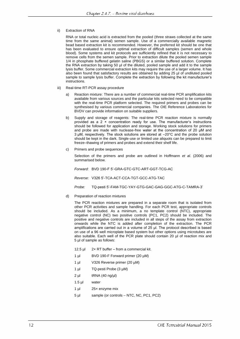
RNA or total nucleic acid is extracted from the pooled (three straws collected at the same time from the same animal) semen sample. Use of a commercially available magnetic bead based extraction kit is recommended. However, the preferred kit should be one that has been evaluated to ensure optimal extraction of difficult samples (semen and whole blood). Some systems and kit protocols are sufficiently refined that it is not necessary to remove cells from the semen sample. Prior to extraction dilute the pooled semen sample 1/4 in phosphate buffered gelatin saline (PBGS) or a similar buffered solution. Complete the RNA extraction by taking 50 µl of the diluted, pooled sample and add it to the sample lysis buffer. Some commercial extraction kits may require the use of a larger volume. It has also been found that satisfactory results are obtained by adding 25 µl of undiluted pooled sample to sample lysis buffer. Complete the extraction by following the kit manufacturer‟s instructions.
iii) Real-time RT-PCR assay procedure
a) Reaction mixture: There are a number of commercial real-time PCR amplification kits available from various sources and the particular kits selected need to be compatible with the real-time PCR platform selected. The required primers and probes can be synthesised by various commercial companies. The OIE Reference Laboratories for BVDV can provide information on suitable suppliers.
b) Supply and storage of reagents: The real-time PCR reaction mixture is normally provided as a 2 × concentration ready for use. The manufacturer‟s instructions should be followed for application and storage. Working stock solutions for primers and probe are made with nuclease-free water at the concentration of 20 μM and 3 μM, respectively. The stock solutions are stored at –20°C and the probe solution should be kept in the dark. Single-use or limited use aliquots can be prepared to limit freeze–thawing of primers and probes and extend their shelf life.
c) Primers and probe sequences
Selection of the primers and probe are outlined in Hoffmann et al. (2006) and
summarised below.
Forward: BVD 190-F 5‟-GRA-GTC-GTC-ART-GGT-TCG-AC
Reverse: V326 5‟-TCA-ACT-CCA-TGT-GCC-ATG-TAC
Probe: TQ-pesti 5‟-FAM-TGC-YAY-GTG-GAC-GAG-GGC-ATG-C-TAMRA-3‟
d) Preparation of reaction mixtures
The PCR reaction mixtures are prepared in a separate room that is isolated from other PCR activities and sample handling. For each PCR test, appropriate controls should be included. As a minimum, a no template control (NTC), appropriate negative control (NC) two positive controls (PC1, PC2) should be included. The positive and negative controls are included in all steps of the assay from extraction onwards while the NTC is added after completion of the extraction. The PCR amplifications are carried out in a volume of 25 μl. The protocol described is based on use of a 96 well microplate based system but other options using microtubes are also suitable. Each well of the PCR plate should contain 20 µl of reaction mix and 5 µl of sample as follows:
12.5 µl 2× RT buffer – from a commercial kit.
1 µl BVD 190-F Forward primer (20 µM)
1 µl V326 Reverse primer (20 µM)
1 µl TQ-pesti Probe (3 µM)
2 µl tRNA (40 ng/µl)
1.5 µl water
1 µl 25× enzyme mix
5 µl sample (or controls – NTC, NC, PC1, PC2)

e) Selection of controls
NTC: usually consists of tRNA in nuclease free water that is added in place of a sample when the PCR reaction is set up.
NC: In practice, many laboratories use PBGS or a similar buffer. Ideally the controls for testing of semen samples should be negative semen, from sero-negative bulls. However, as a minimum, the assay in use should have been extensively validated with negative and positive samples to confirm that it gives reliable extraction and amplification with semen.
PCs: There are two positive controls (PC1=moderate – [Ct 29-32] and PC2=weak [Ct 32–35] positive). Positive semen from naturally infected bulls is preferable as a positive control. However, this is likely to be difficult to obtain. Further, semen from a PI bull is not considered suitable because the virus loads are usually very high and would not give a reliable indication of any moderate reduction in extraction or assay performance. Negative semen spiked with defined quantities of BVDV virus could be used as an alternative. If other samples are used as a routine PC, as a minimum the entire extraction process and PCR assay in use must have been extensively validated using known positive semen from bulls with a PTI or from bulls undergoing an acute infection. If these samples are not available and spiked samples are used for validation purposes, a number of samples spiked with very low levels of virus should be included. On a day to day basis, the inclusion of an exogenous control with each test sample will largely compensate for not using positive semen as a control and will give additional benefits by monitoring the efficiency of the assay on each individual sample. Positive control samples should be prepared carefully to avoid cross contamination from high titred virus stocks and should be prepared in advance and frozen at a „ready to use‟ concentration and ideally „single use‟ volume.
f) Extracted samples are added to the PCR mix in a separate room. The controls should be added last, in a consistent sequence in the following order: NTC, negative and then the two positive controls.
g) Real-time polymerase chain reaction
The PCR plate or tubes are placed in the real-time PCR detection system in a separate, designated PCR room. Some mastermixes have uniform reaction conditions that are suitable for many different assays. As an example, the PCR detection system is programmed for the test as follows:
1× 48°C 10 minutes
1× 95°C 10 minutes
45 × (95°C 15 seconds, 60°C 1 minute)
h) Analysis of real-time PCR data
The software program is usually set to automatically adjust results by compensating for any background signal and the threshold level is usually set according to the manufacturer‟s instructions for the selected analysis software used. In this instance, a threshold is set at 0.05.
i) Interpretation of results
a) Test controls – all controls should give the expected results with positive controls PC1 and PC2 falling within the designated range and both the negative control NC and no template control NTC should have no Ct values.
b) Test samples
1) Positive result: Any sample that has a cycle threshold (Ct) value less than 40 is regarded as positive.
2) Negative result: Any sample that shows no Ct value is regarded as negative. However, before reporting a negative result for a sample, the performance of the exogenous internal control should be checked and shown to give a result within the accepted range for that control (for example, a Ct value no more than 2–3 Ct units higher than the NTC).

Antigen detection by ELISA has become a widely adopted method for the detection of individual PI animals. These assays are not intended for the detection of acutely infected animals (though from to time this may be achieved). Importantly, these assays are not designed for screening of semen or biological materials used in assays or vaccine manufacture. Several methods for the ELISA for antigen detection have been published and a number of commercial kits are available. Most are based on the sandwich ELISA principle, with a capture antibody bound to the solid phase, and a detector antibody conjugated to a signal system, such as peroxidase. Amplification steps such as the use of biotin and streptavidin in the detection system are sometimes used to increase assay sensitivity. Both monoclonal- and polyclonal-based systems are described. The test measures BVD antigen (NS2-3 or ERNS) in lysates of peripheral blood leukocytes; the new generation of antigen-capture ELISAs (ERNS
capture ELISAs) are able to detect BVD antigen in blood as well as in plasma or serum samples. The best of the methods gives a sensitivity similar to virus isolation, and may be preferred in those rare cases where persistent infection is combined with sero-positivity. Due to transient viraemia, the antigen ELISA is less useful for virus detection in acute BVD infections.
The NS2-3 ELISA may be less effective in young calves that have had colostrum due to the presence of BVDV maternal antibodies. The real-time RT-PCR is probably the most sensitive detection method for this circumstance, but the ERNS ELISA has also been shown to be a sensitive and reliable test, particularly when used with skin biopsy (ear-notch) samples (Cornish et al., 2005).
Enzyme-labelled methods are useful to detect BVDV antigen in tissue sections, particularly where suitable MAbs are available. However, these assays are not appropriate to certify animals for international trade and use should be limited to diagnostic investigations. It is important that the reagents and procedures used be fully validated, and that nonspecific reactivity be eliminated. For PI cattle almost any tissue can be used, but particularly good success has been found with lymph nodes, thyroid gland, skin, brain, abomasum and placenta. Skin biopsies, such as ear-notch samples, have shown to be useful for in-vivo diagnosis of persistent BDV infection.
Antibody to BVDV can be detected in cattle sera by a standard VNT or by ELISA, using one of several published methods or with commercial kits (e.g. Edwards, 1990). Serology is used to identify levels of herd immunity, for the detection of the presence of PI animals in a herd, to assist with investigation of reproductive disease and possible involvement of BVDV and to establish the serological status of bulls being used for semen collection and to identify whether there has been a recent infection. ELISA for antibody in bulk milk samples can give a useful indication of the BVD status of a herd (Niskanen, 1993). A high ELISA value (0.8 or more absorbance units) in an unvaccinated herd indicates a high probability of the herd having been exposed to BVDV in the recent past, most likely through one or more persistently viraemic animals being present. In contrast, a very low or negative value (≤0.2) indicates that it is unlikely that persistently viraemic animals are present. However, ELISA values are not always a reliable indicator of the presence of PI animals on farms, due to differing husbandry (Zimmer et al., 2002), recent administration of vaccine and also due to the presence of viral antigen in bulk milk, which may interfere with the antibody assay itself. Determination of the antibody status of a small number of young stock (9–18 months) has also been utilised as an indicator of recent transmission of BVDV in the herd (Houe et al., 1995), but this approach is also dependent on the degree of contact between different groups of animals in the herd and the potential for exposure from neighbouring herds. VN tests are more frequently used for regulatory purposes (e.g. testing of semen donors) while ELISAs (usually in the form of commercially prepared kits) are commonly used for diagnostic applications. Whether ELISA or VNT, control positive and negative standard sera must be included in every test. These should give results within predetermined limits for the test to be considered valid. In the VNT, a „serum control‟ to monitor sample toxicity should also be included for each test sample.
Selection of the virus strain to include in a VNT is very important. No single strain is likely to be ideal for all circumstances, but in practice one should be selected that detects the highest proportion of serological reactions in the local cattle population. Low levels of antibody to BVD type 2 virus may not be detectable by a neutralisation test that uses type 1 strain of the virus, and vice versa (Fulton et al., 1997). It is important that BVD type 1 and BVD type 2 be used in the test and not just the one that the diagnostician thinks is present, as this can lead to under reporting. Because it makes the test easier to read, most laboratories use highly cytopathic, laboratory-adapted strains of BVDV for VN tests. Two widely used cytopathic strains are „Oregon C24V‟ and „NADL‟. However immune-labelling techniques are now available that allow simple detection of the growth or neutralisation of non-cytopathic strains

where this is considered desirable, especially to support the inclusion of a locally relevant virus strain. An outline protocol for a microtitre VN test is given below (Edwards, 1990):
i) The test sera are heat-inactivated for 30 minutes at 56°C.
ii) From a starting dilution of 1/4, serial twofold dilutions of the test sera are made in a cell-culture grade flat-bottomed 96-well microtitre plate, using cell culture medium as diluent. For each sample, three or four wells are used at each dilution depending on the degree of precision required. At each dilution of serum, for each sample one well is left without virus to monitor for evidence of sample toxicity that could mimic viral cytopathology or interfere with virus replication. Control positive and negative sera should also be included in each batch of tests.
iii) An equal volume (e.g. 50 μl) of a stock of cytopathic strain of BVDV containing 100 TCID50
(50%) tissue culture infective dose is added to each well. A back titration of virus stock is also done in some spare wells to check the potency of the virus (acceptance limits 30–300 TCID50).
iv) The plate is incubated for 1 hour at 37°C.
v) A flask of suitable cells (e.g. bovine turbinate, bovine testis) is trypsinized and the cell
concentration is adjusted to 1.5 × 10⁵/ml. 100 μl of the cell suspension is added to each well of the microtitre plate.
vi) The plate is incubated at 37°C for 4–5 days, either in a 5% CO2 atmosphere or with the
plate sealed.
vii) The wells are examined microscopically for CPE or fixed and stained by immunoperoxidase staining using an appropriate monoclonal antibody. The VN titre for each serum is the dilution at which the virus is neutralised in 50% of the wells. This can be calculated by the Spearman–Kärber or Reed Muench methods. A sero-negative animal will show no neutralisation at the lowest dilution (1/4), equivalent to a final dilution of 1/8. For accurate comparison of antibody titres, and particularly to demonstrate significant (more than fourfold) changes in titre, samples should be tested in parallel in the same test.
Both indirect and blocking types of test can be used. A number of commercial kits are available. As with the virus neutralisation test, ELISAs configured using antigen from one genotype of BVD may not efficiently detect antibody induced by another genotype. Tests should therefore be selected for their ability to detect antibody to the spectrum of genotypes and strains circulating in the country where the test is to be performed.
The chief difficulty in setting up the test lies in the preparation of a viral antigen of sufficient potency. The virus must be grown under optimal culture conditions using a highly permissive cell type. Any serum used in the medium must not inhibit growth of BVDV. The optimal time for harvest should be determined experimentally for the individual culture system. The virus can be concentrated and purified by density gradient centrifugation. Alternatively, a potent antigen can be prepared by treatment of infected cell cultures with detergents, such as Nonidet P40, N-decanoyl-N-methylglucamine (Mega 10), Triton X-100 or 1-octylbeta-D-glucopyranoside (OGP). Some workers have used fixed, infected whole cells as antigen. In the future, increasing use may be made of artificial antigens manufactured by expressing specific viral genes in bacterial or eukaryotic systems. Such systems should be validated by testing sera specific to a wide range of different virus strains. In the future, this technology should enable the production of serological tests complementary to subunit or marker vaccines, thus enabling differentiation between vaccinated and naturally infected cattle. An example outline protocol for an indirect ELISA is given below (Edwards, 1990).
i) Roller cultures of secondary calf testis cells with a high multiplicity of infection (about one), are inoculated with BVDV strain Oregon C24V, overlaid with serum-free medium and incubated for 24 hours at 37°C.
ii) The cells are scraped off and pelleted. The supernatant medium is discarded. The pellet is treated with two volumes of 2% OGP in PBS for 15 minutes at 4°C, and centrifuged to remove the cell debris. The supernatant antigen is stored in small aliquots at –70°C, or freeze-dried. Non-infected cells are processed in parallel to make a control antigen.

iii) The antigen is diluted to a predetermined dilution in 0.05 M bicarbonate buffer, pH 9.6. Alternate rows of an ELISA-grade microtitre plate are coated with virus and control antigens overnight at 4°C. The plates are then washed in PBS with 0.05% Tween 20 or Tween 80 (PBST) before use in the test.
iv) Test sera are diluted 1/50 in serum diluent (0.5 M NaCl; 0.01 M phosphate buffer; 0.05% Tween 20; 0.001 M ethylene diamine tetra-acetic acid; 1% polyvinyl pyrrolidone, pH 7.2) and added to virus- and control-coated wells for 1 hour at 37°C. The plates are then washed five times in PBST.
v) Rabbit anti-bovine IgG peroxidase conjugate is added at a predetermined dilution (in serum diluent) for 1 hour at 37°C, then the plates are again washed five times in PBST.
vi) A suitable enzyme substrate is added, such as hydrogen peroxide/tetramethyl benzidine. After colour development, the reaction is stopped with sulphuric acid and the absorbance is read on an ELISA plate reader. The value obtained with control antigen is subtracted from the test reaction to give a net absorbance value for each serum.
vii) It is recommended to convert net absorbance values to sample:positive ratio (or percentage positivity) by dividing net absorbance by the net absorbance on that test of a standard positive serum that has a net absorbance of about 1.0. This normalisation procedure leads to more consistent and reproducible results.
BVDV vaccines are used primarily for disease control purposes although they can convey production advantages especially in intensively managed cattle such as in feedlots. In some countries where BVDV eradication is being undertaken, PI animals are removed and remaining cattle are vaccinated to maintain a high level of infection and prevent the generation of further PI animals. Vaccination to control BVDV infections can be challenging due in part to the antigenic variability of the virus and the occurrence of persistent infections that arise as a result of fetal infection. on-going maintenance of the virus in nature is predominantly sustained by PI animals that are the product of in-utero infection. The goal for a vaccine should be to prevent systemic viraemia and the virus crossing
the placenta, If this is successfully achieved it is likely that the vaccine will prevent the wide range of other clinical manifestations, including reproductive, respiratory and enteric diseases and immunosuppression with its secondary sequelae. There are many different vaccines available in different countries. Traditionally, BVD vaccines fall into two classes: modified live virus or inactivated vaccines. Experimental recombinant subunit vaccines based on BVD viral glycoprotein E2 expressed with baculovirus or transgenic plants and BVDV E2 DNA vaccines have been described but few if any are in commercial production. They offer a future prospect of „marker vaccines‟ when used in connection with a complementary serological test.
Traditionally, BVD vaccines fall into two classes: modified live or inactivated virus vaccines. The essential requirement for both types is to afford a high level of fetal infection. Many of the live vaccines have been based on a cytopathic strain of the virus which is considered to be unable to cross the placenta. However, it is important to ensure that the vaccine virus does not cause fetal infection. In general vaccination of breeding animals should be completed well before insemination to ensure optimal protection and avoid any risk of fetal infection. Live virus vaccine may also be immunosuppressive and precipitate other infections. On the other hand, modified live virus vaccines may only require a single dose. Use of a live product containing a cytopathic strain of BVDV may precipitate mucosal disease by superinfection of persistently viraemic animals. Properly formulated inactivated vaccines are very safe to use but, to obtain satisfactory levels of immunity, they usually require booster vaccinations, which may be inconvenient. A combined vaccination protocol using inactivated followed by live vaccine may reduce the risk of adverse reaction to the live strain. Whether live or inactivated, because of the propensity for antigenic variability, the vaccine should contain strains of BVDV that are closely matched to viruses found in the area in which they are used. For example, in countries where strains of BVDV type 2 are found, it is important for the vaccine to contain a suitable type 2 strain. For optimal immunity against type 1 strains, antigens from the dominant subtypes (e.g. 1a and 1b) should be included. Due to the need to customise vaccines for the most commonly encountered strains within a country or region, it is not feasible to produce a vaccine antigen bank that can be drawn upon globally.

Guidance for the production of veterinary vaccines is given in Chapter 1.1.8 Principles of veterinary vaccine production. The guidelines given here and in chapter 1.1.8 are intended to be general in nature and may be supplemented by national and regional requirements.
For optimal efficacy, it is considered that there should be a close antigenic match between viruses included in a vaccine and those circulating in the target population. BVDV type 2 strains should be included as appropriate. Due to the regional variations in genotypes and subtypes of BVDV, many vaccines contain more than one strain of BVDV to give acceptable protection. A good appreciation of the antigenic characteristics of individual strains can be obtained by screening with panels of MAbs (Paton et al., 1995).
Isolates of cytopathic virus are often mixed with the noncytopathic biotype. The separation and purification of the two biotypes from an initial mixed culture is important to maintain the expected characteristics of the seen and depends on several cycles of a limiting dilution technique for the noncytopathic virus, or plaque selection for the cytopathic virus. Purity of the cytopathic virus should be confirmed by at least one additional passage at limiting dilution. When isolates have been cloned, their identity and key antigenic characteristics should be confirmed. The identity of the seed virus should be confirmed by sequencing. Where there are multiple isolates included in the vaccine, each has to be prepared separately.
While retaining the desirable antigenic characteristics, the strains selected for the seed should not show any signs of disease when susceptible animals are vaccinated. Live attenuated vaccines should not be transmissible to unvaccinated „in-contact‟ animals and should not be able to infect the fetus. Ideally seeds prepared for the production of inactivated vaccines should grow to high titre to minimise the need to concentrate the antigens and there should be a minimal amount of protein from the cell cultures incorporated into the final product. Master stocks for either live or inactivated vaccines should be prepared under a seed lot system involving master and working stocks that can be used for production in such a manner that the number of passages can be limited and minimise antigenic drift. While there are no absolute criteria for this purpose, as a general guide, the seed used for production should not be passaged more than 20 times beyond the master seed and the master seed should be of the lowest passage from the original isolate as is practical.
It is crucial to ensure that all materials used in the preparation of the bulk antigens have been extensively screened to ensure freedom from extraneous agents. This should include master and working seeds, the cell cultures and all medium supplements such as bovine serum. It is particularly important to ensure that any serum used that is of bovine origin is free of both adventitious BVDV of all genotypes and antibodies against BVDV strains because low levels of either virus or antibody can mask the presence of the other. Materials and vaccine seeds should be tested for sterility and freedom from contamination with other agents, especially viruses as described in the chapter 1.1.8 and chapter 1.1.9.
All vaccines should pass standard tests for efficacy. Tests should include as a minimum the demonstration of a neutralising antibody response following vaccination, a reduction in virus shedding after challenge in vaccinated cattle and ideally a prevention of viraemia. Efficacy tests of BVD vaccines by assessing clinical parameters in non-pregnant cattle can be limited by the difficulty of consistently establishing clinical signs but, when employed, clinical parameters such as a reduction in the rectal temperature response and leukopenia should be monitored. Although it can be difficult by using virus isolation in cell culture to consistently demonstrate the low levels of viraemia associated with an acute infection, real-time PCR could be considered as an alternative method to establish the levels of circulating virus.
If a vaccine passes basic tests, the efficacy of vaccination should ultimately be measured by the capacity to prevent transplacental transmission. If there is a substantial reduction and ideally complete prevention of fetal infection, a vaccine would be expected to be highly effective in other situations (for example prevention of respiratory disease). A suitable challenge system

can be established by intranasal inoculation of noncytopathic virus into pregnant cows between 60 and 90 days of gestation (Brownlie et al., 1995). Usually this system will reliably produce persistently viraemic offspring in non-immune cows. In countries where BVDV type 2 viruses are commonly encountered, efficacy in protecting against BVDV2 infections should be measured.
Both cytopathic and noncytopathic biotypes will grow in a variety of cell cultures of bovine origin. Standard procedures may be used, with the expectation for harvesting noncytopathic virus on days 4–7 and cytopathic virus on days 2–4. The optimal yield of infectious virus will depend on several factors, including the cell culture, isolate used and the initial seeding rate of virus. These factors should be taken into consideration and virus replication kinetics investigated to establish the optimal conditions for large scale virus production. Whether a live or inactivated vaccine, the essential aim will be to produce a high-titred virus stock. This bulk antigen preparation can subsequently be prepared according to the type of vaccine being considered.
Most BVDV vaccines are grown in cell cultures of bovine origin that are frequently supplemented with medium components of animal origin. The material of greatest concern is bovine serum due to the potential for contamination with BVD viruses and antibodies to these viruses. These adventitious contaminants not only affect the efficiency of production but also may mask the presence of low levels of infectious BVDV that may have undesirable characteristics. In addition to the virus seeds, all materials should be tested for sterility and freedom from contamination with other agents, especially viruses as described in chapters 1.1.8 and 1.1.9. Further, materials of bovine or ovine origin should originate from a country with negligible risk for transmissible spongiform encephalopathies [TSEs] (see chapter 1.1.9).
In-process controls are part of the manufacturing process. Cultures should be inspected regularly to ensure that they remain free from contamination, and to monitor the health of the cells and the development or absence of CPE, as appropriate. While the basic requirement for efficacy is the capacity to induce an acceptable neutralising antibody response, during production, target concentrations of antigen required to achieve an acceptable response may be monitored indirectly by assessment of the quantity of infectious virus or antigen mass that is produced. Rapid diagnostic assays such as the ELISA are useful to monitor BVDV antigen production. Alternatively, the quality of a batch of antigen may be determined by titration of the quantity of infectious virus present, although this may underestimate the quantity of antigen. For inactivated vaccines, infectivity is evaluated before inactivation. For inactivated vaccines the inactivation kinetics should be established so that a suitable safety margin can be determined and incorporated into the routine production processes. At the end of production, in vitro cell culture assays should be undertaken to confirm that inactivation has been complete. These innocuity tests should include a sufficient number of passages and volume of inoculum to ensure that very low levels of infectious virus would be detected if present.
i) Sterility
Tests of biological materials for sterility and freedom from contamination may be found in Chapter 1.1.9.
ii) Identity
Identity tests should demonstrate that no other strain of BVDV is present when several strains are propagated in a facility producing multivalent vaccines.
iii) Safety
Safety tests shall consist of detecting any abnormal local or systemic adverse reactions to the vaccine by all vaccination route(s). Batch-to-batch safety tests are required unless safety of the product is demonstrated and APPROVED in the registration dossier and production is consistent with that described in chapter 1.1.8.
The safety test is different to the inocuity test (see above).
Live vaccines must either be demonstrated to be safe in pregnant cattle (i.e. no transmission to the fetus), or should be licensed with a warning not to use them in pregnant animals. Live vaccines containing cytopathic strains should have an appropriate warning of the risk of inducing mucosal disease in PI cattle.
iv) Batch potency
BVD vaccines must be demonstrated to produce adequate immune responses, when used in their final formulation according to the manufacturer‟s published instructions. The minimum quantity of infectious virus and/or antigen required to produce an acceptable immune response should be determined. In-vitro assays should be used to monitor individual batches during production.
For registration of a vaccine, all relevant details concerning manufacture of the vaccine and quality control testing should be submitted to the relevant authorities. Unless otherwise specified by the authorities, information should be provided from three consecutive vaccine batches with a volume not less than 1/3 of the typical industrial batch volume.
There is no standard method for the manufacture of a BVD vaccine, but conventional laboratory techniques with stationary, rolled or suspension (micro-carriers) cell cultures may be used. Inactivated vaccines can be prepared by conventional methods, such as binary ethylenimine or beta-propiolactone inactivation (Park & Bolin, 1987). A variety of adjuvants may be used.
In-vivo tests should be undertaken using a single dose, overdose (for live vaccines only) and repeat doses (taking into account the maximum number of doses for primary vaccination and, if appropriate, the first revaccination/booster vaccination) and contain the maximum permitted antigen load and, depending on the formulation of the vaccine, the maximum number of vaccine strains.
i) Target and non-target animal safety
The safety of the final product formulation of both live and inactivated vaccines should be assessed in susceptible young calves that are free of maternally derived antibodies and in pregnant cattle. They should be checked for any local reactions following administration, and, in pregnant cattle, for any effects on the unborn calf. Live attenuated vaccines may contribute to immunosuppression that might increase mortality. It may also contribute to the development of mucosal disease in PI animals that is an animal welfare concern. Therefore vaccination of PI animals with live attenuated vaccines containing cytopathic BVDV should be avoided. Live attenuated vaccines must not be capable of being transmitted to other unvaccinated animals that are in close contact.
ii) Reversion-to-virulence for attenuated/live vaccines and environmental considerations
Virus seeds that have been passaged at least up to and preferably beyond the passage limit specified for the seed should be inoculated into young calves to confirm that there is no evidence of disease. If a live attenuated vaccine has been registered for use in pregnant animals, reversion to virulence tests should also include pregnant animals. Live attenuated vaccines should not be transmissible to unvaccinated „in-contact‟ animals.
iii) Precautions (hazards)
BVDV is not considered to be a human health hazard. Standard good microbiological practice should be adequate for handling the virus in the laboratory. A live virus vaccine should be identified as harmless for people administering the product however adjuvants included in either live or inactivated vaccines may cause injury to people. Manufacturers should provide adequate warnings that medical advice should be sought in the case of self-injection (including for adjuvants, oil-emulsion vaccine, preservatives, etc.) with warnings included on the product label/leaflet so that the vaccinator is aware of any danger.

The potency of the vaccine should be determined by inoculation into seronegative and virus negative calves, followed by monitoring of the antibody response. Antigen content can be assayed by ELISA and adjusted as required to a standard level for the particular vaccine. Standardised assay protocols applicable to all vaccines do not exist. Live vaccine batches may be assayed by infectivity titration. Each production batch of vaccine should undergo potency and safety testing as batch release criteria. BVD vaccines must be demonstrated to produce adequate immune responses, as outlined above, when used in their final formulation according to the manufacturer‟s published instructions.
To date, there are no commercially available vaccines for BVDV that support use of a true DIVA strategy. Experimental subunit vaccines based on baculovirus-expressed BVD viral glycoprotein E2 have been described but are not available commercially. They offer a future prospect of „marker vaccines‟ when used in connection with a complementary serological test. Experimental BVDV E2 DNA vaccines and BVDV E2 subunit vaccines expressed using transgenic plants and alphavirus replicon have also been described.
There are few published data on the duration of antibody following vaccination with a commercial product. Protocols for their use usually recommend a primary course of two inoculations and boosters at yearly intervals. Only limited data are available on the antibody levels that correlate with protection against respiratory infections (Bolin & Ridpath, 1995; Howard et al., 1989) or in-utero infection (Brownlie et al., 1995). However, there are many different commercial formulations and these involve a range of adjuvants that may support different periods of efficacy. Consequently, duration of immunity data must be generated separately for each commercially available product by undertaking challenge tests at the end of the period for which immunity has been claimed.
There are no accepted guidelines for the stability of BVD vaccines, but it can be assumed that attenuated virus vaccine (freeze-dried) should remain potent for at least 1 year if kept at 4°C. Inactivated virus vaccine could have a longer shelf life at 4°C. Lower temperatures could prolong shelf life for either type, but adjuvants in killed vaccine may preclude this. Bulk antigens that have not been formulated into finished vaccine can be reliably stored frozen at low temperatures but the antigen quality should be monitored with in vitro assays prior to incorporation into a batch of vaccine.
BAKER J.C. (1995). The clinical manifestations of bovine viral diarrhea infection. Vet. Clin. North. Am. – Food Animal Practice, 11, 425–445.
BAUERMANN F.V., FLORES E.F. & RIDPATH J.F. (2012). Antigenic relationships between bovine viral diarrhea virus 1 and 2 and HoBi virus: possible impacts on diagnosis and control. J. Vet. Diagn. Invest. (official publication of the American Association of Veterinary Laboratory Diagnosticians), 24, 253–261.
BAUERMANN F.V., HARMON A., FLORES E.F., FALKENBERG S.M., REECY J.M. & RIDPATH J.F. (2013). In vitro
neutralization of HoBi-like viruses by antibodies in serum of cattle immunized with inactivated or modified live vaccines of bovine viral diarrhea viruses 1 and 2. Vet. Microbiol., 166, 242–245.
BOLIN S.R. & RIDPATH J.F. (1992). Differences in virulence between two noncytopathic bovine viral diarrhea viruses in calves. Am. J. Vet. Res., 53, 2157–2163.
BOLIN S.R. & RIDPATH J.F. (1995). Assessment of protection from systemic infection or disease afforded by low to intermediate titers of passively acquired neutralizing antibody against bovine viral diarrhea virus in calves. Am. J. Vet. Res., 56, 755–759.
BROWNLIE J. (1985). Clinical aspects of the bovine virus diarrhoea/mucosal disease complex in cattle. In Practice, 7, 195–202.

BROWNLIE J. (1990). The pathogenesis of bovine virus diarrhoea virus infections. Rev. sci. tech. Off. int. Epiz., 9, 43–59.
BROWNLIE J., CLARKE M.C., HOOPER L.B. & BELL G.D. (1995). Protection of the bovine fetus from bovine viral diarrhoea virus by means of a new inactivated vaccine. Vet. Rec., 137, 58–62.
CORNISH T.E., VAN OLPHEN A.L., CAVENDER J.L., EDWARDS J.M., JAEGER P.T., VIEYRA L.L., WOODARD L.F., MILLER
D.R. & O‟TOOLE D. (2005). Comparison of ear notch immunohistochemistry, ear notch antigen-capture ELISA, and buffy coat virus isolation for detection of calves persistently infected with bovine viral diarrhea virus. J. Vet. Diagn. Invest., 17, 110–117.
DUFFELL S.J. & HARKNESS J.W. (1985). Bovine virus diarrhoea-mucosal disease infection in cattle. Vet. Rec., 117, 240–245.
EDWARDS S. (1990). The diagnosis of bovine virus diarrhoea-mucosal disease in cattle. Rev. sci. tech. Off. int. Epiz., 9, 115–130.
FRAY M.D., MANN G.E., BLEACH E.C.L., KNIGHT P.G., CLARKE M.C. & CHARLESTON B. (2002). Modulation of sex hormone secretion in cows by acute infection with bovine viral diarrhoea virus. Reproduction, 123, 281-289.
FULTON R.W., SALIKI J.T., BURGE L.J., DOFFAY J.M., BOLIN S.R., MAES R.K., BAKER J.C. & FREY M.L. (1997). Neutralizing antibodies to type-1 and type-2 bovine viral diarrhea viruses – detection by inhibition of viral cytopathology and infectivity by immunoperoxidase assay. Clin. Diagn. Lab. Immunol., 4, 380–383.
GIVENS M.D., RIDDELL K.P., WALZ P.H., RHOADES J., HARLAND R., ZHANG Y., GALIK P.K., BRODERSEN B.W., COCHRAN
A.M., BROCK K.V., CARSON R.L. & STRINGFELLOW D.A. (2007). Noncytopathic bovine viral diarrhea virus can persist in testicular tissue after vaccination of peri-pubertal bulls but prevents subsequent infection. Vaccine, 25, 867–876.
HOFFMANN B., DEPNER K., SCHIRRMEIER H. & BEER M. (2006). A universal heterologous internal control system for duplex real-time RT-PCR assays used in a detection system for pestiviruses J. Virol. Methods, 136, 200–209.
HOUE H., BAKER J.C., MAES R.K., RUEGG P.L. & LLOYD J.W. (1995). Application of antibody titers against bovine viral diarrhea virus (BVDV) as a measure to detect herds with cattle persistently infected with BVDV. J. Vet. Diagn. Invest., 7, 327–332.
HOWARD C.J., CLARKE M.C. & BROWNLIE J. (1989). Protection against respiratory infection with bovine virus diarrhoea virus by passively acquired antibody. Vet. Microbiol., 19, 195–203.
LETELLIER C. & KERKHOFS P. (2003). Real-time PCR for simultaneous detection and genotyping of bovine viral diarrhea virus. J. Virol. Methods, 114, 21–27.
MCGOLDRICK A., BENSAUDE E., IBATA G., SHARP G. & PATON D. J. (1999). Closed one-tube reverse transcription nested polymerase chain reaction for the detection of pestiviral RNA with fluorescent probes. J. Virol. Methods, 79, 85–95.
MCGOWAN M.R. & KIRKLAND P.D. (1995). Early reproductive loss due to bovine pestivirus infection. Br. Vet. J., 151, 263–270.
MEYLING A. (1984). Detection of BVD virus in viraemic cattle by an indirect immunoperoxidase technique. In: Recent Advances in Virus Diagnosis (CEC Seminar), McNulty M.S. & McFerran J.B., eds. Martinus Nijhoff, Belfast, UK, 37–46.
MOENNIG V. & LIESS B. (1995). Pathogenesis of intrauterine infections with bovine viral diarrhea virus. Vet. Clin. North. Am., 11, 477–487.
MOENNIG V., HOUE H. & LINDBERG A. (2005). BVD control in Europe: current status and perspectives. Anim. Health Res. Rev., 6, 63–74.
NISKANEN R. (1993). Relationship between the levels of antibodies to bovine viral diarrhoea virus in bulk tank milk and the prevalence of cows exposed to the virus. Vet. Rec., 133, 341–344.

PARK B.K. & BOLIN S.R. (1987). Molecular changes of bovine viral diarrhea virus polypeptides treated with binary ethylenimine, beta-propiolactone and formalin. Res. Rep. Rural Dev. Admin. (L&V) Korea, 29, 99–103.
PATON D.J., SANDS J.J. LOWINGS J.P., SMITH J.E., IBATA G., EDWARDS S. (1995). A proposed division of the pestivirus genus using monoclonal antibodies, supported by cross-neutralisation assays and genetic sequencing. Vet. Res., 26, 92–109.
VILCEK S., PATON D.J., DURKOVIC B., STROJNY L., IBATA G., MOUSSA A., LOITSCH A., ROSSMANITH W., VEGA S., SCICLUNA M.T. & PALFI V. (2001). Bovine viral diarrhoea virus genotype 1 can be separated into at least eleven genetic groups. Arch. Virol., 146, 99–115.
VOGES H., HORNER G.W., ROWE S. & WELLENBERG G.J. (1998). Persistent bovine pestivirus infection localized in the testes of an immuno-competent, non-viremic bull. Vet. Microbiol., 61, 165–175.
ZIMMER G., SCHOUSTRA W. & GRAAT E. A.M. (2002). Predictive values of serum and bulk milk sampling for the presence of persistently infected BVDV carriers in dairy herds. Res. Vet. Sci., 72, 75–82.
*
* *
NB: There are OIE Reference Laboratories for Bovine viral diarrhoea (see Table in Part 4 of this Terrestrial Manual or consult the OIE Web site for the most up-to-date list:
http://www.oie.int/en/our-scientific-expertise/reference-laboratories/list-of-laboratories/ ). Please contact the OIE Reference Laboratories for any further information on
diagnostic tests, reagents and vaccines for bovine viral diarrhoea