identification of non-invasive biomarkers for chronic
TRANSCRIPT

RESEARCH ARTICLE Open Access
Identification of non-invasive biomarkersfor chronic atrophic gastritis from serumexosomal microRNAsHong Liu1,2,5, Pei-wu Li2,4,5, Wei-qin Yang3, Hong Mi4,5, Jing-lin Pan2,5, Yuan-cheng Huang2,5, Zheng-kun Hou2,4,5,Qiu-ke Hou2,4,5, Qi Luo2,4,5 and Feng-bin Liu2,4,5*
Abstract
Background: Serum exosomal microRNAs (miRNAs) have been suggested as novel biomarkers for various diseases,especially gastric cancer (GC). But circulating biomarkers for Chronic atrophic gastritis (CAG) which is defined asprecancrerous lesions of GC remain largely elusive. To investigate serum exosomal miRNAs that are differentlyexpressed in CAG patients and Chronic nonatrophic gastritis (CNAG) may be helpful for its diagnosis and therapy.
Methods: Patients were recruited according to the diagnosis and exclusioncriteria. RNA was extracted from serumexosomes of 30 CAG and 30 CNAG patients. The miRNA expression profiles were analyzed by next generationsequencing and were validated by qRT-PCR. Receiver operating characteristic (ROC) analysis has been used toevaluate the diagnostic value.
Results: 30 CAG patients and 30 CNAG patients were recruited in our study. sRNA-seq results showed that hsa-miR-3591-3p, − 122-3p, and − 122-5p of the top 10 miRNAs (hsa-miR-148a-3p, − 122-3p, − 486-3p, −451a, − 122-5p, −3591-3p, − 486-5p, −151a-3p, −92a-3p, −320a) were significantly upregulated in exosomes from CAG patients versusthose from CNAG patients, but hsa-miR-451a, −151a-3p, and -92a-3p were significantly downregulated.Furthermore, qRT-PCR analysis confirmed that hsa-miR-122-5p and hsa-miR-122-3p were significantly upregulated inCAG samples, but hsa-miR-122-3p hadnot a steable expression. ROC curves showed that the AUC for hsa-miR-122-5p was 0.67 (95% CI 0.52–0.82, SE 62%, SP 86%). A sum of the four miRNAs (panel 1, hsa-miR-122-5p, −451a, −151a-3p, and -92a-3p) did not significantly improve the diagnostic potential (AUC 0.63, 95% CI 0.47 to 0.78). Correlationanalysis showed that the expression of hsa-miR-122-5p differed significantly between patients based on atrophic(Moderate atrophic vs. Absent, P value was 0.036.) and IM (compare moderate-severe, absent and mild P valueswere 0.001 and 0.014, respectively). However, there were no differences between groups based on age, gender,dysplasia, or chronic or active inflammation.
Conclusion: These results suggested that hsa-miR-122-5p in serum exosomes might serve as a potential biomarkerfor CAG diagnosis.
Trial registration: Chinese Clinical Trial Registy (ChiCTR-IOR-16008027, Date of Registration:2016-03-01).
Keywords: Biomarkers, Serum exosomes, microRNAs, Small RNA-sequencing, Chronic atrophic gastritis
* Correspondence: [email protected] University of Chinese Medicine, No.12 Jichang Road,Guangzhou 510405, Guangdong Province, China4The First Affiliated Hospital of Guangzhou University of Chinese Medicine,No.16 Jichang Road, Guangzhou 510405, Guangdong Province, ChinaFull list of author information is available at the end of the article
© The Author(s). 2019 Open Access This article is distributed under the terms of the Creative Commons Attribution 4.0International License (http://creativecommons.org/licenses/by/4.0/), which permits unrestricted use, distribution, andreproduction in any medium, provided you give appropriate credit to the original author(s) and the source, provide a link tothe Creative Commons license, and indicate if changes were made. The Creative Commons Public Domain Dedication waiver(http://creativecommons.org/publicdomain/zero/1.0/) applies to the data made available in this article, unless otherwise stated.
Liu et al. BMC Cancer (2019) 19:129 https://doi.org/10.1186/s12885-019-5328-7

BackgroundChronic atrophic gastritis (CAG) is one of precancerousstage of intestinal type gastric cancer (GC) and has a highprevalence [1]. CAG also usually progresses to intestinalmetaplasia (IM) and dysplasia [2]. Over the last 5 years, theannual incidence of GC was found to be 0.5–1% for CAG,0.6% for mild-to-moderate dysplasia, and 6% for severe dys-plasia [3, 4]. The pathogenesis of CAG remains as acomplicated context, at least including immune-me-diated inflammation, atrophy of mucosal gland, acti-vated serum auto-antibody for gastric parietal cellsand/or intrinsic factors, hypochlorhydria, deficiencyof vitamin B12 and, in some cases, neurologicalsymptoms and diffuse metaplasia [5]. Additionally,the diagnosis and follow-up examination of CAG de-pends on endoscopic examination and histopatho-logic evaluation, which are invasive and time-consuming for patients [1]. There should be biopsiestaken from more sites to avoid misdiagnoses. Evenso, gastrin-17(G-17) and Pepsinogen (PG) I or IImay be helpful for determining atrophy existenceand location. But it had not been widely used withlow stability cases [6, 7]. Thus, it is urgent to inves-tigate some non-invasive, sensitive and reliable bio-markers, which may enrich method of CAGdiagnosis.Biomarkers that can be measured in biofluids,
called as liquid biopsies [8]. It is more comfortablethan endoscopic examination and may reflect theheterogeneity of the disease. Remarkably, exosomes,a new type of liquid biopsy, may show the diseasestatus [9, 10]. Recently, more and more studies havereported that miRNAs are secreted from differenttypes of cells through exsomes (30–100 nm) or otherextracellular vesicles (EVs) such as microvesicles(100–1000 nm) and oncosomes (1–10 μm) [9, 11].They play important roles in cell-to-cell communica-tion, such as in inflammatory response as well as intissue regeneration and tumor progression [12].Serum exosomal miRNAs can be potential bio-markers for various diseases. For example, serumexosomal miR-19b-3p and miR-106a-5p have beenidentified as gastric cancer biomarkers [13]. How-ever, reports of the expression of serum exosomalmiRNAs in CAG have not been found.In the present study, small RNA-sequencing
(sRNA-Seq) was employed to analyze microRNAprofiles in serum exosomes of CAG patients.qRT-PCR and correlation analyses were used to val-idate the results of sRNA-seq. We tried to find somespecific biomarkers for CAG progression. So far aswe know, It might be the first report about serumexosomal miRNAs and potential biomarkers associ-ated with CAG. Our study provides new avenues
and references for determining biomarkers and eluci-dating the underlying mechanisms of CAG.
MethodsPatients and samplesTwo groups of patients were recruited from 2016 to 2017at the First Affiliated Hospital of Guangzhou University ofChinese Medicine in Guangzhou, China. Patients were re-quired to provide medical history, to receive physical ex-aminations and laboratory safety tests and to undergo agastroscopy and biopsies for histological investigation.CAG group was composed of patients who had a diagno-sis of CAG with or without intestinal metaplasia, andChronic non-atrophic gastritis (CNAG) group is com-posed of health honors and patients who had mild ormoderate superficial gastritis. Patients considered to beeligible subjects were between 18 and 70 years old [14].Exclusion criteria included severe dysplasia, suspicion ofgastric cancer or any other malignancy, gastric surgeryhistory, drug allergy to the studied medication, severe sys-temic diseases (cardio-cerebral-vascular disease, hepaticdiseases, blood, kidney, lung or liver disease), administra-tion of non-steroidal anti-inflammatory drugs, pregnancyor lactation in female patients, and unwillingness toundergo repeated endoscopy after treatment.The study was approved by the Ethics Committee of
the First Affiliated Hospital of Guangzhou University ofChinese Medicine, and it was one part of our trial study(ChiCTR-IOR-16008027). All participants provided in-formed consent and were required to have a negative re-ports of 13C-urea from a breath test to show that theywere not infected with H.pylori. Clinical and demo-graphic information was obtained from each study sub-ject using a combination of structured subject interviewsand medical records. Blood (10 ml) and gastric mucosalbiopsies were collected from each patient. Serum wasseparated immediately and stored at − 80 °C before use.
Exosomes isolationExosomes were isolated from serum using ExoQuick™(SBI, USA). Serum was centrifuged at 17,000 g for 10min and filtrated with a 0.22 μm syringe filter (Millipore,USA) to remove cell debris. Then 500 μL of serumsupernatant was moved into a new tube and mixed with125 μL ExoQuick reagent. After incubation at 4 °C over-night, the mixture was centrifuged at 1500 g for 30 min.The pellet containing exosome fractions was then resus-pended in 30-40 μL of PBS or used for the next analysis.
Exosomes characterizationExosomes were characterized with NanoSight analysis andflow cytometry. The size distribution of the exosomes wasanalyzed using the ZETASIZER Nano series-Nano-ZS(Malvern Instruments Ltd., UK) according to the
Liu et al. BMC Cancer (2019) 19:129 Page 2 of 10

manufacturer’s instructions. The exosome markers, CD81and CD63, were identified using flow cytometry. The exo-some pellet was resuspended in 100 μL sterile PBS, thenincubated with CD63-Antibody-FITC (BD557288, USA)and CD81-Antibody-FITC (BD551108, USA) overnight at4 °C. Finally, detection was performed using an Accuri C6flow cytometer (BD Instruments Ltd., USA).
Exosomal RNA isolationBefore RNA was isolated, samples were treated with pro-teinase K and RNase A to degrade potential RNA/proteincomplexes. Exosomal RNA (exo-RNA) was isolated fromexosome fractions using a SeraMir Exosome RNA Purifi-cation Column kit (SBI, USA), with a final elution volumeof 30 μl, according to the manufacturer’s instructions.After RNA isolation, the samples were treated with DNaseto degrade potential DNA. The integrities and concentra-tions of the RNA samples were determined photometric-ally at 260 nm and 280 nm on a NanoDrop (PeqLab,Germany), then 500 ng or 10 ng of exosomal miRNAs wassubjected to ultra-deep sequencing or qRT-PCR.
Small RNA library preparation and sequencing30 RNA samples of each group were randomly allocated intothree sample pools. Small RNA deep sequencing (SRNA-seq)and bioinformatic analyses were used for the construction ofcDNA libraries. Cluster generation and subsequentultra-deep sequencing using the Illumina platform were per-formed at Genomics Institute Tech (BGI, China). BGI wasroutinely used in the bioinformatics analysis process. Briefly,the 49 nt sequence tags from Hiseq sequencing wentthrough the data cleaning analysis first, which included elim-inating the low quality tags and 5′ adaptor contaminantsfrom the 50 nt tags, in order to get credible clean tags. Sub-sequently, the distribution lengths of the clean tags as well asthose of the common and specific sequences between sam-ples were summarized. During standard analysis, the cleantags were allocated into different categories, and those whichcould not be allocated into any category were used to predictthe novel miRNA and the seed edit of potential known miR-NAs. The fragments of rRNA, scRNA, snoRNA, snRNA andtRNA were removed.
miRNAs verification by quantitative real-time PCR (qRT-PCR)qRT-PCR assays were performed using the Mir-X™ miRNAFirst-Strand Synthesis Kit and the SYBR® qRT-PCR Kit(Takara, Japan). The specific primers for miRNAs were syn-thesized by Sangon Biotech Company (Shanghai, China).The PCR reaction mixture contained 2 μL of first-strandcDNA, 10 μL of the 2× SYBR advantage Premix, 0.5 μL ofthe MRQ 3’Primer, 0.5 μL of the miRNA-specific primer(10 μM), and 7 μL ddH2O, having a final volume of 20 μL.After initial denaturation at 95 °C for 10 s, cycling was
performed as follows: 95 °C for 5 s and 60 °C for 20 s for 45cycles in CFX96TM Real-Time PCR Detection System(BioRAD, USA). The qRT-PCR for each miRNA was re-peated three times. The target sequences (5′-3′) of the pri-mer sets are shown in Table S1. U6 snRNA was used fornormalization, as it has previously been used fornormalization in other studies [15]. miRNAs with Cq valuesabove 37 were considered as not having been expressed.
Statistical analysisThe sRNA-seq data were analyzed in J-Express 2011. DEG-seq was used to detect and visualize the intensity-dependentratios of sRNA-seq data. The miRNAs with more than a1.2-fold change or less than a 0.6-fold change and a Q valueof 0.01 compared to a normal control were considered to bedifferentially expressing miRNAs (DEMs). For analysis ofqRT-PCR data, relative miRNA expressions were calculatedusing the 2-ΔCT method (ΔCT=CTmiR - CTreference). The2-ΔCT dataset was then Log2-transformed when necessary.Statistical analysis was executed using a t-test or aMann-Whitney U test. The area under the curve (AUC)used to assess the diagnostic power of the predictors was de-termined using Prism V (Graphpad Software). Logistic re-gression was used to develop a panel of combinedbiomarkers to predict the probability of developing CAG, aspreviously described [16]. Correlation analysis was conductedto evaluate whether the miRNA expression levels were cor-related with clinical characteristics. P< 0.05 was consideredsignificant.
ResultsClinical characteristics of patients30 CNAG and 30 CAG patients participated in our study(The clinical characteristics showed in Additional file 1:Table S2). The ages(Mean ± SD, years old) in CNAG andCAG groups were 48.67±9.12 years old and 52.67±9.74,respectively. The Male:Female ratios in CNAG and CAGgroups were 14:16 and 12:18, respectively. There were nodifferences in age or gender between CNAG and CAGgroups (P > 0.05). The two groups were comparable.
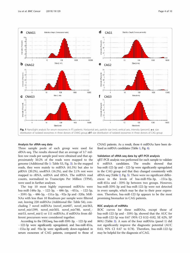
Isolation and identification of exosomesNanoSight analysis revealed that the size distribu-tion of our isolated exosomes ranged from approxi-mately 20 nm to 200 nm in diameter, with a modevalue of 100 nm (Fig. 1). Flow cytometry analysisdemonstrated that the expressions of CD81 andCD63, i.e., the commonly used exosome markers, ineach sample were over 80% (Fig. 2) [17, 18]. So weconcluded that our samples were enriched in exo-somes and co-isolated with other extracellular vesi-cles to some extent.
Liu et al. BMC Cancer (2019) 19:129 Page 3 of 10
Analysis for sRNA-seq dataThree sample pools of each group were used forsRNA-seq. The results showed that an average of 3.7 mil-lion raw reads per sample pool were obtained and that ap-proximately 50.2% of the reads were mapped to thegenome (Additional file 1: Table S3, Fig. 3). In the mappedreads, they were mainly to miRNA (61.5%) but also topiRNA (20.2%), snoRNA (16.2%), and the 2.1% rest weremapped to rRNA, snRNA and tRNA. The miRNA readcounts, normalized to Transcripts Per Million (TPM),were used in further analyses.The top 10 most highly expressed miRNAs were
hsa-miR-148a-3p, − 122-3p, − 486-3p, −451a, − 122-5p,− 3591-3p, − 486-5p, −151a-3p, −92a-3p and -320a. MiR-NAs with less than 10 Readnum per sample were filteredout, leaving 220 miRNAs (Additional file: Table S4), con-cluding 7 novel miRNAs (novel_mir607, novel_mir363,novel_mir1399, novel_mir503, novel_mir780, novel_-mir53, novel_mir1) or 111 miRNAs, if miRNAs from dif-ferent precursors were considered together.According to the DEGseq, hsa-miR-3591-3p, − 122-3p and
− 122-5p were significantly upregulated, and hsa-miR-451a,−151a-3p and -92a-3p were significantly down-regulated inserum exosomes of CAG patients, compared to those of
CNAG patients. As a result, those 6 miRNAs have been de-fined as miRNA candidates (Table 1, Fig. 4).
Validation of sRNA-seq data by qRT-PCR analysisqRT-PCR analysis was performed for each sample to validate6 miRNA candidates. The results showed thathsa-miR-122-3p and− 122-5p were significantly upregulatedin the CAG group and that they changed consistently withsRNA-seq (Table 2, Fig. 5). There were no significant differ-ences in the levels of hsa-miR-92a-3p, −151a-3p,miR-451a and − 3591-3p between two groups. However,hsa-miR-3591-3p and hsa-miR-122-3p were not detectedin every sample, which may be due to their poor expres-sion. Therefore, hsa-miR-122-5p appears to be the mostpromising biomarker in CAG patients.
ROC analysis of miRNAsROC curves for these miRNAs, except those ofhsa-miR-122-3p and − 3591-3p, showed that the AUC forhsa-miR-122-5p was 0.67 (95% CI 0.52–0.82, SE 62%, SP86%) (Table 3). A sum of the four miRNAs (panel 1) didnot significantly improve the diagnostic potential (AUC0.63, 95% CI 0.47 to 0.78). Therefore, hsa-miR-122-5pmay be helpful for the diagnosis of CAG.
a
b
c
d
e
f
Fig. 1 NanoSight analysis for serum exosomes in PS patients. Horizontal axis, particle size (nm); vertical axis, intensity (percent). a-c: sizedistribution of isolated exosomes in three donors of CNAG group; d-f: size distribution of isolated exosomes in three donors of CAG group
Liu et al. BMC Cancer (2019) 19:129 Page 4 of 10

Correlation analysis between hsa-miR-122-5p andclinicopathologic factorsThe results demonstrated that the expression of hsa-miR-122-5p was significantly differs between patients, basedon atrophic (Moderate atrophic vs. Absent, P value was0.036.) and IM (comparing moderate-severe, absent and mildP values were 0.001, 0.014, respectively) values. There were nodifferences among groups based on age, gender, dysplasia, orchronic inflammation or active inflammation (Table 4). Theresults showed that the expression of serum exosomalhsa-miR-122-5p might be related to atrophic and IM.
DiscussionUp to date, the diagnosis of CAG still depends on endo-scopic examination and histopathologic evaluation. Exo-somal miRNA played important roles in cell-to-cellcommunication, tissue regeneration, tumorigenesis andprogression [12]. Expression profiles of exosomalmiRNA are disease- and tissue-specificity, which wereconsidered as candidate biomarkers [19]. In the presentstudy, six miRNAs had been extremely changed inserum exosomes, particularly hsa-miR-122-5p was sig-nificantly upregulated in CAG samples and had the bestAUC value among these miRNAs. Moreover, there was asignificant correlation between hsa-miR-122-5p expres-sion and atrophic /IM existence. Thus, hsa-miR-122-5p
miRNA piRNA snoRNA rRNA snRNA tRNA
miRNA
snoRNA
piRNA
Fig. 3 Pie chart showing the percentage of different kinds of sRNAto the mapped reads
a CNAG1 NC CD63 CD81
b CNAG2 NC CD63 CD81
c CNAG3 NC CD63 CD81
d CAG1 NC CD63 CD81
e CAG2 NC CD63 CD81
f CAG3 NC CD63 CD81
Fig. 2 Flow cytometry analysis using exosome markers CD81 and CD63 antibodies of serum exosomes coupled to 0.4 μm beads. Negativecontrol (NC) corresponds to secondary conjugated antibody. a-c: the expression of exosome markers CD81 and CD63 in three donors'isolatedexosomes of CNAG group; d-f: the expression of exosome markers CD81 and CD63 in three donors'isolated exosomes of CAG group
Liu et al. BMC Cancer (2019) 19:129 Page 5 of 10

in serum exosomes might serve as a potential biomarkerfor CAG diagnosis.Clinically, many volunteers without obvious clinical is-
sues had chronic gastritis more or less when they undergothe gastroscopy. Besides, applying a clinical ethics whichincludes healthy volunteers with a gastroscopy examin-ation is hardly to be approval by the ethics council or beconsented by volunteers themselves. In line with the pre-vious study, CNAG patients who had been diagnosed withnormal, mild or moderate superficial gastritis were se-lected as a control group in the present study [20]. Toscreen the potential biomarker in CAG, we first comparedthe expression profiles of miRNA between CAG andCNAG. Our results showed that there was no significantdifference in clinical characteristics between the CAG and
CNAG, and then we successfully isolated the serum exo-somes from CAG and CNAG, Sequencing analysis resultsfirstly considered the abundance of miRNAs to ensure theeffective progression of a follow-up study as previousstudies [21]. Although there is no report about serum exo-somal biomarker for CAG, these studies show that thosemiRNAs may be involved in the damage, inflammationand even carcinogenesis of gastric mucosa.In the present study, we first investigated aberrant expres-
sion of serum exosomal miRNAs between CAG group andCNAG group. Our results showed that hsa-miR-3591-3p, −122-3p, − 122-5p were upregulated and hsa-miR-451a,−151a-3p, −92a-3p were down-regulated in CAG. So far aswe know, some these following studies related to those miR-NAs. Previous studies had showed that hsa-miR-92a was
Table 1 The DEGseqa results of top 10 highly expressed miRNAs
miRNA id CNAG CAG log2Ratio (CAG/CNAG) Regulation P-value q-value
hsa-miR-122-3p 1,960,665 8,521,642 1.87b UP < 0.0001 < 0.0001
hsa-miR-122-5p 2,114,731 4,290,194 0.77b UP < 0.0001 < 0.0001
hsa-miR-148a-3p 5,804,498 5,150,184 − 0.42 DOWN < 0.0001 < 0.0001
hsa-miR-151a-3p 2,045,393 1,239,824 −0.97b DOWN < 0.0001 < 0.0001
hsa-miR-320a 435,750 552,718 0.10 UP < 0.0001 < 0.00010
hsa-miR-3591-3p 436,401 5,052,588 3.29b UP < 0.0001 < 0.0001
hsa-miR-451a 4,299,557 1,698,145 −1.59b DOWN < 0.0001 < 0.0001
hsa-miR-486-3p 4,269,795 3,349,858 −0.60 DOWN < 0.0001 < 0.0001
hsa-miR-486-5p 2,010,115 1,756,101 −0.44 DOWN < 0.0001 < 0.0001
hsa-miR-92a-3p 688,245 369,669 −1.14b DOWN < 0.0001 < 0.0001
(aBoth SAM and rank product wfere used as statistical analysis. b > 1.2 fold change or < 0.6 fold change and a Q value 0.01 compared with CNAG)
Fig. 4 Top 10 most highly expressed miRNAs in serum exosomes
Liu et al. BMC Cancer (2019) 19:129 Page 6 of 10

upregulated in GC tissues and was also identified as asecretory miRNA in BGC823 and MGC803 cell lines, whichin turn to promotes GC cell growth by targeting E2F1 andHIPK1 [22]. The relationship of miR-451 expression and GCprogression remains as a complicated context. MiR-451 wasdownregulated in gastric cancer, and its proved target wereGATA2, ABCB1, MIF [23, 24]. Moreover, miR-451 mightregulate macrophage migration inhibitory factor productionand the proliferation of gastrointestinal cancer cells [25].
Hsa-miR-3591-3p has been found to be downregulatedmay through regulate RGS2 in anxiety patients [26]. There-fore, these miRNAs not only served as biomarkers of CAG,but also involved in GC progression.Then, we confirmed the sRNA-seq results by real-time
PCR. Firstly, due to the low abundance of miRNAs inexosomes and the limited clinical samples met our inclu-sive criteria, in line with the previous study, we onlychose U6, the widely used normalizer in exosome
Table 2 The qRT-PCR results (log2-△ct value) of miRNAs
Group hsa-miR-92a-3p hsa-miR-122-3p hsa-miR-122-5p hsa-miR-151a-3p hsa-miR-451a hsa-miR-3591-3p
CNAG −0.24 ± 0.46 −1.97 ± 0.82 −0.23 ± 0.51 −0.51 ± 0.35 −0.60 ± 0.66 −1.14 ± 1.89
CAG −0.20 ± 0.75 −0.98 ± 1.26 0.23 ± 0.98 −0.48 ± 0.66 −0.54 ± 0.93 −2.67 ± 1.33
t −0.20 −2.10 − 2.24 − 0.23 −0.27 2.10
P 0.84 0.04* 0.03* 0.82 0.79 0.06
*P < 0.05 versus CNAG group
a b
c d
e f
Fig. 5 The qRT-PCR analysis of miRNAs. a: hsa-miR-92a-3p; b: − 122-3p; c: − 122-5p; d: -151a-3p; e: -451a and f: − 3591-3p were analyzed in CAGpatients and CNAGs. *P < 0.05 versus CNAG group
Liu et al. BMC Cancer (2019) 19:129 Page 7 of 10

studies, to serve as normalizer to economize templatesfor other target genes amplification [15]. In the presentstudy, most of the isolated miRNAs were prior to performthe RNA-seq, and we upregulated the mount of cDNA forqRT-PCR as much as we could. Our showed thathsa-miR-122-3p and hsa-miR-122-5p were significantlyupregulated in CAG group. However, hsa-miR-122-3pand some of the other miRNAs were not detected in everysample. The main reason of these were due to the differ-ence mounts of miRNAs were used in RNA-seq andqRT-PCR, this difference might further increase the vari-ance of results, even reverse them. Therefore, amongthese miRNA, hsa-miR-122-5p might be the more appro-priate biomarker in serum exosomes of CAG patients.The diagnostic value of hsa-miR-122-5p calculated fromPCR results was 0.67 (95% CI 0.52–0.82, SE 62.07%, SP86.21%), and its expression might be correlated with
atrophic and IM existence. The discovery of biomarkersmay present an alternative method for CAG diagnosis,which are urgently needed. In addition, hsa-miR-122-3pand − 122-5p are mature members of the mir-122 family.MiR-122, known as a hepatic-specific miRNA, is relatedto the regulation of TAG pathways in liver cells throughtargeting CTDNEP1/LPIN1 [27–30]. It may could formLINC01296/miR-122/MMP-9 regulation pathways to pro-mote GC cell migration [31]. Remarkably, miR-122 targetsNOD2 to decrease intestinal epithelial cell injury inCrohn’s disease [32]. Thus, miR-122 is a powerful miRNAthat participates in GC initiation and progression.Highly expressed hsa-miR-122-5p in CAG serum exo-
somes may originate from gastric mucosal epithelial cells.Note that levels of exosomal miRNAs do not necessarily rep-resent the cellular levels since the miRNA sorting mechan-ism may affect the incorporation of miRNAs into exosomes[33]. On the disease level, an increase in the level of a circu-lating miRNA is probably not the result of an upregulationof the miRNA in the tissues themselves, rather than a nega-tive effect of the disease on the expression of the miRNA inother cells [34]. Further study is needed to explore the roleand origin of dysregulated hsa-miR-122-5p in CAG progres-sion. Meanwhile, this should be verified in larger patientcohorts due to the low AUC value; the expression ofserum exosomal hsa-miR-122-5p of CAG patients shouldalso be compared with that of gastric cancer patients. It’s
Table 3 ROC curves for those miRNAs
miRNAs AUC STD P value 95%CI SE(%) SP(%)
hsa-miR-92a-3p 0.51 0.08 0.94 0.35 0.66 50.00 70.00
hsa-miR-122-5p 0.67 0.08 0.03* 0.52 0.82 62.07 86.21
hsa-miR-151a-3p 0.51 0.08 0.93 0.35 0.66 27.59 96.67
hsa-miR-451a 0.53 0.08 0.74 0.37 0.68 77.78 40.00
panel 1a 0.63 0.08 0.10 0.47 0.78 46.43 93.33apanel 1 was the sum of hsa-miR-92a-3p, − 122-5p, −151a-3p, −451a. *P < 0.05
Table 4 The expression of serum exosomal hsa-miR-122-5p in groups based on clinicopathologic factors
Variables hsa-miR-122-5p(lg10) P
Mean ± SD
Age < 50 (n = 28) −0.19 ± 0.74 0.09
≥50 (n = 32) 0.16 ± 0.83
Gender Male (n = 26) −0.007 ± 0.76 1.00
Female (n = 34) −0.005 ± 0.87
Atrophic Absent (n = 30) −0.23 ± 0.51 0.01*
Mild (n = 13) 0.22 ± 1.09
Moderate (n = 11) 0.55 ± 0.74*a
Severe (n = 5) −0.46 ± 0.93
Interestnal metaplasia Absent (n = 42) −0.16 ± 0.67*b < 0.01*
Mild (n = 9) −0.11 ± 0.77*b
Moderate-severe (n = 9) 0.79 ± 0.99
Dysplasia Absent (n = 52) −0.04 ± 0.83 0.39
Light-median (n = 8) 0.23 ± 0.55
Chronic inflammation Mild (n = 31) 0.06 ± 0.74 0.68
Moderate (n = 24) −0.02 ± 0.90
Severe (n = 5) −0.24 ± 0.84
Active inflammation No activity (n = 40) 0.01 ± 0.81 0.97
Mild (n = 17) −0.05 ± 0.83
Moderate (n = 3) −0.02 ± 0.80
*aModerate atrophic vs. Absent, P value was 0.036. *bcompare Moderate-severe IM, Absent and Mild IM, P value were 0.001, 0.014, respectively. *P < 0.05
Liu et al. BMC Cancer (2019) 19:129 Page 8 of 10

not likely to guarantee all of the miRNA markers deter-mined with NGS in the discovery stage could be success-fully detected using real-time PCR approaches in thesubsequent validation stage. Relatively larger number ofmiRNA candidates should be seriously considered when amining of miRNA biomarker moves onto the point oftechnical transition [35, 36].In summary, serum exosomal hsa-miR-122-5p appears to
be the most promising biomarker for CAG diagnosis, thoughit remains as a challenge to obtain systematic investigation ofthe pathological process of CAG when using miR-122-5p.Our study provides new determining serum biomarkers,which may be helpful for the diagnosis of CAG and elucidat-ing the underlying mechanisms of CAG.
ConclusionsOur study may be the first to identify 6 differentiallyexpressed circulating exosomal microRNAs (hsa-miR-3591-3p, − 122-3p, − 122-5p, −451a, −151a-3p, −92a-3p) inpatients with CAG with CNAG, hsa-miR-122-5p may beused as a promising biomarker for diagnosis of CAG.
Additional files
Additional file 1: Table S1. The target sequences of the primer. TableS2. Clinical characteristics of patients in this study. Table S3. Summary ofsequencing data for each sample pool. Table S4. Readnum of 220miRNAs for each sample pool. (DOC 573 kb)
AbbreviationsAUC: Area under the curve; CAG: Chorinic atrophy gastris; CNAG: Chorinicnonatrophy gastris; EV: Extracellular vesicle; Exo: Exosomes; G-17: Gastrin;GC: Gastric cancer; IM: Intestinal Metaplasia; miRNAs: microRNAs;PG: Pepsinogen; ROC: Receiver operating characteristic
FundingThis work was supported by grants from the TCM Advantageous DiseaseProject of Guangdong Province (No. [2015]19) and the Construction of ahigh-level university Project of Guangzhou University of Chinese Medicine(No. [2017]10). All funders had no role in the design of the study and collec-tion, analysis, and interpretation of data and in writing the manuscript.
Availability of data and materialsThe datasets used and/or analyzed during the current study are availablefrom the corresponding author on reasonable request.
Authors’ contributionsConceived and designed the experiments: HL FL. Performed theexperiments: WY JP YH for collecting samples, PL HM ZH QL for performinggastroscopy and diagnosis. Analyzed the data: HL QH. Wrote the paper: HL.All authors read and approved the final manuscript.
Ethics approval and consent to participateThe study was approved by the Ethics Committee of the First AffiliatedHospital of Guangzhou University of Chinese Medicine, and it was one partof our trial study (ChiCTR-IOR-16008027). All participants provided a writteninformed consent.
Consent for publicationAll presentations must have consent for publication. I had use ourinstitutional consent form.
Competing interestsThe authors declare that they have no competing interests.
Publisher’s NoteSpringer Nature remains neutral with regard to jurisdictional claims inpublished maps and institutional affiliations.
Author details1The First Affiliated Hospital of Guangdong Pharmaceutical University, No.19Nonglinxia Road, Guangzhou 510080, Guangdong Province, China.2Guangzhou University of Chinese Medicine, No.12 Jichang Road,Guangzhou 510405, Guangdong Province, China. 3The Eight AffiliatedHospital, Sun Yat-sen University, No.3025 Shennanzhong Road, Shenzhen518033, Guangdong Province, China. 4The First Affiliated Hospital ofGuangzhou University of Chinese Medicine, No.16 Jichang Road, Guangzhou510405, Guangdong Province, China. 5Lingnan Medical Research Center ofGuangzhou University of Chinese Medicine, No.12 Jichang Road, Guangzhou510405, Guangdong Province, China.
Received: 15 July 2018 Accepted: 29 January 2019
References1. Sugano K. Screening of gastric cancer in Asia. Best Pract Res Clin
Gastroenterol. 2015;29:895–905.2. Ming SC. Cellular and molecular pathology of gastric carcinoma and
precursor lesions: A critical review. Gastric Cancer. 1999;1:31-50.3. de Vries AC, van Grieken NCT, Looman CWN, Casparie MK, de Vries E, Meijer GA,
et al. Gastric cancer risk in patients with premalignant gastric lesions: anationwide cohort study in the Netherlands. Gastroenterology. 2008;134:945–52.
4. The gastroenterology branch of Chinese medical associati. Consensus onchronic gastritis in China. Zhonghua Xiaohua Zazhi. 2007;27:45–9.
5. Jayavelu ND. Metabolomic studies of human gastric cancer: review. World JGastroentero. 2014;20:8092–101.
6. The gastroenterology branch of Chinese medical associati. Consensus onchronic gastritis in China. Gastroenterology. 2013;18:24–36.
7. SYRJÄNEN K. A panel of serum biomarkers (GastroPanel®) in non-invasivediagnosis of atrophic gastritis. Systematic review and meta-analysis.Anticancer Res. 2016;36:5133–44.
8. Crowley E, Di Nicolantonio F, Loupakis F, Bardelli A. Liquid biopsy: monitoringcancer-genetics in the blood. Nat Rev Clin Oncol. 2013;10:472–84.
9. Torrano V, Royo F, Peinado H, Loizaga-Iriarte A, Unda M, Falcón-Perez JM, etal. Vesicle-MaNiA: extracellular vesicles in liquid biopsy and cancer. CurrOpin Pharmacol. 2016;29:47–53.
10. Fais S, O Driscoll L, Borras FE, Buzas E, Camussi G, Cappello F, et al.Evidence-based clinical use of nanoscale extracellular vesicles innanomedicine. ACS Nano 2016;10:3886–3899.
11. Gibbings DJ, Ciaudo C, Erhardt M, Voinnet O. Multivesicular bodies associatewith components of miRNA effector complexes and modulate miRNAactivity. Nat Cell Biol. 2009;11:1143–9.
12. Alexander M, Hu R, Runtsch MC, Kagele DA, Mosbruger TL, Tolmachova T,et al. Exosome-delivered microRNAs modulate the inflammatory responseto endotoxin. Nat Commun. 2015;6:7321.
13. Wang N, Wang L, Yang Y, Gong L, Xiao B, Liu X. A serum exosomalmicroRNA panel as a potential biomarker test for gastric cancer. BiochemBioph Res Co. 2017;493:1322–8.
14. Schlemper RJ. The Vienna classification of gastrointestinal epithelialneoplasia. Gut. 2000;47:251–5.
15. Pesta M, Klecka J, Kulda V, Topolcan O, Hora M, Eret V, et al. Importance ofmiR-20a expression in prostate cancer tissue. Anticancer Research. 2010;30:3579–83.
16. Zhang ZN, Xu JJ, Fu YJ, Liu J, Jiang YJ, Cui HL, et al. Transcriptomic analysisof peripheral blood mononuclear cells in rapid progressors in early HIVinfection identifies a signature closely correlated with disease progression.Clin Chem. 2013;59:1175–86.
17. Caradec J, Kharmate G, Hosseini-Beheshti E, Adomat H, Gleave M, Guns E.Reproducibility and efficiency of serum-derived exosome extractionmethods. Clin Biochem. 2014;47:1286–92.
18. Li Y, Zhang L, Liu F, Xiang G, Jiang D, Pu X. Identification of endogenouscontrols for analyzing serum exosomal miRNA in patients with hepatitis bor hepatocellular carcinoma. Dis Markers. 2015;2015:1–12.
Liu et al. BMC Cancer (2019) 19:129 Page 9 of 10
19. Huang Y. Circulating microRNAs and long non-coding RNAs in gastric cancerdiagnosis: an update and review. World J Gastroentero. 2015;21:9863–86.
20. Xu Q, Wu Y, Li Y, He C, Sun L, Liu J, et al. SNP-SNP interactions of three newpri-miRNAs with the target gene PGC and multidimensional analysis of H.Pylori in the gastric cancer/atrophic gastritis risk in a Chinese population.Oncotarget. 2016;7:23700–14.
21. Rodríguez M, Bajo-Santos C, Hessvik NP, Lorenz S, Fromm B, Berge V, et al.Identification of non-invasive miRNAs biomarkers for prostate cancer bydeep sequencing analysis of urinary exosomes. Mol Cancer. 2017;16:156.
22. Wu Q, Yang Z, Wang F, Hu S, Yang L, Shi Y, et al. MiR-19b/20a/92a regulatesthe self-renewal and proliferation of gastric cancer stem cells. J Cell Sci.2013;126:4220–9.
23. Kim CH, Kim HK, Rettig RL, Kim J, Lee ET, Aprelikova O, et al. MiRNAsignature associated with outcome of gastric cancer patients followingchemotherapy. BMC Med Genet. 2011;4:79.
24. Ueda T, Volinia S, Okumura H, Shimizu M, Taccioli C, Rossi S, et al. Relationbetween microRNA expression and progression and prognosis of gastriccancer: a microRNA expression analysis. Lancet Oncol. 2010;11:136–46.
25. Bandres E, Bitarte N, Arias F, Agorreta J, Fortes P, Agirre X, et al. MicroRNA-451regulates macrophage migration inhibitory factor production and proliferationof gastrointestinal cancer cells. Clin Cancer Res. 2009;15:2281–90.
26. Hommers L, Raab A, Bohl A, Weber H, Scholz C, Erhardt A, et al. MicroRNAhsa-miR-4717-5p regulates RGS2 and may be a risk factor for anxiety-relatedtraits. Am J Med Genet B Neuropsychiatr Genet. 2015;168:296–306.
27. Naderi M, Pazouki A, Arefian E, Hashemi SM, Jamshidi-Adegani F,Gholamalamdari O, et al. Two triacylglycerol pathway genes, CTDNEPI andLPIN1 are Down-Regulated by hsa-miR-122-5p in hepatocytes. 2017;20:165–71.
28. Raitoharju E, Seppälä I, Lyytikäinen L, Viikari J, Ala-Korpela M, Soininen P, etal. Blood hsa-miR-122-5p and hsa-miR-885-5p levels associate with fatty liverand related lipoprotein metabolism—the young Finns study. Sci Rep-Uk.2016;6:38262.
29. Tan Y, Ge G, Pan T, Wen D, Gan J. Serum MiRNA panel as potentialbiomarkers for chronic hepatitis B with persistently normal alanineaminotransferase. Clin Chim Acta. 2015;451:232–9.
30. TAN Y, LIN B, YE Y, WEN D, CHEN LI. ZHOU X. Differential expression ofserum microRNAs in cirrhosis that evolve into hepatocellular carcinomarelated to hepatitis B virus. Oncol Rep. 2015;33:2863–70.
31. Qin Q, Yin Z, Li Y, Wang B, Zhang M. Long intergenic noncoding RNA01296 aggravates gastric cancer cells progress through miR-122/MMP-9.Biomed Pharmacother. 2018;97:450–7.
32. Chen Y, Wang C, Liu Y, Tang L, Zheng M, Xu C, et al. MiR-122 targets NOD2to decrease intestinal epithelial cell injury in Crohn’s disease. Biochem BiophRes Co. 2013;438:133–9.
33. Valadi H, Ekström K, Bossios A, Sjöstrand M, Lee JJ, Lötvall JO. Exosome-mediated transfer of mRNAs and microRNAs is a novel mechanism ofgenetic exchange between cells. Nat Cell Biol. 2007;9:654–9.
34. Witwer KW. Circulating MicroRNA biomarker studies: pitfalls and potentialsolutions. Clin Chem. 2014;61:56–63.
35. Stefan CP, Koehler JW, Minogue TD. Targeted next-generation sequencingfor the detection of ciprofloxacin resistance markers using molecularinversion probes. Sci Rep. 2016;6:25904.
36. Backes C, et al. Bias in high-throughput analysis of miRNAs and implicationsfor biomarker studies. Anal Chem. 2016;88:2088–95.
Liu et al. BMC Cancer (2019) 19:129 Page 10 of 10