identification of micrornas regulating reprogramming factor
TRANSCRIPT
Identification of MicroRNAs Regulating ReprogrammingFactor LIN28 in Embryonic Stem Cells and Cancer Cells*□S
Received for publication, July 28, 2010, and in revised form, October 14, 2010 Published, JBC Papers in Press, October 14, 2010, DOI 10.1074/jbc.M110.169607
Xiaomin Zhong‡, Ning Li‡, Shun Liang‡, Qihong Huang§, George Coukos‡¶�, and Lin Zhang‡¶1
From the ‡Center for Research on Early Detection and Cure of Ovarian Cancer, the ¶Department of Obstetrics and Gynecology, andthe �Abramson Family Cancer Research Institute, University of Pennsylvania, Philadelphia, Pennsylvania 19104 and the §WistarInstitute, Philadelphia, Pennsylvania 19104
LIN28 (a homologue of the Caenorhabditis elegans lin-28gene) is an evolutionarily conserved RNA-binding protein anda master regulator controlling the pluripotency of embryonicstem cells. Together with OCT4, SOX2, and NANOG, LIN28can reprogram somatic cells, producing induced pluripotentstem cells. Expression of LIN28 is highly restricted to embry-onic stem cells and developing tissues. In human tumors,LIN28 is up-regulated and functions as an oncogene promot-ing malignant transformation and tumor progression. How-ever, the mechanisms of transcriptional and post-transcrip-tional regulation of LIN28 are still largely unknown. Toexamine microRNAs (miRNAs) that repress LIN28 expression,a combined in silico prediction and miRNA library screeningapproach was used in the present study. Four miRNAs directlyregulating LIN28 (let-7,mir-125,mir-9, andmir-30) were ini-tially identified by this approach and further validated byquantitative RT-PCR, Western blot analysis, and a LIN28 3�-UTR reporter assay. We found that expression levels of thesefour miRNAs were clustered together and inversely correlatedwith LIN28 expression during embryonic stem cell differentia-tion. In addition, the expression of these miRNAs was remark-ably lower in LIN28-positive tumor cells compared withLIN28-negative tumor cells. Importantly, we demonstratedthat these miRNAs were able to regulate the expression andactivity of let-7, mediated by LIN28. Taken together, our stud-ies demonstrate that miRNAs let-7,mir-125,mir-9, andmir-30 directly repress LIN28 expression in embryonic stemand cancer cells. Global down-regulation of these miRNAsmay be one of the mechanisms of LIN28 reactivation in humancancers.
LIN28 is an evolutionarily conserved RNA-binding proteinwith two RNA-binding domains (a cold shock domain andretroviral type CCHC zinc finger motif), which was first char-acterized as a critical regulator of developmental timing in
Caenorhabditis elegans (1, 2). The mammalian homologs ofthe C. elegans lin-28 gene (LIN28 and LIN28B) are importantin processes such as embryogenesis (3), skeletal myogenesis(4), germ cell development (5, 6), and neurogliogenesis (7, 8).Genome-wide association studies have implicated the LIN28Blocus in controlling both height and the timing of menarchein humans (9–13). This finding has been successfully pheno-copied in a transgenic mouse model that expresses inducibleLIN28 (14). Increasing evidence suggests that LIN28may alsobe a master regulator controlling the pluripotency of ES cells(15–18). LIN28, together with OCT4, SOX2, and NANOG (the“reprogramming factors”), can reprogram somatic cells toinduced pluripotent stem cells (19). Several reports have dem-onstrated that LIN28 binds to mRNAs, regulating their stabil-ity and translation (4, 16, 17). In addition, LIN28 can bind tothe terminal loops of the precursor of the miRNA let-7,thereby blocking the processing of let-7 into its mature form(7, 8, 20–26). Importantly, expression of LIN28 is highly re-stricted to ES2 cells as well as developing tissues, and its ex-pression dramatically decreases as differentiation proceeds (2,4, 5, 8, 14, 27–29). In human tumors, LIN28/LIN28B is up-regulated/reactivated and functions as an oncogene promot-ing malignant transformation and tumor progression (30–37). However, the transcriptional and post-transcriptionalregulation of mammalian LIN28 is still largely unknown.miRNAs are endogenous �18–25-nucleotide non-coding
small RNAs that regulate gene expression in a sequence-spe-cific manner via degradation of target mRNAs or inhibition ofprotein translation (38–40). With the exception of miRNAswithin the Alu repeats, which are transcribed by RNA poly-merase III (41), most miRNAs are derived from primarymiRNA transcripts transcribed by polymerase II and contain-ing a 5� cap and a poly(A) tail (42, 43). The primary miRNAtranscript is cleaved within the nucleus into a �70-nucleotidehairpin precursor known as pre-miRNA by a multiproteincomplex called Microprocessor, which is composed of theRNase III enzyme Drosha and the double-stranded RNA-binding domain protein DGCR8/Pasha (44–47). Next, thepre-miRNA is exported into the cytoplasm by Exportin-5 viaa Ran-GTP-dependent mechanism (48–50). The pre-miRNAis further cleaved into the mature �22-nucleotide miRNA-miRNA* duplex by an RNase III enzyme, Dicer, in associationwith its partners TRBP/Loquacious and PACT in human cells
* This work was supported, in whole or in part, by National Institutes ofHealth, NCI, Grant R01CA142776 and Ovarian Cancer SPORE P50-CA83638-7951 Project 3. This work was also supported in part by grantsfrom the Breast Cancer Alliance, the Ovarian Cancer Research Fund (LizTilberis Scholar), and the Mary Kay Ash Charitable Foundation and byUnited States Department of Defense Grant W81XWH-10-1-0082.
□S The on-line version of this article (available at http://www.jbc.org) con-tains supplemental Figs. S1 and S2.
1 To whom correspondence should be addressed: Rm. 1209 BRB II/III, 421Curie Blvd., University of Pennsylvania School of Medicine, Philadelphia,PA 19104. Tel.: 215-573-4780; E-mail: [email protected].
2 The abbreviations used are: ES, embryonic stem; miRNA, microRNA; UTR,untranslated region; EB, embryoid body.
THE JOURNAL OF BIOLOGICAL CHEMISTRY VOL. 285, NO. 53, pp. 41961–41971, December 31, 2010© 2010 by The American Society for Biochemistry and Molecular Biology, Inc. Printed in the U.S.A.
DECEMBER 31, 2010 • VOLUME 285 • NUMBER 53 JOURNAL OF BIOLOGICAL CHEMISTRY 41961
by guest on April 4, 2019
http://ww
w.jbc.org/
Dow
nloaded from

(51, 52). Subsequently, an RNA-induced silencing complexcalled RISC is assembled with Argonaute 2 (53, 54). ThemiRNA strand is selectively incorporated into RISC (55, 56)and guides the complex specifically to its mRNA targetsthrough base-pairing interactions.
EXPERIMENTAL PROCEDURES
Cell Culture—The cancer cell lines A2780, T47D, MCF7,and HeLa were cultured in RPMI1640 (Cellgro) supple-mented with 10% fetal bovine serum (FBS; Invitrogen) and1% penicillin/streptomycin (Invitrogen). Mouse embryonicfibroblasts were cultured in Dulbecco’s Modified Eagle’smedium (DMEM; Invitrogen) with 10% FBS and 1% peni-cillin/streptomycin. The mouse embryonic stem cell linesR1 (ATCC) and C57BL/6J-693 (The Jackson Laboratory)were maintained on gelatin-coated flasks with mitomycinC-treated mouse embryonic fibroblasts in DMEM contain-ing 15% ES cell FBS (Invitrogen), 2 mM L-glutamine (In-vitrogen), 1% MEM nonessential amino acids (Invitrogen),1% penicillin/streptomycin, 0.1 mM 2-mercaptoethanol(Invitrogen), and 1000 units/ml mouse leukemia-inhibitoryfactor (Chemicon).Plasmid Construction—A genome-wide miRNA expression
library was generated in the laboratory of Dr. Qihong Huang(The Wistar Institute). The full-length sequence of the humanLIN28 3�-untranslated region (3�-UTR) was cloned from hu-man genomic DNA using the Expand High Fidelity PCR sys-tem (Roche Applied Science). The PCR product was ligatedinto the PCR2.1 TOPO cloning vector (Invitrogen) and thensubcloned into the psiCHECK2 reporter vector (Promega).For mutagenesis of microRNA binding sites on reporter vec-tors, an overlap extension approach by PCR was used as de-scribed previously (57). Briefly, primers encompassing the siteto be mutated were designed to have overlapping ends. Thefirst round of PCR, yielding two fragments with mutated se-quences at either end, was performed using the Expand HighFidelity PCR system with wild type reporter plasmid as tem-plate. The fragments were purified and annealed with eachother for mutually primed extension. A full-length DNA frag-ment with the desired mutation was then produced via a sec-ond round of PCR. Purified DNA was digested and ligatedwith the reporter vector backbone.Western Blot—Cells were lysed in mammalian protein ex-
traction reagent (Pierce). After quantification using a BCAprotein assay kit (Pierce), 15 �g of total protein was separatedby 10% SDS-PAGE under denaturing conditions and trans-ferred to PVDF membranes (Millipore). Membranes wereblocked in 5% nonfat milk (Bio-Rad) and then incubated withan anti-LIN28 primary antibody (1:10,000; Abcam), followedby incubation with an anti-rabbit secondary antibody conju-gated with HRP (1:10,000; Amersham Biosciences) togetherwith an HRP-conjugated primary antibody for �-actin(1:10,000; Sigma). Immunoreactive proteins were visualizedusing the LumiGLO chemiluminescent substrate (CellSignaling).Quantitative Real-time RT-PCR—Total RNA was extracted
using TRIzol reagent (Invitrogen) and reverse-transcribedusing a high capacity RNA-to-cDNA kit (Applied Biosystems)
under conditions provided by the supplier. cDNA was quanti-fied by RT-PCR on an ABI Prism 7900 sequence detectionsystem (Applied Biosystems) using the primers listed in Table1. PCR was performed using SYBR Green PCR core reagents(Applied Biosystems) according to the manufacturer’s in-structions. PCR amplification of the housekeeping geneGAPDH was performed for each sample as a control for sam-ple loading and to allow normalization across samples.TaqMan miRNA Profiling Fluidic Cards—Total RNA was
extracted with TRIzol reagent (Invitrogen). 700 ng of totalRNA was subjected to reverse transcription using the Mega-plex RT primer pool (Applied Biosystems) and the TaqmanmicroRNA reverse transcription kit (Applied Biosystems) ac-cording to the manufacturer’s instructions. cDNA was loadedto the microfluidic card of Taqman miRNA panel A (AppliedBiosystems), and quantitative PCR was performed followingthe manufacturer’s instructions.TaqMan miRNA Assay—Expression of mature miRNAs
were analyzed using the TaqMan miRNA assay (Applied Bio-systems) under conditions defined by the supplier. Briefly,single-stranded cDNA was synthesized from 5 ng of totalRNA in a 15-�l reaction volume, using the TaqMan mi-croRNA reverse transcription kit (Applied Biosystems). Thereactions were incubated first at 16 °C for 30 min and then at42 °C for 30 min and then inactivated by incubation at 85 °Cfor 5 min. Each cDNA generated was amplified by quantita-tive PCR using sequence-specific primers from the TaqManmicroRNA assays on an Applied Biosystems 7900HT se-quence detection system (Applied Biosystems). Each 20-�lPCR included 10 �l of 2� Universal PCR Master Mix (with-out AmpErase UNG), 1 �l of 20� TaqMan microRNA assaymix, and 2 �l of reverse transcription product. The reactionswere incubated in a 384-well plate at 95 °C for 10 min, fol-lowed by 40 cycles of 95 °C for 15 s and 60 °C for 1 min.Cell Transfection—For transient transfections, cells were
plated 24 h before transfection at 50% confluence. Plasmidand miRNA mimic transfections were performed with theFuGENE6 transfection reagent (Roche Applied Science) orLipofectamine RNAiMAX (Invitrogen), respectively.
TABLE 1Primer sequences
GeneAccessionnumber Primer sequence
hLIN28 NM_024674.4 Forward: caaaaggaaagagcatgcagaagReverse: gcatgatgatctagacctccaca
mLin28 NM_145833.1 Forward: gttcggcttcctgtctatgaccReverse: cttccatgtgcagcttgctct
mOct-4 NM_013633.2 Forward: atggcatactgtggacctcaReverse: agcagcttggcaaactgttc
mNanog NM_028016.1 Forward: ctcatcaatgcctgcagtttttcaReverse: ctcctcagggcccttgtcagc
mGata6 NM_010258.3 Forward: acagcccacttctgtgttcccReverse: gtgggttggtcacgtggtacag
mGata4 NM_008092.3 Forward: cctggaagacaccccaatctcReverse: aggtagtgtcccgtcccatct
mBrachyury (T) NM_009309.2 Forward: ctctaatgtcctcccttgttgccReverse: tgcagattgtctttggctactttg
mGSC NM_010351.1 Forward: ttcgggaggagaaggtggaReverse: cggcgaggcttttgagga
mFgf5 NM_010203.3 Forward: aaagtcaatggctcccacgaaReverse: ggcacttgcatggagttttcc
mNestin NM_016701.3 Forward: tgagggtcaggtggttctgReverse: agagcagggagggacattc
MicroRNA Regulates LIN28
41962 JOURNAL OF BIOLOGICAL CHEMISTRY VOLUME 285 • NUMBER 53 • DECEMBER 31, 2010
by guest on April 4, 2019
http://ww
w.jbc.org/
Dow
nloaded from
Lentiviral Transduction and Stable Cell Line Generation—Lentiviral vector and packaging vectors were transfected intothe packaging cell line 293T (ATCC) using the FuGENE6Transfection Reagent (Roche Applied Science). The mediumwas changed 8 h post-transfection, and the medium contain-ing lentivirus was collected 48 h later. Tumor cells were in-fected with lentivirus in the presence of 8 �g/ml Polybrene(Sigma).Reporter Assay—Cells were plated on a 24-well plate and
transfected with 0.125 �g of reporter vector together with0.25 �g of miRNA expression plasmid using the FuGENE6transfection reagent. Alternatively, a sequential transfectionmethod was used as follows. 30 nM miRNA mimics weretransfected using Lipofectamine RNAiMAX; 24 h post-trans-fection, 0.125 �g of reporter vector was transfected using Fu-GENE6 transfection reagent. In both cases, 48 h after reportervector transfection, cells were harvested, and reporter assayswere performed using a dual luciferase reporter assay system(Promega) according to the manufacturer’s instructions. Re-porter activity was measured on the Fluoroskan Ascent FLfluorometer (Thermo Fisher Scientific).Immunohistochemistry and Confocal Image—Sections were
sequentially incubated in 5% normal serum, rabbit anti-LIN28antibody diluted at 1:500 overnight at 4 °C, and Alexa Fluor594 goat anti-rabbit IgG (Invitrogen) at 1:150 for 1.5 h atroom temperature. Sections were counterstained with DAPI(Vector). The image was collected using an Axiovert 200 Minverted microscope (Zeiss).Embryoid Body (EB) Formation Assay—Undifferentiated ES
cells were trypsinized and resuspended in ES cell culture me-dium without mouse leukemia-inhibitory factor. ES cell sus-pensions were applied to gelatin-coated dishes and incubatedat 37 °C for 40 min to remove mouse embryonic fibroblastcells. ES cells were collected and resuspended in ES cell cul-ture medium without mouse leukemia-inhibitory factor. Forsuspension cultures, 1 � 106 cells were placed in 100-mmPetri dishes, and the medium was changed every 2 days.let-7-responsive Sensor Construction and Transfection—An
miRNA-responsive sensor, a technique for monitoringmiRNA activity, has been used to detect let-7 expression inmouse embryos in vivo and in mammary epithelial progenitorcells in vitro. The sensor contains a constitutively expressedreporter bearing sequences complementary to let-7 in thedownstream region of the 3�-UTR of a reporter gene. In cul-tured cells transfected with the sensor, expression of the re-porter is decreased when let-7 is present. Thus, the let-7-re-sponsive sensor is able to monitor let-7 activity in real time.The let-7 sensor was constructed by introducing two copies oflet-7b perfect complement sequences into the 3�-UTR regionof the Renilla luciferase gene in the psiCheck 2 vector (Pro-mega). The let-7-complementary oligonucleotides (sense,TCG AGA ACC ACA CAA CCT ACT ACC TCA GGA TCCAAC CAC ACA ACC TAC TAC CTC AGC; antisense, GGCCGC TGA GGT AGT AGG TTG TGT GGT TGG ATC CTGAGG TAG TAG GTT GTG TGG TTC) were purchased fromIDT and annealed in buffer containing 100 mM potassiumacetate, 30 mM HEPES-KOH, pH 7.4, and 2 mM magnesiumacetate. A2780 cells were seeded in 6-well plates and grown
overnight to 40% confluence prior to transfection. To testlet-7 activity, a total of 1 �g of sense vector was introduced tothe cells using FuGENE6 transfection reagent (Roche AppliedScience). All transfection experiments were done in triplicateand repeated at least twice. Luciferase assays were performed48 h post-transfection with a Fluoroskan Ascent FL fluorome-ter (Thermo Fisher Scientific) using the dual luciferase re-porter assay system (Promega) according to the manufactur-er’s instructions.Statistics—Statistical analysis was performed using the
SPSS statistics software package (SPSS, Chicago, IL). All re-sults are expressed as mean � S.D. and with significance atp � 0.05.
RESULTS
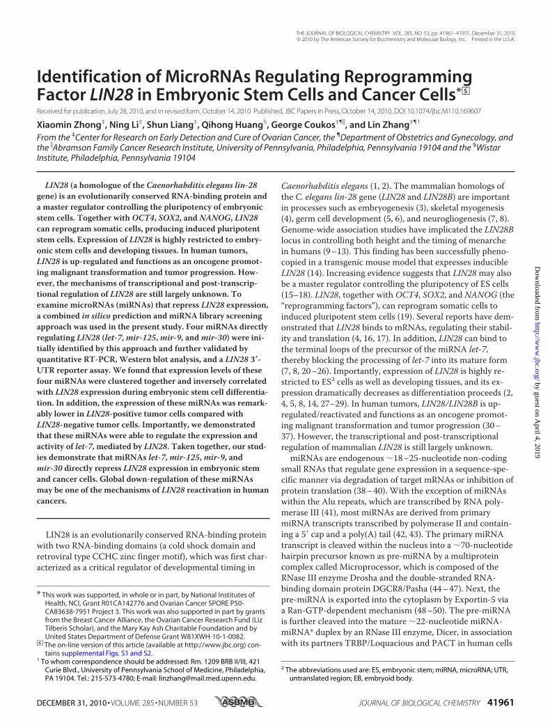
A Combined in Silico Prediction and miRNA LibraryScreening Approach Identified miRNAs Targeting LIN28—Theexpression of LIN28 is highly restricted to ES cells, somaticprogenitor cells, and developing tissues but is not detectablein most adult organs (2, 4, 5, 8, 14, 27–29). However, LIN28 isdramatically up-regulated/reactivated in human cancers (30–35). There is a double negative regulation loop between LIN28and the miRNA let-7 (7, 8, 20–26). To identify miRNAs regu-lating LIN28, our initial screening was performed on a plat-form containing four well characterized cancer cell lines, twoof which (A2780 and T47D) highly expressed endogenousLIN28 and two of which (MCF7 and HeLa) were LIN28-nega-tive (Fig. 1A). Consistent with previous reports (7, 8, 20–26),the levels of let-7 in the LIN28-positive cell lines were remark-ably lower than the levels in LIN28-negative lines (Fig. 1B).
Genome-wide miRNA library screening has been used suc-cessfully to identify miRNAs that regulate certain protein-coding genes (58, 59). Because this method is time- and labor-intensive, we designed a bioinformatics-driven screeningapproach, in which a concentrated miRNA library was gener-ated by miRNA binding site prediction (i.e. we used bioinfor-matics for miRNA target prediction as a filter to generate arestricted/selected miRNA library on which to perform thefunctional screening). This allowed us to perform theexperimental screening in a co-transfection system in which aluciferase reporter was co-transfected with a pre-miRNA-expressing vector (Fig. 1C). The reporter vector contained thefull-length human LIN28 3�-UTR sequence, which was cloneddownstream of the reporter gene Renilla (hRluc), such thatthe reporter gene expression was regulated by the LIN28 3�-UTR. The firefly luciferase (hluc) reporter served as an inter-nal control for transfection efficiency (Fig. 1C). To test ourscreening system, we first chose the miRNA let-7, one of theknown miRNAs regulating LIN28. As expected, let-7 signifi-cantly reduced luciferase activity in LIN28-negative cell linesbut not in LIN28-positive cell lines, in which the pre-miRNAmaturation was blocked by endogenous LIN28 (Fig. 1D).Next, we predicted miRNA binding sites in the 3�-UTR of
the human LIN28 gene using the bioinformatics miRNA tar-get prediction program TargetScan (available on the WorldWide Web) (60). A total of 23 potential miRNA binding sitesthat were broadly conserved among vertebrates were identi-fied (Fig. 1E). The individual miRNA expression vectors for
MicroRNA Regulates LIN28
DECEMBER 31, 2010 • VOLUME 285 • NUMBER 53 JOURNAL OF BIOLOGICAL CHEMISTRY 41963
by guest on April 4, 2019
http://ww
w.jbc.org/
Dow
nloaded from
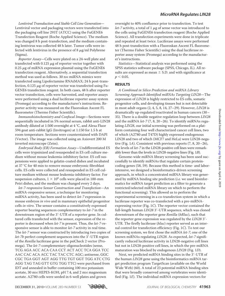
the above miRNAs were selected from our genome-widemiRNA library. A pilot single transfection for each vector wasperformed in cancer cell lines, and enforced miRNA expres-sion was confirmed by RT-PCR (data not shown). ThemiRNA vectors that were not efficiently expressed were ex-cluded from the co-transfection screening. Finally, a total of17 miRNAs were successfully used for the functional screen inall four cell lines. Briefly, nine of these miRNAs significantlyreduced luciferase activity in all four cell lines (marked asgreen in Fig. 1F). To further test whether these 9 candidatemiRNAs led to reduced expression of endogenous LIN28, wegenerated stable cell lines that overexpressed each of thesenine miRNAs individually as well as stable cell lines express-ing five miRNAs (as controls) that did not reduce luciferaseactivity. We found that three of the nine candidate miRNAs(mir-30,mir-125, andmir-9) markedly reduced both LIN28mRNA and protein expression (Fig. 1G). The direct regulationof two miRNAs in the LIN28 3�-UTR (mir-9 andmir-30,which were first identified in our study) were confirmed by amutant LIN28 3�-UTR reporter assay (Fig. 2). The seeding
sequences ofmir-9 ormir-30 in the LIN28 3�-UTR were mu-tated, and overexpression ofmir-9 ormir-30 was shown tosignificantly reduce luciferase activity in the wild type but notthe binding site mutant LIN28 3�-UTR reporters. Althoughmir-18 decreased LIN28 expression at a level comparable withthat ofmir-30 (Fig. 1G), overexpression ofmir-18 was notable to reduce luciferase activity in the wild type and the bind-ing site mutant LIN28 3�-UTR reporters in both A2780 andHeLa cells (data not shown). Taken together, four miRNAs,let-7,mir-30,mir-125, andmir-9, were identified in our initialscreening. Importantly, two miRNAs known to regulateLIN28 in mammalian (let-7 andmir-125) (7, 61) were success-fully identified using our approach.miRNAs Regulate Lin28 in Undifferentiated ES Cells—Next,
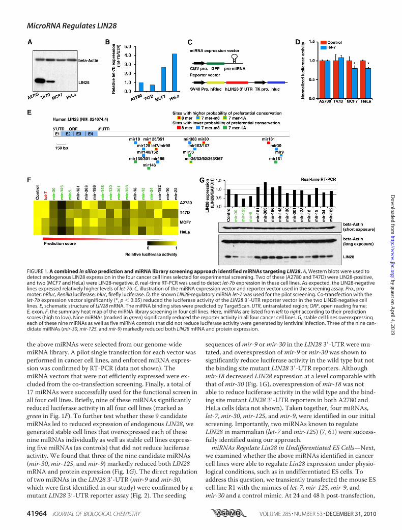
we examined whether the above miRNAs identified in cancercell lines were able to regulate Lin28 expression under physio-logical conditions, such as in undifferentiated ES cells. Toaddress this question, we transiently transfected the mouse EScell line R1 with the mimics of let-7,mir-125,mir-9, andmir-30 and a control mimic. At 24 and 48 h post-transfection,
FIGURE 1. A combined in silico prediction and miRNA library screening approach identified miRNAs targeting LIN28. A, Western blots were used todetect endogenous LIN28 expression in the four cancer cell lines selected for experimental screening. Two of these (A2780 and T47D) were LIN28-positive,and two (MCF7 and HeLa) were LIN28-negative. B, real-time RT-PCR was used to detect let-7b expression in these cell lines. As expected, the LIN28-negativelines expressed relatively higher levels of let-7b. C, illustration of the miRNA expression vector and reporter vector used in the screening assay. Pro., pro-moter; hRluc, Renilla luciferase; hluc, firefly luciferase. D, the known LIN28-regulatory miRNA let-7 was used for the pilot screening. Co-transfection with thelet-7b expression vector significantly (*, p � 0.05) reduced the luciferase activity of the LIN28 3�-UTR reporter vector in the two LIN28-negative celllines. E, schematic structure of LIN28 mRNA. The miRNA binding sites were predicted by TargetScan. UTR, untranslated region; ORF, open reading frame;E, exon. F, the summary heat map of the miRNA library screening in four cell lines. Here, miRNAs are listed from left to right according to their predictionscores (high to low). Nine miRNAs (marked in green) significantly reduced the reporter activity in all four cancer cell lines. G, stable cell lines overexpressingeach of these nine miRNAs as well as five miRNA controls that did not reduce luciferase activity were generated by lentiviral infection. Three of the nine can-didate miRNAs (mir-30, mir-125, and mir-9) markedly reduced both LIN28 mRNA and protein expression.
MicroRNA Regulates LIN28
41964 JOURNAL OF BIOLOGICAL CHEMISTRY VOLUME 285 • NUMBER 53 • DECEMBER 31, 2010
by guest on April 4, 2019
http://ww
w.jbc.org/
Dow
nloaded from
total RNA and protein were collected, and the endogenousLin28 expression was examined by RT-PCR and Westernblots. We found that the expression of both Lin28 protein andmRNA was significantly decreased in the cells transfectedwith miRNA mimics compared with the control transfections(Fig. 3, A and B). For the two miRNAs (mir-9 andmir-30) thatwere first identified as LIN28 regulators in this study, we alsoconfirmed the protein expression and localization by immu-nohistochemical staining. Consistent with the Western blotresults, bothmir-9 andmir-30 led to a reduction of endoge-
nous Lin28 in ES cells, but they had no effect on the cellulardistribution of Lin28 in these cells (Fig. 3C).Finally, we asked whether the effects of the four miRNA
families on LIN28 expression are cumulative or if the effectof one miRNA is dominant over the others during ES cell dif-ferentiation. We transfected undifferentiated ES cells witheach individual miRNA as well as a combination of all fourmiRNAs. We found thatmir-125more efficiently suppressedLIN28 expression compared with the other three miRNAs(supplemental Fig. S1A) and that the transfection of all four
FIGURE 2. LIN28-regulatory function of mir-9 and mir-30 was validated by the 3�-UTR reporter assay. A, schematic diagram of the mir-125, let-7, mir-30,and mir-9 binding sites in the LIN28 3�-UTR. The seeding sequences (marked in gray) were broadly conserved among different species. Hsa, Human; Ptr,chimpanzee; Mml, Rhesus; Mmu, mouse; Rno, Rat; Cpo, Pig; Ocu, rabbit; Eeu, Hedgehog; Cfa, Dog; Eca, Horse; Bta, Cow; Dno, armadillo; Laf, elephant; Mdo,opossum. B and C, summary of the reporter assays on the wild type and mir-9 binding site mutant reporter (B) and mir-30 binding site mutant reporter (C) inA2780 (LIN28-positive) and HeLa (LIN28-negative) cells. WT, wild type LIN28 3�-UTR reporter; mut9, mir-9 binding site mutant LIN28 3�-UTR reporter; mut30,mir-30 binding site mutant LIN28 3�-UTR reporter. Overexpression of mir-9 or mir-30 was able to significantly (*, p � 0.05) reduce luciferase activity in thewild type but not the binding site mutant LIN28 3�-UTR reporters.
FIGURE 3. miRNAs regulate Lin28 in undifferentiated ES cells. The miRNA mimics (30 nM) of let-7, mir-125, mir-9, mir-30, and a control mimic were tran-siently transfected into the mouse ES cell line R1. At 24 and 48 h post-transfection, total RNA and protein were collected, and the endogenous Lin28 expres-sion was examined by real-time RT-PCR (A) and Western blots (B). A, the Lin28 mRNA expression was significantly (*, p � 0.05) decreased in the cells trans-fected with miRNA mimics compared with the control transfections. B, Lin28 protein expression was markedly decreased in the cells transfected withmiRNA mimics compared with the control transfections. C, immunohistochemical staining further confirmed that mir-9 and mir-30 decreased LIN28 expres-sion in ES cells.
MicroRNA Regulates LIN28
DECEMBER 31, 2010 • VOLUME 285 • NUMBER 53 JOURNAL OF BIOLOGICAL CHEMISTRY 41965
by guest on April 4, 2019
http://ww
w.jbc.org/
Dow
nloaded from
miRNAs combined had a similar efficiency on the suppressionof LIN28 expression asmir-125 alone (supplemental Fig.S1A). Serial combination transfections further demonstratedthatmir-125 was indeed the major functional miRNA in the
combination transfection and that the effect ofmir-125 isdominant on LIN28 expression when these miRNA are pres-ent in equal concentrations in ES cells (supplemental Fig.S1B). However, the mature miRNA expression levels during
MicroRNA Regulates LIN28
41966 JOURNAL OF BIOLOGICAL CHEMISTRY VOLUME 285 • NUMBER 53 • DECEMBER 31, 2010
by guest on April 4, 2019
http://ww
w.jbc.org/
Dow
nloaded from
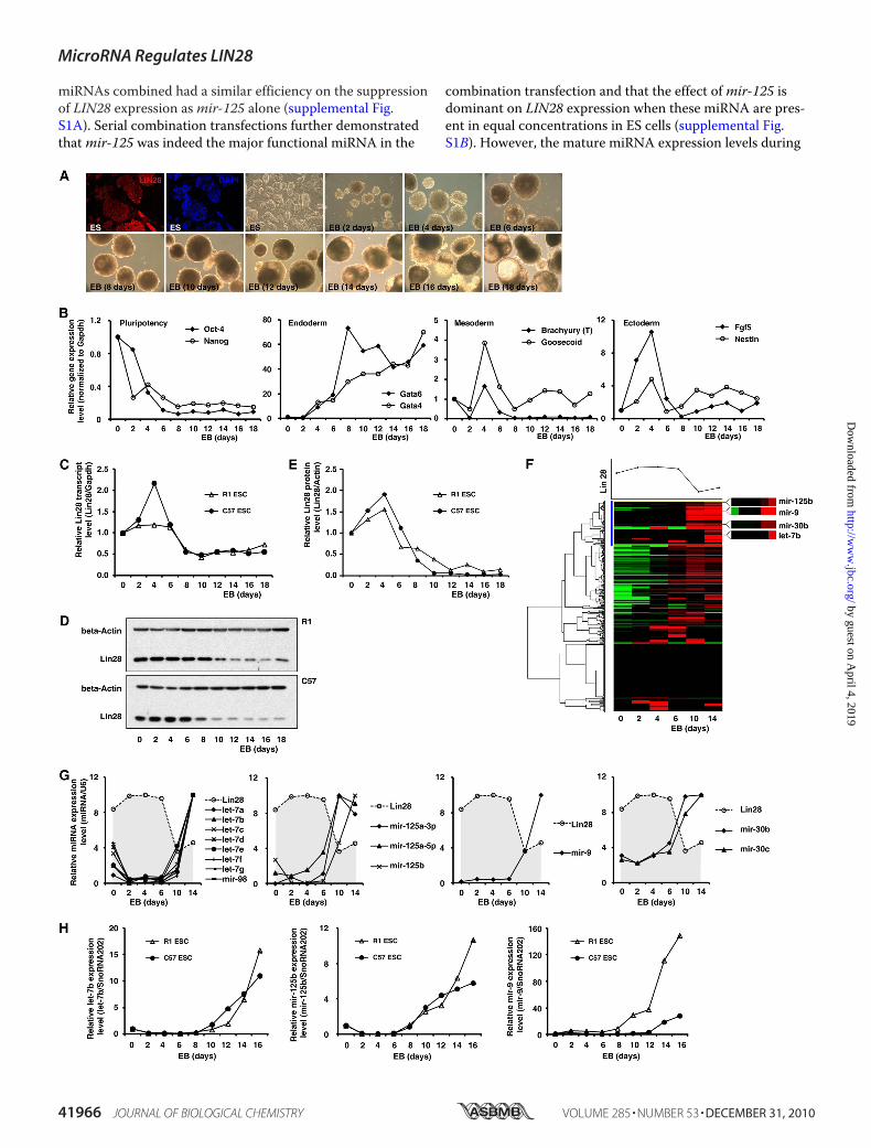
ES cell differentiation will be another important factor affect-ing LIN28 expression. To address this question, we retrievedpreviously published miRNA profiling data that was obtainedby deep sequencing of small RNA libraries during ES cell dif-ferentiation (62). In differentiated ES cells,mir-125a/b yielded1,557 sequence reads, and let-7a/b/c/d/e/f/g/i,mir-9, andmir-30a/b/c/d/e yielded 669, 2, and 4,284 sequence reads,respectively (supplemental Fig. S1C). We transfected the fourmiRNAs in the above ratios into undifferentiated ES cells inwhich all four miRNAs were absent. Consistently, we foundthatmir-125 was most efficient at suppressing endogenousLIN28 expression (supplemental Fig. S1D). Based on theabove results, we conclude that themir-125 family is the mostdominant miRNA regulating LIN28 expression during ES celldifferentiation, and that the let-7 andmir-30 families may alsoplay important roles in the suppression of LIN28 expressionin the early stages of differentiation. Because the transfectionof miRNA mimics only transiently affects miRNA expression,it is technically difficult to manipulate the levels of miRNAsthroughout the entire process of ES cell differentiation.Therefore, to address the same question at later stages of EScell differentiation, we retrieved three published miRNA pro-filing data sets from ES cell differentiation studies (63–65).We found that during ES cell differentiation, let-7 is ubiqui-tously up-regulated/expressed in most somatic progenitorcells, whereas the other three miRNAs are probably tissuetype-specific. For example, during the differentiation of EScells to neurons, let-7 is remarkably up-regulated, andmir-9 isalso significantly increased (63–65). This suggests that duringthe late differentiation stages in ES cells, these four miRNAsmay serve distinct functions in different tissue lineages andthat one or two of them may be more dominant at suppress-ing LIN28 expression.Expression Levels of LIN28 and Its Regulatory miRNAs Are
Inversely Correlated during ES Cell Differentiation—Expres-sion of LIN28 is highly restricted to ES cells as well as devel-oping tissues, and its expression dramatically decreases asdifferentiation proceeds (2, 4, 5, 8, 14, 27–29). Therefore, weexamined how the expression levels of LIN28 and itsregulatory miRNAs dynamically changed during ES cell dif-ferentiation. ES cells are commonly differentiated in vitro byspontaneously self-assembling in suspension culture intothree-dimensional cell aggregates called EBs, which modelmany of the hallmarks of early embryonic development. Wechose two mouse ES cell lines (R1 and C57) and induced themto differentiate and form EBs (Fig. 4A). The germ layer makerexpression patterns during EB formation (days 0–18) weremonitored by real-time RT-PCR (Fig. 4B), and the expressionof Lin28 was examined by real-time RT-PCR and Westernblots. Lin28mRNA levels were markedly decreased at day 6 of
ES differentiation and EB formation (Fig. 4C), and LIN28 pro-tein expression was reduced at days 6–8 of differentiation(Fig. 4, D and E). To examine the global miRNA expressionchanges during EB formation, a low density TaqMan assaycontaining 381 miRNA probes was used (Fig. 4F). Interest-ingly, unsupervised cluster analysis indicated that all of theabove four miRNA families (let-7,mir-125,mir-9, andmir-30)identified by our screening were clustered in the same groupand shared similar expression patterns (Fig. 4F, blue cluster).Importantly, their expression levels were nearly non-detect-able in undifferentiated ES cells and remarkably increasedfrom day 6 at the same time that Lin28 expression was de-creased. Their expression levels then remained high in thedifferentiated EB cells (Fig. 4, F and G). This high throughputresult was further validated by real-time RT-PCR assays in thetwo ES cell lines (Fig. 4H). In summary, during ES cell differ-entiation and EB formation, the expression level of Lin28 de-creases, whereas its regulatory miRNAs (let-7,mir-125,mir-9,andmir-30) display an inversely correlated expression pat-tern. This result strongly suggests that these four miRNAsplay a functional role in Lin28 regulation under physiologicalconditions such as early development.LIN28-regulatory miRNAs Are Globally Down-regulated in
LIN28-positive Cancer Cell Lines—LIN28 has also been iden-tified as a putative oncogene up-regulated/reactivated in5–15% of human tumors (30–37). We examined whether theLIN28-regulatory miRNAs were also deregulated in these tu-mors. First, we reanalyzed a publicly available miRNA data setcontaining 218 specimens (normal control tissues, n � 46;tumors, n � 172) (66), and we successfully retrieved 16miRNAs from the four LIN28-regulatory miRNA families.Interestingly, we found that 11 of these 16 miRNAs weremarkedly down-regulated (decreased more than 20%) in hu-man tumors (Fig. 5, A and B). This indicates that the LIN28-regulatory miRNAs are deregulated in cancer. To providefurther experimental evidence of this, we chose two cancercell lines (A2780 and T47D) that highly express LIN28 as wellas 17 cancer cell lines in which LIN28 protein was not detect-able (Fig. 5C). The expression of LIN28-regulatory miRNAswas analyzed in these cell lines by real-time RT-PCR. Asshown in Fig. 5C, all four miRNA families (let-7,mir-9,mir-30, andmir-125) displayed lower levels of expression in theLIN28-positive lines compared with the LIN28-negative lines.The negative correlation of LIN28 expression with its sup-pressing miRNAs suggests that the global deregulation ofthese miRNAs may be an important mechanism of oncogenicLIN28 up-regulation/reactivation in human cancer.Next, we tested whether miRNA inhibitors were able to
rescue/up-regulate endogenous LIN28 expression in tumorcells. We transfected each individual miRNA inhibitor and a
FIGURE 4. Expression levels of Lin28 and its regulatory miRNAs are inversely correlated during ES cell differentiation. A, ES cells were differentiatedin vitro by spontaneously self-assembling in suspension culture into three-dimensional cell aggregates (EBs). Immunostaining demonstrated that Lin28 washighly expressed in the undifferentiated ES cells. B, pluripotency and differentiation markers during differentiation and EB formation were monitored byreal-time RT-PCR. C, Lin28 mRNA expression during EB formation was analyzed by real-time RT-PCR. D and E, Lin28 protein expression during EB formationwas analyzed using Western blots. F, the global miRNA expression profile during EB formation was analyzed using a TaqMan miRNA assay. An unsupervisedcluster analysis indicated that all four Lin28-regulatory miRNAs grouped together. G, detailed miRNA expression changes during EB formation identified bythe TaqMan miRNA assay. All four Lin28-regulatory miRNAs were markedly up-regulated from day 6 of EB formation. H, real-time RT-PCR validations of theTaqMan miRNA assay in two ES cell lines.
MicroRNA Regulates LIN28
DECEMBER 31, 2010 • VOLUME 285 • NUMBER 53 JOURNAL OF BIOLOGICAL CHEMISTRY 41967
by guest on April 4, 2019
http://ww
w.jbc.org/
Dow
nloaded from
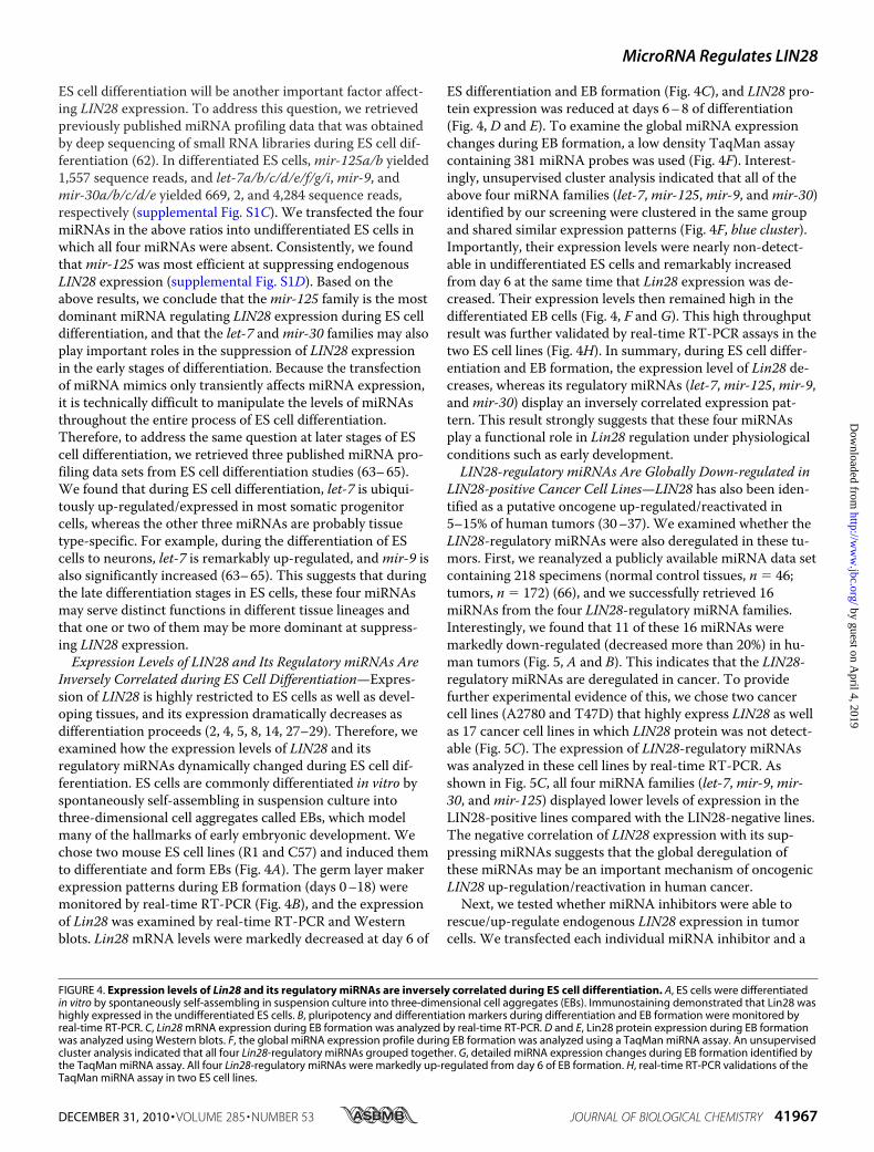
combination of these inhibitors as well as control oligonu-cleotides to both LIN28-positive (T47D) and LIN28-negative(HeLa) cell lines. We found that in the LIN28-positive cell lineT47D, all four inhibitors, both individually and in a combi-nation, were able to increase LIN28 protein expression.However, in the LIN28-negative cell line HeLa, none ofthese activated LIN28 expression to detectable levels (sup-plemental Fig. S2). Blocking all four miRNAs did not acti-vate endogenous LIN28 expression in LIN28-negative tu-mor cells. This suggests that the silencing of LIN28 in adulttissues may be controlled by multiple mechanisms and notonly by miRNA suppression. In LIN28-positive tumors,there may be at least two LIN28-suppressing pathways thatare not functioning (e.g. miRNA suppression and epige-netic silencing).miRNAs Regulate let-7 ExpressionMediated by LIN28—
Our finding suggested a novel regulatory mechanism in whichone miRNA may indirectly regulate another miRNA via aprotein coding gene; for example,mir-125,mir-9, andmir-30could regulate let-7 expression via repression of LIN28 ex-
pression. To test this hypothesis, we transfectedmir-9 andmir-125mimics into a LIN28-positive cell line (T47D). Asexpected, bothmir-9 andmir-125 significantly reduced en-dogenous LIN28 expression as detected by real-time RT-PCR(Fig. 6A). We then examined endogenous let-7b expression byreal-time RT-PCR. As shown in Fig. 6B, bothmir-9 andmir-125 significantly increased let-7b expression in T47D cells.Importantly, when we transfectedmir-9 ormir-125 into aLIN28-negative cell line (MCF7), the let-7b expression levelswere not affected (Fig. 6B). Finally, in order to monitor let-7activity, a let-7 sensor (Fig. 6C) was co-transfected with themir-9 andmir-125mimics. As shown in Fig. 6D,mir-9 andmir-125 significantly reduced the luciferase activity of thelet-7 sensor in T47D cells, indicating that the endogenouslet-7 activity was increased by themir-9 andmir-125mimics.In the LIN28-negative line MCF7, the mimic transfectionconsistently did not change the activity of the let-7 sensor.Taken together, our results demonstrate a novel regulatorymechanism where LIN28-regulatory miRNAs regulate let-7expression and activity via the protein-coding gene LIN28.
FIGURE 5. LIN28-regulatory miRNAs are globally down-regulated in LIN28-positive cancer cell lines. A, a publicly available miRNA microarray data setwas retrieved from the Broad Institute (66). Normalized expression levels of miRNAs regulating LIN28 were analyzed and are shown as a heat map. Wefound that 11 of 16 LIN28-regulatory miRNAs (marked in green) were markedly down-regulated (more than 20% reduction) in human tumors comparedwith normal control tissues. B, summary of the average expression levels of the miRNAs regulating LIN28 in normal and tumor specimens in the publicmiRNA microarray data set. C, LIN28 protein levels in 19 human cancer cell lines detected by Western blot. Two of these cell lines (10.5%) were LIN28-posi-tive. The LIN28-regulatory miRNA expression levels were analyzed by real-time RT-PCR in these 19 cell lines. Shown is a summary of the average expressionof each individual miRNA in the LIN28-positive lines (green) and LIN28-negative lines (white).
FIGURE 6. miRNAs regulate let-7 expression mediated by LIN28. A, the transfection of mir-9 and mir-125 mimics significantly (*, p � 0.05) reduced LIN28mRNA expression in T47D cells. B, transfection of mir-9 and mir-125 mimics significantly (*, p � 0.05) increased let-7b expression in the LIN28-positive cellline T47D but not in the LIN28-negative cell line MCF7. C, a miRNA-responsive sensor, a technique for monitoring miRNA activity, bearing sequences com-plementary to let-7b in the downstream region of the 3�-UTR of a constitutively expressed reporter gene. D, transfection of mir-9 and mir-125 mimics signifi-cantly (*, p � 0.05) decreased the let-7b-responsive sensor in the LIN28-positive cell line T47D but not in the LIN28-negative cell line MCF7.
MicroRNA Regulates LIN28
41968 JOURNAL OF BIOLOGICAL CHEMISTRY VOLUME 285 • NUMBER 53 • DECEMBER 31, 2010
by guest on April 4, 2019
http://ww
w.jbc.org/
Dow
nloaded from
DISCUSSION
It is estimated that the human genome contains �1,000miRNAs (67), more than 900 of which have already been identi-fied, according to the latest version of miRBase. miRNAsare predicted to target up to one-third of humanmRNAs (60).EachmiRNA can target hundreds of transcripts (68–71) andproteins (70, 71) directly or indirectly, andmore than onemiRNA can converge on a single transcript target (72). There-fore, the potential regulatory circuitry afforded bymiRNA isenormous, but identification of miRNAs regulating protein-cod-ing genes still remains challenging. Both bioinformatic predic-tionmethods and whole genome-wide genetic screening havebeen used to characterize miRNA targets (58, 59). Here, using acombinatorial approach, we successfully identified four miRNAfamilies, including two newly identified families (mir-9 andmir-30), which regulate LIN28 expression in ES cells and cancer cells.However, similar to the results obtained whenmiRNA libraryscreening has been performed on other protein coding genes (58,59), many predicted LIN28-regulatory miRNAs showed no sig-nificant effect on endogenous LIN28 expression. For example,
using a whole genome-widemiRNA library, Sage et al. (58) re-ported that only themir-221/mir-222 family regulated p27Kip1,and Nagel et al. (59) also found that only themir-135 family sup-pressedAPC expression. This suggests that protein-coding genesmay be regulated by a smaller number of miRNAs than has pre-viously been thought. However, in this study, we only in-cluded miRNAs that were predicted to bind to the LIN283�-UTR in our screening. Therefore, it is possible that othermiRNAs may target other regions of LIN28. All four miRNAfamilies identified in our study contain multiple members (e.g.the human let-7 family contains 12 members; themir-125family and themir-9 family each contain three members; andthemir-30 family contains six members) (Table 2). Thesefamilies are located in distinct chromosomal locations, sug-gesting that their expression is regulated by different 5�-UTRregulatory sequences and transcription factors. In addition,several LIN28-regulatory miRNAs are located in the samegenomic loci and clustered together (e.g. let-7e andmir-125aare located in the same genomic locus, as aremir-30b andmir-30d) (Table 2). This suggests that they may be processedfrom the same primary miRNA transcript, and could coordi-nately regulate LIN28 expression. Therefore, the potentialregulatory circuitry afforded by these four miRNA families (atleast 24 members) on LIN28 is extensive and complex.
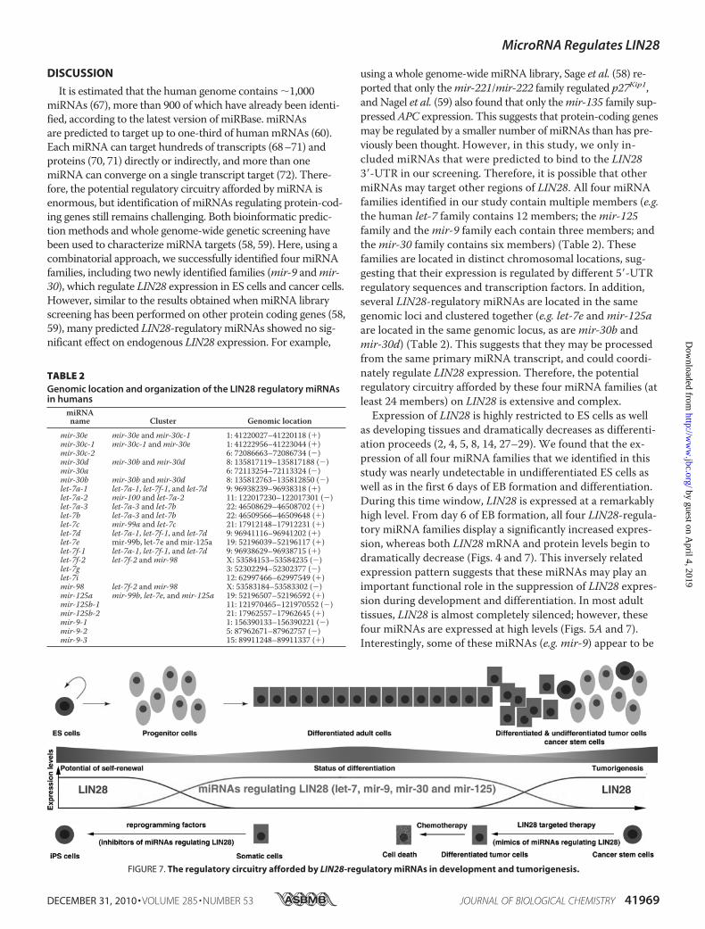
Expression of LIN28 is highly restricted to ES cells as wellas developing tissues and dramatically decreases as differenti-ation proceeds (2, 4, 5, 8, 14, 27–29). We found that the ex-pression of all four miRNA families that we identified in thisstudy was nearly undetectable in undifferentiated ES cells aswell as in the first 6 days of EB formation and differentiation.During this time window, LIN28 is expressed at a remarkablyhigh level. From day 6 of EB formation, all four LIN28-regula-tory miRNA families display a significantly increased expres-sion, whereas both LIN28mRNA and protein levels begin todramatically decrease (Figs. 4 and 7). This inversely relatedexpression pattern suggests that these miRNAs may play animportant functional role in the suppression of LIN28 expres-sion during development and differentiation. In most adulttissues, LIN28 is almost completely silenced; however, thesefour miRNAs are expressed at high levels (Figs. 5A and 7).Interestingly, some of these miRNAs (e.g. mir-9) appear to be
FIGURE 7. The regulatory circuitry afforded by LIN28-regulatory miRNAs in development and tumorigenesis.
TABLE 2Genomic location and organization of the LIN28 regulatory miRNAsin humans
miRNAname Cluster Genomic location
mir-30e mir-30e andmir-30c-1 1: 41220027–41220118 (�)mir-30c-1 mir-30c-1 andmir-30e 1: 41222956–41223044 (�)mir-30c-2 6: 72086663–72086734 (�)mir-30d mir-30b andmir-30d 8: 135817119–135817188 (�)mir-30a 6: 72113254–72113324 (�)mir-30b mir-30b andmir-30d 8: 135812763–135812850 (�)let-7a-1 let-7a-1, let-7f-1, and let-7d 9: 96938239–96938318 (�)let-7a-2 mir-100 and let-7a-2 11: 122017230–122017301 (�)let-7a-3 let-7a-3 and let-7b 22: 46508629–46508702 (�)let-7b let-7a-3 and let-7b 22: 46509566–46509648 (�)let-7c mir-99a and let-7c 21: 17912148–17912231 (�)let-7d let-7a-1, let-7f-1, and let-7d 9: 96941116–96941202 (�)let-7e mir-99b, let-7e and mir-125a 19: 52196039–52196117 (�)let-7f-1 let-7a-1, let-7f-1, and let-7d 9: 96938629–96938715 (�)let-7f-2 let-7f-2 andmir-98 X: 53584153–53584235 (�)let-7g 3: 52302294–52302377 (�)let-7i 12: 62997466–62997549 (�)mir-98 let-7f-2 andmir-98 X: 53583184–53583302 (�)mir-125a mir-99b, let-7e, andmir-125a 19: 52196507–52196592 (�)mir-125b-1 11: 121970465–121970552 (�)mir-125b-2 21: 17962557–17962645 (�)mir-9-1 1: 156390133–156390221 (�)mir-9-2 5: 87962671–87962757 (�)mir-9-3 15: 89911248–89911337 (�)
MicroRNA Regulates LIN28
DECEMBER 31, 2010 • VOLUME 285 • NUMBER 53 JOURNAL OF BIOLOGICAL CHEMISTRY 41969
by guest on April 4, 2019
http://ww
w.jbc.org/
Dow
nloaded from
tissue-specific, whereas some of them (e.g. mir-30) are ex-pressed in nearly all adult tissues. This indicates that there is acomplex temporal and spatial regulatory network of miRNAsaffecting LIN28 expression under physiological conditions.Finally, given that LIN28 can act as a reprogramming factortogether with OCT4, SOX2, and NANOG to reprogram so-matic cells to induced pluripotent stem cells (19), the ques-tion arises as to whether co-transfection of LIN28-regulatorymiRNA inhibitors with these reprogramming factors couldincrease the efficiency of reprogramming (Fig. 7). Supportingthis hypothesis, Melton et al. (73) have recently reported thatinhibition of let-7 promotes dedifferentiation of somatic cellsto induced pluripotent stem cells.In human cancer, LIN28 is up-regulated/reactivated in
about 5–15% of patients and functions as an oncogene (30–37). Interestingly, we found that these four LIN28-regulatorymiRNAs are globally down-regulated in tumors (Figs. 5 and7), possibly due to genetic and/or genomic alterations in thesemiRNAs or deregulation of their biogenesis pathways (66).Notably, in the LIN28-positive tumor cell lines, these miRNAswere expressed at relatively lower levels compared with theLIN28-negative tumor cell lines. Therefore, down-regulationof LIN28-regulatory miRNAs may be an important mecha-nism of LIN28 reactivation in human cancer (Fig. 5C). Recentstudies have demonstrated that LIN28 serves as an oncogenepromoting malignant transformation and tumor growth (30–37). Importantly, LIN28may contribute to the maintenance ofcancer stem cells, a relatively rare subpopulation of tumorcells having the unique ability to initiate and perpetuate tu-mor growth (34). Rapidly accumulating evidence indicatesthat miRNAs are involved in the initiation and progression ofcancer (66, 74–80). Interestingly, two of the LIN28-regulatorymiRNAs (let-7 andmir-125) have been demonstrated as tu-mor suppressor genes. For example, the tumor suppressorrole of let-7 in cancer was first demonstrated by the Slack lab-oratory (76). It was found that the let-7 family negatively reg-ulates let-60/RAS in C. elegans by binding to multiple let-7-complementary sites in its 3�-UTR (76). Moreover, the findingthat let-7 expression is lower in lung tumors than in normallung tissue, whereas RAS protein is significantly higher inlung tumors, proposes let-7 as a tumor suppressor gene (76,81–84). Therefore, our study could lead to new therapeuticstrategies for cancer treatment (Fig. 7). For example, nanopar-ticle-delivered LIN28-regulatory miRNA mimics may be anattractive therapeutic method to target the LIN28-positivecancer stem cell population in tumors.
REFERENCES1. Ambros, V., and Horvitz, H. R. (1984) Science 226, 409–4162. Moss, E. G., Lee, R. C., and Ambros, V. (1997) Cell 88, 637–6463. Yokoyama, S., Hashimoto, M., Shimizu, H., Ueno-Kudoh, H., Uchibe, K.,
Kimura, I., and Asahara, H. (2008) Gene Expr. Patterns 8, 155–1604. Polesskaya, A., Cuvellier, S., Naguibneva, I., Duquet, A., Moss, E. G., and
Harel-Bellan, A. (2007) Genes Dev. 21, 1125–11385. West, J. A., Viswanathan, S. R., Yabuuchi, A., Cunniff, K., Takeuchi, A.,
Park, I. H., Sero, J. E., Zhu, H., Perez-Atayde, A., Frazier, A. L., Surani,M. A., and Daley, G. Q. (2009) Nature 460, 909–913
6. Zheng, K., Wu, X., Kaestner, K. H., and Wang, P. J. (2009) BMC Dev.Biol. 9, 38
7. Rybak, A., Fuchs, H., Smirnova, L., Brandt, C., Pohl, E. E., Nitsch, R., and
Wulczyn, F. G. (2008) Nat. Cell. Biol. 10, 987–9938. Balzer, E., Heine, C., Jiang, Q., Lee, V. M., and Moss, E. G. (2010) Devel-
opment 137, 891–9009. Lettre, G., Jackson, A. U., Gieger, C., Schumacher, F. R., Berndt, S. I.,
Sanna, S., Eyheramendy, S., Voight, B. F., Butler, J. L., Guiducci, C., Illig,T., Hackett, R., Heid, I. M., Jacobs, K. B., Lyssenko, V., Uda, M., Boe-hnke, M., Chanock, S. J., Groop, L. C., Hu, F. B., Isomaa, B., Kraft, P.,Peltonen, L., Salomaa, V., Schlessinger, D., Hunter, D. J., Hayes, R. B.,Abecasis, G. R., Wichmann, H. E., Mohlke, K. L., and Hirschhorn, J. N.(2008) Nat. Genet. 40, 584–591
10. Ong, K. K., Elks, C. E., Li, S., Zhao, J. H., Luan, J., Andersen, L. B., Bing-ham, S. A., Brage, S., Smith, G. D., Ekelund, U., Gillson, C. J., Glaser, B.,Golding, J., Hardy, R., Khaw, K. T., Kuh, D., Luben, R., Marcus, M., Mc-Geehin, M. A., Ness, A. R., Northstone, K., Ring, S. M., Rubin, C., Sims,M. A., Song, K., Strachan, D. P., Vollenweider, P., Waeber, G., Water-worth, D. M., Wong, A., Deloukas, P., Barroso, I., Mooser, V., Loos, R. J.,and Wareham, N. J. (2009) Nat. Genet. 41, 729–733
11. Sulem, P., Gudbjartsson, D. F., Rafnar, T., Holm, H., Olafsdottir, E. J.,Olafsdottir, G. H., Jonsson, T., Alexandersen, P., Feenstra, B., Boyd,H. A., Aben, K. K., Verbeek, A. L., Roeleveld, N., Jonasdottir, A.,Styrkarsdottir, U., Steinthorsdottir, V., Karason, A., Stacey, S. N., Gud-mundsson, J., Jakobsdottir, M., Thorleifsson, G., Hardarson, G., Gulcher,J., Kong, A., Kiemeney, L. A., Melbye, M., Christiansen, C., Tryggvadot-tir, L., Thorsteinsdottir, U., and Stefansson, K. (2009) Nat. Genet. 41,734–738
12. He, C., Kraft, P., Chen, C., Buring, J. E., Pare, G., Hankinson, S. E.,Chanock, S. J., Ridker, P. M., Hunter, D. J., and Chasman, D. I. (2009)Nat. Genet. 41, 724–728
13. Perry, J. R., Stolk, L., Franceschini, N., Lunetta, K. L., Zhai, G., McArdle,P. F., Smith, A. V., Aspelund, T., Bandinelli, S., Boerwinkle, E., Cherkas,L., Eiriksdottir, G., Estrada, K., Ferrucci, L., Folsom, A. R., Garcia, M.,Gudnason, V., Hofman, A., Karasik, D., Kiel, D. P., Launer, L. J., vanMeurs, J., Nalls, M. A., Rivadeneira, F., Shuldiner, A. R., Singleton, A.,Soranzo, N., Tanaka, T., Visser, J. A., Weedon, M. N., Wilson, S. G.,Zhuang, V., Streeten, E. A., Harris, T. B., Murray, A., Spector, T. D., De-merath, E. W., Uitterlinden, A. G., and Murabito, J. M. (2009) Nat.Genet. 41, 648–650
14. Zhu, H., Shah, S., Shyh-Chang, N., Shinoda, G., Einhorn, W. S.,Viswanathan, S. R., Takeuchi, A., Grasemann, C., Rinn, J. L., Lopez,M. F., Hirschhorn, J. N., Palmert, M. R., and Daley, G. Q. (2010) Nat.Genet. 42, 626–630
15. Darr, H., and Benvenisty, N. (2009) Stem Cells 27, 352–36216. Xu, B., Zhang, K., and Huang, Y. (2009) RNA 15, 357–36117. Qiu, C., Ma, Y., Wang, J., Peng, S., and Huang, Y. (2010) Nucleic Acids
Res. 38, 1240–124818. Xu, B., and Huang, Y. (2009) Nucleic Acids Res. 37, 4256–426319. Yu, J., Vodyanik, M. A., Smuga-Otto, K., Antosiewicz-Bourget, J., Frane,
J. L., Tian, S., Nie, J., Jonsdottir, G. A., Ruotti, V., Stewart, R., Slukvin,I. I., and Thomson, J. A. (2007) Science 318, 1917–1920
20. Viswanathan, S. R., Daley, G. Q., and Gregory, R. I. (2008) Science 320,97–100
21. Newman, M. A., Thomson, J. M., and Hammond, S. M. (2008) RNA 14,1539–1549
22. Heo, I., Joo, C., Cho, J., Ha, M., Han, J., and Kim, V. N. (2008)Mol. Cell.32, 276–284
23. Piskounova, E., Viswanathan, S. R., Janas, M., LaPierre, R. J., Daley,G. Q., Sliz, P., and Gregory, R. I. (2008) J. Biol. Chem. 283, 21310–21314
24. Heo, I., Joo, C., Kim, Y. K., Ha, M., Yoon, M. J., Cho, J., Yeom, K. H.,Han, J., and Kim, V. N. (2009) Cell 138, 696–708
25. Lehrbach, N. J., Armisen, J., Lightfoot, H. L., Murfitt, K. J., Bugaut, A.,Balasubramanian, S., and Miska, E. A. (2009) Nat. Struct. Mol. Biol. 16,1016–1020
26. Hagan, J. P., Piskounova, E., and Gregory, R. I. (2009) Nat. Struct. Mol.Biol. 16, 1021–1025
27. Viswanathan, S. R., and Daley, G. Q. (2010) Cell 140, 445–44928. Moss, E. G., and Tang, L. (2003) Dev. Biol. 258, 432–44229. Yang, D. H., and Moss, E. G. (2003) Gene Expr. Patterns 3, 719–72630. Viswanathan, S. R., Powers, J. T., Einhorn, W., Hoshida, Y., Ng, T. L.,
MicroRNA Regulates LIN28
41970 JOURNAL OF BIOLOGICAL CHEMISTRY VOLUME 285 • NUMBER 53 • DECEMBER 31, 2010
by guest on April 4, 2019
http://ww
w.jbc.org/
Dow
nloaded from
Toffanin, S., O’Sullivan, M., Lu, J., Phillips, L. A., Lockhart, V. L., Shah,S. P., Tanwar, P. S., Mermel, C. H., Beroukhim, R., Azam, M., Teixeira,J., Meyerson, M., Hughes, T. P., Llovet, J. M., Radich, J., Mullighan,C. G., Golub, T. R., Sorensen, P. H., and Daley, G. Q. (2009) Nat. Genet.41, 843–848
31. Chang, T. C., Zeitels, L. R., Hwang, H. W., Chivukula, R. R., Wentzel,E. A., Dews, M., Jung, J., Gao, P., Dang, C. V., Beer, M. A., Thomas-Tik-honenko, A., and Mendell, J. T. (2009) Proc. Natl. Acad. Sci. U.S.A. 106,3384–3389
32. Iliopoulos, D., Hirsch, H. A., and Struhl, K. (2009) Cell 139, 693–70633. Dangi-Garimella, S., Yun, J., Eves, E. M., Newman, M., Erkeland, S. J.,
Hammond, S. M., Minn, A. J., and Rosner, M. R. (2009) EMBO J. 28,347–358
34. Peng, S., Maihle, N. J., and Huang, Y. (2010) Oncogene 29, 2153–215935. Guo, Y., Chen, Y., Ito, H., Watanabe, A., Ge, X., Kodama, T., and Abura-
tani, H. (2006) Gene 384, 51–6136. Lu, L., Katsaros, D., Shaverdashvili, K., Qian, B., Wu, Y., de la Longrais,
I. A., Preti, M., Menato, G., and Yu, H. (2009) Eur. J. Cancer. 45,2212–2218
37. Wang, Y. C., Chen, Y. L., Yuan, R. H., Pan, H. W., Yang, W. C., Hsu,H. C., and Jeng, Y. M. (2010) Carcinogenesis 31, 1516–1522
38. Lee, R. C., Feinbaum, R. L., and Ambros, V. (1993) Cell 75, 843–85439. Lagos-Quintana, M., Rauhut, R., Lendeckel, W., and Tuschl, T. (2001)
Science 294, 853–85840. Lau, N. C., Lim, L. P., Weinstein, E. G., and Bartel, D. P. (2001) Science
294, 858–86241. Borchert, G. M., Lanier, W., and Davidson, B. L. (2006) Nat. Struct. Mol.
Biol. 13, 1097–110142. Lee, Y., Kim, M., Han, J., Yeom, K. H., Lee, S., Baek, S. H., and Kim, V. N.
(2004) EMBO J. 23, 4051–406043. Cai, X., Hagedorn, C. H., and Cullen, B. R. (2004) RNA 10, 1957–196644. Lee, Y., Ahn, C., Han, J., Choi, H., Kim, J., Yim, J., Lee, J., Provost, P.,
Rådmark, O., Kim, S., and Kim, V. N. (2003) Nature 425, 415–41945. Denli, A. M., Tops, B. B., Plasterk, R. H., Ketting, R. F., and Hannon, G. J.
(2004) Nature 432, 231–23546. Gregory, R. I., Yan, K. P., Amuthan, G., Chendrimada, T., Doratotaj, B.,
Cooch, N., and Shiekhattar, R. (2004) Nature 432, 235–24047. Landthaler, M., Yalcin, A., and Tuschl, T. (2004) Curr. Biol. 14,
2162–216748. Yi, R., Qin, Y., Macara, I. G., and Cullen, B. R. (2003) Genes Dev. 17,
3011–301649. Lund, E., Guttinger, S., Calado, A., Dahlberg, J. E., and Kutay, U. (2004)
Science 303, 95–9850. Bohnsack, M. T., Czaplinski, K., and Gorlich, D. (2004) RNA 10,
185–19151. Hutvagner, G., McLachlan, J., Pasquinelli, A. E., Balint, E., Tuschl, T.,
and Zamore, P. D. (2001) Science 293, 834–83852. Ketting, R. F., Fischer, S. E., Bernstein, E., Sijen, T., Hannon, G. J., and
Plasterk, R. H. (2001) Genes Dev. 15, 2654–265953. Gregory, R. I., Chendrimada, T. P., Cooch, N., and Shiekhattar, R. (2005)
Cell 123, 631–64054. Maniataki, E., and Mourelatos, Z. (2005) Genes Dev. 19, 2979–299055. Schwarz, D. S., Hutvagner, G., Du, T., Xu, Z., Aronin, N., and Zamore,
P. D. (2003) Cell 115, 199–20856. Du, T., and Zamore, P. D. (2005) Development 132, 4645–465257. Ho, S. N., Hunt, H. D., Horton, R. M., Pullen, J. K., and Pease, L. R.
(1989) Gene 77, 51–5958. le Sage, C., Nagel, R., Egan, D. A., Schrier, M., Mesman, E., Mangiola, A.,
Anile, C., Maira, G., Mercatelli, N., Ciafre, S. A., Farace, M. G., andAgami, R. (2007) EMBO J. 26, 3699–3708
59. Nagel, R., le Sage, C., Diosdado, B., van der Waal, M., Oude Vrielink,J. A., Bolijn, A., Meijer, G. A., and Agami, R. (2008) Cancer Res. 68,5795–5802
60. Lewis, B. P., Burge, C. B., and Bartel, D. P. (2005) Cell 120, 15–2061. Wu, L., and Belasco, J. G. (2005)Mol. Cell. Biol. 25, 9198–920862. Bar, M., Wyman, S. K., Fritz, B. R., Qi, J., Garg, K. S., Parkin, R. K., Kroh,
E. M., Bendoraite, A., Mitchell, P. S., Nelson, A. M., Ruzzo, W. L., Ware,C., Radich, J. P., Gentleman, R., Ruohola-Baker, H., and Tewari, M.(2008) Stem Cells 26, 2496–2505
63. Krichevsky, A. M., Sonntag, K. C., Isacson, O., and Kosik, K. S. (2006)Stem Cells 24, 857–864
64. Wu, H., Xu, J., Pang, Z. P., Ge, W., Kim, K. J., Blanchi, B., Chen, C., Sud-hof, T. C., and Sun, Y. E. (2007) Proc. Natl. Acad. Sci. U.S.A. 104,13821–13826
65. Marson, A., Levine, S. S., Cole, M. F., Frampton, G. M., Brambrink, T.,Johnstone, S., Guenther, M. G., Johnston, W. K., Wernig, M., Newman,J., Calabrese, J. M., Dennis, L. M., Volkert, T. L., Gupta, S., Love, J., Han-nett, N., Sharp, P. A., Bartel, D. P., Jaenisch, R., and Young, R. A. (2008)Cell 134, 521–533
66. Lu, J., Getz, G., Miska, E. A., Alvarez-Saavedra, E., Lamb, J., Peck, D.,Sweet-Cordero, A., Ebert, B. L., Mak, R. H., Ferrando, A. A., Downing,J. R., Jacks, T., Horvitz, H. R., and Golub, T. R. (2005) Nature 435,834–838
67. Zamore, P. D., and Haley, B. (2005) Science 309, 1519–152468. Bartel, D. P., and Chen, C. Z. (2004) Nat. Rev. Genet. 5, 396–40069. Lim, L. P., Lau, N. C., Garrett-Engele, P., Grimson, A., Schelter, J. M.,
Castle, J., Bartel, D. P., Linsley, P. S., and Johnson, J. M. (2005) Nature433, 769–773
70. Selbach, M., Schwanhausser, B., Thierfelder, N., Fang, Z., Khanin, R.,and Rajewsky, N. (2008) Nature 455, 58–63
71. Baek, D., Villen, J., Shin, C., Camargo, F. D., Gygi, S. P., and Bartel, D. P.(2008) Nature 455, 64–71
72. Krek, A., Grun, D., Poy, M. N., Wolf, R., Rosenberg, L., Epstein, E. J.,MacMenamin, P., da Piedade, I., Gunsalus, K. C., Stoffel, M., and Rajew-sky, N. (2005) Nat. Genet. 37, 495–500
73. Melton, C., Judson, R. L., and Blelloch, R. (2010) Nature 463, 621–62674. Calin, G. A., Dumitru, C. D., Shimizu, M., Bichi, R., Zupo, S., Noch, E.,
Aldler, H., Rattan, S., Keating, M., Rai, K., Rassenti, L., Kipps, T., Ne-grini, M., Bullrich, F., and Croce, C. M. (2002) Proc. Natl. Acad. Sci.U.S.A. 99, 15524–15529
75. Calin, G. A., Sevignani, C., Dumitru, C. D., Hyslop, T., Noch, E., Yen-damuri, S., Shimizu, M., Rattan, S., Bullrich, F., Negrini, M., and Croce,C. M. (2004) Proc. Natl. Acad. Sci. U.S.A. 101, 2999–3004
76. Johnson, S. M., Grosshans, H., Shingara, J., Byrom, M., Jarvis, R., Cheng,A., Labourier, E., Reinert, K. L., Brown, D., and Slack, F. J. (2005) Cell120, 635–647
77. O’Donnell, K. A., Wentzel, E. A., Zeller, K. I., Dang, C. V., and Mendell,J. T. (2005) Nature 435, 839–843
78. Voorhoeve, P. M., le Sage, C., Schrier, M., Gillis, A. J., Stoop, H., Nagel,R., Liu, Y. P., van Duijse, J., Drost, J., Griekspoor, A., Zlotorynski, E.,Yabuta, N., De Vita, G., Nojima, H., Looijenga, L. H., and Agami, R.(2006) Cell 124, 1169–1181
79. Calin, G. A., and Croce, C. M. (2006) Nat. Rev. Cancer 6, 857–86680. Esquela-Kerscher, A., and Slack, F. J. (2006) Nat. Rev. Cancer. 6,
259–26981. Kumar, M. S., Erkeland, S. J., Pester, R. E., Chen, C. Y., Ebert, M. S.,
Sharp, P. A., and Jacks, T. (2008) Proc. Natl. Acad. Sci. U.S.A. 105,3903–3908
82. Yu, F., Yao, H., Zhu, P., Zhang, X., Pan, Q., Gong, C., Huang, Y., Hu, X.,Su, F., Lieberman, J., and Song, E. (2007) Cell 131, 1109–1123
83. Johnson, C. D., Esquela-Kerscher, A., Stefani, G., Byrom, M., Kelnar, K.,Ovcharenko, D., Wilson, M., Wang, X., Shelton, J., Shingara, J., Chin, L.,Brown, D., and Slack, F. J. (2007) Cancer Res. 67, 7713–7722
84. Trang, P., Medina, P. P., Wiggins, J. F., Ruffino, L., Kelnar, K., Omotola,M., Homer, R., Brown, D., Bader, A. G., Weidhaas, J. B., and Slack, F. J.(2010) Oncogene 29, 1580–1587
MicroRNA Regulates LIN28
DECEMBER 31, 2010 • VOLUME 285 • NUMBER 53 JOURNAL OF BIOLOGICAL CHEMISTRY 41971
by guest on April 4, 2019
http://ww
w.jbc.org/
Dow
nloaded from
Xiaomin Zhong, Ning Li, Shun Liang, Qihong Huang, George Coukos and Lin ZhangEmbryonic Stem Cells and Cancer Cells
inLIN28Identification of MicroRNAs Regulating Reprogramming Factor
doi: 10.1074/jbc.M110.169607 originally published online October 14, 20102010, 285:41961-41971.J. Biol. Chem.
10.1074/jbc.M110.169607Access the most updated version of this article at doi:
Alerts:
When a correction for this article is posted•
When this article is cited•
to choose from all of JBC's e-mail alertsClick here
Supplemental material:
http://www.jbc.org/content/suppl/2010/10/14/M110.169607.DC1
http://www.jbc.org/content/285/53/41961.full.html#ref-list-1
This article cites 84 references, 28 of which can be accessed free at
by guest on April 4, 2019
http://ww
w.jbc.org/
Dow
nloaded from