identification of four novel cytochrome p4501b1 … of four novel cytochrome p4501b1 mutations...
TRANSCRIPT

Identification of four novel cytochrome P4501B1 mutations(p.I94X, p.H279D, p.Q340H, and p.K433K) in primary congenitalglaucoma patients
Mukesh Tanwar,1 Tanuj Dada,2 Ramanjit Sihota,2 Rima Dada1
1Laboratory For Molecular Reproduction and Genetics, Department of Anatomy, All India Institute of Medical Sciences, AnsariNagar, New Delhi, India; 2Dr. R.P. Centre for Ophthalmic Sciences, All India Institute of Medical Sciences, Ansari Nagar, NewDelhi, India
Purpose: Primary congenital glaucoma (PCG) is an autosomal recessive eye disorder that is postulated to result fromdevelopmental defects in the anterior eye segment. Mutations in the cytochrome P4501B1 (CYP1B1) gene are apredominant cause of congenital glaucoma. In this study we identify CYP1B1 mutations in PCG patients.Methods: Twenty-three unrelated PCG patients and 50 healthy controls were enrolled in the study. CYP1B1 was screenedfor mutations by PCR and DNA sequencing.Results: DNA sequencing revealed a total of 15 mutations. Out of these, four (p.I94X, p.H279D, p.Q340H, and p.K433K)were novel mutations and five were known pathogenic mutations. Five coding single nucleotide polymorphisms and oneintronic single nucleotide polymorphism (rs2617266) were also found. Truncating mutations (p.I94X and p.R355X) wereassociated with the most severe disease phenotype. It is possible that patients with two null alleles with no catalytic activitymay present with a more severe phenotype of the disease compared to patients with one null allele (heterozygous). Thedisease phenotype of patients with CYP1B1 mutations was more severe compared with the clinical phenotype of patientsnegative for CYP1B1 mutations.Conclusion: Mutations in CYP1B1 are a major cause for PCG in our patients. Identifying mutations in subjects at risk ofdeveloping glaucoma, particularly among relatives of PCG patients, is of clinical significance. These developments mayhelp in reducing the disease frequency in familial cases. Such studies will be of benefit in the identification of pathogenicmutations in different populations and will enable us to develop simple and rapid diagnostic tests for analyzing such cases.
Primary congenital glaucoma (PCG; OMIM 231300) isan autosomal recessive disorder of the eye. In this disease thetrabecular meshwork (TM) and anterior chamber of the eyeare affected, leading to impairment in the aqueous drainage,increased intraocular pressure (IOP), and optic nerve damage.PCG occurs during the neonatal or early infantile period [1].The term PCG is reserved for those cases in which the onlyanatomic defect observed is isolated trabeculodysgenesis.This increased IOP results in ocular enlargement(buphthalmos), corneal clouding, and rapid optic nervecupping. Progressive degeneration of the retinal ganglion cells(RGCs) results in the characteristic optic nerve atrophy andvisual field defects found in glaucoma. Most cases of PCG aresporadic, but familial cases have also been reported. PCG isthe most common type of pediatric glaucoma and accounts for55% of such cases. Its expression and penetrance vary from40–100%. Its incidence varies substantially from onepopulation to another. It is estimated to occur in 1 in 10,000
Correspondence to: Dr. Rima Dada, Associate Professor, LaboratoryFor Molecular Reproduction and Genetics, Department of Anatomy,All India Institute of Medical Sciences, Ansari Nagar, New Delhi,India 110029; Phone: +91-11-26546716; FAX: +91-11-26588663;email: [email protected]
births in Europe and 1 in 3,300 births in Andhra Pradesh, India[2,3].
Recently a putative PCG locus, GLC3A, was linked tomarkers on the short arm of chromosome 2 in 11 Turkishfamilies [4]. Six other families did not show linkage to thislocus, suggesting locus heterogeneity for this disease. AnotherPCG locus, GLC3B, was localized on chromosome 1p36 insome families but did not show linkage to chromosome 2markers [5]. Other subsets of families that did not showlinkage to these two loci provide evidence for at least a thirdof the unmapped loci [5]. Recently Stoilov et al. [6] identifiedthree different mutations in the cytochrome P4501B1(CYP1B1) gene in five unrelated Turkish families in whichthe disease had been linked to the 2p21 locus [6]. Even thoughthree different loci have been mapped for PCG, mutations inCYP1B1 (GLC3A) are the most predominant cause of diseaseand are reported in various ethnic backgrounds [6-15].Further, it is estimated that all the known loci/genes ofglaucoma account for a minority of the total cases of glaucoma[4,5], and hence many other genes remain to be identified.
CYP1B1 is located on chromosome 2 and consists of threeexons and two introns. The coding region of CYP1B1 starts atthe 5′ end of exon 2 and ends within exon 3. It codes for a 543-amino acid protein and is expressed in the ocular tissues, such
Molecular Vision 2009; 15:2926-2937 <http://www.molvis.org/molvis/v15/a310>Received 26 November 2009 | Accepted 24 December 2009 | Published 30 December 2009
© 2009 Molecular Vision
2926

as the anterior chamber, and in several nonocular tissues[16]. CYP1B1 is a member of the cytochrome P450superfamily of drug-metabolizing enzymes. It catalyzesseveral oxidative reactions, some of which are biosynthetic,producing necessary hormones or compounds of intermediarymetabolism in most living organisms and substrates, includingmany xenobiotics, vitamins, and steroids [17]. CYP1B1 alsometabolizes vitamin A in two steps to all-trans-retinal and all-trans-retinoic acid. The latter is a potent morphogen andregulates in utero development of tissue growth anddifferentiation. CYP1B1 is involved in the metabolism ofendogenous and exogenous substrates that take part in earlyocular differentiation [18-20]. In the present study wescreened all coding exons of CYP1B1 in 23 unrelatedcongenital glaucoma patients.
METHODSClinical evaluation and patient selection: Primary congenitalglaucoma cases presenting at the Dr. R. P. Centre forOphthalmic Sciences (AIIMS, New Delhi, India), wereenrolled for this study. Six patients were female and 17 weremale. Mean age of presentation was 15.17 months (range 1.5– 132 months). After ethical approval of the InstitutionalReview Board (IRB00006862; All India Institute of MedicalSciences, New Delhi, India), 23 PCG cases were enrolled inthis study. The diagnosis involved clinical ocular and systemicexamination. Inclusion criteria of the patients were increasedcorneal diameter (>12.0 mm) and raised IOP (>21 mmHg)with presence/absence of Haab’s striae and optic disc changes(where examination was possible). Symptoms of epiphora andphotophobia were the additional inclusion factors. The age ofonset ranged from birth to 3 years. Detailed family historiesup to three generations were taken, and pedigree charts wereconstructed. The history of ocular or other hereditarydisorders was recorded. Glaucoma cases other than PCG wereexcluded. Fifty ethnically matched normal individualswithout any ocular disorders were enrolled as controls.Peripheral blood samples were collected from patients andcontrols by venipuncture after informed consent. Bloodsamples were collected in EDTA vaccutainer and stored in -80°C until DNA isolation.Mutation screening and sequence analysis: Genomic DNAwas isolated from peripheral blood by the phenol chloroform
method. The entire coding region, including exon–intronboundaries of CYP1B1, from patients and controls wasamplified and screened for mutations by using three sets ofoverlapping primers (Table 1) [7,21]. The primers used wereset I (1F–1R, 786 bp) [12], set II (2F–2R, 787 bp) [13], andset III (3F–3R, 885 bp) [12]. PCR amplifications for primersets I and II were performed in a 40 µl volume containing 1.0µl of 20 µM stock solution for each primer, 100 ng of genomicDNA, 1 unit of Taq polymerase (Banglore Genei), 0.1 mM ofeach dNTP, 4 µl of 10X PCR buffer (with 15 mM MgCl2) and4 µl of dimethyl sulphoxide (Sigma), by means of 35 cyclesof amplification, each consisting of 30 s denaturation at 94°C, 30 s annealing at 56 °C and 1 min extension at 72 °C [12],while conditions for set III were initial denaturation at 94 °Cfor 3 min followed by 30 cycles each at 94 °C for 30 s, 60 °Cfor 30 s, and 72 °C for 1 min.
Amplified PCR products were purified using a gel/PCRDNA fragments extraction kit (number DF100; GeneaidBiotech Ltd., Sijhih City, Taiwan). Purified PCR productswere sent for sequencing to MCLAB (Molecular CloningLaboratories, South San Francisco, CA). DNA sequenceswere analyzed against the CYP1B1 reference sequenceENSG00000138061 using ClustalW2 (multiple sequencealignment program for DNA; European Molecular BiologyLaboratory (EMBL) – European Bioinformatics Institute(EBI)).Computational assessment of missense mutations: Twohomology-based programs PolyPhen (polymorphismphenotyping; Division of Genetics, Department of Medicine,Brigham and Women’s Hospital/Harvard Medical School,Boston, MA) and SIFT (sorting intolerant from tolerant; theJ. Craig Venter Institute Rockville, MD and La Jolla, CA)were used to predict the functional impact of missensechanges identified in this study. PolyPhen structurallyanalyzes an amino acid polymorphism and predicts whetherthat amino acid change is likely to be deleterious to proteinfunction [22-24]. The prediction is based on the position-specific independent counts (PSIC) score derived frommultiple sequence alignments of observations. PolyPhenscores of >2.0 indicate the polymorphism is probablydamaging to protein function. Scores of 1.5–2.0 are possiblydamaging, and scores of <1.5 are likely benign. SIFT is asequence homology-based tool that sorts intolerant from
TABLE 1. THE PRIMERS USED FOR PCR AMPLIFICATION.
Primer sequence Product size (bp)1F-5′-TCTCCAGAGAGTCAGCTCCG-3′ 7861R-5′-GGGTCGTCGTGGCTGTAG-3′ 2F-5′-ATGGCTTTCGGCCACTACT-3′ 7872R-5′-GATCTTGGTTTTGAGGGGTG-3′ 3F-5′-TCCCAGAAATATTAATTTAGTCACTG-3′ 8853R-5′-TATGGAGCACACCTCACCTG-3′
Molecular Vision 2009; 15:2926-2937 <http://www.molvis.org/molvis/v15/a310> © 2009 Molecular Vision
2927

tolerant amino acid substitutions and predicts whether anamino acid substitution in a protein will have a phenotypiceffect [25-28]. SIFT is based on the premise that proteinevolution is correlated with protein function. Positionsimportant for function should be conserved in an alignmentof the protein family, whereas unimportant positions shouldappear diverse in an alignment. Positions with normalizedprobabilities <0.05 are predicted to be deleterious and those≥0.05 are predicted to be tolerated.
RESULTSAll cases were found to be sporadic in origin. A total of 15nucleotide changes were observed in this study. Out of these,five were previously reported coding single nucleotidepolymorphisms (SNPs) and one was already reported as anintronic SNP; five were known pathogenic CYP1B1mutations. Four novel nucleotide changes (twononsynonymous, one frameshift, and one synonymousmutation) were also found in this study. Details of allnucleotide changes are presented below.Identification of four novel mutations:
Isoleucine94stop (p.I94X) mutation—In this mutationa single base guanine (G) deletion (Figure 1) was observed atgenomic position 38302285, coding nucleotide number c.247.This caused a frameshift after codon 82 and introduced a stopcodon (TAG) at position 94 in the protein. This mutationproduced a truncated CYP1B1 protein of 93 amino acids. Thischange was identified as a homozygous mutation in the patient(P55).
Histidine279aspartic acid (p.H279D) mutation—Inthis mutation a single base cytosine (C) was replaced by G(Figure 2) at genomic position 38301697, coding nucleotide
Figure 1. DNA sequence chromatogram of CYP1B1 exon 2equivalent to codon 81–85. A: The reference sequence derived fromcontrol is shown. B: Sequence derived from congenital glaucomapatient P55 shows the homozygous deletion of G at c.247, whichcaused a p.asp83thrfsX12 (p.I94X) mutation.
number c.835. This resulted in a codon change from CAC toGAC and an amino acid change from histidine to aspartic acid(p.H279D), a nonsynonymous mutation in the CYP1B1protein. This mutation was identified in one patient (P61) andwas heterozygous.
Glutamine340histidine (p.Q340H) mutation—In thismutation a single base G was replaced by thymine (T) (Figure3) at genomic position 38301512, coding nucleotide numberc.1020. This resulted in a codon change from CAG to CATand an amino acid change from glutamine to histidine(p.Q340H), a nonsynonymous mutation in the CYP1B1protein. This mutation was identified in one patient (P56) andwas heterozygous and present with the p.R390H mutation inthis patient.
Lysine433lysine (p.K433K) mutation—In thismutation a single base G was replaced with adenine (A)(Figure 4) at genomic position 38298198, coding nucleotidenumber c.1299. This resulted in a codon change from AAGto AAA and resulted in no amino acid change (lysine). Thiswas a neutral mutation (p.K433K) in patient P69.
All four novel mutations p.I94X, p.H279D, p.Q340H,and p.K433K have been registered in GenBank with accessionnumbers GQ925803, GQ925804, GQ925805, andGQ925806, respectively.Other previously reported pathogenic CYP1B1 mutations:
Glutamic acid229lysine (p.E229K) mutation—Thismutation resulted in G being replaced with A at genomicposition 38301847 (rs57865060), coding nucleotide numberc.685. This resulted in a codon change from GAA to AAA andan amino acid change from glutamic acid to lysine (p.E229K),a nonsynonymous mutation in the CYP1B1 protein. Thischange was found in one patient (P65) and was heterozygous.
Figure 2. DNA sequence chromatogram of CYP1B1 exon 2equivalent to codon 277–280. A: The reference sequence derivedfrom control is shown. B: Sequence derived from congenitalglaucoma patient P55 shows heterozygous c.835C>G, whichpredicts a codon change of CAC>GAC and a p.H279D mutation.
Molecular Vision 2009; 15:2926-2937 <http://www.molvis.org/molvis/v15/a310> © 2009 Molecular Vision
2928

Arginine355stop (p.A355X) mutation—In thismutation a single base C was replaced by T (Figure 5) atgenomic position 38298434, coding nucleotide number c.1063. This resulted in a codon change from CGA to TGA(p.R355X), a nonsense mutation in the CYP1B1 protein. Thisresulted in a truncated CYP1B1 protein of 355 amino acids.The p.R355X mutation was described only once in theliterature [29]. This change was homozygous in one patient(P70).
Figure 3. DNA sequence chromatogram of CYP1B1 exon 2equivalent to codon 339-342. A: The reference sequence derivedfrom control is shown. B: Sequence derived from congenitalglaucoma patient P56 shows heterozygous c.1020G>T change,which predicts a codon change CAG>CAT and heterozygousp.Q340H mutation.
Figure 4. DNA sequence chromatogram of CYP1B1 exon 3equivalent to codon 431-434. A: The reference sequence derivedfrom control is shown. B: Sequence derived from congenitalglaucoma patient P69 shows heterozygous c.1294C>G andheterozygous c.1299G>A, which predicts codon change CTG>GTGand AAG>AAA and heterozygous p.L432V and p.K433Kmutations, respectively.
Arginine368histidine (p.R368H) mutation—In thismutation a single base G was replaced by A at genomicposition 38298394 (rs28936414), coding nucleotide numberc.1103. This resulted in a codon change from CGT to CATand an amino acid change from arginine to histidine(p.R368H), a nonsynonymous mutation. This change washomozygous in one patient (P68).
Arginine390cysteine (p.R390C) mutation— In thismutation a single base C was replaced by T at genomicposition 38298329 (rs56010818), coding nucleotide numberc.1168. This resulted in a codon change from CGC to TGCand an amino acid change from arginine to cysteine(p.R390C), a nonsynonymous mutation. This mutation wasidentified in one patient (P64) and was heterozygous.
Arginine390histidine (p.R390H) mutation—In thismutation a single base G was replaced by A (Figure 6) atgenomic position 38298328, coding nucleotide number c.1169. This resulted in a codon change from CGC to CAC andan amino acid change from arginine to histidine (p.R390H), anonsynonymous mutation. This mutation was identified inone patient (P56) and was heterozygous.Nonpathogenic CYP1B1 single nucleotide polymorphisms:
In addition to these pathogenic mutations, six previouslyreported single nucleotide polymorphisms [8] were identifiedin a less conserved region of the CYP1B1 protein. Details ofthese polymorphisms are provided below.
Cytisine (C) to thymine (T) change in intron 1—In thismutation, C was replaced by T at genomic position 38302544,nucleotide position 780 in CYP1B1 (rs2617266) in intron I.This was observed in 13 patients but was absent in controls.
Figure 5. DNA sequence chromatogram of CYP1B1 exon 3equivalent to codon 353-356. A: The reference sequence derivedfrom control is shown. B: Sequence derived from congenitalglaucoma patient P70 shows homozygous c.1063C>T, whichpredicts a codon change CGA>TGA and p.R355X nonsensemutation.
Molecular Vision 2009; 15:2926-2937 <http://www.molvis.org/molvis/v15/a310> © 2009 Molecular Vision
2929

Arginine48glycine (p.R48G)—In this mutation, C wasreplaced by guanine (G) at genomic position 38302390(rs10012), coding nucleotide number c.142. This resulted ina codon change from CGG to GGG and an amino acid changefrom arginine to glycine (p.R48G), a nonsynonymousmutation in the CYP1B1 protein. This change was also presentin controls.
Alanine119serine (p.A119S)—In this mutation G wasreplaced by T at genomic position 38302177 (rs1056827),coding nucleotide number c.355. This resulted in a codonchange from GCC to TCC and an amino acid change fromalanine to lysine (p.A119S), a nonsynonymous mutation inthe CYP1B1 protein. This change was found in patients P55and P73 but absent in controls.
Leucine432valine (p.L432V)—In this mutation a singlebase C was replaced by G at genomic position 38298203(rs1056836), coding nucleotide number c.1294. This resultedin a codon change from CTG to GTG and an amino acidchange from leucine to valine (p.L432V), a nonsynonymousmutation in the CYP1B1 protein. This mutation was identifiedin four patients; it was homozygous in three patients (P52,P55, and P68) and heterozygous in one patient (P69) and wasalso present in controls.
Aspartic acid449aspartic acid (p.D449D)—In thismutation a single base T was replaced by C at genomicposition 38298150 (rs1056837), nucleotide position 5174 inthe gene, coding nucleotide number c.1347. This resulted ina codon change from GAT to GAC and no change in the aminoacid (aspartic acid) (p.D449D), a synonymous mutation in theCYP1B1 protein. This mutation was identified in 20 patientsand was homozygous in all. This change was also present incontrols.
Figure 6. DNA sequence chromatogram of CYP1B1 exon 3equivalent to codon 388-391. A: The reference sequence derivedfrom control is shown. B: Sequence derived from congenitalglaucoma patient P56 shows homozygous c.11169G>A, whichpredicts a codon change CGC>CAC and p.R390H mutation.
Asparagine453serine (p.N453S)—In this mutation asingle base A was replaced by G at genomic position38298139 (rs1800440), coding nucleotide number c.1358.This resulted in a codon change from AAC to AGC and anamino acid change from asparagine to serine (p.N453S), anonsynonymous mutation in the CYP1B1 protein. Thep.N453S mutation was present in two patients (P51 and P62)but absent in controls.
The clinical manifestations of PCG patients have beentabulated (Table 2), and the CYP1B1 sequence variantsidentified in the various studies to date have been summarized(Table 3). The clinical phenotype of the cases with pathogenicCYP1B1 mutations was more severe compared to caseswithout CYP1B1 mutations. The mean IOP of cases withpathogenic CYP1B1 mutations was 30.21 mmHg comparedto 23.96 mmHg in mutation-negative cases; the difference issignificant (p value <0.005). The mean corneal diameter inpatients without the CYP1B1 mutations was 12.625×12.181mm (left eye) and 12.406×12.781 mm (right eye), whereas itwas 13.833×13.750 mm (left eye) and 13.416×15.50 mm(right eye) in mutation-negative cases. Haab’s striae werepresent in two cases (P56 and P61), which were positive forthe CYP1B1 mutations.
DISCUSSIONStructural/functional implications of mutations:
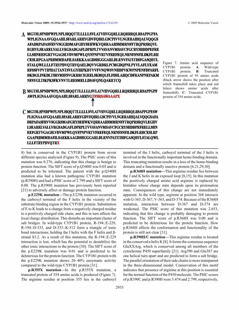
p.I94X mutation—In the isoleucine94stop mutation (p.194X) mutation a truncated protein of 93 amino acids isproduced in which only the first 82 amino acids are the sameas the wild-type CYP1B1 protein (Figure 7). This truncatedprotein lacks all functional domains of the CYP1B1 proteinand is a nonfunctional protein [6,21,29,30].
p.H279D mutation—This histidine residue lies in thecarboxyl terminal of the G helix in the CYP1B1 protein.Replacement of an aromatic, weak basic, amino acid histidinewhose charge state depends upon its protonation state with analiphatic, strong acidic, and negatively charged aspartic acidat this locus. This in turn affects the local charge distribution,and hence the structure of the protein is disturbed. Histidineis conserved at this locus in the CYP1A1 protein from 12different species (Figure 8) and in the CYP1B1 protein fromseven different species (Figure 9) analyzed, suggesting thathistidine performs some important functions at this locus. Noother known pathogenic mutation was present in the patient(P61), and the PSIC score of this mutation was 2.628,indicating that this change is probably damaging to the proteinfunction. The SIFT score of p.H279D was 0.00 and ispredicted to be deleterious for the protein function.
p.Q340H mutation—This glutamine residue lies in thecarboxyl terminal of the I helix. Replacement of a polaruncharged amino acid (glutamine) with a weak basic aminoacid (histidine) may or may not alter the structure/function ofthe protein. Glutamine is not conserved at this locus in theCYP1A1 protein from 12 different species analyzed (Figure
Molecular Vision 2009; 15:2926-2937 <http://www.molvis.org/molvis/v15/a310> © 2009 Molecular Vision
2930

TAB
LE 2
. CLI
NIC
AL
MA
NIF
ESTA
TIO
NS O
F PC
G PA
TIEN
TS.
Pt.
IDA
ge o
fon
set o
fdi
seas
e
Sex
Age
at p
rese
ntat
ion/
sam
plin
gC
orne
al D
iam
eter
(mm
) OS/
OD
and
clar
ity a
tdi
agno
sis
Bup
htha
lmos
IOP
OS/
OD
(mm
Hg)
At
pres
enta
tion
Haa
bs’
stri
aeL
ast C
up D
isc
ratio
OS/
OD
Phot
o-ph
obia
Mut
atio
nsT
reat
men
ts
P51
By
birth
F36
mon
ths
11x1
1.5/
13x1
3;O
U m
ild e
dem
aO
U;O
D>O
S22
/28
no0.
8:1/
0.9:
1Y
es—
Med
ical
and O
U T
rab/
Trab
+MM
C; O
Uca
tara
ct su
rger
yP5
2B
y B
irth
F2
mon
ths
12.5
x13/
12.5
x13;
OS
mild
ede
ma
OU
20/2
0no
Haz
y m
edia
/0.
5:1
Yes
—M
edic
al an
d OU
Tra
b/Tr
ab+M
MC
P53
By
birth
F4
mon
ths
11.5
x12/
12x1
2;O
U m
ild e
dem
aO
U; O
D>O
S40
/23
noH
azy
med
iaY
es—
Med
ical
and O
U T
rab/
Trab
+MM
CP5
4B
y bi
rthM
9 m
onth
s15
x14.
5/15
x14.
5;no
ede
ma
OU
26/2
6no
No
glow
No
—M
edic
al a
nd O
UTr
ab/
Trab
+MM
CP5
5B
y bi
rthM
8 m
onth
sPh
this
ic ey
e/12
x12;
OU
seve
re e
dem
aO
D; O
S Ph
this
icey
eN
A/3
7no
NA
/0.9
:1N
op.
I94X
(H)
Med
ical
and O
D T
rab/
Trab
+MM
CP5
6B
y bi
rthF
12 m
onth
s14
.5x1
4.5/
14x1
4;O
U se
vere
ede
ma
OU
; OS>
OD
30/2
8O
U +
veH
azy
med
iaN
op.
Q34
0H (H
)+
p.R
390H
(H)
Med
ical
and O
U T
rab/
Trab
+MM
CP5
7B
y bi
rthM
3 m
onth
s13
x13/
13.5
x13.
5;N
o ed
ema
OU
; OD
>OS
28/3
0no
0.7:
1/0.
7:1
No
—M
edic
al an
d OU
Tra
b/Tr
ab+M
MC
P58
By
birth
M15
mon
ths
14x1
4/12
.5x1
2.5;
No
edem
aO
U; O
S>O
D20
/16
noto
tal c
uppi
ng/
0.5:
1N
o—
Med
ical
and O
U T
rab/
Trab
+MM
CP5
9B
y bi
rthF
10 m
onth
s14
x14.
5/13
.5x1
4;O
S ed
ema
OU
; OS>
OD
31/3
1O
S +v
eN
ot a
vaila
ble
No
—M
edic
al an
d OU
Tra
b/Tr
ab+M
MC
P60
7 m
onth
sF
41 m
onth
s14
x14/
13x1
3.5;
no
edem
aO
U; O
S>O
D24
/18
no0.
4:1/
0.6:
1Y
es—
Med
ical
and O
U T
rab/
Trab
+MM
CP6
1B
y bi
rthM
4 m
onth
s15
.5x1
5/14
x14;
OU
mild
ede
ma
OU
; OS>
OD
30/3
4O
U +
veN
ot v
isib
le/0
.9:1
No
p.H
279D
(h)
Med
ical
and O
U T
rab/
Trab
+MM
CP6
2B
y bi
rthM
8 m
onth
s11
x11.
5/10
x11.
5;O
D e
dem
aO
U; O
S>O
D22
/16
no0.
6:1/
0.6:
1Y
es—
Med
ical
and O
U T
rab/
Trab
+MM
CP6
33
mon
ths
M12
mon
ths
12x1
2/11
x12.
5; N
oed
ema
OU
; OS>
OD
22/2
3no
0.4:
1/0.
4:1
No
—M
edic
al an
d OU
Tra
b/Tr
ab+M
MC
P64
By
birth
M1
mon
th12
x12/
11.5
x11.
5;O
D se
vere
ede
ma
OU
; OS>
OD
28/2
4no
Not
vis
ible
/0.7
:1Y
esp.
R39
0C (h
)M
edic
al an
d OU
Tra
b/Tr
ab+M
MC
P65
By
birth
M6
mon
ths
13x1
3/12
.5x1
3;O
U e
dem
aO
U; O
S>O
D25
/26
no0.
7:1/
0.7:
1N
op.
E229
K (h
)M
edic
al an
d OU
Tra
b/Tr
ab+M
MC
P66
3 m
onth
sM
13 m
onth
s12
.5x1
2/12
.5x1
2;N
o ed
ema
OU
22/2
4no
0.4:
1/0.
4:1
no—
Med
ical
and O
U T
rab/
Trab
+MM
CP6
711
mon
ths
M13
2 m
onth
s12
x13/
13x1
4; O
Um
ild e
dem
aO
U; O
D>O
S21
/24
noN
ot v
isib
leY
es—
Med
ical
and O
U T
rab/
Trab
+MM
CP6
8B
y bi
rthM
6 m
onth
s13
x13/
13.5
x14;
OU
seve
re e
dem
aO
U; O
D>O
S26
/28
no0.
8:1/
0.8:
1no
p.R
368H
(H)
Med
ical
and O
U T
rab/
Trab
+MM
CP6
9B
y bi
rthM
4 m
onth
s13
x13/
12x1
3; N
oed
ema
OU
; OS>
OD
24/2
6no
0.5:
1/0.
6:1
no—
Med
ical
and O
U T
rab/
Trab
+MM
CP7
0B
y bi
rthM
45 d
ays
15x1
5/15
x14.
5;O
U se
vere
ede
ma
OU
/; O
S>O
D30
/40
Not
visi
ble
Not
vis
ible
nop.
R35
5X (H
)M
edic
al an
d OU
Tra
b/Tr
ab+M
MC
P71
13 m
onth
sM
18 m
onth
s12
x2/1
2x12
; No
edem
aO
U22
/23
no0.
4:1/
0.4:
1Y
es—
Med
ical
and O
U T
rab/
Trab
+MM
CP7
2B
y bi
rthM
45 d
ays
12x1
2/12
x11.
5;O
U m
ild e
dem
aO
U; O
S>O
D20
/24
noN
o gl
owY
es—
Med
ical
and O
U T
rab/
Trab
+MM
CP7
3B
y m
onth
M2
mon
ths
12.5
x13/
11x1
2;O
U m
ild e
dem
aO
U; O
S>O
D22
/22
noH
azy
med
iano
—M
edic
al an
d OU
Tra
b/Tr
ab+M
MC
Foot
note
: M- m
ale;
F- f
emal
e; H
-hom
ozyg
ous;
h-h
eter
ozyg
ous;
X- t
imes
; Tra
b/Tr
ab+M
MC
- com
bine
d tra
becu
loto
my
trabe
cule
ctom
y an
d m
itom
ycin
C tr
eatm
ent;
OD
- rig
ht e
ye; O
S- le
ft ey
e; O
U- b
oth
eyes
; mut
atio
ns in
bol
d le
tters
- nov
el m
utat
ions
.
Molecular Vision 2009; 15:2926-2937 <http://www.molvis.org/molvis/v15/a310> © 2009 Molecular Vision
2931

T AB
LE 3
. SU
MM
AR
Y O
F TH
E SE
QU
ENC
E V
AR
IAN
TS ID
ENTI
FIED
IN V
AR
IOU
S STU
DIE
S TIL
L D
ATE
.S.
No.
Patie
nt n
umbe
rG
enom
iclo
catio
nN
ucle
otid
ech
ange
Cod
on c
hang
eT
ype
ofm
utat
ion
Loc
atio
n in
prot
ein
Mut
atio
nid
entif
ied
Obs
erva
tiona
lhi
stor
y of
mut
atio
ns in
diff
eren
t dis
ease
s
Ori
gin
(ref
eren
ce)
1P5
1, P
53, P
55, P
58-P
65,
P71,
P72
, P73
g.38
3025
44C
>TN
AIn
troni
cN
AN
APC
GIn
dia,
Sau
dia
Ara
bia,
Om
an, B
razi
l [7,
13,3
5,36
]
2P5
1, P
53, P
54, P
57-P
61,
P63-
P65,
P71
-P73
g.38
3023
90c.
142
C>G
CG
G>G
GG
Mis
sens
e48
p.ar
g48
gly
(p.R
48G
)PC
GSa
udia
Ara
bia,
Indi
a, Ja
pan
[8,1
1,15
]
3P5
5g.
3830
2285
c.24
7del
. GFS
FSFS
afte
r 82
p.as
p83t
hrfs
X12
(p.I9
4X)
PCG
This
stud
y
4P5
5, P
73g.
3830
2177
c.35
5G>T
GC
C>T
CC
Mis
sens
e11
9p.
ala1
19se
r(p
.A11
9S)
PCG
Saud
ia A
rabi
a, Ja
pan,
Indi
a [7
,11,
15]
5P6
5g.
3830
1847
c.68
5G>
AG
AA
>AA
AM
isse
nse
229
p.gl
u229
lys
(p.E
229K
)PC
G, P
OA
GFr
ance
, Ind
ia, G
erm
any
[7,2
9,31
]
6P6
1g.
3830
1697
c.83
5C>G
CA
C>G
AC
Mis
sens
e27
9p.
his2
79as
p(p
.H27
9D)
PCG
This
stud
y
7P5
6g.
3830
1512
c.10
20G
>TC
AG
>CA
TM
isse
nse
340
p.gl
n340
his
(p.Q
340H
)PC
GTh
is st
udy
8P7
0g.
3829
8434
c.10
63C
>TG
GA
>TG
AN
on-s
ense
355
p.al
a355
stop
(p.A
355X
)PC
GG
erm
any,
Indi
a [2
8] th
is st
udy
9P6
8g.
3829
8394
c.11
03G
>AC
GT>
CA
TM
isse
nse
368
p.ar
g368
his
(p.R
368H
)PC
G, P
A, P
OA
GSa
udia
Ara
bia,
Indi
a. F
ranc
e [7
,15,
32]
10P6
4g.
3929
8329
c.11
68C
>TC
GC
>TG
CM
isse
nse
390
p.ar
g390
cys
(p.R
390C
)PC
GEc
uado
r, In
dia
[8,3
7]
11P5
6g.
3829
8328
c.11
69G
>AC
GC
>CA
CM
isse
nse
390
p.ar
g390
his
(p.R
390H
)PC
G, P
OA
GPa
kist
an, I
ndia
, Fra
nce
[7,2
1,32
]
12P5
2, P
55, P
68, P
69g.
3829
8203
c.12
94C
>GC
TG>G
TGM
isse
nse
432
p.le
u432
lys
(p.L
432V
)PC
G, P
AIn
dia,
Japa
n, T
urke
y [7
,11,
29]
13P6
9g.
3829
8198
c.12
99G
>AA
AG
>AA
AN
eutra
l43
3p.
lys4
33ly
s(p
.K43
3K)
PCG
This
stud
y
14P5
1, P
53-P
67, P
69-P
71,
P73
g.38
2983
50c.
1347
T>C
GA
T>G
AC
Neu
tral
449
p.as
p449
asp
(p.D
449D
)PC
GJa
pan,
Indi
a [7
,11]
15P5
6, P
67g.
3829
8139
c.13
58A
>GA
AC
>AG
CM
isse
nse
453
p.as
p453
ser
(p.N
453S
)PC
GFr
ance
, Ind
ia [7
,12]
Foot
note
: PC
G- P
rimar
y co
ngen
ital g
lauc
oma;
PO
AG
- Prim
ary
open
ang
le g
lauc
oma;
PA
-Pet
er’s
ano
mal
y; F
S-fr
ames
hift;
X- s
top
codo
n; N
A- n
ot a
pplic
able
;m
utat
ions
in b
old
lette
rs- n
ovel
mut
atio
ns.
Molecular Vision 2009; 15:2926-2937 <http://www.molvis.org/molvis/v15/a310> © 2009 Molecular Vision
2932
8) but is conserved in the CYP1B1 protein from sevendifferent species analyzed (Figure 9). The PSIC score of thismutation was 0.276, indicating that this change is benign toprotein function. The SIFT score of p.Q340H was 0.05 and ispredicted to be tolerated. The patient with the p.Q340Hmutation also had a known pathogenic CYP1B1 mutation(p.R390H) and had a PSIC score of 2.799 and a SIFT score of0.00. The p.R390H mutation has previously been reported[21] to adversely affect or damage protein function.
p.E229K mutation—The p.E229k mutation occurred inthe carboxyl terminal of the F helix in the vicinity of thesubstrate-binding region in the CYP1B1 protein. Substitutionof E to K leads to a change from a negatively charged residueto a positively charged side chain, and this in turn affects thelocal charge distribution. This disturbs an important cluster ofsalt bridges. In wild-type CYP1B1 protein, R-194::E-229,R-194::D-333, and D-333::K-512 form a triangle of ionicbond interactions, holding the I helix with the F helix and β-strand S3.2. As a result of this mutation, the R-194::E-229interaction is lost, which has the potential to destabilize theother ionic interactions in the protein [30]. The SIFT score ofthe p.E229K mutation was 0.01 and is predicted to bedeleterious for the protein function. The CYP1B1 protein withthe p.E229K mutation shows 20–40% enzymatic activitycompared to the wild-type CYP1B1 protein [31].
p.R355X mutation—In the p.R355X mutation, atruncated protein of 354 amino acids is produced (Figure 7).The arginine residue at position 355 lies in the carboxyl
terminal of the J helix, carboyxl terminal of the J helix isinvolved in the functionally important heme-binding domain.This truncating mutation results in a loss of the heme-bindingdomain and a functionally inactive protein [6,21,29,30].
p.R368H mutation—This arginine residue lies betweenthe J and K helix in an exposed loop [8,15]. In this mutationthe positively charged amino acid arginine is replaced byhistidine whose charge state depends upon its protonationstate. Consequences of this change are not immediatelyapparent. In the wild type, arginine at position 368 interactswith G-365, D-367, V-363, and D-374. Because of the R368Hmutation, interaction between D-367 and D-374 areweakened. The PSIC score of this mutation was 2.653,indicating that this change is probably damaging to proteinfunction. The SIFT score of p.R368H was 0.00 and ispredicted to be deleterious for the protein function. Howp.R368H affects the conformation and functionality of theprotein is still not clear [31].
p.R390H/C mutation—This arginine residue is locatedin the conserved α helix K [8]. It forms the consensus sequenceGluXXArg, which is conserved among all members of thecytochrome P450 superfamily [21]. Arg390 and Glu387 areone helical turn apart and are predicted to form a salt bridge.The parallel orientation of their side chains is more transparentin the three-dimensional model. Conservation of this motifindicates that presence of arginine at this position is essentialfor the normal function of the P450 molecule. The PSIC scoresof p.R390C and p.R390H were 3.474 and 2.799, respectively,
Figure 7. Amino acid sequence ofCYP1B1 protein. A: Wild-typeCYP1B1 protein. B: TruncatedCYP1B1 protein of 93 amino acids(black arrow shows the position afterwhich frameshift takes place and redletters shows amino acids afterframeshift). C: Truncated CYP1B1protein of 354 amino acids.
Molecular Vision 2009; 15:2926-2937 <http://www.molvis.org/molvis/v15/a310> © 2009 Molecular Vision
2933

indicating that both these changes are probably damaging toprotein function. The SIFT score of p.R390H/C was 0.00 andis predicted to be deleterious for the protein function.
The PSIC scores of the nonpathogenic single nucleotidepolymorphisms were <2 for p.R48G, p.A119S, p.N453S, andp.L432V, indicating that all these changes were benign toprotein function. The SIFT scores of the nonpathogenic singlenucleotide polymorphisms were >0.05 for p.R48G, p.L432V,p.K433K, and p.D449D, indicating that all these changes weretolerated in the protein.
PCG is a clinically and genetically heterogeneousdisorder. More than 50 different mutations have been reportedin the entire coding region of CYP1B1 from variouspopulations. We screened the entire coding region ofCYP1B1 in 23 congenital glaucoma patients by using primersdescribed elsewhere [8]. Of all mutations identified herein,the frameshift mutation (c.247delG) and nonsense mutation(c.1063C>T) resulted in the most severe disease phenotype.
The patient (P55) with the p.I94X (homozygous)mutation is a male child of a consanguineous marriage withoutany family history of glaucoma; he presented at 8 months ofage. He was born at full term through a normal vaginaldelivery. He had severe bilateral corneal edema at birth. Atthe age of 2 months he had congestion with discharge in theleft eye and was diagnosed to have a left corneal ulcer and wastreated with antibiotics; the left eye consequently developedphthisis. The right eye dimensions increased, and he wasdiagnosed as having buphthalmos at the age of 8 months.Combined trabeculotomy and trabeculectomy withmitomycin C was performed in his right eye. He wasdiagnosed as having 100% blindness at 8 months. His parentswere also screened for CYP1B1 mutations by DNAsequencing but were found to be negative for any pathogenicCYP1B1 mutations.
Patient P70 has a p.R355X (homozygous) mutation andis a male offspring of a non-consanguineous marriage; hepresented at 45 days. At birth, he had bilateral congenital
Figure 8. Multi sequence alignment ofthe human CYP1B1 protein with theCYP1A1 protein from different species.Red Underlined amino acids shows theconserved residues in human CYP1B1and different CYP1A1 protein fromdifferent species (when mutated)causing primary congenital glaucomaphenotype. While Red letter showsamino acid conserved in differentCYP1A1 protein from different speciesbut not present in human CYP1B1protein.
Figure 9. Multisequence alignment ofthe human CYP1B1 protein with theCYP1B1 protein from different species.Underlined red amino acids show theconserved residues (when mutated)causing the primary congenitalglaucoma phenotype. Red coloredamino acid shows the non-conservationof glutamic acid at this locus inZebrafish CYP1B1. Blue-coloredamino acids show the less conservedresidues in CYP1B1 protein fromdifferent species.
Molecular Vision 2009; 15:2926-2937 <http://www.molvis.org/molvis/v15/a310> © 2009 Molecular Vision
2934

glaucoma and had IOPs of 30 and 40 mmHg in his left andright eye, respectively. He had severe corneal clouding in botheyes, at birth, and therefore the fundus was not visualized.Combined trabeculotomy and trabeculectomy withmitomycin C was performed in both eyes. He had no lightperception and was visually blind since 45 days of age. Hisparents were also negative for the pathogenic CYP1B1mutations. The absence of mutations in the parents of P55 andP70 could be due to a parental germline mutation, whichcannot be tested by using peripheral leukocytes.
Patient P56 is a female child of a non-consanguineousmarriage; she presented at the age of 1 year. She has p.Q340H(heterozygous) and p. R390H (homozygous) mutations. Shehad bilateral congenital glaucoma since birth. She had acorneal diameter of 14.0×14.0 mm (right eye) and14.5×14.5 mm (left eye) and IOPs of 28 and 30 mmHg in theright and left eye, respectively. She had Haab’s striae in botheyes, and the fundus was not visible. Combinedtrabeculotomy and trabeculectomy with mitomycin C wasperformed in both eyes.
Patient P61 is a male child of a non-consanguineousmarriage; he presented at the age of 4 months and has ap.H279D (heterozygous) mutation. He had bilateralcongenital glaucoma at birth. At presentation corneal diameterand IOPs of his left and right eye were 15.5×15.0 mm and14.0×14.0 mm and 30 mmHg and 34 mmHg, respectively. Thecup to disc ratio of the left eye was not visible due to the hazymedia and that of the right eye was 0.9:1. He had Haab’s striaein both eyes. Combined trabeculotomy and trabeculectomywith mitomycin C was performed in both eyes.
An intriguing finding that apparently does not match atypical recessive pattern of inheritance is the presence of aheterozygous CYP1B1 mutation in PCG patients. Thissituation has been previously reported [7,29]. A heterozygousp.Y81N mutation has also been described in PCG patientsfrom Germany, and a heterozygous p.E229K mutation hasbeen identified in unrelated French and Indian patients [7,32]. Few heterozygous CYP1B1 mutations were associatedwith the milder, primary, open-angle glaucoma phenotypes inpatients from Spain, France, and India. The presence of aheterozygous CYP1B1 mutation in PCG suggests thepossibility of other loci, yet undetected, that may be involvedin anterior chamber formation. Recently the presence ofdouble heterozygote variants CYP1B1 and FOXC1 has beendescribed in two PCG cases, although the role of possibledigenic inheritance in disease causation is yet to be established[33]. Defective variants of modifier genes and/orenvironmental factors have an additive effect with loss-of-function CYP1B1 alleles to produce the disease phenotype.However further work is required to understand thismechanism.
Previous studies have reported that the age of diseaseonset in PCG patients with CYP1B1 mutations is younger thanin patients without CYP1B1 mutations [34]. Our data show
that the onset age in three patients (P60, P66, and P67) was 7,3, and 11 months, while the rest of the patients presented atbirth. In these 20 patients there is no significant difference inthe age of disease onset in CYP1B1 mutation-positive andmutation-negative cases, although clinical phenotypes ofpatients (P55, P56, P61, P68, and P70) with homozygousCYP1B1 mutations were more severe compared to patients(P64 and P65) who were heterozygous for the CYP1B1mutations (Table 1). It is possible that patients with two nullalleles with no catalytic activity may present with a moresevere phenotype of the disease compared to patients with onenull (heterozygote) allele. The disease phenotype of patientswith homozygous/heterozygous CYP1B1 mutations was moresevere compared to the clinical phenotype of patients negativefor the CYP1B1 mutations.
We also observed a higher mean IOP in a group ofpatients with CYP1B1 mutations. In accordance with the ideaof associating the severe phenotypes with the null CYP1B1allele, the percentage of severe phenotypes in at least one eyehas been reported to be associated with various mutationsranging 80-100% for a frameshift mutation (e.g., c.376insA)and truncating mutations [11]. Three different truncationmutations (p.C280X, p.E281X, and p.R355X) producing atruncated protein of 279, 280, and 354 amino acids,respectively, have also been associated with more severedisease phenotypes [11,21,29]. In patient P55 with ahomozygous p.I94X mutation, a truncated protein of 93 aminoacids is produced that has the first 82 amino acids similar tothe wild-type CYP1B1 protein. The disease phenotype of thispatient is severe with a left phthisic and a right buphthalmiceye with a cup to disc ratio of 0.9:1. He is visually blind.Another patient (P70) with a p.R355X mutation had bilateralbuphthalmos with severe corneal edema and a cornealdiameter of 15.0×15.0 mm and 15.0×14.5 mm in the left andright eye, respectively. He was blind at the age of 45 days.Patient P61 with a p.H279D mutation had bilateralbuphthalmos with mild edema in both eyes and a cornealdiameter of 15.5×15.5 mm and 14.0×14.0 mm in the left andright eye, respectively. He was blind at the age of 4 months.The range of percentages of severe phenotypes in at least oneeye is 62–83% for different mutations, such as p.G61E,p.E229K, p.R368H, and p.R390C [9].
Membrane-bound cytochromes, such as CYP1B1, havea molecular structure containing a transmembrane domainlocated at the N-terminal end of the molecule. This is followedby a proline-rich “hinge” region, which permits flexibilitybetween the membrane-spanning domain and the cytoplasmicportion of the protein molecule. The COOH-terminal ends arehighly conserved among different members of the cytochromeP450 superfamily [17]. This family contains a set of conservedcore structures responsible for the heme-binding region ofthese molecules. The heme-binding region is essential for thenormal function of every P450 molecule. Between the hingeregion and the conserved core structure lies a less conserved
Molecular Vision 2009; 15:2926-2937 <http://www.molvis.org/molvis/v15/a310> © 2009 Molecular Vision
2935

substrate-binding region. The cytochrome P450 proteinfunctions like any classical enzyme molecule [18,19].Mutations affecting such enzymes generally producerecessive phenotypes because in heterozygous subjects thenormal allele is capable of compensating for the mutant allele.Mutations in the CYP1B1protein interfere with the integrityof the CYP1B1 protein as well as its ability to adopt a normalconformation and to bind heme; for example, inducedmutations in the hinge region have previously been reportedto interfere with the heme-binding properties of thecytochrome P450 molecules
Thus mutations of CYP1B1 are a major cause of PCG inour study as well as various other studies [6-15,35-37]. Thisstudy confirms genetic heterogeneity of the disease. Weidentified four novel mutations in this study in addition to oneprevious reported [7]. Studies of pathogenic sequence variantsof CYP1B1 in different populations will contribute to a betterunderstanding of the pathogenesis of PCG and will aid inanalyzing the structure–function relationship of differentCYP1B1 mutations Identifying mutations in subjects at risk ofdeveloping glaucoma, particularly among relatives of PCGpatients, is of clinical significance. Monitoring vision in thesefamilies would be helpful. These developments may help inreducing the disease frequency in familial cases. Such studieswill also help in understanding the pathogenic mutations inour patient populations and enable us to develop simple andrapid diagnostic tests for analyzing such cases. This may leadto the development of novel therapies in the management ofcongenital glaucoma.
ACKNOWLEDGMENTSThis work was financially supported by Department ofBiotechnology, Government of India. The authors would liketo thank the families of the patients for their cooperationwithout this study was not possible. The author MukeshTanwar thanks University Grants Commission (UGC), Govt.of India for providing Senior Research Fellowship (SRF).
REFERENCES1. deLuise VP, Anderson DR. Primary infantile glaucoma
(congenital glaucoma). Surv Ophthalmol 1983; 28:1-19.[PMID: 6353647]
2. Francois J. Congenital glaucoma and its inheritance.Ophthalmologica 1980; 181:61-73. [PMID: 7219964]
3. Dandona L, Williams JD, Williams BC, Rao GN. Population-based assessment of childhood blindness in southern India.Arch Ophthalmol 1998; 116:545-6. [PMID: 9565065]
4. Sarfarazi M, Akarsu AN, Hossain A, Turacli ME, Aktan SG,Barsoum-Homsy M, Chevrette L, Sayli BS. Assignment of alocus (GLC3A) for primary congenital glaucoma(buphthalmos) to 2p21and evidence for geneticheterogeneity. Genomics 1995; 30:171-7. [PMID: 8586416]
5. Akarsu AN, Turacli ME, Aktan SG, Barsoum-Homsy M,Chevrette L, Sayli BS, Sarfarazi M. A second locus (GLC3B)for primary congenital glaucoma (buphthalmos) maps to the1p36 region. Hum Mol Genet 1996; 5:1199-203. [PMID:8842741]
6. Stoilov I, Akarsu AN, Sarfarazi M. Identification of threedifferent truncating mutations in cytochrome P450B1(CYP1B1) as the principal cause of primary congenitalglaucoma (buphthalmos) in families linked to the GLC3Alocus on chromosome 2p21. Hum Mol Genet 1997;6:641-7. [PMID: 9097971]
7. Tanwar M, Dada T, Sihota R, Das TK, Yadav U, Dada R.Mutation spectrum of CYP1B1 in north Indian Congenitalglaucoma patients. Mol Vis 2009; 15:1200-9. [PMID:19536304]
8. Panicker SG, Reddy AB, Mandal AK, Ahmed N, NagarajaramHA, Hasnain SE, Balasubramanian D. Identification of novelmutations causing familial primary congenital glaucoma inIndian pedigrees. Invest Ophthalmol Vis Sci 2002;43:1358-66. [PMID: 11980847]
9. Panicker SG, Mandal AK, Reddy ABM, Gothwal VK, HasnainSE. Correlations of Genotype with Phenotype in IndianPatients with Primary Congenital Glaucoma. InvestOphthalmol Vis Sci 2004; 45:1149-56. [PMID: 15037581]
10. Stoilov IR, Costa VP, Vascincellos JP, Melo MB, Betinjane AJ,Carani JCE, Oltrogge EV, Sarfarazi M. Molecular genetics ofprimary congenital glaucoma in Brazil. Invest OphthalmolVis Sci 2002; 43:1820-7. [PMID: 12036985]
11. Mashima Y, Suzuki Y, Sergeev Y, Ohtake Y, Tanino T, KimuraI, Miyata H, Aihara M, Tanihara H, Inatani M, Azuma N,Iwata T, Araie M. Novel Cytochrome p.4501B1 (CYP1B1)gene mutations in Japanese patients with Primary CongenitalGlaucoma. Invest Ophthalmol Vis Sci 2001; 42:2211-6.[PMID: 11527932]
12. Colomb E, Kaplan J, Garchon HJ. Novel cytochrome P450(CYP1B1) mutations in patients with primary congenitalglaucoma in France. Hum Mutat 2003; 22:496-500. [PMID:14635112]
13. Bejjani BA, Lewis RA, Tomey K, Anderson KL, Dueker DK,Jabak M, Astle WF, Otterud B, Leppert M, Lupski JR.Mutations in CYP1B1, the gene for cytochrome P4501B1, arethe predominant cause of primary congenital glaucoma inSaudi Arabia. Am J Hum Genet 1998; 62:325-33. [PMID:9463332]
14. Plasilova M, Stoilov I, Sarfarazi M, Kadasi L, Ferakova E,Ferak V. Identification of a single ancestral CYP1B1mutation in Slovak gypsies (Roms) affected with primarycongenital glaucoma. J Med Genet 1999; 36:290-4. [PMID:10227395]
15. Bejjani BA, Stockton DW, Lewis RA, Tomey KF, Duekar DK,Jabak M, Astle WF, Lupski JR. Multiple CYP1B1 mutationsand incomplete penetrance in an inbred populationsegregating primary congenital glaucoma suggest frequent denovoevents and a dominant modifier locus. Hum Mol Genet2000; 9:367-74. [PMID: 10655546]
16. Sutter TR, Tang YM, Hayes CL, Wo YP, Jabs EW, Li X, YinH, Cody CW, Greenlee WF. Complete cDNA Sequence of aHuman Dioxin-inducible mRNA Identifies a New GeneSubfamily of Cytochrome P450 That Maps to Chromosome2. J Biol Chem 1994; 269:13092-9. [PMID: 8175734]
17. Yamazaki H, Inui Y, Yun CH, Guengerich FP, Shimada T.Cytochrome P450 2E1 and 2A6 enzymes as major catalystsfor metabolic activation of N-nitrosodialkylamines andtobacco-related nitrosamines in human liver microsomes.Carcinogenesis 1992; 13:1789-94. [PMID: 1423839]
Molecular Vision 2009; 15:2926-2937 <http://www.molvis.org/molvis/v15/a310> © 2009 Molecular Vision
2936

18. Chen H, Howald WN, Juchau MR. Biosynthesis of all trans-retinol: catalysis of all-trans-retinol oxidation by humanp-450 cytochrome. Drug Metab Dispos 2000;28:315-22.http://www.ncbi.nlm.nih.gov/sites/entrez?cmd=Retrieve&db=PubMed&list_uids=10681373&dopt=Abstract [PMID: 10681373]
19. Swindell EC, Eichele G. Retinoid metabolizing enzymes indevelopment. Biofactors 1999; 10:85-9. [PMID: 10609867]
20. Nebert DW. Proposed role of drug-metabolizing enzymes:regulation of steady state levels of the ligands that affectgrowth, homeostasis, differentiation and neuroendocrinefunctions. Mol Endocrinol 1991; 5:1203-14. [PMID:1663211]
21. Stoilov I, Akarsu AN, Alozie I, Child A, Barsoum-Homsy M,Turacli ME, Or M, Lewis RA, Ozdemir N, Brice G, AktanSG, Chevrette L, Coca-Prados M, Sarfarazi M. Sequenceanalysis and homology modeling suggest that primarycongenital glaucoma on 2p21 results from mutationsdisrupting either the hinge region or the conserved corestructures of cytochrome P4501B1. Am J Hum Genet 1998;62:573-84. [PMID: 9497261]
22. Sunyaev S, Ramensky V, Koch I, Lathe W III. Kondrashiv,Bork P. Prediction of deleterious human alleles. Hum MolGenet 2001; 10:591-7. [PMID: 11230178]
23. Ramensky V, Bork P, Sunyaev S. Human non-synonymousSNPs: server and survey. Nucleic Acids Res 2002;30:3894-900. [PMID: 12202775]
24. Sunyaev S, Ramensky V. Towards a structural basis of humannon-synonymous single nucleotide polymorphisms. TrendsGenet 2000; 16:198-200.Bork P [PMID: 10782110]
25. Kumar P, Henikoff S, Ng PC. Predicting the effects of codingnon-synonymous variants on protein function using the SIFTalgorithm. Nat Protoc 2009; 4:1073-81. [PMID: 19561590]
26. Ng PC, Henikoff S. SIFT: predicting amino acid changes thataffect protein function. Nucleic Acids Res 2003; 31:3812-4.[PMID: 12824425]
27. George Priya Doss C. Rajasekaran R, Sudandiradoss C,Ramanathan K, Purohit R, Sethumadhavan R. A novelcomputational and structural analysis of nsSNPs in CFTRgene. Genomic Med 2008; 2:23-32. [PMID: 18716917]
28. Gale CP, Grant PJ. The characterisation and functional analysisof the human glyoxalase-1 gene using methods ofbioinformatics. Gene 2004; 340:251-60. [PMID: 15475166]
29. Michels-Rautenstrauss KG, Mardin CV, Zenker M, Jordan N,Gusek-Schneider GC, Rautenstrauss BW. Primary congenitalglaucoma: three case reports on novel mutations andcombinations of mutations in the GLC3A (CYP1B1) gene. JGlaucoma 2001; 10:354-7. [PMID: 11558822]
30. Choudhary D, Jansson I, Sarfarazi M, Schenkman JB.Characterization of biochemical and structural phenotypes offour CYP1B1 mutations observed in individuals with primarycongenital glaucoma. Pharmacogenet Genomics 2008;18:665-76. [PMID: 18622259]
31. Campos-Mollo E, Lopez-Garrido MP, Blanco-Marchite C,Garcia-Feijoo J, Peralta J, Belmonte-Martinez J, Ayuso C,Escribano J. CYP1B1 mutations in Spanish patients withprimary congenital glaucoma: phenotypic and functionalvariability. Mol Vis 2009; 15:417-31. [PMID: 19234632]
32. Melki R, Colomb E, Lefort N, Brezin AP, Garchon HJ. CYP1B1mutations in French patients with early-onset primary open-angle glaucoma. J Med Genet 2004; 41:647-51. [PMID:15342693]
33. Chakrabarti S, Kaur K, Rao KN, Mandal AK, Kaur I, ParikhRS, Thomas R. The transcription factor gene FOXC1 exhibitsa limited role in primary congenital glaucoma. InvestOphthalmol Vis Sci 2009; 50:75-83. [PMID: 18708620]
34. Ohtake Y, Tanino T, Suzuki Y, Miyata H, Taomoto M, AzumaN, Tanihara H, Araie M, Mashima Y. Phenotype ofcytochrome P450B1 gene (CYP1B1) mutations in Japanesepatients with primary congenital glaucoma. Br J Ophthalmol2003; 87:302-4. [PMID: 12598442]
35. El-Gayar S, Ganesh A, Chavarria-Soley G, Al-Zuhaibi S, Al-Mjeni R, Raeburn S, Bialasiewicz AA. Molecular analysis ofCYP1B1 in Omani patients with primary congenitalglaucoma: a pilot study. Mol Vis 2009; 15:1325-31. [PMID:19597567]
36. Paolera MD, Cabral de Vasconcellos JP, Umbelino CC,Kasahara N, Rocha MN, Richeti F, Costa VP, Tavares A, deMelo MB. CYP1B1 gene analysis in primary congenitalglaucoma Brazilian patients: Novel mutations and associationwith poor prognosis. J Glaucoma. 2009 [PMID: 19528825]
37. Curry SM, Daou AG, Hermanns P, Molinari A, Lewis RA,Bejjani BA. Cytochrome P4501B1 mutations cause only partof primary congenital glaucoma in Ecuador. OphthalmicGenet 2004; 25:3-9. [PMID: 15255109]
Molecular Vision 2009; 15:2926-2937 <http://www.molvis.org/molvis/v15/a310> © 2009 Molecular Vision
The print version of this article was created on 27 December 2009. This reflects all typographical corrections and errata to thearticle through that date. Details of any changes may be found in the online version of the article.
2937