identification of clinical phenotypes in knee
TRANSCRIPT
RESEARCH ARTICLE Open Access
Identification of clinical phenotypes in kneeosteoarthritis: a systematic review of theliteratureA. Dell’Isola*, R. Allan, S. L. Smith, S. S. P. Marreiros and M. Steultjens
Abstract
Background: Knee Osteoarthritis (KOA) is a heterogeneous pathology characterized by a complex and multifactorialnature. It has been hypothesised that these differences are due to the existence of underlying phenotypes representingdifferent mechanisms of the disease.
Methods: The aim of this study is to identify the current evidence for the existence of groups of variables which pointtowards the existence of distinct clinical phenotypes in the KOA population. A systematic literature search in PubMedwas conducted. Only original articles were selected if they aimed to identify phenotypes of patients aged 18 years orolder with KOA. The methodological quality of the studies was independently assessed by two reviewers and qualitativesynthesis of the evidence was performed. Strong evidence for existence of specific phenotypes was considered presentif the phenotype was supported by at least two high-quality studies.
Results: A total of 24 studies were included. Through qualitative synthesis of evidence, six main sets of variablesproposing the existence of six phenotypes were identified: 1) chronic pain in which central mechanisms (e.g. centralsensitisation) are prominent; 2) inflammatory (high levels of inflammatory biomarkers); 3) metabolic syndrome (highprevalence of obesity, diabetes and other metabolic disturbances); 4) Bone and cartilage metabolism (alteration in localtissue metabolism); 5) mechanical overload characterised primarily by varus malalignment and medial compartmentdisease; and 6) minimal joint disease characterised as minor clinical symptoms with slow progression over time.
Conclusions: This study identified six distinct groups of variables which should be explored in attempts to betterdefine clinical phenotypes in the KOA population.
Keywords: Knee, Osteoarthritis, Phenotype, Sub-group, Clinical
BackgroundOsteoarthritis is the most common form of arthritis; itconstitutes a leading cause of disability in the adultpopulation [1] with the knee the most affected joint.Knee Osteoarthritis (KOA) is a heterogeneous pathologycharacterized by a complex and multifactorial nature [2].This multifactorial aetiology contributes to the broadvariation in symptoms presentation and treatmentresponse that characterize the KOA subjects and consti-tutes a challenge for the identification of personalized andeffective interventions. Therefore, in order to optimizetreatment effect in KOA, the intervention should address
this variability and should be tailored to specific subgroupsor phenotypes as highlighted in the NICE guidelines onKOA [3–6]. A phenotype in KOA can be defined as a col-lection of observable traits (i.e. aetiologic factors, risk fac-tors) that can identify and characterize a subgroup in adefined population. The presence of distinct phenotypeswithin the KOA patient population would suggest distinctunderlying causes and mechanisms, which could be highlyrelevant for understanding and treating the disease [7, 8].Previous attempts to identify distinctive KOA pheno-
types used different perspectives. Some researchers useddisease progression to determine KOA phenotypes, whileothers looked at pain perception or the degenerationpattern of the cartilage [9–15]. Potentially, hundreds ofphenotypes may be identified depending on the definition
* Correspondence: [email protected] of Applied Health Research/School of Health and Life Sciences,Glasgow Caledonian University, Glasgow G4 0BA, Scotland, UK
© 2016 The Author(s). Open Access This article is distributed under the terms of the Creative Commons Attribution 4.0International License (http://creativecommons.org/licenses/by/4.0/), which permits unrestricted use, distribution, andreproduction in any medium, provided you give appropriate credit to the original author(s) and the source, provide a link tothe Creative Commons license, and indicate if changes were made. The Creative Commons Public Domain Dedication waiver(http://creativecommons.org/publicdomain/zero/1.0/) applies to the data made available in this article, unless otherwise stated.
Dell’Isola et al. BMC Musculoskeletal Disorders (2016) 17:425 DOI 10.1186/s12891-016-1286-2

of phenotypes and on the variables selected. Each ap-proach can be considered equally valid depending on thescope. Only studies focusing on the identification of clin-ical subgroups characterized by different disease mecha-nisms can be considered useful to improve treatmentallocation and clinical management of the disease. If, ashypothesized, treatments Are highly effective only in onesub-type; the therapeutic effect of the intervention will belost if tested in KOA population as a whole [4]. Therefore,the identification of risk and aetiologic factors that canidentify specific clinical subtypes of KOA is an importantstarting point for the implementation of phenotypingresearch in clinical practice and may be critical for theimprovement of treatment allocation and for the develop-ment of new treatment strategies.The aim of this review is therefore to synthesize the
current evidence for the existence of distinct sets ofvariables that may suggest the existence of clinical KOAphenotypes characterized by the presence of differentrisk and aetiologic factors.
MethodsInformation sourcesA systematic literature search was conducted in PubMed(Medline) for the period from 01/01/1984 to 29/04/2016. An additional manual search was completed byADI from the references of the selected papers.The research strategy was built up using the following
key words: osteoarthritis, knee, phenotyp*, subgroup,cluster, “factor analysis”. These terms were combined inthe following way: osteoarthritis AND knee AND (phe-notyp* OR subgroup OR cluster OR “factor analysis”)(for further details see Additional file 1-A).
Inclusion criteriaArticles were included if: (1) the population involved(a subgroup of ) patients over 18 years of age; (2) thepopulation consisted of patients diagnosed with KOA;(3) the aim was to identify clinical phenotypes of pa-tients with KOA; (4) the methodology and analysiswere designed to identify phenotypes (e.g. cluster ana-lysis using clinical variables) ; (5) the article was anoriginal research report. Previous systematic reviewswere excluded. In addition to the second criterion,studies that included patients with hip or hand OAother than KOA were included if: (1) they used bio-markers or other measures that are not joint specific,(2) the KOA subgroup represented more than 60 % ofthe sample.
Data selection processArticle selection was made independently by two reviewers(ADI and MS) based on title and abstract according to theinclusion criteria. The final selection was made by the same
two independent reviewers based on the full text. Disagree-ments between the two reviewers were resolved by theintervention of a third reviewer (SS); this procedure wasadopted for both selection steps.
Assessment of methodological quality (risk of bias)The methodological quality of the papers was assessedusing an adaption of the standardized Hayden score [16](Table 1) to identify the risk of bias affecting the validityof findings. All papers were reviewed by ADl and MS,with additional proportional reviews performed by RA,SM, SS using block allocation with each reviewing 2/3 ofthe final papers.The risk of bias for each area was rated as low, moder-
ate or high [16]. Studies that had a high risk of bias in atleast one of the area assessed were considered to havean overall high risk of bias and regarded as sources oflow quality evidence used only to support the findings ofother (i.e. high quality) studies. Studies with low to mod-erate risk of bias and appropriate design were consideredsources of strong evidence.
Data-extraction analysisThe data from each study were extracted by two reviewers(ADI, MS) and included number of patients, clusteringmethod and subgroups identified. Additionally, the preva-lence of each phenotype was extracted where possible.
Identification of phenotypesIn this systematic review we adopted a tailored data ana-lysis process in order to deal with the broad variation inthe methodologies of the studies included. This processshares some similarities with the directed content ana-lysis method [17]. Key variables for each phenotypereported in the included studies were extracted. Usingthe theory and previous evidence, we assigned each keyvariable to a category (e.g. biomechanical, inflammatory,metabolic) indicating the underlying disease mechanismrepresented by that specific variable.. Variables (e.g.radiographic features, pain sensitization) were consid-ered to suggest similar disease mechanisms and classi-fied in the same category if: (1) It was specifically statedby the author of the paper (e.g. two subgroups extractedfrom two different studies were reported by the respect-ive authors as representing the same phenotype); (2) theassociation of the reported characteristic to a specificpathophysiologic mechanism had been reported in previ-ous studies investigating disease mechanisms and riskfactors (e.g. malalignment consistent with compartmentdegeneration has previously been reported as a biomech-anical mechanism responsible for KOA development).Each phenotype was then classified in the category indi-cated by the variable that characterized it. A phenotypewas considered supported by evidence when at least two
Dell’Isola et al. BMC Musculoskeletal Disorders (2016) 17:425 Page 2 of 12
studies with low risk of bias identified a phenotypeunder the same category. If a phenotype was reported inonly a single study this was not considered sufficient evi-dence to include this phenotype in the final list of phe-notypes identified in this review.
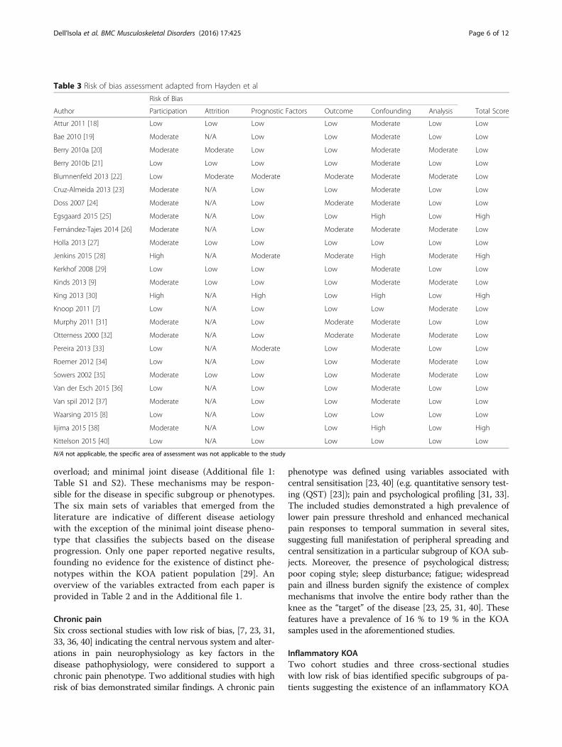
ResultsDescription of the included studiesThe initial literature search identified 841 articles. Threeadditional papers were identified through a manual searchof the references. After screening for title and abstract,781 papers were excluded. The full texts for the remaining63 articles were assessed for inclusion. From this list, 25articles matched the inclusion criteria and were includedin the systematic review (Fig. 1) [7–9, 18–39]. For an over-view of the studies included see Table 2. (For furtherdetails see Additional file 1).
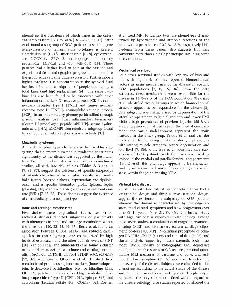
Quality assessmentThe quality assessment resulted in 21 papers with low ormoderate risk of bias (considered to be high qualitystudies) and four papers with a high risk of bias for atleast one of the six areas reviewed, which were consid-ered to be low quality studies (Table 3).
PhenotypesA total of 79 phenotypes were reported in the includedstudies. Of those, 42 phenotypes were reported in a singlestudy only and therefore not taken forward into the quali-tative evidence synthesis. The remaining 37 subgroupswere matched and combined into six main groups of vari-ables that suggest the existence of different mechanismsin the KOA population: chronic pain; inflammatorymechanisms; metabolic mechanisms of bone and cartil-age local to the joint, metabolic syndrome; mechanical
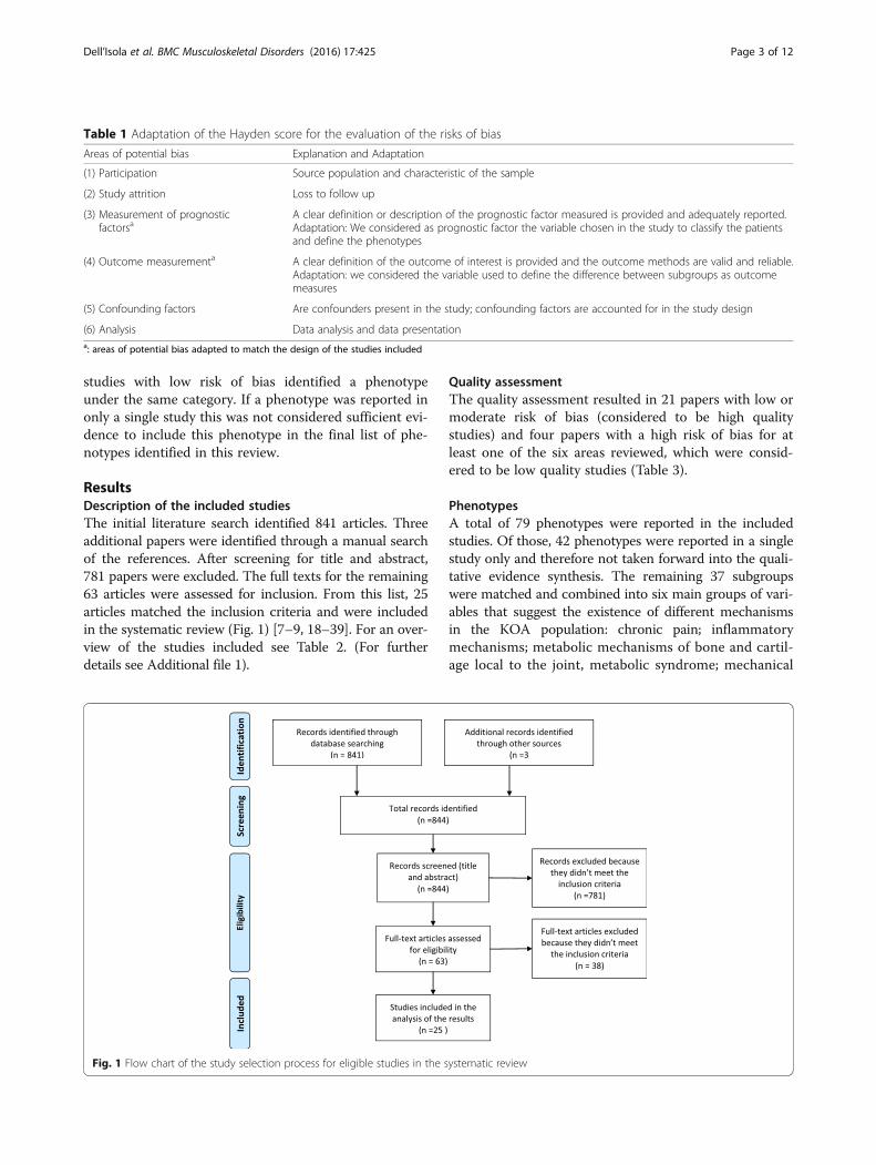
Table 1 Adaptation of the Hayden score for the evaluation of the risks of bias
Areas of potential bias Explanation and Adaptation
(1) Participation Source population and characteristic of the sample
(2) Study attrition Loss to follow up
(3) Measurement of prognosticfactorsa
A clear definition or description of the prognostic factor measured is provided and adequately reported.Adaptation: We considered as prognostic factor the variable chosen in the study to classify the patientsand define the phenotypes
(4) Outcome measurementa A clear definition of the outcome of interest is provided and the outcome methods are valid and reliable.Adaptation: we considered the variable used to define the difference between subgroups as outcomemeasures
(5) Confounding factors Are confounders present in the study; confounding factors are accounted for in the study design
(6) Analysis Data analysis and data presentationa: areas of potential bias adapted to match the design of the studies included
Fig. 1 Flow chart of the study selection process for eligible studies in the systematic review
Dell’Isola et al. BMC Musculoskeletal Disorders (2016) 17:425 Page 3 of 12

Table 2 Description of the papers
Author Type of research Type of study Analysis Participants Control Subgoups
Chronic pain Inflammatory Metabolicsyndrome
Bone andcartilagemetabolism
Mechanicaloverload
Minimaljointdisease
Attur 2011 [18] Genetic/geneexpression
Cohort (prosp) complete-linkagehierarchicalclustering
1: 41a
2: 36a
3: 86a
1: 25a
2: 0a
3: 12a
- 1: 16/41 = 39 %.2: 8/36 = 22 %,3: 33/86 = 38 %
- - - -
Bae 2010 [19] Imaging(photography)
Cross sectional K-meansclusteranalysis
127 - - - - - 20 %b -
Berry 2010a [20] Biomarker Cohort (prosp) Mann–Whitneyu, χ2, Multipleregression analysis
117 - - - - Prevalencenot reported
- -
Berry 2010b [21] Biomarker Cohort (prosp) Mann–Whitney u,Multiple regressionand logistic regressionanalysis
117 - - - - - - Prevalencenot reported
Blumnenfeld2013 [22]
Biomarker Cohort (prosp) Binary logisticregression analysis
Different indifferentanalysis
Differentin differentanalysis
- - - Prevalencenot reported
- -
Cruz-Almeida2013 [23]
Lab experimental(non-biomech)
Cross-sectional Hierarchical clusteranalysys
194 - 32/194 = 16 % - - - - -
Doss 2007 [24] Biomarker Cross-sectional Mann–Whitney 49 - - 8/49 = 16 % - - - -
Egsgaard2015 [25]
Biomarker Case control Principal componentanalysis/Hierarchicalcluster analysis
216 64 41/212 = 19 % - - - - -
Fernández-Tajes2014 [26]
Genetics Case control Cluster analysys(unsupervised)
23 18 - 7/23 = 30 % - - - -
Holla 2013 [27] Epidemiology Cohort (prosp) Latent classgrowth analysis
697 - - - - - - 330/697 = 47 %
Jenkins 2015[28]
Epidemiology Secondary dataanalysis
Hierarchical andk -means clusteranalysis
75 - - - - - - Prevalence notreported
Kerkhof 2008[29]
Genetics Cross sectional χ2, OR, ANCOVA,meta-analysis ofexisting cohorts
4993 - - - - - - -
Kinds 2013 [9] Imaging Cohort (prosp) Hierarchicalcluster analysys
336 - - - - - - 108/417 = 26 %
King 2013 [30] Lab experimental(non-biomech)
Case control ANCOVA 209 107 Subgroupssplitted usingmean value ofwomac(percentagenot reliable)
- - - - -
Dell’Isola
etal.BM
CMusculoskeletalD
isorders (2016) 17:425
Page4of
12

Table 2 Description of the papers (Continued)
Knoop 2011 [7] Epidemiology Secondarydata analysis
K-means lusteranalysis
842 - 83/841 = 10 %(onlydepression)
- 168/841 = 22 %(only obese)
- 189/841 = 22 % 140/841 = 17 %
Murphy2011 [31]
Epidemiology Cross-sectional Hierarchicalcluster analysis
129 - 45/125 = 36 % - - - - -
Otterness2000 [32]
Biomarker Case control Principalcomponentanalysis
39 21 - Prevalence notreported
- Prevalence notreported
- -
Pereira2013 [33]
Epidemiology Cross-sectional T-test, OR,logisticregression
663 - Prevalencenot reported
- - - - -
Roemer2012 [34]
Imaging Cross sectional OR 1248 - - - - 1248 subjects/0,2 %hypertrophic-1.3 % atrophic
- -
Sowers2002 [35]
Biomarker Cohort ANOVA, χ2 1025 - - - 11 %b - - -
Van der Esch2015 [36]
Epidemiology Secondarydata anlysis
K-meanscluster analysis
551 - 86/551 = 15.6 %(only depression)
- 81/551 = 15 %(only obese)
- 114/551 =20.6 %
154/551 = 28 %
Van Spil2012 [37]
Biomarker Cohort (prosp) Principalcomponentanalysis, multiplelinear regression(interaction terms)
1002 - - Prevalence notreported
- Prevalence notreported
- -
Waarsing2015 [8]
Epidemiology Secondarydata analysis
Latent classcluster analysis
518 - - - 27 % (group withhypertension andhigher BMI)
- 15 % (lateraldegeneration)12 %(previousinjuries)
47 %b
Iijima 2015[38]
Epidemiology Cross sectional Multiple Logisticregression Analysis
266 - - - - - 26/266 = 9.7 %(static + dinamicmalalignment)
-
Kittelson2015 [40]
Epidemiology Secondarydata analysis
Latent class analysis 3494 - 337/3494 = 9.6 % - - - - -
a: this study is composed of 3 cohorts, the results obtained in the first cohort were replicated in the other two to validate the resultsb: Only percentage reported
Dell’Isola
etal.BM
CMusculoskeletalD
isorders (2016) 17:425
Page5of
12
overload; and minimal joint disease (Additional file 1:Table S1 and S2). These mechanisms may be respon-sible for the disease in specific subgroup or phenotypes.The six main sets of variables that emerged from theliterature are indicative of different disease aetiologywith the exception of the minimal joint disease pheno-type that classifies the subjects based on the diseaseprogression. Only one paper reported negative results,founding no evidence for the existence of distinct phe-notypes within the KOA patient population [29]. Anoverview of the variables extracted from each paper isprovided in Table 2 and in the Additional file 1.
Chronic painSix cross sectional studies with low risk of bias, [7, 23, 31,33, 36, 40] indicating the central nervous system and alter-ations in pain neurophysiology as key factors in thedisease pathophysiology, were considered to support achronic pain phenotype. Two additional studies with highrisk of bias demonstrated similar findings. A chronic pain
phenotype was defined using variables associated withcentral sensitisation [23, 40] (e.g. quantitative sensory test-ing (QST) [23]); pain and psychological profiling [31, 33].The included studies demonstrated a high prevalence oflower pain pressure threshold and enhanced mechanicalpain responses to temporal summation in several sites,suggesting full manifestation of peripheral spreading andcentral sensitization in a particular subgroup of KOA sub-jects. Moreover, the presence of psychological distress;poor coping style; sleep disturbance; fatigue; widespreadpain and illness burden signify the existence of complexmechanisms that involve the entire body rather than theknee as the “target” of the disease [23, 25, 31, 40]. Thesefeatures have a prevalence of 16 % to 19 % in the KOAsamples used in the aforementioned studies.
Inflammatory KOATwo cohort studies and three cross-sectional studieswith low risk of bias identified specific subgroups of pa-tients suggesting the existence of an inflammatory KOA
Table 3 Risk of bias assessment adapted from Hayden et al
Risk of Bias
Author Participation Attrition Prognostic Factors Outcome Confounding Analysis Total Score
Attur 2011 [18] Low Low Low Low Moderate Low Low
Bae 2010 [19] Moderate N/A Low Low Moderate Low Low
Berry 2010a [20] Moderate Moderate Low Low Moderate Moderate Low
Berry 2010b [21] Low Low Low Low Moderate Low Low
Blumnenfeld 2013 [22] Low Moderate Moderate Moderate Moderate Moderate Low
Cruz-Almeida 2013 [23] Moderate N/A Low Low Moderate Low Low
Doss 2007 [24] Moderate N/A Low Moderate Moderate Low Low
Egsgaard 2015 [25] Moderate N/A Low Low High Low High
Fernández-Tajes 2014 [26] Moderate N/A Low Moderate Moderate Moderate Low
Holla 2013 [27] Moderate Low Low Low Low Low Low
Jenkins 2015 [28] High N/A Moderate Moderate High Moderate High
Kerkhof 2008 [29] Low Low Low Low Moderate Low Low
Kinds 2013 [9] Moderate Low Low Low Moderate Moderate Low
King 2013 [30] High N/A High Low High Low High
Knoop 2011 [7] Low N/A Low Low Low Moderate Low
Murphy 2011 [31] Moderate N/A Low Moderate Moderate Low Low
Otterness 2000 [32] Moderate N/A Low Moderate Moderate Moderate Low
Pereira 2013 [33] Low N/A Moderate Low Moderate Low Low
Roemer 2012 [34] Low N/A Low Low Moderate Moderate Low
Sowers 2002 [35] Moderate Low Low Low Moderate Moderate Low
Van der Esch 2015 [36] Low N/A Low Low Moderate Low Low
Van spil 2012 [37] Moderate N/A Low Low Moderate Low Low
Waarsing 2015 [8] Low N/A Low Low Low Low Low
Iijima 2015 [38] Moderate N/A Low Low High Low High
Kittelson 2015 [40] Low N/A Low Low Low Low Low
N/A not applicable, the specific area of assessment was not applicable to the study
Dell’Isola et al. BMC Musculoskeletal Disorders (2016) 17:425 Page 6 of 12
phenotype, the prevalence of which varies in the differ-ent samples from 16 % to 30 % [18, 24, 26, 32, 37]. Atturet al. found a subgroup of KOA patients in which a geneoverexpression of inflammatory cytokines is present(Interleukin-1B [IL-1β], Interleukin-8 [IL-8], cycloxygen-ase 2[COX-2], GRO 2, macrophage inflammatoryprotein-1α [MIP-1α] and -1β [MIP-1β]) [18]. Thesepatients had a higher level of pain at the baseline andexperienced faster radiographic progression compared tothe group with cytokine underexpression. Furthermore ahigher cytokine IL-6 concentration in the synovial fluidhas been found in a subgroup of people undergoing atotal knee (and hip) replacement [24]. The same cyto-kine has also been found to be associated with otherinflammation markers (C-reactive protein [CR-P], tumornecrosis receptor type I [TNFI] and tumor necrosisreceptor type II [TNFII], eosinophilic cationic protein[ECP]) in an inflammatory phenotype identified througha serum analysis [32]. Other inflammatory biomarkers(Serum III procollagen peptide [sPIIINP], serum hyalur-onic acid [sHA], sCOMP) characterise a subgroup foundby van Spil et al. with a higher synovial activity [37].
Metabolic syndromeA metabolic phenotype characterized by variables sug-gesting that a systemic metabolic syndrome contributessignificantly to the disease was supported by the litera-ture Two longitudinal studies and two cross-sectionalstudies, all with low risk of bias (Tables 2, 3 and 4)[7, 35–37], suggest the existence of specific subgroupsof patients characterized by a higher prevalence of meta-bolic factors (obesity, diabetes, hypertension and dyslipid-emia) and a specific biomarker profile (plasma leptin[pLeptin], High-Sensitivity C-RP, erythrocyte sedimentationrate [ESR]) [7, 35–37]. These findings suggest the existenceof a metabolic syndrome phenotype.
Bone and cartilage metabolismsFive studies (three longitudinal studies; two cross-sectional studies) reported subgroups of participantswith alterations in bone and cartilage metabolism withinthe knee joint [20, 22, 32, 34, 37]. Berry et al. found anassociation between CTX-I; NTX-I and reduced cartil-age lost in two subgroups, one characterized by highlevels of osteocalcin and the other by high levels of PINP[20]. Van Spil et al. and Blumenfeld et al. found a clusterof biomarkers associated with bone and cartilage metab-olism (uCTX-I, uCTX-II, uNTX-I, sPINP, sOC, sCOMP)[22, 37]. Additionally, Otterness et al. identified threemetabolic subgroups using bone markers (bone sialopro-tein, hydroxylysyl pyridinoline, lysyl pyridinoline [BSP,HP, LP], putative markers of cartilage anabolism (car-boxypropeptide of type II [CPII], HA, epitope 846) andcatabolism (keratan sulfate [KS], COMP) [32]. Roemer
et al. used MRI to identify two rare phenotypes charac-terised by hypertrophic and atrophic reactions of thebone with a prevalence of 0.2 %-1.3 % respectively [34].Evidence from these papers also suggests this mayconstitute more than a single phenotype, including somerare variations.
Mechanical overloadFour cross sectional studies with low risk of bias andone with high risk of bias reported biomechanicalfactors as main mechanisms of the disease in specificKOA populations [7, 8, 19, 36]. From the dataextracted, these mechanisms seem responsible for thedisease in 12 %-22 % of the KOA population. Waarsinget al. identified two subgroups in which biomechanicalstressors appear to be responsible for the disease [8].One subgroup was characterized by degeneration of thelateral compartment, valgus alignment, and lower BMIwhile a high prevalence of previous injuries (55 %), asevere degeneration of cartilage in the medial compart-ment and varus malalignment represent the mainfeatures in the other group. Knoop et al. and van derEsch et al. found, using cluster analysis, a phenotypewith strong muscle strength, severe degeneration andlow BMI [7, 36], while Bae et al. identified two sub-groups of KOA patients with full thickness cartilagelesions in the medial and patella-femoral compartments[19]. Overall, this phenotype appears to be character-ized by excessive mechanical forces acting on specificareas within the joint, causing KOA.
Minimal joint diseaseSix studies with low risk of bias, of which three had alongitudinal design and three a cross sectional design,suggest the existence of a subgroup of KOA patientswhereby the disease is characterised by low degener-ation, mild clinical symptoms and slow progression overtime (2–10 years) [7–9, 21, 27, 36]. One further studywith high risk of bias reported similar findings. Amongthese seven studies, a combination of magnetic resonanceimaging (MRI) and biomarkers (serum cartilage oligo-meric protein [sCOMP] , N-terminal propeptide of colla-gen IIA [PIIANP]) [21]; x ray and clinical data [9, 27], andcluster analysis (upper leg muscle strength, body massindex (BMI), severity of radiographic OA, depressivemood, radiographic scores of OA features, regional quan-titative MRI measures of cartilage and bone, and self-reported knee symptoms) [7, 36] were used to determinethe severity of the disease. Subjects were classified in thisphenotype according to the actual status of the diseaseand the long term outcome (2–10 years). This phenotyperepresents the only subgroup defined without regard tothe disease aetiology. Five studies reported or allowed the
Dell’Isola et al. BMC Musculoskeletal Disorders (2016) 17:425 Page 7 of 12

calculation of the prevalence in the KOA population ofthese features that varied between 17 % and 47 %.
DiscussionThe aim of the present study was to synthesize the currentevidence for the existence of clinical phenotypes in theKOA population. Six main groups of variables which sug-gest the existence of different underlying disease mecha-nisms in the KOA population were identified after aqualitative data analysis. These sets of variables should befurther explored in order to confirm and better define theKOA phenotypes emerging from the literature.In the chronic pain phenotype, high prevalence of
widespread pain and psychological disturbs suggests thatcentral sensitization plays a fundamental role in the dis-ease process. Severe pain is often reported in associationwith low or moderate degeneration of the local jointstructures. In these subjects, the joint disease alone is
not sufficient to explain the complex symptomatology,thus it is likely that these subjects belong to a specificKOA phenotype rather than to a stage of the disease[7, 36]. Due to the reversibility of central sensitizationcombined with the lack of longitudinal studies, it isnot yet clear if membership of this subgroup is stableover time. Despite this uncertainty, when patientspresent symptoms consistent with a chronic painphenotype, they may need and respond to treatmentsthat differ from those targeted towards joint pain [4].Cognitive-behavioural therapy and pain education canbe worthwhile in this phenotype and may optimizethe results of other traditional intervention such asexercise therapy and joint replacement [23].In recent years, a growing body of evidence supports
the involvement of local inflammatory mediators in thedisease pathogenesis [41]. Signs of inflammation havebeen found in a large part of the KOA population. In
Table 4 Appraisal of the evidence
Phenotypes
Author/year Chronic pain Inflammatory Metabolic syndrome Metabolic bone/cartilage Mechanical overload Minimal joint disease
Attur 2011 [18] ++
Bae 2010 [19] ++
Berry 2010a [20] ++
Berry 2010b [21] ++
Blumnenfeld 2013 [22] ++
Cruz-Almeida 2013 [23] ++
Doss 2007 [24] ++
Egsgaard 2015 [25] +
Fernández-Tajes 2014 [26] ++
Holla 2013 [27] ++
Jenkins 2015 [28] +
Kerkhof 2008 [29]
Kinds 2013 [9] ++
King 2013 [30] +
Knoop 2011 [7] ++ ++ ++ ++
Murphy 2011 [31] ++
Otterness 2000 [32] ++ ++
Pereira 2013 [33] ++
Roemer 2012 [34] ++
Sowers 2002 [35] ++
Van der Esch 2015 [36] ++ ++ ++ ++
Van Spil 2012 [37] ++ ++
Waarsing 2015 [8] ++ ++ ++
Iijima 2015 [38] +
Kittelson 2015 [40] ++
Total number of studies 6 (2) 5 4 5 4 (1) 6 (1)
+ high risk of bias, ++ low risk of biasTotal Number of Studies: low risk of bias (high risk of bias)
Dell’Isola et al. BMC Musculoskeletal Disorders (2016) 17:425 Page 8 of 12

many cases these signs seem only to characterize specificphases of the disease [42]. From this literature reviewemerged evidence that a subgroup of the KOA subjectspresents specific inflammatory mechanisms as determinantof the disease. Attur et al. identified a group of KOA sub-jects with a gene overexpression of inflammatory cytokinesin a study with longitudinal design [18]. This finding sug-gests that KOA subgroups characterized by specific inflam-mation mechanisms may exist regardless of disease stage,as found in other studies [43, 44]. Treatments targeting theinflammation process may be particularly effective in thesesubjects [45].Metabolic alterations seem key factors in two sub-
groups in which the alterations are present at a systemiclevel or with regards only to bone and cartilage metabol-ism in the affected knee joint [46, 47]. The includedstudies reporting a metabolic syndrome as key characteris-tic of a specific KOA subgroup used BMI; blood; and serumbiomarkers in their identification process. The use of thesefeatures is supported by previous non-phenotyping studiesthat identified an association between high BMI and OAlesions in non-weight-bearing joints suggesting an under-lying systemic pathway [48]. Moreover, recent studiesshowed that the combination of cardio-metabolic disturb-ance and obesity increases the risk of OA and identified anassociation between OA and hypertension, dyslipidaemia,and hyperglycaemia [46, 49–51]. These findings indicatethat systemic metabolic alterations could be one of themain causes for the disease in a specific subgroup of sub-jects. A multi-stages disease model cannot fully explain theexistence of a metabolic syndrome subgroup that insteadcould be explained as a separate KOA phenotype.Metabolic alterations in the KOA population have
been reported not only at a systemic level, but as specificalterations in cartilage and bone metabolism. Biomarkeranalysis represents the gold standard for the identifica-tion of metabolic alterations in bone and cartilage. Theidentification of specific biomarkers profiles in the KOApopulation, as emerged from the studies included in thisreview, which represents strong evidence in support ofthe existence of a phenotype in which bone and cartilagemetabolism are of primary importance as a determinantof the disease. Drugs aiming to influence bone and car-tilage metabolism may see their effect improved if testedin this specific phenotype [4].The possibility of a mechanical overload phenotype
emerged from this systematic review; however, a large gapin the evidence regarding the existence of this phenotypeemerged, due also to the lack of studies with longitudinaldesign. Among the studies included, malalignment andmuscle strength were the biomechanical variables used todefine biomechanical phenotypes [7, 8, 36] in combinationwith cartilage degeneration, BMI, and previous injuries.Malalignment has been shown to be strongly associated
with disease progression and cartilage degeneration inspecific compartments of the knee (e.g. varus malalign-ment is closely associated with medial tibiofemoralcompartment disease) [52]; while high muscle strengthhas been reported as a protective factor against symp-tomatic but not radiographic KOA [53]. The studies in-cluded in this systematic review reported subgroups ofKOA subjects with high levels of muscle strength. Theauthors suggested that the presence of high level ofmuscle strength in combination with other factors (e.g.malalignment, previous injury, BMI) could signify agroup of people with high level of physical activity andbiomechanical overload [8]. Therefore, malalignment incombination with other known factors (e.g. musclestrength, previous injury) may confer high local stressin the correspondent joint compartment supporting thehypothesis of biomechanical mechanisms responsiblefor the disease. For this reason, it is likely that thesesubjects would respond to, biomechanical interventions(e.g. wedged insoles, knee braces) rather than to drugtreatments aiming to protect the cartilage [4].Although our study aimed to identify phenotypes based
on different disease mechanisms, from the literature agroup of subjects with low degeneration and mild clinicalsymptoms emerged. These subjects seem to suggest theexistence of a KOA subgroup characterized by minimaljoint disease. Although these features could be consideredrepresenting an early stage of the disease; three of theincluded studies showed stability over time (2–10 years)[9, 20, 27], supporting the consideration of this sub-group as a phenotype rather than a stage of the disease.Subjects were classified in this group based on the se-verity and the outcome of the disease regardless of pos-sible mechanisms or aetiology. Despite this, the clinicalcharacteristics of the subjects classified in this subgroupseem to suggest different underling mechanisms of thedisease. The inclusion of outcomes in the classificationprocess makes the identification of subjects belongingto this phenotype difficult in clinical practice. Strongevidence of a clinical variable able to predict the non-progression of the disease is still missing.In this systematic review, six groups of variables that
can indicate the presence of six main phenotypes havebeen identified. These sets of variables seem to suggestthe existence of different disease mechanisms and aeti-ology in specific subgroups of the KOA population. Noneof the studies analysed here explored the possibility of anoverlap between the suggested subgroups. Consideringthe variables used to identify phenotypes and the patho-physiology of the disease, there is no reason to exclude thepossibility of an overlap. For example, patients withchronic pain could present characteristics considered keyfactors of other phenotypes like metabolic alterations ormalalignment. Therefore, while these phenotypes may be
Dell’Isola et al. BMC Musculoskeletal Disorders (2016) 17:425 Page 9 of 12

distinct, they are not necessarily mutually exclusive. It canbe hypothesized that patients with features consistent withmore than one phenotype may be more severely affectedby the disease and could be regarded as more complexclinical cases.Another implication of the overlap between phenotypes
is the possibility that the phenotypes identified here donot exist as separate entities in the KOA population, butonly as result of the choice of specific variables, samplesand analysis in the phenotyping process. This represents alimitation of the review that is not able to conclude ifthese phenotypes can be regarded as separate entities.Therefore, studies that try to identify KOA phenotypeswith different disease mechanisms within the same sampleare needed to study the possibility and the entity of over-lap between phenotypes and verify the existence of pheno-types as distinct groups. Moreover, studies identifying anoverlap between phenotypes may be important in theidentification of complex KOA cases that may benefitfrom a combined treatment approach.Among the 25 studies included, four had a strong risk
of bias [25, 28, 30]. The main source of bias was thepresence of confounding factors; of all the studies in-cluded in the review, only four studies presented a lowrisk of bias in that specific area [7, 8, 27, 40]. Diseaseduration was the main confounding factor taken into ac-count in this systematic review, whereby differences be-tween patients due to them being in different stages ofthe same disease process could potentially identify sub-groups. These disease-stage subgroups did not fit thedefinition of phenotypes for the purpose of this review.Therefore, studies in which there were significant differ-ences in disease duration between identified subgroupswere regarded as at high risk of bias in this area.Two of the included studies using blood and serum bio-
markers in order to identify phenotypes had a mixed sam-ple of KOA and hip OA [24, 37]. In both the samples morethan 70 % of the subject had a diagnosis of KOA, butnevertheless findings from these studies should be inter-preted with caution when applied to the KOA population.Another important source of bias was the selection of
the study sample. Studies that tried to identify specificphenotype may have oversampled high-risk patients,thus leading to elevated prevalence rates. A similar biaswas the inclusion of only patients listed for joint replace-ment [24, 26, 28]. Furthermore, the evidence presentedin this review is limited by the research focus of pub-lished studies and their quality. The criterion used toidentify a phenotype required the support of two studieswith low or moderate risk of bias. This approach impliesthe possibility that some important phenotypes have notbeen reported due to a limited number of appropriatestudies (as was hypothesized to be the case for themechanical overload phenotype to some extent).
Because OA is a heterogeneous disease, identifyingsubgroups for treatments is probably one of the promis-ing ways forward in clinical research [2]. This can onlybe achieved when the correct methodology to identifysuch subgroups is used. For this reason, we focused onlyon studies that had as a main focus the identification ofKOA phenotypes. Some studies looking at the influenceof specific risk factors of disease progression and out-come were excluded. We are aware that results emer-ging from these studies may identify useful evidence,especially in generating new hypotheses regarding phe-notypes. Nevertheless, the aim of this review was theidentification of phenotypes which have already beenbroadly studied in the literature and that are supportedby evidence emerging from these studies. The absenceof a post traumatic KOA as an identified phenotype maywork as an example. Only Waarsing et al. analysed therate of knee injuries to characterize their phenotype.Despite the strong evidence that identifies injuries as animportant risk factor in the development of KOA; stud-ies investigating whether patients can be meaningfullygrouped based on a history of traumatic injury are ab-sent. It may be that subjects with a history of traumaticknee injury constitute a separate phenotype. Alterna-tively, injuries may predispose patients to KOA throughmore than one underlying pathway, and may thereforenot be a meaningful phenotypic identifier in itself.The lack of a clear definition of phenotypes makes
synthesis of the current literature difficult; therefore, aclear and shared definition of KOA phenotypes wouldhelp to better direct future research in the field. Tocombine studies, we relied on what was reported by theauthor and on previous research on KOA risk andaetiologic factors. This approach has intrinsic risks andmay be affected by a decisional bias. However, all thedata used to draw the conclusions have been reported(see Additional file 1) in the attempt to make the deci-sion process as transparent as possible. We found thismethodology the best compromise to deal with the largevariability in the field and to provide useful evidence.Finally, the six sets of variables identified in this reviewmay not be able to fully explain heterogeneity of thepatient population. Future research may yet lead to theidentification of different disease mechanisms suggestingthe existence of new phenotypes.
ConclusionsSix main sets of variables suggesting the existence of sixclinical phenotypes of KOA characterized by differentdisease mechanisms were identified in this systematic re-view: chronic pain; inflammatory; metabolic syndrome;bone and cartilage metabolism; mechanical overload andminimal joint disease. This represents a good startingpoint for future research aiming to better identify KOA
Dell’Isola et al. BMC Musculoskeletal Disorders (2016) 17:425 Page 10 of 12

phenotypes. Furthermore, this process of synthesis ofevidence may be relevant in the development of bettertreatment allocation and clinical disease management.
Additional file
Additional file 1: A. Research strategy. Table S1. Key characteristics ofsubgroups/phenotypes extracted from each study. Table S2. Phenotypename reported in the original paper. Table S3. Resume of prevalence ofthe different phenotypes. [7–9, 18–38, 40]. (DOCX 100 kb)
AbbreviationsBMI: Body mass index; BSP: Bone sialoprotein; COX-2: Cycloxygenase 2;ECP: Eosinophilic cationic protein; ESR: Erythrocyte sedimentation rate;HP: Hydroxylysyl pyridinoline; hsCRP: High-Sensitivity C-reactive protein;IL-1β: Interleukin-1B; IL-8: Interleukin-8; KOA: Knee osteoarthritis;KS: Keratan sulfate; LP: Lysyl pyridinoline; Mets: Metabolic syndrome;MIP-1α: Macrophage inflammatory protein-1α; MIP-1β: Macrophageinflammatory protein-1β; MRI: Magnetic resonance imaging;OA: Osteoarthritis; PIIANP: N-terminal propeptide of collagen IIA;pLeptin: Plasma leptin; pLeptin: Plasma leptin; QST: Quantitative sensorytesting; sCOMP: Serum cartilage oligomeric protein; TNFI: Tumor necrosisreceptor type I; TNFII: Tumor necrosis receptor type II
AcknowledgmentsWe would like to kindly acknowledge Martin van der Esch and Joost Dekker fortheir advice during the writing of the paper and the “KNEEMO – Preventionand personalized treatments in knee osteoarthritis: an Initial Training Network”for the training.
FundingThe research leading to these results has received funding from theEuropean Union’s Seventh Framework Programme (FP7-PEOPLE-2013-ITN)under grant agreement n° 607510.
Availability of data and materialsAll of the data for this study are contained within the manuscript and itsAdditional file 1.
Authors’ contributionAll persons designated as authors qualify for authorship. ADI, RA, SS, SM, MSparticipated in the work and made substantial contributions to all of followingsections below:(1) The conception and design of the study, or analysis andinterpretation of data. (2) Drafting the article or revising it critically for importantintellectual content. (3) Final approval of the version to be submitted. AndreaDell’Isola ([email protected]) takes responsibility for the integrity of thiswork. All authors read and approved the final manuscript.
Competing interestsThe authors declare that they have no competing interests.
Consent for publicationNot applicable.
Ethics approval and consent to participateNot applicable.
Received: 27 May 2016 Accepted: 7 October 2016
References1. Hunter DJ, Lo GH. The management of osteoarthritis: an overview and call
to appropriate conservative treatment. Rheum Dis Clin North Am. 2008;34:689–712.
2. Bierma-Zeinstra SM, Verhagen AP. Osteoarthritis subpopulations andimplications for clinical trial design. Arthritis Res Ther. 2011;13:213.doi:10.1186/ar3299.
3. Driban JB, Sitler MR, Barbe MF, Balasubramanian E. Is osteoarthritis aheterogeneous disease that can be stratified into subsets? Clin Rheumatol.2010;29:123–31. doi:10.1007/s10067-009-1301-1.
4. Felson DT. Identifying different osteoarthritis phenotypes throughepidemiology. Osteoarthr Cartil. 2010;18:601–4. doi:10.1016/j.joca.2010.01.007.
5. Hinman RS, Crossley KM. Patellofemoral joint osteoarthritis: an importantsubgroup of knee osteoarthritis. Rheumatology (Oxford). 2007;46:1057–62.
6. National Institute for Health and Clinical Excellence (NICE). Osteoarthritis:Care and Management. London: NICE; 2014. Accessible at: https://www.nice.org.uk/guidance/cg177. Accessed 09 Aug 2016 (UK) NCGC.
7. Knoop J, Van Der Leeden M, Thorstensson CA, Roorda LD, Lems WF, KnolDL, et al. Identification of phenotypes with different clinical outcomes inknee osteoarthritis: Data from the osteoarthritis initiative. Arthritis Care Res.2011;63:1535–42. http://dx.doi.org/10.1002/acr.20571.
8. Waarsing JH, Bierma-Zeinstra SM, Weinans H. Distinct subtypes of kneeosteoarthritis: data from the Osteoarthritis Initiative. Rheumatology (Oxford).2015;54:1650-8.
9. Kinds MB, Marijnissen ACA, Viergever MA, Emans PJ, Lafeber FPJG,Welsing PMJ. Identifying phenotypes of knee osteoarthritis by separatequantitative radiographic features may improve patient selection formore targeted treatment. J Rheumatol. 2013;40:891–902. http://dx.doi.org/10.3899/jrheum.121004.
10. Karsdal MA, Bihlet A, Byrjalsen I, Alexandersen P, Ladel C, Michaels M, et al.OA phenotypes, rather than disease stage, drive structural progression -identification of structural progressors from 2 phase III randomized clinicalstudies with symptomatic knee OA. Osteoarthr Cartil. 2015;23:550–8.
11. Wesseling J, Bierma-Zeinstra SM, Kloppenburg M, Meijer R, Bijlsma JW.Worsening of pain and function over 5 years in individuals with “early” OAis related to structural damage: data from the Osteoarthritis Initiative andCHECK (Cohort Hip & Cohort Knee) study. Ann Rheum Dis. 2015;74:347–53.doi:10.1136/annrheumdis-2013-203829.
12. Ibrahim SA, Burant CJ, Mercer MB, Siminoff LA, Kwoh CK. Older patients’perceptions of quality of chronic knee or hip pain: differences by ethnicityand relationship to clinical variables. J Gerontol A, Biol Sci Med Sci. 2003;58:M472–7.
13. Riddle DL, Stratford PW. Knee pain during daily tasks, knee osteoarthritisseverity, and widespread pain. Phys Ther. 2014;94:490–8. doi:10.2522/ptj.20130331.
14. Weidow J, Pak J, Karrholm J. Different patterns of cartilage wear in medialand lateral gonarthrosis. Acta Orthop Scand. 2002;73:326–9. doi:10.1080/000164702320155347.
15. van der Esch M, Knol DL, Schaffers IC, Reiding DJ, van Schaardenburg D,Knoop J, et al. Osteoarthritis of the knee: multicompartmental orcompartmental disease? Rheumatology (Oxford). 2014;53:540–6. doi:10.1093/rheumatology/ket393.
16. Hayden JA, Cote P, Bombardier C. Evaluation of the quality of prognosisstudies in systematic reviews. Ann Intern Med. 2006;144:427–37.
17. Hsieh H-F, Shannon SE. Three approaches to qualitative content analysis.Qual Health Res. 2005;15:1277–88. doi:10.1177/1049732305276687.
18. Attur M, Belitskaya-Levy I, Oh C, Krasnokutsky S, Greenberg J, Samuels J, et al.Increased interleukin-1beta gene expression in peripheral blood leukocytes isassociated with increased pain and predicts risk for progression ofsymptomatic knee osteoarthritis. Arthritis Rheum. 2011;63:1908–17. doi:10.1002/art.30360.
19. Bae WC, Payanal MM, Chen AC, Hsieh-Bonassera ND, Ballard BL, Lotz MK, et al.Topographic Patterns of Cartilage Lesions in Knee. Osteoarthr Cartil.2010;1:10–9. doi:10.1177/1947603509354991.
20. Berry PA, Maciewicz RA, Cicuttini FM, Jones MD, Hellawell CJ, Wluka AE.Markers of bone formation and resorption identify subgroups of patientswith clinical knee osteoarthritis who have reduced rates of cartilage loss. JRheumatol. 2010;37:1252–9. doi:10.3899/jrheum.091055.
21. Berry PA, Maciewicz RA, Wluka AE, Downey-Jones MD, Forbes A, HellawellCJ, et al. Relationship of serum markers of cartilage metabolism to imagingand clinical outcome measures of knee joint structure. Ann Rheum Dis.2010;69:1816–22. doi:10.1136/ard.2009.124420.
22. Blumenfeld O, Williams FMK, Hart DJ, Spector TD, Arden N, Livshits G.Association between cartilage and bone biomarkers and incidence ofradiographic knee osteoarthritis (RKOA) in UK females: A prospective study.Osteoarthr Cartil. 2013;21:923–9. doi:10.1016/j.joca.2013.04.009.
23. Cruz-Almeida Y, King CD, Goodin BR, Sibille KT, Glover TL, Riley JL, et al.Psychological profiles and pain characteristics of older adults with knee
Dell’Isola et al. BMC Musculoskeletal Disorders (2016) 17:425 Page 11 of 12
osteoarthritis. Arthritis Care Res (Hoboken). 2013;65:1786–94. doi:10.1002/acr.22070.
24. Doss F, Menard J, Hauschild M, Kreutzer H-JJ, Mittlmeier T, Müller-SteinhardtM, et al. Elevated IL-6 levels in the synovial fluid of osteoarthritis patientsstem from plasma cells. Scand J Rheumatol. 2007;36:136–9.
25. Egsgaard LL, Eskehave TN, Bay-Jensen AC, Hoeck HC, Arendt-Nielsen L.Identifying specific profiles in patients with different degrees of painful kneeosteoarthritis based on serological biochemical and mechanistic painbiomarkers: a diagnostic approach based on cluster analysis. Pain. 2015;156:96–107. doi:10.1016/j.pain.0000000000000011.
26. Fernandez-Tajes J, Soto-Hermida A, Vazquez-Mosquera ME, Cortes-Pereira E,Mosquera A, Fernandez-Moreno M, et al. Genome-wide DNA methylationanalysis of articular chondrocytes reveals a cluster of osteoarthritic patients.Ann Rheum Dis. 2014;73:668–77. doi:10.1136/annrheumdis-2012-202783.
27. Holla JF, van der Leeden M, Heymans MW, Roorda LD, Bierma-ZeinstraSM, Boers M, et al. Three trajectories of activity limitations in earlysymptomatic knee osteoarthritis: a 5-year follow-up study. Ann RheumDis. 2014;73:1369–75. doi:10.1136/annrheumdis-2012-202984.
28. Jenkins JB, McCoy TP. Symptom clusters, functional status, and quality of lifein older adults with osteoarthritis. Orthop Nurs. 2015;34:34–6. doi:10.1097/NOR.0000000000000112.
29. Kerkhof JM, Uitterlinden AG, Valdes AM, Hart DJ, Rivadeneira F, Jhamai M, et al.Radiographic osteoarthritis at three joint sites and FRZB, LRP5, and LRP6polymorphisms in two population-based cohorts. Osteoarthr Cartil. 2008;16:1141–9. doi:10.1016/j.joca.2008.02.007.
30. King CD, Sibille KT, Goodin BR, Cruz-Almeida Y, Glover TL, Bartley E, et al.Experimental pain sensitivity differs as a function of clinical pain severity insymptomatic knee osteoarthritis. Osteoarthr Cartil. 2013;21:1243–52. doi:10.1016/j.joca.2013.05.015.
31. Murphy SL, Lyden AK, Phillips K, Clauw DJ, Williams DA. Subgroups of olderadults with osteoarthritis based upon differing comorbid symptompresentations and potential underlying pain mechanisms. Arthritis Res Ther.2011;13:R135. doi:10.1186/ar3449.
32. Otterness IG, Swindell AC, Zimmerer RO, Poole AR, Ionescu M, Weiner E. Ananalysis of 14 molecular markers for monitoring osteoarthritis: segregationof the markers into clusters and distinguishing osteoarthritis at baseline.Osteoarthr Cartil. 2000;8:180–5. doi:10.1053/joca.1999.0288.
33. Pereira D, Severo M, Barros H, Branco J, Santos RA, Ramos E. The effect ofdepressive symptoms on the association between radiographicosteoarthritis and knee pain: a cross-sectional study. BMC MusculoskeletDisord. 2013;14:214. doi:10.1186/1471-2474-14-214.
34. Roemer FW, Guermazi A, Niu J, Zhang Y, Mohr A, Felson DT. Prevalence ofmagnetic resonance imaging-defined atrophic and hypertrophicphenotypes of knee osteoarthritis in a population-based cohort. ArthritisRheum. 2012;64:429–37. doi:10.1002/art.33344.
35. Sowers M, Jannausch M, Stein E, Jamadar D, Hochberg M, Lachance L. C-reactive protein as a biomarker of emergent osteoarthritis. Osteoarthr Cartil.2002;10:595–601.
36. van der Esch M, Knoop J, van der Leeden M, Roorda LD, Lems WF, Knol DL,et al. Clinical phenotypes in patients with knee osteoarthritis: a study in theAmsterdam osteoarthritis cohort. Osteoarthr Cartil. 2015;23:544–9. doi:10.1016/j.joca.2015.01.006.
37. van Spil WEE, Jansen NWDW, Bijlsma JWJW, Reijman M, DeGroot J, WelsingPMJM, et al. Clusters within a wide spectrum of biochemical markers forosteoarthritis: data from CHECK, a large cohort of individuals with very earlysymptomatic osteoarthritis. Osteoarthr Cartil. 2012;20:745–54. doi:10.1016/j.joca.2012.04.004.
38. Iijima H, Fukutani N, Aoyama T, Fukumoto T, Uritani D, Kaneda E, et al.Clinical Phenotype Classifications Based on Static Varus Alignment andVarus Thrust in Japanese Patients With Medial Knee Osteoarthritis. ArthritisRheumatol (Hoboken, NJ). 2015;67:2354–62. doi:10.1002/art.39224.
39. Baert IA, Mahmoudian A, Nieuwenhuys A, Jonkers I, Staes F, Luyten FP, et al.Proprioceptive accuracy in women with early and established kneeosteoarthritis and its relation to functional ability, postural control, and musclestrength. Clin Rheumatol. 2013;32:1365–74. doi:10.1007/s10067-013-2285-4.
40. Kittelson AJ, Stevens-Lapsley JE, Schmiege SJ. Determination of PainPhenotypes in Knee Osteoarthritis: A Latent Class Analysis using Data fromthe Osteoarthritis Initiative Study. Arthritis Care Res (Hoboken). 2015. doi:10.1002/acr.22734.
41. Pelletier JP, Martel-Pelletier J, Abramson SB. Osteoarthritis, an inflammatorydisease: potential implication for the selection of new therapeutic targets.
Arthritis Rheum. 2001;44:1237–47. doi:10.1002/1529-0131(200106)44:6 < 1237::AID-ART214 > 3.0.CO;2-F [doi].
42. Benito MJ, Veale DJ, FitzGerald O, van den Berg WB, Bresnihan B. Synovialtissue inflammation in early and late osteoarthritis. Ann Rheum Dis. 2005;64:1263–7.
43. Haywood L, McWilliams DF, Pearson CI, Gill SE, Ganesan A, Wilson D, et al.Inflammation and angiogenesis in osteoarthritis. Arthritis Rheum. 2003;48:2173–7. doi:10.1002/art.11094.
44. Smith MD, Triantafillou S, Parker A, Youssef PP, Coleman M. Synovialmembrane inflammation and cytokine production in patients with earlyosteoarthritis. J Rheumatol. 1997;24:365–71.
45. McCabe PS, Parkes MJ, Maricar N, Hutchinson CE, Freemont A, O’Neill TW, et al.Synovial Fluid White Cell Count in Knee Osteoarthritis: Association withStructural Findings and Treatment Response. NJ): Arthritis Rheumatol(Hoboken; 2016. doi:10.1002/art.39829.
46. Lee S, Kim TN, Kim SH, Kim YG, Lee CK, Moon HB, et al. Obesity, metabolicabnormality, and knee osteoarthritis: a cross-sectional study in Koreanwomen. Mod Rheumatol. 2015;25:292–7. doi:10.3109/14397595.2014.939393.
47. Hardcastle SA, Dieppe P, Gregson CL, Arden NK, Spector TD, Hart DJ, et al.Individuals with high bone mass have an increased prevalence ofradiographic knee osteoarthritis. Bone. 2015;71:171–9. doi:10.1016/j.bone.2014.10.015.
48. Yusuf E, Nelissen RG, Ioan-Facsinay A, Stojanovic-Susulic V, DeGroot J, vanOsch G, et al. Association between weight or body mass index and handosteoarthritis: a systematic review. Ann Rheum Dis. 2010;69:761–5. doi:10.1136/ard.2008.106930.
49. Eymard F, Parsons C, Edwards MH, Petit-Dop F, Reginster JY, Bruyere O, et al.Diabetes is a risk factor for knee osteoarthritis progression. Osteoarthr Cartil.2015;23(6):851–9.
50. Sowers M, Karvonen-Gutierrez CA, Palmieri-Smith R, Jacobson JA, Jiang Y,Ashton-Miller JA. Knee osteoarthritis in obese women with cardiometabolicclustering. Arthritis Rheum. 2009;61:1328–36. doi:10.1002/art.24739.
51. Zhuo Q, Yang W, Chen J, Wang Y. Metabolic syndrome meets osteoarthritis.Nat Rev. 2012;8:729–37. doi:10.1038/nrrheum.2012.135.
52. Sharma L, Song J, Felson DT, Cahue S, Shamiyeh E, Dunlop DD. The role ofknee alignment in disease progression and functional decline in kneeosteoarthritis. JAMA. 2001;286:188–95.
53. Segal NA, Glass NA, Felson DT, Hurley M, Mei Y, Nevitt M, et al. Effect ofQuadriceps Strength and Proprioception on Risk for Knee Osteoarthritis.Med Sci Sport Exerc. 2010;42:2081–8. doi:10.1249/MSS.0b013e3181dd902e.
• We accept pre-submission inquiries
• Our selector tool helps you to find the most relevant journal
• We provide round the clock customer support
• Convenient online submission
• Thorough peer review
• Inclusion in PubMed and all major indexing services
• Maximum visibility for your research
Submit your manuscript atwww.biomedcentral.com/submit
Submit your next manuscript to BioMed Central and we will help you at every step:
Dell’Isola et al. BMC Musculoskeletal Disorders (2016) 17:425 Page 12 of 12