identification of candidate susceptibility and resistance genes of
TRANSCRIPT

Identification of Candidate Susceptibility and ResistanceGenes of Mice Infected with Streptococcus suis Type 2Jie Rong, Wei Zhang, Xiaohui Wang, Hongjie Fan, Chengping Lu, Huochun Yao*
Key Lab of Animal Bacteriology, Ministry of Agriculture, Nanjing Agricultural University, Nanjing, China
Abstract
Streptococcus suis type 2 (SS2) is an important swine pathogen and zoonosis agent. A/J mice are significantly moresusceptible than C57BL/6 (B6) mice to SS2 infection, but the genetic basis is largely unknown. Here, alterations in geneexpression in SS2 (strain HA9801)-infected mice were identified using Illumina mouse BeadChips. Microarray analysisrevealed 3,692 genes differentially expressed in peritoneal macrophages between A/J and B6 mice due to SS2 infection.Between SS2-infected A/J and control A/J mice, 2646 genes were differentially expressed (1469 upregulated; 1177downregulated). Between SS2-infected B6 and control B6 mice, 1449 genes were differentially expressed (778 upregulated;671 downregulated). These genes were analyzed for significant Gene Ontology (GO) categories and signaling pathwaysusing the Kyoto Encylopedia of Genes and Genomes (KEGG) database to generate a signaling network. Upregulated genesin A/J and B6 mice were related to response to bacteria, immune response, positive regulation of B cell receptor signalingpathway, type I interferon biosynthesis, defense and inflammatory responses. Additionally, upregulated genes in SS2-infected B6 mice were involved in antigen processing and presentation of exogenous peptides, peptide antigenstabilization, lymphocyte differentiation regulation, positive regulation of monocyte differentiation, antigen receptor-mediated signaling pathway and positive regulation of phagocytosis. Downregulated genes in SS2-infected B6 mice playedroles in glycolysis, carbohydrate metabolic process, amino acid metabolism, behavior and muscle regulation. Microarrayresults were verified by quantitative real-time PCR (qRT-PCR) of 14 representative deregulated genes. Four genesdifferentially expressed between SS2-infected A/J and B6 mice, toll-like receptor 2 (Tlr2), tumor necrosis factor (Tnf), matrixmetalloproteinase 9 (Mmp9) and pentraxin 3 (Ptx3), were previously implicated in the response to S. suis infection. This studyidentified candidate genes that may influence susceptibility or resistance to SS2 infection in A/J and B6 mice, providingfurther validation of these models and contributing to understanding of S. suis pathogenic mechanisms.
Citation: Rong J, Zhang W, Wang X, Fan H, Lu C, et al. (2012) Identification of Candidate Susceptibility and Resistance Genes of Mice Infected with Streptococcussuis Type 2. PLoS ONE 7(2): e32150. doi:10.1371/journal.pone.0032150
Editor: Eliane Namie Miyaji, Instituto Butantan, Brazil
Received July 25, 2011; Accepted January 23, 2012; Published February 27, 2012
Copyright: � 2012 Rong et al. This is an open-access article distributed under the terms of the Creative Commons Attribution License, which permitsunrestricted use, distribution, and reproduction in any medium, provided the original author and source are credited.
Funding: This work was supported by Cloning and Identification of the Resistance Genes of Swine Against Major Pathogenic Picroorganism (2009ZX08009-1546),The Foundation of National Natural Science Foundation of China (No. 30671558), and The Priority Academic Program Development of Jiangsu Higher EducationInstitutions. The funders had no role in study design, data collection and analysis, decision to publish, or preparation of the manuscript.
Competing Interests: The authors have declared that no competing interests exist.
* E-mail: [email protected]
Introduction
Streptococcus suis, a Gram-positive encapsulated coccus, is
considered to be an important swine pathogen, which not only
causes septicemia but also affects the central nervous system (CNS)
and other tissues, leading to meningitis, endocarditis, pneumonia
and arthritis [1,2]. Although 33 serotypes have been recognized on
the basis of capsular antigens, serotype 2 is still the most frequently
isolated from diseased animals [3]. S. suis does not only cause
disease in pigs but also affects humans. Human infection with S.
suis mainly occur in people with occupational exposure to infected
pigs or raw pork products and have been reported in different
Asian and European countries, as well as in New Zealand,
Australia, Argentina and Canada [4,5,6,7].
The pathogenesis of both systemic and CNS infections caused
by S. sius is poorly understood. To induce clinical disease in swine,
it is believed that S. suis enter through the respiratory route and
remain localized in the tonsils. In humans, however, the route of
infection is mainly through skin injuries when bacteria may gain
access to the bloodstream, where they disseminate freely or as cell-
bound bacteria attached to phagocytes [2] until reaching the CNS.
Septicemia and meningitis may be related to an exacerbated or
uncontrolled inflammatory response that is also, in the case of
meningitis, accompanied by an increase in the permeability or
breakdown of the blood-brain barrier [2]. For example, S. suis can
upregulate expression of adhesion molecules on monocytes,
thereby increasing leukocyte recruitment to infection sites and
boosting the inflammatory response [8]. It was reported that
human and murine monocytes/macrophages recognize the intact
S. suis or its purified cell wall components through a toll-like
receptor 2 (Tlr2)-dependent pathway, with the possible participa-
tion of CD14, and release of cytokines and chemokines [9,10,11].
Animal models are essential to obtaining a better understanding
of pathogenesis of S. suis, and mice have been used as an
experimental model for evaluation of S. suis virulence [12,13,14].
Research by Williams et al. showed that the behavior of S. suis type
2 (SS2) in infected mice resembles that in pigs [12]. Previous
research indicated that BALB/c and SS strains of mice are useful
as experimental models of SS2 infections in pigs. The type strain
and isolates of this S. suis type from diseased pigs produce
septicemia and meningitis in BALB/c and SS mice inoculated
with 108 colony forming units (CFU) of the bacteria and 60 to
PLoS ONE | www.plosone.org 1 February 2012 | Volume 7 | Issue 2 | e32150

100% of these infected mice die. In BALB/c mice that die or
develop nervous signs due to SS2 infection, purulent meningoen-
cephalitis, myocarditis, ophthalmitis, labyrinthitis and otitis media
were observed [14]. Recently, a hematogenous model of S. suis
infection in adult CD1 outbred mice was developed by
Dominguez-Punaro and colleagues, and this experimental model
may be useful for studying the mechanisms underlying sepsis and
meningitis during bacterial infection [15]. Their further research
demonstrated that A/J mice are significantly more susceptible to S.
suis infection than C57BL/6 (B6) mice, especially during the acute
septic phase of infection [16]. Assessment of susceptibility to S. suis
using animal models has long been limited to monitoring mortality
rates and histopathological studies, but the genetic basis of
susceptibility to S. suis infection is largely unknown. Therefore,
we used Illumina mouse BeadChips in this study to identify
alterations in gene expression of mice injected with SS2 strain
HA9801. Such whole transcriptome analyses would contribute to
future studies of transmission, virulence and pathogenesis of S. suis.
Materials and Methods
Ethics statementAll animals used in this study, and animal experiments, were
approved by Department of Science and Technology of Jiangsu
Province. The license number was SYXK(SU) 2010-0005.
Bacterial strains and culture conditionsSS2 HA9801, originally isolated by our laboratory, is consid-
ered a virulent strain [17,18,19,20]. Bacteria were grown
overnight on sheep blood agar plates at 37uC, and isolated
colonies were inoculated into 5 mL cultures of Todd-Hewitt broth
(THB) (Oxoid), which were incubated for 12 h at 37uC with
agitation. Working cultures were prepared by transferring 300 ml
of the 12 h cultures into 30 mL of THB, which were further
incubated for 3–4 h at 37uC with agitation. Late log phase
bacteria were washed twice in phosphate-buffered saline (PBS)
(pH 7.4). The pelleted bacteria were then resuspended and
adjusted to a concentration of 56108 CFU/mL. The inoculum
for experimental infection was diluted in THB to obtain a final
concentration of 16108 CFU/mL. This final suspension was
plated onto blood agar to accurately determine the CFU/mL.
Mice and experimental infectionSpecific pathogen-free mice of the B6 and A/J strains were
purchased from the Model Animal Research Center of Nanjing
University. Female mice of 8–14 weeks of age were acclimated to
standard laboratory conditions of a 12-h light/12-h dark cycle with
free access to rodent chow and water. A preliminary study was
performed to verify the 50% lethal dose (LD50) of the HA9801
strain and to determine the optimal bacterial dose and time points.
For the microarray experiment, experimental and mock infections
of mice were performed by intraperitoneal inoculations according to
the following groups: Five A/J and five B6 mice were each injected
with a 200 mL volume of the SS2 HA9801 bacterial suspension
(16108 CFU/mL); Five A/J and five B6 control mice were each
injected with a 200 mL volume of the vehicle solution (sterile THB).
Extraction of peritoneal macrophagesControl and SS2-infected A/J and B6 mice (three in each group)
were sacrificed at 9 h post-infection. The peritoneal macrophages
were harvested according to a procedure reported elsewhere [21].
Resident peritoneal macrophages were collected from A/J and B6
mice by flushing of the peritoneal cavity with 5 mL ice-cold Hank’s
balanced salt solution containing 10 U/mL of heparin. Peritoneal
cells were plated at a density of 16106 cells/cm2 in RPMI medium
supplemented with 10% FBS, and macrophages were left to adhere
for 2 h in a humidified atmosphere at 37uC with 5% CO2. Non-
adherent cells were washed off the plate, and the adherent cells were
considered macrophages.
RNA preparationThe peritoneal macrophages of each mouse were lysed, and
total RNA was extracted using Trizol reagent (Invitrogen). The
partial RNA from the peritoneal macrophages of each of three
mice from each group were pooled to minimize biological
variation in gene expression within a group [22]. The left RNA
samples were remained for qRT-PCR. One sample of pooled
RNA for each group was further purified using an RNeasy Mini
kit (Qiagen) according to the manufacturer’s instructions and
submitted for microarray hybridization. The integrity of the
isolated RNA was assessed both before and after pooling by
formaldehyde denaturation gel electrophoresis. RNA concentra-
tion and purity were determined by spectrophotometry. Only
those samples that had an OD260/OD280 ratio of approximately
2.0 and showed no degradation (ratio approaching 2:1 for the 28S
and 18S bands) were used to generate labeled targets.
Illumina BeadChip gene expression and data analysisThe RNA samples were sent to Biostar Genechip Inc. (Shanghai,
China) for microarray hybridization. The pooled RNA sample from
each group was hybridized to one Illumina mouse Genome
Beadchip Array (catalog number 5022612022, Mouse WG-6_V2,
Illumina). Therefore, four BeadChips were used in total, one for
each of the A/J and B6 infected and control mice groups. Biotin-
labeled cRNA preparation and hybridization were performed as
described previously [23]. The arrays were scanned on an Illumina
BeadStation 500 System and the hybridization data analyzed using
Illumina BeadStudio software. The following filtering criteria were
used for selection of differentially expressed genes: positive gene in
either test or control, and test DiffScore $+20 or #220. The
differentially expressed genes were selected by comparing the
following groups: SS2-infected A/J vs. SS2-infected B6; control A/J
vs. control B6; SS2-infected A/J vs. control A/J; SS2-infected B6 vs.
control B6. All data were MIAME compliant, and the raw data has
been deposited in ArrayExpress database along with normalized
data. The accession assigned is E-MTAB-745.
Gene ontogeny (GO) category and pathway analysisThe differentially expressed genes between SS2-infected A/J
and control A/J mice were intersected with those between SS2-
infected A/J and SS2-infected B6. The same process was carried
out with the differentially expressed genes between SS2-infected
B6 and control B6. The differentially expressed genes between
control A/J and control B6 mice were eliminated, as they were
considered the genes that were inherently different between A/J
and B6 mice. The remaining set of differentially expressed genes
were analyzed for inclusion in GO categories and pathways. The
concrete treatment for four groups of data is presented in Figure 1.
Categorization in significant biological processes was performed
using tools of the Gene Ontology project (http://www.geneontol-
ogy.org) [24]. The test of statistical significance considers the
number of differentially expressed genes found in each category
compared with the total number of genes in the category
represented on the chip. The pathway analysis was carried out
using the Kyoto Encyclopedia of Genes and Genomes (KEGG)
database [25]. Two-sided Fisher’s exact test and x2 test were used
to classify the GO category and pathway, and the false discovery
rate (FDR) was calculated to correct the P value. P value,0.05
Candidate Susceptibility and Resistance Genes
PLoS ONE | www.plosone.org 2 February 2012 | Volume 7 | Issue 2 | e32150

and FDR,0.05 were used as a threshold to select significant GO
categories and KEGG pathways.
Gene network analysisThe gene network analysis of the differentially expressed genes
involved in significant pathways was carried out using the KEGG
database. Interactions of genes in the database were analyzed, and
gene networks were established. The degree of connectivity was
used to evaluate the role of genes in the network.
Confirmation of BeadChip results by quantitativereal-time PCR (qRT-PCR)
Total RNA from each of three mice of each group was treated
as same as the pooled RNA for BeadChips and the integrity was
assessed. One microgram of total RNA from each of three mice of
each group was used in a reverse transcription reaction of 20 mL
total volume to synthesize first strand cDNA using Transcriptor
First Strand cDNA Synthesis Kit (Roche) according to the
manufacturer’s instructions. According to the relative researches
and network analysis results, the specific genes were selected for
verification. Primers were designed to amplify sequences of 75–
250 base pairs (bp) (Table 1). For real-time PCR, the 7300 Real-
Time PCR System (ABI) and FastStart Universal SYBR Green
Master (Roche) were used. Each reaction contained 1 mL cDNA
template and 9 mL SYBR Green Master. Amplification conditions
were 95uC for 10 m, followed by 40 cycles of 95uC for 15 s and
60uC for 60 s. Each sample and no template controls were run in
duplicate. Glyceraldehyde-3-phosphate dehydrogenase (GAPDH)
was also amplified under the same conditions as the internal
control to normalize reactions. After completion of the PCR
amplification, the relative fold change after infection was
calculated based on the 22DDCT method [26].
Results
Determination of LD50 of strain HA9801 andexperimental infection for microarray analysis
The LD50 of strain HA9801 was determined by injecting mice
with various doses, and mortality was monitored until 7 days post-
Figure 1. The process of treatment of four groups of data for GO, pathway and gene network analysis. (a) The differentially expressedgenes between control A/J and control B6 mice were eliminated from those between SS2-infected A/J and SS2-infected B6 mice. (b) The remain ofdifferential genes between SS2-infected A/J and SS2-infected B6 were intersected with differentially expressed genes between SS2-infected A/J andcontrol A/J mice. (c) The remaining set of differentially expressed genes were analyzed for inclusion in GO categories and pathways. The same processwas carried out with the differentially expressed genes between SS2-infected B6 and control B6 mice.doi:10.1371/journal.pone.0032150.g001
Candidate Susceptibility and Resistance Genes
PLoS ONE | www.plosone.org 3 February 2012 | Volume 7 | Issue 2 | e32150
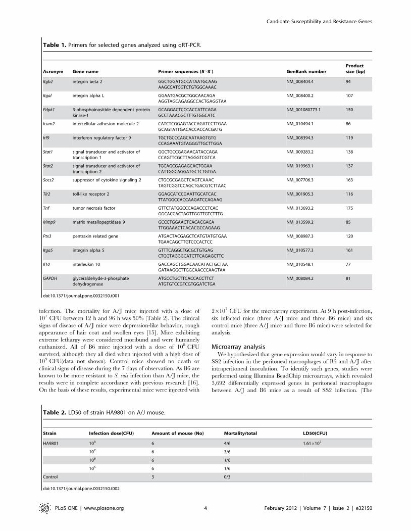
infection. The mortality for A/J mice injected with a dose of
107 CFU between 12 h and 96 h was 50% (Table 2). The clinical
signs of disease of A/J mice were depression-like behavior, rough
appearance of hair coat and swollen eyes [15]. Mice exhibiting
extreme lethargy were considered moribund and were humanely
euthanized. All of B6 mice injected with a dose of 108 CFU
survived, although they all died when injected with a high dose of
109 CFU(data not shown). Control mice showed no death or
clinical signs of disease during the 7 days of observation. As B6 are
known to be more resistant to S. suis infection than A/J mice, the
results were in complete accordance with previous research [16].
On the basis of these results, experimental mice were injected with
26107 CFU for the microarray experiment. At 9 h post-infection,
six infected mice (three A/J mice and three B6 mice) and six
control mice (three A/J mice and three B6 mice) were selected for
analysis.
Microarray analysisWe hypothesized that gene expression would vary in response to
SS2 infection in the peritoneal macrophages of B6 and A/J after
intraperitoneal inoculation. To identify such genes, studies were
performed using Illumina BeadChip microarrays, which revealed
3,692 differentially expressed genes in peritoneal macrophages
between A/J and B6 mice as a result of SS2 infection. (The
Table 1. Primers for selected genes analyzed using qRT-PCR.
Acronym Gene name Primer sequences (59-39) GenBank numberProductsize (bp)
Itgb2 integrin beta 2 GGCTGGATGCCATAATGCAAGAAGCCATCGTCTGTGGCAAAC
NM_008404.4 94
Itgal integrin alpha L GGAATGACGCTGGCAACAGAAGGTAGCAGAGGCCACTGAGGTAA
NM_008400.2 107
Pdpk1 3-phosphoinositide dependent proteinkinase-1
GCAGGACTCCCACCATTCAGAGCCTAAACGCTTTGTGGCATC
NM_001080773.1 150
Icam2 intercellular adhesion molecule 2 CATCTCGGAGTACCAGATCCTTGAAGCAGTATTGACACCACCACGATG
NM_010494.1 86
Irf9 interferon regulatory factor 9 TGCTGCCCAGCAATAAGTGTGCCAGAAATGTAGGGTTGCTTGGA
NM_008394.3 119
Stat1 signal transducer and activator oftranscription 1
GGCTGCCGAGAACATACCAGACCAGTTCGCTTAGGGTCGTCA
NM_009283.2 138
Stat2 signal transducer and activator oftranscription 2
TGCAGCGAGAGCACTGGAACATTGGCAGGATGCTCTGTGA
NM_019963.1 137
Socs2 suppressor of cytokine signaling 2 CTGCGCGAGCTCAGTCAAACTAGTCGGTCCAGCTGACGTCTTAAC
NM_007706.3 163
Tlr2 toll-like receptor 2 GGAGCATCCGAATTGCATCACTTATGGCCACCAAGATCCAGAAG
NM_001905.3 116
Tnf tumor necrosis factor GTTCTATGGCCCAGACCCTCACGGCACCACTAGTTGGTTGTCTTTG
NM_013693.2 175
Mmp9 matrix metallopeptidase 9 GCCCTGGAACTCACACGACATTGGAAACTCACACGCCAGAAG
NM_013599.2 85
Ptx3 pentraxin related gene ATGACTACGAGCTCATGTATGTGAATGAACAGCTTGTCCCACTCC
NM_008987.3 120
Itga5 integrin alpha 5 GTTTCAGGCTGCGCTGTGAGCTGGTAGGGCATCTTCAGAGCTTC
NM_010577.3 161
Il10 interleukin 10 GACCAGCTGGACAACATACTGCTAAGATAAGGCTTGGCAACCCAAGTAA
NM_010548.1 77
GAPDH glyceraldehyde-3-phosphatedehydrogenase
ATGCCTGCTTCACCACCTTCTATGTGTCCGTCGTGGATCTGA
NM_008084.2 81
doi:10.1371/journal.pone.0032150.t001
Table 2. LD50 of strain HA9801 on A/J mouse.
Strain Infection dose(CFU) Amount of mouse (No) Mortality/total LD50(CFU)
HA9801 108 6 4/6 1.616107
107 6 3/6
106 6 1/6
105 6 1/6
Control 3 0/3
doi:10.1371/journal.pone.0032150.t002
Candidate Susceptibility and Resistance Genes
PLoS ONE | www.plosone.org 4 February 2012 | Volume 7 | Issue 2 | e32150

Candidate Susceptibility and Resistance Genes
PLoS ONE | www.plosone.org 5 February 2012 | Volume 7 | Issue 2 | e32150

differentially expressed genes between control A/J and control B6
mice were used to exclude those genes which were thought to be
inherently different between A/J and B6 mice.) Between the SS2-
infected A/J and control A/J mice, 2646 genes were identified to
be differentially expressed, of which 1469 genes were upregulated
and 1177 genes downregulated. Between the SS2-infected B6 and
control B6 mice, 1449 genes were differentially expressed, of
which 778 genes were upregulated and 671 genes downregulated.
The differentially expressed genes of the four groups and the group
of 3,692 differentially expressed genes are summarized in Table
S1.
GO categorizationThe differentially expressed genes of A/J and B6 mice after
infection with strain HA9801 were classified into different
functional categories according to the Gene Ontology project for
biological processes. The main GO categories for significantly
upregulated genes between SS2-infected A/J and control A/J mice
were positive regulation of T-helper 1 type immune response,
regulation of interleukin-12 biosynthetic process, positive regula-
tion of B cell receptor signaling pathway, type I interferon
biosynthetic process, defense response to bacteria, immune
response, ion transport and inflammatory cell apoptosis. The
main GO categories for significantly downregulated genes
between SS2-infected A/J and control A/J mice included negative
regulation of interleukin-2 production, negative regulation of ab-T
cell proliferation, protein heterotetramerization and heparan
sulfate proteoglycan biosynthetic process (Fig. 2A).
The primary GO categories for significantly upregulated genes
between SS2-infected B6 and control B6 mice were antigen
processing and presentation of exogenous peptide antigen, positive
regulation of T-helper 1 type immune response, peptide antigen
stabilization, positive regulation of B cell receptor signaling
pathway, regulation of lymphocyte differentiation, positive regu-
lation of monocyte differentiation, antigen receptor-mediated
signaling pathway, positive regulation of interleukin-12 biosyn-
thetic process, type I interferon biosynthetic process, platelet
activation, positive regulation of phagocytosis, immune response,
defense response to bacterium and apoptosis. The primary GO
categories for significantly downregulated genes between SS2-
infected B6 and control B6 mice were pointed-end actin filament
capping(The specific gene involved in this GO was tmod3, which
was related to movement.), vitamin K biosynthetic process, GDP-
L-fucose biosynthetic process, negative regulation of collagen
binding, GDP-mannose metabolic process, negative regulation of
nucleotide metabolic process, positive regulation of glycolysis,
positive regulation of fatty acid biosynthetic process, negative
regulation of alpha-beta T cell proliferation and glutamine
metabolic process (Fig. 2B). The differentially expressed genes
from this study classified into significant GO categories are
summarized in Table S2.
Pathway analysisThe pathway analysis based on the KEGG database was
performed on the genes selected as described above. Significantly
upregulated genes between SS2-infected A/J and control A/J mice
were mainly involved in the toll-like receptor signaling pathway,
cytokine-cytokine receptor interaction, T cell receptor signaling
pathway, B cell receptor signaling pathway, natural killer cell
mediated cytotoxicity, antigen processing and presentation,
leukocyte transendothelial migration. Significantly downregulated
genes between SS2-infected A/J and control A/J mice were
involved in only one pathway, olfactory transduction (Fig. 3A).
The KEGG pathway analysis for significantly upregulated genes
between SS2-infected B6 and control B6 mice showed that the
genes were related to toll-like receptor signaling pathway,
leukocyte transendothelial migration, cytokine-cytokine receptor
interaction, B cell receptor signaling pathway, natural killer cell
mediated cytotoxicity and antigen processing and presentation.
The KEGG pathway analysis for significantly downregulated
genes between SS2-infected B6 and control B6 mice showed that
the genes were related to tryptophan and tyrosine metabolism,
phenylalanine, tyrosine and tryptophan biosynthesis, fructose and
mannose metabolism, fatty acid metabolism, aminoacyl-tRNA
biosynthesis and renin-angiotensin system (Fig. 3B). The differen-
tially expressed genes involved in significant pathways are
summarized in Table S3.
Gene network analysisThe differentially expressed genes involved in significant
pathways were analyzed for their interaction, and the networks
of genes involved in signal transduction during SS2 infection were
established utilizing the KEGG database. In the gene network
comprised of the differentially expressed genes involved in
significant pathways of A/J mice infected with SS2, genes with a
high of degree of connectivity, such as Socs2, Sta1, Stat2, were in
the core axis of the network. Genes were regulated by their
upstream genes when their outdegrees were zero (e.g., Ccnd2), or
they regulated expression of downstream genes when their
indegrees were zero (e.g., Cish). The key genes regulated by SS2
infection in the A/J mice were mainly involved in the Jak-STAT
signaling pathway and related to cell apoptosis (Fig. 4A, Table 3).
In the gene network composed of the differentially expressed
genes involved in significant pathways of B6 mice infected with
SS2, some of the genes with a high of degree of connectivity in the
core axis were Icam2, Itgal, Itgb2. Ptk2b with an outdegree of zero is
an example of a gene regulated by upstream genes, while Rxra with
an indegree of zero represents a gene which regulated expression
of other downstream genes.
On the whole, the gene network could be divided into five parts,
three of which were related to cell apoptosis in the left top, left
bottom and middle bottom of the gene network (Fig. 4B, Table 4).
Four genes (H2-T10, H2-Q6, Tapbp, Tap1) constituted a small
signal transduction network associated with immune responses
(center), and three genes (Plxnb2, Sema4a, Sema4d) composed a
small nervous system net (bottom right) (Fig. 4B, Table 4).
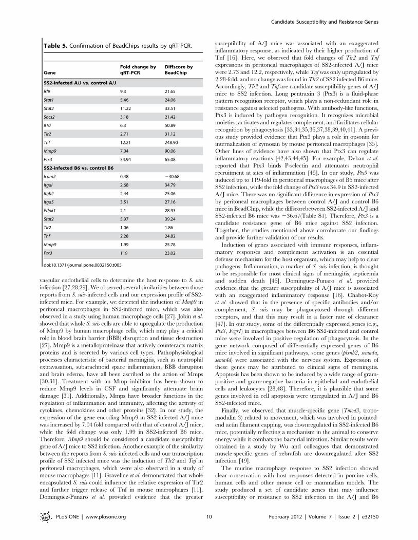
Confirmation of BeadChips results by qRT-PCRIn order to verify the data obtained by microarray analysis,
qRT-PCR was performed. We tested 9 genes differentially
expressed between SS2-infected A/J and control A/J mice, and
10 genes differentially expressed between SS2-infected B6 and
control B6 mice. As shown in Table 5, the qRT-PCR results
largely confirmed the data from the microarray. Notably, the
diffscore is the filtering criteria of Illumina for selection of
differentially expressed genes. There is no direct relationship with
fold change by qRT-PCR. But they have the similar tendency.
Figure 2. GO categories of biological processes for significantly differentially expressed genes. (A) between SS2-infected A/J and controlA/J mice and (B) between SS2-infected B6 and control B6 mice. P value,0.05 and FDR,0.05 were used as thresholds to select significant GOcategories.doi:10.1371/journal.pone.0032150.g002
Candidate Susceptibility and Resistance Genes
PLoS ONE | www.plosone.org 6 February 2012 | Volume 7 | Issue 2 | e32150

Figure 3. KEGG pathway analysis for significantly differentially expressed genes (A) between SS2-infected A/J and control A/J miceand (B) between SS2-infected B6 and control B6 mice. P value,0.05 and FDR,0.05 were used as thresholds to select significant KEGGpathways. LgP is the base 10 logarithm of the P value.doi:10.1371/journal.pone.0032150.g003
Candidate Susceptibility and Resistance Genes
PLoS ONE | www.plosone.org 7 February 2012 | Volume 7 | Issue 2 | e32150

Comparison of gene expressionThe expression level of toll-like receptor 2 (Tlr2) and tumor
necrosis factor (Tnf ) of A/J mice after infection with SS2 were
obviously upregulated. There were no changes in Tlr2 of B6 mice,
and the upregulated expression of Tnf of B6 mice was significant
lower than that of A/J mice after infection with SS2. The
Figure 4. Gene networks of differentially expressed genes involved in significant pathways. The gene networks comprised of thedifferentially expressed genes involved in significant pathways of (A) A/J mice infected with SS2 and (B) B6 mice infected with SS2 are shown. Legend:each circle represents a gene; red, upregulation; blue, downregulation; line segment, interaction of genes; arrow, activation (a), flat-ended arrow,inhibition (inh); straight, binding (b); dashed line, indirect effect (ind); P, phosphorylation; dp, dephosphorylation; ex, expression; u, ubiquitination.doi:10.1371/journal.pone.0032150.g004
Candidate Susceptibility and Resistance Genes
PLoS ONE | www.plosone.org 8 February 2012 | Volume 7 | Issue 2 | e32150

pentraxin 3 (Ptx3) genes of both A/J mice and B6 mice were
upregulated, but its expression level in B6 mice was obviously
higher than that of A/J mice. The expression of matrix
metalloproteinase 9 (Mmp9) in macrophages of B6 mice was lower
than that in A/J mice post-infection (Fig. 5).
Discussion
Gene expression profile analysis was used in this study to
identify the candidate genes of susceptibility or resistance to SS2
infection in mice models. While several studies have been
performed to evaluate host responses to SS2 infection, this was
the first time that the genetic basis of susceptibility to SS2 infection
has been studied at the whole transcriptome level.
To confirm host genetic differences in susceptibility to HA9801
infection, A/J and B6 mice were used to determine mortality and
clinical signs after infection. We determined that the LD50 of
HA9801 in A/J mice was 16107 CFU between 12 h and 96 h
(Table 2), and chose to use just twice the LD50 (26107 CFU) for
subsequent microarray analysis. The inoculated mice showed
expected clinical signs of disease such as depression-like behavior,
rough appearance of hair coat and swollen eyes [15]. B6 mice
injected with a dose of 108 CFU survived and were still active, while
a high dose of 109 CFU was required for 100% mortality. The
results confirmed that A/J mice were more susceptible to HA9801
infection than B6 mice, consistent with previous research [16].
Several studies have used human or mouse macrophages, porcine
choroid plexus epithelial cells (PCPEC), or porcine brain micro-
Table 4. Degree of key genes in gene network of SS2-infected B6 mice.
vertex degree indegree outdegree description
Icam2 10 5 5 intercellular adhesion molecule 2
Itgal 7 2 5 integrin alpha L
Itgb2 7 2 5 integrin beta 2
Tapbp 6 3 3 TAP binding protein
Crkl 5 4 1 v-crk sarcoma virus CT10 oncogene homolog (avian)-like
Itga5 5 3 2 integrin alpha 5 (fibronectin receptor alpha)
Pdpk1 5 4 1 3-phosphoinositide dependent protein kinase-1
Pik3cg 5 2 3 phosphoinositide-3-kinase, catalytic, gamma polypeptide
Ptk2b 4 4 0 PTK2 protein tyrosine kinase 2 beta
Plxnb2 2 2 0 plexin B2
Cdkn2b 2 2 0 cyclin-dependent kinase inhibitor 2B (p15, inhibits CDK4)
Rxra 4 0 4 retinoid X receptor alpha
Smad3 3 0 3 MAD homolog 3 (Drosophila)
doi:10.1371/journal.pone.0032150.t004
Table 3. Degree of key genes in gene network of SS2-infected A/J mice.
Vertex degree indegree outdegree description
Socs2 10 2 8 suppressor of cytokine signaling 2
Stat1 6 2 4 signal transducer and activator of transcription 1
Stat2 5 2 3 signal transducer and activator of transcription 2
Irf9 4 2 2 interferon regulatory factor 9
Cdkn2b 3 1 2 cyclin-dependent kinase inhibitor 2B (p15, inhibits CDK4)
Smad3 2 1 1 MAD homolog 3 (Drosophila)
Cish 2 0 2 Cytokine inducible SH2-containing protein
Igf1r 1 0 1 insulin-like growth factor I receptor
Tgfb2 1 0 1 transforming growth factor, beta 2
Ccnd2 3 3 0 cyclin D2
Il28ra 2 2 0 interleukin 28 receptor alpha
Il15ra 2 2 0 interleukin 15 receptor, alpha chain
Il11ra1 2 2 0 interleukin 11 receptor, alpha chain 1
Il10rb 2 2 0 interleukin 10 receptor, beta
Cxcl10 1 1 0 chemokine (C-X-C motif) ligand 10
Gna13 1 1 0 guanine nucleotide binding protein, alpha 13
5830411I20 1 1 0 Data not found
doi:10.1371/journal.pone.0032150.t003
Candidate Susceptibility and Resistance Genes
PLoS ONE | www.plosone.org 9 February 2012 | Volume 7 | Issue 2 | e32150
vascular endothelial cells to determine the host response to S. suis
infection [27,28,29]. We observed several similarities between those
reports from S. suis-infected cells and our expression profile of SS2-
infected mice. For example, we detected the induction of Mmp9 in
peritoneal macrophages in SS2-infected mice, which was also
observed in a study using human macrophage cells [27]. Jobin et al.
showed that whole S. suis cells are able to upregulate the production
of Mmp9 by human macrophage cells, which may play a critical
role in blood brain barrier (BBB) disruption and tissue destruction
[27]. Mmp9 is a metalloproteinase that actively counteracts matrix
proteins and is secreted by various cell types. Pathophysiological
processes characteristic of bacterial meningitis, such as neutrophil
extravasation, subarachnoid space inflammation, BBB disruption
and brain edema, have all been ascribed to the action of Mmps
[30,31]. Treatment with an Mmp inhibitor has been shown to
reduce Mmp9 levels in CSF and significantly attenuate brain
damage [31]. Additionally, Mmps have broader functions in the
regulation of inflammation and immunity, affecting the activity of
cytokines, chemokines and other proteins [32]. In our study, the
expression of the gene encoding Mmp9 in SS2-infected A/J mice
was increased by 7.04 fold compared with that of control A/J mice,
while the fold change was only 1.99 in SS2-infected B6 mice.
Therefore, Mmp9 should be considered a candidate susceptibility
gene of A/J mice to SS2 infection. Another example of the similarity
between the reports from S. suis-infected cells and our transcription
profile of SS2 infected mice was the induction of Tlr2 and Tnf in
peritoneal macrophages, which were also observed in a study of
mouse macrophages [11]. Graveline et al. demonstrated that whole
encapsulated S. suis could influence the relative expression of Tlr2
and further trigger release of Tnf in mouse macrophages [11].
Dominguez-Punaro et al. provided evidence that the greater
susceptibility of A/J mice was associated with an exaggerated
inflammatory response, as indicated by their higher production of
Tnf [16]. Here, we observed that fold changes of Tlr2 and Tnf
expressions in peritoneal macrophages of SS2-infected A/J mice
were 2.73 and 12.2, respectively, while Tnf was only upregulated by
2.28-fold, and no change was found in Tlr2 of SS2 infected B6 mice.
Accordingly, Tlr2 and Tnf are candidate susceptibility genes of A/J
mice to SS2 infection. Long pentraxin 3 (Ptx3) is a fluid-phase
pattern recognition receptor, which plays a non-redundant role in
resistance against selected pathogens. With antibody-like functions,
Ptx3 is induced by pathogen recognition. It recognizes microbial
moieties, activates and regulates complement, and facilitates cellular
recognition by phagocytosis [33,34,35,36,37,38,39,40,41]. A previ-
ous study provided evidence that Ptx3 plays a role in opsonin for
internalization of zymosan by mouse peritoneal macrophages [35].
Other lines of evidence have also shown that Ptx3 can regulate
inflammatory reactions [42,43,44,45]. For example, Deban et al.
reported that Ptx3 binds P-selectin and attenuates neutrophil
recruitment at sites of inflammation [45]. In our study, Ptx3 was
induced up to 119-fold in peritoneal macrophages of B6 mice after
SS2 infection, while the fold change of Ptx3 was 34.9 in SS2-infected
A/J mice. There was no significant difference in expression of Ptx3
by peritoneal macrophages between control A/J and control B6
mice in BeadChip, while the diffscorebetween SS2-infected A/J and
SS2-infected B6 mice was 236.67(Table S1). Therefore, Ptx3 is a
candidate resistance gene of B6 mice against SS2 infection.
Together, the studies mentioned above corroborate our findings
and provide further validation of our results.
Induction of genes associated with immune responses, inflam-
matory responses and complement activation is an essential
defense mechanism for the host organism, which may help to clear
pathogens. Inflammation, a marker of S. suis infection, is thought
to be responsible for most clinical signs of meningitis, septicemia
and sudden death [46]. Dominguez-Punaro et al. provided
evidence that the greater susceptibility of A/J mice is associated
with an exaggerated inflammatory response [16]. Chabot-Roy
et al. showed that in the presence of specific antibodies and/or
complement, S. suis may be phagocytosed through different
receptors, and that this may result in a faster rate of clearance
[47]. In our study, some of the differentially expressed genes (e.g.,
Ptx3, Fcgr1) in macrophages between B6 SS2-infected and control
mice were involved in positive regulation of phagocytosis. In the
gene network composed of differentially expressed genes of B6
mice involved in significant pathways, some genes (plxnb2, sema4a,
sema4d) were associated with the nervous system. Expression of
these genes may be attributed to clinical signs of meningitis.
Apoptosis has been shown to be induced by a wide range of gram-
positive and gram-negative bacteria in epithelial and endothelial
cells and leukocytes [28,48]. Therefore, it is plausible that some
genes involved in cell apoptosis were upregulated in A/J and B6
SS2-infected mice.
Finally, we observed that muscle-specific gene (Tmod3, tropo-
modulin 3) related to movement, which was involved in pointed-
end actin filament capping, was downregulated in SS2-infected B6
mice, potentially reflecting a mechanism in the animal to conserve
energy while it combats the bacterial infection. Similar results were
obtained in a study by Wu and colleagues that demonstrated
muscle-specific genes of zebrafish are downregulated after SS2
infection [49].
The murine macrophage response to SS2 infection showed
clear conservation with host responses detected in porcine cells,
human cells and other mouse cell or mammalian models. The
study produced a set of candidate genes that may influence
susceptibility or resistance to SS2 infection in the A/J and B6
Table 5. Confirmation of BeadChips results by qRT-PCR.
GeneFold change byqRT-PCR
Diffscore byBeadChip
SS2-infected A/J vs. control A/J
Irf9 9.3 21.65
Stat1 5.46 24.06
Stat2 11.22 33.51
Socs2 3.18 21.42
Il10 6.3 50.89
Tlr2 2.71 31.12
Tnf 12.21 248.90
Mmp9 7.04 90.06
Ptx3 34.94 65.08
SS2-infected B6 vs. control B6
Icam2 0.48 230.68
Itgal 2.68 34.79
Itgb2 2.44 25.06
Itga5 3.51 27.16
Pdpk1 2.1 28.93
Stat2 5.97 39.24
Tlr2 1.06 1.86
Tnf 2.28 24.82
Mmp9 1.99 25.78
Ptx3 119 23.02
doi:10.1371/journal.pone.0032150.t005
Candidate Susceptibility and Resistance Genes
PLoS ONE | www.plosone.org 10 February 2012 | Volume 7 | Issue 2 | e32150

mouse models. Among these, Mmp9, Tlr2 and Tnf were identified
as candidate susceptibility genes of A/J mice and Ptx3 as a
candidate resistance gene of B6 mice against SS2 infection. In
future work, we will continue searching for infection markers using
these models in order to provide leads for further investigation of
S. suis pathogenesis.
Supporting Information
Table S1
(XLS)
Table S2
(XLS)
Table S3
(XLS)
Acknowledgments
We thank HaoDan Zhu for useful suggestions and ShuJian Zhang, Di Gao
and WenChao Song for assistance.
Author Contributions
Conceived and designed the experiments: HY HF WZ JR. Performed the
experiments: JR XW. Analyzed the data: JR HY WZ. Contributed
reagents/materials/analysis tools: HY CL HF. Wrote the paper: JR.
References
1. Staats JJ, Feder I, Okwumabua O, Chengappa MM (1997) Streptococcus suis:
past and present. Vet Res Commun 21: 381–407.
2. Gottschalk M, Segura M (2000) The pathogenesis of the meningitis caused by
Streptococcus suis: the unresolved questions. Vet Microbiol 76: 259–272.
3. Hill JE, Gottschalk M, Brousseau R, Harel J, Hemmingsen SM, et al. (2005)
Biochemical analysis, cpn60 and 16S rDNA sequence data indicate that
Streptococcus suis serotypes 32 and 34, isolated from pigs, are Streptococcusorisratti. Vet Microbiol 107: 63–69.
4. Lun ZR, Wang QP, Chen XG, Li AX, Zhu XQ (2007) Streptococcus suis: anemerging zoonotic pathogen. Lancet Infect Dis 7: 201–209.
5. Fittipaldi N, Gottschalk M, Vanier G, Daigle F, Harel J (2007) Use of selectivecapture of transcribed sequences to identify genes preferentially expressed by
Streptococcus suis upon interaction with porcine brain microvascular endothe-
lial cells. Appl Environ Microbiol 73: 4359–4364.
6. Mai NT, Hoa NT, Nga TV, Linh le D, Chau TT, et al. (2008) Streptococcus
suis meningitis in adults in Vietnam. Clin Infect Dis 46: 659–667.
7. Yu H, Jing H, Chen Z, Zheng H, Zhu X, et al. (2006) Human Streptococcus suis
outbreak, Sichuan, China. Emerg Infect Dis 12: 914–920.
8. Al-Numani D, Segura M, Dore M, Gottschalk M (2003) Up-regulation of
ICAM-1, CD11a/CD18 and CD11c/CD18 on human THP-1 monocytes
stimulated by Streptococcus suis serotype 2. Clin Exp Immunol 133: 67–77.
9. Segura M, Stankova J, Gottschalk M (1999) Heat-killed Streptococcus suis
capsular type 2 strains stimulate tumor necrosis factor alpha and interleukin-6production by murine macrophages. Infect Immun 67: 4646–4654.
10. Segura M, Vadeboncoeur N, Gottschalk M (2002) CD14-dependent and-independent cytokine and chemokine production by human THP-1 monocytes
stimulated by Streptococcus suis capsular type 2. Clin Exp Immunol 127:
243–254.
11. Graveline R, Segura M, Radzioch D, Gottschalk M (2007) TLR2-dependent
recognition of Streptococcus suis is modulated by the presence of capsularpolysaccharide which modifies macrophage responsiveness. Int Immunol 19:
375–389.
12. Williams AE, Blakemore WF, Alexander TJ (1988) A murine model of
Streptococcus suis type 2 meningitis in the pig. Res Vet Sci 45: 394–399.
13. Beaudoin M, Higgins R, Harel J, Gottschalk M (1992) Studies on a murine
model for evaluation of virulence of Streptococcus suis capsular type 2 isolates.FEMS Microbiol Lett 78: 111–116.
14. Kataoka Y, Haritani M, Mori M, Kishima M, Sugimoto C, et al. (1991)
Experimental infections of mice and pigs with Streptococcus suis type 2. J VetMed Sci 53: 1043–1049.
15. Dominguez-Punaro MC, Segura M, Plante MM, Lacouture S, Rivest S, et al.
(2007) Streptococcus suis serotype 2, an important swine and human pathogen,induces strong systemic and cerebral inflammatory responses in a mouse model
of infection. J Immunol 179: 1842–1854.
16. Dominguez-Punaro Mde L, Segura M, Radzioch D, Rivest S, Gottschalk M(2008) Comparison of the susceptibilities of C57BL/6 and A/J mouse strains to
Streptococcus suis serotype 2 infection. Infect Immun 76: 3901–3910.
17. Wu Z, Zhang W, Lu C (2008) Comparative proteome analysis of secretedproteins of Streptococcus suis serotype 9 isolates from diseased and healthy pigs.
Microb Pathog 45: 159–166.
18. Wang K, Lu C (2007) Adhesion activity of glyceraldehyde-3-phosphate
dehydrogenase in a Chinese Streptococcus suis type 2 strain. Berl MunchTierarztl Wochenschr 120: 207–209.
19. Zhang W, Lu CP (2007) Immunoproteomics of extracellular proteins of Chinese
virulent strains of Streptococcus suis type 2. Proteomics 7: 4468–4476.
Figure 5. Comparative analysis of gene expression in peritoneal macrophages. Expression levels of Tlr2, Tnf, Ptx3 and Mmp9 in A/J and B6mice were measured by qRT-PCR and normalized to the housekeeping gene GAPDH. Differences between A/J and B6 mice were statisticallysignificant with a P value of ,0.05 as determined by one-way ANOVA, except with the Tlr2 gene.doi:10.1371/journal.pone.0032150.g005
Candidate Susceptibility and Resistance Genes
PLoS ONE | www.plosone.org 11 February 2012 | Volume 7 | Issue 2 | e32150

20. Zhang W, Lu CP (2007) Immunoproteomic assay of membrane-associated
proteins of Streptococcus suis type 2 China vaccine strain HA9801. Zoonoses
Public Health 54: 253–259.
21. de Jonge WJ, van der Zanden EP, The FO, Bijlsma MF, van Westerloo DJ, et al.
(2005) Stimulation of the vagus nerve attenuates macrophage activation by
activating the Jak2-STAT3 signaling pathway. Nat Immunol 6: 844–851.
22. Lemay AM, Haston CK (2005) Bleomycin-induced pulmonary fibrosis
susceptibility genes in AcB/BcA recombinant congenic mice. Physiol Genomics
23: 54–61.
23. Chemnitz JM, Driesen J, Classen S, Riley JL, Debey S, et al. (2006)
Prostaglandin E2 impairs CD4+ T cell activation by inhibition of lck:
implications in Hodgkin’s lymphoma. Cancer Res 66: 1114–1122.
24. Ashburner M, Ball CA, Blake JA, Botstein D, Butler H, et al. (2000) Gene
ontology: tool for the unification of biology. The Gene Ontology Consortium.
Nat Genet 25: 25–29.
25. Ogata H, Goto S, Sato K, Fujibuchi W, Bono H, et al. (1999) KEGG: Kyoto
Encyclopedia of Genes and Genomes. Nucleic Acids Res 27: 29–34.
26. Livak KJST (2001) Analysis of relative gene expression data using realtime
quantitative PCR and the 2(-Delta Delta C(T)) method. Methods 25: 402–408.
27. Jobin MC, Gottschalk M, Grenier D (2006) Upregulation of prostaglandin E2
and matrix metalloproteinase 9 production by human macrophage-like cells:
synergistic effect of capsular material and cell wall from Streptococcus suis.
Microb Pathog 40: 29–34.
28. Tenenbaum T, Essmann F, Adam R, Seibt A, Janicke RU, et al. (2006) Cell
death, caspase activation, and HMGB1 release of porcine choroid plexus
epithelial cells during Streptococcus suis infection in vitro. Brain Res 1100: 1–12.
29. Vanier G, Fittipaldi N, Slater JD, de la Cruz Dominguez-Punaro M, Rycroft AN,
et al. (2009) New putative virulence factors of Streptococcus suis involved in
invasion of porcine brain microvascular endothelial cells. Microb Pathog 46:
13–20.
30. Kolb SA, Lahrtz F, Paul R, Leppert D, Nadal D, et al. (1998) Matrix
metalloproteinases and tissue inhibitors of metalloproteinases in viral meningitis:
upregulation of MMP-9 and TIMP-1 in cerebrospinal fluid. J Neuroimmunol
84: 143–150.
31. Leib SL, Leppert D, Clements J, Tauber MG (2000) Matrix metalloproteinases
contribute to brain damage in experimental pneumococcal meningitis. Infect
Immun 68: 615–620.
32. Parks WC WC, Lopez-Boado YS (2004) Matrix metalloproteinases as
modulators of inflammation and innate immunity. Nat Rev Immunol 4:
617–629.
33. Garlanda C, Bottazzi B, Bastone A, Mantovani A (2005) Pentraxins at the
crossroads between innate immunity, inflammation, matrix deposition, and
female fertility. Annu Rev Immunol 23: 337–366.
34. Jeannin P, Bottazzi B, Sironi M, Doni A, Rusnati M, et al. (2005) Complexity
and complementarity of outer membrane protein A recognition by cellular andhumoral innate immunity receptors. Immunity 22: 551–560.
35. Diniz SN, et al. (2004) PTX3 function as an opsonin for the dectin-1-dependent
internalization of zymosan by macrophages. J Leukoc Biol 75: 649–656.36. Garlanda C, Hirsch E, Bozza S, Salustri A, De Acetis M, et al. (2002) Non-
redundant role of the long pentraxin PTX3 in anti-fungal innate immuneresponse. Nature 420: 182–186.
37. Soares AC, Souza DG, Pinho V, Vieira AT, Nicoli JR, et al. (2006) Dual
function of the long pentraxin PTX3 in resistance against pulmonary infectionwith Klebsiella pneumoniae in transgenic mice. Microbes Infect 8: 1321–1329.
38. Deban L, Jarva H, Lehtinen MJ, Bottazzi B, Bastone A, et al. (2008) Binding ofthe long pentraxin PTX3 to factor H: interacting domains and function in the
regulation of complement activation. J Immunol 181: 8433–8440.39. Nauta AJ, et al. (2003) Biochemical and functional characterization of the
interaction between pentraxin 3 and C1q. Eur J Immunol 33: 465–473.
40. Cotena A, Maina V, Sironi M, Bottazzi B, Jeannin P, et al. (2007) Complementdependent amplification of the innate response to a cognate microbial ligand by
the long pentraxin PTX3. J Immunol 179: 6311–6317.41. Garlanda C, BB, Bastone A, Mantovani A (2005) Pentraxins at the crossroads
between innate immunity, inflammation, matrix deposition, and female fertility.
Annu Rev Immunol 23: 337–366.42. Salio M, Chimenti S, De Angelis N, Molla F, Maina V, et al. (2008)
Cardioprotective function of the long pentraxin PTX3 in acute myocardialinfarction. Circulation 117: 1055–1064.
43. Ravizza T, Moneta D, Bottazzi B, Peri G, Garlanda C, et al. (2001) Dynamicinduction of the long pentraxin PTX3 in the CNS after limbic seizures: evidence
for a protective role in seizure-induced neurodegeneration. Neuroscience 105:
43–53.44. Dias AA, et al. (2001) TSG-14 transgenic mice have improved survival to
endotoxemia and to CLP-induced sepsis. J Leukoc Biol 69: 928–936.45. Deban L, Russo RC, Sironi M, Moalli F, Scanziani M, et al. (2010) Regulation
of leukocyte recruitment by the long pentraxin PTX3. Nat Immunol 11:
328–334.46. Segura M, Vanier G, Al-Numani D, Lacouture S, Olivier M, et al. (2006)
Proinflammatory cytokine and chemokine modulation by Streptococcus suis in awhole-blood culture system. FEMS Immunol Med Microbiol 47: 92–106.
47. Chabot-Roy G, Willson P, Segura M, Lacouture S, Gottschalk M (2006)Phagocytosis and killing of Streptococcus suis by porcine neutrophils. Microb
Pathog 41: 21–32.
48. Fink SL, Cookson BT (2005) Apoptosis, pyroptosis, and necrosis: mechanisticdescription of dead and dying eukaryotic cells. Infect Immun 73: 1907–1916.
49. Wu Z, Zhang W, Lu Y, Lu C (2010) Transcriptome profiling of zebrafishinfected with Streptococcus suis. Microb Pathog 48: 178–187.
Candidate Susceptibility and Resistance Genes
PLoS ONE | www.plosone.org 12 February 2012 | Volume 7 | Issue 2 | e32150