identification and cloning of afur homolog from neisseria...
TRANSCRIPT

INFECrION AND IMMUNITY, Nov. 1993, p. 4599-4606 Vol. 61, No. 110019-9567/93/114599-08$02.00/0Copyright X 1993, American Society for Microbiology
Identification and Cloning of a fur Homolog fromNeisseria gonorrhoeae
SALLY A. BERISH, SHAMBHAVI SUBBARAO, CHENG-YEN CHEN,DAVID L. TREES, AND STEPHEN A. MORSE*
Division of Sexually Transmitted Diseases Laboratory Research, National CenterforInfectious Diseases, Centers for Disease Control and Prevention,
Atlanta, Georgia 30333
Received 7 May 1993/Returned for modification 28 June 1993/Accepted 2 August 1993
The promoter region of the major iron-regulated protein ofNeisseria gonorrhoeae, Fbp, has two regions thatexhibit homology with the Escherichia coli consensus Fur-binding sequences. Gel retardation assays suggestedthat purified E. coli Fur bound to two sites within the Fbp promoter. The presence of a gonococcal Fur homologwas suggested by Southern hybridization under conditions of low stringency, which revealed a DNA locus thatexhibited homology to the E. colifur gene. Oligonucleotides derived from the conserved regions offur genes ofextremely diverse bacteria were used to amplify a 140-bp fragment of a putative gonococcal fur gene. Thisfragment was used to identify clones containing the entire gonococcalfiur gene. After sequencing the gonococcalfur gene and its promoter region, we found that gonococcal Fur exhibited 50%o identity with E. coli Fur at theamino acid level; however, it complemented two E. coli Fur- mutants. The presence of a Fur homolog in N.gonorrhoeae suggests that Fur-regulated genes are widely distributed among extremely diverse bacteria.
Gonorrhea is a sexually transmitted disease of worldwideimportance caused by the obligate human pathogen Neisse-ria gonorrhoeae. N. gonorrhoeae generally causes symp-tomatic or asymptomatic infections of mucosal surfaceslined by columnar epithelial cells (35). However, dissemi-nated infections may occur, either by extension to adjacentorgans (e.g., pelvic inflammatory disease) or by bacteremicspread (e.g., skin lesions and tenosynovitis) (35). Thus,gonococci infect sites that may differ widely with respect toenvironmental and nutritional factors.
Iron is essential for the growth and multiplication of nearlyall microorganisms (5), and the acquisition of this nutrient bymicrobial pathogens is critical to their ability to causedisease. N. gonorrhoeae, like many other pathogenic micro-organisms, is able to utilize host iron sources such astransferrin (TF), lactoferrin (LF), hemoglobin, and heme forin vitro growth (27, 28, 49). However, unlike many otherpathogenic microorganisms, N. gonorrhoeae does not syn-thesize siderophores to compete with host iron-bindingproteins such as TF and LF but instead utilizes a receptor-mediated mechanism that requires direct contact betweenTF and LF and specific cell surface receptors (26, 39).
In humans and other vertebrates, growth-essential iron iswithheld from invading microorganisms (46). In serum andsecretions, free extracellular iron is bound to TF and LF,respectively. The amount of free iron that remains in equi-librium with these proteins is ca. 10-18 M, which is far toolow to support bacterial growth (5). Like other microorgan-isms, N. gonorrhoeae synthesizes a unique set of proteinswhen confronted with such an iron-restricted environment;these proteins are not present in appreciable quantities underconditions of iron excess. Some of these iron-regulatedproteins are thought to function in the acquisition of iron bythese microorganisms (8, 34). In Escherichia coli and Sal-monella typhimurium, the expression of iron-regulated genesis controlled by the intracellular ferric uptake regulator
* Corresponding author.
protein (Fur) (2). Genetically diverse microorganisms appearto utilize a similar mechanism to control the expression ofiron-regulated genes. Molecular homologs of Fur have beenshown to exist in Yersinia pestis (42), Pseudomonas aerug-inosa (37), Vibrio cholerae (22), and Vibrio vulnificus (23).
Indirect evidence suggests that a similar Fur homologexists in N. gonorrhoeae. The 37,000-Da major iron-regu-lated protein (Fbp) is expressed by all strains of N. gonor-rhoeae when grown under iron-restricted conditions (29, 30).Little if any of this protein is present when gonococci aregrown in iron-replete medium. Fbp mRNA is not detectablein gonococci grown in the presence of excess iron (33),suggesting that Fbp synthesis is regulated at the level oftranscription. The 5' flanking sequence of the Fbp genecontains two regions which share considerable homologywith the E. coli consensus Fur-binding iron boxes (3, 33).One of the potential iron boxes overlaps the Fbp promoter.From these data, we postulated the existence of a gonococ-cal Fur homolog. This report describes the identification,cloning, and sequence of the gonococcal Fur homolog. Ourresults show that E. coli Fur recognizes putative gonococcalFur-binding sequences and that cloned gonococcal Fur com-plements two E. coli Fur- mutants. Additional data on theregulation of Fur in N. gonorrhoeae are presented.
MATERIALS AND METHODS
Bacterial strains, plasmids, and medium. N. gonorrhoeaeF62 was obtained from R. P. Williams (Baylor College ofMedicine, Houston, Tex.). E. coli 71.18 and TB1 wereprovided by K. Birkness (Centers for Disease Control andPrevention, Atlanta, Ga.). E. coli XL1 Blue was purchasedfrom Stratagene, La Jolla, Calif., and E. coli H1681 waskindly provided by K. Hantke (Universitat Tubingen, Tub-ingen, Germany) (19). E. coli W3110 fur' (lacU169 Tna2)and W3110 fur (lacU169 Tna2 fir::TnS) (13) were providedby J. B. Neilands (University of California, Berkeley).pUC9, pUC19, and pBR322 were purchased from BethesdaResearch Laboratories, Gaithersburg, Md. Plasmid pLAFR3
4599
on May 25, 2018 by guest
http://iai.asm.org/
Dow
nloaded from

4600 BERISH ET AL.
TABLE 1. Primer sequences of oligonucleotides used in this study
Primerdriesignatn Sequence Used for:designation
Si TGTTGCTGAATTTTTCCA Primer extension analysisMB1 ATGGAAAAATTCAGCAACATTGCG Amplification of DNA for nick translationF76 CAACTCATAAACCGCCTTGCC Amplification of DNA for nick translation and SSP-PCRF43 CTGCTGGAAGAGGGCGTGGAA SSP-PCRS100 AGGTAGCACTGCTTGAAT Amplification of DNA to generate plasmid clone pSBFW13FEN TTTAACGTTTGCCCTTGGCCTG Same as aboveF90 CGGCCGTTTGCGCCGGCGATC Sequence flanking N-terminal HinclI siteF91 GACCGCAACCCGATAGCTGC Sequence flanking C-terminal HincIl siteFUR1 ATGACTGATAACAATACCGCC Amplification of E. coli fur for nick translationFUR2 GGTTGGCTTTTCTCGTTCAGG Same as aboveFHA1 AATAACATCCCATCTAAGATATT Amplification of E. coli fhuA promoter for gel shift assayFHA2 GAGCAGTTTTGGAACGCGCCAT Same as aboveFPRO1 CCGCCGCTTCCGCCTTACGGG Amplification ofJbp promoter for gel shift assayFPRO2 TCGGATAGATGTTTTCATATC Same as aboveFS1 GACATTACCGTGTACAACGGC Amplification of bases 1-198 in fbp structural geneFS2 CGGAAAGGGTGGCGAGTGCCG Same as above
was provided by Christine Litwin (Harvard Medical School,Boston, Mass.) (22).N. gonorrhoeae was grown as described previously (30).
E. coli was grown overnight on Luria broth (LB) mediumwith antibiotics (ampicillin [50 p,g/ml], tetracycline [12.5,ug/ml], kanamycin [50 p,g/ml], and streptomycin [20 p,g/ml])as indicated. 5-Bromo-4-chloro-3-indolyl-o-D-galactopyra-noside (X-Gal) was added to the LB agar at a concentrationof 40 p,g/ml.
Gel retardation assay. The binding of purified Fur protein(obtained from J. B. Neilands) to DNA was assessed by themethod of de Lorenzo et al. (14). A 200-bp fragment of thefhuA4 promoter (9) and a 200-bp fragment of thefbp structuralgene (3) were amplified by polymerase chain reaction (PCR),using primers (Table 1) derived from the published se-quences, for use as positive and negative controls, respec-tively. A similar-size fragment containing the fbp promoterwas also amplified by PCR (Table 1). The amplicons were 5'end labeled with 32p, using standard techniques (24). Thegels were electrophoresed at a constant voltage of 180 V for3 h, dried, and exposed to XAR5 film (Eastman Kodak Co.,Rochester, N.Y.).
In vitro DNA manipulations. Genomic DNA was purifiedfrom N. gonorrhoeae F62 as described by Marmur (25) anddigested with various restriction endonucleases as instructedby the manufacturer. The resulting fragments were sepa-rated by agarose gel electrophoresis, transferred accordingto the method of Southern (41), and hybridized to 32P-labeledDNA. Afuir gene probe was prepared by PCR amplificationof the E. coli fur gene by using primers (Table 1) derivedfrom the published sequence (38). A N. gonorrhoeae furgene probe was prepared by PCR amplification of gonococ-cal DNA by using primers FSB2 and FSB5 as describedbelow. All probes were 32p labeled by nick translation.Low-stringency hybridization of E. coli DNA to gonococcalDNA was performed at 55°C in 1 M NaCl-50 mM sodiumphosphate-5 mM EDTA-0.5% sodium dodecyl sulfate(SDS)-lOx Denhardt's solution. High-stringency conditionswere used for the hybridization of the gonococcal fur probeto N. gonorrhoeae DNA {50% formamide, 5 x Denhardt'ssolution, 0.1% SDS, 5x SSPE (lx SSPE is 0.18 M NaCl, 10mM NaPO4, and 1 mM EDTA [pH 7.7])} at 420C.
Cloning strategy. PCR primers for preparation of a gono-coccal fur gene probe were synthesized by using inosine andselected bases derived from gonococcal codon usage (47).
The primer sequences were based on two regions of aminoacids that are conserved among the fir genes of E. coli (38),Y pestis (43), and P. aeruginosa (37) (see Fig. 1 and 7).These regions are located between amino acids 10 to 19 and51 through 57 of E. coli. PCR amplification of gonococcalgenomic DNA by using primers FSB2 and FSB5 (Fig. 1)derived from these common regions resulted in a product ofapproximately 140 bp. PCR conditions consisted of 25 cyclesof denaturation at 94°C for 1.5 min, annealing at 60°C for 2min, and extension at 72°C for 4 min followed by 8 cycles ofdenaturation at 94°C for 1 min, annealing at 50°C for 1 min,and extension at 72°C for 2 min. The amplified fragment wasnick translated and used to screen a HincII minilibrary ofgonococcal DNA from N. gonorrhoeae F62.To construct a HinclI minilibrary, genomic DNA was
digested with HincII and then size fractionated (1,200 to1,500 bp). EcoRI linkers were added, and the DNA wasrestricted with EcoRI and ligated into pUC19. The ligationmixture was transformed into E. coli XL1 Blue. Two hun-dred colonies were screened by the method of Grunstein andHogness (17) under conditions of high stringency with thenick-translated 140-bp gonococcal fur probe. Several posi-tive clones were identified and found to contain a 950-bpinsert. The insert was subsequently cloned into M13 andsequenced. Single-specific-primer PCR (SSP-PCR) (40) wasused to obtain sequences internal to the 3' HinclI site.Briefly, genomic DNA was cut with HincII and ligated into
"1 noacids10-19 K A GILIKIVITILIPIR
oligo FSB2
aminoaci ds51-57 G L AlT VIYIR
3'-CCI GAC CGI TGI CAI ATG GC-5'
oligo FSB5FIG. 1. Two regions of amino acids that are conserved among E.
coli (38), Y. pestis (42), V. cholerae (22), and P. aeruginosa (37) Furproteins. Oligonucleotide sequences chosen for PCR are listedbelow the amino acids.
INFECT. IMMUN.
on May 25, 2018 by guest
http://iai.asm.org/
Dow
nloaded from
GONOCOCCAL fir HOMOLOG 4601
the SmaI site of pUC9. The ligation mixture was used toprime a PCR using the F43 (Table 1) primer and the M13reverse primer. The resulting single band was gel purifiedand directly sequenced from both ends (48).
Primer F91 (Table 1) was derived from the gonococcalDNA sequence obtained from sequencing with the reverseprimer. PCR was repeated with primers F43 (Table 1) andF91. This PCR product was also purified and sequenced.Primer extension analysis. Total RNA was isolated (1) from
N. gonorrhoeae F62 grown at 35°C in GC broth (30) underconditions of either iron limitation (25 ,uM Desferal) or ironexcess (50 ,uM ferric nitrate). An 18-bp oligonucleotidedesignated Si (Table 1), complementary to the 5' end of thenoncoding strand of the fir structural gene, was 5' endlabeled with [,y-32PJATP by using T4 polynucleotide kinase(24). Annealing of the labeled primer and reactions usingreverse transcriptase were carried out as described by Moran(32). The samples were run on a 6% sequencing gel along witha sequencing ladder generated by the same 18-bp primer,using a gonococcalfur gene plasmid clone (pSBFW13) as thetemplate.
Construction of plasmids. Primers S100 and FEN (Table 1)were used to amplify the fur promoter and entire structuralgene. The resulting 629-bp fragment was ligated into theSmaI site of pUC9. Clones containing the insert in E. coliTB1 were obtained in both orientations. Clone pSBFW13and pUC9 (no insert) were transformed into E. coli H1681and H1780, and transformants were selected on LB mediumcontaining ampicillin and tetracycline or ampicillin and ka-namycin, respectively. Clone pSBFW13 was digested withBsmI, which cuts between bp 127 and 128 and bp 201 and 202in the coding region of fur, and the fl fragment was inserted(36). After transformation into E. coli H1681 and H1780,streptomycin-resistant transformants were obtained on LBmedium containing ampicillin, tetracycline, and streptomy-cin or ampicillin, kanamycin, and streptomycin, respec-tively. Plasmid minipreps were prepared by the method ofBirnboim and Doly (4) to confirm the additional 2 kb ofDNAin clone pSBFW13QI.Western blot (immunoblot) analysis. Whole-cell lysates of
E. coli and N. gonorrhoeae were fractionated by SDS-polyacrylamide gel electrophoresis (12.5% acrylamide), us-ing the buffer system of Laemmli (21). Proteins were trans-ferred onto a nitrocellulose membrane (Schleicher andSchuell, Keene, N.H.), using a semidry electroblotter (Inte-grated Separation System). The nitrocellulose membranewas incubated in 1% bovine serum albumin containing 0.5%Tween 20 at room temperature for 1 h.ICR (Institute of Cancer Research) mice received four
immunizations, intradermally, with 100 ,ug of E. coli Furprotein mixed with incomplete Freund's adjuvant at inter-vals of 1 to 2 weeks. The mice were bled after 4 weeks, andthe serum was aliquoted and stored at -70°C until used.Mouse polyclonal anti-Fur antiserum was added at a dilutionof 1:200, and the membrane was incubated overnight at roomtemperature on a rocker. After washing, the nitrocellulosemembrane was incubated with horseradish peroxidase-con-jugated goat anti-mouse immunoglobulin (Bio-Rad) at roomtemperature for 1 h and developed by using hydrogenperoxide and 4-chloro-1-naphthol (Bio-Rad).
13-Galactosidase activity measurements. E. coli H1780 andH1681 were grown in LB to early log phase (optical densityat 600 nm = 0.1 to 0.4). I-Galactosidase expression wasquantitated by using 0.5-ml aliquots of culture treated withchloroform and SDS as described by Miller (31).
Nucleotide sequence accession number. The nucleotide and
B
A
'..
-^.A -
c
...n. so is no me:? low
Fur(nm)
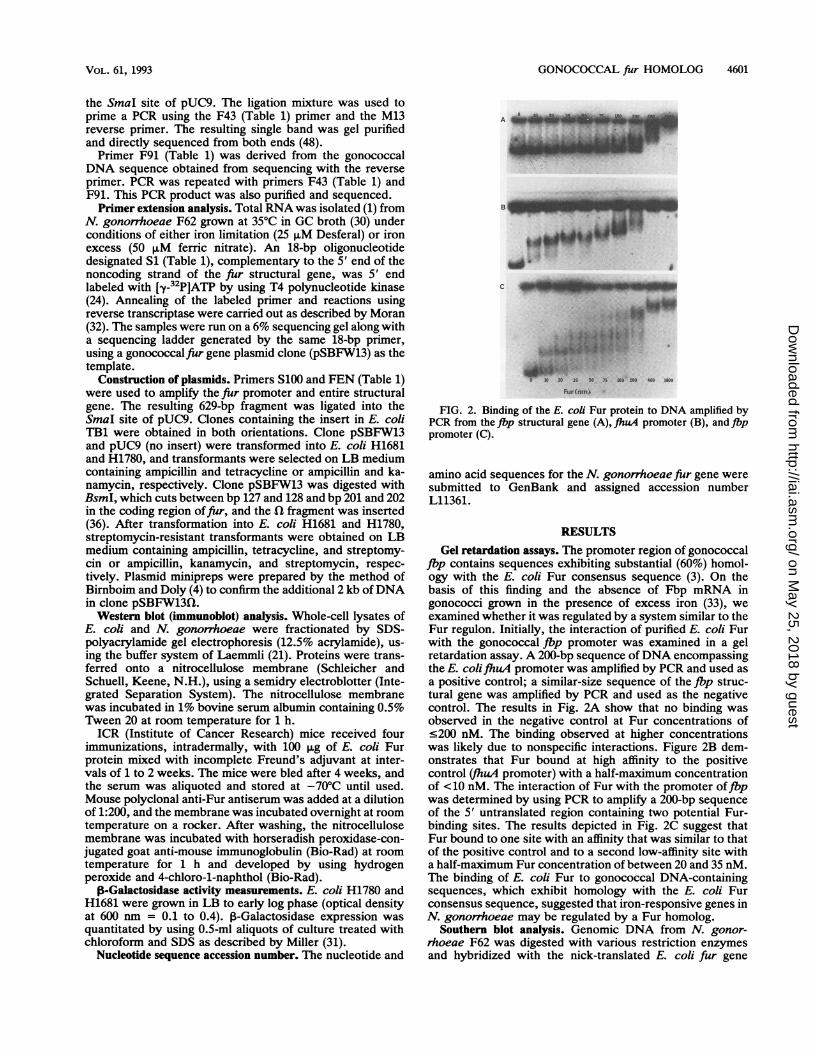
FIG. 2. Binding of the E. coli Fur protein to DNA amplified byPCR from theJbp structural gene (A), liuA promoter (B), and fbppromoter (C).
amino acid sequences for the N. gonorrhoeae fur gene weresubmitted to GenBank and assigned accession numberL11361.
RESULTSGel retardation assays. The promoter region of gonococcal
flp contains sequences exhibiting substantial (60%) homol-ogy with the E. coli Fur consensus sequence (3). On thebasis of this finding and the absence of Fbp mRNA ingonococci grown in the presence of excess iron (33), weexamined whether it was regulated by a system similar to theFur regulon. Initially, the interaction of purified E. coli Furwith the gonococcal fbp promoter was examined in a gelretardation assay. A 200-bp sequence of DNA encompassingthe E. colifhuA promoter was amplified by PCR and used asa positive control; a similar-size sequence of the fbp struc-tural gene was amplified by PCR and used as the negativecontrol. The results in Fig. 2A show that no binding wasobserved in the negative control at Fur concentrations of.200 nM. The binding observed at higher concentrationswas likely due to nonspecific interactions. Figure 2B dem-onstrates that Fur bound at high affinity to the positivecontrol (fhuA promoter) with a half-maximum concentrationof <10 nM. The interaction of Fur with the promoter offbpwas determined by using PCR to amplify a 200-bp sequenceof the 5' untranslated region containing two potential Fur-binding sites. The results depicted in Fig. 2C suggest thatFur bound to one site with an affinity that was similar to thatof the positive control and to a second low-affinity site witha half-maximum Fur concentration of between 20 and 35 nM.The binding of E. coli Fur to gonococcal DNA-containingsequences, which exhibit homology with the E. coli Furconsensus sequence, suggested that iron-responsive genes inN. gonorrhoeae may be regulated by a Fur homolog.
Southern blot analysis. Genomic DNA from N. gonor-rhoeae F62 was digested with various restriction enzymesand hybridized with the nick-translated E. coli fur gene
VOL. 61, 1993
on May 25, 2018 by guest
http://iai.asm.org/
Dow
nloaded from

4602 BERISH ET AL.
a A B C D E F G H J K
bp
6108- A;lw::::' '.
3054-. .:
2036 -1636-
1018 -
517-
b A B C D E F G H J K
bp.. . ... .._
6108-
3054-
2036
1636
1.:.
1 01 8-
517 -
FIG. 3. (a) Southern hybridization analysis of nick-translated E.coli fur under low-stringency conditions with DNA from N. gonor-rhoeae F62. Genomic DNA digests were as follows: lane A, HincII;lane B, SmaI; lane C, Sau3A; lane D, AccI; lane E, AluI; lane F,RsaI; lane G, DdeI; lane H, PvuII; lane I, HindIII; lane J, StuI; laneK, EcoRI. Lane L contains 300 ng of the E. coli fur DNA. (b)Southern hybridization analysis of the 140-bp nick-translated gono-coccal fiur probe under high-stringency conditions with DNA fromN. gonorrhoeae F62. The DNA probe was generated by PCRamplification with primers FSB2 and FSB5 (Fig. 1). Genomic DNAdigests were the same as described for Fig. 3A.
under both high- and low-stringency conditions. The probeconsisted of the entire E. coli fur structural gene and 27bases of 3' flanking DNA. No hybridization was observedunder high-stringency conditions (data not shown); how-ever, moderate hybridization of the probe to several bandswas observed under low-stringency conditions (Fig. 3a).To assist in identifying a restriction fragment containing
the gonococcal fur homolog, we used PCR to amplify a
sequence of gonococcal DNA, using primers derived fromtwo conserved regions of Fur located between amino acids10 to 19 and 51 to 57 (Fig. 1). This 140-bp DNA fragment waslabeled and used to probe Southern transfers of N. gonor-rhoeae F62 chromosomal DNA digested with various re-
LHc I Ssp I Ssp I EcoR I S3A HcIIi; * o *+A# 10 141111110111111111ll11,llElllllllllll1l
-- -
100 bp
FIG. 4. Physical map of the gonococcal fiur gene sequence lo-cated on a 1.4-kb HincII (HcII) fragment. The coding region of the,fur gene is boxed. Primers used for sequencing, SSP-PCR, andconstruction of pSBFW13 are labeled (0, MB1; Cl, F76; *, F43; A,S100; *, FEN; 0, F90; *, F91). Arrows indicate the regions thatwere sequenced. S3A, Sau3A.
striction enzymes under conditions of high stringency. Theresults (Fig. 3b) showed that the probe hybridized stronglyto a single HincII band about 1.4 kb in size (lane A). Asimilar-size band was observed under low-stringency condi-tions when the E. coli fur gene was used as the probe (Fig.3a, lane A). A fragment of 1.4 kb was hypothesized to belarge enough to contain the fur structural gene and flankingDNA on both ends.
Cloning and sequencing. A HincII minilibrary was pre-pared as described in Materials and Methods and screenedwith the 140-bp fur probe. Several positive clones wereidentified from the HincII minilibrary and found to contain a950-bp insert. One of the inserts was cloned into M13 mpl8and mp19, and both strands were sequenced. The physicalmap, areas of the 950-bp insert that were sequenced, and thelocation of the putative fur gene are shown in Fig. 4. Therewas an internal EcoRI site in the structural gene, whichexplained why the insert was 950 bp instead of 1.4 kb. Thepromoter region and an open reading frame predicting theN-terminal amino acid sequence through amino acid residue103 were identified in the 950-bp insert.Fragments containing the C-terminal portion of the gene
and the 3' flanking region could not be directly cloned. Toobtain the C-terminal portion of the gene, primers F43 andF91 were derived as described in Materials and Methods andused to amplify a 680-bp sequence from chromosomal DNA(Fig. 4). The PCR product was purified and directly se-quenced. The sequence obtained encompassed amino acids45 through 146 and an additional 90 bases of the 3' flankingregion.
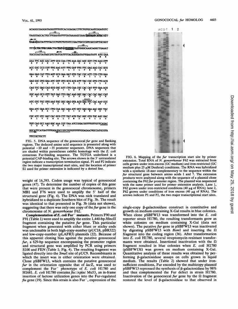
Features of the DNA sequence. The nucleotide sequenceand predicted amino acid sequence of the putative gonococ-cal Fur are shown in Fig. 5. The open reading frame has aputative Shine-Dalgarno sequence just upstream of the ini-tiating methionine. Two -10 and -35 promoter regions wereidentified. Primer extension analysis (Fig. 6) suggested thatthere were two transcriptional start sites. In addition, therewere two 19-bp sequences within the promoter region thatexhibited 68 and 58% similarity to the E. coli Fur protein-binding consensus sequence (13). A similar organization ofthe Fur protein-binding sequences was reported for Fbp (3).A potential catabolite activator protein (CAP)-binding site islocated 65 bp upstream from the start codon. A similar site islocated in the promoter region of E. colifur (13). A putativerho-independent transcriptional terminator was found 18 bpafter the stop codon. The gonococcal Fur was determined toconsist of 146 amino acids with a calculated molecular
INFECT. IMMUN.
0
on May 25, 2018 by guest
http://iai.asm.org/
Dow
nloaded from
GONOCOCCAL fir HOMOLOG 4603
-35 -10
TAGGTAGCACTGCTT GAATATTTTATAAGCGAACG¢rb-P2*MGtGGTATTTTACCCTGTCTGTGTCCGACTGCCCGTCGCCGTTGCGGT
-35
TTT-.iTAGCG.CTGCWA TAGTGGGCAAGCAGGCTGAAAT-10 APi SD
TGCGTATTATAGCGTCTATTATTTTACAGGGATATTGAATATT ATG GAAN I
MATTCAAGC AAC ATT GCG CAA CTG AAA GAC AGC GGT CTG AAG GTTK P S N I A Q L X D S a L X V
ACC GGC CCG CGT TTGAAG ATT TTG GAT TTGTTC GAGMG CAC GCGT G P R L K I L D L P I X H A
GAA GAG CAT TTG AGT GCG GAA GAT GTG TAC CGC ATT CTG CTG GAAE E H L s A I D V Y R I L L E
GAG GGC GTG GAA ATC GGT GTG GCG ACG ATT TAC CGC GTG CTG ACG1 0 v I I G V A T I Y R V L T
CAG TTC GAG CAG GCG GGC ATT CTG CAA CGC CAC CAT TTT GAA ACGQ P I Q A G I L Q R H H P E T
GGC AAG GCG GTT TAT GAG TTG GAC AAG GGC GAC CAC CAC GAC CATG X A V Y I L D X G D H H D H
EcoRIATC GTC TGC GTG AAG TGC GGC GAG GTA ACG GAA TTC CAC MTCCC
I V C V K C G 1 V T I P H N P
GA ATC GM GCC CTGCM GAC AM ATC GCC GAG GAA AAC GGC TACI I K A L Q D x I A Z Z N 0 Y
CGC ATC GTC GAT CAC GCG CTT TAT ATG TAC GGC GTG TGC AGC GACR I V D H A L Y M Y G V C S D
TGT CAG GCC AAG GGC MA CGT TAA ATCCGGACGGTTTGTTGTTCAGACC Q A K G X R ...
TTCAGATTTGA CGTTGTTTTTGGGAATGCCATG,CGTA
TTCCGCTGCTT
FIG. 5. DNA sequence of the gonococcalflur gene and flankingregions. The deduced amino acid sequence is presented along withpotential -10 and -35 promoter sequences. DNA sequences thatare shaded within parentheses exhibit homology with the E. coliconsensus Fur-binding sequence. The TGTGA underlined is apotential CAP-binding site. The arrows shown in the 3' untranslatedregion indicate a transcription termination signal. P1 and P2 indicatethe two major transcriptional start sites, and the location of primerSi used for primer extension is indicated by a dotted line.
weight of 16,393. Codon usage was typical of gonococcalgenes (47). To determine the number of copies of this genethat were present in the gonococcal chromosome, primersMB1 and F76 were used to amplify the 5' half of thestructural gene (Fig. 4). The DNA was nick translated andhybridized to a duplicate Southern blot of Fig. 3b. The resultwas identical to that presented in Fig. 3b (data not shown),suggesting that there was only one copy of thefur gene in thechromosome of N. gonorrhoeae F62.Complementation ofE. coli Fur- mutants. Primers F90 and
F91 (Table 1) were used to amplify the entire 1,440-bp HincIIfragment containing the putative fur gene. This particularfragment when generated with either blunt or sticky endswas unclonable in both high-copy-number (pUC18, pBR322)and low-copy-number (pLAFR3) plasmids (22). Because ofthis apparent cloning bias against the putative gonococcalfur, a 629-bp sequence encompassing the promoter regionand structural gene was amplified by PCR using primersS100 and FEN (Table 1; Fig. 4). The resulting fragment wasligated directly into the SmaI site of pUC9. Recombinants inwhich the insert was in either orientation were obtained.Clone pSBFW13, which contains the putative gonococcalfur in the orientation opposite that of lacZ, was used tocomplement the Fur- phenotype of E. coli H1780 andH1681. E. coli H1780 contains fiu::Xplac Mu53, an in-frameinsertion of lactose utilization genes into the Fur-regulatedfiu gene (19). Since this strain is also Fur-, expression of the
ACG T 1 2
4~~~~iurw
FIG. 6. Mapping of the _fur transcription start site by primerextension. Total RNA of N. gonorrhoeae F62 was extracted fromcells grown under iron-excess (GC medium) and iron-restricted (GCmedium plus 25 pM Desferal) conditions. The RNA was hybridizedwith a synthetic 18-mer complementary to the sequence within thefur structural gene between amino acids 1 and 7. The extensionproducts were analyzed along with the sequence of a plasmid clonecontaining the F62.fur promoter region. The plasmid was sequencedwith the same primer used for primer extension analysis. Lane 1,F62 grown under iron-restricted conditions (40 p.g of RNA); lane 2,F62 grown under conditions of iron excess (40 ~±g of RNA). Thearrows indicate P1 and P2, the two major transcriptional start sites.
single-copy 13-galactosidase construct is constitutive andgrowth on medium containing X-Gal results in blue colonies.When clone pSBFW13 was transformed into the E. colireporter strain H1780, the resulting transformants grew aswhite colonies on medium containing X-Gal (data notshown). The putative fur gene in pSBFW13 was inactivatedby digesting pSBFW13 with BsmI and inserting the ilfragmnent into the coding region (36). After transformationinto E. coli H1780, several streptomycin-resistant transfor-mants were obtained. Insertional inactivation with the flfragmnent resulted in blue colonies when E. coli H1780(pSBFW13fl was grown on medium containing X-Gal.Quantitative analysis of these results was obtained by per-forming 13-galactosidase assays on cells grown in liquidmedium. The results (Table 2) showed that under iron-sufficient conditions, Fur encoded by the multicopy plasmidpSBFW13 repressed the synthesis of 03-galactosidase by 98%and thus complemented the Fur defect in strain H1780.Inactivation of the gonococcal fur gene by the fl fragmentrestored the level of 0-galactosidase to that observed in
VOL. 61, 1993
on May 25, 2018 by guest
http://iai.asm.org/
Dow
nloaded from

4604 BERISH ET AL.
TABLE 2. P-Galactosidase activities of E. coli H1780 and H1681containing three different plasmidsa
Miller units (% of control)'
Plasmid Strain H1780c Strain H1681d
Fe sufficient Fe limited (Fe sufficient)
pUC9 1,134 (100) 956 (100) 2,198 (100)pSBFW13 23 (2) 124 (13) 1,624 (74)pSBFW13Q7 1,004 (89) 898 (94) 2,289 (104)
a Ferric chloride (50 ,uM) or 2,2'-dipyridyl (200 ,uM) was added to LB toachieve Fe-sufficient or Fe-limited conditions, respectively.
b Miller units of samples taken at A6,s of 0.15 to 0.2 for strain H1681 and0.3 to 0.4 for strain H1780.
c araD139 (argF-lac) U169 rpsL150 reL41 flbBS301 deoCI ptsF25 rbsR furfiu::Xplac Mu53.
d thr lac galK hsdR fhuA fur-31 zbf::TnlO fhuF::Xplac Mu.
strain H1780 containing pUC9. Under iron-limiting con-ditions, ,B-galactosidase activity levels in strain H1780(pSBFW13) were sixfold higher than those observed underiron-sufficient conditions and indicated that gene activitywas modulated by iron. An intermediate level of comple-mentation was observed with E. coli H1681. Colonies ofH1681 containing pSBFW13 were light blue when grown onmedium containing X-Gal (data not shown). This partialcomplementation was confirmed by quantitative enzymeanalysis indicating that gonococcal Fur repressed 3-galacto-sidase levels in strain H1681 by only 26%; inactivation of thegene by insertional mutagenesis restored enzyme levels tothat observed in strain H1681 containing pUC9 (Table 2).Attempts to insertionally inactivate far. In an attempt to
construct a Fur- mutant of N. gonorrhoeae F62, we choseto insertionally inactivate the cloned gonococcal fur gene.The fur coding region contained in pSBFW13 was inacti-vated by using the Q fragment as described in Materials andMethods. Plasmid (pSBFW13Q) or PCR-amplified DNA,consisting of thefur gene with the inserted fl fragment, wasused to transform piliated cells of N. gonorrhoeae F62 withselection for streptomycin resistance. No streptomycin-resistant transformants were recovered at a selection level ofeither 30, 50, 70, or 100 ,ug of streptomycin per ml. Theinability to obtain transformants suggests that the loss of Furmay be lethal to gonococci.Western blot analysis. The molecular weight of gonococcal
Fur, based on the consensus amino acid sequence of thepredicted protein, is 16,393. This value is in general agree-ment with the estimated molecular weight of ca. 17,000which was observed when Western blots of whole-celllysates of N. gonorrhoeae F62, E. coli W3110fur', and E.coli H1780(pSBFW13) were probed with polyclonal anti-serum to E. coli Fur (data not shown). No reactive band wasobserved with whole-cell lysates of E. coli W3110 furand E. coli H1780(pUC9).
DISCUSSION
In 1978, Ernst et al. (15) characterized a mutation in S.typhimurium which allowed constitutive overexpression ofiron-regulated proteins. This mutation was termed fur, forferric uptake regulation. To date,fur genes have been clonedand sequenced from E. coli (38), V. cholerae (22), V.vulnificus (23), Y pestis (43), and most recently P. aerugi-nosa (37). Undoubtedly, many pathogenic organisms ex-press a Fur-like gene product that is involved in regulation ofvirulence determinants and in iron transport systems (15,
18). In this study, we identify a gonococcal Fur homolog thatis functionally similar to that of E. coli but somewhatdivergent in structure.
Gel retardation analysis showed that purified E. coli Furbound to the promoter sequence of gonococcal iron-regu-lated gene, flp (Fig. 2). Sequence analysis suggested thatthere were two potential Fur-binding sites within the fbppromoter sequence (3). One site, designated the primaryFur-binding site, consists of the sequence -GATAATAACAAATJ'7JAAAA- and contains 12 of 19 bases of the E. coliconsensus Fur-binding sequence (13). Immediately down-stream of the primary Fur site is another sequence,-ATAATTA1IT7lGCATT-, which shows considerable ho-mology to a secondary Fur-binding site that has been char-acterized in the iucA promoter in E. coli (13). This consensussecondary binding site is defined by the sequence motifATAATnnnnATnATT (2, 13). Binding of E. coli Fur to eachof these two sites may account for the two mobility shiftsseen with the fbp promoter (Fig. 2) and correspond toDNA-protein complexes with either one or two sites occu-pied by Fur. It is presently not known which of the two sitesin the fbp promoter is the high-affinity binding site, nor is itknown how these two sites function in vivo.DNA hybridization analysis under low-stringency condi-
tions revealed that N. gonorrhoeae possessed a DNA locuswith homology to the E. coli fur gene (Fig. 3a). The gono-coccal fur gene could not be directly cloned in a plasmidvector; most likely, fragments containing the 3' flankingsequence were lethal in the E. coli host. Similar observationshave been made for gonococcal genes encoding Fbp (3),protein I (6), and the outer membrane protein macromolec-ular complex (45). A clone containing the promoter andN-terminal amino acid sequence through amino acid 103 wasobtained by screening a HincII minilibrary with a 140-bp Furprobe derived from PCR amplification of a highly conservedregion of previously characterized Fur genes. SSP-PCR wasused to amplify a DNA sequence encompassing the C-ter-minal portion of the gene and the 3' flanking region. Directsequencing of the ends of this fragment enabled us to obtainadditional sequence from which oligonucleotides were de-rived and subsequently used to amplify a smaller sequencewhich, when partially sequenced, was found to containamino acids 45 through 146 and 90 bases of the 3' flankingregion. The sequence encompassing the promoter region andstructural gene was amplified by PCR, and the resulting629-bp fragment was ligated into the SmaI site of pUC9. Aclone (pSBFW13) containing the putative gonococcal fur inthe orientation opposite that of lacZ was obtained so thatfurtranscription was dependent on its own promoter and wasused to complement the Fur- phenotype of E. coli H1780and H1681 (Table 2). The data suggest that N. gonorrhoeaepossesses a functional Fur regulatory protein capable ofinteracting with the E. coli Fur system. Quantitative 3-ga-lactosidase assays indicated that gonococcal Fur repressedenzyme levels to a greater extent in strain H1780 than instrain H1681. Comparisons of the promoter regions offhuF(H1681) and fiu (H1780) cannot be made because sequencedata are not available. However, E. coli Fur exhibits a higheraffinity for the fiu::plac fusion, whereas the fhuF::plac Mufusion is already derepressed at a mild iron stress (17a).The gonococcal fiur promoter sequence has two potential
Fur-binding sites and the sequence TGTGA, which may be apotential CAP-binding site. Fur-binding sites were not iden-tified in the V. cholerae fur promoter (22). However, tran-scription of E. coli fur is autoregulated by Fur as well asregulated by CAP (12). Thus, the transcription of gonococcal
INFEcr. IMMUN.
on May 25, 2018 by guest
http://iai.asm.org/
Dow
nloaded from
GONOCOCCAL fiur HOMOLOG 4605
N. gonorrhoeaeV. choleraeY. pestisE. coliP. aeruginosa
MEKFSNIAQLKDSGLKVTGPRLKILDLFEKHAEEHLSAEDVYRILLEEGVEIMSD.NQA ..DA L EVLQQPECQ.1 .. EL.KK.IDLSE..
MTD.NKA ..NA..L EVLQNPACH.V L.I. .IDI.E..NTD.NTA ..KA..L EVLQEPDNH.VV ... LICRDN.E..
MVE.SELR-KA. L.V .QLDSAEQR.N...... KA.N.A.EDV
GVATIYRVLTQFEQAGILQRHHFET6KAVYELDKGDHHDHIVCVKCGEVT.L..V W .DD VT. .S.F. .STQH.. L.LD.... I.L..V..CSE..DD...VT..N..G..S.F..TQQH ..LI.LD..K.I
.L..V...LN..D ...VT.NVA6..S.F..TQQH....LI.LD..K.I
.L.V .LVV. N.DG.H .F. .ADSG M. .DT I
EFHNPEIEALQDKIAEENGYRIVDHALY?YGVCS DCQAK GKR
..SDWV ..QR.KE. .AKYNVQLTN.S. .L. .K.GSDGSCKDNPNAH.PKK
..S.ES. .S. .RE. .KQH.IKLTN.S. .L. .H.ETGN.REDESAS..
..SWS ...RR.RE. .AKH.I.LTN.S. .L. .H.AEG. .REDEHAHEGK
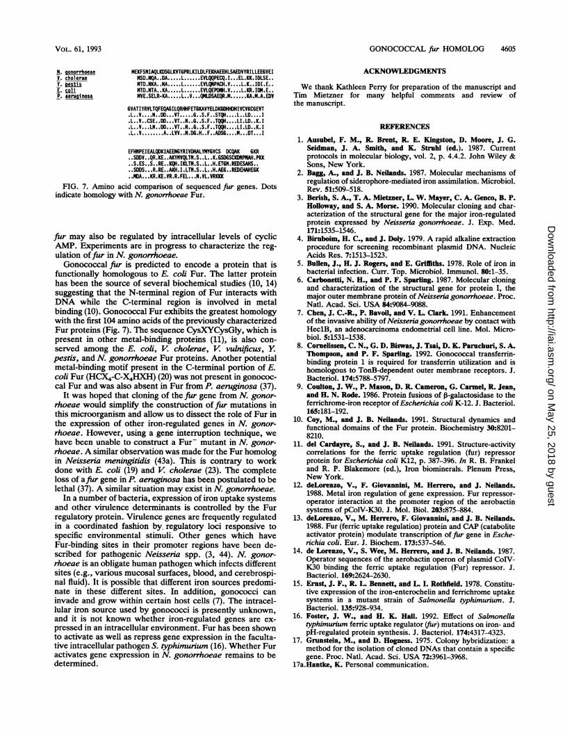
. DA....KR.KE.VR.R.FEL. . eN.VL.VRKKKFIG. 7. Amino acid comparison of sequenced _fur genes. Dots
indicate homology with N. gonornhoeae Fur.
fur may also be regulated by intracellular levels of cyclicAMP. Experiments are in progress to characterize the reg-ulation of fur in N. gonorrhoeae.Gonococcal fur is predicted to encode a protein that is
functionally homologous to E. coli Fur. The latter proteinhas been the source of several biochemical studies (10, 14)suggesting that the N-terminal region of Fur interacts withDNA while the C-terminal region is involved in metalbinding (10). Gonococcal Fur exhibits the greatest homologywith the first 104 amino acids of the previously characterizedFur proteins (Fig. 7). The sequence CysXYCysGly, which ispresent in other metal-binding proteins (11), is also con-served among the E. coli, V. cholerae, V. vulnificus, Ypestis, and N. gonorrhoeae Fur proteins. Another potentialmetal-binding motif present in the C-terminal portion of E.coli Fur (HCX4-C-X4HXH) (20) was not present in gonococ-cal Fur and was also absent in Fur from P. aeruginosa (37).
It was hoped that cloning of thefur gene from N. gonor-rhoeae would simplify the construction of fur mutations inthis microorganism and allow us to dissect the role of Fur inthe expression of other iron-regulated genes in N. gonor-rhoeae. However, using a gene interruption technique, wehave been unable to construct a Fur- mutant in N. gonor-rhoeae. A similar observation was made for the Fur homologin Neisseria meningitidis (43a). This is contrary to workdone with E. coli (19) and V cholerae (23). The completeloss of afiur gene in P. aeruginosa has been postulated to belethal (37). A similar situation may exist in N. gonorrhoeae.
In a number of bacteria, expression of iron uptake systemsand other virulence determinants is controlled by the Furregulatory protein. Virulence genes are frequently regulatedin a coordinated fashion by, regulatory loci responsive tospecific environmental stimuli. Other genes which haveFur-binding sites in their promoter regions have been de-scribed for pathogenic Neissenia spp. (3, 44). N. gonor-rhoeae is an obligate human pathogen which infects differentsites (e.g., various mucosal surfaces, blood, and cerebrospi-nal fluid). It is possible that different iron sources predomi-nate in these different sites. In addition, gonococci caninvade and grow within certain host cells (7). The intracel-lular iron source used by gonococci is presently unknown,and it is not known whether iron-regulated genes are ex-
pressed in an intracellular environment. Fur has been shownto activate as well as repress gene expression in the faculta-tive intracellular pathogen S. typhimurium (16). Whether Furactivates gene expression in N. gonorrhoeae remains to bedetermined.
ACKNOWLEDGMENTS
We thank Kathleen Perry for preparation of the manuscript andTim Mietzner for many helpful comments and review ofthe manuscript.
REFERENCES
1. Ausubel, F. M., R. Brent, R. E. Kingston, D. Moore, J. G.Seidman, J. A. Smith, and K. Struhl (ed.). 1987. Currentprotocols in molecular biology, vol. 2, p. 4.4.2. John Wiley &Sons, New York.
2. Bagg, A., and J. B. Neilands. 1987. Molecular mechanisms ofregulation of siderophore-mediated iron assimilation. Microbiol.Rev. 51:509-518.
3. Berish, S. A., T. A. Mietzner, L. W. Mayer, C. A. Genco, B. P.Holloway, and S. A. Morse. 1990. Molecular cloning and char-acterization of the structural gene for the major iron-regulatedprotein expressed by Neisseria gonorrhoeae. J. Exp. Med.171:1535-1546.
4. Birnboim, H. C., and J. Doly. 1979. A rapid alkaline extractionprocedure for screening recombinant plasmid DNA. NucleicAcids Res. 7:1513-1523.
5. Bullen, J., H. J. Rogers, and E. Griffiths. 1978. Role of iron inbacterial infection. Curr. Top. Microbiol. Immunol. 80:1-35.
6. Carbonetti, N. H., and P. F. Sparling. 1987. Molecular cloningand characterization of the structural gene for protein I, themajor outer membrane protein of Neisseria gonorrhoeae. Proc.Natl. Acad. Sci. USA 84:9084-9088.
7. Chen, J. C.-R., P. Bavoil, and V. L. Clark 1991. Enhancementof the invasive ability of Neisseria gonorrhoeae by contact withHeclB, an adenocarcinoma endometrial cell line. Mol. Micro-biol. 5:1531-1538.
8. Cornelissen, C. N., G. D. Biswas, J. Tsai, D. K. Paruchuri, S. A.Thompson, and P. F. Sparling. 1992. Gonococcal transferrin-binding protein 1 is required for transferrin utilization and ishomologous to TonB-dependent outer membrane receptors. J.Bacteriol. 174:5788-5797.
9. Coulton, J. W., P. Mason, D. R. Cameron, G. Carmel, R. Jean,and H. N. Rode. 1986. Protein fusions of 3-galactosidase to theferrichrome-iron receptor of Eschenichia coli K-12. J. Bacteriol.165:181-192.
10. Coy, M., and J. B. Neilands. 1991. Structural dynamics andfunctional domains of the Fur protein. Biochemistry 30:8201-8210.
11. del Cardayre, S., and J. B. Neilands. 1991. Structure-activitycorrelations for the ferric uptake regulation (fur) repressorprotein for Escherichia coli K12, p. 387-396. In R. B. Frankeland R. P. Blakemore (ed.), Iron biominerals. Plenum Press,New York.
12. deLorenzo, V., F. Giovannini, M. Herrero, and J. Neilands.1988. Metal iron regulation of gene expression. Fur repressor-operator interaction at the promoter region of the aerobactinsystems of pColV-K30. J. Mol. Biol. 203:875-884.
13. deLorenzo, V., M. Herrero, F. Giovannini, and J. B. Neilands.1988. Fur (ferric uptake regulation) protein and CAP (cataboliteactivator protein) modulate transcription of fur gene in Esche-richia coli. Eur. J. Biochem. 173:537-546.
14. de Lorenzo, V., S. Wee, M. Herrero, and J. B. Neilands. 1987.Operator sequences of the aerobactin operon of plasmid CoIV-K30 binding the ferric uptake regulation (Fur) repressor. J.Bacteriol. 169:2624-2630.
15. Ernst, J. F., R. L. Bennett, and L. I. Rothfield. 1978. Constitu-tive expression of the iron-enterochelin and ferrichrome uptakesystems in a mutant strain of Salmonella typhimurium. J.Bacteriol. 135:928-934.
16. Foster, J. W., and H. K Hall. 1992. Effect of Salmonellatyphimurium ferric uptake regulator (fur) mutations on iron- andpH-regulated protein synthesis. J. Bacteriol. 174:4317-4323.
17. Grunstein, M., and D. Hogness. 1975. Colony hybridization: amethod for the isolation of cloned DNAs that contain a specificgene. Proc. Natl. Acad. Sci. USA 72:3961-3968.
17a.Hantke, K. Personal communication.
VOL. 61, 1993
on May 25, 2018 by guest
http://iai.asm.org/
Dow
nloaded from

4606 BERISH ET AL.
18. Hantke, K. 1984. Cloning of the repressor protein gene ofiron-regulated systems in Eschenchia coli K-12. Mol. Gen.Genet. 197:337-341.
19. Hantke, K. 1987. Selection procedure for deregulated irontransport mutants (fur) in Escherichia coli: fur not only affectsiron metabolism. Mol. Gen. Genet. 210:135-139.
20. Hennecke, H. 1990. Regulation of bacterial gene expression bymetal-protein complexes. Mol. Microbiol. 4:1621-1628.
21. Laemmli, U. K. 1970. Cleavage of structural proteins during theassembly of the head of bacteriophage T4. Nature (London)227:680-685.
22. Litwin, C. M., S. A. Boyko, and S. B. Calderwood. 1992.Cloning, sequencing, and transcriptional regulation of the Vibriocholerae fur gene. J. Bacteriol. 174:1897-1903.
23. Litwin, C. M., and S. B. Calderwood. 1993. Cloning and geneticanalysis of the Vibrio vulnificus _fur gene and construction of afur mutant by in vivo marker exchange. J. Bacteriol. 175:706-715.
24. Maniatis, T., E. F. Fritsch, and J. Sambrook. 1982. Molecularcloning: a laboratory manual. Cold Spring Harbor Laboratory,Cold Spring Harbor, N.Y.
25. Marmur, J. 1961. A procedure for the isolation of deoxyribo-nucleic acid from microorganisms. J. Mol. Biol. 3:208-218.
26. McKenna, W. R., P. A. Mickelson, P. F. Sparling, and D. W.Dyer. 1988. Iron uptake from lactoferrin and transferrin byNeisseria gonorrhoeae. Infect. Immun. 56:785-791.
27. Mickelsen, P. A., E. Blackman, and P. F. Sparling. 1982. AbilityofNeisseria gonorrhoeae, Neisseria meningitidis, and commen-sal Neisseria species to obtain iron from lactoferrin. Infect.Immun. 35:915-920.
28. Mickelsen, P. A., and P. F. Sparling. 1981. Ability of Neisseriagonorrhoeae, Neisseria meningitidis, and commensal Neisseriaspecies to obtain iron from transferrin and iron compounds.Infect. Immun. 33:555-564.
29. Mietzner, T. A., R. C. Barnes, Y. A. JeanLouis, W. M. Shafer,and S. A. Morse. 1986. Distribution of an antigenically relatediron-regulated protein among the Neisseria spp. Infect. Immun.51:60-68.
30. Mietzner, T. A., G. H. Luginbuhi, E. C. Sandstrom, and S. A.Morse. 1984. Identification of an iron-regulated 37,000-daltonprotein in the cell envelope of Neisseria gonorrhoeae. Infect.Immun. 45:410-416.
31. Miller, J. H. 1972. Experiments in molecular genetics. ColdSpring Harbor Laboratory, Cold Spring Harbor, N.Y.
32. Moran, C. P. 1990. Measuring gene expression in Bacillus, p.267-293. In C. R. Harwood and S. M. Cutting (ed.), Molecularbiology methods for Bacillus. John Wiley & Sons, Ltd., Lon-don.
33. Morse, S. A., S. A. Berish, C.-Y. Chen, D. L. Trees, T. A.Mietzner, C. A. Genco, and D. Kapczynski. 1991. Structure,function, and regulation of the iron-binding protein, Fbp, p.
453-458. In M. Achtman, P. Kohl, C. Marchal, G. Morelli, A.Seiler, and B. Thiesen (ed.), Neisseriae 1990. Walter deGruyter, New York.
34. Morse, S. A., C.-Y. Chen, A. LeFaou, and T. A. Mietzner. 1988.A potential role for the major iron-regulated protein expressedby pathogenic Neisseria spp. Rev. Infect. Dis. 10:s306-s310.
35. Morse, S. A., and K K. Holmes. 1989. Gonococcal infection, p.639-657. In P. D. Hoeprich and M. C. Jordan (ed.), Infectiousdiseases, 4th ed. J. B. Lippincott Co., Philadelphia.
36. Prentki, P., and H. Krisch. 1984. In vitro insertional mutagene-sis with a selectable DNA fragment. Gene 29:303-313.
37. Prince, R. W., C. D. Cox, and M. Vasil. 1993. Coordinateregulation of siderophore and exotoxin A production: molecularcloning and sequencing of the Pseudomonas aeruginosa furgene. J. Bacteriol. 175:2589-2598.
38. Schaffer, S., K. Hantke, and V. Braun. 1985. Nucleotide se-quence of the iron regulatory gene fur. Mol. Gen. Genet.200:110-113.
39. Schryvers, A. B., and B. C. Lee. 1989. Comparative analysis ofthe transferrin and lactoferrin binding proteins in the familyNeisseriaceae. Can. J. Microbiol. 35:409-415.
40. Shyamala, V., and G. Ames. 1989. Genome walking by single-specific-primer polymerase chain reaction: SSP-PCR. Gene84:1-8.
41. Southern, E. M. 1975. Detection of specific sequences amongDNA fragments separated by gel electrophoresis. J. Mol. Biol.98:503-517.
42. Staggs, T. M., and R. D. Perry. 1991. Identification and cloningof afur regulatory gene in Yersinia pestis. J. Bacteriol. 173:417-425.
43. Staggs, T. M., and R. D. Perry. 1992. Fur regulation in Yersiniaspecies. Mol. Microbiol. 6:2507-2516.
43a.Thomas, C. Personal communication.44. Thompson, S. A., L. L. Wang, A. West, and P. F. Sparling. 1993.
Neisseria meningitidis produces iron-regulated proteins relatedto the RTX family of exoproteins. J. Bacteriol. 175:811-818.
45. Tsai, W., S. H. Larsen, and C. E. Wilde III. 1989. Cloning andDNA sequence of the omc gene encoding the outer membraneprotein-macromolecular complex from Neisseria gonorrhoeae.Infect. Immun. 57:2653-2659.
46. Weinberg, E. D. 1978. Iron and infection. Microbiol. Rev.42:45-66.
47. West, S. E. H., and V. L. Clark. 1989. Genetic loci and linkageassociations in Neisseria gonorrhoeae and Neisseria meningit-idis. Clin. Microbiol. Rev. 2(Suppl.):S92-5103.
48. Winship, P. R. 1989. An improved method for directly sequenc-ing PCR amplified material using dimethyl sulphoxide. NucleicAcids Res. 17:1266.
49. Yancy, R. J., and R. A. Finkelstein. 1981. Assimilation of iron bypathogenic Neisseria spp. Infect. Immun. 32:592-599.
INFECT. IMMUN.
on May 25, 2018 by guest
http://iai.asm.org/
Dow
nloaded from