icosadeltahedral geometry of geodesic domes, fullerenes
TRANSCRIPT

symmetryS S
Article
Icosadeltahedral Geometry of Geodesic Domes,Fullerenes and Viruses: A Tutorial on the T-Number
Antonio Šiber
Institute of Physics, Bijenicka cesta 46, 10000 Zagreb, Croatia; [email protected]
Received: 24 January 2020; Accepted: 12 March 2020; Published: 4 April 2020�����������������
Abstract: The Caspar–Klug (CK) classification of viruses is discussed by parallel examination ofgeometry of icosahedral geodesic domes, fullerenes, and viruses. The underlying symmetry of allstructures is explained and thoroughly visually represented. Euler’s theorem on polyhedra is used tocalculate the number of vertices, edges, and faces in domes, number of atoms, bonds, and pentagonaland hexagonal rings in fullerenes, and number of proteins and protein–protein contacts in viruses.The T-number, the characteristic for the CK classification, is defined and discussed. The superpositionof fullerene and dome designs is used to obtain a representation of a CK virus with all the proteinsindicated. Some modifications of the CK classifications are sketched, including elongation of theCK blueprint, fusion of two CK blueprints, dodecahedral view of the CK shapes, and generalizedCK designs without a clearly visible geometry of the icosahedron. These are compared to cases ofexisting viruses.
Keywords: virus; fullerene; geodesic dome; T-number
1. Introduction
Although the fullerene molecules were theoretically predicted and discussed [1], it wasnevertheless a surprise for the largest part of the scientific community when Kroto et al. publisheda paper announcing their experimental discovery [2]. Some of the surprise can be attributed to animpressive and amazingly symmetrical arrangement of carbon atoms in the most abundant of all thefullerene molecules—Buckminsterfullerene or C60. Kroto and colleagues named this molecule afterRichard Buckminster Fuller, who was well known in his time for his far reaching ideas, envisioningnew ways of living and working that are popular and perhaps more relevant today than they ever were.Still, he is perhaps [2] most recognized for popularizing geodesic domes—networks of interconnectedstruts forming a (hemi)spherical grid. Fuller constructed them as an alternative to typical 1960’sarchitecture in the USA [3]. Perhaps the most famous Fuller’s dome housed the American pavilionfor the 1967 World Expo exhibition in Montreal. Yet, the basic geometry and symmetry behind suchstructures was known at least since 1937 when Goldberg discussed a class of polyhedra, now oftencalled Goldberg polyhedra, with only pentagonal and hexagonal faces [4].
Fuller’s geodesic domes were not chiral [5] (i.e., they were equal to their mirror images), and evenbefore the discovery of fullerenes, it was known that achiral domes that Fuller constructed belongto a larger class of mathematical structures, which also includes chiral (or skew) objects. It wasalready known that fullerian-like designs are not only artificial constructions but are realized instructures occurring in nature. Pollen grains were observed to sometimes have dome-like shapes,formed apparently by many similar elements or modules, or at least with their surface divided in such away to exhibit similarity to geodesic domes [6,7]. From Haeckel’s beautiful and precise illustration [8,9],some Radiolaria were known to have geodesic-dome-like structures, although, in addition to hexagonaland pentagonal faces, they contained some heptagonal faces [9,10], which do not appear in Fuller’sdesign and are not a feature of icosadeltahedral constructions that will be discussed in the following.
Symmetry 2020, 12, 556; doi:10.3390/sym12040556 www.mdpi.com/journal/symmetry

Symmetry 2020, 12, 556 2 of 22
Perhaps it was then not so surprising that viruses were proposed to have a similar modular, dome-likestructure. Extending Fuller’s design ideas [11,12], in the year 1962, Donald Caspar and Aaron Klugconstructed the first theory that explained the features of most of the so-called ”spherical” (icosahedral)viruses known at the time [13].
It is intriguing that objects so different in size (fullerenes—1 nm, viruses—100 nm, and geodesicdomes—100 m), in addition to history, also share the design and symmetry. A partial reason forthis must have something to do with the fact that all these objects are built up of identical ornearly identical elements or modules—carbon atoms in fullerenes, proteins in viruses, and strutsin geodesic domes—which arrange so to fully enclose the object interior, i.e., to form a cage-likestructure. The nearly identical elements are in nearly identical surroundings, which is the essence ofthe concept used to rationalize the appearance of such shapes in viruses—the quasiequivalence concept.Caspar and Klug note that the appearance of a (nearly) symmetrical structure is a necessary outcomeof the arrangement of (nearly) identical units in (nearly) identical environments [13].
The aim of this paper is to explain the design principles and symmetries of a large subset ofthese objects, which can be called icosadeltahedral. The term was used by Caspar and Klug in theirseminal paper [13] and will be explained and often used in what follows. The language of the paper is,thus, perhaps most similar to the one used by Caspar and Klug [5,13]. The objects of interest are theicosahedral geodesic domes, fullerenes, and viruses, and a parallel examination of these objects will beused to illustrate the universality and versatility of CK blueprint and provide a wide view of the CKquasiequivalent construction.
There have been many reviews of virus structure in the past (see, e.g., [14]), and it wouldbe impractical to count them all here—perhaps one of the best references to quickly grasp thebasic ideas and forms is still the early paper by Caspar and Klug [13]. The parallels betweenthe fullerenes, geodesic domes, and viruses have also been discussed [10,11,15,16], which leavesa restricted space for the originality of this work. Yet, the basic Fuller–Caspar–Klug [1,13] blueprintcan be extended to account for more complicated structures, although the extension may be clumsy,especially when not resorting to the powerful group-theoretical methods [17]. It is intriguing that manystructures based on the modified or, at least, slightly reformulated blueprints have been observed,some relatively recently. These include different types of viruses, which require at least a subtlereformulation of the blueprint. Perhaps the most famous and well known such case is the one whichalready bothered D. Caspar [18]—the case of the SV-40 polyoma virus, which required only a slight,yet important modification of the general construction put forth by Caspar and Klug (CK). In recentyears, it was proposed that such and similar cases (e.g., bovine papilloma virus) could be betterviewed as dodecahedral structures or dodecahedral tessellations of the sphere [19]. There are alsoelongated viruses, typically bacteriophages, which require a different sort of modification allowingfor elongation of the CK design [20]; somewhat similarly, the carbon nanotubes can be obtained byformally “elongating” the fullerenes. And yet, there are structures that are difficult to think of interms of the CK construction but still fulfill some of the universal features guaranteed for the classof Goldberg polyhedra [4,17]—such is the case of the HIV virus [21,22] and fullerenes with lowersymmetry [17]. Why and how such structures arise is still not entirely clear, yet it is important to notethem, especially in the context of violation of the CK construction. Some extensions and violations ofthe CK construction, which are manifested in real objects, will also be briefly discussed, without theambition of fully classifying them.
The article is certainly not a novel research contribution to the field but is neither a typical researchreview. A mathematician may view it as inadequate in terms of rigor and application of the full-blownmachinery of group-theoretical symmetry concepts. A theoretical chemist, especially one doing workin graph-theoretical fields, may think the same, while a chemist dealing with electron structure mayfind it lacking substance with respect to quantum theoretical considerations explaining the stabilityand activity of different fullerenes. A structural virologist may find the discussion of geodesic domesand fullerenes irrelevant in the context of virus structure and may also note that the article does not
Symmetry 2020, 12, 556 3 of 22
cover the intricacies of protein structure, which enable the formation of the dome-like virus proteincoatings. She/he may also note that the article does not discuss the biological and evolutionary reasonsfor the CK design and its violations, in particular. A physicist may note that the article does not gosufficiently deep in explaining why the same motif appears in many different systems and that it musthave to do something with the overarching thermodynamic concepts that favor the formation of suchstructures in quite different circumstances. All of these remarks would be true. However, to covereven a single of these fields would require several thorough and extensive reviews. Nevertheless,the paper was written with the hope to clarify the different languages and approaches of authorsworking in different fields, yet on the objects that have the same symmetry and share many importantcharacteristics that are fixed by the symmetry. It is surely of interest to add here that the key formuladerived for viruses by CK (Equation (2)) [13] was in fact a rediscovery of the expression published25 years earlier by M. Goldberg in a completely abstract, mathematical context [12].
The language of this work is simple in mathematical terms. This level of mathematical languagewas adopted with the aim of making the paper readable to a wide audience of researchers and students.That is also why the visualizations of structures and concepts are often used in place of mathematicalformulas and theorems. This article is a significantly extended and appropriately updated rewriteof my unpublished manuscript, deposited in the year 2007 in the ArXiv database [23]. In particular,the parallels between the dome, fullerene, and virus designs have been additionally interlinked inSection 4.1. The virus is, both visually and mathematically represented as a sort of union of a geodesic(sub)dome in which triangular faces correspond to individual proteins and fullerenes in which bondsindicate the borders between the pentameric and hexameric clusters in viruses. In Section 4.2, possibleambiguities of the CK designs are discussed, particularly in the view that a distribution of proteindensity in a capsid may visually look like a distribution of protein dimers or trimers, rather thanpentamers and hexamers, which is typically observed in CK shapes. The dependence of the virus sizeon its T-number is discussed in Section 4.3. The aim of this section is to provide a rough link betweenthe CK structures of various T-numbers and the size and shape of their material manifestations inthe form of virus capsids. A long yet non-exhaustive discussion of some of the structures requiring amodification of the CK rules is presented in Section 5. These include elongated CK designs, the fusionof two incomplete CK designs, dodecahedral view of the CK designs, and generalized CK designswithout a clearly visible geometry of the icosahedron. All of these are compared to cases of existingviruses. The discussion also briefly touches some more recent studies of virus geometry, for example,the alternative and the so-called quasi-crystal approaches to virus tilings [19,24,25]. These are, however,covered only partially and to the extent that is compatible with the concept of this article.
2. Icosa(delta)hedral Geodesic Domes
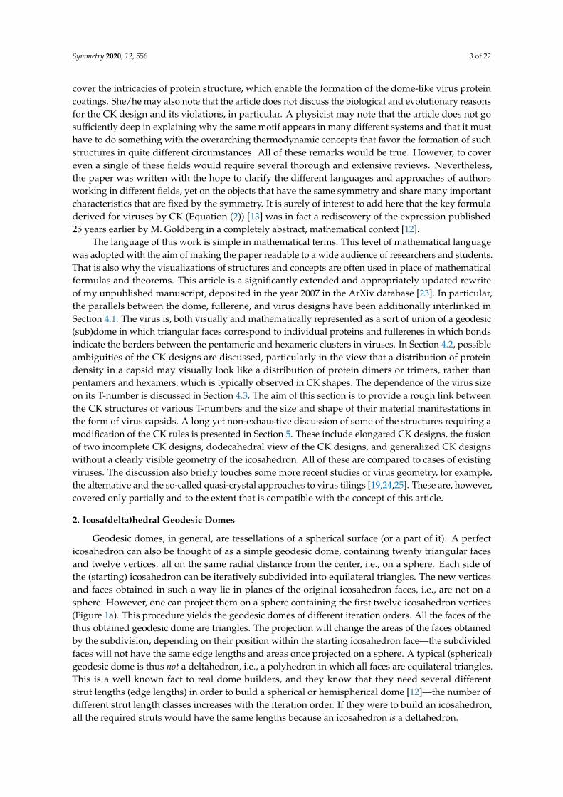
Geodesic domes, in general, are tessellations of a spherical surface (or a part of it). A perfecticosahedron can also be thought of as a simple geodesic dome, containing twenty triangular facesand twelve vertices, all on the same radial distance from the center, i.e., on a sphere. Each side ofthe (starting) icosahedron can be iteratively subdivided into equilateral triangles. The new verticesand faces obtained in such a way lie in planes of the original icosahedron faces, i.e., are not on asphere. However, one can project them on a sphere containing the first twelve icosahedron vertices(Figure 1a). This procedure yields the geodesic domes of different iteration orders. All the faces of thethus obtained geodesic dome are triangles. The projection will change the areas of the faces obtainedby the subdivision, depending on their position within the starting icosahedron face—the subdividedfaces will not have the same edge lengths and areas once projected on a sphere. A typical (spherical)geodesic dome is thus not a deltahedron, i.e., a polyhedron in which all faces are equilateral triangles.This is a well known fact to real dome builders, and they know that they need several differentstrut lengths (edge lengths) in order to build a spherical or hemispherical dome [12]—the number ofdifferent strut length classes increases with the iteration order. If they were to build an icosahedron,all the required struts would have the same lengths because an icosahedron is a deltahedron.

Symmetry 2020, 12, 556 4 of 22
Figure 1. (a) The icosahedron with subdivided faces (left) and the geodesic dome (right), with verticesall lying on a sphere. (b) The unwrapped and flattened icosahedron with subdivided faces, i.e., the net ofthe polyhedron. The small red dots denote the twelve pentavalent vertices. (c) The Coxeter constructionwith (m, n) = (2,1), (3,3), (4,0).
The polyhedron can be unwrapped, i.e., cut along certain edges and flattened so that all of thepolyhedron faces lie in the same plane—this is called a net (an unsolved mathematical problem iswhether every (convex) polyhedron has a net, but this is not really important for our purposes). A netof a geodesic dome would contain triangles of different sizes, but it is easier to think of a net of anicosahedron whose faces have been subdivided/triangulated prior to projecting them on the sphere—sucha polyhedron has a simple net, with the triangles/faces that are all equal and equilateral and whichcan be grouped within 20 larger triangles, the faces of the starting icosahedron (see Figure 1b). This iswhat is meant by the icosadeltahedral—the triangular subdivision of icosahedron faces that producespolyhedra with a larger number of triangular faces, which become unequal once the subdivided facesare projected on a sphere (Note here that the icosahedron with subdivided faces is not strictly convexand that there are neighboring triangular(sub)faces with a dihedral angle of π.). Such geodesic domesare thus triangulations of the sphere with the “icosahedral backbone”. The starting icosahedron can bedetected in the subdivided polyhedra by exactly twelve vertices in which exactly five faces meet (thesetwelve vertices are called pentavalent)—these are the vertices of the starting icosahedron, and theyhave five nearest neighboring vertices (see Figure 1). Exactly six faces and edges meet in all othervertices (the remaining vertices are called hexavalent—they all have six nearest neighboring vertices).The “backbone” is, thus, a sort of the fixation of the subdivided structure in twelve points, i.e., thetwelve vertices of the starting icosahedron. These twelve vertices can always be recognized in thesubdivided structure, no matter how dense and fine the subdivision.
There are many ways to triangulate a sphere so that there are twelve points that make the verticesof an icosahedron and have five nearest neighbors, all other points having six nearest neighboringpoints. The different triangulation patterns can be obtained by overlapping the one face of theicosahedron—the equilateral triangle—with the triangular mesh, so that the three vertices of theicosahedron face coincide with the mesh points (see Figure 1c). This is also known as a Coxeterconstruction [17,26].
A three-dimensional illustration of the geometry discussed is shown in Figure 2. The neighboringpoints of the icosahedral vertices are outlined as thick pentagons. As already discussed, the icosadeltahedralgeodesic domes are not (icosa)deltahedra since the triangles that they consist of are not equilateral.The requirement that all of the polyhedral faces be equilateral or nearly equilateral triangles necessarilyproduces aspherical (spiky) polyhedra (see Figure 2), quite different from the geodesic domes,which can also be considered as polyhedral approximates of a sphere. In the following, the word dome

Symmetry 2020, 12, 556 5 of 22
is used only for an icosadeltahedral geodesic dome, but one can obviously devise different polyhedralapproximates, with non-icosahedral symmetries (e.g., octahedral or tetrahedral).
The icosadeltahedral triangulation can be defined in terms of the triangular mesh base vectors,a1 and a2 (Figure 1c). The oriented icosahedron edge, E, in such a mesh can be written as
E = ma1 + na2. (1)
Each of the domes can be, thus, characterized by two nonnegative integers, m and n. These canalso be thought of as numbers of ”jumps” through the vertices of a dome that need to be performedin order to reach a pentavalent vertex from its closest pentavalent vertex. Except for (m, 0) dome,the jumps need to be directed along two different spherical geodesics (the shortest lines between twopoints on a sphere)—m jumps along one of them, and n along another one. The two geodesics makean angle of 60 degrees in the unwrapped, flattened, and deltahedral projection of the dome. In orderto be definite, we need to specify whether the ”jumper” needs to turn left or right after the m jumpsalong the first spherical geodesic. In what follows, the left turn shall be assumed and the type of theicosadeltahedral structures will be denoted by (m, n). Were the other convention chosen (turningright by 60 degrees after first set of jumps), the (m, n) dome in our convention would correspondto (n, m) dome in the alternative convention. The domes with m 6= n; m, n > 0 are chiral (or skew).A mirror image of (m, n) dome is (n, m) dome. This is illustrated in the upper-right corner of Figure 2for (3, 2) dome.
Figure 2. Gallery of icosadeltahedral geodesic domes for m > n and m < 5. For the sake ofclarity, the back sides of geodesic domes are not shown, i.e., only half of the dome and four oftwelve icosahedral vertices can be seen. The upper-right corner of the figure contains comparisonbetween chiral (3, 2) and (2, 3) domes. Note that they are mirror images. The spiky shape is a (3, 2)icosadeltahedron in which triangular faces are very nearly equal and equilateral—note that such arequirement produces a very aspherical polyhedron.
Symmetry 2020, 12, 556 6 of 22
From m and n, one can calculate the number of triangles in a dome. The number of triangles,t, in one face of the icosahedron can be obtained by dividing the area of the face,
√3/4a2(m2 +mn+ n2),
with the area of a mesh triangle,√
3/4a2, where a = |a1| = |a2|. This gives [4,17,26]
t = m2 + n2 + mn. (2)
The total number of triangles (or faces) in an icosadeltahedron is
f = 20t. (3)
Integer number t is called the triangulation number or simply the t-number. It adopts specialinteger values, t = 1, 3, 4, 7, 9, 12, 13, 16, 19, . . .. Instead of m and n integers, the t-number can be used toclassify the icosadeltahedral order. The problem with this choice is that it doesn’t discriminate between(m, n) and (n, m) domes. That is why the t-number is sometimes used in combination with words laevo(left) and dextro (right) to resolve this ambiguity. For example, (2, 1) structure in our convention wouldbe, in this case, denoted as t = 7laevo or t = 7l or simply t = 7, while (1, 2) structure would be denotedas t = 7dextro or t = 7d [14]. In our convention, when m > n (m < n), the domes are laevo (dextro).There is an additional problem when using only the t-number to classify the dome and that is thatdifferent pairs, (m, n) and (p, q) may produce the same t [4]. In particular, Goldberg has shown that([3P + Q][P−Q], 4PQ) domes have the same t-number as (3P2 + Q2, 0) domes. The lowest t-numberfor which a duplicity occurs is 133—the same t-number corresponds to (9,4) and (11,1) domes inthis case. Although it may seem as a quite large triangulation order, larger t’s are realized in nature.For example, a recent study [27] reported on a virus that has t = 169d. Intriguingly, this is again thet-number that allows for duplicity of structures. The virus has a (7,8) [27] rather than (13,0) structure,which would not require dextro or laevo classification. Larger t-numbers may produce even highermultiplicities. For example, the t = 8281 class of domes contains five different dome types: (91, 0),(85, 11), (80, 19), (65, 39), and (56, 49) [4].
From the known number of the polyhedron faces ( f ) one can proceed to find the number of itsvertices (v) and edges (e) by using Euler’s formula for polyhedra [17,28], which relates these nonzerointeger quantities as
v− e + f = 2. (4)
This equation is valid for polyhedra, which are homeomorphic to the sphere [17],i.e., their topology is the same as that of a sphere, which means that they do not have holes liketori or coffee cups—this is the case of interest to us. In the icosadeltahedral domes, twelve verticesbelong to five edges, and these are located at the vertices of an icosahedron. All the other verticesbelong to six edges, i.e., six edges meet at those vertices. Each edge is bounded by two vertices, and allthese facts together can be used to relate e and v as [17]
2e = 5 · 12 + 6 · (v− 12) = 60 + 6(v− 12). (5)
In combination with Euler’s formula, one obtains that
v =f2+ 2 = 10t + 2, (6)
ande =
3 f2
= 30t. (7)
3. Icosahedral Fullerenes
Fullerene molecules are carbon cages in which all carbon rings are either pentagonal or hexagonal,and all carbon atoms make three covalent bonds with their nearest neighbors (sp2 bonding). There are

Symmetry 2020, 12, 556 7 of 22
many different structures that can be made of carbon atoms connected with sp2 bonds, at leastconceptually (see, for example [29–32]). Quite a different question is whether such structures can beexperimentally obtained. The icosadeltahedral structure of geodesic domes is characteristic of a class ofespecially symmetric fullerene molecules, sometimes called icosahedral fullerenes, or giant icosahedralfullerenes in the case that molecules contain more than about 100 carbon atoms. Buckminsterfullerenebelongs to this class. Its “companion” molecule C70 that was discovered simultaneously [2] does not,however. These carbon molecules, at least in their proper symmetry, if not an energy-minimizingstate, can be obtained from icosadeltahedral domes by placing carbon atoms in the (bary)centers ofevery triangle in the dome. The newly obtained set of points (carbon atoms) is then interlinked sothat each point becomes connected to its three nearest neighbors, i.e., the carbon–carbon bonds areestablished (this procedure is illustrated in the upper-right corner of Figure 3). The basic chemicalrequirement for carbon atoms in the sp2 bonding electronic configuration are obviously established,as each vertex in the new structure (i.e., the carbon atom) is three-valent, i.e., connected with threeedges (sp2 bonds) to its neighbors—this is a simple consequence of the fact that every triangle in adome has three neighboring dome triangles.
Figure 3. Gallery of icosahedral fullerenes for m > n and m < 5. For the sake of clearer representation,the back sides of fullerenes are not shown. The average carbon–carbon bond length in all fullerenes isabout 0.142 nm [30], and this can be used to estimate their size. The upper-right corner of the figurecontains a comparison between (1, 1) dome and (1, 1) icosahedral fullerene (buckminsterfullerene,not to scale with other depicted fullerenes). Note that these polyhedra are dual to each other.
The construction produces a dual tessellation of the sphere—the centers of triangles in the geodesicdome become vertices of the dual structure. The geodesic domes and icosahedral fullerenes can thusbe considered as dual polyhedra. Icosahedral fullerenes contain twelve pentagonal carbon rings(pentagons) and a certain number of hexagonal carbon rings (hexagons), depending on the t-numberof the dome. The (m, n) pair that was characteristic of the dome will also be characteristic of its dualfullerene-like polyhedron, but now the “jumping” that characterizes icosadeltahedral ordering isallowed only through the centers of pentagons and hexagons (not along the carbon–carbon bonds—see

Symmetry 2020, 12, 556 8 of 22
Figure 4). I used the term “fullerene-like polyhedron” to emphasize that the true fullerene moleculeswill in general be different from the polyhedron obtained by a simple mathematical dualization of the(spherical) dome. The shape of the fullerene molecule is governed by the energetics of carbon–carboninteractions. Carbon–carbon bonds are much easier to bend than to stretch [30], so the shape of thefullerene molecule will be such to keep the nearest-neighbor carbon–carbon distances as uniform aspossible and as close to their equilibrium value as possible (the equilibrium length of carbon–carbonbonds in an infinite graphene plane is about 1.42 Å, but in fullerenes, the bonds in the pentagons arelonger than the bonds fusing hexagons together [33]). This means that large enough fullerenes willnecessarily be aspherical, looking more like an icosahedron with vertices slightly above the centersof carbon pentagons as the molecules get larger (One should be a bit careful when using the termspentagon (hexagon) and pentagonal (hexagonal) carbon rings as synonyms. All of the carbon atomsin a pentagonal or hexagonal ring need not necessarily lie in a plane.). The pentagons can also beunderstood as effective sources of curvature in the fullerenes, and exactly twelve of them are requiredto completely close a flat piece of graphene and produce a cage-like carbon molecule [30,32,34].
Figure 3 displays a gallery of icosahedral fullerenes. Their shape is not merely a mathematicalconstruction obtained by dualization of a dome, but a true minimum of energy, calculated by usingthe realistic model of energetics of carbon sp2 bonding [35], as described in [30]. Note that thebuckminsterfullerene (1, 1) is almost perfectly spherical, i.e., all of its carbon atoms are almost equallydistanced from the geometrical center of the molecule. A high degree of sphericity is also present in(2, 0) fullerene, but already in (2, 1) fullerene, a visible icosahedral shape of the molecule develops,and this becomes more prominent in larger molecules.
A way to better comprehend the symmetry of fullerenes is to unfold them so that they becomepolygonal pieces of graphene. Alternatively, and analogously to the case of geodesic domes, one canalso think of this procedure, illustrated in Figure 4, as a way to construct these molecules. The polygonalshape consisting of 20 equilateral triangles outlined by thick lines is cut out from the grapheneplane. The polygon is then creased along the edges shared by the triangles and folded into a perfecticosahedron. Thus, the obtained shape is not a physical fullerene since the details of its shape arewrong, but it has the same connectivity and number of carbon atoms as the icosahedral fullerene does.The shape of a physical fullerene can be obtained by a relaxation of the mathematically constructedentity towards the minimum of (chemical) energy [30]. The integers m and n that characterize theshape can now be interpreted as components of a two-dimensional vector E in a basis of graphene unitcell vectors A1 and A2 denoted in Figure 4,
E = MA1 + NA2, M, N > 0. (8)
The E vector is directed along the side of one of the twenty triangles making the icosahedron,as illustrated in Figure 4. In this convention, the unit cell vectors A1 and A2 need to be chosen sothat their cross product (A1 × A2) points from the paper towards the reader. This reproduces thejumping-to-the-left convention discussed in the previous section.
An important piece of information on fullerene molecules can again be obtained from the Euler’stheorem on polyhedra [17,36]. Since exactly three bonds (or polyhedron edges) finish at each of the carbonatoms (polyhedron vertices) and the bond (edge) is shared by two atoms (vertices), it follows that
2e = 3v. (9)
By definition, the fullerenes contain only pentagonal and hexagonal faces (carbon rings). Let usdenote the number of a pentagonal and hexagonal faces by f5 and f6, respectively. The total number offaces is obviously given by
f = f5 + f6. (10)

Symmetry 2020, 12, 556 9 of 22
Pentagonal and hexagonal faces are bounded by five and six vertices (atoms), respectively,and each vertex (atom) belongs to exactly three faces. This means that
5 f5 + 6 f6 = 3v. (11)
Figure 4. Cut-and-fold construction of (2, 1) icosahedral fullerene. The vectors a1, a2, and E discussedin the text are denoted. The triangular faces (Coxeter constructions) of (1, 1), (2, 0), (3, 1), and (4, 2)fullerene-like icosahedra are shown in the bottom of the figure. The figure also illustrates the conceptof ‘’jumping” over pentagons and hexagons in order to determine the t-number of the structure.
Combining these equations with Euler’s theorem in Equation (4), one obtains that [17]
f5 = 12. (12)
This is obviously true for the icosahedral fullerenes discussed so far, but the equation holdsfor general fullerenes as long as they are topologically equivalent to a sphere (including, e.g., C70).In other words, every network of pentagonal and hexagonal ring of carbon atoms with sphericaltopology necessarily has five pentagonal rings. A relation between the number of carbon atoms infullerenes and the number of hexagonal faces can also be obtained from the above consideration,
v = 2( f6 + 10). (13)
This means that the number of carbon atoms in the fullerene molecules is necessarily even (this canalso be seen from the fact that e = 3v/2, and since the number of edges must be an integer, the numberof vertices must be even) [17]. This, at first puzzling, piece of information was observed already inthe mass spectra of carbon clusters obtained by laser vaporization from the graphitic sample [37].Only signatures of clusters containing an even number of carbon atoms were detected, which can benicely explained by assuming that the clusters detected were, in fact, fullerenes. Specifying now thediscussion to the case of icosahedral fullerenes, the total number of carbon atoms in these molecules is
v = 20(M2 + MN + N2) = 20T, (14)
and the number of carbon–carbon bonds is
e = 30T. (15)
As shown earlier, there are exactly twelve pentagonal carbon rings and
f6 = 10(T− 1) (16)
hexagonal carbon rings.

Symmetry 2020, 12, 556 10 of 22
4. Caspar–Klug Classification of Viruses: The T-Number
The simplest viruses are particles made of DNA or RNA molecules (genome) protected by acoating made of proteins (capsid). Some viruses are additionally enveloped by parts of their hostcell membranes decorated by virus (glyco)proteins. A typical diameter of a virus is about 50 nm.For example, the diameter of a herpes simplex virus is ∼125 nm, while polio virus is about 32 nm indiameter [14].
In 1956, Crick and Watson [38] proposed that the spherical protein coating probably has a platonicpolyhedral symmetry, i.e., that it is built of identical proteins or protein subunits assembled in apolyhedral shell. They developed this notion by noting that the quantity of information containedin the viral genome is quite small, so that only a few different proteins can be produced fromit. Thus, they reasoned that the capsid is most likely constructed from a single subunit, which isrepeated many times to form a protein shell. Caspar and Klug considered different polyhedral shellsmade of identical proteins with tetrahedral, octahedral, and icosahedral symmetry and deduced thaticosadeltahedral ordering provides a structure in which all of the proteins are in surroundings thatare to the best approximation equal (quasiequivalent) of all the choices considered [13]. They calledthis the principle of quasiequivalence. Their proposition is based on the idea that the identical proteinsubunits have a certain degree of flexibility in their bonding to their neighbors but that only a certaindegree of their deformation when packed can be tolerated in this respect. The idea can be explained byexamining the subdivisions starting from different polyhedra. If the subdivision of sides is performed,for example, on an octahedron, all the vertices would be hexavalent, except for the six vertices of thestarting octahedron, which would be four-valent. The combination of pentavalent and hexavalentvertices in the case of the icosadeltahedral arrangement is obviously more uniform, in fact, the mostuniform of all deltahedral subdivisions of Platonic polyhedra—all the sides and vertices of suchpolyhedra are to the best approximation equivalent, i.e., quasiequivalent. In addition, the CK alsoemphasize the energy of the structure proposed—it enables formation of the maximum number ofthe most stable bonds between the units [13]. This is in part due to a dense packing of the proteinson a sphere and, in part, to small deviations of the different units from their ideal, minimum energypositions, so that the binding energy is still favorable.
The geometry behind the principle of quasiequivalence is the one already discussed in the casesof icosadeltahedral geodesic domes and fullerenes. A historical review focusing on the determinationof the structure of viruses and their relation to geodesic domes can be found in [11].
In most of the “spherical” viruses (Not all viruses are “spherical”. For example, the coating of thetobacco mosaic virus has a shape of a cylinder with viral RNA in its interior [39]. Even in such cases,there are strong parallels between the viruses and similarly structured shapes of sp2 carbon—see,e.g., [32].), the proteins are typically grouped in clusters called capsomers. The capsomers consist of five(pentamers; pentons) and six (hexamers; hexons) proteins. This is very often the case even if not allproteins are equal, i.e., when the capsid consists of several types of proteins [14]. In the assembledspherical capsids, the twelve pentamers occupy the same spatial positions as carbon pentagons infullerenes. The hexamers are analogous to hexagonal carbon rings in fullerenes. Viruses, thus, with allthe individual proteins delineated, can be constructed by connecting each of the vertices in pentagonaland hexagonal rings of the fullerenes with the ring centers and interpreting the thus obtained divisionsof the pentagons and hexagons as the dividing lines between the viral proteins in a capsomere.The (fullerene) vertices need not be connected necessarily to the centers of the rings but to pointslying on approximate normals to the pentagonal and hexagonal faces and passing through centers ofthe faces. This corresponds to capping the pentagons and hexagons with pentagonal and hexagonalpyramids, respectively, but the capping (i.e., modeling of protein pentamers and hexamers) can also beperformed in many other ways. The thus obtained polyhedron may be called omnicapped fullerene(all (omni) of the fullerene faces capped by pyramids or some other polyhedra). This procedure isillustrated in Figure 5a,b.

Symmetry 2020, 12, 556 11 of 22
Figure 5. Panels (a,b) represent polyhedral models of T = 3 viruses. These polyhedra can be termedas omnicapped truncated icosahedra or omnicapped Buckminsterfullerenes. The “pentamers” arecolored in a darker tone and borders (contacts) between “capsomers” are represented by thicker lines.The polyhedron in panel (b) is quite similar to turnip yellow mosaic virus [40]. Panel (c) represents amodel T = 1 (pT3) virus in which the building block is a “protein trimer” outlined by dashed lines.
The viral proteins have a certain shape, which is of course three-dimensional [41]. Representationof protein capsomers by pyramids or any other polyhedron is, thus, approximate. Any three-dimensional shape erected above the hexagon (pentagon) and having a six-fold (five-fold) symmetrywith respect to rotations around the hexagon (pentagon) normal may serve as a representation of aviral hexamer (pentamer). The classification of the symmetry of the capsid, however, does not dependon the shape of individual protein but only on the characteristics of the arrangement of all the proteinsin the capsid (at least when all proteins are equal, see below). The symmetry of viruses is characterizedin the same way as in the case of fullerenes: M and N integers are counted by “jumping” through thecenters of the capsomers and using the convention of turning left after the first M jumps. If M ≥ N,the virus is classified as a member of Tlaevo = M2 + MN + N2 class (or simply T), and if otherwise(M < N), the virus is classified as a member of the Tdextro (or Td) class. The virus-like polyhedradepicted in panels (a) and (b) of Figure 4 both belong to T = 3 class, although the details of theirshapes are quite different. The cut-and-fold constructions, shown in Figure 4 for fullerenes, can also beeasily applied to viruses—each carbon hexagon (pentagon) is to be interpreted as protein hexamer(pentamer) [42].
4.1. Viruses as Overlapping Dome and Fullerene Designs
The total number of capsomers (c) in a T-class virus is the same as the number points in theicosadeltahedral dome with triangulation number T,
c = 10T + 2. (17)
It is instructive to overlap the hexagonal net of fullerenes with a triangular net of a correspondingdome in which every triangle corresponds to an individual protein subunit, as in Figure 6. The fullerenebonds in this representation indicate the borders between the protein capsomers. The base vectors of ahexagonal mesh, A1 and A2 can be expressed using the base vectors of triangular (sub)mesh, a1 and a2,
A1 = a1 + a2
A2 = −a1 + 2a2, (18)
and the icosahedron edge vector E can then be expressed in both bases as
E = MA1 + NA2 = (M− N)a1 + (M + 2N)a2. (19)

Symmetry 2020, 12, 556 12 of 22
Figure 6. The overlapping of hexagonal and triangular meshes (left) and CK (1,1) shape and (3,0)geodesic dome (right). The base vectors of the triangular (a1, a2) and hexagonal (A1, A2) meshesare denoted.
The pair of integers characterizing the dome submesh is, thus, (m, n) = (M − N, M + 2N).The t-number of the (sub)dome is then
t = m2 + mn + n2 = 3(M2 + MN + N2) = 3T. (20)
The total number of proteins in a virus (p) is a sum of 60 proteins in 12 pentamers and 60(T− 1)proteins in 10(T− 1) hexamers [13]. Alternatively (see Figure 4), one can deduce that p for a virus inclass T should be the same as the number of faces in a dome in class t = 3T, i.e.,
p = 60T, (21)
so that both approaches give the same answer. There are 3T proteins, analogous to dome triangles,per icosahedron side, but not all of them are in different settings on a sphere—only a third of these areindeed mutually different, so that there are T different classes of protein surroundings in a virus of Tclass. The individual proteins need to be sufficiently flexible to adopt all of these T slightly different(quasiequivalent) positions in a completed virus shell [43]. It is, in this respect, remarkable to notethat CK construction applies even to huge viruses, with T numbers in the range of 103—such are,for example, the mimiviruses [44] for which T ≈ 1150, i.e., there are more than 1000 different butquasiequivalent positions in such viruses.
It is of interest to count the number of protein–protein contacts in a viral capsid since thisdetermines a large part of its energy [45]. It makes sense to separate the contacts to those that pertainto proteins belonging to the same capsomer (intra-capsomer; e1) and those between the proteins indifferent capsomers (inter-capsomer; e2). These contacts may be expected to have different associationenergies. Capsomers are typically more strongly bound and formation of capsomers from individualproteins is observed, at least in some viruses (see, for example [46]), to precede their assembly in thevirus capsid. There are five (six) protein–protein contacts per pentamer (hexamer), so that
e1 = 5 · 12 + 6 · 10(T− 1) = 60T. (22)
The number of inter-capsomer protein–protein contacts is the same as the number of carbon–carbonbonds in a fullerene of the same T-number as the virus in question (see Figure 5), so that
e2 = 30T. (23)
These are perhaps the simplest of the results, which can be easily obtained by employing theanalogy between domes, fullerenes, and viruses.
4.2. Possible Ambiguities of the CK Designs
Caspar and Fuller presented the quasiequivalent construction slightly differently [13]. They startfrom the dome and put three proteins in symmetric positions in each triangle of the dome, which is yet

Symmetry 2020, 12, 556 13 of 22
another and alternative way to think of correspondence between the domes and the viruses. In such aconstruction, if the proteins are situated near the triangle vertices, then they form clusters of five andsix around the dome vertices, i.e., pentamers and hexamers (see Figure 7). However, if the proteinsare clustered near the center of the dome triangles, then one can think of structure as consisting oftrimeric clusters. When the proteins are near the midpoints of the edges, one most easily recognizesthe dimeric clusters. Real proteins can be thought of as (atomic) density distributions, and they can,thus, not be represented as points in a triangle. In viruses, one can typically visually observe bothpentameric-hexameric and trimeric or dimeric signatures of capsid protein density, depending on thepoint of view taken (see, for example [47] for the case of the Zika virus)—this is illustrated in Figure 7.It may happen that the morphological features that are most easily discerned are not the physicalsubunits but, for example, trimeric-like density signatures of a pentameric-hexameric arrangement.
Figure 7. (a) Three different arrangements of three proteins on the faces of the geodesic dome. When thethree proteins are near the dome vertices (left), one first recognizes hexamers and pentamers. When theyare near the center of the dome faces (middle), one first recognizes trimers. When they are near the centersof the dome edges (right), dimers can be most easily observed. (b,c) The protein density in a virus canoften be thought of both as consisting of trimers or dimers (left) and of pentamers and hexamers (right).This is illustrated on a simplified geometrical model of protein density on a T = 3 virus.
There may also occur problems in identifying the capsid class when several different proteinsform a capsid, for example, when building blocks of the capsid are not individual protein subunitsbut trimers (clusters of three proteins). A typical situation is illustrated by the shape in panel (c) ofFigure 5. The building block of this shape is a protein trimer outlined by thick dashed lines. It consistsof a darker triangular protein (denoted by 1) and two brighter kite-shaped proteins (denoted by 2and 3). This structure could be identified as belonging to T = 1 class, with only twelve “pentons”(outlined by thick full lines in Figure 5c)) composed of five protein trimers (180 proteins in total).On the other hand, one could, at least conceptually, arrange the proteins in pentamers and hexamers asindicated by thick dash-dotted lines in Figure 5c). In this case, the hexamers would contain three pairsof 2- and 3-proteins from three different trimers, while pentamers would consist of five 1-proteinsfrom five different trimers. If the proteins 1, 2, and 3 are reasonably similar, as they often are in viruses,

Symmetry 2020, 12, 556 14 of 22
the pentamers and hexamers would almost look as if they were made of the same protein repeated fiveand six times, respectively. This would implicate that the shape belongs to T = 3 class. Such viruses arecalled pseudo-T3 (or pT3) viruses. The problem with the ambiguity of identification could be resolvedon physical grounds—if the binding energy between the proteins of the trimer is larger than betweenthe proteins from different trimers, it makes sense to speak about the trimer as the basic building blockand to identify the structure as belonging to T = 1 class. However, the problem in the mathematicalsense occurs when there are two T numbers that can be divided without remainder (e.g., T = 21 andT = 7). In the most trivial case, every capsid with T-number T1 could be interpreted as a T = 1 capsidconsisting of T1-mers. In addition, every (M, M) capsid could, in principle, be thought of as a (M, 0)capsid made of trimers. The important question is again whether the conceptually obtained proteinmultimers make any sense as the strongly bonded elementary units. A further complication canoccur if the particularly stable protein cluster is a trimer that geometrically looks just like a hexamer(hexagon). Those quasi-hexamers can, thus, tile the sides of the icosadeltahedron as already described;the quasi-pentamers in such situations are typically special, i.e., formed differently (see, e.g., [48]).
4.3. The T-Number and the Capsid Size and Shape
Viruses differ very much in size, depending on the type [14]. The variation in the mean radiusof a virus capsid could, in general, arise from the variation in size of the proteins that make thecapsid. This means that two capsids having the same T-number may be quite different in size dueto the difference in the size of their coat (capsid) proteins. On the other hand, if the size of thecapsid proteins were similar across different virus families, the variation in virus size would arisedominantly from the change in T-number of different viruses—the viruses with the same T-numberwould, in this case, have similar radii. As the total number of capsid proteins is proportional toT (see Equation (21)), the area of the capsid is also proportional to T, and the (mean) radius of thecapsid should then be proportional to
√T, across the different virus families. To test whether this is
indeed the case, a statistical analysis of 130 capsid entries deposited by the end of the year 2012 inVIPERdb database [49] was performed [50]. The mean capsid radius, R, was calculated for each of theviruses analyzed, as explained in [50], and plotted against its T-number. The results of the study werereanalyzed and redrawn in Figure 8 so to emphasize the T-number dependence of the mean radii andthe spread of radii within a particular T-number class.
1 2 3 4 7 13 21T
R [n
m]
10
15
20
30
25
Figure 8. The mean radius of the capsids plotted against their T-number. The vertical gray lines weredrawn between the minimum and the maximum value of the mean radius in each T-class. The thick lineshows a square-root dependence of mean radius, as discussed in the text, R = r0
√T, with r0 = 8 nm.
Adapted from [50].

Symmetry 2020, 12, 556 15 of 22
A large spread of radii is, in general, observed within a T-number class, but an overall trend ofmean radius increasing proportionally to
√T, R = r0
√T is visible. An area in which a “typical” coat
protein contributes to the overall mean capsid area, ap, can be estimated from
ap = r20
π
15≈ 13 nm2. (24)
The thicknesses of the virus capsid also differ quite a lot. This is in part due to the fact that theexternal morphology of the capsid, its grooves, and ridges play a part in the binding of the virus tocellular receptors and its subsequent entry. Some viruses have, thus, quite pronounced morphologicalfeatures, including long spikes located often at pentamers [50]. Nevertheless, a mean capsid thickness,which could be considered as typical for many viruses, is about 3 nm [50].
The overall shape and the (a)sphericity of viruses have been a subject of several studies,and deliberately simplified theories representing the capsid as a thin elastic shell predict that smallerviruses should be spherical while larger should show pronounced faceting, i.e., should look more likean icosahedron than a sphere, similar to the case of fullerenes. This has indeed been observed to be thecase, at least to a certain extent [50,51]. The interactions between the proteins in a virus are much morecomplicated than those characterizing carbon atoms in fullerenes, and the theoretical descriptionsof shape depend on much more details, which also include the presence of ions in the solution,which modify and screen the protein–protein interaction [52,53]. Functional viruses also contain thegenome molecule, RNA or DNA, and the interaction of the coat proteins with the nucleic acid mayalso influence the shape of the capsid. In particular, the packed DNA can be to some approximationoften described as effective physical pressure acting outwards, to expand the capsid [54]. The internalpressure both increases the mean radius of the capsid, inducing a kind of swelling, and smooths outthe polygonal nature of the capsid a bit [54,55]. The virus proteins can also have shapes and activebinding regions which promote spontaneous curvatures of their assemblies. Such proteins will thenprefer a certain radius [56], and a certain T-number will be effectively encoded in the details of theprotein shape. This is a point in stark contrast when compared to the case fullerenes. All of these,physically and biologically relevant considerations go beyond the simplest description of a virus as athin icosadeltahedral shell.
5. Some Modifications and Variations of the Icosadeltahedral Geometry in Viruses
Caspar and Klug quasiequivalence principle predicts that there are only twelve pentamericcapsomers, all other being hexameric. In the year 1982, it became clear that there were viruses(e.g., SV-40 virus from polyomavirus genus) composed only of pentamers but still retaining theicosadeltahedral ordering of their arrangement [57]. The concept of T-number is still valid in thatcase, and the number of capsomers is still given by Equation (17), but the total number of proteinsis no longer given by Equation (21) because there are no hexamers in the structure. For such viruses,the total number of proteins is
p = 5c = 5(10T + 2). (25)
This is a seemingly innocent modification of the CK principle, yet it is important in the light ofcut-and-fold constructions of the CK structures [13,43]. Namely, the missing material at the position ofcuts where there are pentamers in the standard CK construction acts as a source of curvature aroundthese positions [30,32,34]. If all the capsomers are the same, the source of curvature must be equallydistributed in all of them, so that the formation of a spherical structure is not surprising [56]. It is,however, intriguing that even in this case, such a structure has an icosadeltahedral order and can be,to a large degree, described by CK construction. In such a structure, not all capsomers are in identicalsurroundings. All the capsomers have six nearest neighboring capsomers (these would be hexamers instandard CK construction, but are pentamers in SV-40 viruses [57]), except for the twelve of them atthe position of icosahedron vertices (these would be pentamers in standard CK construction and areindeed pentamers in SV-40 viruses [57]).

Symmetry 2020, 12, 556 16 of 22
The correspondence between CK shapes and geodesic domes expressed in Equation (20) indicatesthat there are domes that cannot be represented by pentameric and hexameric clusters of proteins(or triangles). Such are all the domes for which it is not divisible by three, for example, (1,0), (2,0), (2,1),(2,2), and (4,1) domes. Yet, these domes can still be thought of as representing different clusters of virusproteins, as illustrated in Figure 9. Triangles are (2,0), and (2,1) domes can be organized in pentagons(pentamers) and individual triangles (protein monomers). The (2,2) domes can be reorganized andtiled by pentagons, rectangles, and triangles, which may, for example, represent different types ofcapsid proteins or protein clusters. The (4,1) domes can be thought of as being tiled by pentamers(pentagons) and hexamers (hexagons), with an additional layer of monomers (triangles) surroundingeach pentamer and hexamer. The reorganization is not unique, and several different designs may begiven for a particular dome. Such modifications of the CK approach are being studied and applied toreal viruses [24,25,58].
Figure 9. An alternative view of (2,0), (2,1), (2,2), and (4,1) geodesic domes. The dome triangles havebeen united or replaced by other polygons, so that (2,0) and (2,1) domes consist of pentagons andtriangles, (2,2) domes of pentagons, rectangles, and triangles, and (4,1) domes of pentagons, hexagons,and triangles.
A simplest modification of the icosadeltahedral cut-and-fold blueprint is to elongate it or shortenit along a five-fold axis of symmetry by a distance b, which needs to conform to the lattice distances ofthe net, as illustrated in Figure 10 [30]. Very elongated icosadeltahedra can be thought of as cylinderscapped by two pentagonal pyramids, i.e., capped tubes. This, of course, reminds one of (capped)carbon nanotubes [30]. It is intriguing that Buckminster Fuller also designed elongated forms of hisdomes; his design for the entrance pavilion of the Union Tank Car Company dome appears to bea dual of the capped carbon nanotube [59]. Elongated (prolate) viruses are well known, and sucha geometry is fairly typical for bacteriophage viruses [60,61]. Oblate viruses, i.e., those shortenedalong the five-fold axis of symmetry do not appear to be the native shape of any virus. However,some of the aberrant capsids of mutant T4 bacteriophages have been proposed to have an oblate(also termed shortened) icosahedral shape [61]. Note that the vector b may be non-parallel to thefive-fold axis of symmetry of the pentagonal pyramids that cap the shape. The phage T4 [60] hassuch a symmetry. The isometric icosadeltahedral shapes can also be thought of as two pentagonalpyramids capping the middle portion (midsection) of the icosahedron. This middle portion, of whichtwo caps have been cut off, is called the uniform pentagonal antiprism. It consists of 10 triangularsides and two pentagonal bases rotated with respect to each other by 36 degrees (2π/10). The vector bdefines the elongation (shortening) of the midsection as well as the angle of mutual rotation of thetwo capping pentagonal pyramids. The elongated structure is typically characterized in terms of twoT-numbers, one specifying the triangulation of the caps, i.e., smaller triangular faces, Tend and the otherspecifying the triangulation of the middle portion, i.e., elongated triangular faces, Tmid. For example,in phage φ29, Tend = 3, Tmid = 5, and many different cases can be found in [20]. The two numbers canbe elegantly defined by specifying an additional coordinate system for the elongated faces, rotated by60 degrees with respect to the one used to define the T-number for the caps [20,62]. If one decided todefine b on the same basis as E, b = PA1 + QA2, Tmid could be expressed as Tmid = Tend + MP− NQ.Such relations can be derived by calculating the area of the triangular faces, i.e., by performing thecross products of the vectors defining the edges of the faces. For example, for the elongated shape in

Symmetry 2020, 12, 556 17 of 22
Figure 10, the capping pyramid is a part of the (2,2) CK shape, so T = 12. Vector b can be expressedas b = 2A1 − 2A2, so P = 2 and Q = −2, which gives Tmid = 12 + 8 = 20. The total number ofproteins in the shape is now 30Tend in the two pentagonal pyramids and 30Tmid in the midsection [20],which means that it is increased with respect to isometric shape by 30(20− 12) = 240. That this isindeed the case can, in this case, be easily checked by calculating the area of the strip of hexagonalmesh of height |b| inserted in the midsection of the isometric shape.
Figure 10. The prolate (top) and oblate (middle) icosadeltahedral designs obtained by elongatingand shortening the isometric icosadeltahedral shape along the five-fold axis of symmetry by b = |b|.The influence of elongation vector b on shapes is illustrated by the three shapes shown at the bottom ofthe figure. Adapted from [30].
Quasiequivalence in elongated capsids is valid to a lesser degree than in isometric,CK capsids—this can also be seen from the fact that two T numbers are required to describe theconstruction. In particular, the two pentamers at the top and the bottom of the shape, i.e., at the apicesof the pyramidal caps, are in different positions than the ten positioned on the two borderlines betweenthe caps and the midsection. The curvature of the capsid may be quite different in these positions,the difference growing with |b| [20].
There is another type of elongated design in which the capsid can be thought of as consisting oftwo fused, incomplete icosahedral capsids. Such a capsid could be constructed as a modification ofthe cut-and-fold plan, as illustrated in Figure 11. The structure has 2× 11 = 22 pentavalent vertices,which may host twelve protein pentamers. This obviously contradicts the standard CK construction,which predicts only 12 of them. Note, however, that in addition to pentavalent and hexavalent vertices,the fused structure also contains octavalent (eightfold coordinated) vertices, five of them aroundthe ring, which fuses the two incomplete capsids—these restore the validity of the Euler formula inEquation (4). Such design can be found in geminiviruses that consist of 22 pentamers, i.e, they can bethought of as two incomplete T = 1 capsids fused together. In these viruses, the two halves are slightlyrotated with respect to each other around the five-fold axis of symmetry of the virus. Such details ofarchitecture depend crucially on the flexibility of the proteins that make the capsid and the details

Symmetry 2020, 12, 556 18 of 22
of their conformations [63]. The gemini capsid has an obvious advantage in that it can pack moreDNA than a single capsid. It also appears as if the DNA is essential for the assembly of the capsid asno empty geminivirus capsids have been reported [63]. The capsids could also be thought of as twofused dodecahedra (Figure 11)—the faces of dodecahedra are, in this case, tiled by protein pentamers.Erecting a pentagonal pyramid on each of the dodecahedron faces produces a T = 1 CK structure [13].
Figure 11. The two nets for the two halves of the capsid of geminiviruses and the folded shapes,which may be thought of both as.
The duality between the icosahedra and dodecahedra, observed here for the case of geminivirus,has been employed in an alternative description of the virus architecture [19]. Some viruses,for example, bovine papilloma virus, can perhaps be better thought of as dodecahedra in whichfaces are tiled by protein pentamers. Protein density maxima in pentagonal faces exhibit patternssimilar to those found in chiral pentagonal quasicrystals [19].
Another modification of the icosadeltahedral design is to form a structure similar to a truncatedcone, which is capped by two pyramid-like shapes of different sizes [20]—the larger connected to thelarger, bottom base of the cone, and the smaller to the smaller, top base of the cone (see Figure 12). This isapproximately the structure of the HIV virus, although it seems that the HIV virus is quite polymorphicand that functional HIV viruses may have many different coat structures [64]. The coat structure doesnot, thus, appear to be strongly fixed, but varies in details, although there are obvious constraints thatthe structure must fulfill; it must, in the first place, be spacious enough to pack properly its own RNAmolecule. There are obviously many different ways to form a closed polyhedral structure containingonly hexagonal and pentagonal sides, but the number of pentagons must, in all cases, be 12, as thederivation leading to Equation (12) demonstrates. Any such polyhedron can be represented by a neton a lattice of hexagons. The net will necessarily consist of exactly twelve cuts, i.e., positions where thewedge of exactly 60 degrees has been removed by cutting. Pentagons will be formed at these positionsonce the net is folded in order to make the polyhedron. However, none of the faces of the polyhedronneeds to be an equilateral triangle. This was already seen in the case of elongated icosahedral designs,where the mid-faces can be isosceles triangles, or their edge lengths can be even all different.
Twelve cuts of 60 degrees can be positioned in many different ways in a lattice, as Figure 12suggests. All of these nets correspond in general to different polyhedra. All such polyhedra could,in principle, be thought of as modifications of the perfect icosa(delta)hedron, as they bear the signatureof the twelve vertices of the icosahedron where the twelve pentagons reside. Furthermore, all suchstructures could be considered as if obeying a relaxed variant of the principle of quasiequivalenceput forth by Caspar and Klug since all of the vertices and faces in such polyhedra are in nearlyequivalent surroundings—a fact of essential importance in rationalizing the appearance of perfecticosadeltahedral geometry [13]. The reservations are in order when the two pentagons (pentamers)are very close to each other. In such cases, the effective interactions acting in the regions betweenthe two pentagons may be quite different from the interactions far away, in large patches consistingexclusively of hexagons. Due to a loss of symmetry of the folding pattern, the number of differentprotein surroundings increases, and the concept of T-number loses its strength as the structures becomemore complex.

Symmetry 2020, 12, 556 19 of 22
Caspar has noted the usefulness of icosadeltahedral cut-and-fold schemes in constructing differentfullerenes and has proposed many different polyhedral nets of deltahedra dual to fullerenes [5].Note here that one can conceptually construct an octahedral, fullerene-like shape by putting pairsof pentagons near the octahedron vertices—the necessary twelve pentagons are now distributedin pairs of six near the six octahedron vertices. Putting triples of pentagons near four tetrahedronvertices again produces a fullerene-like structure with a tetrahedral shape (see also [32]). The pairsand triples of pentagons in these shapes define the effective vertices of the fullerene-octahedron andfullerene-tetrahedron. The “vertices” are, in fact, the smooth, curved regions of the lattice of whichcurvature is determined by the number of pentagons (two or three)—the more pentagons, the largerthe curvature of the effective vertex [32]. The pentagons in each pair or triple should be near eachother if the effective octahedra and tetrahedra are to have fairly pointed effective vertices. In suchsituations, the notions of quasiequivalence may fail.
Figure 12. A possible way to construct a polyhedron similar to a capsid of the HIV virus. Note thatthe top pyramid-like shape is smaller than the bottom pyramid-like shape, which can be seen by thedifferent sizes of the triangles in the polyhedron net (adapted from [65]). The folded polyhedron isalso shown in two different views. Such constructions can be obviously varied in many different ways.One of the representative structures of an HIV capsid is also shown (adapted from [21]).
6. Conclusions
This completes a brief introduction to the geometry of icosadeltahedral structures, but manyquestions, of course, remain. In the case of structures made of carbon, one would like to know whichfactors govern the appearance of different shapes and can the process of production of these shapesbe tuned to produce predominantly one type of myriad of possible closed structures. In the case ofviruses, one is, of course, interested in the assembly processes that produce such large and complexobjects. It is even more intriguing that some of them can be assembled spontaneously, even outsidea living cell. This was first observed already in the year 1955 for a tubular tobacco mosaic virus [66].Some large viruses use scaffolding proteins to guide the process of assembly, perhaps somewhatsimilar to what workers assembling a geodesic dome would do—build a scaffold to stand on [67].Some viruses (e.g., adenovirus) produce proteins that are packed in their interior together with theDNA. It appears that these proteins reduce the pressure in the capsid due to electrostatic self-repulsionof the DNA, perhaps stabilizing the structure [68]. These mechanisms are actively researched and stillnot understood completely. As viruses are simple structures and yet subject to evolution, they seem tobe ideal systems to better understand the evolutionary processes and mechanisms. It would certainlybe of importance to construct some sort of the evolutionary narrative explaining the appearance oficosadeltahedral structures in the living world and their assembly. The field of virology is currentlyexperiencing a sort of revival, as scientists from very different backgrounds show interest in viruses andapproach the subject from their perspective [69,70]. There is also a somber aspect of this revival—newand potentially deadly viruses are constantly entering the human population. Powerful diagnostic andgenome sequencing tools enable us to quickly detect them and assess their pandemic potential—justas I am writing this sentence, whole countries are put under quarantine due to the outbreaks of

Symmetry 2020, 12, 556 20 of 22
the coronavirus COVID-19 infection throughout the world [71]. New ideas and approaches to themonitoring and control of viruses are needed. A better understanding of virus stability and assemblymay help in devising new anti-viral drugs. This paper is hoped to be of some use to the newcomers inthis interesting interdisciplinary and quickly-developing field of research.
Funding: This research received no external funding.
Conflicts of Interest: The author declares no conflict of interest.
References
1. Kroto, H. Symmetry, space, stars and C60. Rev. Mod. Phys. 1997, 69, 703–722. [CrossRef]2. Kroto, H.W.; Heath, J.R.; O’Brien, S.C.; Curl, R.F.; Smalley, R.E. C60-Buckminsterfullerene. Nature 1985,
318, 162–163. [CrossRef]3. Ananthasuresh, G.K. Buckminster Fuller and his Fabulous Designs. Resonance 2015, 20, 98–122. [CrossRef]4. Goldberg, M. A class of multi-symmetric polyhedra. Tohoku Math. J. 1937, 43, 104–108.5. Caspar, D.L.D. Deltahedral views of fullerene polymorphism. Philos. Trans. R. Soc. Lond. A 1993, 343, 133–144.6. Fritzsche, C.J. Ueber den Pollen; Kais, Ed.; Academie der Wissenschaften: St. Petersburg, Russia, 1837.7. Halbritter, H.; Ulrich, S.; Grímsson, F.; Weber, M.; Zetter, R.; Hesse, M.; Buchner, R.; Svojtka, M.;
Frosch-Radivo, A. Illustrated Pollen Terminology, 2nd ed.; Springer: Cham, Switzerland, 2018.8. Haeckel, E. Die Radiolarien; G. Reimer: Berlin, Germany, 1862.9. Thompson, D.W. On Growth and Form; Dover Publications Inc.: New York, NY, USA, 1992.10. Darvas, G. Symmetry: Cultural-Historical and Ontological Aspects of Science-Arts Relations; Birkhauser Verlag
AG: Basel, Switzerland, 2007.11. Morgan, G.J. Historical review: Viruses, crystals and geodesic domes. Trends Biochem. Sci. 2003, 28, 86–90.
[CrossRef]12. Morgan, G.J. Virus Design, 1955–1962: Science Meets Art. Phytopathology 2006, 96, 1287–1291. [CrossRef]13. Caspar, D.L.D.; Klug, A. Physical principles in the construction of regular viruses. Cold Spring Harb. Symp.
Quant. Biol. 1962, 27, 1–24. [CrossRef]14. Baker, T.S.; Olson, N.H.; Fuller, S.D. Adding the third dimension to virus life cycles: Three-dimensional
reconstruction of icosahedral viruses from cryo-electron micrographs. Microbiol. Mol. Biol. Rev. 1999, 63, 862–922.[CrossRef]
15. Popko, E.S. Geodesics and the Orderly Subdivision of the Sphere; CRC Press, Taylor & Francis Group: Boca Raton,FL, USA, 2012.
16. Brinkmann, G.; Goetschalckx, P.; Schein, S. Comparing the constructions of Goldberg, Fuller, Caspar, Klug andCoxeter, and a general approach to local symmetry-preserving operations. Proc. R. Soc. A 2017, 473, 20170267.[CrossRef]
17. Fowler, P.W.; Manolopoulos, D.E. An Atlas of Fullerenes; Clarendon: Oxford, UK, 1995.18. Makowski, L. An Unreasonable Man in a Quasi-Equivalent World. Biophys. J. 1998, 74, 534–536. [CrossRef]19. Konevtsova, O.V.; Rochal, S.B.; Lorman, V.L. Chiral Quasicrystalline Order and Dodecahedral Geometry in
Exceptional Families of Viruses. Phys. Rev. Lett. 2012, 108, 038102. [CrossRef] [PubMed]20. Moody, M.F. Geometry of Phage Head Construction. J. Mol. Biol. 1999, 293, 401–433. [CrossRef] [PubMed]21. Ganser, B.K.; Li, S.; Klishko, V.Y.; Finch, J.T.; Sundquist, W.I. Assembly and analysis of conical models for the
HIV-1 core. Science 1999, 283, 80–83. [CrossRef] [PubMed]22. Zhao, G.; Perilla, J.R.; Yufenyuy, E.L.; Meng, X.; Chen, B.; Ning, J.; Ahn, J.; Gronenborn, A.M.; Schulten, J.;
Aiken, C.; Aiken, C.; et al. Mature HIV-1 capsid structure by cryo-electron microscopy and all-atom moleculardynamics. Nature 2013, 497, 643–646. [CrossRef]
23. Šiber, A. Icosadeltahedral geometry of fullerenes, viruses and geodesic domes. arXiv 2007, arXiv:0711.3572.24. Pimonov, V.V.; Konevtsova, O.V.; Rochal, S.B. Anomalous small viral shells and simplest polyhedra with
icosahedral symmetry: The rhombic triacontahedron case. Acta Cryst. A 2019, 75, 135–141. [CrossRef]25. Twarock, R.; Luque, A. Structural puzzles in virology solved with an overarching icosahedral design principle.
Nat. Commun. 2019, 10, 4414. [CrossRef]

Symmetry 2020, 12, 556 21 of 22
26. Coxeter, H.S.M. Virus macromolecules and geodesic domes. In A Spectrum of Mathematics; Essays Presented toH.G. Forder; Butcher, J.C., Ed.; Oxford University Press: Oxford, UK, 1971; pp. 98–107.
27. Fang, Q.; Zhu, D.; Agarkova, I.; Adhikari, J.; Klose, T.; Liu, Y.; Chen, Z.; Sun, Y.; Gross, M.L.; Van Etten, J.L.;et al. Near-atomic structure of a giant virus. Nat. Commun. 2019, 10, 388. [CrossRef]
28. Posamentier, A.S. Math Wonders to Inspire Teachers and Students; Association for Supervision and CurriculumDevelopment: Alexandria, VA, USA, 2003; pp. 195–197.
29. Osawa, E.; Yoshida, M.; Fujita, M. Shape and Fantasy of Fullerenes. MRS Bull. 1994, 19, 33–36. [CrossRef]30. Šiber, A. Energies of sp2 carbon shapes with pentagonal disclinations and elasticity theory. Nanotechnology
2006, 17, 3598–3606. [CrossRef] [PubMed]31. Schwerdtfeger, P.; Wirz, L.N.; Avery, J. The topology of fullerenes. Wiley Interdiscip. Rev. Comput. Mol. Sci.
2015, 5, 96–145. [CrossRef] [PubMed]32. Šiber, A. Continuum and all-atom description of the energetics of graphene nanocones. Nanotechnology 2007,
18, 375705. [CrossRef]33. Hedberg, K.; Hedberg, L.; Bethune, D.S.; Brown, C.A.; Dorn, H.C.; Johnson, R.D.; De Vries, M. Bond Lengths in
Free Molecules of Buckminsterfullerene, C60, from Gas-Phase Electron Diffraction. Science 1991, 254, 410–412.[CrossRef]
34. Seung, H.S.; Nelson, D.R. Defects in flexible membranes with crystalline order. Phys. Rev. A 1988, 38, 1005–1018.[CrossRef]
35. Brenner, D.W.; Shenderova, O.A.; Harrison, J.A.; Stuart, S.J.; Ni, B.; Sinnott, S.B. A second-generation reactiveempirical bond order (REBO) potential energy expression for hydrocarbons. J. Phys. Condens. Matter 2002,14, 783–802. [CrossRef]
36. King, R.B. Some aspects of the symmetry and topology of possible carbon allotrope structures. J. Math. Chem.1997, 23, 197–227. [CrossRef]
37. Curl, R.F., Jr. Dawn of the fullerenes: Experiment and conjecture. Rev. Mod. Phys. 1997, 69, 691–702. [CrossRef]38. Crick, F.H.C.; Watson, J.D. Structure of small viruses. Nature 1956, 177, 473–475. [CrossRef]39. Klug, A. The tobacco mosaic virus particle: Structure and assembly. Philos. Trans. R. Soc. Lond. B 1999,
354, 531–535. [CrossRef]40. Canady, M.A.; Larson, S.B.; Day, J.; McPherson, A. Crystal structure of turnip yellow mosaic virus.
Nat. Struct. Biol. 1996, 3, 771–781. [CrossRef] [PubMed]41. Dokland, T. Freedom and restraint: Themes in virus capsid assembly. Structure 2000, 8, R157–R162. [CrossRef]42. Kellenberger, E. Form determination of the heads of bacteriophages. Eur. J. Biochem. 1990, 190, 233–248.
[CrossRef] [PubMed]43. Caspar, D.L.D. Movement and self-control in protein assemblies: Quasiequivalence revisited. Biophys. J. 1980,
32, 103–138. [CrossRef]44. Xiao, C.; Chipman, P.R.; Battisti, A.J.; Bowman, V.D.; Renesto, R.; Raoult, D.; Rossmann, M.G. Cryo-electron
Microscopy of the Giant Mimivirus. J. Mol. Biol. 2005, 353, 493–496. [CrossRef]45. Kegel, W.K.; van der Schoot, P. Competing Hydrophobic and Screened-Coulomb Interactions in Hepatitis B
Virus Capsid Assembly. Biophys. J. 2004, 86, 3905–3913. [CrossRef]46. Mukherjee, S.; Abd-El-Latif, M.; Bronstein, M.; Ben-nun-Shaul, O.; Kler, S.; Oppenheim, A. High Cooperativity
of the SV40 Major Capsid Protein VP1 in Virus Assembly. PLoS ONE 2007, 2, e765. [CrossRef]47. Sirohi, D.; Chen, Z.; Sun, L.; Klose, T.; Pierson, T.C.; Rossmann, M.G.; Kuhn, R.J. The 3.8 Å resolution cryo-EM
structure of Zika Virus. Science 2016, 352, 467–470. [CrossRef]48. Laanto, E.; Mantynen, S.; De Colibus, L.; Marjakangas, J.; Gillum, A.; Stuart, D.I.; Ravantti, J.J.; Huiskonen, J.T.;
Sundberg, L.-R. Virus found in a boreal lake links ssDNA and dsDNA viruses. Proc. Natl. Acad. Sci. USA2017, 114, 8378–8383. [CrossRef]
49. Carrillo-Tripp, M.; Shepherd, C.M.; Borelli, I.A.; Venkataraman, S.; Lander, G.; Natarajan, P.; Johnson, J.E.;Brooks, C.L., III; Reddy, V.S. Viperdb2: An enhanced and web API enabled relational database for structuralvirology. Nucleic Acids Res. 2009, 37, D436–D442. [CrossRef]
50. Lošdorfer Božic, A.; Šiber, A.; Podgornik, P. Statistical analysis of sizes and shapes of virus capsids and theirresulting elastic properties. J. Biol. Phys. 2013, 39, 215–228. [CrossRef] [PubMed]
51. Lidmar, J.; Mirny, L.; Nelson, D.R. Virus shapes and buckling transitions in spherical shells. Phys. Rev. E 2003,68, 051910. [CrossRef] [PubMed]

Symmetry 2020, 12, 556 22 of 22
52. Šiber, A.; Lošdorfer Božic, A.; Podgornik, R. Energies and pressures in viruses: Contribution of nonspecificelectrostatic interactions. Phys. Chem. Chem. Phys. 2012, 14, 3746–3765. [CrossRef] [PubMed]
53. Zandi, R.; Dragnea, B.; Travesset, A.; Podgornik, R. On virus growth and form. Phys. Rep. 2019. [CrossRef]54. Šiber, A. Buckling transition in icosahedral shells subjected to volume conservation constraint and pressure:
Relations to virus maturation. Phys. Rev. E 2006, 73, 061915. [CrossRef]55. Lošdorfer Božic, A.; Šiber, A. Electrostatics-Driven Inflation of Elastic Icosahedral Shells as a Model for
Swelling of Viruses. Biophys. J. 2018, 115, 822–829. [CrossRef]56. Šiber, A.; Majdandžic, A. Spontaneous curvature as a regulator of the size of virus capsids. Phys. Rev. E 2009,
80, 021910. [CrossRef]57. Rayment, I.; Baker, T.S.; Caspar, D.L.D.; Murakami, W.T. Polyoma virus capsid structure at 22.5 Å resolution.
Nature 1982, 295, 110–115. [CrossRef]58. Sinkovits, R.S.; Baker, T.S. A tale of two symmetrons: Rules for construction of icosahedral capsids from
trisymmetrons and pentasymmetrons. J. Struct. Biol. 2010, 170, 109–116. [CrossRef]59. Harris, P.J.F. Carbon Nanotubes and Related Structures: New Materials for the Twenty-First Century; Cambridge
University Press: Cambridge, UK, 1999.60. Fokine, A.; Chipman, P.R.; Leiman, P.G.; Mesyanzhinov, V.V.; Rao, V.B.; Rossmann, M.G. Molecular
architecture of the prolate head of bacteriophage T4. Proc. Natl. Acad. Sci. USA 2004, 101, 6003–6008.[CrossRef]
61. Keller, B.; Dubochet, J.; Adrian, M.; Maeder, M.; Wurtz, M.; Kellenberger, E. Length and Shape Variants ofthe Bacteriophage T4 Head: Mutations in the Scaffolding Core Genes 68 and 22. J. Virol. 1980, 62, 2960–2969.[CrossRef]
62. Luque, A.; Zandi, R.; Reguera, D. Optimal architectures of elongated viruses. Proc. Natl. Acad. Sci. USA 2010,107, 5323–5328. [CrossRef] [PubMed]
63. Hesketh, E.L.; Saunders, K.; Fisher, C.; Potze, J.; Stanley, J.; Lomonossoff, G.P.; Ranson, N.A. The 3.3 Åstructure of a plant geminivirus using cryo-EM. Nat. Commun. 2018, 9, 2369. [CrossRef] [PubMed]
64. Briggs, J.A.G.; Wilk, T.; Welker, R.; Krausslich, H.-G.; Fuller, S.D. Structural organization of authentic,mature HIV-1 virions and cores. EMBO J. 2003, 22, 1707–1715. [CrossRef] [PubMed]
65. Nguyen, T.T.; Bruinsma, R.F.; Gelbart, W.M. Elasticity theory and shape transitions of viral shells. Phys. Rev. E2005, 72, 051923. [CrossRef] [PubMed]
66. Fraenkel-Conrat, H.; Williams, R.C. Reconstitution of active tobacco mosaic virus from its inactive proteinand nucleic acid components. Proc. Natl. Acad. Sci. USA 1955, 41, 690–698. [CrossRef]
67. Li, S.; Roy, P.; Travesset, A.; Zandi, R. Why large icosahedral viruses need scaffolding proteins. Proc. Natl.Acad. Sci. USA 2018, 115, 10971–10976. [CrossRef]
68. Martin-Gonzalez, N.; Hernando-Perez, M.; Condezo, G.N.; Perez-Illana, M.; Šiber, A.; Reguera, D.;Ostapchuk, P.; Hearing, P.; San Martin, C.; de Pablo, P.J. Adenovirus major core protein condenses DNAin clusters and bundles, modulating genome release and capsid internal pressure. Nucleic Acids Res. 2019,47, 9231–9242. [CrossRef]
69. Zlotnick, A. Viruses and the physics of soft condensed matter. Proc. Natl. Acad. Sci. USA 2004, 101, 15549–15550.[CrossRef]
70. Greber, U.F. (Ed.) Physical Virology: Virus Structure and Mechanics; Springer: Cham, Switzerland, 2019.71. World Health Organization. Coronavirus Disease 2019 (COVID-19) Situation Report—49; World Health Organization:
Geneva, Switzerland, 2020.
c© 2020 by the author. Licensee MDPI, Basel, Switzerland. This article is an open accessarticle distributed under the terms and conditions of the Creative Commons Attribution(CC BY) license (http://creativecommons.org/licenses/by/4.0/).