hypoxia-mediated impairment of the mitochondrial respiratory chain
TRANSCRIPT
Hypoxia-Mediated Impairment of the Mitochondrial RespiratoryChain Inhibits the Bactericidal Activity of Macrophages
Melanie Wiese,a Roman G. Gerlach,c Isabel Popp,a Jasmin Matuszak,a Mousumi Mahapatro,a Kirstin Castiglione,a
Dipshikha Chakravortty,e Carsten Willam,b Michael Hensel,d Christian Bogdan,a and Jonathan Jantscha
Microbiology Institute–Clinical Microbiology, Immunology, and Hygienea and Department of Nephrology and Hypertension,b University Hospital of Erlangen andFriedrich-Alexander University, Erlangen-Nuremberg, Germany; Junior Research Group 3, Robert Koch Institute, Wernigerode, Germanyc; Department of Microbiology,University of Osnabrueck, Osnabrueck, Germanyd; and Department of Microbiology and Cell Biology, Centre for Infectious Disease Research and Biosafety Laboratories,Indian Institute of Science, Bangalore, Indiae
In infected tissues oxygen tensions are low. As innate immune cells have to operate under these conditions, we analyzed the abil-ity of macrophages (M�) to kill Escherichia coli or Staphylococcus aureus in a hypoxic microenvironment. Oxygen restrictiondid not promote intracellular bacterial growth but did impair the bactericidal activity of the host cells against both pathogens.This correlated with a decreased production of reactive oxygen intermediates (ROI) and reactive nitrogen intermediates. Experi-ments with phagocyte NADPH oxidase (PHOX) and inducible NO synthase (NOS2) double-deficient M� revealed that in E. coli-or S. aureus-infected cells the reduced antibacterial activity during hypoxia was either entirely or partially independent of thediminished PHOX and NOS2 activity. Hypoxia impaired the mitochondrial activity of infected M�. Inhibition of the mitochon-drial respiratory chain activity during normoxia (using rotenone or antimycin A) completely or partially mimicked the defectiveantibacterial activity observed in hypoxic E. coli- or S. aureus-infected wild-type M�, respectively. Accordingly, inhibition of therespiratory chain of S. aureus-infected, normoxic PHOX�/� NOS2�/� M� further raised the bacterial burden of the cells, whichreached the level measured in hypoxic PHOX�/� NOS2�/� M� cultures. Our data demonstrate that the reduced killing of S. au-reus or E. coli during hypoxia is not simply due to a lack of PHOX and NOS2 activity but partially or completely results from animpaired mitochondrial antibacterial effector function. Since pharmacological inhibition of the respiratory chain raised the gen-eration of ROI but nevertheless phenocopied the effect of hypoxia, ROI can be excluded as the mechanism underlying the anti-microbial activity of mitochondria.
Although the content of oxygen in inhaled air is ca. 20%, thephysiologic oxygen levels encountered by cells in vivo are
rarely above 12% O2 and are, for the most part, between 3 to 5%O2 (12, 13, 45, 77). Certain layers of the skin as well as cells fromthe epithelial lining may even encounter oxygen tensions below 3to 5% under physiological conditions (40, 58). In pathologicallyaltered tissues, in contrast, the oxygen tensions may drop to valueswell below 2%. In order to avoid nomenclatural confusion, Herzen-berg and coworkers suggested use of the term “hypoxic environ-ment” for oxygen tensions below 2% O2 and the term “physiologicaloxygen level” for oxygen tensions between 2 and 12% O2 (5).
Well-established pathological factors leading to severe tissuehypoxia include cancer and ischemia (55, 63, 74, 76). Further-more, infections in a living organism are frequently associatedwith very low oxygen tensions in the afflicted tissues (32, 56, 65,71). This raises the possibility that hypoxia may alter the ability ofthe immune system to combat the invading pathogen.
Paul Ehrlich hypothesized in 1885 that “the protoplasm (of thehost cell), in its avidity for oxygen, may cut off the oxygen supplyof the bacteria and [. . .] thus remove the essential factor of theirlife” (22). Nevertheless, to the best of our knowledge, no detailedstudies have been performed in macrophages (M�) in order toconfirm or refute this hypothesis. However, there are several linesof evidence suggesting that hypoxia may negatively or positivelyaffect the ability of the host to control infections. Supporting thenotion that hypoxia may favor the host’s ability to clear off invad-ing pathogens, hypoxia was found to increase the production ofthe antimicrobial peptide cathelicidin in mouse blood leukocytes(60) and, in the presence of Toll-like receptor (TLR)-dependent
stimulation, to upregulate the expression of inducible nitric oxidesynthase (iNOS or NOS2) in M� (54) in a hypoxia-inducible fac-tor 1� (HIF1� or HIF1A)-dependent manner. The transcriptionfactor HIF1A plays a key role in allowing cells to adapt to hypoxicconditions (39). Interestingly, hypoxia increased phagocytosis bythe RAW264.7 macrophage-like cell line in a HIF1A-dependentmanner (3). Intriguingly, even under normoxic conditions theexposure of M�, dendritic cells, or granulocytes to bacteria orlipopolysaccharide (LPS) led to an accumulation of HIF1A pro-tein comparable to that seen with hypoxic stimulation. Further-more, under normoxic conditions, inflammatory HIF1A was re-quired for (i) the proinflammatory function of myeloid cells (16,35, 59), (ii) the gene expression of NOS2 (21, 53), and (iii) thecontrol of infections with group A streptococci (16, 60). By boost-ing HIF1A activity using a pharmacologic approach, the clinicalcourse of an infection with Staphylococcus aureus was improved(86). Together, these data favor the hypothesis that bacterial in-
Received 21 September 2011 Returned for modification 21 October 2011Accepted 28 December 2011
Published ahead of print 17 January 2012
Editor: B. A. McCormick
Address correspondence to Jonathan Jantsch, [email protected].
Supplemental material for this article may be found at http://iai.asm.org/.
Copyright © 2012, American Society for Microbiology. All Rights Reserved.
doi:10.1128/IAI.05972-11
0019-9567/12/$12.00 Infection and Immunity p. 1455–1466 iai.asm.org 1455
Dow
nloa
ded
from
http
s://j
ourn
als.
asm
.org
/jour
nal/i
ai o
n 23
Feb
ruar
y 20
22 b
y 18
6.10
5.11
9.10
1.

fections in the presence of hypoxia may boost HIF1A activity andthereby promote the antibacterial capacity of myeloid cells.
However, oxygen deprivation not only augments the accumu-lation and transcriptional activity of HIF1A but also inhibits theactivity of the oxygen-dependent enzymes NOS2 and phagocyteNADPH oxidase (PHOX) (2, 18, 41, 44, 66, 81). Since both en-zymes are of paramount importance to controlling certain viral,bacterial, protozoan or fungal infections in vivo (8, 9, 75), hypoxiamay inhibit the ability of myeloid cells to kill ingested microbes. Inlung and skin infection models with S. aureus it has been demon-strated that systemic hypoxia inhibits the clearance of S. aureus (29,38). In line with this observation granulocytes showed a reduced abil-ity to kill bacteria (including S. aureus and Escherichia coli) underhypoxic conditions (49). However, to the best of our knowledge, theability of primary M� to kill bacteria under hypoxic conditions hasnot yet been studied. Furthermore, there are no reports on compar-ative analyzes of the possible mechanisms by which hypoxia mightimpair the bactericidal activity of myeloid cells.
In the present study we tested whether a reduced oxygen supplyaffects the ability of M� to kill Gram-negative (E. coli) or Gram-positive (S. aureus) bacteria. We found that under hypoxic conditionsM� are impaired to kill both pathogens. Unexpectedly, this defectiveantibacterial activity could not be solely attributed to a reduced activ-ity of PHOX or NOS2, but also resulted from an inhibition of themitochondrial respiratory function during hypoxia.
MATERIALS AND METHODSMouse strains. C57BL/6 wild-type (WT) mice were purchased fromCharles River Breeding Laboratories (Sulzfeld, Germany). Breeding pairsof Cybb�/� Nos2�/� mice were kindly provided by W.-D. Hardt (ETHZurich, Switzerland), where Cybb represents cytochrome b-245 beta chain(gp91phox). Cybb�/� Nos2�/� mice were generated by crossing B6.129S6-Cybbtm1Din/J40 mice and B6;129P2-Nos2tm1Lau/J41 mice (both fromJackson Laboratory) as described by Ackermann et al. (1). The Cybb�/�
Nos2�/� mice were bred at the Franz-Penzoldt Animal Center of theFriedrich-Alexander-University Erlangen-Nürnberg. All mice were keptunder specific-pathogen-free conditions.
Preparation of M�. M� were grown from bone marrow tissues ofC57BL/6 WT and Cybb�/� Nos2�/� mice as described previously (85). Tocontrol the purity of bone marrow (BM)-derived M�, the cells were sub-jected to flow cytometry (FACSCalibur; BD Biosciences, Heidelberg, Ger-many) after surface staining with fluorochrome-labeled antibodies (allfrom BD Biosciences, unless otherwise stated): anti-CD11b (clone M1/70) and anti-F4/80 (clone CI:A3; Serotec, Düsseldorf, Germany). Thespecificity of the stainings was verified by the use of isotype control mono-clonal antibodies. At day 7 of BM culture, M� were harvested and rou-tinely yielded a population of �90% CD11bhigh F4/80high M�. The cellswere allowed to settle for at least 2 h in conventional polystyrene plates(Greiner Bio One, Frickenhausen, Germany; Corning Costar, Amster-dam, Netherlands). Where indicated, the cells were seeded in gas-perme-able plates (Lumox Multiwell; Sarstedt, Nürnbrecht, Germany) or low-attachment plates (Corning, Wiesbaden, Germany).
Bacterial strains and growth conditions. E. coli strain HB101 and S.aureus strain ATCC 25923 were used to infect M�. All bacteria were rou-tinely grown in Luria-Bertani (LB) broth or on Mueller-Hinton plates at37°C. Plasmid pDiGc was kindly provided by David Holden, London,United Kingdom. The E. coli strain harboring the pDiGc plasmid wasgrown in LB supplemented with carbenicillin (50 �g/ml) or LB brothcontaining 0.2% arabinose where indicated.
Bacterial infection of M�. M� were infected with E. coli HB101 or S.aureus ATCC 25293 grown to stationary phase. The concentrations of thebacterial suspensions were adjusted by reading the optical density at 600nm (OD600). The actual multiplicity of infection (MOI) of each experi-
ment was assessed by plating dilutions of the infection inocula onto agarplates for the determination of the number of CFU. For synchronizationof infection, centrifugation at 1,400 rpm for 5 min was performed. M�were infected at an MOI of 10 for 60 min at 37°C in 5% CO2. Afterinfection, the cells were washed twice with phosphate-buffered saline(PBS) to remove noninternalized bacteria. To kill the residual extracellu-lar bacteria, the cells were treated with RPMI 1640 medium supplementedwith 10% fetal calf serum, 0.05 mmol of 2-mercaptoethanol (2-ME)/liter,and 10 mM HEPES containing gentamicin at a concentration of 100�g/ml for 1 h, followed by treatment with 25 �g of gentamicin/ml for therest of the experiment. Two hours after infection, the cells were culturedunder normoxic conditions in a regular humidified incubator (37°C, 5%CO2, 21% O2) or under hypoxic conditions (37°C, 5% CO2, 0.5% O2)using an adjustable hypoxic humidified workbench suitable for cell cul-ture experiments (invivo300; Ruskinn Technology, West Yorkshire,United Kingdom). To examine the influence of reoxygenation, the cellswere first incubated for 8 h under hypoxic conditions and then kept undernormoxic conditions for the rest of the experiment. Where indicated,rotenone dissolved in chloroform (R8875 [Sigma-Aldrich, Deisenhofen,Germany], with a 100 mM concentration of stock solution) or antimycinA dissolved in 100% ethanol (A8674 [Sigma-Aldrich], with a 50-mg/mlconcentration of stock solution) was added at a final concentration of 100�M (rotenone) or 4 �g/ml (antimycin A) 2 h after infection. In theseexperiments, cells incubated with the respective concentrations of chlo-roform or ethanol alone served as controls.
Cells were lysed 2 and 24 h after infection using 0.1% Triton X-100 inPBS to recover intracellular bacteria. The number of intracellular bacteriawas determined by serial 10-fold dilutions in 0.05% Tween 80 in PBS andsubsequent plating on a Mueller-Hinton (MH) agar plate to enumeratethe CFU. The killing rate of myeloid cells is given as a percentage of thesurviving bacteria and was calculated as follows: (average CFU at 24 h/av-erage CFU at 2 h) � 100.
Monitoring of the growth of E. coli and S. aureus in LB media. Theproliferative behavior of E. coli and S. aureus under both normoxic andhypoxic conditions was investigated by monitoring the growth of a di-luted bacterial suspension. An overnight bacterial culture was diluted inLB or cell culture medium in the same way as it was done for the infectionof M� (see above). The bacteria were incubated under normoxic or hy-poxic conditions. To investigate the effect of rotenone and antimycin A,each was added to the growth medium at a final concentration of 100 �M(rotenone) and 4 �g/ml (antimycin A). At different time points, theCFU/ml of each sample was determined by plating serial dilutions of analiquot of the bacterial suspension on MH agar plates.
Fluorescence dilution assays to detect the proliferation of intracel-lular bacteria. To monitor the proliferative activity of intracellular E. coli,M� were infected with an E. coli HB101 strain harboring a dual fluores-cence reporter plasmid (pDIGc) (31). This strain shows a constitutiveexpression of green fluorescent protein (GFP), whereas the expression ofthe DsRed protein (red fluorescent protein) is arabinose inducible. There-fore, the growth of the bacteria can be monitored by measuring the fluo-rescence intensity of DsRed after transferring an arabinose-induced bac-terial suspension to arabinose-free conditions. The GFP expression wasused to identify the bacteria during flow cytometric analysis. E. coli HB101pDiGc strain was grown overnight in LB broth containing 0.2% arabi-nose. Prior to infection, the bacteria were washed twice with PBS, and allfurther steps were performed under arabinose-free conditions. At differ-ent time points after infection, the cells were lysed with 0.1% PBS–TritonX-100, and the DsRed fluorescence intensity of GFP-positive bacterium-sized particles was measured via flow cytometry (DsRed, FL-2; GFP, FL-1[10,000 events in the FL-1 gate]). As a control, the same amount of bac-teria used to infect the M� was incubated in cell culture medium for 6 hunder normoxic and hypoxic conditions. As expected, an increase in bac-terial counts was accompanied by a dilution of DsRed fluorescence in theGFP-positive fraction under both conditions.
To analyze the replication dynamics of intracellular S. aureus, the bacteria
Wiese et al.
1456 iai.asm.org Infection and Immunity
Dow
nloa
ded
from
http
s://j
ourn
als.
asm
.org
/jour
nal/i
ai o
n 23
Feb
ruar
y 20
22 b
y 18
6.10
5.11
9.10
1.
were stained prior to infection with carboxyfluorescein diacetate (CFDA)succinimidyl ester (CFSE) mixed isomers (C1157; Invitrogen, Carlsbad, CA).To this end, 500 �l of an overnight grown S. aureus culture was washed twicewith PBS and incubated with PBS containing 5 �M CFSE for 15 min at 37°Cin the dark. Afterwards, the bacteria were washed twice with ice-cold PBScontaining 5% fetal calf serum (FCS) and then resuspended in cold PBS. Thecells were infected as described above using the CFSE-stained S. aureus. At theindicated time points, the infected M� were lysed with PBS–0.1% TritonX-100. To distinguish between cell debris and bacteria, the lysates werestained with an S. aureus-specific antibody (AP00865PU-N, 1:100 rabbit anti-S. aureus [Acris Antibodies, Herford, Germany]) in PBS containing 1% bo-vine serum albumin (BSA) and 10% FCS for 1 h at 4°C. After incubation ofthe lysates with a goat anti-rabbit IgG (H�L) Alexa Fluor 647-coupled sec-ondary antibody (A21245; Invitrogen), diluted 1:500 in PBS containing 1%BSA and 10% FCS for 1 h at 4°C in the dark, the CFSE fluorescence intensityof Alexa Fluor 647-positive bacterial particles was analyzed by flow cytometry(CFSE, FL-1; Alexa Fluor 647, FL-4 [10,000 events in the FL-4 gate]). As acontrol, the same amount of bacteria that was used for infecting M� wasincubated in cell culture medium for 6 h under normoxic and hypoxic con-ditions. As expected, an increase in bacterial counts was accompanied by adilution of CFSE fluorescence in the Alexa Fluor 647-positive fraction underboth conditions.
siRNA duplexes. Nonsilencing small interfering RNA (ns-siRNA) du-plexes (catalog no.1027281 [Qiagen, Hilden, Germany]) were directedagainst the following nonsense target sequence: AATTCTCCGAACGTGTCACGT (sense, UUCUCCGAACGUGUCACGUdTdT; antisense, ACGUGACACGUUCGGAGAAdTdT). Hif1a-specific, silencing siRNA mole-cules were obtained from Dharmacon’s prevalidated siRNA database(Hif1a-specific, prevalidated siRNA; catalog no. L040638). The duplexeswere dissolved in siRNA suspension buffer (Qiagen) to a final concentra-tion of 0.3 �g/�l (20 �M), heated for 1 min to 90°C, and incubated at 37°Cfor 60 min. Resolved duplexes were stored in aliquots at �80°C.
RNA interference studies. RNA interference was performed as de-scribed previously (35, 36, 85). M� were harvested, washed four timeswith Opti-MEM (Invitrogen), and resuspended at a concentration of 4 �107 cells/ml. Then, 20-�l portions of a 20 �M solution of the respectivesiRNA duplexes were transferred to a 4-mm cuvette (Peqlab, Erlangen,Germany), and the final volume was adjusted to 50 �l with Opti-MEM. Atotal of 50 �l of the cell suspension (containing 2 � 106 cells) was addedand pulsed in a Gene Pulser Xcell apparatus (Bio-Rad). The pulse condi-tions were 400 V, 150 �F, and 100 �. After electroporation, the cells weretransferred into serum-free RPMI 1640 cell culture medium. After 1 h, anequal amount of RPMI 1640 medium supplemented with 20% FCS wasadded. After 24 h, electroporated M� were infected as described above.
RNA extraction and cDNA synthesis. At various time points, totalRNA was isolated by phenol-chloroform extraction using Trifast (Peqlab)according to the manufacturer’s instructions. Briefly, the cells werewashed with PBS and rinsed off the plate with Trifast. The phenolic sus-pension was mixed with a 1/5 volume of chloroform and centrifuged for15 min at 12,000 � g and 4°C. The upper, aqueous phase was transferredinto a new reaction tube. By adding 1 volume of isopropanol and incu-bating the sample for 10 min at room temperature, the RNA was precip-itated and pelleted by centrifugation (12,000 � g) at 4°C for 15 min. Afterthe pellet was washed with 75% ethanol, the RNA was resuspended inRNase-free water and incubated at 60°C for 10 min. Then, 1 to 2 �g oftotal RNA was reverse transcribed using a high-capacity cDNA archive kit(Applied Biosystems, Darmstadt, Germany).
Real-time PCR. After RNA extraction and cDNA synthesis, real-timePCR was performed using an ABI Prism 7900 sequence detector (AppliedBiosystems) with TaqMan Universal Mastermix and Assays-on-Demand(Applied Biosystems), which include forward and reverse primers andthe FAM-labeled probe for the target gene, respectively. The followingassays were used: murine hypoxanthine guanine phosphoribosyl trans-ferase 1 (Hprt1; Mm00446968_m1), phosphoglycerate kinase 1 (Pgk1;Mm01225301_m1), and Hif1a (Mm01283760). Each cDNA was ampli-
fied and measured in duplicates or triplicates with 50 to 100 ng of cDNA/well in a reaction volume of 15 �l and the following cycle conditions: 2min at 50°C, 10 min at 95°C, and then 15 s at 95°C and 60 s at 60°C for 40cycles. mRNA levels were calculated using SDS 2.1 software (Applied Bio-systems). The amount of mRNA for each gene was normalized to thehousekeeping gene Hprt1.
Detection of reactive nitrogen intermediates (RNI). The productionof reactive nitrogen species was investigated by measuring the nitrite ac-cumulation of M� 24 h after infection by the Griess reaction using sodiumnitrite as a standard (20).
Measurement of intracellular ROI using CM-H2DCFDA. For the de-tection of intracellular reactive oxygen intermediates (ROI), M� wereinfected as described above. After 24 h, the cells were stained with CM-H2DCFDA [5-(and 6)-chloromethyl-2=,7=-dichlorodihydrofluoresceindiacetate, acetyl ester; Invitrogen], a fluorescent dye that shows a higherfluorescence intensity when reacting with intracellular ROI. To avoid re-oxygenation of the hypoxic samples, all staining steps were performed inthe hypoxia chamber, and all buffers and reagents were equilibrated for atleast 6 h to hypoxic conditions. After the cells were washed twice with PBS,the M� were loaded with ROI-sensitive dye by incubation in PBS contain-ing 20 �M CM-H2DCFDA for 15 min at 37°C and 5% CO2 under hypoxicor normoxic conditions. Loading of the cells was stopped by two washeswith PBS. The cells were then covered with RPMI 1640 culture mediumcontaining 10% FCS, 0.05 mM 2-ME, and 10 mM HEPES and placed for15 min at 37°C, 5% CO2, and either normoxia or hypoxia. As a positivecontrol, some of the cells were treated with phorbol myristate acetate (100ng/ml) for the final 15 min. The samples were fixed with 3.5% paraform-aldehyde (PFA), and the CM-H2DCFDA fluorescence intensity of wholecells was analyzed by flow cytometry (10,000 events; FL-1).
Analysis of the mitochondrial membrane potential. Changes in themitochondrial membrane potential (��M) were measured using JC-1 dye(JC-1 mitochondrial membrane potential assay; Biomol, Hamburg, Ger-many). A total of 100,000 macrophages were seeded in a black-pigmented,flat-bottom 96-well plate with a transparent bottom (Brand Plates,Wertheim, Germany). M� were infected as described above and subjected tohypoxia or treated with 100 �M rotenone. At least 24 h after infection, JC-1staining was performed according to the manufacturer’s instructions in 100�l of medium under the respective pO2. Briefly, 10 �l of JC-1 staining solu-tion was added to the cells for 15 min. The cells were washed twice with theassay buffer; finally, 100 �l of assay buffer was added, and the 96-well platewas sealed with an adhesive clear seal suitable for quantitative (real-time)reverse transcriptase PCR (qRT-PCR) applications (4titude, Wotton, UnitedKingdom) in order to preserve the respective atmospheric condition. Imme-diately thereafter, the fluorescence was read at an excitation of 485 nm and anemission of 538 nm (green) and at an excitation of 530 nm and an emission of590 nm (red) in a Fluoroskan Ascent FL microplate fluorescent reader (Lab-systems, Frankfurt, Germany). The ratio of red to green was calculated and isgiven as the ��M in arbitrary units.
Immunoblotting. At the indicated time points, M� cell monolayerswere lysed using a PE lysis buffer (6.65 M urea, 10% glycerin, 1% sodiumdodecyl sulfate [SDS], 10 mM Tris-HCl [pH 6.8], 5 mM dithiothreitol) in thepresence of a protease inhibitor cocktail (Roche Diagnostics, Mannheim,Germany). Lysates were diluted with SDS-PAGE sample buffer. Then, 60 �gof protein was separated by SDS-PAGE and transferred onto a polyvinylidenedifluoride membrane (Millipore, Schwalbach, Germany). NOS2 (iNOS) wasdetected using an NOS2-specific antibody (U.S. Biologicals, catalog no.N5350-10B.100). Actin was detected by using an actin-specific antibody fromSigma-Aldrich (catalog no. A2066). HIF1A (HIF-1�) was detected by usingan HIF1A-specific antibody (Cayman Chemical, Ann Arbor, MI, catalog no.10006421). HIF2A (HIF-2�) was detected by using an HIF2A-specific anti-body (Novus Biologicals, distributed by Acris Antibodies, Germany, catalogno. NB 100-122). Bound antibodies were visualized by using enhancedchemiluminescence technology.
LDH release assay. To analyze the viability of the cells under the cer-tain conditions, the release of the cytosolic lactate dehydrogenase (LDH)
Hypoxic Impairment of Bactericidal Activity
April 2012 Volume 80 Number 4 iai.asm.org 1457
Dow
nloa
ded
from
http
s://j
ourn
als.
asm
.org
/jour
nal/i
ai o
n 23
Feb
ruar
y 20
22 b
y 18
6.10
5.11
9.10
1.

was measured. Therefore, the supernatants of the cells were collected 24 hafter infection, and the extracellular LDH content was determined byusing a cell death detection kit (Roche) according to the manufacturer’sinstructions. To take into account that an intracellular induction of theLDH expression might lead to an increase in the release of this enzyme, theintracellular LDH activity was also investigated after lysing the cells using0.1% Triton X-100. To evaluate the relative LDH release, the OD492 of thesupernatant was divided by the OD492 of the cell lysate derived from thesame sample. Afterward, the relative LDH release of the control sample(untreated cells under normoxic conditions) was set to 1. Referring to thisvalue, the change in the relative release of LDH was calculated.
Annexin V and propidium iodide assay. Approximately 500,000 cellswere stained in 100 �l of 1� annexin V binding buffer (BD Biosciences)with annexin V APC (1:100; BD Biosciences) for 15 min at room temper-ature in the dark. Immediately before analysis of the cells with a flowcytometer, 1 �g of propidium iodide (Sigma-Aldrich)/ml was added.
Statistical analysis. Statistical analysis was performed using the Stu-dent t test.
RESULTSM� are impaired in their ability to kill ingested S. aureus and E.coli under hypoxic conditions. S. aureus and E. coli are known tobe readily killed by murine M� under normoxic conditions (47,
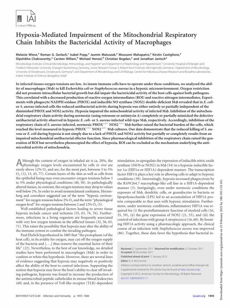
61). We assumed that directly after a bacterial infection in vivophysiological oxygen tensions will prevail, whereas during theprogression of the infection the oxygen tension will progressivelydrop to low levels. In order to mimic this situation in vitro, wesubjected infected M� to hypoxic conditions from 2 h after infec-tion with E. coli HB101 or S. aureus onward until the time point ofreadout (24 h after infection) (Fig. 1A). Under hypoxic conditionswe recovered more bacteria from M� than under normoxic con-ditions (Fig. 1B; see also Table S1 in the supplemental material).However, the amount of intracellular bacteria retrieved from cellsthat had been exposed to hypoxia for 8 h and then subjected tonormoxic conditions for 16 h (reoxygenation) was indistinguish-able from infected cells that had only been cultivated under nor-moxic conditions (Fig. 1B; see also Table S1 in the supplementalmaterial). This indicates that the effect of hypoxia on the survivalof intracellular bacteria is fully reversible.
Hypoxia does not promote the bacterial growth within M�.Next, we wanted to clarify whether the increased survival of S.aureus and E. coli was due to an increased bacterial proliferationwithin the host cells under hypoxic conditions or to the impair-ment of an antibacterial mechanism. Therefore, we analyzed the
FIG 1 Impaired ability of murine bone marrow-derived macrophages (M�) to kill intracellular bacteria under hypoxic conditions. (A) Schematic representationof the experimental setup for assaying intracellular survival of E. coli and S. aureus under normoxic and hypoxic conditions. M� were infected with either E. colior S. aureus at an MOI of 10. One hour after infection extracellular bacteria were removed by a wash with PBS, followed by gentamicin treatment. At 2 h afterinfection the cells were exposed to either normoxia (N) or hypoxia (H; 0.5% oxygen). For reoxygenation (Re) the cells were first incubated for 8 h under hypoxicconditions and then kept in normoxia. Cell lysates were prepared 2 and 24 h after infection to determine the amount of CFU inside the cells. (B) The graphs showsthe percentage of intracellular bacteria in M� under the indicated conditions. Relative survival was calculated by dividing the amount of intracellular bacteriarecovered 24 h after infection related to the amount of intracellular bacteria determined 2 h after infection. The data are means � standard errors of the mean(SEM) of five independent experiments. *, P � 0.05; **, P � 0.01; ***, P � 0.001.
Wiese et al.
1458 iai.asm.org Infection and Immunity
Dow
nloa
ded
from
http
s://j
ourn
als.
asm
.org
/jour
nal/i
ai o
n 23
Feb
ruar
y 20
22 b
y 18
6.10
5.11
9.10
1.

bacterial growth of E. coli and S. aureus grown under normoxicand hypoxic conditions in LB broth. Hypoxia did not increase thegrowth of E. coli or S. aureus compared to normoxic conditions(see Fig. S1 in the supplemental material). Next, we analyzed theintracellular replication dynamics of E. coli and S. aureus in M�.To this end, we used (i) an E. coli strain harboring a reporterplasmid (pDiGc) and (ii) CFSE-labeled S. aureus. The dual fluo-rescence reporter plasmid (pDiGc) encodes a reporter system inwhich the production of DsRed protein is arabinose inducible,whereas the expression of eGFP is constitutive (31). The prolifer-ation of pDiGc-harboring E. coli can be monitored by measuringthe dilution of DsRed fluorescence after the bacterial suspension istransferred to arabinose-free conditions for 6 h (Fig. 2A). Afterlabeling of S. aureus with CFSE, the replication of S. aureus can beassayed by dilution of CFSE-fluorescence for 6 h (Fig. 2C). WhenM� were infected with pDiGc harboring E. coli or CFSE-labeled S.aureus, we could not detect a dilution of DsRed or CFSE fluores-
cence irrespective of whether the infected cells were cultured un-der normoxic or hypoxic conditions (Fig. 2B and D). Taken to-gether, these findings demonstrate that hypoxia does not promoteintracellular bacterial proliferation but impairs the host’s antibac-terial capacity.
HIF1A accumulation in S. aureus-infected M� does not ac-count for the impaired killing of S. aureus under hypoxic con-ditions. HIF1A accumulation has been demonstrated to be a com-mon feature of infected tissues (83) and has been associated with anantibacterial function of myeloid cells (16, 60). Furthermore, it hasbeen shown that macrophages accumulate HIF2A as well and thatHIF2A plays an important role in the regulation of M� function (33,79). Therefore, we analyzed the status of both HIF-� isoforms(HIF1A and HIF2A) in E. coli- and S. aureus-infected M�.
nfection with either pathogen under normoxic conditions re-sulted in the accumulation of HIF1A and HIF2A (Fig. 3A). Hy-poxic incubation of infected cells did not further induce HIF2A
FIG 2 Hypoxia does not improve the bacterial growth within M�. (A and B) To study the replication dynamics of E. coli, a strain was used harboring a dualfluorescence reporter plasmid (pDiGc) in which the production of DsRed protein is arabinose inducible, whereas the expression of eGFP is constitutive. Theproliferation of bacteria can be monitored by measuring the dilution of DsRed fluorescence after placing the bacterial suspension to arabinose-free conditions.Growing E. coli (pDiGc) strains were analyzed immediately (0 h) after placement into arabinose-free medium (RPMI) and 6 h thereafter. (A) GFP-positive,bacterium-sized particles were identified by flow cytometry and in that population the DsRed fluorescence was analyzed. (B) M� were infected with pDiGc-containing E. coli. After different time points, the cells were lysed, and the DsRed fluorescence of GFP-positive, bacterium-sized particles was measured by flowcytometry. The results of a representative experiment out of at least two similar experiments are displayed. (C and D) The replication of S. aureus was investigatedby labeling the bacteria with CFSE. (C) Bacteria were identified after an S. aureus-specific staining. The proliferation of CFSE-labeled S. aureus in medium (RPMI)was accompanied by a reduction in CFSE fluorescence intensity. (D) M� were infected with CFSE-labeled S. aureus. At different time points, the cells were lysed,the bacteria were identified by S. aureus-specific staining, and S. aureus-positive, bacterium-sized particles were analyzed for CFSE fluorescence. The results of arepresentative experiment out of at least two similar experiments are displayed.
Hypoxic Impairment of Bactericidal Activity
April 2012 Volume 80 Number 4 iai.asm.org 1459
Dow
nloa
ded
from
http
s://j
ourn
als.
asm
.org
/jour
nal/i
ai o
n 23
Feb
ruar
y 20
22 b
y 18
6.10
5.11
9.10
1.

accumulation. Therefore, we conclude that it is very unlikely thatHIF2A accounts for the impaired antibacterial capacity under hy-poxic conditions.
Under normoxic conditions infection of M� with E. coli re-sulted in a higher HIF1A protein content compared to an infec-tion of M� with S. aureus. This resulted in an increased expressionof the HIF1A target gene Pgk1 (Fig. 3A and B). In S. aureus-in-fected cells hypoxia further unregulated the level of HIF1A pro-tein, which was not the case in E. coli-infected cells (Fig. 3A).
Accordingly, hypoxia increased the expression of the HIF1A targetgene Pgk1 mRNA in S. aureus infected M� but not in E. coli-infected host cells (Fig. 3B).
Since the impaired antibacterial capacity of the host cells underhypoxic conditions correlated with an increased accumulation ofHIF1A in S. aureus-infected cells, we investigated whether HIF1Aaccounts for this hypoxia-induced phenotype using an RNA in-terference approach. Silencing of Hif1a in S. aureus-infected M�failed to restore the antibacterial activity under hypoxic condi-tions (Fig. 3C). As described earlier (35–37), knockdown effi-ciency was evaluated by analyzing HIF1A protein, Hif1a mRNA,and the HIF1A-dependent metabolic target gene Pgk1 (see Fig. S2in the supplemental material). There was also no effect of Hif1asilencing on the killing of S. aureus by normoxic host cells (Fig.3C). From these data we conclude that the impaired antibacterialcapacity of M� under hypoxic conditions is independent of theincreased HIF1A accumulation.
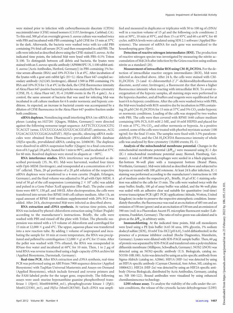
Hypoxia abrogates the production of ROI and RNI by M� inresponse to infection. Since NOS2 and PHOX are oxygen-depen-dent enzymes (2, 18, 41, 44, 51, 66, 81), we analyzed the produc-tion of ROI and RNI in infected M�. As expected, infection of M�with E. coli under normoxic conditions induced a robust genera-tion of NO, which was severely impaired in a hypoxic environ-ment. However, if the cells were reoxygenated for 16 h after aninitial 8 h period of hypoxia, NO levels reached normoxic levelsagain (Fig. 4A). Next, we tested whether the impaired NO produc-tion under hypoxia results from a reduced NOS2 activity or froma diminished NOS2 protein induction under hypoxic conditions.Infection with E. coli led to a comparable NOS2 protein expressionunder both normoxic and hypoxic conditions, strongly suggestingthat hypoxia incapacitates the enzyme activity of NOS2. In con-trast to infection with E. coli, infection of M� with S. aureus hardlycaused an induction of NOS2 protein and NO production (Fig. 4Aand B).
Next, we quantified the production of ROI by M� using theCM-H2DCFDA fluorochrome, which is a nonselective detector ofvarious reactive oxygen species. In order to assess the ROI produc-tion under hypoxic conditions and to avoid any assay related re-oxygenations, we were particularly careful to ensure that the entirestaining procedure of the hypoxia-treated samples was performedin the hypoxia chamber and that all buffers and reagents wereequilibrated to hypoxic conditions. After loading of the dye andincubation under the respective oxygen tension, the samples werefixed with equilibrated PFA and subjected to flow cytometry. Fix-ation with PFA neither generated fluorescence signals nor inhib-ited the fluorescence of CM-H2DCFDA (data not shown). Underhypoxic conditions we could not detect any production of ROIafter infection with E. coli and S. aureus, whereas ROI were readilydetectable under normoxic conditions (Fig. 4C). Together, ourdata show that hypoxia prevents the production of RNI and ROIin response to a bacterial infection of M�, as expected from theoxygen dependency of NOS2 and PHOX.
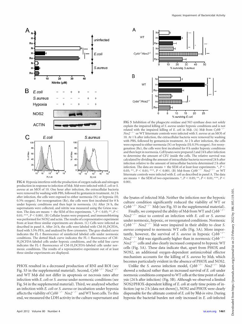
A PHOX- and NOS2-independent, oxygen-dependent anti-microbial mechanism contributes to the control of E. coli and S.aureus in M�. In order to investigate whether the reduced anti-bacterial capacity of M� during hypoxia simply results from anabsent NOS2 and PHOX activity, we infected M� deficient forboth NOS2 and PHOX (Cybb�/� Nos2�/�) and tested the impactof hypoxia versus normoxia on the survival of E. coli or S. aureus inthese cells. First, we confirmed that a deficiency in NOS2 and
FIG 3 Hypoxia augmented the HIF1A activity of S. aureus-infected M� butdoes not contribute to the impaired killing of S. aureus under hypoxic condi-tions. (A) Cellular lysates were prepared of M� infected with E. coli or S. aureusunder normoxic or hypoxic conditions for 24 h, and immunoblotting forHIF1A and HIF2A was performed. Equal loading is demonstrated in HIF1Aimmunoblots by incubating the blots with an actin-specific antibody. To dem-onstrate equal loading in blots probed for HIF2A, a nonspecific band (n.s.) ofthe HIF2A antibody is shown. The results of a representative experiment out ofat least three similar experiments are displayed. (B) M� were infected with E.coli or S. aureus under normoxic (N) or hypoxic (H) conditions for 24 h, andqRT-PCR was performed with Pgk1 as the target and Hprt1 serving as theinternal control. The data are means � the SEM of four experiments. *, P �0.05; **, P � 0.01; ***, P � 0.001. (C) ns-siRNA (ns) or Hif1a-specific (Hif1a)siRNA was transferred into M�, or the cells were left untreated (untreated).After 24 h, the cells were infected with S. aureus and subjected to normoxic (N)or hypoxic (H) conditions. Cell lysates were prepared 2 and 24 h after infectionto determine the CFU count inside the cells. The relative survival was calcu-lated by dividing the amount of intracellular bacteria recovered 24 h afterinfection related to the amount of intracellular bacteria determined 2 h afterinfection. The data are means � the SEM of four experiments. *, P � 0.05; **,P � 0.01; ***, P � 0.001.
Wiese et al.
1460 iai.asm.org Infection and Immunity
Dow
nloa
ded
from
http
s://j
ourn
als.
asm
.org
/jour
nal/i
ai o
n 23
Feb
ruar
y 20
22 b
y 18
6.10
5.11
9.10
1.
PHOX resulted in a decreased production of RNI and ROI (seeFig. S3 in the supplemental material). Second, Cybb�/� Nos2�/�
and WT M� did not differ in apoptosis or necrosis rates afterinfection with E. coli or S. aureus under normoxic conditions (seeFig. S4 in the supplemental material). Third, we analyzed whetheran infection with E. coli or S. aureus or incubation under hypoxiaaffects the viability of Cybb�/� Nos2�/� and WT host cells. To thisend, we measured the LDH activity in the culture supernatant and
the lysates of infected M�. Neither the infection nor the hypoxicculture condition significantly reduced the viability of WT orCybb�/� Nos2�/� M� (see Fig. S5 in the supplemental material).
Finally, we compared the ability of M� from WT and Cybb�/�
Nos2�/� mice to control an infection with E. coli or S. aureusunder normoxic, hypoxic, or reoxygenated conditions. NormoxicCybb�/� Nos2�/� M� were impaired in their ability to clear S.aureus compared to normoxic WT cells (Fig. 5A). More impor-tantly, however, the survival of S. aureus in hypoxic Cybb�/�
Nos2�/� M� was significantly higher than in normoxic Cybb�/�
Nos2�/� cells and also clearly increased compared to hypoxic WTcells (Fig. 5A). These data indicate that, apart from PHOX andNOS2, an additional oxygen-dependent antimicrobial effectormechanism accounts for the killing of S. aureus by M�, whichbecomes particularly evident in the absence of PHOX and NOS2.
Unlike the S. aureus infection model, Cybb�/� Nos2�/� M�showed a reduced rather than an increased survival of E. coli undernormoxic conditions compared to WT cells at the time point of anal-ysis (24 h after infection) (Fig. 5B). Although we observed a limitedNOS2/PHOX-dependent killing of E. coli at early time points of in-fection (up to 2 h [data not shown]), NOS2 and PHOX were clearlydispensable for the ultimate control of E. coli by � in vitro. Duringhypoxia the bacterial burden not only increased in E. coli-infected
FIG 4 Hypoxia interferes with the production of oxygen radicals and nitrogenproduction in response to infection of M�. M� were infected with E. coli or S.aureus at an MOI of 10. One hour after infection, the extracellular bacteriawere removed by washing with PBS, followed by gentamicin treatment. At 2 hafter infection, the cells were exposed to either normoxia (N) or hypoxia (H;0.5% oxygen). For reoxygenation (Re), the cells were first incubated for 8 hunder hypoxic conditions and then kept in normoxia. (A) After 24 h, thesupernatants were collected, and nitrite was measured using the Griess reac-tion. The data are means � the SEM of five experiments. *, P � 0.05; **, P �0.01; ***, P � 0.001. (B) Cellular lysates were prepared, and immunoblottingwas performed for NOS2 and actin. The results of a representative experimentfrom at least three similar experiments are shown. (C) Cells were infected asdescribed in panel A. After 24 h, the cells were labeled with CM-H2DCFDA,fixed with 3.5% PFA, and analyzed by flow cytometry. The gray-shaded curveindicates the FL-1 fluorescence of uninfected labeled cells under normoxicconditions. The dotted black curve indicates the FL-1 fluorescence of CM-H2DCFDA-labeled cells under hypoxic conditions, and the solid line curveindicates the FL-1 fluorescence of CM-H2DCFDA-labeled cells under nor-moxic conditions. The results of a representative experiment out of at leastthree similar experiments are displayed.
FIG 5 Inhibition of the phagocyte oxidase and NO synthase does not solelyexplain the impaired killing of S. aureus under hypoxic conditions and is notrelated with the impaired killing of E. coli in M�. (A) M� from Cybb�/�
Nos2�/� or WT littermate controls were infected with S. aureus at an MOI of10. At 1 h after infection, the extracellular bacteria were removed by washingwith PBS, followed by gentamicin treatment. At 2 h after infection, the cellswere exposed to either normoxia (N) or hypoxia (H; 0.5% oxygen). For reoxy-genation (Re), the cells were first incubated for 8 h under hypoxic conditionsand then kept in normoxia. Cell lysates were prepared 2 and 24 h after infectionto determine the amount of CFU inside the cells. The relative survival wascalculated by dividing the amount of intracellular bacteria recovered 24 h afterinfection relative to the amount of intracellular bacteria determined 2 h afterinfection. The data are means � the SEM of at least four experiments. *, P �0.05; **, P � 0.01; ***, P � 0.001. (B) M� from Cybb�/� Nos2�/� or WTlittermate controls were infected with E. coli as described in panel A. The dataare means � the SEM of two experiments. *, P � 0.05; **, P � 0.01; ***, P �0.001.
Hypoxic Impairment of Bactericidal Activity
April 2012 Volume 80 Number 4 iai.asm.org 1461
Dow
nloa
ded
from
http
s://j
ourn
als.
asm
.org
/jour
nal/i
ai o
n 23
Feb
ruar
y 20
22 b
y 18
6.10
5.11
9.10
1.

WT M� but also in Cybb�/� Nos2�/� M� (Fig. 5B). From these data,we conclude that the control of E. coli by M� is due to a PHOX- andNOS2-independent, but oxygen-dependent effector mechanism.
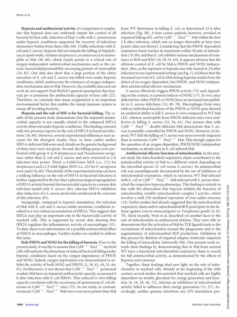
Hypoxia inhibits the activity of the mitochondrial respira-tory chain in E. coli- or S. aureus-infected M�. Mitochondrialactivity is impaired under hypoxia (14). Sonoda et al. demonstratedthat M� that are genetically defective for mitochondrial ROI produc-tion show a decreased capacity to degrade intracellular L. monocyto-genes compared to WT M�, even in the presence of an inhibitor ofNOS2 or PHOX (78). In order to determine the mitochondrial activ-ity of infected M�, we determined the mitochondrial membrane po-tential (��M) by using JC-1 dye. The lipophilic, cationic dye JC-1 canselectively enter into mitochondria and reversibly change color fromgreen to red as the membrane potential increases. In cells with a high��M, JC-1 spontaneously forms complexes known as J-aggregateswith intense red fluorescence. In cells with a low ��M, JC-1 remainsin the monomeric form, which shows only green fluorescence. Theratio of this red/green fluorescence is independent of mitochondrialshape, density, or size but depends only on the membrane potential
(15). Since this dye is not suitable for fixation, we sealed the platesharboring the infected cells with an adhesive clear seal suitable forqRT-PCR applications in order to preserve the respective normoxicand hypoxic condition. We observed that in infected M� the ��M
decreased in rotenone- and hypoxia-treated cells (Fig. 6A). However,the hypoxic impairment of ��M was not due to toxic effects becausewithin 5 min of reoxygenation (i.e., removal of the adhesive seal), weobserved a substantial increase of��M in S. aureus-infected (data notshown) and E. coli-infected (see Fig. S6 in the supplemental material)M�. Thus, we conclude that hypoxia works in analogy to rotenoneand interferes with mitochondrial activity.
Inhibition of the mitochondrial respiratory chain com-pletely or partially mimics the effect of hypoxia on the killing ofE. coli and S. aureus, respectively. Next, we analyzed the fate of E.coli in M� after inhibition of mitochondrial respiration. For thatpurpose, we used rotenone that binds to the ubiquinone bindingsite of complex I and thereby interferes with electron transport.We used different concentrations of rotenone (10 �M, 100 �M,and 1 mM) and correlated this treatment with the bactericidal
FIG 6 Hypoxia impairs mitochondrial activity in M�, and inhibition of the mitochondrial respiratory chain completely or partially mimics the effect of hypoxiaon the killing of E. coli or S. aureus by M�, respectively. (A) M� were infected with E. coli or S. aureus at an MOI of 10. At 1 h after infection, extracellular bacteriawere removed by washing with PBS, followed by gentamicin treatment. At 2 h after infection, the cells were exposed to either normoxia (N) or hypoxia (H; 0.5%oxygen) in the absence or presence of rotenone (100 �M). After at least 24 h, the cells were stained with JC-1. The mitochondrial membrane potential (��M) wasdetermined and is given in arbitrary units (AU). The data are means � the SD out of three similar experiments performed in triplicate. *, P � 0.05; **, P � 0.01;***, P � 0.001. (B and C) Cells were infected with E. coli (B) or S. aureus (C) and treated as described in panel A. Cell lysates were prepared 2 and 24 h afterinfection to determine the number of CFU inside the cells. The relative survival was calculated by dividing the amount of intracellular bacteria recovered 24 h afterinfection related to the amount of intracellular bacteria determined 2 h after infection. The data are means � the SEM of six experiments. *, P � 0.05; **, P � 0.01;***, P � 0.001. (D and E) Cells were infected with E. coli (D) or S. aureus (E). At 2 h after infection, the cells were exposed to either normoxia (N) or hypoxia (H;0.5% oxygen) in the absence or presence of antimycin A (4 �g/ml). Cell lysates were prepared 2 and 24 h after infection to determine the number of CFU insidethe cells. The relative survival was calculated by dividing the amount of intracellular bacteria recovered 24 h after infection related to the amount of intracellularbacteria determined 2 h after infection. The data are means � the SEM of at least four experiments. *, P � 0.05; **, P � 0.01; ***, P � 0.001. (F) M� were leftuntreated or treated with rotenone (100 �M) and subjected to normoxic or hypoxic conditions. After 24 h, the cells were labeled with CM-H2DCFDA, fixed with3.5% PFA, and analyzed by flow cytometry. The gray-shaded curve indicates the FL-1 fluorescence of labeled cells under normoxic conditions. The black dottedcurve indicates the FL-1 fluorescence of CM-H2DCFDA-labeled cells after stimulation with rotenone and incubation under hypoxic conditions, and the blacksolid curve demonstrates the FL-1 fluorescence of CM-H2DCFDA-labeled cells after stimulation with rotenone and incubation under normoxic conditions.
Wiese et al.
1462 iai.asm.org Infection and Immunity
Dow
nloa
ded
from
http
s://j
ourn
als.
asm
.org
/jour
nal/i
ai o
n 23
Feb
ruar
y 20
22 b
y 18
6.10
5.11
9.10
1.

activity of E. coli-infected M�. Rotenone at 10 �M barely inhib-ited the bactericidal activity. However, 100 �M rotenone robustlyimpaired the antibacterial capacity (see Fig. S7 in the supplemen-tal material). Rotenone at 1 mM appeared to be toxic to the hostcells. For further experiments, we therefore used a 100 �M con-centration, which proved to sufficiently inhibit mitochondrial ac-tivity (Fig. 6A). For the control of host cell viability, we analyzedthe relative LDH release rate and NO production under normoxicand hypoxic conditions in the absence or presence of rotenone. Atthe concentrations used we could not detect any toxicity of rote-none in LDH release assays (see Fig. S5 in the supplemental ma-terial). Moreover, inhibition of mitochondrial respiration did notaffect NO production upon infection (see Fig. S8 in the supple-mental material). In order to exclude that rotenone inhibits bac-terial cell division and viability in vitro, we analyzed the growth ofE. coli in the absence or presence of rotenone. We found that E. coligrew equally well in the presence or absence of rotenone (see Fig.S9 in the supplemental material) as described previously (27).When rotenone was added to E. coli-infected M� under normoxicconditions, we found that the bacterial survival rate exactlyreached the level observed in hypoxic M� without rotenone. Im-portantly, the addition of rotenone to hypoxic cultures did notfurther inhibit the antimicrobial activity of M� (Fig. 6B).However, treatment of S. aureus-infected WT M� with rote-none under normoxic conditions did not lead to the same in-crease of S. aureus, as seen in hypoxic M� cultures in the ab-sence of rotenone (Fig. 6C).
Furthermore, we used antimycin A to inhibit the coenzymeQ/cytochrome c/oxidoreductase, which is located in complex IIIof the respiratory chain. Antimycin A did not interfere with bac-terial cell division and viability in vitro (see Fig. S11 in the supple-mental material). After treatment of E. coli-infected macrophageswith antimycin A, bacterial survival reached exactly the level ofhypoxia-treated macrophages (Fig. 6D). However, treatment of S.aureus-infected WT M� with antimycin A did not lead to the sameincrease of S. aureus as seen in hypoxic M� cultures in the absenceof antimycin A (Fig. 6E). Therefore, we conclude that inhibition ofmitochondrial activity completely or partially mimicked the de-fective antibacterial capacity observed in hypoxic E. coli- or S.aureus-infected WT M�.
Inhibition of the mitochondrial respiratory chain and notmitochondrial ROI account for rotenone’s effects on bacteri-cidal activity. Next, we wanted to clarify whether mitochondrialrespiration or mitochondrial ROI accounts for our observation.In accordance with the findings from West et al. (84), we observedan increased production of ROI after treatment of M� with rote-none under normoxic conditions (Fig. 6F). However, we did notobserve a significant ROI production of M� infected with E. coli orS. aureus under hypoxic conditions (see Fig. S3 in the supplemen-tal material; Fig. 4C). Thus, treatment of macrophages with rote-none and hypoxic treatment led to a change in ROI levels in op-posite directions. Since hypoxia and rotenone induced similareffects on the mitochondrial and bactericidal killing, we concludethat mitochondrial respiration and not mitochondrial ROI ac-counts for our findings.
Inhibition of the mitochondrial respiratory chain is thePHOX- and NOS2-independent component of the impairedkilling of S. aureus by M� during hypoxia. Having seen that alack of PHOX and NOS2 activity only partially explains the re-duced antibacterial activity of M� against S. aureus during hyp-
oxia (Fig. 5), we performed the reverse experimental approachand tested the effect of inhibition of mitochondrial respiratorychain activity on the survival of S. aureus. Treatment of S. aureus-infected WT M� with rotenone under normoxic conditions didnot lead to the same increase of S. aureus, as seen in hypoxic M�cultures in the absence of rotenone. Furthermore, exposure of S.aureus-infected WT M� to rotenone during hypoxia caused anadditional significant increase in the bacterial load compared tothe normoxic or rotenone-free hypoxic controls (Fig. 7). InCybb�/� Nos2�/� M� cultures rotenone treatment raised the bac-terial numbers by a factor of 2.5 during normoxia but was ineffec-tive under hypoxic conditions. These data demonstrate thatPHOX and NOS2, as well as an oxygen-dependent mitochondrialantibacterial killing mechanism, contribute to the control of S.aureus in M�.
DISCUSSION
To the best of our knowledge, the present study demonstrates forthe first time that hypoxia impairs the bactericidal activity of pri-mary M�. A related observation has previously been made withhuman neutrophils, which turned out to be poor killers of E. coliand S. aureus under anaerobic conditions (49). McGovern et al.attributed this reduced killing of granulocytes to an inhibition ofthe PHOX-dependent respiratory burst (51). Our present dataobtained with primary M� illustrate that, in addition to PHOXand NOS2, the mitochondrial respiratory chain is highly relevantfor the antibacterial activity of phagocytes. Collectively, these invitro observations with myeloid cells offer an explanation for theaggravated course of S. aureus lung and skin infections followingsystemic hypoxia in vivo (29, 30, 38). Interestingly, hypoxia—amilieu often encountered in estuarine marine habitats—also in-hibits the elimination of Vibrio campbellii in the economicallyimportant shellfish species Crassostera virginica (48). This indi-cates that oxygen availability is of general importance for the con-trol of bacterial pathogens by host organisms.
FIG 7 Inhibition of the mitochondrial respiratory chain is the PHOX- andNOS2-independent component of the impaired killing of S. aureus by M�during hypoxia. M� from Cybb�/� Nos2�/� or WT littermate controls wereinfected with S. aureus. At 1 h after infection, extracellular bacteria were re-moved by washing with PBS, followed by gentamicin treatment. At 2 h afterinfection, the cells were exposed to either normoxia (N) or hypoxia (H; 0.5%oxygen) in the absence or presence of rotenone (100 �M). Cell lysates wereprepared 2 and 24 h after infection to determine the amount of CFU inside thecells. The relative survival was calculated by dividing the amount of intracel-lular bacteria recovered 24 h after infection relative to the amount of intracel-lular bacteria determined 2 h after infection. The data are means � the SEM ofat least four experiments. *, P � 0.05; **, P � 0.01; ***, P � 0.001.
Hypoxic Impairment of Bactericidal Activity
April 2012 Volume 80 Number 4 iai.asm.org 1463
Dow
nloa
ded
from
http
s://j
ourn
als.
asm
.org
/jour
nal/i
ai o
n 23
Feb
ruar
y 20
22 b
y 18
6.10
5.11
9.10
1.
Hypoxia and antibacterial activity. It is important to empha-size that hypoxia does not uniformly impair the control of allbacteria by host cells. Infection of Hep-2 cells with C. pneumoniaeunder hypoxic conditions supported the recovery of infectiouselementary bodies from these cells (68). Unlike infections with E.coli and S. aureus, hypoxia did not impede the killing of Staphylo-coccus epidermidis, viridans streptococci, or enterococci in neutro-phils or M� (49, 60), which clearly points to a critical role ofoxygen-independent antimicrobial mechanisms such as the cat-ionic bactericidal/permeability-increasing protein of neutrophils(24, 82). Our data also show that a large portion of the entireinoculum of E. coli and S. aureus was killed even under hypoxicconditions, which underscores the existence of oxygen-indepen-dent mechanisms also in M�. However, the available data and ourwork do not support Paul Ehrlich’s general assumption that hyp-oxia per se promotes the antibacterial capacity of host cells (22).Therefore, we conclude that tissue oxygenation is an importantenvironmental factor that enables the innate immune system tosweep off invading bacteria.
Hypoxia and the role of HIF-� for bacterial killing. The re-sults of the present study demonstrate that the impaired antimi-crobial capacity is not causally related to the enhanced HIF1Aactivity observed under hypoxic conditions. This finding contrastswith two previous reports on the role of HIF1A in bacterial infec-tions (16, 60). However, several experimental differences may ac-count for the divergent results. First, in these earlier studiesHIF1A-deficient M� were used; details on the genetic backgroundof these mice were not given. Second, the killing assays were per-formed with group A or B streptococci and Pseudomonas aerugi-nosa rather than E. coli and S. aureus and were restricted to 2-hinfection time points. Third, a 4-fold-lower MOI (i.e., 2.5) forstreptococci and a 2.5-fold-higher MOI (i.e., 25) for P. aeruginosawere used (16, 60). That details of the experimental setup can havea striking influence on the role of HIF1A in bacterial infections isfurther supported by the fact that a pharmacological upregulationof HIF1A activity boosted the bactericidal capacity in a mouse skininfection model with S. aureus (86), whereas HIF1A inhibitionafter induction of a S. aureus peritonitis ameliorated the outcomeof this infection (83).
Intriguingly, compared to hypoxic stimulation, the infectionof M� with E. coli and S. aureus under normoxic conditions re-sulted in a very robust accumulation of HIF2A. This suggests thatHIF2A may play an important role in the bactericidal activity ofmyeloid cells. This is supported by recent data showing thatHIF2A regulates the inflammatory activity of macrophages (33).To date, there is no information on a possible antimicrobial effectof HIF2A in macrophages. Further studies are needed to addressthis issue.
Role PHOX and NOS2 for the killing of bacteria. Prior to thepresent study, it was fair to assume that Cybb�/� Nos2�/� myeloidcells will replicate the phenotype of reduced bacterial killing underhypoxic conditions based on the oxygen dependency of PHOXand NOS2. Indeed, oxygen deprivation was demonstrated to in-hibit the activity of both NOS2 and PHOX (2, 18, 41, 44, 51, 66,81). Furthermore, it was shown that Cybb�/� Nos2�/� peritonealexudate M� have an impaired antibacterial capacity, as assessed 4h after infection with E. coli HB101. This reduced in vitro killingcapacity correlated with the occurrence of spontaneous E. coli ab-scesses in Cybb�/� Nos2�/� mice (75). In our study, in contrast,normoxic Cybb�/� Nos2�/� M� were even more potent than M�
from WT littermates in killing E. coli, as determined 24 h afterinfection (Fig. 5B). A time course analysis, however, revealed animpaired killing of E. coli by Cybb�/� Nos2�/� M� within the first2 h after infection, which was no longer detectable at later timepoints (data not shown). Considering that the PHOX-dependentrespiratory burst reaches its maximum within 30 min of stimula-tion (19, 80) and that E. coli exhibits various mechanisms of resis-tance to ROI and RNI (10, 50, 51, 64), it appears obvious that theultimate control of E. coli by M� is PHOX and NOS2 indepen-dent. Also, as the exposure to hypoxia was only started at 2 h afterinfection in our experimental setting (see Fig. 1), it follows that theincreased survival of E. coli in M� during hypoxia results from thedefect of an oxygen-dependent, but PHOX- and NOS2-indepen-dent antimicrobial effector mechanism.
S. aureus effectively triggers PHOX activity (73) and, depend-ing on the context, is a potent inducer of NOS2 (17). In vivo, micedeficient for either PHOX or NOS2 have an increased susceptibil-ity to S. aureus infections (52, 69, 70). Macrophages from micewith an individual knockout of the PHOX or NOS2 gene showedan unaltered ability to kill S. aureus in vitro compared to WT M�(42), whereas neutrophils from PHOX-deficient mice were inef-fective in killing S. aureus (23, 34, 62). Our present data withCybb�/� Nos2�/� double-deficient M� demonstrate that S. au-reus is partially controlled by PHOX and NOS2. However, in hy-poxic WT M� the killing of S. aureus was more severely impairedthan in normoxic Cybb�/� Nos2�/� M�. This strongly argues forthe operation of an oxygen-dependent, PHOX/NOS2-independentmechanism, as already seen in E. coli-infected M�.
Antibacterial effector function of mitochondria. In the pres-ent study the mitochondrial respiratory chain contributed to theantimicrobial activity of M� to a different extent depending onthe microbial species (E. coli versus S. aureus). Its antimicrobialrole was unambiguously documented by the use of inhibitors ofmitochondrial respiration, which in normoxic WT M� infectedwith E. coli or Cybb�/� Nos2�/� M� infected with S. aureus mim-icked the respective hypoxia phenotype. This finding is entirely inline with the observation that hypoxia inhibits the function ofmitochondria, notably mitochondrial complex I activity, whichinvolves a miR-210-mediated repression of iron-sulfur enzymes(14). Earlier studies had already suggested that the mitochondrialrespiratory chain and/or mitochondrial ROI participate in the de-fense against Listeria monocytogenes or Toxoplasma gondii (4, 67,78). Most recently, West et al. described yet another facet to therole of mitochondria in antibacterial defense. They were able todemonstrate that the activation of M� by TLR ligands leads to therecruitment of mitochondria toward the phagosome and to theaugmentation of mitochondrial ROI production. Inhibition ofthis process by deletion of required adaptor molecules impairedthe killing of intracellular Salmonella (84). Our present work ex-tends these findings by demonstrating that in M� from normalWT mice a functional mitochondrial respiratory chain is crucialfor full antimicrobial activity, as demonstrated by the effects ofhypoxia and rotenone.
Together, these findings shed new light on the role of mito-chondria in myeloid cells. Already at the beginning of the 20thcentury several studies documented that myeloid cells are highlydependent on aerobic glycolysis for energy generation and func-tion (6, 16, 28, 46, 72), whereas an inhibition of mitochondrialactivity failed to influence their energy generation (11, 57). Ac-cordingly, neither gamma interferon nor LPS stimulation pro-
Wiese et al.
1464 iai.asm.org Infection and Immunity
Dow
nloa
ded
from
http
s://j
ourn
als.
asm
.org
/jour
nal/i
ai o
n 23
Feb
ruar
y 20
22 b
y 18
6.10
5.11
9.10
1.

moted fatty acid �-oxidation (43, 78) but instead increased mito-chondrial ROI production in M� (25, 78). This strongly suggeststhat mitochondrial function may be beyond energy generation.We propose that in the presence of ample oxygen myeloid cells canutilize mitochondria for antimicrobial defense because they canshift the energy generation from mitochondria to aerobic glycol-ysis resembling a metabolic process induced in transformed cells(“Warburg effect”).
There are at least two clinical situations in which an impairedmitochondrial activity in M� may account for the occurrence ofsevere infections. First, the Barth syndrome, a rare X-linked reces-sive disorder, is associated with mitochondrial dysfunction. Car-diac failure and septicemia are the leading causes of death of chil-dren suffering from this disease (7). Until now neutropenia hasbeen considered as the main factor predisposing these patients tosepticemia. However, mitochondrial dysfunction of M� andhence a reduced bactericidal activity may very well contribute tothe fatal infections. Second, mitochondrial damage has been asso-ciated with poor outcome in sepsis patients (26). Mitochondrialdysfunction may be a consequence of hypoxia and hypoxic signal-ing, which is commonly encountered in the tissues of septic pa-tients, or induced by pathogens, their products, and/or inflamma-tory mediators. It is tempting to speculate that the predispositionof septic patients to opportunistic infections in intensive careunits may be associated with the dysfunction of mitochondria inmononuclear phagocytes, which normally keep commensal bac-teria at bay.
In summary, hypoxia shuts down not only the enzyme activityof PHOX and NOS2 but also the antibacterial effector function ofmitochondria. Further studies are needed to precisely investigate themechanisms by which mitochondria promote oxygen-dependentbacterial clearance and to determine how hypoxia interferes with thatprocess in mononuclear phagocytes.
ACKNOWLEDGMENTS
We thank Anja Lührmann for stimulating discussions.This study was supported by grants to C.B., M.H., and J.J. from the
Deutsche Forschungsgemeinschaft (Bo996/3-3, SFB643 project A6, JA1993/1-1), by grants to C.B., M.H., C.W., and J.J. from the Interdiszi-plinäres Zentrum für Klinische Forschung (projects A24, A28, and A49),and by a grant to J.J. from the ELAN program (Az. 11.01.26.1) at theUniversitätsklinikum Erlangen and Friedrich-Alexander-Universität Er-langen-Nürnberg.
REFERENCES1. Ackermann M, et al. 2008. Self-destructive cooperation mediated by
phenotypic noise. Nature 454:987–990.2. Albina JE, Henry WL, Jr, Mastrofrancesco B, Martin BA, Reichner JS.
1995. Macrophage activation by culture in an anoxic environment. J.Immunol. 155:4391– 4396.
3. Anand RJ, et al. 2007. Hypoxia causes an increase in phagocytosis bymacrophages in a HIF-1�-dependent manner. J. Leukoc. Biol. 82:1257–1265.
4. Arsenijevic D, et al. 2000. Disruption of the uncoupling protein-2 gene inmice reveals a role in immunity and reactive oxygen species production.Nat. Genet. 26:435– 439.
5. Atkuri KR, Herzenberg LA, Niemi AK, Cowan T, Herzenberg LA. 2007.Importance of culturing primary lymphocytes at physiological oxygenlevels. Proc. Natl. Acad. Sci. U. S. A. 104:4547– 4552.
6. Bakker A. 1927. Einige Ubereinstimmungen im Stoffwechsel der Carci-nomzellen und Exsudatleukocyten. Klin. Wochenschr. 6:252.
7. Barth PG, et al. 1999. X-linked cardioskeletal myopathy and neutropenia(Barth syndrome) (MIM 302060). J. Inherit. Metab. Dis. 22:555–567.
8. Bogdan C. 2011. Reactive oxygen and reactive nitrogen intermediates in
the immune system, p 69 – 84. In Kaufmann SH, Rouse B, Sacks D (ed),Immunology of infectious diseases. ASM Press, Washington, DC.
9. Bogdan C, Rollinghoff M, Diefenbach A. 2000. Reactive oxygen andreactive nitrogen intermediates in innate and specific immunity. Curr.Opin. Immunol. 12:64 –76.
10. Bonamore A, Boffi A. 2008. Flavohemoglobin: structure and reactivity.IUBMB Life 60:19 –28.
11. Borregaard N, Herlin T. 1982. Energy metabolism of human neutrophilsduring phagocytosis. J. Clin. Invest. 70:550 –557.
12. Caldwell CC, et al. 2001. Differential effects of physiologically relevanthypoxic conditions on T lymphocyte development and effector functions.J. Immunol. 167:6140 – 6149.
13. Campbell JA. 1925. The influence of O2 tension in the inspired air uponthe O2 tension in the tissues. J. Physiol. 60:20 –29.
14. Chan SY, et al. 2009. MicroRNA-210 controls mitochondrial metabolismduring hypoxia by repressing the iron-sulfur cluster assembly proteinsISCU1/2. Cell Metab. 10:273–284.
15. Chazotte B. September 2011. Labeling mitochondria with JC-1. ColdSpring Harbor Protoc. doi:10.1101/pdb.prot065490.
16. Cramer T, et al. 2003. HIF-1� is essential for myeloid cell-mediatedinflammation. Cell 112:645– 657.
17. Cunha FQ, Assreuy J, Moncada S, Liew FY. 1993. Phagocytosis andinduction of nitric oxide synthase in murine macrophages. Immunology79:408 – 411.
18. Daniliuc S, et al. 2003. Hypoxia inactivates inducible nitric oxide syn-thase in mouse macrophages by disrupting its interaction with alpha-actinin 4. J. Immunol. 171:3225–3232.
19. De la Harpe J, Nathan CF. 1985. A semi-automated micro-assay forH2O2 release by human blood monocytes and mouse peritoneal macro-phages. J. Immunol. Methods 78:323–336.
20. Ding AH, Nathan CF, Stuehr DJ. 1988. Release of reactive nitrogenintermediates and reactive oxygen intermediates from mouse peritonealmacrophages: comparison of activating cytokines and evidence for inde-pendent production. J. Immunol. 141:2407–2412.
21. Dlaska M, Weiss G. 1999. Central role of transcription factor NF-IL6 forcytokine and iron-mediated regulation of murine inducible nitric oxidesynthase expression. J. Immunol. 162:6171– 6177.
22. Ehrlich P. 1885. Das Sauerstoff-Bedürfniss des Organismus, vol 1. Spring-er-Verlag, Berlin, Germany.
23. Ellson CD, et al. 2006. Neutrophils from p40phox�/� mice exhibit severedefects in NADPH oxidase regulation and oxidant-dependent bacterialkilling. J. Exp. Med. 203:1927–1937.
24. Elsbach P, Weiss J. 1985. Oxygen-dependent and oxygen-independentmechanisms of microbicidal activity of neutrophils. Immunol. Lett. 11:159 –163.
25. Emre Y, et al. 2007. Mitochondria contribute to LPS-induced MAPKactivation via uncoupling protein UCP2 in macrophages. Biochem. J. 402:271–278.
26. Exline MC, Crouser ED. 2008. Mitochondrial mechanisms of sepsis-induced organ failure. Front. Biosci. 13:5030 –5041.
27. Finel M. 1996. Genetic inactivation of the H�-translocating NADH:ubiquinone oxidoreductase of Paracoccus denitrificans is facilitated by in-sertion of the ndh gene from Escherichia coli. FEBS Lett. 393:81– 85.
28. Fleischmann W, Kubowitz F. 1927. Uber den Stoffwechsel der Leuko-cyten. Biochem. Z. 181:395.
29. Green GM, Kass EH. 1964. Factors influencing the clearance of bacteriaby the lung. J. Clin. Invest. 43:769 –776.
30. Harris GD, Johanson WG, Jr, Pierce AK. 1977. Determinants of lungbacterial clearance in mice after acute hypoxia. Am. Rev. Respir. Dis. 116:671– 677.
31. Helaine S, et al. 2010. Dynamics of intracellular bacterial replication atthe single cell level. Proc. Natl. Acad. Sci. U. S. A. 107:3746 –3751.
32. Hunt TK, Twomey P, Zederfeldt B, Dunphy JE. 1967. Respiratory gastensions and pH in healing wounds. Am. J. Surg. 114:302–307.
33. Imtiyaz HZ, et al. 2010. Hypoxia-inducible factor 2alpha regulates mac-rophage function in mouse models of acute and tumor inflammation. J.Clin. Invest. 120:2699 –2714.
34. Jackson SH, Gallin JI, Holland SM. 1995. The p47phox mouse knockoutmodel of chronic granulomatous disease. J. Exp. Med. 182:751–758.
35. Jantsch J, et al. 2008. Hypoxia and hypoxia-inducible factor-1 alphamodulate lipopolysaccharide-induced dendritic cell activation and func-tion. J. Immunol. 180:4697– 4705.
36. Jantsch J, et al. 2008. Small interfering RNA (siRNA) delivery into murine
Hypoxic Impairment of Bactericidal Activity
April 2012 Volume 80 Number 4 iai.asm.org 1465
Dow
nloa
ded
from
http
s://j
ourn
als.
asm
.org
/jour
nal/i
ai o
n 23
Feb
ruar
y 20
22 b
y 18
6.10
5.11
9.10
1.
bone marrow-derived dendritic cells by electroporation. J. Immunol.Methods 337:71–77.
37. Jantsch J, et al. Toll-like receptor activation and hypoxia use distinctsignaling pathways to stabilize hypoxia-inducible factor 1� (HIF1A) andresult in differential HIF1A-dependent gene expression. J. Leukoc. Biol.90:551–562.
38. Jonsson K, Hunt TK, Mathes SJ. 1988. Oxygen as an isolated variableinfluences resistance to infection. Ann. Surg. 208:783–787.
39. Kaelin WG, Jr, Ratcliffe PJ. 2008. Oxygen sensing by metazoans: thecentral role of the HIF hydroxylase pathway. Mol. Cell 30:393– 402.
40. Karhausen J, et al. 2004. Epithelial hypoxia-inducible factor-1 is protec-tive in murine experimental colitis. J. Clin. Invest. 114:1098 –1106.
41. Kim N, et al. 1993. Oxygen tension regulates the nitric oxide pathway:physiological role in penile erection. J. Clin. Invest. 91:437– 442.
42. Kohler J, et al. NADPH-oxidase but not inducible nitric oxide synthasecontributes to resistance in a murine Staphylococcus aureus Newmanpneumonia model. Microbes Infect. 13:914 –922.
43. Krawczyk CM, et al. 2010. Toll-like receptor-induced changes in glyco-lytic metabolism regulate dendritic cell activation. Blood 115:4742– 4749.
44. Kwon NS, et al. 1990. L-Citrulline production from L-arginine by mac-rophage nitric oxide synthase: the ureido oxygen derives from dioxygen. J.Biol. Chem. 265:13442–13445.
45. Laser H. 1937. Tissue metabolism under the influence of low oxygentension. Biochem. J. 31:1671–1676.
46. Levene PA, Meyer GM. 1912. The action of leucocytes on glucose, secondcommunication. J. Biol. Chem. 12:265–273.
47. Lissner CR, Weinstein DL, O’Brien AD. 1985. Mouse chromosome 1 Itylocus regulates microbicidal activity of isolated peritoneal macrophagesagainst a diverse group of intracellular and extracellular bacteria. J. Immu-nol. 135:544 –547.
48. Macey BM, Achilihu IO, Burnett KG, Burnett LE. 2008. Effects ofhypercapnic hypoxia on inactivation and elimination of Vibrio campbelliiin the Eastern oyster, Crassostrea virginica. Appl. Environ. Microbiol. 74:6077– 6084.
49. Mandell GL. 1974. Bactericidal activity of aerobic and anaerobic poly-morphonuclear neutrophils. Infect. Immun. 9:337–341.
50. Mason MG, et al. 2009. Cytochrome bd confers nitric oxide resistance toEscherichia coli. Nat. Chem. Biol. 5:94 –96.
51. McGovern NN, et al. Hypoxia selectively inhibits respiratory burst activ-ity and killing of Staphylococcus aureus in human neutrophils. J. Immunol.186:453– 463.
52. McInnes IB, Leung B, Wei XQ, Gemmell CC, Liew FY. 1998. Septicarthritis following Staphylococcus aureus infection in mice lacking induc-ible nitric oxide synthase. J. Immunol. 160:308 –315.
53. Melillo G, et al. 1997. Functional requirement of the hypoxia-responsiveelement in the activation of the inducible nitric oxide synthase promoterby the iron chelator desferrioxamine. J. Biol. Chem. 272:12236 –12243.
54. Mi Z, et al. 2008. Synergystic induction of HIF-1� transcriptional activityby hypoxia and lipopolysaccharide in macrophages. Cell Cycle 7:232–241.
55. Nangaku M, Eckardt KU. 2007. Hypoxia and the HIF system in kidneydisease. J. Mol. Med. 85:1325–1330.
56. Niinikoski J, Grislis G, Hunt TK. 1972. Respiratory gas tensions andcollagen in infected wounds. Ann. Surg. 175:588 –593.
57. O’Flaherty JT, Kreutzer DL, Showell HJ, Ward PA. 1977. Influence ofinhibitors of cellular function on chemotactic factor-induced neutrophilaggregation. J. Immunol. 119:1751–1756.
58. Peyssonnaux C, et al. 2008. Critical role of HIF-1� in keratinocyte de-fense against bacterial infection. J. Invest. Dermatol. 128:1964 –1968.
59. Peyssonnaux C, et al. 2007. Cutting edge: essential role of hypoxia induc-ible factor-1� in development of lipopolysaccharide-induced sepsis. J.Immunol. 178:7516 –7519.
60. Peyssonnaux C, et al. 2005. HIF-1� expression regulates the bactericidalcapacity of phagocytes. J. Clin. Invest. 115:1806 –1815.
61. Pfeifer JD, Wick MJ, Russell DG, Normark SJ, Harding CV. 1992.Recombinant Escherichia coli express a defined, cytoplasmic epitope that isefficiently processed in macrophage phagolysosomes for class II MHCpresentation to T lymphocytes. J. Immunol. 149:2576 –2584.
62. Pollock JD, et al. 1995. Mouse model of X-linked chronic granulomatousdisease, an inherited defect in phagocyte superoxide production. Nat.Genet. 9:202–209.
63. Pouyssegur J, Dayan F, Mazure NM. 2006. Hypoxia signalling in cancerand approaches to enforce tumour regression. Nature 441:437– 443.
64. Rada BK, Geiszt M, Kaldi K, Timar C, Ligeti E. 2004. Dual role ofphagocytic NADPH oxidase in bacterial killing. Blood 104:2947–2953.
65. Remensnyder JP, Majno G. 1968. Oxygen gradients in healing wounds.Am. J. Pathol. 52:301–323.
66. Robinson MA, Baumgardner JE, Good VP, Otto CM. 2008. Physiolog-ical and hypoxic O2 tensions rapidly regulate NO production by stimu-lated macrophages. Am. J. Physiol. 294:C1079 –C1087.
67. Rousset S, et al. 2006. The uncoupling protein 2 modulates the cytokinebalance in innate immunity. Cytokine 35:135–142.
68. Rupp J, et al. 2007. Chlamydia pneumoniae directly interferes withHIF-1� stabilization in human host cells. Cell Microbiol. 9:2181–2191.
69. Sakiniene E, Bremell T, Tarkowski A. 1997. Inhibition of nitric oxidesynthase (NOS) aggravates Staphylococcus aureus septicemia and septicarthritis. Clin. Exp. Immunol. 110:370 –377.
70. Sasaki S, et al. 1998. Protective role of nitric oxide in Staphylococcusaureus infection in mice. Infect. Immun. 66:1017–1022.
71. Sawyer RG, Spengler MD, Adams RB, Pruett TL. 1991. The peritonealenvironment during infection: the effect of monomicrobial and polymi-crobial bacteria on pO2 and pH. Ann. Surg. 213:253–260.
72. Sbarra AJ, Karnovsky ML. 1959. The biochemical basis of phagocytosis.I. Metabolic changes during the ingestion of particles by polymorphonu-clear leukocytes. J. Biol. Chem. 234:1355–1362.
73. Segal AW, Geisow M, Garcia R, Harper A, Miller R. 1981. The respira-tory burst of phagocytic cells is associated with a rise in vacuolar pH.Nature 290:406 – 409.
74. Semenza GL. 2010. Defining the role of hypoxia-inducible factor 1 incancer biology and therapeutics. Oncogene 29:625– 634.
75. Shiloh MU, et al. 1999. Phenotype of mice and macrophages deficient inboth phagocyte oxidase and inducible nitric oxide synthase. Immunity10:29 –38.
76. Shohet RV, Garcia JA. 2007. Keeping the engine primed: HIF factors askey regulators of cardiac metabolism and angiogenesis during ischemia. J.Mol. Med. 85:1309 –1315.
77. Sitkovsky M, Lukashev D. 2005. Regulation of immune cells by local-tissue oxygen tension: HIF1� and adenosine receptors. Nat. Rev. Immu-nol. 5:712–721.
78. Sonoda J, et al. 2007. Nuclear receptor ERR� and coactivator PGC-1� areeffectors of IFN-�-induced host defense. Genes Dev. 21:1909 –1920.
79. Takeda N, et al. 2010. Differential activation and antagonistic function ofHIF-� isoforms in macrophages are essential for NO homeostasis. GenesDev. 24:491–501.
80. VanderVen BC, Yates RM, Russell DG. 2009. Intraphagosomal measure-ment of the magnitude and duration of the oxidative burst. Traffic 10:372–378.
81. Wang JF, Komarov P, Sies H, de Groot H. 1992. Inhibition of superox-ide and nitric oxide release and protection from reoxygenation injury byEbselen in rat Kupffer cells. Hepatology 15:1112–1116.
82. Weiss J, Victor M, Stendhal O, Elsbach P. 1982. Killing of gram-negativebacteria by polymorphonuclear leukocytes: role of an O2-independentbactericidal system. J. Clin. Invest. 69:959 –970.
83. Werth N, et al. 2010. Activation of hypoxia inducible factor 1 is a generalphenomenon in infections with human pathogens. PLoS One 5:e11576.
84. West AP, et al. TLR signalling augments macrophage bactericidal activitythrough mitochondrial ROS. Nature 472:476 – 480.
85. Wiese M, et al. 2010. Small interfering RNA (siRNA) delivery into murinebone marrow-derived macrophages by electroporation. J. Immunol.Methods 353:102–110.
86. Zinkernagel AS, Peyssonnaux C, Johnson RS, Nizet V. 2008. Pharma-cologic augmentation of hypoxia-inducible factor-1� with mimosineboosts the bactericidal capacity of phagocytes. J. Infect. Dis. 197:214 –217.
Wiese et al.
1466 iai.asm.org Infection and Immunity
Dow
nloa
ded
from
http
s://j
ourn
als.
asm
.org
/jour
nal/i
ai o
n 23
Feb
ruar
y 20
22 b
y 18
6.10
5.11
9.10
1.