hypoxia induces macrophage polarization and re-education ... · moreover, once in the tissue, mf...
TRANSCRIPT

Hypoxia induces macrophage polarizationand re-education toward an M2 phenotype
in U87 and U251 glioblastoma modelsMarine M Leblond1,2,3,4, Aur�elie N G�erault1,2,3,4, Aur�elien Corroyer-Dulmont1,2,3,4, Eric T MacKenzie1,2,3,4, Edwige Petit1,2,3,4,
Myriam Bernaudin1,2,3,4, and Samuel Valable1,2,3,4,*
1CNRS; UMR6301-ISTCT; CERVOxy Group; GIP CYCERON; Bd Henri Becquerel; Caen Cedex, France; 2Universit�e de Caen Basse-Normandie; Caen, France; 3CEA; DSV/I2BM;
Caen, France; 4Normandie Universit�e; Caen, France
Keywords: brain tumor, hypoxia, M2 macrophages, polarization, re-education
Abbreviations: Arg1, arginase-1; CAIX, carbonic anhydrase IX; GBM, glioblastoma; iNOS, inducible nitric oxide synthase; MF,macrophage; NO, nitric oxide; O2, oxygen; PIMO, pimonidazole; ptO2, tissue partial pressure of oxygen; SDF-1a, stromal cell-derived factor 1 a; SN, supernatant; TC, tumor core; TS, tumor shell; [18F]-FMISO, 3-[18F]-fluoro-1-(2-nitro-1-imidazolyl)-2-
propanol.
Hypoxia is a common feature of solid tumors, particularly in glioblastoma (GBM), and known to be a poor prognosisfactor in GBM patients. The growth of GBM is also associated with a marked inflammation partially characterized by anaccumulation of macrophage (MF) of the M2 phenotype. However, the transition between M1 MF (antitumoral) andM2 MF (protumoral) phenotypes is a dynamic process. We made the assumption that oxygen (O2) availability could bea major regulator of this transition and that the intratumoral O2 gradient is of importance. We evaluated, in vivo, theimpact of hypoxia on MF tropism and polarization in two models of human GBM, well differentiated by their degree ofhypoxia. MF migration in the tumor was more pronounced in the more hypoxic tumor of the two GBM models. In themore hypoxic of the models, we have shown that MF migrated at the tumor site only when hypoxia takes place.We also demonstrated that the acquisition of the M2 phenotype was clearly an evolving phenomenon with hypoxia asthe major trigger for this transition. In support of these in vivo finding, M0 but also M1 MF cultured in moderate orsevere hypoxia displayed a phenotype close to that of M2 MF whose phenotype was further reinforced by severehypoxia. These results highlight the role of hypoxia in the aggressiveness of GBM, in part, by transforming MF such thata protumoral activity is expressed.
Introduction
GBM are the most frequent and aggressive primary braintumors of the adult. Despite surgery, radiotherapy and chemother-apy, the median survival of patients is dismally low.1 GBM arehighly heterogeneous tumors which can be considered as a multi-compartmental system divided into the tumor cells, the vasculatureand the microenvironment. Commonly, the tumor microenviron-ment is a hypoxic milieu in which cell types other than tumoral cellsare observed inside or close to the tumor mass such as fibroblasts,stromal cells and multiple components of the immune system.2,3
Hypoxia, formally defined as an inadequacy between O2 sup-ply and demand, is the consequence, in tumor, of a functionallyinappropriate vascularization, and/or irregular blood flow relativeto the high-proliferation rate of tumor cells. Hypoxia is one mainfeature of GBM relative to lower grade glioma4 with a tissue par-tial pressure of oxygen (ptO2) demonstrated to be inferior to
10 mmHg in the tumor core (TC).5,6 Hypoxia triggers manyintracellular modifications allowing adaptation to the low O2
availability as well as the potentiation of the infiltration andmigra-tion of tumor cells.7 Hypoxia is known to induce angiogenesis andto promote resistance to therapies8 and has been shown to be anindependent factor for a reserved prognosis in GBM patients.9
In solid tumors, MF are the most abundant infiltrativeinflammatory cells present in and around tumors.10 Circulatingmonocytes can enter tumors11 under the effect of chemokinessynthetized by several cell types, including the cancerous cellsthemselves.12 Once entered the tissues, monocytes differentiateinto MF under the influence of cytokines, and accumulate inspecific tumor regions, such as hypoxic/necrotic areas.12 InGBM, MF can migrate in the tumor13,14 and have beendescribed as the most abundant infiltrative inflammatory cells.2,15
More interestingly, it has been demonstrated that the number ofMF is inversely correlated with the survival of GBM patients.16
© Marine M Leblond, Aur�elie N Gerault, Aur�elie Corroyer-Dulmont, Eric T MacKenzie, Edwige Petit, Myriam Bernaudin, and Samuel Valable*Correspondence to: Samuel Valable; Email: [email protected]: 03/13/2015; Revised: 05/22/2015; Accepted: 05/24/2015http://dx.doi.org/10.1080/2162402X.2015.1056442
This is an Open Access article distributed under the terms of the Creative Commons Attribution-Non-Commercial License (http://creativecommons.org/licenses/by-nc/3.0/), which permits unrestricted non-commercial use, distribution, and reproduction in any medium, provided the original work is properly cited. Themoral rights of the named author(s) have been asserted.
www.tandfonline.com e1056442-1OncoImmunology
OncoImmunology 5:1, e1056442; January 2016; Published with license by Taylor & Francis Group, LLCORIGINAL RESEARCH

Moreover, once in the tissue, MF are likely to undergo arapid phenotype switching effected by local environmental sig-nals. MF can be polarized into principally two main distinctphenotypes, M1 or M2, which have been respectively proposedto either restrict or promote tumor development.17,18 M1 MF,characterized by the expression of the inducible type of nitricoxide synthase (iNOS) are pro-inflammatory MF implicated inan antitumoral activity, especially by their phagocytic propertiesand their capacity to activate pro-inflammatory syntheses. MFare polarized into M1 phenotype by the presence of diverse pro-inflammatory cytokines, such as interferong (IFNg), or micro-bial molecules, such as lipopolysaccharide (LPS).19 On the con-trary, M2 MF, essentially characterized by a potent arginase-1(Arg1) activity, are anti-inflammatory MF not only known topromote tumor development by tissue remodeling, cell prolifera-tion and immunoregulation but also by promoting angiogene-sis.20 MF can acquire an M2 phenotype by the presence ofseveral anti-inflammatory molecules such as the interleukins:IL4, IL10 and/or IL13.19 The phenotype of MF present intumors is not yet fully established but it seems to be a dynamicprocess. It has been hypothesized that M1 MF are found at theinitiation of tumor development and that MF are polarized intoan M2 phenotype along with the growth of the tumor.21,22 Inthe instance of GBM, a recent study has demonstrated that thepresence of M2 MF increases with glioma grade and that aninverse relationship exists between patient survival and the con-centration of M2 MF.23
Despite these results, the factors responsible for MF migra-tion and polarization in GBM remain obscure. However, MFare likely to have an important impact on tumor development.For example, it has been recently shown that the re-education ofM2 MF to M1 MF has a marked beneficial impact on gliomagrowth.24
Hypoxia has been proposed to be at the origin of the migra-tion of MF into the TC.12 Consistent with this, the most hyp-oxic areas of numerous solid tumors are known to have apronounced congregation of M2-like MF.25-27 Hypoxia has alsobeen suggested to be a possible cause of MF polarization. Indeed,hypoxia may induce the synthesis of protumoral molecules by thetumor cells which, in turn, would promote MF to polarize intothe M2 phenotype.28-30 Moreover, in vitro studies have alsoimplied that hypoxia may directly act on MF to induce a polari-zation into an M2 phenotype.30,31 However, up until now, thedetailed relationship between hypoxia and the M1 to M2 transi-tion has never been formally analyzed in vivo and warrants an in-depth investigation. Using robust and pertinent models of GBM,previously shown to develop severe hypoxia32 such as thatobserved in patients,33 we evaluated whether GBM hypoxia, inaddition to its well described effect on the infiltration of MF,could affect the acquisition of an M2 phenotype.
In conclusion, even if hypoxia is assumed to polarize MF intothe M2 form, the intrinsic ability of hypoxia to drive a conver-sion of M1, suspected to be present at the onset of GBM devel-opment, to M2 has never been tested, to the best of ourknowledge. Accordingly, we analyzed the impact of hypoxia tore-educate M1 MF into the M2 phenotype.
Results
Hypoxia-related MF migrationTo determine whether hypoxia influences MF migration in
GBM, we used two models of human GBM, U87 and U251,known to be non-hypoxic and severely hypoxic, respectively.32
CD68 immunostaining was performed to visualize the MF/microglial cells present in the tumor. 23 § 5% of CD68C cells inthe tumor of the hypoxic U251 model were observed compared toonly 12§ 6% in the less hypoxic tumor implanted with the U87-MG cells (Fig. 1Aa and Ab). CD14 immunostaining was thenused to differentiate MF (CD68C/CD14C) from microglia(CD68C/CD14¡).34 21 § 2% of CD68C cells were also CD14C
in the U87 tumor compared to 58 § 29% in the U251 tumor(Fig. 1Aa and Ac). To evaluate whether this migration was essen-tially hypoxia-dependent and not intrinsic to the tumor cellsthemselves, we examined the MFmigration in U251 tumor priorto the development of hypoxia. The [18F]-FMISO mPET analy-sis, performed to estimate the level of hypoxia, showed no [18F]-FMISO uptake in the relatively small tumor but, in contrary, asustained [18F]-FMISO uptake in the later stage of tumor devel-opment (Fig. 1Ba). This was also confirmed with the carbonicanhydrase IX (CAIX) (Fig. 1Bb) and the stromal cell-derived fac-tor 1 a (SDF-1a) immunostainings (Fig. S1A), known to be up-regulated in hypoxia (Fig. S1B).35,36 Immunostainings showedthat 67 § 28% of CD68C cells were also CD14C in the hypoxictumor while only 9 § 13% were found in the pre-hypoxic tumor(Fig. 1Bc and Bd). When computing the number of MF(CD68C/CD14C) as a function of tumor volume, a sigmoidalrelationship was observed (data not shown). These results indicatethat MF are found in the U251 tumor at the hypoxic state. Withthe hypoxic U251 tumors, we also confirmed that the accumula-tion of MF occurs mainly in the hypoxic regions (Fig. S1C)which can be inferred from the SDF-1a expression marginal tothe hypoxic core (Fig. S1D).
GBM oxygenation-related MF polarizationWe then analyzed the relationships between hypoxia and MF
polarization. Accordingly, M1 and M2 MF were differentiatedby iNOS and Arg1 immunostaining, respectively. In the U87tumor, iNOS staining co-localized with the CD68 signal (31§13% were CD68C/iNOSC) while Arg1 staining was not detected(2 § 2% were CD68C/Arg1C) (Fig. 2A). In the hypoxic U251tumor, no iNOS signal was apparent (0.6 § 0.4% were CD68C/iNOSC) but the Arg1 signal co-localized with CD68 (55 § 31%were CD68C/Arg1C) (Fig. 2C). MF polarization was also deter-mined in the pre-hypoxic U251 tumor to ascertain that the pres-ence of M2 MF is not intrinsic to the GBM cell typesthemselves but rather may be a function of O2 availability. NoiNOS staining was visualized (1.5 § 2% were CD68C/iNOSC)but the signal for Arg1C, though detected, was not significant(10 § 16% were CD68C/Arg1C) (Fig. 2B).
Although M2 MF were found in the hypoxic tumor, thequestion that arises is whether M2 MF aggregation has a locali-zation specific to the most hypoxic areas. The images of the entirevolume of the hypoxic U251 tumor were taken and were divided
e1056442-2 Volume 5 Issue 1OncoImmunology

into two regions: the TC known to beseverely hypoxic and the tumor shell(TS) known to be less hypoxic andmore vascularized (Fig. 3A). On thisbasis, iNOS and Arg1 stainings werelocalized and compared to that of theCD68 signal. The CD68 staining waspresent in the TC but also in the TS ofthe U251 tumor (Fig. 3B and C). Inthe whole tumor, iNOS signal wasobserved in the TS (19 § 9% wereCD68C/iNOSC) (Fig. 3B). Arg1 sig-nal was preferentially present in theTC (63 § 28% were CD68C/Arg1C),more particularly around pseudopali-sades (Fig. 3C), and only localized inpimonidazole positive areas of thistumor (Fig. 3D). These results indicatenot only that the MF present at theshell of this tumoral model are M0(CD68C/iNOS¡/Arg1¡) but alsoM1 MF (CD68C/iNOSC/Arg1¡),though the MF detected in the hyp-oxic areas present an M2 phenotype(CD68C/iNOS¡/Arg1C).
Our in vivo result suggest that hyp-oxia is responsible for the acquisitionof an M2 phenotype. However, hyp-oxia may drive this polarization by alsoacting on tumor cells. To test this latterhypothesis, we performed in vitro stud-ies, as described below.
Effect of hypoxia-exposed GBMcells supernatants on M0 MFpolarization
U87-MG and U251 cells were cul-tured in 1% O2 for 24h and the
Figure 1. MF migration toward tumormodels of human GBM. (A) CD68, CD14and Hoechst 33342 immunofluorescenceimages (a) and their respective quantifica-tions (b, c) in the U87 and U251 tumors.Scale bars: 100 mm. (B) T2w mMRI and[18F]-FMISO mPET images (a) and CAIXand Hoechst 33342 immunofluorescenceimages (b) of the U251 tumors at two dif-ferent stages of tumor development.CD68, CD14 and Hoechst 33342 immuno-fluorescence images (c) and their respec-tive quantifications (d) of pre-hypoxic andhypoxic U251 tumors. n D 3 animals pergroup and per time. Statistical significancewas achieved when p < 0.05 (*) or p <
0.01 (**).
www.tandfonline.com e1056442-3OncoImmunology

supernatant (SN) was transferred onto M0 MF for 24h(Fig. 4). No changes in M0 MF morphology were observedwhen the SN from U87-MG and U251 cells either in normoxiaor in hypoxia was applied (Fig. 4A). In parallel, no significanteffects on the expression of iNOS and Arg1 mRNA wereobserved in M0 MF cultured either with SN from U87-MG orU251 cultured in 1% or 0.2% O2 (Fig. 4B). Functionally, nosignificant changes in NO production were observed inM0 MF cultured either with SN from U87-MG or U251 cul-tured in 1% or 0.2% O2 for 24 h (Fig. 4C). Moreover, nochanges in Arg1 activity were observed when M0 MF were cul-tured with SN from U87-MG and U251 cells cultured in 1%O2. However, M0 MF cultured with SN from U251 in 0.2%O2 had a significant increase in Arg1 activity (36 § 7 mg urea/h per 106 cells), which was not observed with the U87-MGSN, compared to M0 MF cultured with SN from U251 in
normoxia (23 § 4 mg urea/h per 106 cells) (Fig. 4C). The cyto-kine profile of the U87-MG and U251 cells cultured in hypoxiafailed to show any change in the expression of IFNg and IL4both well-known to induce the polarization of M1 andM2 MF, respectively37 (Fig. S2). These results indicate thatsevere hypoxia applied to tumor cells induced Arg1 activity inM0 MF, only in one GBM cell type, and independently ofIFNg and IL4 productions. However, to conclude on the hyp-oxic cytokine production in GBM cells, a large genomicapproach would be of need to evaluate. Consequently, we pos-tulated that hypoxia could directly influence MF polarization.
Hypoxia polarizes M0 toward M2 MF independentlyof GBM cells
M0 MF were directly cultured at different degrees of hypoxiaover different time periods (Figs. 5 and S3). After 24 h in 1% or
Figure 2. MF polarization in tumor models of human GBM. Representative Arg1, iNOS, CD68 and Hoechst 33342 immunofluorescence images and theirrespective quantifications in the U87 (A), pre-hypoxic (B) and hypoxic (C) U251 tumors. Scale bars: 100 mm. n D 3 animals per group and per time. Statis-tical significance was achieved when p < 0.05 (*) or p< 0.01 (**), otherwise it was not significant (NS).
e1056442-4 Volume 5 Issue 1OncoImmunology

0.2% O2, M0 MF presented a cell mor-phology similar to that of M2 MF(Fig. 5A). Of note is that iNOS mRNAlevels increased in M0 MF after 24 h in1% O2, at a level significantly less thanM1 MF (about 70 compared to 105
times greater, respectively, when normal-ized to M0 MF in normoxia) but not sig-nificantly different from M2 MF.M0 MF cultured in 0.2% O2 for 24 hdid not express iNOS mRNA as M2 MF(Fig. 5B). In the contrary, Arg1 mRNAwas clearly expressed in M2 MF andM0 MF cultured in 1% or 0.2% O2
when normalized to M0 MF in nor-moxia (between 102 to 103 times greater)but not in M1 MF (Fig. 5B). Moreover,the increase in exposure time decreasedthe iNOS mRNA expression andincreased the Arg1 mRNA expression inM0 MF (Fig. S3A). These results showthat M0 MF cultured in hypoxia increasethe expression of Arg1 mRNA but notiNOS mRNA. Functionally, NO wasproduced in M1 MF (230 § 61 mM ofNO per 106 cells) and was significantlydifferent from the level of M2 MF andM0 MF cultured in 1% or 0.2% O2
(24 § 2, 26 § 3 and 23 § 5 mM of NOper 106 cells, respectively). However,M0 MF cultured after 24h in 1% or0.2% O2 had Arg1 activities not signifi-cantly different from M2 MF (33 § 1,62 § 12 and 43 § 8 mg urea/h per 106
cells, respectively) (Fig. 5C). Further-more, the longer the time of exposure ofhypoxia increased the Arg1 activity butnot the NO production in M0 MF(Fig. S3B). These results strengthen thepossibility that hypoxia directly polarizesM0 into M2 MF with a more efficienteffect than through GBM cells.
Hypoxia reinforces the M2 phenotypeand re-educates M1 MF toward an M2phenotype
We have previously shown thatM2 MF are found in hypoxic tumorsand that hypoxia may drive the polariza-tion of M0 MF. However, M2 MFcould arise from the re-education of resi-dent M1 MF in the tumor.
After having demonstrated that hyp-oxia is more potent to induce the M2phenotype directly than via GBM cells,we therefore studied the question whether
Figure 3. M2 MF distribution in the hypoxic U251 tumor. (A) Representative region of interest ofthe TC and TS overlayed on T2w and fCBV maps. Representative iNOS (B), Arg1 (C), CD68 andHoechst 33342 immunofluorescence images and their quantifications in the hypoxic U251 tumor.Arrows indicate pseudopalisaded areas in the tumor. (D) Representative co-localization of Arg1C
immunofluorescence with pimonidazole staining in the hypoxic U251 tumor. Scale bars: 500 mm. nD 3 animals. Statistical significance was achieved when p < 0.05 (*).
www.tandfonline.com e1056442-5OncoImmunology

hypoxia could also directly re-educate M1 into M2 MF. MFwere pre-polarized to either M1 or M2 phenotype for 24 h andthen cultured in 1% or 0.2% O2 for different times (Fig. 6 andFig. S4). Morphologically, no changes were observed whenM2 MF were cultured in 1% O2 for 24 h, but M1 MF seemedsmaller and more readily defined under phase contrast after hyp-oxia exposure (Fig. 6A). At the transcriptional level (Fig. 6B), nosignificant changes were detected in M1 MF cultured in 1% O2
for 24 h. However, a significant decrease in iNOS mRNA expres-sion were observed in M1 MF cultured in 0.2% O2 (about60 times greater compared to M0 in normoxia) compared toM1 MF in normoxia (about 104 times greater), with a level closeto that of M2 MF in normoxia (about 30 times greater). Arg1
mRNA expression was increased in M1 MF cultured in 0.2%O2 (about 500 times greater), at a level close to that of M2 MF(about 103 times greater). Exposure of M2 MF to 1% or 0.2%O2 did not induce significant changes in the expression of Arg1mRNA (Fig. 6B). Furthermore, the increase in the time the cellswere exposed to hypoxia, decreased the iNOS mRNA expressionin M1 MF and increased the Arg1 mRNA expression inM2 MF (Fig. S4A). Functionally, NO production in M1 MFsignificantly decreased with the decrease in O2 level (200 § 35,122 § 30 and 84 § 17 mM of NO for 106 cells in M1 MF innormoxia, 1% and 0.2% O2, respectively) (Fig. 6C). In the con-trary, Arg1 activity increased in M1 MF cultured in hypoxia (38§ 7 and 46 § 8 mg urea/h per 106 cells in M1 MF in 1% and
Figure 4. Effect of SN of hypoxia-exposed GBM cells on MF polarization. (A) Phase contrast microscopy of M0 MF cultured with common media or withSN withdrawn from U87-MG or U251 cells cultured either in normoxia (20% O2) or in 1% or 0.2% O2 for 24 h. Scale bar: 25 mm. iNOS and Arg1 mRNA rel-ative expressions (compared to M0 MF cultured with common media) (B) and NO concentration (mM per 106 cells) and Arg1 activity (mg urea/h per 106
cells) (C) in M0 MF cultured with SN withdrawn from U87-MG or U251 cells cultured either in normoxia (20% O2) or in 1% or 0.2% O2 for 24 h. Datawere represented as the mean§ SD, n D 3 per group. Statistical significance was achieved when p< 0.05 (*), otherwise it was not significant (NS).
e1056442-6 Volume 5 Issue 1OncoImmunology

0.2% O2, respectively) at a level not significantly different fromM2 MF (88 § 47 mg urea/h per 106 cells). A significantlyincrease in Arg1 activity was observed in M2 MF after 24 h of0.2% O2 (192 § 28 mg urea/h per 106 cells) compared to nor-moxia (Fig. 6C). Moreover, the Arg1 activity increased inM2 MF with longer the time of exposure to hypoxia (Fig. S4B).These results show that hypoxia seems to decrease iNOS mRNA
expression/NO production in M1 MF and to reinforce M2markers in M2 MF. Furthermore, M0 (Fig. S5A), M1 and M2(Fig. S5B) MF cultured in 1% or 0.2% O2 and then re-oxygen-ated in 20% O2 for 24 h did not show a difference of iNOS andArg1 activity compared to MF maintained in hypoxia. Theseresults indicates that re-oxygenation fails to change the MFphenotype acquired in hypoxia.
Figure 5. Effect of hypoxia on MF polarization. (A) Phase contrast microscopy of M0, M1 and M2 MF cultured in normoxia (20% O2) and M0 MF in thepresence of 1% or 0.2% O2 for 24 h. Scale bar: 20 mm. iNOS and Arg1 mRNA relative expressions (compared to M0 MF in normoxia) (B) and NO concen-tration (mM per 106 cells) and Arg1 activity (mg urea/h per 106 cells) (C) in M1 and M2 MF in normoxia (20% O2) and M0 MF in 1% or 0.2% O2 for 24 h.Data were represented as the mean §SD, n D 3 per group and per time. Statistical significance was achieved when p < 0.05 (*), otherwise it was not sig-nificant (NS).
www.tandfonline.com e1056442-7OncoImmunology

Discussion
Recent reports have demonstrated that hypoxia plays animportant role in tumor development,6,7 treatments resis-tance7,8 and tumor inflammation.28,38 The inflammatorymicroenvironment is essential for tumoral development; par-ticularly MF, markedly present in GBM.2 In this study, wehave shown, in vivo, in a model of GBM that progressivelyevolves toward severe hypoxia, and in vitro, that GBM hyp-oxia is responsible for MF polarization, for a re-education of
M1 MF toward an M2 phenotype and for fine-tuning theM2 phenotype.
Numerous studies have tried to correlate MF with hypoxiaand it seems, as noted in the present investigation, that this rela-tionship may be model or cell lines dependent.39 It should beunderlined that the hypoxic signal, observed in the present study,seems to be the most important trigger for MF migration as wellas for polarization rather than the cell type. We observed thatMF were the most abundant in the U251 tumor that best repli-cates the hypoxic characteristics of human GBM.32 It is not
Figure 6. Effect of hypoxia on MF re-education. (A) Phase contrast microscopy of M0, M1 and M2 MF in normoxia (20% O2) or in 1% O2 for 24 h. Scalebar: 20 mm. iNOS and Arg1 mRNA relative expressions (compared to M0 MF in normoxia) (B) and NO concentration (mM per 106 cells) and Arg1 activity(mg urea/h per 106 cells) (C) in M1 and M2 MF in normoxia (20% O2) or in 1% or 0.2% O2 for 24 h; Data were represented as the mean § SD, n D 3 pergroup and per time. Statistical significance was achieved when p < 0.05 (*), otherwise it was not significant (NS).
e1056442-8 Volume 5 Issue 1OncoImmunology
unlikely that the two cell lines we employed may produce differ-ent cytokines and/or chemokines. Accordingly, we analyzed theevolution of MF migration as a function of tumoral growth withincreasingly severe hypoxia. We demonstrated that this MFmigration is oxygen-dependent. Few MF were observed at theearly phase of U251 tumor development when hypoxia wasundetectable. This oxygen-dependent phenomenon couldexplain the increase in MF numbers with increasing grade of gli-omas,23,40 and is consistent with the observation that ptO2 valuesalso fall as the glioma grade increases.5,6 Altogether, these dataindicate that MF can accumulate in hypoxic areas of GBM andonly by the presence of hypoxia.
Based on publications that have studied the relationshipbetween the number of M2 MF and the grade of glioma,23,40
we have examined the possibility that hypoxia may also drive theacquisition of an M2 phenotype. Indeed, the MF polarization ina tumor is a relatively recent and not yet fully established con-cept. Here, we have shown marked differences between the twodifferent models of glioma employed. In the early developmentof the U251 tumor, a time at which hypoxia had not yet devel-oped, the proportion of M1 or M2 markers were very low. Incontrast, in the late phase of the U251 tumor, MF present at theless hypoxic and more vascularized TS, or margin, were both M0and M1 MF as described in mammary tumors,41 the only MFpresent in the highly hypoxic TC exhibited discrimatory factorsfor the M2 phenotype. This study demonstrate a pronouncedtransformation of MF, concomitant to the hypoxic shift, andthis within the evolution of a single type of tumor.
After having shown the association between the normoxic/hypoxic transition and the acquisition of the M2 phenotype, thenext question was to know whether these M2 MF were derivedfrom (i) M0 MF, (ii) newly arrived M2 MF or (iii) re-educatedfrom M1 MF as initially suspected.17 Although the effect ofhypoxia on MF gene expression in tumor development has beenstudied,42-45 no one has described the direct effect of the O2 gra-dient on MF polarization. We cultured M0 MF in moderate(1% O2) and severe hypoxia (0.2% O2) to mimic the O2 gradientobserved in vivo. Despite that several studies have demonstratedthat MF polarization toward an M2 phenotype was induced byhypoxic tumor cells,27,29,30 we have shown that this hypoxiaeffect is independent on GBM cells. Indeed, when we performedthe co-culture experiments, the production of urea was observedonly with one GBM cell type and the amount of urea was nomore than 36 § 7 mg urea/h per 106 cells. These results speakabout a direct effect of hypoxia on MF polarization. Further-more, we observed that the more severe the hypoxia, the moreimportantly the M0 MF expressed M2 MF markers. Theseresults could explain our in vivo observations in which circulatingmonocytes enter the tumor as M0 MF and then acquire M2markers with decreased O2 levels and consequently becomeexclusively M2 MF in parallel with their accumulation in thehypoxic zones within the tumor. Factors responsible for this hyp-oxia-dependent MF polarization could be the hypoxia induciblefactors, HIF-1a and HIF-2a. It has been suggested that HIF-1awas stable in M1 MF while HIF-2a was stabilized in M2 MF31
but also that HIF-1a was stable in acute hypoxia compared to
HIF-2a which was stable under chronic hypoxia.46 Thus, HIF-2a seems to be involved in the establishment of an M2 pheno-type under hypoxia but this needs to be demonstrated. A recentstudy has also indicated that M2 MF were promoted by the lac-tic acid produced by hypoxic tumor cells.30 Another hypothesisis that hypoxic M2 MF could also produce lactic acid which inturn reinforces the M2 phenotype. In this way, it could be inter-esting to focalize on this pathway and try to inhibit lactic acidproduction47 or intracellular transport48 in cells to inhibit or re-educate M2 MF in GBM.
However, as proposed, M2 MF may originate from a re-edu-cation of M1 MF originally present at the initial stage of tumoraldevelopment21,22 and this re-education would be induced by cyto-kine stimuli.49,50,37 Here, we hypothesize that hypoxia maydirectly re-educateM1 MF, which has never been assessed in vitroto the best of our knowledge. We demonstrated that the moresevere the hypoxia and the more the M1 markers were decreased,then the greater the increase in M2 markers. We also observed anaugmented expression of M2 markers on M2 MF as an inversefunction of O2 availability. These results not only indicate thathypoxia could induce the re-education from antitumoral MFtoward protumoral MF but also that severe hypoxia fine-tunesthe protumoral phenotype as already described in lung carci-noma.26 In this study, we have demonstrated that hypoxia directlydifferentiates M0 MF, but also re-educates M1 MF toward anM2 phenotype. These results differ from those of Laoui and cow-orkers (2014),26 who demonstrated (in a model of pulmonary car-cinoma) that hypoxia is without influence on the polarization ofM0 MF and the re-education of M1 MF. Although varioushypotheses could explain this discrepancy, we postulate that amajor possibility might be the degree of hypoxia. Indeed, in ourstudy, we clearly show from both in vivo and in vitro experimentsthat the more pronounced the hypoxia, the more the M2 pheno-type is present. In GBM, it has been demonstrated that, even if themedian ptO2 is 7.4 mmHg, the ptO2 around pseudopalisadedareas is lower than 2.5 mmHg and even attaining near zero in thecore. It is in these severe hypoxic (or near anoxic) conditions thatwe observe the M2 phenotype. In the study from Laoui and col-leagues (2014),26 one might note that the initial ptO2 was around6 mmHg in their tumor model, a degree of hypoxia described asbeing “moderate” in a general review on this subject.46 Ratherthan reinforce hypoxia, Laoui and coworkers induced a minor re-oxygenation (from 6 to 10 mmHg), whereas our results show thata complete re-oxygenation of MF failed to change the phenotypeacquired in hypoxia. Concomitant to the effects of hypoxia-induced changes in the metabolism of glioma cells and also inangiogenesis, the potentiation of a protumoral inflammationshould also be considered as an explanation of the results thatshow that hypoxia is an independent, negative prognostic factor inGBM.9,23
As a conclusion, in our study, we made the assumption thathypoxia should not be considered as a binary process but as aslowly evolving process which may directly drive the acquisitionof a protumoral immunity. We used two models of GBM, highlydifferent in terms of oxygenation and benefited from the abilityto assess intratumoral hypoxia as a function of time to show that
www.tandfonline.com e1056442-9OncoImmunology

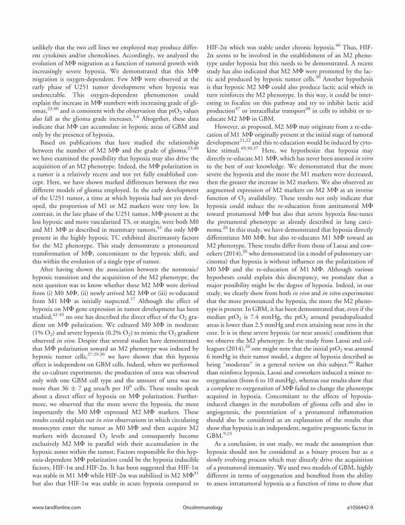
MF migration into the tumor as well as MF polarization towarda protumoral phenotype are related to the depth of hypoxia. Asthe GBM grows, the volume of hypoxia expands and its severityincreases, hence the acquisition of the M2 phenotype (Fig. 7).However, up to now, we have been unable to characterize theeffects of M2 MF in this model.
As large amounts of NO, produced by M1 MF,17 has beendemonstrated to induce cell damage51,52 and inhibit tumorangiogenesis,53 several attempts have been made to re-educateprotumoral MF toward antitumoral MF24,54-56 rather than thedepletion of MF. Our present investigation encourages a combi-nation of these recently developed therapies for GBM withapproaches designed to alleviate intratumoral hypoxia to furtherpotentiate the efficacy of available chemotherapeutic agents. Forinstance, low dose antiangiogenic therapy,57 low dose irradia-tion,58 or hyperoxic gases59 could all be pertinent strategies tomilitate against the tumoral accumulation of M2 MF.
Materials and Methods
GBM cell cultureHuman GBM cell lines, U87-MG (ATCC) and U251 (NCI),
were cultured in 1g/L glucose Dulbecco’s Modified Eagle’sMedium (DMEM, Sigma) supplemented with 10% fetal calfserum (Eurobio), 1 mg/mL penicillin/streptomycin (P/S, Sigma)and 2 mM glutamine (Gln, Sigma) at 37�C in wet atmosphere.
Tumor modelsTumor models consist of an orthotopic injection of
human GBM cells in athymic rats (200–250 g, Charles River
Laboratory). The animal investigations were performed underthe current European directive (2010/63/EU). The license toinvestigate was given to SV (14–55) in authorized housing andlaboratories (B14118001) and with the permission of theregional committee on animal ethics (CENOMEXA, 0611-02).The rats were maintained in specific pathogen-free housing andwere fed g-irradiated laboratory food and water ad libitum.
Rats were operated under anesthesia (induction in 5% andmaintenance in 2% of isoflurane in 70% NO2/30% O2) andU87-MG and U251 cells were injected (5.104 cells in 3 mL in2 mM Gln/PBS) in the right caudate-putamen.
ImagingFor all imaging experiments, animals were anesthetized as
described above, and maintained in position by ear and toothbars. Micro-magnetic resonance imaging (mMRI) was performedon a 7 Tesla horizontal magnet (Pharmascan�, Bruker, Ger-many). Micro-positron emission tomography (mPET) was per-formed on an Inveon PET-CT small animal imaging system(Siemens Healthcare) with the use of the tracer, 3-[18F]-fluoro-1-(2-nitro-1-imidazolyl)-2-propanol ([18F]-FMISO), to detecthypoxia. Imaging protocols are detailed in supplementary data.
ImmunohistochemistryAt the end of the protocol, the rats were deeply anesthetized
and were transcardially perfused with a 0.2 M phosphate buffer(PB)/4% paraformaldehyde (PFA, Sigma) solution 2 h after theintraperitoneal injection (80 mg/kg) of pimonidazole (Hypoxyp-robe Incorporation, USA). The brain was removed and placed in30% sucrose for 48 h and 30 mm thick freezing microtome sec-tions were realized. Slices were blocked with PBS, 0.5% Triton,
3% BSA (Sigma) for 2 h and then incu-bated overnight with primary antibodies(Table S1) in PBS, 0.5% Triton, 1%BSA at 4�C. Sections were then incu-bated with fluorochrome-conjugated sec-ondary antibodies (Table S1) andHoechst 33342 (Sigma, 10 mg/mL) inPBS solution as described above.
Image analysisImages were analyzed by in-house
macros based on ImageJ software(http://imagej.nih.gov/ij/). MF/micro-glial cell density was determined as thenumber of positive pixels for CD68divided by the total tumor area. MFdensity was differentiated from that ofmicroglia by the ratio of CD14 positivepixels to CD68 positive pixels. M1 andM2 MF densities were characterized bythe number of either iNOS or Arg1 posi-tive pixels, respectively, within the popu-lation of CD68 positive pixels.
Figure 7. Cross-section through a theoretical GBM to indicate the position and identity of the sub-types of MF as a function of hypoxia development. At the onset of tumor development, only micro-glia was present. When hypoxia begin to take place, MF were attracted to the tumor site and presentM0 and M1 phenotypes at the shell of the tumor. Then, M0 and M1 MF migrated toward hypoxiczones where they increased the M2 markers. Once arrived to hypoxic zones, MF were M2 cells andhypoxia fine-tunes this phenotype.
e1056442-10 Volume 5 Issue 1OncoImmunology

Bone marrow-derived MF culture and activationBone marrow was isolated from femora and tibiae of nude
mice (20–25 g, Charles River Laboratory) by flushing the boneswith 1 mL of Iscove’s Modified Dulbecco’s Media (IMDM,Sigma) containing 60% Fetal Clone II (FCII, Thermo Scientific)and 1 mg/mL P/S (Sigma). The marrow was passed through a70 mm strainer and MF (M0) were selected and cultured inIMDM enriched with 15% FCII, 1 mg/mL P/S, 10 ng/mLrecombinant mouse MF colony-stimulating factor (M-CSF,Miltenyi Biotec) and 10 ng/mL recombinant mouse Fms-relatedtyrosine kinase three ligand (Flt3-Ligand, Miltenyi Biotec) at37�C in a humid atmosphere. M1 MF were obtained by cultur-ing cells in 1 g/L glucose DMEM (Sigma) supplied with 15%FCII, 1 mg/mL P/S, 2 mM Gln (Sigma), 100 ng/mL LPS(Sigma) and 10 U/mL recombinant mouse IFNg (eBioscience).M2 MF were obtained by culturing cells with 1g/L glucoseDMEM supplemented with 15% FCII, 1% P/S, 2mM Gln(Sigma) and 50 ng/mL recombinant mouse IL4 (MiltenyiBiotec).
Cell and hypoxic culturesThe normoxic state (20% O2) was a humidified 5% CO2/air
atmosphere in an incubator and hypoxia was obtained with ahumidified 5% CO2/balance N2 gas mixture in a hypoxic cham-ber (Invivo2 500, Ruskinn, Awel) at 37
�C. For the polarizationexperiments, M0 MF were cultured either in normoxia or inhypoxia (1% or 0.2% O2) for different time periods. For the re-education experiments, M1 and M2 MF were activated 24 hand then cultured in hypoxia (1% or 0.2% O2) with their respec-tive conditioning media for different time periods. GBM celllines were cultured in the M0 MF growth medium, as describedabove, in normoxia or in 1% or 0.2% O2 for 24 h.
Reverse transcription and real-time quantitative PCRTotal RNA was isolated by the Nucleospin� RNA kit
(Macherey-Nagel) following manufacter’s instructions andreverse transcribed into cDNA (cDNA) with AMV� reverse tran-scriptase (Promega). cDNA were then quantified by real-timePCR with SYBR� Green master mix (Bio-Rad) with primersdesigned by Eurogentec (Table S2). Samples were run in tripli-cate with an amplification profile as follows: an activation stageat 95�C for 3 min following by 40 cycles at 95�C, 15sec and60�C, 30 sec. Expression levels were determined by the DCtmethod.
Determination of nitric oxide (NO) productionNO measurement in the SN of MF cultures was performed
by the Griess reaction.60 Briefly, the Griess reagent was preparedby mixing 2% sulphanilamide (Sigma) in 10% phosphoric acid(Sigma) and 0.2% naphthylethylene-diamine-dihydrochloride(Sigma). The reagent was added to SN and the mixture was incu-bated 10 min at room temperature in the dark. Each sample wasassayed in duplicate, the absorbance was measured at 540 nmand NO concentration was determined with sodium nitrite as astandard.
Determination of Arg1 activityArg1 activity was determined by a standard colorimetric
method60 in cell lysates. Briefly, cells were lysed by adding 0.1%Triton X-100 (Sigma) and 50 mM Tris-HCl (pH 7.5). 10 mMMnCl2 (Sigma) to cell samples, then heated at 56�C for 7 min toactivate the enzyme. Hydrolysis of L-arginine by Arg1 was per-formed by incubating the mixture with 50 mmol of L-arginine(pH 9.7; Sigma) at 37�C for 2 h, and the reaction was stoppedby adding an acid solution (H2SO4; H3PO4; H2O). For thedetermination of urea production, a-isonitrosopropiophenone(Sigma) was added and the mixture was incubated at 95�C for30 min and then 4�C for 30 min. Each sample was assayed induplicate, the absorbance was measured at 540 nm and urea pro-duction was determined with urea as a standard.
Statistical analysesAll data are presented as the mean § standard deviation (SD).
Statistical analyses were performed with the JMP� program (SASinstitute, USA) and, unless otherwise stated, significances werecalculated by nonparametric Kruskall–Wallis for multiple com-parisons or Mann–Whitney U tests. Statistical significance wasachieved when p < 0.05 (*) or p < 0.01 (**), otherwise they werenot significant (NS).
Disclosure of Potential Conflicts of Interest
No potential conflicts of interest were disclosed.
Acknowledgements
The authors wish to thank the LDM-TEP team for the[18F]-FMISO production.
Funding
This study was supported by the grants from the ConseilR�egional de Basse-Normandie, the Universit�e de Caen Basse-Normandie (UCBN), the French Minist�ere de l’EnseignementSup�erieur et de la Recherche (MESR), the Center National de laRecherche Scientifique (CNRS), the TC2N ‘Trans ChannelNeuroscience Network’ Interreg IV A 2 Mers Seas Zee€ens pro-gram, “Investing in your future” crossborder cooperation pro-gram 2007–2014 part financed by the European Union(European Regional Development Fund), the French NationalAgency for Research called “Investissements d’Avenir” no. ANR-11-LABEX-0018-01, the F�ed�eration pour la Recherche sur leCerveau (FRC), the Institut National contre le Cancer (INCa),and the Advanced Resource Center for Hadrontherapy in Europe(Archade).
Supplemental Material
Supplemental data for this article can be accessed on thepublisher’s website.
www.tandfonline.com e1056442-11OncoImmunology

References
1. Stupp R, Mason WP, van den Bent MJ, Weller M,Fisher B, Taphoorn MJB, Belanger K, Brandes AA,Marosi C, Bogdahn U et al. Radiotherapy plus con-comitant and adjuvant temozolomide for glioblastoma.N Engl J Med 2005; 352:987-96; PMID:15758009;http://dx.doi.org/10.1056/NEJMoa043330
2. Charles NA, Holland EC, Gilbertson R, Glass R, Ket-tenmann H. The brain tumor microenvironment. Glia2011; 59:1169-80; PMID:21446047; http://dx.doi.org/10.1002/glia.21136
3. Clavreul A, Etcheverry A, Chassevent A, Quillien V,Avril T, Jourdan M-L, Michalak S, Francois P, Carr�e J-L, Mosser J et al. Isolation of a new cell population inthe glioblastoma microenvironment. J Neurooncol2012; 106:493-504; PMID:21928115; http://dx.doi.org/10.1007/s11060-011-0701-7
4. Hirata K, Terasaka S, Shiga T, Hattori N, Magota K,Kobayashi H, Yamaguchi S, Houkin K, Tanaka S,Kuge Y et al. 18F-Fluoromisonidazole positron emis-sion tomography may differentiate glioblastoma multi-forme from less malignant gliomas. Eur J Nucl MedMol Imaging 2012; 39:760-70; PMID:22307533;http://dx.doi.org/10.1007/s00259-011-2037-0
5. Rampling R, Cruickshank G, Lewis AD, FitzsimmonsSA, Workman P. Direct measurement of pO2 distribu-tion and bioreductive enzymes in human malignantbrain tumors. Int J Radiat Oncol Biol Phys 1994;29:427-31; PMID:8005794; http://dx.doi.org/10.1016/0360-3016(94)90432-4
6. Evans SM, Judy KD, Dunphy I, Jenkins WT, HwangW, Nelson PT, Lustig RA, Jenkins K, Magarelli DP,Hahn SM et al. Hypoxia is important in the biologyand aggression of human glial brain tumors. Clin Can-cer Res 2004; 10:8177-84; PMID:15623592; http://dx.doi.org/10.1158/1078-0432.CCR-04-1081
7. Kaur B, Khwaja FW, Severson EA, Matheny SL, BratDJ, Van Meir EG. Hypoxia and the hypoxia-inducible-factor pathway in glioma growth and angiogenesis.Neuro Oncol 2005; 7:134-53; PMID:15831232;http://dx.doi.org/10.1215/S1152851704001115
8. Vaupel P, Mayer A. Hypoxia in cancer: significance andimpact on clinical outcome. Cancer Metastasis Rev2007; 26:225-39; PMID:17440684; http://dx.doi.org/10.1007/s10555-007-9055-1
9. Spence AM, Muzi M, Swanson KR, O’Sullivan F,Rockhill JK, Rajendran JG, Adamsen TCH, Link JM,Swanson PE, Yagle KJ et al. Regional hypoxia in glio-blastoma multiforme quantified with ; [18F]Fluoromi-sonidazole positron emission tomography beforeradiotherapy: correlation with time to progression andsurvival. Clin Cancer Res 2008; 14:2623-30;PMID:18451225; http://dx.doi.org/10.1158/1078-0432.CCR-07-4995
10. Lewis CE, Pollard JW. Distinct role of macrophages indifferent tumor microenvironments. Cancer Res 2006;66:605-12; PMID:16423985; http://dx.doi.org/10.1158/0008-5472.CAN-05-4005
11. Franklin RA, Liao W, Sarkar A, Kim MV, Bivona MR,Liu K, Pamer EG, Li MO. The cellular and molecularorigin of tumor-associated macrophages. Science 2014;344:921-5; PMID:24812208; http://dx.doi.org/10.1126/science.1252510
12. Murdoch C, Giannoudis A, Lewis CE. Mechanismsregulating the recruitment of macrophages into hypoxicareas of tumors and other ischemic tissues. Blood 2004;104:2224-34; PMID:15231578; http://dx.doi.org/10.1182/blood-2004-03-1109
13. Badie B, Schartner JM. Flow cytometric characteriza-tion of tumor-associated macrophages in experimentalgliomas. Neurosurgery 2000; 46:957-62;PMID:10764271; http://dx.doi.org/10.1097/00006123-200004000-00035
14. Strik HM, Stoll M, Meyermann R. Immune cell infil-tration of intrinsic and metastatic intracranial tumours.Anticancer Res 2004; 24:37-42; PMID:15015573
15. Hussain SF, Yang D, Suki D, Aldape K, Grimm E,Heimberger AB. The role of human glioma-infiltrat-ing microglia/macrophages in mediating antitumor
immune responses. Neuro Oncol 2006; 8:261-79;PMID:16775224; http://dx.doi.org/10.1215/15228517-2006-008
16. Lu-Emerson C, Snuderl M, Kirkpatrick ND, Goveia J,Davidson C, Huang Y, Riedemann L, Taylor J, Ivy P,Duda DG et al. Increase in tumor-associated macro-phages after antiangiogenic therapy is associated withpoor survival among patients with recurrent glioblas-toma. Neuro Oncol 2013; 15:1079-87;PMID:23828240; http://dx.doi.org/10.1093/neuonc/not082
17. Mantovani A, Sozzani S, Locati M, Allavena P, Sica A.Macrophage polarization: tumor-associated macro-phages as a paradigm for polarized M2 mononuclearphagocytes. Trends Immunol 2002; 23:549-55;PMID:12401408; http://dx.doi.org/10.1016/S1471-4906(02)02302-5
18. Chang CI, Liao JC, Kuo L. Macrophage arginase pro-motes tumor cell growth and suppresses nitric oxide-mediated tumor cytotoxicity. Cancer Res 2001;61:1100-6; PMID:11221839
19. Sica A,Mantovani A.Macrophage plasticity and polariza-tion: in vivo veritas. J Clin Invest 2012; 122:787-95;PMID:22378047; http://dx.doi.org/10.1172/JCI59643
20. Biswas SK, Chittezhath M, Shalova IN, Lim J-Y. Mac-rophage polarization and plasticity in health and dis-ease. Immunol Res 2012; 53:11-24; PMID:22418728;http://dx.doi.org/10.1007/s12026-012-8291-9
21. Biswas SK, Sica A, Lewis CE. Plasticity of macrophagefunction during tumor progression: regulation by dis-tinct molecular mechanisms. J Immunol 2008;180:2011-7; PMID:18250403; http://dx.doi.org/10.4049/jimmunol.180.4.2011
22. Huang Y, Snuderl M, Jain RK. Polarization of tumor-associated macrophages: a novel strategy for vascularnormalization and antitumor immunity. Cancer Cell2011; 19:1-2; PMID:21251607; http://dx.doi.org/10.1016/j.ccr.2011.01.005
23. Prosniak M, Harshyne LA, Andrews DW, Kenyon LC,Bedelbaeva K, Apanasovich TV, Heber-Katz E, CurtisMT, Cotzia P, Hooper DC. Glioma grade is associatedwith the accumulation and activity of cells bearing M2monocyte markers. Clin Cancer Res 2013; 19:3776-86; PMID:23741072; http://dx.doi.org/10.1158/1078-0432.CCR-12-1940
24. Pyonteck SM, Akkari L, Schuhmacher AJ, BowmanRL, Sevenich L, Quail DF, Olson OC, Quick ML,Huse JT, Teijeiro V et al. CSF-1R inhibition altersmacrophage polarization and blocks glioma progres-sion. Nat Med 2013; 19:1264-72; PMID:24056773;http://dx.doi.org/10.1038/nm.3337
25. Lewis C, Murdoch C. Macrophage responses to hyp-oxia: implications for tumor progression and anti-can-cer therapies. Am J Pathol 2005; 167:627-35;PMID:16127144; http://dx.doi.org/10.1016/S0002-9440(10)62038-X
26. Laoui D, Van Overmeire E, Di Conza G, Aldeni C,Keirsse J, Morias Y, Movahedi K, Houbracken I,Schouppe E, Elkrim Y et al. Tumor hypoxia does notdrive differentiation of tumor-associated macrophagesbut rather fine-tunes the M2-like macrophage popula-tion. Cancer Res 2014; 74:24-30; PMID:24220244;http://dx.doi.org/10.1158/0008-5472.CAN-13-1196
27. Zhang J, Cao J, Ma S, Dong R, Meng W, Ying M,Weng Q, Chen Z, Ma J, Fang Q et al. Tumor hypoxiaenhances non-small cell lung cancer metastasis by selec-tively promoting macrophage M2 polarization throughthe activation of ERK signaling. Oncotarget2014;5:9664-77; PMID:25313135
28. Tafani M, Di Vito M, Frati A, Pellegrini L, De SantisE, Sette G, Eramo A, Sale P, Mari E, Santoro A et al.Pro-inflammatory gene expression in solid glioblastomamicroenvironment and in hypoxic stem cells fromhuman glioblastoma. J Neuroinflammation 2011;8:32; PMID:21489226; http://dx.doi.org/10.1186/1742-2094-8-32
29. Tripathi C, Tewari BN, Kanchan RK, Baghel KS, Nau-tiyal N, Shrivastava R, Kaur H, Bhatt MLB, BhadauriaS. Macrophages are recruited to hypoxic tumor areas
and acquire a pro-angiogenic M2-polarized phenotypevia hypoxic cancer cell derived cytokines Oncostatin Mand Eotaxin. Oncotarget 2014; 5:5350-68;PMID:25051364
30. Colegio OR, Chu N-Q, Szabo AL, Chu T, RhebergenAM, Jairam V, Cyrus N, Brokowski CE, EisenbarthSC, Phillips GM et al. Functional polarization oftumour-associated macrophages by tumour-derived lac-tic acid. Nature 2014; 513:559-63; PMID:25043024;http://dx.doi.org/10.1038/nature13490
31. Takeda N, O’Dea EL, Doedens A, Kim J, WeidemannA, Stockmann C, Asagiri M, Simon MC, Hoffmann A,Johnson RS. Differential activation and antagonisticfunction of HIF-a isoforms in macrophages are essen-tial for NO homeostasis. Genes Dev 2010; 24:491-501; PMID:20194441; http://dx.doi.org/10.1101/gad.1881410
32. Corroyer-Dulmont A, P�er�es EA, Petit E, Durand L,Marteau L, Toutain J, Divoux D, Roussel S, MacKen-zie ET, Barr�e L et al. Noninvasive assessment of hyp-oxia with 3-; [18F]-fluoro-1-(2-nitro-1-imidazolyl)-2-propanol (; [18F]-FMISO): a PET study in two experi-mental models of human glioma. Biol Chem 2013;394:529-39; PMID:23399636; http://dx.doi.org/10.1515/hsz-2012-0318
33. Hou H, Krishnamurthy Nemani V, Du G, MontanoR, Song R, Gimi B, Swartz HM, Eastman A, Khan N.Monitoring oxygen levels in orthotopic human gliomaxenograft following carbogen inhalation and chemo-therapy by implantable resonator-based oximetry. Int JCancer 2014; 136:1688-96; PMID:25111969; http://dx.doi.org/10.1002/ijc.29132
34. Guillemin GJ, Brew BJ. Microglia, macrophages, peri-vascular macrophages, and pericytes: a review of func-tion and identification. J Leukoc Biol 2004; 75:388-97;PMID:14612429; http://dx.doi.org/10.1189/jlb.0303114
35. Rempel SA, Dudas S, Ge S, Guti�errez JA. Identificationand localization of the cytokine SDF1 and its receptor,CXC chemokine receptor 4, to regions of necrosis andangiogenesis in human glioblastoma. Clin Cancer Res2000; 6:102-11; PMID:10656438
36. Zagzag D, Esencay M, Mendez O, Yee H, Smirnova I,Huang Y, Chiriboga L, Lukyanov E, Liu M, NewcombEW. Hypoxia- and vascular endothelial growth factor-induced stromal cell-derived factor-1a/CXCR4 expres-sion in glioblastomas: one plausible explanation ofscherer’s structures. Am J Pathol 2008; 173:545-60;PMID:18599607; http://dx.doi.org/10.2353/ajpath.2008.071197
37. Pelegrin P, Surprenant A. Dynamics of macrophagepolarization reveal new mechanism to inhibit IL-1betarelease through pyrophosphates. EMBO J 2009;28:2114-27; PMID:19536133; http://dx.doi.org/10.1038/emboj.2009.163
38. Murat A, Migliavacca E, Hussain SF, Heimberger AB,Desbaillets I, Hamou M-F, R€uegg C, Stupp R, Delor-enzi M, Hegi ME. Modulation of angiogenic andinflammatory response in glioblastoma by hypoxia.PLoS One 2009; 4:e5947; PMID:19536297; http://dx.doi.org/10.1371/journal.pone.0005947
39. Chiang C-S, Fu SY, Wang S-C, Yu C-F, Chen F-H,Lin C-M, Hong J-H. Irradiation promotes an M2 mac-rophage phenotype in tumor hypoxia. Front Oncol2012; 2:89; PMID:22888475; http://dx.doi.org/10.3389/fonc.2012.00089
40. Komohara Y, Ohnishi K, Kuratsu J, Takeya M. Possi-ble involvement of the M2 anti-inflammatory macro-phage phenotype in growth of human gliomas. J Pathol2008; 216:15-24; PMID:18553315; http://dx.doi.org/10.1002/path.2370
41. Movahedi K, Laoui D, Gysemans C, Baeten M,Stang�e G, Van den Bossche J, Mack M, PipeleersD, In’t Veld P, De Baetselier P et al. Differenttumor microenvironments contain functionally dis-tinct subsets of macrophages derived from Ly6C(high) monocytes. Cancer Res 2010; 70:5728-39;PMID:20570887; http://dx.doi.org/10.1158/0008-5472.CAN-09-4672
e1056442-12 Volume 5 Issue 1OncoImmunology

42. Leek RD, Talks KL, Pezzella F, Turley H, Campo L,Brown NS, Bicknell R, Taylor M, Gatter KC, HarrisAL. Relation of hypoxia-inducible factor-2a (HIF-2a)expression in tumor-infiltrative macrophages to tumorangiogenesis and the oxidative thymidine phosphory-lase pathway in human breast cancer. Cancer Res 2002;62:1326-9; PMID:11888900
43. Burke B, Giannoudis A, Corke KP, Gill D, Wells M,Ziegler-Heitbrock L, Lewis CE. Hypoxia-induced geneexpression in human macrophages: implications forischemic tissues and hypoxia-regulated gene therapy.Am J Pathol 2003; 163:1233-43; PMID:14507633;http://dx.doi.org/10.1016/S0002-9440(10)63483-9
44. Fang H-Y, Hughes R, Murdoch C, Coffelt SB, BiswasSK, Harris AL, Johnson RS, Imityaz HZ, Simon MC,Fredlund E et al. Hypoxia-inducible factors 1 and 2 areimportant transcriptional effectors in primary macro-phages experiencing hypoxia. Blood 2009; 114:844-59;PMID:19454749; http://dx.doi.org/10.1182/blood-2008-12-195941
45. Doedens AL, Stockmann C, Rubinstein MP, Liao D,Zhang N, DeNardo DG, Coussens LM, Karin M,Goldrath AW, Johnson RS. Macrophage expression ofHIF-1a suppresses T cell function and promotes tumorprogression. Cancer Res 2010; 70:7465-75;PMID:20841473; http://dx.doi.org/10.1158/0008-5472.CAN-10-1439
46. Koh MY, Powis G. Passing the baton: the HIFswitch. Trends Biochem Sci 2012; 37:364-72;PMID:22818162; http://dx.doi.org/10.1016/j.tibs.2012.06.004
47. Sonveaux P, V�egran F, Schroeder T, Wergin MC, Ver-rax J, Rabbani ZN, De Saedeleer CJ, Kennedy KM,Diepart C, Jordan BF et al. Targeting lactate-fueledrespiration selectively kills hypoxic tumor cells in mice.J Clin Invest 2008; 118:3930-42; PMID:19033663;http://dx.doi.org/10.1172/JCI36843
48. Miranda-Goncalves V, Honavar M, Pinheiro C, Mar-tinho O, Pires MM, Pinheiro C, Cordeiro M, BebianoG, Costa P, Palmeirim I et al. Monocarboxylate trans-porters (MCTs) in gliomas: expression and exploitationas therapeutic targets. Neuro Oncol 2013; 15:172-88;PMID:23258846; http://dx.doi.org/10.1093/neuonc/nos298
49. Stout RD, Jiang C, Matta B, Tietzel I, Watkins SK,Suttles J. Macrophages sequentially change theirfunctional phenotype in response to changes inmicroenvironmental influences. J Immunol 2005;175:342-9; PMID:15972667; http://dx.doi.org/10.4049/jimmunol.175.1.342
50. Dehne N, Tausendsch€on M, Essler S, Geis T,Schmid T, Br€une B. IL-4 reduces the proangiogeniccapacity of macrophages by down-regulating HIF-1a translation. J Leukoc Biol 2014; 95:129-37;PMID:24006507; http://dx.doi.org/10.1189/jlb.0113045
51. Goodman JE, Hofseth LJ, Hussain SP, Harris CC.Nitric oxide and p53 in cancer-prone chronic inflam-mation and oxyradical overload disease. Environ MolMutagen 2004; 44:3-9; PMID:15199542; http://dx.doi.org/10.1002/em.20024
52. Weigert A, Br€une B. Nitric oxide, apoptosis and mac-rophage polarization during tumor progression. NitricOxide 2008; 19:95-102; PMID:18486631; http://dx.doi.org/10.1016/j.niox.2008.04.021
53. Singh S, Gupta AK. Nitric oxide: role in tumour biol-ogy and iNOS/NO-based anticancer therapies. CancerChemother Pharmacol 2011; 67:1211-24;PMID:21544630; http://dx.doi.org/10.1007/s00280-011-1654-4
54. Hagemann T, Lawrence T, McNeish I, Charles KA,Kulbe H, Thompson RG, Robinson SC, Balkwill FR.“Re-educating” tumor-associated macrophages by tar-geting NF-kappaB. J Exp Med 2008; 205:1261-8;
PMID:18490490; http://dx.doi.org/10.1084/jem.20080108
55. Buhtoiarov IN, Sondel PM, Wigginton JM, Buhtoiar-ova TN, Yanke EM, Mahvi DA, Rakhmilevich AL.Anti-tumour synergy of cytotoxic chemotherapy andanti-CD40 plus CpG-ODN immunotherapy throughrepolarization of tumour-associated macrophages.Immunology 2011; 132:226-39; PMID:21039467;http://dx.doi.org/10.1111/j.1365-2567.2010.03357.x
56. Garris C, Pittet MJ. Therapeutically reeducating mac-rophages to treat GBM. Nat Med 2013; 19:1207-8;PMID:24100977; http://dx.doi.org/10.1038/nm.3355
57. Huang Y, Yuan J, Righi E, Kamoun WS, AncukiewiczM, Nezivar J, Santosuosso M, Martin JD, Martin MR,Vianello F et al. Vascular normalizing doses of antian-giogenic treatment reprogram the immunosuppressivetumor microenvironment and enhance immunother-apy. Proc Natl Acad Sci U S A 2012; 109:17561-6;PMID:23045683; http://dx.doi.org/10.1073/pnas.1215397109
58. Klug F, Prakash H, Huber PE, Seibel T, Bender N,Halama N, Pfirschke C, Voss RH, Timke C, UmanskyL et al. Low-dose irradiation programs macrophage dif-ferentiation to an iNOSC/M1 phenotype that orches-trates effective T cell immunotherapy. Cancer Cell2013; 24:589-602; PMID:24209604; http://dx.doi.org/10.1016/j.ccr.2013.09.014
59. Stuhr LEB, Raa A, Oyan AM, Kalland KH, SakariassenPO, Petersen K, Bjerkvig R, Reed RK. Hyperoxiaretards growth and induces apoptosis, changes in vascu-lar density and gene expression in transplanted gliomasin nude rats. J Neurooncol 2007; 85:191-202;PMID:17557137; http://dx.doi.org/10.1007/s11060-007-9407-2
60. Reiner NE, editor. Macrophages and Dendritic Cells:Methods and Protocols. New York, NY: HumanaPress; 2009.
www.tandfonline.com e1056442-13OncoImmunology