hypertonicity is involved in redirecting the aquaporin-2 ... · bas w.m. van balkom1, marcel van...
TRANSCRIPT

basolateral localization of AQP2 with hypertonicity
1
Hypertonicity is involved in redirecting the aquaporin-2 water channel into
the basolateral, instead of the apical, plasma membrane of renal epithelial
cells
Bas W.M. van Balkom1, Marcel van Raak1, Sylvie Breton2, Nuria Pastor-Soler2, Richard
Bouley2, Peter van der Sluijs3, Dennis Brown2 and Peter M.T. Deen1
1Dept of Cell Physiology, NCMLS, UMC St Radboud, Nijmegen, the Netherlands,
2Program in Membrane Biology and Renal Unit, Dept of Medicine, Massachusetts
General Hospital and Harvard Medical School, Boston, MA, USA and 3Dept of Cell
Biology, University of Utrecht, Utrecht, the Netherlands.
Running title:
Correspondence to: Peter M.T. Deen, Ph.D.
160, Dept of Cell Physiology, UMC St Radboud Nijmegen
PO Box 9101, 6500 HB Nijmegen
The Netherlands
Telephone: +31 24 3617347, Fax: +31 24 3616413
E-mail: [email protected]
Copyright 2002 by The American Society for Biochemistry and Molecular Biology, Inc.
JBC Papers in Press. Published on October 8, 2002 as Manuscript M207339200 by guest on Septem
ber 12, 2018http://w
ww
.jbc.org/D
ownloaded from

basolateral localization of AQP2 with hypertonicity
2
Summary
In renal collecting ducts, vasopressin increases the expression of and redistributes
aquaporin-2 (AQP2) water channels from intracellular vesicles to the apical membrane,
leading to urine concentration. However, basolateral membrane expression of AQP2, in
addition to AQP3 and AQP4, is often detected in inner medullary principal cells in vivo.
Here, potential mechanisms that regulate apical versus basolateral targeting of AQP2
were examined. The lack of AQP2-4 association into heterotetramers and the complete
apical expression of AQP2 when highly expressed in MDCK cells indicated that neither
heterotetramerization of AQP2 with AQP3 and/or AQP4, nor high expression levels of
AQP2 explained basolateral AQP2 localization. However, long-term hypertonicity, a
feature of the inner medullary interstitium, resulted in an insertion of AQP2 into the
basolateral membrane of MDCK cells after acute forskolin stimulation. Similarly, a
marked insertion of AQP2 into the basolateral membrane of principal cells was observed
in the distal inner medulla from normal rats and Brattleboro rats after acute vasopressin
treatment of tissue slices that had been chronically treated with vasopressin to increase
interstitial osmolality in the medulla, but not in tissues from vasopressin-deficient
Brattleboro rats. These data reveal for the first time that chronic hypertonicity can
program cells in vitro and in vivo to change the insertion of a protein into the basolateral
membrane instead of the apical membrane.
Keywords: hypertonicity, water channels, sorting, plasma membrane
by guest on September 12, 2018
http://ww
w.jbc.org/
Dow
nloaded from

basolateral localization of AQP2 with hypertonicity
3
Introduction
The renal collecting duct is involved in urine concentration via a process that is regulated
by the antidiuretic hormone arginine-vasopressin (AVP). After binding to its receptor on
target cells in the kidney collecting duct, AVP initiates an intracellular signaling cascade
that increases cytosolic cAMP and calcium levels (1-3). Upon activation of protein kinase
A (PKA), aquaporin-2 (AQP2) is phosphorylated and is rapidly redistributed from
intracellular vesicles to the apical membrane of collecting duct principal cells. Driven by
an osmotic gradient, water then moves into the cell apically via AQP2, and exits across
the basolateral membrane via AQP3 and/or AQP4 (4;5). In addition to this short-term
effect, increased circulating AVP levels also lead to an increased expression of AQP2
protein, which is mediated via a cAMP responsive element in the AQP2 gene promoter
(6-8). Additionally, the expression of AQP3 is increased, but the level of AQP4 remains
unchanged (9;10).
Although the majority of AQP2 is located in the apical plasma membrane under
“steady-state” conditions in normally-hydrated animals, immunocytochemical studies
have shown that AQP2 antigenicity can also be detected in the basolateral plasma
membrane of collecting duct principal cells in these rats. This basolateral staining pattern
becomes more prominent with increased AVP levels or water deprivation in rats, and is
especially prominent in principal cells of the inner medulla (11).
The factors and mechanisms that determine the partitioning of AQP2 between the
apical and basolateral membrane of principal cells in the kidney are unknown, and the
goal of the present study was to investigate this process further. Three hypotheses that
by guest on September 12, 2018
http://ww
w.jbc.org/
Dow
nloaded from

basolateral localization of AQP2 with hypertonicity
4
might explain basolateral AQP2 targeting were tested using renal tissue, as well as
oocytes and cultured renal epithelial cells heterologously expressing AQP2, AQP3 and/or
AQP4. We considered a) that heterotetramer formation among differentially-targeted
aquaporins might be involved, b) that higher levels of AQP2 expression both in vivo and
in vitro might cause AQP2 to traffic to the membrane in both apical and basolateral
pathways, and c) that a hypertonic environment such as that found in the renal medullary
interstitium could play a role. Our data indicate that long-term exposure of cells to
hypertonicity primes epithelial cells to insert AQP2 into the basolateral membrane upon
acute stimulation with AVP and/or forskolin.
by guest on September 12, 2018
http://ww
w.jbc.org/
Dow
nloaded from

basolateral localization of AQP2 with hypertonicity
5
Experimental procedures
Plasmids
To stably-express AQP2 in MDCK cells in high amounts, the pBS-AQP2 (12)
construct was digested with XbaI, blunted, and cut with HindIII. Subsequently, the full-
length human AQP2 cDNA was isolated and ligated into the blunted BglII site and
HindIII site of the eucaryotic expression vector pCB6 (13), thereby generating pCB6-
AQP2. To generate the oocyte expression construct pT7Ts-AQP4, a pBluescript vector
containing the entire cDNA of human AQP4a (pBS-AQP4a (14)) was digested with
EcoRV and XbaI, full-length AQP4a cDNA was isolated, and cloned into the EcoRV and
SpeI sites of pT7Ts. The pSPORT-AQP3 construct, encoding full-length rat AQP3 (15),
was kindly donated by M. Echevarria, Spain.
AQP expression in oocytes
Xenopus laevis oocytes were isolated and cultured as described (16). To generate
AQP2, AQP3 and AQP4 cRNAs, pT7Ts-AQP2 (17) was linearized with SalI, while
pSPORT-AQP3 and pT7Ts-AQP4 were linearized with XbaI. Synthesis of G-capped
cRNA transcripts and determination of their integrity and concentration were done as
described (17). Two days after injection, oocytes were subjected to assays described
below.
Culturing and transfection of MDCK cells
All cells used in this study were derived from MDCK type I cells (18) and were
grown in DME medium supplemented with 5% (v/v) fetal calf serum at 37°C in 5% CO2.
by guest on September 12, 2018
http://ww
w.jbc.org/
Dow
nloaded from

basolateral localization of AQP2 with hypertonicity
6
Transfected cells used were those stably expressing human AQP2 (Wt10 cells (19)). To
obtain MDCK cells expressing high levels of AQP2, MDCK cells stably transfected with
the pCB6-AQP2 construct were generated as described (20).
To test the effect of hypertonicity on the steady state localization of AQP2, cells were
seeded on 1.13 cm2 filters at 3.0 x 10⁵ cells per cm2 and grown in medium for 8 h.
Subsequently, the osmolarity of the medium was increased from 297 to 672 mOsm/kg
H2O in three steps of 125 mOsm/kg H2O at t = 8, 24 and 32 hours using NaCl, sucrose,
raffinose or mannitol as osmolytes. The MDCK cells were analyzed at three days after
seeding, which meant that the cells were exposed to hypertonicity for about 64 hours
(starting 8 hours after seeding) and to a full hypertonicity (672 mOsm/kg) for 40 hours.
Control cells were seeded at 1.5 x 10⁵ cells per cm2. Three days after seeding, the cells
were directly prepared for confocal laser scanning microscopy analysis or first incubated
for 45 minutes in hypertonic medium containing 1 x 10⁻⁵ M forskolin to induce
translocation of AQP2 to the plasma membrane (19).
Isolation of membranes
Total membranes of oocytes were isolated as described previously (16). For
membranes of renal cells, kidneys were removed from control or 24 h water-deprived rats
and homogenized in 5 ml HbA per 350 mg wet tissue. After removing nuclei and
unbroken cells by centrifugation at 1000 g at 4°C for 10 min, each supernatant was
centrifuged at 100,000 g for 1 h to pellet the membranes.
Subsequently, oocyte (20 µl/oocyte) and kidney (5 ml/sample) membranes were
incubated for 30 min at 37oC in solubilization buffer (4% Na-desoxycholate, 20 mM Tris
by guest on September 12, 2018
http://ww
w.jbc.org/
Dow
nloaded from
basolateral localization of AQP2 with hypertonicity
7
(pH 8.0), 5 mM EDTA, 10% glycerol, 1 mM phenylmethylsulfonyl fluoride (PMSF), 5
µg/ml leupeptin and pepstatin) to dissolve the membranes. Next, undissolved membranes
were removed with a centrifugation step at 100,000 g at 4°C for 1 h.
Antibodies
For analysis of AQP2, rabbit (17) and guinea pig (21) antibodies, raised against a
synthetic peptide corresponding to the last 15 amino acids of rat AQP2 were used. AQP3
antibodies were raised against a peptide based on the predicted 15 COOH-terminal amino
acids of rat AQP3, which was conjugated to keyhole limpet hemocyanin (17). By passing
whole serum over a BSA-AQP3-coupled Affi-gel 15 column, affinity-purified antibodies
were isolated (Pharmacia Biotech, Uppsala, Sweden). Antibodies were eluted with 0.1 M
glycine (pH 2.8) and directly neutralized.
To generate AQP4 antibodies, a fragment of 389 nucleotides encoding the entire
C-terminal tail of AQP4a was isolated from pBS-AQP4a, by digestion with EcoRI, and
cloned into the EcoRI site of the pGEX1 vector (Pharmacia Biotech, Uppsala, Sweden).
After transformation of DH5á bacteria with this construct and induction of protein
expression with IPTG, the soluble glutathion S-transferase-AQP4 fusion protein was
isolated using Glutathion Sepharose 4B (Amersham Pharmacia Biotech AB, Uppsala,
Sweden). Antibodies raised in rabbit and guinea pig were affinity-purified as described
above.
by guest on September 12, 2018
http://ww
w.jbc.org/
Dow
nloaded from

basolateral localization of AQP2 with hypertonicity
8
Immunoprecipitations and sucrose gradient analysis
Immunoprecipitations were performed as previously described (16). Samples
(300 µl) of solubilized renal and oocyte membranes were loaded onto a 3.2 ml 5 to 17.5%
linear sucrose gradient (in 20 mM Tris (pH 8.0), 5 mM EDTA, 0.1% Triton X-100, 1 mM
PMSF, 5 µg/ml of leupeptin and pepstatin). Gradients were centrifuged at 100,000 g in a
Beckman SW-60 rotor for 16 h at 8°C. After centrifugation, 200 µl fractions, annotated
A-Q, were taken from the top and 15 µl samples were analyzed by immunoblotting. As
size markers, BSA (68 kDa), phosphorylase B (97 kDa), yeast alcohol dehydrogenase
(150 kDa) and β-amylase (200 kDa) were centrifuged in a parallel tube. To determine the
peak fractions of marker proteins, fractions were analyzed by SDS PAGE, after which the
proteins were visualized using Coomassie Brilliant Blue staining.
Immunoblotting
To prepare Wt10 cell lysates, cells from a 1.13 cm2 filter were incubated in 170 µl
Laemmli buffer for 30 minutes, after which the DNA was sheared by pulling the sample
through a 30G needle 3 times. When indicated, N-linked sugar groups were removed
with PNGase F according to the manufacturer (New England Biolabs, Beverly, MA,
U.S.A.). Samples were denatured for 30 min at 37°C in Laemmli buffer and subjected to
13% SDS-PAGE electrophoresis. Proteins were transferred to PVDF membranes
(Millipore Corporation, Bedford, MA, U.S.A.) as described (17). For immunoblot
analysis, AQP3 antibodies were biotinylated with Sulfo-NHS biotin (Pierce, Rockford,
IL) according the manufacturer. Membranes were incubated with 1:4000 diluted guinea
pig AQP2 antibodies in TBST (20 mM Tris, 140 mM NaCl, 0.2% Tween-20, pH 7.6),
by guest on September 12, 2018
http://ww
w.jbc.org/
Dow
nloaded from

basolateral localization of AQP2 with hypertonicity
9
1:3000 diluted rabbit AQP2 antibodies, 1:1000 biotinylated rabbit AQP3 antibodies or
guinea pig AQP4 antibodies or 1:1000 mouse monoclonal AQP1 antibodies (22), which
were all diluted in TBST with 1% non-fat dried milk (NFDM). As secondary antibodies,
goat anti-rabbit (1:5000 in TBST, Sigma, St. Louis, MO, U.S.A.), sheep anti-mouse
(1:2000 in TBST, Sigma, St. Louis, MO, U.S.A.) goat anti-guinea pig (1:10 000 in
TBST, Sigma, St. Louis, MO, U.S.A.) or streptavidin (1:8000 in TBST with 1% NFDM;
Jackson Immunoresearch, West Grove, PA, USA), all coupled to horseradish peroxidase,
were used. Proteins were visualized using enhanced chemiluminescence (Pierce,
Rockford, IL, U.S.A.).
Confocal Laser Scanning Microscopy on cell cultures
Preparation of MDCK cells for CLSM analysis was done as described (20). The
filters were incubated overnight with 1:100 diluted affinity-purified rabbit anti-AQP2,
followed by an incubation with a 1:100 diluted Alexa-594 coupled goat anti rabbit
antibodies (Molecular probes, Eugene, OR). When co-stained for AQP3, 1:100 dilutions
of affinity-purified rabbit anti-AQP3 and guinea pig anti-AQP2 antibodies were used,
followed by 1:100 dilutions of affinity-purified goat anti-rabbit or guinea pig IgGs,
coupled to Alexa-488 or Alexa-594 (Molecular Probes, Leiden, The Netherlands),
respectively. Using Adobe Photoshop, all signals were maximally expanded over the
intensity range. All figures shown are representative images of at least three independent
experiments.
by guest on September 12, 2018
http://ww
w.jbc.org/
Dow
nloaded from

basolateral localization of AQP2 with hypertonicity
10
Whole animal studies
Animal experiments were approved by the Institutional Committee on Research
Animal Care of the Massachusetts General Hospital, in accordance with the NIH guide
for the Care and Use of Laboratory Animals. Male adult Sprague Dawley and
homozygous, vasopressin-deficient Brattleboro rats were purchased from Harlan
(Indianapolis, IN).
Chronic vasopressin treatment of Brattleboro rats
Adult male Brattleboro homozygous rats weighing 300-360 g were used to study
the effects of chronic AVP pre-treatment on the polarity of AQP2 membrane insertion
after acute AVP treatment in the tissue slice preparation. The Brattleboro rats were
divided into two groups (3 animals/group). One group of animals was not treated
(control) and the other group received the vasopressin analogue 1-desamino-8-D-arginine
vasopressin (dDAVP) at a rate of 5 µl/h via osmotic minipumps as described (23) This
dose has been shown to produce comparable plasma vasopressin levels to those achieved
in normal rats during water restriction (24). All Brattleboro rats had free access to food
and water for the duration of the studies. A 5500 Wescor Vapor Pressure Osmometer
(Wescor, Logan, Utah) was used to measure the urine osmolarity. Urine samples were
collected by "clean catch" before and after the implantation of the minipumps, and at
various times during the 11 day treatment period.
by guest on September 12, 2018
http://ww
w.jbc.org/
Dow
nloaded from

basolateral localization of AQP2 with hypertonicity
11
Acute effects of vasopressin in control rat kidney slices
Adult Brattleboro rats, pretreated for 11 days (see above) and male Sprague
Dawley rats were anesthetized with an injection of sodium pentobarbital (65 mg/kg; IP).
Both kidneys were removed from the rats, cut into ~ 2-3 mm thick slices using a razor
blade and quickly placed in Hank's balanced salt solution (HBSS) pH 7.4 at 37°C
equilibrated with 5%CO2/95%O2. Slices of 0.5 mm were then cut as described (25). The
thin slices were first incubated at 37°C for 15 min in equilibrated HBSS only (Vial A) to
washout endogenous AVP and cause internalization of cell-surface AQP2. These slices
were then simultaneously transferred to fresh vials containing either arginine AVP (10
nM) plus forskolin (10 µM) (Sigma, St. Louis, MO) in HBSS (Vial B) or to vials
containing HBSS alone (Vial C) for 15 min at 37°C. Next, all slices were fixed by
immersion in periodate-lysine containing 4% paraformaldehyde (PLP) as described (25).
Slices were then rinsed several times in PBS and stored in PBS containing 0.02 % NaN3
at 4°C. To determine the localization of AQP2 prior to the treatment with AVP plus
forskolin, some slices from Vial A were fixed immediately. Additionally, some kidney
slices from Brattleboro rats were fixed in PLP immediately after preparation (i.e. before
(dD)AVP washout).
Immunocytochemistry on tissue slices
Kidney cryosections (4 µm thick) were prepared as described (26). After re-
hydration in PBS for 15 min, sections were treated with 1% SDS for antigen retrieval
(27). Blocking and immunostaining of the sections was done as described (26), except
that affinity-purified antiserum raised against the second extracellular loop of AQP2 (28)
by guest on September 12, 2018
http://ww
w.jbc.org/
Dow
nloaded from

basolateral localization of AQP2 with hypertonicity
12
and goat anti-rabbit IgG conjugated to indocarbocyanine (CY3) (2 µg/ml; Jackson
Immuno Research, West Grove, PA) were used as primary and secondary antibodies
respectively. Sections were mounted in Vectashield diluted 1:1 in 1.5 M Tris-HCl, pH
8.9. Sections were examined using a Bio-Rad Radiance 2000 confocal laser scanning
microscope (Bio-Rad Microscience Ltd, Hemel Hempstead, UK) or a Nikon 800
epifluorescence microscope coupled to a Hamamatsu Orca CCD camera and IP Lab
Spectrum software (Scanalytics, Vianna, VA).
Image quantification analysis.
To create an objective index for basolateral versus apical expression of AQP2, the
integrated optical density (IOD) of equal basolateral or apical membrane segments within
a fixed square area was determined using Image-Pro Plus analysis software (Media
Cybernetics, Silver Spring, USA). Background IOD values, determined within the
nucleus area of the particular cell, were subtracted from the obtained basolateral and
apical IOD values. The B/Asorting index is defined as the IOD of the basolateral
membrane segment divided by the IOD of the apical membrane segment. Of 8 or 15
independent cells (indicated) of representative images, and 3 segments of the basolateral
and apical membrane per cell, the mean B/Asorting index ± SEM was determined. The
significance of a change in sorting index between two experimental settings was
determined with an independent two population T-test.
by guest on September 12, 2018
http://ww
w.jbc.org/
Dow
nloaded from

basolateral localization of AQP2 with hypertonicity
13
Results
AQP2, AQP3 and AQP4 do not form heterotetramers
Some membrane proteins that are mostly expressed as homomultimeric proteins,
can also form heteromultimeric complexes consisting of related, but distinct, subunits
(29;30). The function, trafficking and regulation of such heteromeric complexes, such as
the heterodimeric GABAB1-2 receptor (31), AMPA receptor (32) and the opioid receptor
(33) are different from that of homomeric complexes. Within the AQP family of proteins,
altered trafficking of wt-AQP2 has been found upon formation of heterotetramers with
the AQP2 mutants AQP2-E258K or AQP2-delG, and this provided the explanation for
the occurrence of a dominant form of NDI (16;34). While all AQPs tested up to now
(AQP0-AQP2 and AQP4) are expressed as homotetramers (16;35-37), the possibility
existed that some AQP2 might be targeted to the basolateral membrane due to the
formation of heterotetramers with AQP3 and/or AQP4, which are both basolateral
membrane proteins.
Aquaporin-3 is expressed as tetramers: To be able to address this hypothesis, it is
essential that AQP2, AQP3 and AQP4 are all expressed as tetramers and that the
tetramers are not disrupted by the membrane isolation and extraction procedure. While it
has been shown that AQP2 and AQP4 form homotetramers (16;37), this has not been
reported for AQP3. To allow these analyses, antibodies were raised against AQP3 and
AQP4, affinity-purified and tested for their AQP-specificity. Immunoblotting of total
membranes from AQP2, AQP3 or AQP4-expressing oocytes revealed that each of these
by guest on September 12, 2018
http://ww
w.jbc.org/
Dow
nloaded from

basolateral localization of AQP2 with hypertonicity
14
antibodies specifically recognized the AQP against which it was raised (not shown).
Next, membranes of oocytes expressing AQP2, AQP3 or AQP4 were isolated, solubilized
and sedimented through a sucrose gradient. Immunoblotting of fractions taken from these
gradients revealed that AQP3 peaked in fraction J (Fig. 1) between the 97 and 150 kDa
marker proteins that were run in parallel. Because an AQP3 monomer has a calculated
molecular mass of 31.4 kDa, the observed sedimentation is consistent with the presence
of an AQP3 homotetramer. AQP4 bands of 32 and 34 kDa were obtained, which are, as
shown before (38), derived from the use of alternative translational starting methionines
(M1 and M23), both of which are contained in the AQP4 cDNA construct used here.
AQP2 and AQP4, with monomeric molecular masses of 29 and 32/34 kDa, respectively,
also peaked in fraction J, which also supports the presence of homotetramers. These data
revealed that, besides AQP2 and AQP4, also AQP3 is expressed as homotetramers and
that the tetrameric structure remains intact upon solubilization of membranes with
desoxycholate.
Do aquaporins form heterotetramers? To investigate whether AQP2, 3 and 4 form
heterotetramers in vivo, rats either received water ad libitum, or were water deprived for
24 h to increase the expression of AQP2 and AQP3, and to maximize basolateral
expression of AQP2 (11). Total kidney membranes were isolated, solubilized and
subjected to AQP-specific immunoprecipitation. As shown in Fig. 2A, none of the other
AQPs co-precipitated with the immunoprecipitated AQP. However, AQP2, AQP3 and, to
a lesser extent, AQP4 were readily detectable in the lanes containing total kidney
membranes. These results indicate that none of the AQPs form heterotetramers. Note that
by guest on September 12, 2018
http://ww
w.jbc.org/
Dow
nloaded from
basolateral localization of AQP2 with hypertonicity
15
only the 32 kDa isoform of AQP4 was detected in these experiments, which is more
abundant in the kidney (37).
In collecting duct cells of water deprived rats, the majority of AQP2 is still located in the
apical membrane and a relative small number of AQP2/AQP3 or AQP2/AQP4
heterotetramers might have been overlooked. Therefore, to examine this further, the three
aquaporins were co-expressed in oocytes, where they are all located in the plasma
membrane (15;17;38). Immunoblotting of the immunoprecipitates for the different AQPs
clearly demonstrated that the AQPs did not co-precipitate, even though each AQP was
expressed at a high level in this system (Fig. 2B, TM). These data show that AQPs 2, 3
and 4 are not able to form heteroligomers and, therefore, strongly suggest that the
basolateral localization of AQP2 is not a result of heterotetramerization of AQP2 with
AQP3 or AQP4. The ~32 kDa AQP3 band on blots disappeared upon digestion with
endoglycosidase F, indicating that this band corresponded to glycosylated AQP3 (not
shown).
Increased expression of AQP2 does not lead to its basolateral localization
Although most AQP2 is still found in the apical membrane, basolateral expression
of AQP2 is especially prominent in antidiuresis. Since in this condition AQP2 expression
is increased (6;11), we speculated that this might lead to expression in the basolateral
membrane due to saturation of the apical sorting pathway, and ‘overflow’ of AQP2 into
the basolateral pathway. This hypothesis was tested using MDCK cells that express
AQP2 at moderate levels, driven by an SV40 promoter (Wt10 cells), or at high levels
driven by a strong CMV promoter. As previously reported (19), confocal microscopy
by guest on September 12, 2018
http://ww
w.jbc.org/
Dow
nloaded from
basolateral localization of AQP2 with hypertonicity
16
showed that cells with a moderate expression level inserted AQP2 into the apical
membrane upon stimulation with forskolin (Fig. 3A). Similarly, cells with high AQP2
expression also inserted AQP2 apically (Fig. 3B). Semi-quantification of the AQP2
expression in the basolateral versus apical membrane in wt10 cells, resulted in a
B/Asorting index of 0.07±0.00 (n=8), which indicated that at these detection levels nearly
20-times more AQP2 was present in the apical than basolateral membrane. In the cells in
which AQP2 expression was derived from the CMV promoter, no basolateral membrane
AQP2 expression was detected, while the AQP2 expression in the apical membrane was
saturated. These results indicate that basolateral localization of AQP2 is probably not due
to saturation of the apical sorting pathway, at least in this culture system.
Hypertonicity results in the basolateral localization of AQP2 in MDCK cells
Since in antidiuresis, the tonicity of the inner medullary collecting duct is
increased (39), we tested whether basolateral targeting of AQP2 could be induced by
hypertonicity. The osmolarity of the medium of Wt10 cells was gradually increased with
NaCl from 297 mOsm/kg H2O to 672 mOsm/kg H2O over a three-day period. Cells were
either untreated or exposed to forskolin, fixed and examined by immunocytochemistry.
Confocal analysis revealed that without forskolin, AQP2 was mainly localized in vesicles
(Fig. 4C1, H1). With forskolin stimulation, however, hypertonicity indeed resulted in a
pronounced basolateral localization of AQP2 (Fig. 4H2), which was underscored by its
co-localization with AQP3 (Fig. 4H-AQP3). As reported (40), AQP3 appeared to be
induced in its expression in MDCK cells by hypertonicity (not shown) and has shown to
be localized in the basolateral membrane (40;41). The B/Asorting index was 1.02±0.04
by guest on September 12, 2018
http://ww
w.jbc.org/
Dow
nloaded from

basolateral localization of AQP2 with hypertonicity
17
(n=8; Fig. 4H2), which indicated that with hypertonicity the basolateral versus apical
expression of AQP2 was about 14-fold increased compared to wt10 cells grown under
isotonic conditions (0.07±0.00; n=8; p<0.001; Fig. 4C2). To further test whether this was
osmolyte-dependent, Wt10 cells were treated as above, using different sugars as
osmolytes. Confocal analysis of these cells and subsequent determination of B/Asorting
indexes revealed that, compared to cells grown under isotonic conditions, basolateral
versus apical expression of AQP2 was 15-17 fold increased for mannitol (index of
1.20±0.14; n=8), sucrose (1.08±0.04; n=8) or raffinose (1.22±0.05; n=8) upon forskolin
treatment (p<0.001; shown for mannitol in Fig. 4H3).
Basolateral localization of AQP2 in kidney principal cells in situ
Previous studies have demonstrated that AQP2 can be detected on the basolateral
plasma membrane of collecting duct principal cells in situ, and that this basolateral
location is a) more prominent in the inner medulla and b) increased after vasopressin
treatment or dehydration. To examine this further, experiments on normal rats and
vasopressin-deficient Brattleboro rats were performed.
Effect of acute vasopressin treatment on AQP2 distribution in normal rat kidney slices:
Kidney slices were prepared from normally hydrated Sprague Dawley rats, and were
incubated in vitro with and without vasopressin/forskolin. Confocal analysis revealed a
heterogeneous pattern of AQP2 distribution in collecting duct principal cells that was
different in various regions of the kidney. After removal from the animal and incubation
in vitro in buffer alone to wash out endogenous vasopressin, plasma membrane staining
by guest on September 12, 2018
http://ww
w.jbc.org/
Dow
nloaded from

basolateral localization of AQP2 with hypertonicity
18
was considerably reduced (Figs. 5A, C) compared to tissues that had been exposed to
vasopressin/forskolin (Figs. 5B, D). However, a remarkable difference in the polarity of
AQP2 insertion was seen between the proximal third and the distal portion of the inner
medullary collecting duct. As seen in Fig. 5B, a strong apical staining was induced after
15 min treatment in the initial portion of the inner medulla, although basolateral staining
was also detectable (B/Asorting index of 0.39±0.01; n=15). In more distal regions of the
collecting duct, however, the basolateral over apical staining was more than two-fold
higher than in the initial portion (0.87±0.08; n=15; p<0.001; Fig. 5D). In the inner stripe
of the outer medulla, AQP2 staining was predominantly apical after vasopressin/forskolin
treatment (not shown), as we have previously described (26). These data support the
hypothesis that hypertonicity could be involved in determining the polarity of AQP2
distribution, because the most intense basolateral staining was seen in the region of the
inner medulla that in which interstitial osmolarity is the highest.
Effect of acute VP stimulation on AQP2 distribution in kidney slices from Brattleboro
rats: To test this hypothesis further, we used vasopressin-deficient homozygous
Brattleboro rats, which are unable to concentrate their urine, and in which the interstitial
osmolarity is reduced compared to normal rats (42). The acute AQP2 insertion response
to AVP/forskolin was compared in tissue slices from control, diuretic Brattleboro rats and
from Brattleboro rats whose concentration defect had been “corrected” by administration
of dDAVP by osmotic minipump for 11 days. As shown in Fig. 6A, AQP2 was located at
the apical plasma membrane of principal cells from the inner medulla of diuretic
Brattleboro rats, although a granular staining was also detected in the basolateral pole of
by guest on September 12, 2018
http://ww
w.jbc.org/
Dow
nloaded from

basolateral localization of AQP2 with hypertonicity
19
some cells. After acute stimulation of tissue slices from the group of rats chronically
treated with dDAVP prior to the in vitro experiment, a different AQP2 staining pattern
was found. In addition to some apical staining, a very marked basolateral staining was
induced, with the staining being especially pronounced along the lateral aspects of
principal cells (Fig. 6C). This lateral staining was especially evident in tangential sections
of tubules in which a bright honeycomb staining pattern, indicative of basolateral AQP2
insertion, was seen in the dDAVP-treated rats (Fig. 6D), but not the untreated rats (Fig.
6B). These data also indicate that the differential polarized insertion of AQP2 between
the two groups resulted from an effect of chronic dDAVP treatment, and did not result
from an acute effect of osmolarity, because the osmolarity of the buffer in which the
slices were incubated was the same (isotonic) for all experimental groups.
by guest on September 12, 2018
http://ww
w.jbc.org/
Dow
nloaded from

basolateral localization of AQP2 with hypertonicity
20
Discussion
Basolateral localization of AQP2 in MDCK cells
The present study was designed to investigate potential factors or mechanisms
that might be involved in determining basolateral AQP2 localization. We clearly showed
that, besides AQP2 and AQP4, AQP3 is also expressed as a homotetramer. However,
while co-expressed in the same cells, AQP2, AQP3 and AQP4 appeared not to co-
precipitate, even when overexpressed in Xenopus oocytes. Also, immunoprecipitation of
AQP2 from wt10 cells treated with hypertonic medium, a condition at AQP2 and AQP3
co-localize and at which endogenous AQP3 expression is induced, did not result in co-
precipitation AQP3 (not shown). No AQP4 expression was detected in wt10 cells treated
with hypertonic medium. Although we can’t rule out the possibility that, in contrast to
AQP2-4 homotetramers, AQP2/3 or AQP2/4 heterotetramers disintegrate in 4%
desoxycholate, our data strongly indicate that the basolateral routing of AQP2 in
principal cells is probably not due to the formation of heterotetramers of AQP2 with
basolaterally-targeted AQP3 or AQP4. In addition, analysis of MDCK cells expressing
high levels of AQP2 driven by the CMV promoter did not reveal any detectable staining
of the basolateral membrane, which indicates that overexpression of AQP2 is also not a
likely cause for its basolateral targeting. This finding is consistent with the fact that
basolateral AQP2 expression in the kidney is segment-specific, and is not readily seen in
the outer medulla, despite an increase in AQP2 expression levels in all collecting duct
segments during dehydration or chronic vasopressin treatment.
by guest on September 12, 2018
http://ww
w.jbc.org/
Dow
nloaded from
basolateral localization of AQP2 with hypertonicity
21
Hypertonicity, however, appeared to alter the trafficking of AQP2 in epithelial
cells, because AQP2-expressing MDCK cells grown in hypertonic medium for nearly 3
days showed a 14-18 times increase in basolateral over apical membrane insertion of
AQP2 after acute forskolin stimulation compared to control cells grown in isotonic
medium. In fact, this change in ratio is even underestimated, because, to obtain a
detectable apical membrane signal for AQP2 in hypertonic wt10 cells, the AQP2 signal in
the basolateral membrane was often saturated. In contrast to AQP2, the distribution of the
basolateral protein AQP3 was not altered by growth in hypertonic media.
Basolateral localization of AQP2 in the kidney
The role of hypertonicity in AQP2 trafficking was also evaluated using an
established in vitro kidney slice model in which AVP-induced AQP2 membrane insertion
has been previously demonstrated (25;26). In kidney slices from normally-hydrated
Sprague-Dawley rats, treatment with a AVP/forskolin cocktail resulted a more than two-
fold increase of the AQP2 B/Asorting index of principal cells of the distal inner medulla
compared to the proximal inner medulla of the same tissue slice. The first segment is the
kidney region, which, in vivo, is exposed to the highest interstitial osmolarity (up to 1200
mOsm/kg). We have previously shown that in the outer medulla, regulated AQP2
insertion is almost exclusively apical (26). Thus, the polarity of acute insertion of AQP2
in medullary collecting ducts is segment specific, and can be correlated with the level of
hypertonicity to which the tubule segments had been exposed in vivo.
The data from dDAVP-treated and non-treated Brattleboro rats support this
contention. When tissue slices from control, vasopressin-deficient Brattleboro rats were
by guest on September 12, 2018
http://ww
w.jbc.org/
Dow
nloaded from
basolateral localization of AQP2 with hypertonicity
22
challenged in vitro with an AVP/forskolin cocktail, apical insertion was seen, although
some staining remained at the basolateral pole of the cells. This situation resembled that
found in the proximal inner medulla of normal rats. However, the striking basolateral
AQP2 insertion detected in the distal inner medulla of slices from Brattleboro rats after
chronic pre-treatment with dDAVP, supports the idea that a high interstitial osmolarity is
necessary for this process to occur. Because only tubules from the distal inner medulla
showed this marked basolateral staining, a direct effect of dDAVP on this process is
unlikely, since principal cells in all kidney regions were exposed to circulating dDAVP.
In addition, even after approximately 45 minutes of bathing in an isotonic incubation
medium, the in vivo environment modulates the subsequent response of collecting ducts
in excised tissue slices. This clearly indicated that the effect is not a direct and rapid
effect of exposure of cells to an increased osmolarity, but probably reflects a longer term
adaptation of the cell to the high interstitial osmolarity that occurs in vivo (or nearly three
days adaptation of MDCK cells to hypertonicity in vitro).
Cellular changes with hypertonicity.
In the short term, a hypertonicity-induced cell volume de crease is followed by a
regulated volume increase, which is achieved by an influx of inorganic solutes. Over the
long term, cells adapt to increased extracellular osmolarity by an intracellular
accumulation of organic osmolytes, such as myo-inositol, glycerophosphorylcholine,
taurine, betaine and sorbitol (43-45), which is brought about by an increased expression
of their transporters. This slow process of adaptation protects the cells from growth
retardation and apoptosis, which has been reported to occur with acute high-salt
by guest on September 12, 2018
http://ww
w.jbc.org/
Dow
nloaded from

basolateral localization of AQP2 with hypertonicity
23
challenge in IMCD cells (46). Indeed, acute treatment of our MDCK cells with 375
mOsm of extra NaCl resulted in a significant loss of cells, which precluded the analysis
of the short-term effect of a hyperosmotic/tonic agent on the localization of AQP2. In
addition to the accumulation of inorganic or organic solutes, the process of adaptation to
hypertonicity also affects basic cellular functions, such as alterations in cell metabolism,
cell growth and differentiation, transcriptional activation or repression of specific genes
and reorganization of cellular structure (via the cytoskeleton; 47).
These different forms of adaptation to a hypertonic environment are likely to play
a role in the redistribution of AQP2 from the apical to the basolateral membrane. In wt10
cells, the basolateral translocation was observed with membrane-impermeant solutes
(NaCl, sucrose, raffinose), which indicated that hypertonicity (osmotic gradient) and not
simply hyperosmolarity (increased solute content) induced the effect. The requirement
for hypertonic, rather than hyperosmolar, conditioning provides some insight in to
upstream events, as it suggests that perturbation of the cell membrane or cytoskeleton
may be necessary for AQP2 translocation to occur. Indeed, several studies have reported
that osmomechanical stress can activate numerous membrane-associated events including
activation of plasma membrane ion channels, calcium signaling events and
phosphatidylinositol turnover (48), which are known to play critical roles in membrane
trafficking and cytoskeleton reorganization (49). Of particular relevance to our present
observations is that in renal proximal tubule cells, hypoosmotic stress induces exocytosis
followed by endocytosis of vesicles at the basolateral membrane and a basolateral-to-
apical translocation of vesicles and ion channels (48). Possibly, hyperosmotic stress has
by guest on September 12, 2018
http://ww
w.jbc.org/
Dow
nloaded from
basolateral localization of AQP2 with hypertonicity
24
the opposite effect of inducing an apical-to-basolateral translocation of vesicles and
associated membrane proteins.
Physiological relevance of basolateral AQP2 in the renal medulla.
In mammals, only cells of the renal medulla are subject to substantial fluctuations
in extracellular solute concentrations, because in antidiuresis, medullary cells are
confronted with high extracellular NaCl and urea concentrations, which then fall rapidly
during the onset of water loading (50). Our present model does not, therefore, explain the
basolateral localization of AQP2 in connecting tubules, which are not exposed to
significant hypertonicity.
While other factors may be involved in the targeting process, our study suggests
that an increased hypertonicity of the renal medulla might be fundamental to the
pronounced basolateral localization of AQP2 in principal cells in antidiuresis. The
physiological relevance of this redistribution remains unclear, but three scenarios are
possible. First, basolateral AQP2 insertion might be required to increase the water
permeability of the basolateral membrane under high flow conditions, despite the
presence of AQP3 and AQP4. Second, diversion of AQP2 to the basolateral membrane
may be a protective mechanism to limit the apical flow of water and to prevent
hypervolemia during prolonged antidiuresis and/or hypernatremia. Third, basolateral
AQP2 insertion may represent a transient part of an indirect trafficking pathway in which
AQP2 is first delivered basolaterally, followed by internalization and re-routing to the
apical membrane by transcytosis. Such an indirect pathway of apical membrane protein
insertion has been described for several membrane proteins in other cell types (51).
by guest on September 12, 2018
http://ww
w.jbc.org/
Dow
nloaded from

basolateral localization of AQP2 with hypertonicity
25
It is now clear that the same protein, including AQP2, can be inserted into
different membrane domains when expressed in different cell types, implying that
targeting signals on proteins are not interpreted identically by the sorting machineries of
all cells (20;52-54). However, it is unusual for the polarity of any given protein to be
modified under normal physiological, non-pathological conditions. The kidney is largely
responsible for maintaining body fluid, electrolyte and acid/base homeostasis, and the
remarkable plasticity of epithelial cells in some kidney regions may reflect a continual
need to monitor prevailing physiological conditions, and adapt to them by modulating
vectorial transport processes across the epithelium. In this organ, systemic acid-base
alterations can lead to altered polarity of the H+ATPase in collecting duct intercalated
cells (55;56), and we now report that hypertonicity can modify the polarity of AQP2
insertion in principal cells of some regions of the collecting duct. Thus, hypertonicity
represents a novel regulatory factor involved in modifying the polarity of membrane
protein insertion in the kidney, and possibly in other cell types that are exposed to
alterations in their extracellular osmotic environment. While these findings might explain
the basolateral localization of AQP2 in the inner medullary collecting duct cells of the
kidney, factors involved in basolateral AQP2 expression in the cortex, as well as the
physiological/cell biological role of basolateral AQP2 insertion remain to be determined.
by guest on September 12, 2018
http://ww
w.jbc.org/
Dow
nloaded from

basolateral localization of AQP2 with hypertonicity
26
Acknowledgements
We thank Dr W.J.H. Koopman, Dept Cell Physiology, UMC St Radboud, Nijmegen for
his help with setting up the image quantification analysis. This study was supported by
grant from the Dutch Organization of Scientific Research (NWO-MW 902-18-092) to
PMTD and PvdS, from the EU (QLRT-2000-00778) to PMTD. A grant from the National
Institutes of Health, DK38452 supported DB and SB, and RB was partially supported by
a grant from the National Kidney Foundation. An NRSA award from the NIH (HD08684)
supported NP-S.
by guest on September 12, 2018
http://ww
w.jbc.org/
Dow
nloaded from

basolateral localization of AQP2 with hypertonicity
27
Figure legends
Figure 1. AQP3 is expressed as a homotetramer. Two days after injection, total
membranes of oocytes expressing AQP2, AQP3 or AQP4 were isolated, solubilized in
desoxycholate and subjected to sucrose gradient centrifugation. Fractions of 200 µl were
taken, of which fractions C to P (indicated) were immunoblotted for AQP2, AQP3 or
AQP4. AQP3 fractions were treated with PNGase F before loading. To estimate the mass
of the AQP complexes, sedimentation marker proteins BSA (68 kDa), phosphorylase B
(97 kDa), yeast alcohol dehydrogenase (150 kDa) and β-amylase (200 kDa) were
sedimented in a parallel tube. Their peak fractions are indicated at the bottom. The mass
of a marker protein (in kDa) is given on the left. All aquaporins sedimented between the
97 and 150 kD markers, indicating a tetrameric assembly, which was maintained
throughout the extraction and centrifugation procedure.
Figure 2. AQP2, AQP3 and AQP4 do not form heterotetramers in vivo and in vitro.
A. In vivo. Membranes of renal medulla from control (C) or water-deprived (D) rats were
isolated, solubilised in desoxycholate and subjected to immunoprecipitation (IP) with
AQP2, AQP3 or AQP4 specific antibodies. The proteins of the precipitates were
separated by SDS-PAGE and immunoblotted for AQP2 (upper panel), AQP3 (middle
panel) or AQP4 (lower panel). Proteins from total membranes (TM) of renal medulla’s
were taken as controls.
by guest on September 12, 2018
http://ww
w.jbc.org/
Dow
nloaded from
basolateral localization of AQP2 with hypertonicity
28
B. In vitro. Oocytes were injected with a mix of cRNAs encoding AQP2, 3 and 4. Two
days after injection, membranes were isolated, solubilized in desoxycholate and analyzed
as under A. Proteins from total membranes (TM) of non-injected (C) or AQP2-4
expressing oocytes were taken as negative and positive controls, respectively. In addition
to AQP2 and AQP4, unglycosylated (u-AQP3) and glycosylated AQP3 (g-AQP3) are
indicated. The mass of a marker protein (in kDa) is given on the left.
These data indicate that AQP2, AQP3 and AQP4 do not form heterotetramers in renal
medulla’s and in oocytes expressing all three aquaporins, since only the aquaporin that
was specifically immunoprecipitated is detectable in the respective lanes.
Figure 3. Intracellular localization of AQP2 with overexpression. X-Z images of
AQP2 localization in MDCK cells with moderate expression of AQP2 (Wt10 cells; panel
A) and pooled colonies of MDCK expressing high levels of AQP2 (derived from pCB6-
AQP2 construct; panel B). The cells were grown to confluence in normal medium,
treated with forskolin, fixed and subjected to AQP2 immunocytochemistry and confocal
laser scanning microscopy. For both figures, identical CLSM settings were used.
Figure 4. With hypertonicity, forskolin induces the translocation of AQP2 to the
basolateral instead of apical membrane of MDCK. X-Z images of AQP2 or AQP3 in
MDCK cells are shown. Both in isotonic (C1) and hypertonic (H1) conditions, AQP2
resides within intracellular vesicles in non-stimulated cells, but after acute forskolin
stimulation, AQP2 is mainly present in the apical membrane under isotonic culturing
conditions (C2), and in the apical and basolateral membrane when cultured in hypertonic
by guest on September 12, 2018
http://ww
w.jbc.org/
Dow
nloaded from

basolateral localization of AQP2 with hypertonicity
29
medium using NaCl (H2) or mannitol (H3) as osmolytes. In hypertonic conditions, AQP2
(H2) co-localizes with AQP3 (indicated) in the basolateral membrane.
Figure 5. Localization of AQP2 in tissue slices from normal rat kidney. Panel A
shows AQP2 immunostaining in the proximal inner medulla from a kidney slice that was
incubated in buffer alone (for 15 min) to washout endogenous vasopressin. Internalized
AQP2 is distributed throughout the cytoplasm of principal cells, although the vesicles
tend to be more concentrated towards the apical pole of the cells. After acute (15 min)
treatment with vasopressin/forskolin, a bright apical membrane staining and a weaker
basolateral membrane staining is detectable in principal cells from the proximal inner
medulla (Panel B). In Panel C, a collecting duct from the distal inner medulla is shown
after 15 min incubation in buffer alone. AQP2 is distributed throughout the cytoplasm of
principal cells. After stimulation with vasopressin/forskolin for 15 min, a more than two-
fold increase in basolateral versus apical membrane staining is seen in collecting ducts
from the distal inner medulla (Panel D - arrows), compared to that in more proximal
regions of the collecting duct (Panel B). L = lumen of the collecting duct. Bar = 10 µM.
Figure 6. Effect of chronic pre-treatment with dDAVP on polarity of AQP2 insertion
in tissue slices from Brattleboro rats. Panel A shows AQP2 localization in principal
cells from the distal inner medullary collecting duct of a control Brattleboro rat, after
incubation of kidney slices in vitro with vasopressin/forskolin. Staining of the apical pole
of the epithelial cells is evident, and some granular staining is also located in the basal
by guest on September 12, 2018
http://ww
w.jbc.org/
Dow
nloaded from

basolateral localization of AQP2 with hypertonicity
30
region of some cells. Panel B shows an oblique section through an inner medullary
collecting duct from the same slice to illustrate the low level of basolateral membrane
staining under these conditions. Panels C and D show similar sections through collecting
ducts from the distal inner medulla of Brattleboro rats that were pre-treated for 11 days
with dDAVP prior to the in vitro studies. After vasopressin/forskolin treatment of tissues
from these rats, a marked basolateral plasma membrane staining was observed (arrows),
in addition to the apical membrane staining that was evident in some cells (Panel C). The
basolateral, honeycomb pattern is especially evident in the obliquely sectioned collecting
duct shown in Panel D. This strong basolateral insertion of AQP2 is virtually
undetectable in Brattleboro rats that were not pre-treated with vasopressin (Panel B). Bars
= 10 µM.
by guest on September 12, 2018
http://ww
w.jbc.org/
Dow
nloaded from

basolateral localization of AQP2 with hypertonicity
31
References
1. Chou, C. L., Yip, K. P., Michea, L., Kador, K., Ferraris, J. D., Wade, J. B., and
Knepper, M. A. (2000) J Biol Chem 275, 36839-36846
2. Brown, D. and Nielsen, S. (2000) The cell biology of vasopressin action. In
Brenner, B., editor. The Kidney, Saunders Co., Orlando
3. Deen, P. M. T. and Brown, D. (2001) Trafficking of native and mutant
mammalian MIP proteins. In Hohmann, S., Agre, P., and Nielsen, S., editors.
Academic Press, San Diego, CA, USA
4. Deen, P. M. T. and van Os, C. H. (1998) Curr Opin Cell Biol 10, 435-442
5. Nielsen, S., Frokiaer, J., Marples, D., Kwon, T. H., Agre, P., and Knepper, M.
A. (2002) Physiol Rev. 82, 205-244
6. van Os, C. H., Deen, P. M. T., and Dempster, J. A. (1994) Biochim Biophys
Acta 1197, 291-309
7. Matsumura, Y., Uchida, S., Rai, T., Sasaki, S., and Marumo, F. (1997) J Am Soc
Nephrol 8, 861-867
by guest on September 12, 2018
http://ww
w.jbc.org/
Dow
nloaded from
basolateral localization of AQP2 with hypertonicity
32
8. Hozawa, S., Holtzman, E. J., and Ausiello, D. A. (1996) Am J Physiol 39,
C1695-C1702
9. Ishibashi, K., Sasaki, S., Fushimi, K., Yamamoto, T., Kuwahara, M., and
Marumo, F. (1997) Am J Physiol 41, F235-F241
10. Terris, J., Ecelbarger, C. A., Marples, D., Knepper, M. A., and Nielsen, S.
(1995) Am J Physiol 38, F775-F785
11. Nielsen, S., Digiovanni, S. R., Christensen, E. I., Knepper, M. A., and Harris, H.
W. (1993) Proc Natl Acad Sci U S A 90, 11663-11667
12. Deen, P. M. T., Verdijk, M. A. J., Knoers, N. V. A. M., Wieringa, B., Monnens,
L. A. H., van Os, C. H., and van Oost, B. A. (1994) Science 264, 92-95
13. Brewer, C. B. and Roth, M. G. (1991) J Cell Biol 114, 413-421
14. Lu, M., Lee, M. D., Smith, B. L., Jung, J. S., Agre, P., Verdijk, M. A. J., Merkx,
G., Rijss, J. P. L., and Deen, P. M. T. (1996) Proc Natl Acad Sci U S A 93,
10908-10912
15. Echevarria, M., Windhager, E. E., Tate, S. S., and Frindt, G. (1994) Proc Natl
Acad Sci U S A 91, 10997-11001
by guest on September 12, 2018
http://ww
w.jbc.org/
Dow
nloaded from

basolateral localization of AQP2 with hypertonicity
33
16. Kamsteeg, E. J., Wormhoudt, T. A., Rijss, J. P. L., van Os, C. H., and Deen, P.
M. T. (1999) EMBO J 18, 2394-2400
17. Deen, P. M. T., Croes, H., van Aubel, R. A., Ginsel, L. A., and van Os, C. H.
(1995) J Clin Invest 95, 2291-2296
18. Richardson, J. C., Scalera, V., and Simmons, N. L. (1981) Biochim Biophys
Acta 673, 26-36
19. Deen, P. M. T., Rijss, J. P. L., Mulders, S. M., Errington, R. J., van Baal, J., and
van Os, C. H. (1997) J Am Soc Nephrol 8, 1493-1501
20. Deen, P. M. T., van Balkom, B. W., Savelkoul, P. J., Kamsteeg, E. J., Van
Raak, M., Jennings, M. L., Muth, T. R., Rajendran, V., and Caplan, M. J. (2002)
Am J Physiol Renal Physiol 282, F330-F340
21. Mulders, S. M., Bichet, D. G., Rijss, J. P. L., Kamsteeg, E. J., Arthus, M. F.,
Lonergan, M., Fujiwara, M., Morgan, K., Leijendekker, R., van der Sluijs, P.,
van Os, C. H., and Deen, P. M. T. (1998) J Clin Invest 102, 57-66
22. Deen, P. M. T., Nielsen, S., Bindels, R. J. M., and van Os, C. H. (1997) Pflugers
Arch 433, 780-787
by guest on September 12, 2018
http://ww
w.jbc.org/
Dow
nloaded from

basolateral localization of AQP2 with hypertonicity
34
23. Shayakul, C., Smith, C. P., Mackenzie, H. S., Lee, W. S., Brown, D., and
Hediger, M. A. (2000) Am.J Physiol Renal Physiol 278, F620-F627
24. Gellai, M., Silverstein, J. H., Hwang, J. C., LaRochelle, F. T., Jr., and Valtin, H.
(1984) Am J Physiol 246, F819-F827
25. Breton, S. and Brown, D. (1998) J Am Soc Nephrol 9, 155-166
26. Bouley, R., Breton, S., Sun, T., McLaughlin, M., Nsumu, N. N., Lin, H. Y.,
Ausiello, D. A., and Brown, D. (2000) J Clin Invest 106, 1115-1126
27. Brown, D., Lydon, J., McLaughlin, M., Stuart-Tilley, A., Tyszkowski, R., and
Alper, S. (1996) Histochem Cell Biol 105, 261-267
28. Gustafson, C. E., Levine, S., Katsura, T., McLaughlin, M., Aleixo, M. D.,
Tamarappoo, B. K., Verkman, A. S., and Brown, D. (1998) Histochem Cell Biol
110, 377-386
29. Krapivinsky, G., Gordon, E. A., Wickman, K., Velimirovic, B., Krapivinsky, L.,
and Clapham, D. E. (1995) Nature 374, 135-141
30. Bouvier, M. (2001) Nat Rev Neurosci 2, 274-286
31. Ng, G. Y., Clark, J., Coulombe, N., Ethier, N., Hebert, T. E., Sullivan, R.,
Kargman, S., Chateauneuf, A., Tsukamoto, N., McDonald, T., Whiting, P.,
by guest on September 12, 2018
http://ww
w.jbc.org/
Dow
nloaded from

basolateral localization of AQP2 with hypertonicity
35
Mezey, E., Johnson, M. P., Liu, Q., Kolakowski, L. F., Jr., Evans, J. F., Bonner,
T. I., and O'neill, G. P. (1999) J Biol Chem 274, 7607-7610
32. Shi, S., Hayashi, Y., Esteban, J. A., and Malinow, R. (2001) Cell 105, 331-343
33. Jordan, B. A. and Devi, L. A. (1999) Nature 399, 697-700
34. Marr, N., Bichet, D. G., Lonergan, M., Arthus, M. F., Jeck, N., Seyberth, H. W.,
Rosenthal, W., van Os, C. H., Oksche, A., and Deen, P. M. T. (2002) Hum Mol
Genet 11, 779-789
35. Hasler, L., Walz, T., Tittmann, P., Gross, H., Kistler, J., and Engel, A. (1998) J
Mol Biol 279, 855-864
36. Walz, T., Hirai, T., Murata, K., Heymann, J. B., Mitsuoka, K., Fujiyoshi, Y.,
Smith, B. L., Agre, P., and Engel, A. (1997) Nature 387, 624-627
37. Neely, J. D., Christensen, B. M., Nielsen, S., and Agre, P. (1999) Biochem 38,
11156-11163
38. Jung, J. S., Bhat, R. V., Preston, G. M., Guggino, W. B., Baraban, J. M., and
Agre, P. (1994) Proc Natl Acad Sci U S A 91, 13052-13056
39. Flamion, B., Spring, K. R., and Abramow, M. (1995) Am J Physiol 268, F53-
F63
by guest on September 12, 2018
http://ww
w.jbc.org/
Dow
nloaded from

basolateral localization of AQP2 with hypertonicity
36
40. Matsuzaki, T., Suzuki, T., and Takata, K. (2001) Am J Physiol Cell Physiol 281,
C55-C63
41. Ecelbarger, C. A., Terris, J., Frindt, G., Echevarria, M., Marples, D., Nielsen, S.,
and Knepper, M. A. (1995) Am J Physiol 38, F663-F672
42. Bondy, C. A., Lightman, S. L., and Lightman, S. L. (1989) Mol Endocrinol 3,
1409-1416
43. Kwon, H. M. and Handler, J. S. (1995) Curr Opin Cell Biol 7, 465-471
44. Yancey, P. H., Clark, M. E., Hand, S. C., Bowlus, R. D., and Somero, G. N.
(1982) Science 217, 1214-1222
45. Sarkadi, B. and Parker, J. C. (1991) Biochim Biophys Acta 1071, 407-427
46. Michea, L., Ferguson, D. R., Peters, E. M., Andrews, P. M., Kirby, M. R., and
Burg, M. B. (2000) Am J Physiol Renal Physiol 278, F209-F218
47. Lang, F., Busch, G. L., Ritter, M., Volkl, H., Waldegger, S., Gulbins, E., and
Haussinger, D. (1998) Physiol Rev 78, 247-306
48. Reid, J. M. and O'Neil, R. G. (2000) Biochem Biophys Res Commun 271, 429-
434
by guest on September 12, 2018
http://ww
w.jbc.org/
Dow
nloaded from

basolateral localization of AQP2 with hypertonicity
37
49. Dove, S. K., McEwen, R. K., Cooke, F. T., Parker, P. J., and Michell, R. H.
(1999) Biochem Soc Trans 27, 674-677
50. Beck, F. X., Burger-Kentischer, A., and Muller, E. (1998) Pflugers Arch 436,
814-827
51. Brown, D. and Stow, J. L. (1996) Physiol Rev 76, 245-297
52. Roush, D. L., Gottardi, C. J., Naim, H. Y., Roth, M. G., and Caplan, M. J.
(1998) J Biol Chem 273, 26862-26869
53. Pathak, R. K., Yokode, M., Hammer, R. E., Hofmann, S. L., Brown, M. S.,
Goldstein, J. L., and Anderson, R. G. (1990) J Cell Biol 111, 347-359
54. Katsura, T., Verbavatz, J. M., Farinas, J., Ma, T., Ausiello, D. A., Verkman, A.
S., and Brown, D. (1995) Proc Natl Acad Sci U S A 92, 7212-7216
55. Sabolic, I., Brown, D., Gluck, S. L., and Alper, S. L. (1997) Kidney Int 51, 125-
137
56. Bastani, B., Purcell, H., Hemken, P., Trigg, D., and Gluck, S. (1991) J Clin
Invest 88, 126-136
by guest on September 12, 2018
http://ww
w.jbc.org/
Dow
nloaded from

AQP2
AQP3
AQP4
C D JI K L M N O PG HE F
68 97 150 232
Figure 1
-30
-30
-30
by guest on September 12, 2018
http://ww
w.jbc.org/
Dow
nloaded from

Figure 2A
TM AQP2 AQP3 AQP4 IPC D C D C D C D
AQP2
AQP4
AQP3
by guest on September 12, 2018
http://ww
w.jbc.org/
Dow
nloaded from
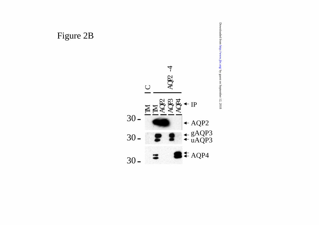
Figure 2B
AQP2
uAQP3
AQP4
gAQP3
IP
TM AQ
P4A
QP2
-4A
QP3
C
AQ
P2TM
-30
-30
-30
by guest on September 12, 2018
http://ww
w.jbc.org/
Dow
nloaded from
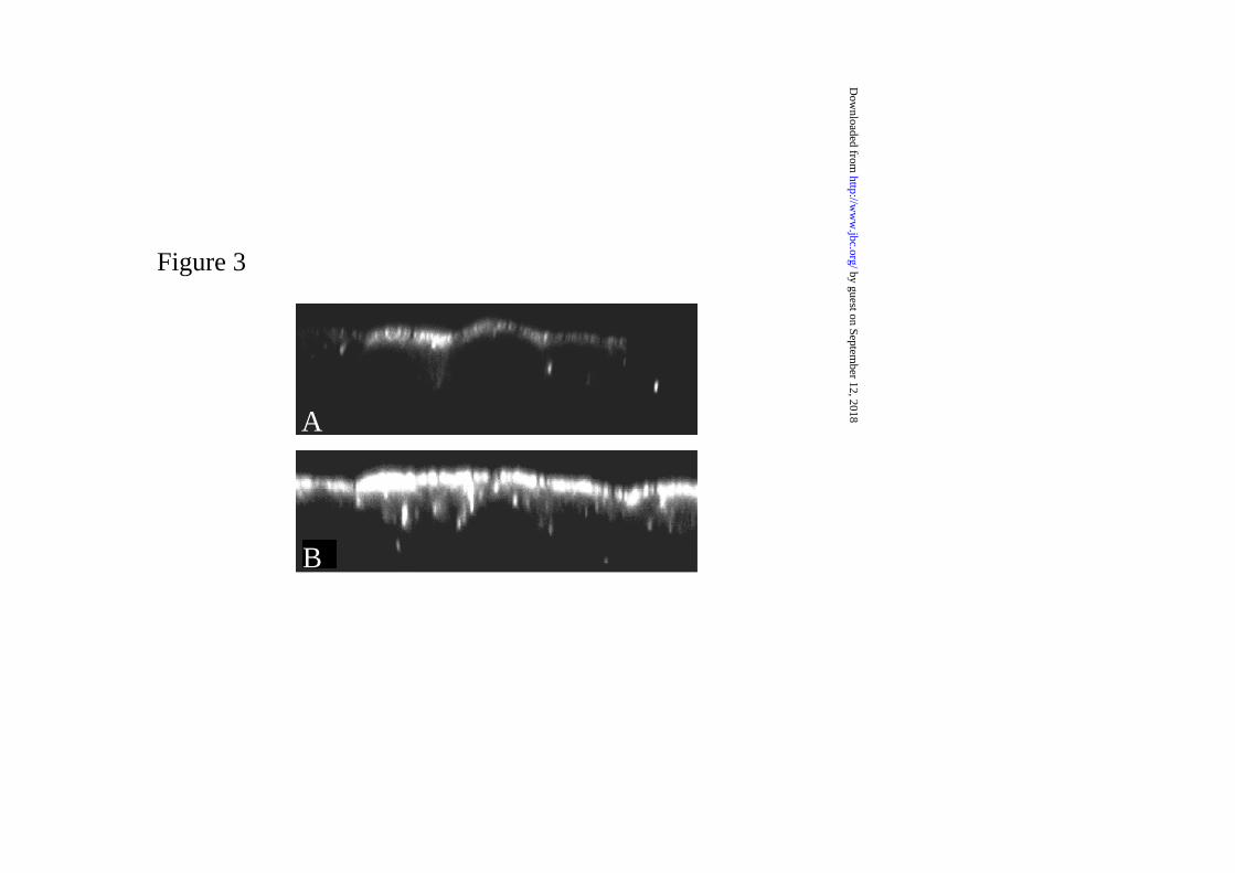
Figure 3
A
B
by guest on September 12, 2018
http://ww
w.jbc.org/
Dow
nloaded from

C HFigure 4
1 1
2 2
AQP3
3
by guest on September 12, 2018
http://ww
w.jbc.org/
Dow
nloaded from


Bouley, Peter Van der Sluijs, Dennis Brown and Peter M.T. DeenBas W.M. Van Balkom, Marcel Van Raak, Sylvie Breton, Nuria Pastor-Soler, Richard
basolateral, instead of the apical, plasma membrane of renal epithelial cellsHypertonicity is involved in redirecting the aquaporin-2 water channel into the
published online October 8, 2002J. Biol. Chem.
10.1074/jbc.M207339200Access the most updated version of this article at doi:
Alerts:
When a correction for this article is posted•
When this article is cited•
to choose from all of JBC's e-mail alertsClick here
by guest on September 12, 2018
http://ww
w.jbc.org/
Dow
nloaded from