hyperaemia and congestionpeople.upei.ca/smartinson/circ2-congest_-16_sam_final.pdf · hyperemia and...
TRANSCRIPT

Circulatory Disturbances 2: Hyperemia and Congestion
Shannon Martinson, Feb 2016
http://people.upei.ca/smartinson/ VPM 152 General Pathology

CIRCULATORY DISTURBANCES
Edema Congestion and Hyperemia
Hemorrhage Thrombosis and Embolism
Infarction Shock

Learning Objectives
• Define congestion and hyperemia
• Differentiate between the two with regard to:
• Mechanisms / underlying causes
• Appearance (gross and histologic)
• Effects
• Understand / differentiate between the 2 types of hyperemia
• Know what kind of congestion is seen with right and left heart failure
• Be able to differentiate acute and chronic pulmonary congestion (gross and histology)
• Know the consequences of chronic pulmonary congestion
• Be able to recognize and describe hepatic congestion and know under what condition it occurs
HYPEREMIA AND CONGESTION

Hyperemia and congestion indicate a local increase in blood within a tissue
• An excess of blood within blood vessels in a part of the body due to an active process Hyperemia
• An excess of blood within blood vessels in a part of the body due to a passive process Congestion
ALTERATIONS IN BLOOD FLOW & PERFUSION

• Active process
• Increased blood entering tissue via arterial flow
• Oxygenated (red) Hyperemia
ALTERATIONS IN BLOOD FLOW & PERFUSION - HYPEREMIA

• Active engorgement of vascular beds due to
increased arteriolar inflow
• Response to stimulus → Can be physiologic or
pathologic
ALTERATIONS IN BLOOD FLOW & PERFUSION - HYPEREMIA

Physiologic Hyperemia
Digestion
Exercise
Dissipate heat
Neurovascular (blushing)
ALTERATIONS IN BLOOD FLOW & PERFUSION - HYPEREMIA

• Due to an underlying pathologic process → INFLAMMATION
• Arteriolar dilation in response to inflammatory stimuli / mediators
• Often accompanied by edema
Pathologic Hyperemia
ALTERATIONS IN BLOOD FLOW & PERFUSION - HYPEREMIA

Pathologic Hyperemia = Hyperemia of Inflammation
ALTERATIONS IN BLOOD FLOW & PERFUSION - HYPEREMIA
One of the cardinal signs of inflammation is reddening =
“rubor”

Gross Findings • Increased redness in
the affected tissue • Swelling, warmth
ALTERATIONS IN BLOOD FLOW & PERFUSION - HYPEREMIA
• Always a localized change* • Best appreciated in the living
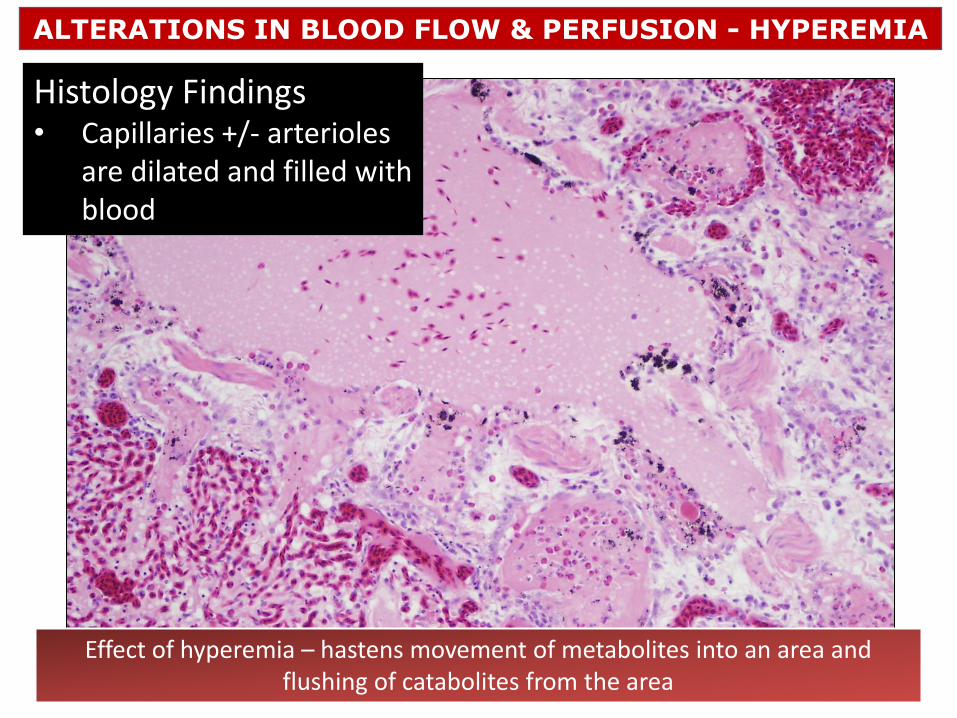
Histology Findings • Capillaries +/- arterioles
are dilated and filled with blood
ALTERATIONS IN BLOOD FLOW & PERFUSION - HYPEREMIA
Effect of hyperemia – hastens movement of metabolites into an area and flushing of catabolites from the area

• Passive process
• Decreased outflow of blood
• Deoxygenated (blue) Congestion
ALTERATIONS IN BLOOD FLOW & PERFUSION - CONGESTION

• Passive process
• Decreased outflow of blood
• Deoxygenated (blue) Congestion
• Passive engorgement of vascular beds
• Decreased/obstructed venous return
• Tissue dark red to blue / black (cyanotic)
• Poorly oxygenated hemoglobin
ALTERATIONS IN BLOOD FLOW & PERFUSION - CONGESTION

Gross Appearance
• Dark red to blue / black depending on degree of stagnation of blood
• Tissues might be cooler than normal
• Cut surfaces ooze blood and are often wet due to accompanying edema
ALTERATIONS IN BLOOD FLOW & PERFUSION - CONGESTION

Chronic Congestion • Hypoxia (O2)
• Atrophy
• Degeneration / necrosis
Histopathology
Acute Congestion • Engorged capillaries
• Some edema
ALTERATIONS IN BLOOD FLOW & PERFUSION - CONGESTION
Effect of congestion • Leads to hypoxia and accumulation of catabolites • Often edema occurs • Interference with normal tissue function • May get thrombosis of congested veins • +/- proliferation of connective tissue if chronic

Two factors used to define types of congestion
ALTERATIONS IN BLOOD FLOW & PERFUSION - CONGESTION
1. Duration
• Acute (sudden)
• Chronic (long-term)
2. Extent
• Localized – Change is confined to a discrete area
• Generalized – Indicates systemic change (eg heart failure)
Acute localized
congestion
Acute generalized congestion
Chronic generalized congestion
Chronic localized
congestion

ALTERATIONS IN BLOOD FLOW & PERFUSION - CONGESTION
Localized congestion
• Local obstruction to venous drainage • Such as when
and organ twists
Intestinal torsion
Intestinal intussusception

• Sudden death due to heart failure or euthanasia
with barbiturates
• Blood accumulates in lung, spleen and liver
ALTERATIONS IN BLOOD FLOW & PERFUSION - CONGESTION
Acute generalized congestion

• Sudden death due to heart failure or euthanasia
with barbiturates
• Blood accumulates in lung, spleen and liver
ALTERATIONS IN BLOOD FLOW & PERFUSION - CONGESTION
Acute generalized congestion
Generalized congestion occurs with pathology of the heart or lung • Left heart failure • Right heart failure • 1◦ Pulmonary disease
ALTERATIONS IN BLOOD FLOW & PERFUSION - CONGESTION
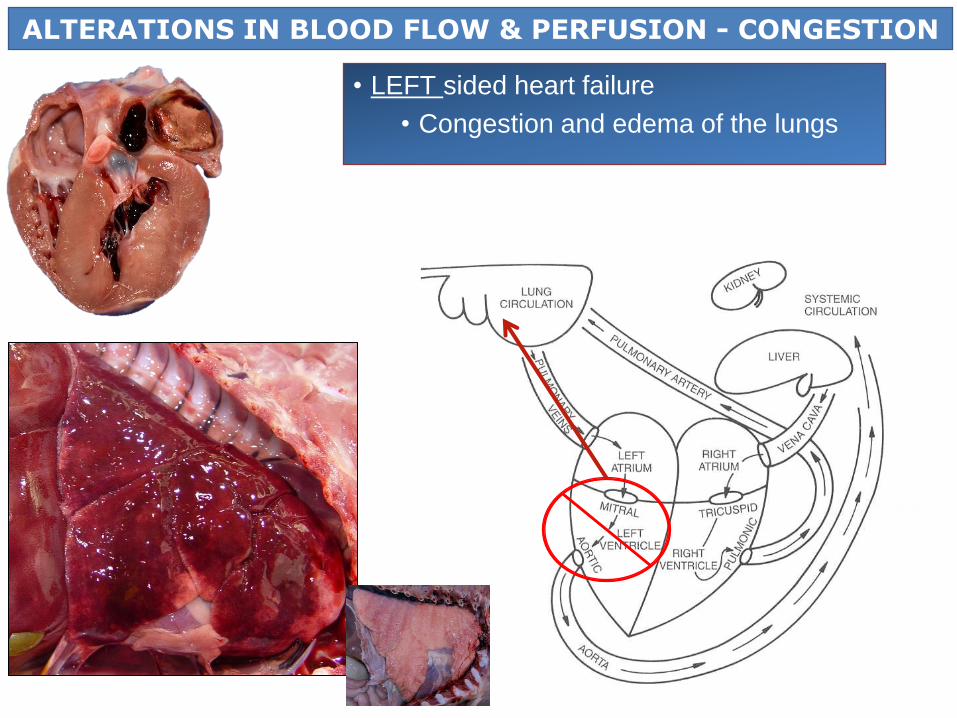
• LEFT sided heart failure
• Congestion and edema of the lungs

ALTERATIONS IN BLOOD FLOW & PERFUSION - CONGESTION
Gross
Histo
•Pulmonary congestion • Usually due to LEFT heart failure
• When acute →lungs are red (congestion), wet (edema) and heavy

ALTERATIONS IN BLOOD FLOW & PERFUSION - CONGESTION
•Pulmonary congestion • Usually due to LEFT heart failure
• When chronic →lungs can be light brown due to hemosiderin accumulation

ALTERATIONS IN BLOOD FLOW & PERFUSION - CONGESTION
Consequences of Chronic Pulmonary Congestion
1. Intra-alveolar hemorrhages • “Heart failure cells”
2. Pulmonary Edema • Interferes with gas exchange
3. Interstitial Fibrosis
4. Pulmonary Hypertension • ↑ Pressure in pulmonary
artery • +/- Cor pulmonale

ALTERATIONS IN BLOOD FLOW & PERFUSION - CONGESTION
RIGHT sided heart failure
• Systemic congestion – Liver especially
• Generalized edema (SQ, ascites, hydrothorax)

ALTERATIONS IN BLOOD FLOW & PERFUSION - CONGESTION
Primary pulmonary disease • Lung disease → progressive damage of pulmonary vascular bed → increased
resistance /pulmonary hypertension → RIGHT heart failure
• Generalized edema (SQ, ascites, hydrothorax) and hepatic congestion
• Right heart failure resulting from pulmonary disease Cor pulmonale

ALTERATIONS IN BLOOD FLOW & PERFUSION - CONGESTION
• Hepatic Congestion • Most often due to right heart failure • Less often secondary to pulmonary hypertension and cor pulmonale

ALTERATIONS IN BLOOD FLOW & PERFUSION - CONGESTION
• Hepatic Congestion • Gross appearance:
• Liver is enlarged and dark brown with rounded edges

ALTERATIONS IN BLOOD FLOW & PERFUSION - CONGESTION
• Hepatic Congestion • Gross appearance:
• Liver is enlarged and dark brown with rounded edges

ALTERATIONS IN BLOOD FLOW & PERFUSION - CONGESTION
• Hepatic Congestion • Gross appearance:
• Cut surface may have a reticular / zonal pattern (= nutmeg liver)

1 = caudal vena cava
21 = hepatic veins
38 = portal vein
ALTERATIONS IN BLOOD FLOW & PERFUSION - CONGESTION
Image: Dyce, K. M.. Textbook of Veterinary Anatomy, 4th Edition

ALTERATIONS IN BLOOD FLOW & PERFUSION - CONGESTION

ALTERATIONS IN BLOOD FLOW & PERFUSION - CONGESTION
Histopathology – Acute hepatic congestion
Zone 1 - relatively normal
Zone 2 - fatty change (partial hypoxia)
Zone 3 - congested sinusoids, hepatocyte degeneration/necrosis
/loss

ALTERATIONS IN BLOOD FLOW & PERFUSION - CONGESTION
Histopathology – Acute hepatic congestion
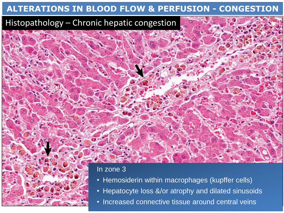
In zone 3
• Hemosiderin within macrophages (kupffer cells)
• Hepatocyte loss &/or atrophy and dilated sinusoids
• Increased connective tissue around central veins
ALTERATIONS IN BLOOD FLOW & PERFUSION - CONGESTION
Histopathology – Chronic hepatic congestion

Congestion / Hyperemia – RBCs are within the blood vessels (*)
Hemorrhage – RBCs are outside vessels ( )
Hyperemia/Congestion vs Hemorrhage
*
*
ALTERATIONS IN BLOOD FLOW & PERFUSION

Questions?
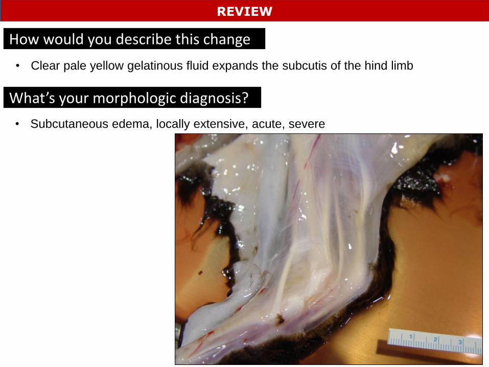
How would you describe this change
REVIEW
What’s your morphologic diagnosis?
• Clear pale yellow gelatinous fluid expands the subcutis of the hind limb
• Subcutaneous edema, locally extensive, acute, severe

What do these two findings tell you about the lungs?
REVIEW
• Froth in the trachea and exuding from the nares indicates that is pulmonary edema

What is your morphologic diagnosis?
REVIEW
• Hydropericardium, moderate
Possible cause? • Hypoproteinemia, congestive heart failure
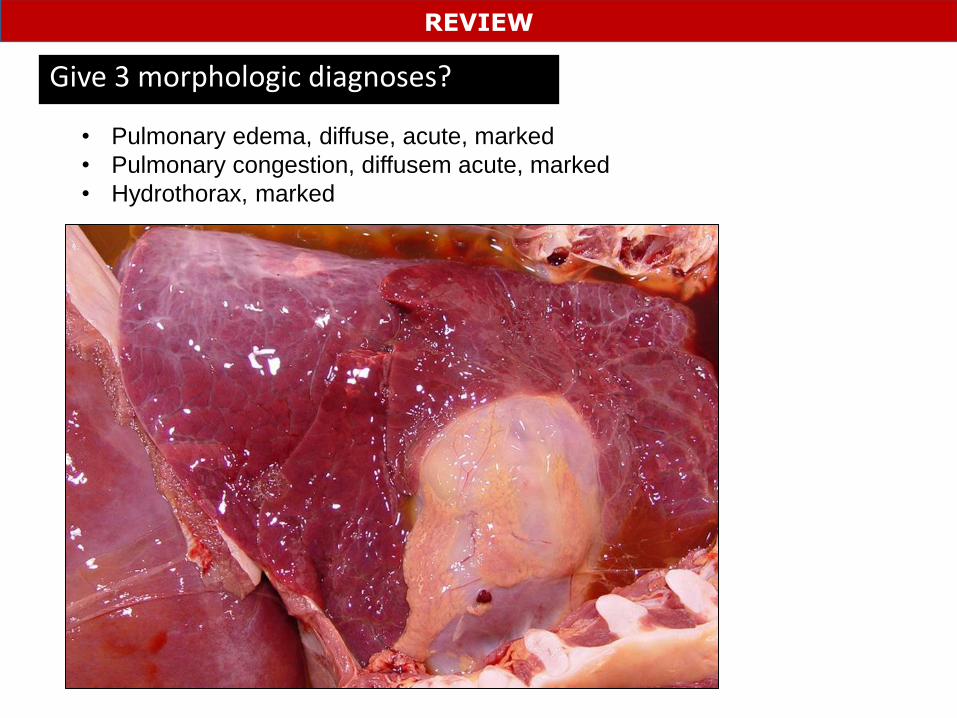
Give 3 morphologic diagnoses?
REVIEW
• Pulmonary edema, diffuse, acute, marked
• Pulmonary congestion, diffusem acute, marked
• Hydrothorax, marked