hydrogel adhesion: a supramolecular synergy of chemistry, … adhesion.pdf · 2019-04-11 ·...
TRANSCRIPT

REVIEWwww.afm-journal.de
© 2019 WILEY-VCH Verlag GmbH & Co. KGaA, Weinheim1901693 (1 of 27)
Hydrogel Adhesion: A Supramolecular Synergy of Chemistry, Topology, and Mechanics
Jiawei Yang, Ruobing Bai, Baohong Chen, and Zhigang Suo*
Adhering hydrogels to various materials is fundamental to a large array of established and emerging applications. The last few years have seen transformative advances in achieving strong hydrogel adhesion, which is a supramolecular phenomenon. Two adherends connect through covalent bonds, noncovalent complexes, polymer chains, polymer networks, or nanoparticles. Separating the adherends dissipates energy through cascading events across length scales, including bond cleavage, chain retraction, and bulk hysteresis. A unifying principle has emerged: strong hydrogel adhesion requires the synergy of chemistry of bonds, topology of connection, and mechanics of dissipation. This synergy characterizes hydrogel adhesion to various materials (another hydrogel, tissue, elastomer, plastic, metal, glass, and ceramic) in various operations (cast, coat, print, attach, pierce, and glue). Strong adhesion can be made permanent, reversible, degradable, or on-demand detachable. The development of hydrogel adhesion and its applications adheres disciplines, discovers interlinks, and forges cohesion. Discussed throughout the review are immediate opportunities for fundamental studies and practical applications.
DOI: 10.1002/adfm.201901693
Dr. J. Yang, Dr. R. Bai, B. Chen, Prof. Z. SuoJohn A. Paulson School of Engineering and Applied SciencesKavli Institute for Nanobio Science and TechnologyHarvard UniversityCambridge, MA 02138, USAE-mail: [email protected]
The ORCID identification number(s) for the author(s) of this article can be found under https://doi.org/10.1002/adfm.201901693.
hydrogel form a sparse and strong network, of mesh size on the order of 10 nm, much larger than a water molecule. This primary polymer network acts as an entropic spring, enabling elasticity. Some other polymers in the hydrogel may form a secondary network, enabling inelasticity. The water molecules in the hydrogel are in the liquid state, changing neighbors readily, and trans-mitting force negligibly. The abundance of water in a hydrogel poses a particular chal-lenge to adhesion. Strong adhesion can never be achieved through binding water molecules in the hydrogel to the other material. Even if the material binds strongly with a monolayer of water molecules, water molecules inside the hydrogel are still as weakly bonded as those in liquid water. Indeed, a hydrogel typically does not adhere strongly to any material, not even to another piece of the same hydrogel.[29,77,78]
Last few years have seen transformative advances in achieving strong hydrogel
adhesion.[5,51,70–72,78–86] A unifying principle has emerged: strong hydrogel adhesion is achieved through the synergy of chemistry, topology, and mechanics (Figure 1). Weak adhesion inevitably originates from missing one or more parts of this synergy. Because the majority constituent of a hydrogel—the water—transmits negligible force, strong adhesion must rely on the minority constituent of the hydrogel—the primary polymer network. The primary polymer network in the hydrogel must connect to the other material through covalent bonds, noncovalent complexes, polymer chains, polymer networks, nanoparticles, etc. The topology of this connection must ensure that the separation of the two adherends dissipates energy through breaking strong bonds on the crack plane, as well as eliciting inelasticity inside one or both of the adherends. We use this unifying principle to organize this review, and to describe individual methods of adhesion.
When adhering a hydrogel to another stretchable material (i.e., another hydrogel, a soft tissue, or a hydrophobic elastomer), which also has a strong and sparse polymer network, the adhe-sion is often required not to harden the interface. A commonly used adhesive, cyanoacrylate, forms a glassy phase, which does harden the interface.[1,87] To achieve strong and stretchable adhesion, the connection between the polymer networks of the two adherends must be strong and sparse.[5,71,78,82–86]
This review first describes functions of hydrogel adhesion in medicine and engineering (Section 2). We then briefly outline common methods to adhere dry materials, and note if they
Hydrogels
1. Introduction
Two materials adhered together are called adherends. Their adhesion is often, but not always, via a third material called an adhesive. By hydrogel adhesion we mean that at least one adherend is a hydrogel, and the other adherend can be any material. Hydrogel adhesion is central to many applications in medicine and engineering (Table 1).
This review focuses on strong hydrogel adhesion—that is, a hydrogel itself has appreciable cohesion, and its adhesion to another material is comparable to the cohesion. The art of adhe-sion dates back to the beginning of civilization, but has been mostly devoted to adhering dry materials (e.g., metals, ceramics, plastics, and elastomers).[76] A hydrogel, either natural or syn-thetic, is wet. The hydrogel is an aggregate of water and polymers, with the water being the majority constituent, and the polymers being the minority constituent. Some or all of the polymers in the
Adv. Funct. Mater. 2019, 1901693

www.afm-journal.dewww.advancedsciencenews.com
1901693 (2 of 27) © 2019 WILEY-VCH Verlag GmbH & Co. KGaA, Weinheim
are applicable to hydrogel adhesion (Section 3). We describe chemistries of bonds (Section 4), topologies of connection (Section 5), and mechanics of dissipation (Section 6). We then describe recently developed methods of strong hydrogel adhe-sion (Section 7), and illustrate their applications in various operations: cast, coat, print, attach, pierce, and glue (Section 8). Strong hydrogel adhesion can be made permanent, reversible, degradable, or on-demand detachable (Section 9). We conclude with a call to action (Section 10).
2. Functions of Hydrogel Adhesion in Medicine and Engineering
2.1. Hydrogels
Most tissues of animals and plants are natural hydrogels; their evolution must have intertwined with that of life. Synthetic hydro-gels, however, are relatively new materials. The development of synthetic hydrogels started in 1960s and has been vibrant since.[88,89] The enthusiasm for hydrogels is reminiscent of the adolescent years of the rubber and plastic industries.[90] Soft tissues (e.g., skin, mucosa, brain, axon, cartilage, blood vessels, and muscle) are natural hydrogels, containing about 60%–90% of water.[91] Synthetic hydrogels can mimic soft tissues with high fidelity, chemically, mechanically, and electrically. This biomimicry has enabled an enormous and fast-expanding field of biointegration and bioinspiration. Well-established applica-tions include contact lenses,[92,93] superabsorbent diapers,[94] cell culture,[95,96] tissue scaffolds,[97,98] wound closure,[1–5] and drug delivery.[13,18,97] Emerging applications include ionotronics,[28,29] bioelectronics,[27,99] water matrix composites (WMCs),[57–67] soft robots,[33,34,100,101] soft power sources,[56] muscle-like adaptive materials,[102] and expansion microscopy.[24,25]
In a hydrogel, the mesh size of the polymer network is much larger than a water molecule. Within a mesh, water molecules move relative to one other, behaving as a liquid. Beyond a mesh, the polymer network deforms but retains crosslinks, behaving as a solid. The hybrid solid–liquid attributes make hydrogels a unique class of materials. Water molecules in a hydrogel dissolve many other molecules and transport them through the hydrogel. The polymer network of a synthetic hydrogel is readily engineered to mimic functions of natural hydrogels (i.e., tissues of animals and plants). When the polymer network has reversible moieties, the hydrogel heals after damage.[103,104] When the polymer network has stimuli-responsive groups, the hydrogel changes its volume, transparency, color, etc., in response to stimuli (e.g., chemicals, temperature, pH, humidity, mechanical forces, light of a certain frequency range, electrical and magnetic fields).[20,74,75,105–107] Such a hydrogel enables actuators,[108,109] sensors,[110,111] drug vehicles,[18,20] and switchable surfaces.[112] When the polymer network has degradable moieties, the hydrogel degrades spon-taneously over time, or degrades on-demand in response to a trigger. Such a hydrogel has been used in wound management and controlled release of therapeutics and cells.[18,113,114] Hydro-gels can be stretchable, transparent, ionic conductors, enabling ionotronics and bioelectronics.[29,30,37,38] Furthermore, hydrogels can be printed to replicate the complex structures of organs,[115] vascular networks,[116,117] and aortic valves.[118,119]
Jiawei Yang is currently a Ph.D. student in the John A. Paulson School of Engineering and Applied Sciences at Harvard University. He received his M.S. from Harvard University in 2016 in Engineering Sciences, and Ph.D. and B.S. from Tongji University in 2015 and 2009, both in Engineering Mechanics. His
research focuses on exploring mechanics and chemistry of materials, and investigating broad topics at the interface of physics, chemistry engineering, and medicine, including soft active materials, adhesion and fracture, mechanical instabilities, complex rheology, soft wearable devices, and biomedical engineering.
Ruobing Bai is a postdoctoral fellow in the Department of Mechanical and Civil Engineering at California Institute of Technology. He received his Ph.D. in Engineering Sciences from Harvard University in 2018. He received his B.S. degree in Theoretical and Applied Mechanics from Peking University in
2012. His research focuses on fracture, adhesion, and mechanical instability of materials, soft active materials such as hydrogels and liquid crystal elastomers, as well as multiphysics and functionalities of materials combining mechanics, thermodynamics, chemistry, optics, and electromagnetism.
Zhigang Suo is Allen E. and Marilyn M. Puckett Professor of Mechanics and Materials at Harvard University. He earned a bachelor’s degree from Xi’an Jiaotong University in 1985, and a Ph.D. degree from Harvard University in 1989. Suo joined the faculty of the University of California at Santa Barbara in 1989,
Princeton University in 1997, and Harvard University in 2003. His research centers on the mechanics of materials. Applications include electronics, composites, and stretch-able devices.
Adv. Funct. Mater. 2019, 1901693

www.afm-journal.dewww.advancedsciencenews.com
1901693 (3 of 27) © 2019 WILEY-VCH Verlag GmbH & Co. KGaA, Weinheim
2.2. Hydrogel–Tissue Adhesion
We next describe the functions of adhering hydrogels to various materials (Table 1). Sutures and staples are commonly used to
close wounds, but inflict secondary damage to tissues and possibly cause inflamma-tion.[120–122] Hydrogels that strongly adhere to tissues can supplement or even replace sutures and staples. Moreover, a hydrogel can support the growth of a damaged cartilage,[7] and seal defects on a lung or artery.[10] The abundant water in hydrogels enables them to encapsulate drugs. In contact with tis-sues, the hydrogels regulate the release of the drugs in space and time.[18] The drug-loaded hydrogels are often required to adhere to tis-sues. It is also conceivable that hydrogels will be developed as human–machine interfaces to stimulate neurons chemically, electrically, and optically, as well as monitor health in real time.[27–29]
Hydrogels of proteins, polysaccharides, and synthetic polymers are frequently used as tissue adhesives. Fibrin hydrogels derive their two major components—fibrinogen and thrombin—from human plasma or bovine
proteins. Other protein hydrogels include gelatin, collagen, and albumin hydrogels. Alginate and chitosan are polysaccha-ride hydrogels derived from seaweeds and shells of shrimp. Synthetic hydrogel adhesives include poly(ethylene glycol) and poly(vinyl alcohol). These hydrogels adhere to tissues through covalent bonds or noncovalent complexes. All these hydrogels are mechanically much weaker than most tissues,[123,124] and cannot be applied in the environment where high pressure is present, such as inside the cardiac chambers and major blood vessels.[122]
A commonly used tissue glue is cyanoacrylate.[87,125] When applied at the interface between two tissues, cyanoacrylate instantly polymerizes into a glassy polymer, which forms covalent bonds with the polymer networks of the tissues.[1] Cyanoacrylate has many issues: i) it works poorly in the presence of blood, as it quickly sets before adhering the tissues; ii) the adhesive layer is stiff and constrains the movements of the soft tissues; iii) the polycyanoacrylate degrades and releases cyanoacetate and formaldehyde, which are cytotoxic.[126,127]
2.3. Mucoadhesion
Mucus covers the surfaces of cavities and organs in a body, and is a natural hydrogel consisting of over 90% of water and a polymer network of glycoproteins.[128,129] Since the 1980s, mucoadhesives have been developed to adhere with mucosal surfaces for drug delivery.[21,23,130] Common mucoad-hesives include polyacrylate, alginate, chitosan, and cellulose derivatives. They are processed into dry forms (tablets, films and patches) and wet forms (hydrogels and ointment).[130] When a mucoadhesive contacts with mucus, the polymer chains in the mucoadhesive and the mucin glycoprotein inter-penetrate to a thickness of nanometers to micrometers.[131] Interpolymer complexes form through noncovalent bonds and establish adhesion within seconds.[23,132,133] The existing mucoadhesives, however, are mechanically weak.
Adv. Funct. Mater. 2019, 1901693
Table 1. Applications enabled by adhering hydrogels to various materials.
Hydrogel–tissue Hydrogel–elastomer Hydrogel–hydrogel
Hydrogel–plastic Hydrogel–ceramic Hydrogel–bone
Hydrogel–metal
• Wound dressing[1–5]
• Tissue repair[5–10]
• Tissue regeneration[11,12]
• Medical implant[13–15]
• Transdermal drug delivery[16–20]
• Transmucosal drug delivery[18–23]
• Expansion microscopy[24,25]
• Implosion fabrication[26]
• Bioelectronics[27–29]
• Artificial muscles[30–34]
• Artificial skins[35–37]
• Artificial axons[38,39]
• Ionic textiles[39]
• Ionotronic luminescence[40–42]
• Ionotronic liquid crystal
devices[43]
• Ionic touchpads[44]
• Ionic diodes[45]
• Triboelectric generators[46–48]
• Biomedical devices[49–51]
• Stretchable and interactive
seal[52,53]
• Microfluidics[54]
• Hydrogel–hydrogel
composites[55]
• Soft power sources[56]
• Water matrix composites[57–67]
• Implantable devices[68,69]
• Bone repair[70]
• Soft electronics[71–73]
• Chemical sensors[74,75]
Figure 1. Chemistries of bonds, topologies of connection, and mechanics of dissipation. A hydrogel may connect to an adherend through many molecular topologies. Drawn here is the stitch-bond topology. The hydrogel has a preexisting covalent network (black). A species of polymer chains form a new network (red), in topological entanglement with the preexisting polymer network of the hydrogel. The new network also forms strong bonds (blue) with the other adherend. The separation of the two adherends dissipates energy through breaking the strong bonds at the interface and eliciting inelasticity inside the hydrogel. Inelasticity requires the bulk of the hydrogel to have sacrificial bonds (green). Inelasticity inside the bottom adherend is also possible, but is not drawn here.

www.afm-journal.dewww.advancedsciencenews.com
1901693 (4 of 27) © 2019 WILEY-VCH Verlag GmbH & Co. KGaA, Weinheim
2.4. Hydrogel–Elastomer Adhesion
In open air, a hydrogel coated with a hydrophobic elastomer dehydrates more slowly than a naked hydrogel.[39,83,86] Such an elastomer-coated hydrogel may enable the development of stretchable and conductive textiles to endure wear, wash, and iron.[39,84] Hydrophilic brushes and hydrogels have been used to coat polydimethylsiloxane (PDMS) microfluidic channels to enhance surface wettability, channel filling, and cell attachment.[54,134] In one example, a hydrogel coating on elastomeric microfluidic channels serves as a semipermeable membrane, which encapsulates the embedded bacteria in the channels from leaking, and allows chemicals from environment to diffuse through the hydrogel coating to the encapsulated bacteria. The bacteria responds to chemicals and gives fluo-rescent feedbacks.[52,53] These hydrogel seals are capable of real-time environmental sensing despite being stretched and twisted. An ionotronic device functions with both mobile ions and mobile electrons. Familiar examples include batteries and fuel cells.[135–137] By contrast, hydrogel ionotronics are soft and stretchable.[29,30,38] In hydrogel ionotronics, hydrogels are ionic conductors and elastomers are dielectrics. A variety of hydrogel ionotronics have been developed, including artifi-cial muscles,[30–34] artificial skins,[35–37] artificial axons,[38,39] ionotronic luminescent devices,[40–42] liquid crystal devices,[43] touchpads,[44] triboelectric and ionotronic generators,[46–48] and biomedical devices.[49–51]
VHB (3M), PDMS (Dow Corning), Ecoflex (Smooth-on), and butyl rubber are mostly used in hydrogel–elastomer adhesion. VHB (4910) is highly stretchable (stretchability ≈ 9), transparent (transmittance >90%), and relatively tough (≈1000 J m−2).[138] However, VHB is crosslinked as received, and cannot be reshaped into other geometries or reprocessed with different mechanical properties. PDMS has a stretchability of about 3, good transparency (transmittance >90% at a thickness of 1 mm), but lower fracture toughness (≈100 J m−2). Ecoflex has a stretchability of about 7, and becomes opaque when the thickness is larger than 1 mm. Both PDMS and Ecoflex are synthesized from precursors, easily shaped into com-plex geometries, and tuned into desired mechanical prop-erties. Of all elastomers, butyl rubber has the lowest water permeability.[39,139]
2.5. Hydrogel–Hydrogel Adhesion
Hydrogel–hydrogel adhesion has also found significant applications. Hydrogel–hydrogel adhesion is often used as a model system in the preliminary development of methods for hydrogel–tissue adhesion. Dissimilar polyelectrolytes in contact function as ionic diodes and soft power source.[45,56] Adhesion is not evaluated in these works, but strong adhe-sion will be important in developing integrated hydrogel devices of more complexity. Many integrated hydrogel devices are conceivable by adhering dissimilar hydrogels responding to dissimilar stimuli. Microgel-reinforced hydrogels are both tough and amenable to cast, coat, and print.[51,55,140,141] The microgels and the hydrogel matrix adhere strongly through topological entanglement.
2.6. Hydrogel–Metal, Hydrogel–Ceramic, and Hydrogel–Plastic Adhesion
Elastomers, plastics, glasses, ceramics, and metals are dry materials. Fibers and sheets of dry materials have been embedded in hydrogels to create a new class of materials: WMCs. Their stiffness, strength, and toughness are orders of magnitude higher than those of pristine hydrogels.[57–62] Typ-ical WMCs include hydrogel–fabric composites,[63] hydrogel–glass composites,[57] hydrogel–metal composites,[58,59,65] and hydrogel–plastic composites.[60–62,64–67] WMCs are developed for applications that require materials to be resilient, energy absorbing, and load bearing, such as materials for impact-resistant helmets[142] and materials mimicking load-bearing tissues.[143]
An implantable medical device coated with a layer of hydrogel changes the surface to be hydrophilic, biocompatible, and mechanically compatible to biological tissues.[68,69] It not only circumvents the adverse effects brought by the rigid metal components, but also offers additional functions such as anti-microbe, low friction, and wear resistance.
In soft electronics, a hydrogel serves as a stretchable and biocompatible substrate, and is intended to contact biological tissues.[71–73] Rigid integrated electronic circuits adhere to the hydrogel for data requisition, computing, and output. Chem-ical sensors have been developed using a sub-micrometer-thick hydrogel coating, which detect metal ions, biomolecules (e.g., protein and enzyme), and volatile organic compounds with ultrahigh sensitivity and selectivity.[74,75]
3. Methods to Adhere Two Dry Materials
Dry materials include glasses, ceramics, elastomers, plastics, and metals. Their adhesion has been practiced since antiq-uity.[76,144–146] This section briefly describes methods of dry adhesion, and note if they are applicable for hydrogel adhe-sion. An understanding of dry adhesion helps to appreciate the requirements for hydrogel adhesion.
3.1. Direct Wafer Bond
When two solids, sufficiently clean and flat, are brought into contact, interatomic or intermolecular attraction zips them into adhesion (Figure 2a). This method of adhesion, called direct wafer bond, has been extensively developed in the semi-conductor industry.[147,148] Similar method has also been used to adhere other materials, such as mica[149] and rubber.[150] The surfaces of the solids need not be smooth at the atomic or molecular scale, so long as the reduction of surface energy exceeds the gain of elastic energy.[151]
Whereas direct wafer bond can achieve strong adhe-sion between dry materials, it usually cannot achieve strong hydrogel adhesion. The water molecules in a hydrogel change neighbors readily and transmit force negligibly. Strong adhe-sion can only be achieved when complementary functional groups exist to bind the polymers in the hydrogel strongly to the other adherend.
Adv. Funct. Mater. 2019, 1901693

www.afm-journal.dewww.advancedsciencenews.com
1901693 (5 of 27) © 2019 WILEY-VCH Verlag GmbH & Co. KGaA, Weinheim
3.2. Cold Weld
When surfaces of two solids are rough, they cannot reach atomic contact by elastic deformation. To close the gap, atoms must change neighbors. A ductile metal can achieve atomic contact with the other adherend through plastic deforma-tion, usually under pressure comparable to the yield strength of the metal (Figure 2b). This method of adhesion is called cold weld.[152] Cold weld cannot achieve strong adhesion for hydrogels, elastomers, and thermoset plastics. The permanent crosslinks in these materials prohibit polymer chains from changing connectivity.
3.3. Diffusion Bond
Glasses, ceramics, and refractory metals do not plastically deform appreciably at room temperature. To close the gaps, the materials are heated to a temperature about 50–80% of the abso-lute melting temperature of one of the materials. At such a tem-perature, the adherends can still retain their macroscopic shape, but at the contact, atoms change neighbors by plastic creep or self-diffusion (Figure 2c). This method of adhesion is called dif-fusion bond, or sinter.[153–155] If the two materials can form a solid solution or a new phase, atoms can diffuse from one solid into the other to from an interphase. Solubility and interphase, however, are not required for strong diffusion bond. Diffusion bond is also used to adhere thermoplastics.[156–158] Diffusion bond cannot achieve strong adhesion for hydrogels, elastomers, and thermoset plastics. The permanent crosslinks in these mate-rials prohibit polymer chains from changing connectivity.
3.4. Surface Modification
Surfaces of adherends are often modified to enable strong interlinks (Figure 2d). For example, when a precursor of polybutadiene rubber is cast on a vinylsilane treated glass
substrate, in situ free radical polymerization crosslinks the rubber and interlinks the rubber and the glass substrate.[159] Adherends can also interlink through complementary func-tional groups. For example, PDMS is usually plasma-treated to oxidize methyl groups into silanol groups.[160] These silanol groups can condensate to covalent bonds with appropriate groups (e.g., hydroxyl, carboxylic acid, ketone) on the other adherend.[161] For PDMS and glass, this reaction yields siloxane bonds after condensation. Surface modification has been used to achieve strong hydrogel adhesion.[68,72,80,81,83,84,86,162]
3.5. Topological Adhesion
Two elastomers without any functional groups can strongly adhere through topological entanglement (Figure 2e). A species of polymer chains diffuse into the two preexisting elastomer networks, and crosslink into a third polymer network in situ, in topological entanglement with the two preexisting elastomer networks. We have suggested that this method of adhesion be called topological adhesion, or topohesion for short.[85]
When two partially crosslinked elastomers (e.g., polybuta-diene and poly(ethylene-co-propylene)) are pressed into an intimate contact, the uncrosslinked polymer chains from one elastomer diffuse into the other, and then crosslink in situ, in topological entanglement.[163] This method is also applicable for adhering two pieces of PDMS or two pieces of dissimilar elasto-mers.[164,165] Topological adhesion of PDMS fibers and a softer PDMS matrix has led to an elastomer composite of high tough-ness and low hysteresis.[166]
An elastomer and a plastic may also topohere. A precursor, consisting of a suitable solvent, monomers, crosslinkers, and initiators, is placed at the interface of an elastomer and a plastic. The precursor diffuses into both adherends, polymerizes, and crosslinks into a new network, in topolog-ical entanglement with the networks of both adherends.[167] Topological adhesion can also achieve strong hydrogel adhesion.[85,86,168]
Adv. Funct. Mater. 2019, 1901693
Figure 2. Methods of dry adhesion. a) Direct wafer bond. b) Cold weld. c) Diffusion bond. d) Surface modification. e) Topological adhesion. f) Physical entanglement. g) Mechanical interlock. h) Adhesive bond.

www.afm-journal.dewww.advancedsciencenews.com
1901693 (6 of 27) © 2019 WILEY-VCH Verlag GmbH & Co. KGaA, Weinheim
3.6. Physical Entanglement
Two thermoplastics can adhere strongly through physical entanglement of polymer chains (Figure 2f). Polymer chains are spread at the interface of the thermoplastics, and are often annealed above their glass transition temperatures to allow polymer chains to diffuse into them. Upon cooling, the two ther-moplastics become glassy polymers again, and the penetrating polymers are locked inside.[169] Such polymer chains in phys-ical entanglement can be slowly pulled out, and the adhesion strength depends on the rates of separation.[170,171]
For a soft material of a pre-existing polymer network, strong adhesion requires that the entanglement be topological. To sepa-rate two such materials in topohesion, at least one of the three networks must rupture. By contrast, if the diffusing chains are in physical entanglement with the preexisting polymer networks, the adhesion is rate-dependent, and vanishes at low debond speed, because these diffusing chains can be slowly pulled out from the polymer networks.[172] Adhesion between soft materials by physical entanglement is like a suture without knots.
3.7. Mechanical Interlock
Mechanical interlock is a well-known mechanism of adhesion.[76,145] Adhesion increases with the roughness of adherends,[173] possibly through the increase of energy dissipation and interfacial area. Indeed, for surfaces with a spe-cial shape (e.g., Figure 2g), a liquid adhesive fills the asperities on the surface of both materials and then solidifies to a rigid adhesive layer. Because the adhesive layer may be mechanically as strong as that of adherends, the force required to separate the two adherends is large. However, most surfaces may not be very rough, and mechanical interlock may not be the major contributor to adhesion. Mechanical interlock usually cannot achieve strong hydrogel adhesion, because hydrogels are soft, and can be pulled out by deformation. An exception exists, where the adherend has open pores; a hydrogel fills the pores, and can only be pulled out by rupture.[79] In such a case, the method may be called topological interlock.
3.8. Adhesive Bond
Adhesive bond is perhaps the most common method of adhesion in daily life. One form of adhesives is a tape, called a pressure-sensitive adhesive, consisting of lightly crosslinked or uncrosslinked polymer chains.[174,175] The adhesive is sufficiently soft to conform to the surfaces of adherends and fill gaps. To resist separation, the adhesive dissipates energy by viscoelasticity. When the adherends are pulled apart, the adhesive often forms fibrils. Another form of adhesives is a solu-tion of reactive prepolymers or monomers. Examples include acrylic, silicone, polyurethane, poly(vinyl acetate), epoxy, and cyanoacrylate.[76] The liquid wets the interface of two adherends, and cures into a dense glassy polymer. Even though the glassy polymer may not form covalent bonds with the adherends, adhesion can still be appreciable, so long as the noncovalent interaction is dense packed—that is, every atom or monomer
unit at the interface carries load (Figure 2h). Adhesive bonding has been demonstrated for strong hydrogel adhesion.[5,71,78,85,86]
4. Chemistries of Bonds
Hydrogel adhesion is a supramolecular interplay of chemistry, topology, and mechanics (Figure 1). Strong hydrogel adhesion may result from various covalent and noncovalent bonds (Table 2). Individual covalent bonds are strong. They link mon-omer units into a polymer chain, crosslink similar polymer chains into a polymer network, and interlink two polymer networks into adhesion. By contrast, individual noncovalent bonds are weak (Table 3). An aggregate of noncovalent bonds, however, can be strong.[85,176,177] Polymer chains can interact strongly with each other through aggregates of noncovalent bonds, forming a polymer complex.[178,179] Polymer complexes have enabled hydro-gels of high toughness and self-heal,[176,180–182] as well as supra-molecular assembly of intricate structures.[183,184] Life thrives on supramolecular adhesion through noncovalent bonds between covalent molecules. Familiar examples include DNA replication, protein folding, antigen–ligand binding, and cell adhesion.[185,186] Polymer complexes are widely used in hydrogel adhesion. Examples include mucoadhesion,[187] underwater adhesion,[188] polyampholyte adhesion,[82] reversible adhesion,[189,190] and topo-logical adhesion.[85,86]
After breaking, a bond may reform under suitable conditions, such as long time, elevated temperature, proper pH, presence of an enzyme, and exposure to light of a specific range of frequency. Some covalent bonds and many noncovalent bonds are revers-ible (dynamic) under common experimental conditions. We regard a bond as irreversible (static) if its reformation requires conditions beyond those for common operations in adhesion. In the following, we briefly describe representative bonds.
4.1. Static Covalent Bonds
Static covalent bonds are stable and strong. To form covalent interlinks between a hydrogel and another adherend, the hydrogel and adherend should have complementary functional groups. Otherwise, the adherends are chemically modified to create the requisite functional groups. Static covalent bonds commonly used in hydrogels include carbon–carbon,[72,81,83] amide,[5,80] siloxane,[84] and carbon–nitrogen.[201] These bonds are irreversible. Once broken during the separation of the adherends, these covalent bonds do not reform.
4.2. Dynamic Covalent Bonds
Dynamic covalent bonds reversibly dissociate and reform in response to pH, heat, light, or redox condition.[202–204] The time of bond reformation vary from minutes to hours. Example dynamic covalent bonds include disulfide,[133] imine,[7,9,86] acylhydra-zone,[205,206] phenylboronate ester,[207] and bonds formed by Diels–Alder reactions.[208] The strong yet reversible covalent bonds have enabled self-healing hydrogels. For example, acylhydrazone bonds heal the crack within 24 to 48 h,[205] dextran–poly(ethylene
Adv. Funct. Mater. 2019, 1901693

www.afm-journal.dewww.advancedsciencenews.com
1901693 (7 of 27) © 2019 WILEY-VCH Verlag GmbH & Co. KGaA, Weinheim
glycol) hydrogel heals the crack after 7 h at 37 °C,[208] and a tough alginate–polyacrylamide hydrogel containing ionic bonds and disulfide bonds can be on-demand degraded using ethyl-enediaminetetraacetic acid and glutathione.[114] Because mucus contains abundant cystines, hydrogels carrying thiol groups can form mucoadhesion through disulfide bonds,[23] which improves
cohesive properties of the adhesive layer, and provides stability for long-term drug delivery.
4.3. Ionic Bonds
An ionic bond is formed by electrostatic interaction. For example, the negatively charged carboxylate groups on two alginate chains form ionic bonds through divalent calcium ions.[196,209] The ionic bonds in the Ca-alginate complex, once broken, are almost irre-versible even after one day at room temperature, but partially reform at a temperature of 60 °C for one day.[210] The strength and recoverability of ionic bonds are improved using trivalent cations such as Al3+ and Fe3+.[211] Trivalent cations interact with three carboxylic groups at the same time. The elastic moduli of Al-alginate complex and Fe-alginate complex are almost one order of magnitude higher than that of Ca-alginate complex. There-fore, the iron–polyacrylate network is broadly used to toughen a hydrogel[84,86,181,212] or to serve as a stitching network for strong adhesion.[168] When the iron–polyacrylate complex is ruptured,
Adv. Funct. Mater. 2019, 1901693
Table 2. Representative chemistries of bonds.
Bond types Representative bonds
Static covalent bonds
Dynamic covalent bonds
Ionic bonds
Hydrogen bonds
Other noncovalent interactions
a)Reproduced with permission.[191] Copyright 2013, American Chemical Society; b)Reproduced with permission.[192] Copyright 2013, Wiley-VCH Verlag GmbH & Co. KGaA, Weinheim.
Table 3. Bond energy of representative interactions (kT = 4.11 × 10⁻²¹ J at room temperature).
Interactions Bond energy (kT)
Covalent bond 80–320 (ref. [193])
Hydrogen bond 1–50 (ref. [194,195])
Ionic bond 1–10 (ref. [196,197])
Hydrophobic interaction 1–10 (ref. [198])
π–π stack 1–10 (ref. [199])
Host–guest interaction 1–10 (ref. [200])
van der Waals interaction ≈1 (ref. [78,194])

www.afm-journal.dewww.advancedsciencenews.com
1901693 (8 of 27) © 2019 WILEY-VCH Verlag GmbH & Co. KGaA, Weinheim
the ionic bonds can reform and restore the complex to its initial mechanical properties at room temperature after a few hours.[181]
A polycation and a polyanion can form a polyelectrolyte com-plex.[213,214] For example, a negatively charged sodium alginate and a positively charged chitosan form a polyelectrolyte com-plex in water.[215] Polyelectrolyte complexes can also form with polyampholytes—polymers that bear both cationic and anionic mono mer units. For example, polymer chains carrying positively charged trimethylammonium and negatively charged sulfonate can lead to a stable polymer network crosslinked by random for-mation of ionic bonds of different strength.[180,216] The strong ionic bonds improve the modulus to 1 MPa and toughness over 1000 J m−2, and enable self-heal of mechanical properties within 30 min at room temperature. Polyelectrolyte complexes have been used to enable strong hydrogel adhesion.[82]
4.4. Hydrogen Bonds
A hydrogen bond is formed between a hydrogen atom bound to a more electronegative atom and another atom bearing a lone pair of electrons.[195] A single hydrogen bond is weak, with the bond strength about 1–10 kT. However, two polymer chains can form a hydrogen-bonded complex through dense arrays of hydrogen bonds. Strong hydrogen-bonded complexes are formed through hydroxyl groups on poly(vinyl alcohol) chains[217] and on cellulose chains,[85,218] amine and hydroxyl groups on chitosan chains,[85,182] carboxylic acid and hydroxyl groups on alginate chains,[85] as well as amine and phenyl groups on poly(4-aminostyrene) chains.[85] Hydrogen-bonded interpolymer complexes include poly(ethylene glycol) and poly(acrylic acid), as well as polyacrylamide and poly(acrylic acid).[219,220] Hydrogen-bonded complexes have been used to enable hydrogel adhesion.[177,221] Hydrogen bonds between nucleobases enable the molecular recognition in DNA and RNA, and the nucleobases have been copolymerized in polymer chains to enable hydrogel adhesion.[222]
4.5. Hydrophobic Interaction
Hydrophobic molecules tend to aggregate in water. When hydro-phobic molecules are chemically linked to hydrophilic polymer chains, two or more such polymer chains tend to aggregate at the hydrophobic sites, and the hydrophobic sites serve as crosslinks. A typical hydrophobic moiety is an alkyl chain with length of 12 and 22 carbon atoms. The hydrophobic interaction has been used to make tough and self-healing hydrogels,[223–225] but has not been used to create strong hydrogel adhesion.
4.6. π–π Stack
π–π stack refers to interaction between aromatic rings. Aromatic rings are insoluble in water. Aromatic rings can be incorpo-rated in a hydrogel by chemically linking to polymer chains with highly hydrophilic monomer units, such as amine, amide, and hydroxyl. π–π stack has been used both in hydrogels[226–228] and in hydrogel adhesion.[85,86]
4.7. Dipole–Dipole Interaction
Dipolar molecules interact electrostatically. For example, the interaction between two nitrile groups is strong, and ensures the success of a famous instant glue—cyanoacrylate, which forms a glassy polymer network though interchain nitrile inter-action.[229] Nitrile group has also been used to make hydrogels of high strength, toughness, and fatigue resistance.[230,231]
4.8. Host–Guest Interaction
Host–guest interaction refers to two or more molecules or ions that held together by noncovalent bonds.[232] Macrocyclic molecules and their derivatives have been developed as hosts, such as calixarenes, crown ethers, cyclodextrins, cyclophanes, cucurbit[n]urils, and pillar[n]arenes. The cavity of a macro-cyclic molecule can encapsulate guests like small molecules, proteins, and polymer chains. Self-healing hydrogels are made with phenylalanine and cucurbit[8],[233,234] cyclodextrins and adamantan,[191] and cyclodextrins and ferrocene.[235] In addi-tion, the cucurbit[7]uril and aminomethylferrocene host–guest complex is designed to make supramolecular Velcro for reversible underwater adhesion.[192]
4.9. Catechol Chemistry
Catechol chemistry is inspired by mussel adhesion.[236–238] Catechol has a benzene ring and two neighboring hydroxyl groups, and forms either covalent bonds or noncovalent com-plexes with many other functional groups (Figure 3).[201,239] The hydroxyl groups of catechol form hydrogen bonds with hydro-philic surfaces, and electrostatic interaction with metal oxide surfaces. The benzene ring of catechol forms π–π stack, hydro-phobic interaction with hydrophobic surface, and cation–π interaction with positively charged surface. In addition, when catechols are oxidized to become quinones, they form a strong coordination complex with metal oxide surface, and covalently
Adv. Funct. Mater. 2019, 1901693
Figure 3. Catechol chemistry. a) Chemical structure of a catechol. b) Possible interactions between catechol and various functional groups.
www.afm-journal.dewww.advancedsciencenews.com
1901693 (9 of 27) © 2019 WILEY-VCH Verlag GmbH & Co. KGaA, Weinheim
bond to surfaces containing nucleophiles (e.g., amine and thiol) through possible Michael addition reaction.[201,240] Catechol even strongly adheres to inert metals such as gold.[241] Catechol has been added into hydrogels to enable adhesion with various materials, by chemical modification of the hydrogel network or physically mixing polymer chains bearing catechols in the hydrogel network.[68,242,243] However, the adhesion achieved so far is not strong.
5. Topologies of Connection
By a topology of connection, we mean a way to connect two adherends through bonds, polymer chains, polymer networks, nanoparticles, bulk materials, or their combinations. Connection through bulk materials is a macroscopic topology, commonly called mechanical interlock. The rest of the examples are mole-cular topologies. Significantly, numerous molecular topologies are possible, and many are unexplored. The diversity of topolo-gies is expected to expand the field of innovation greatly in hydrogel adhesion.[5,70–72,79–86]
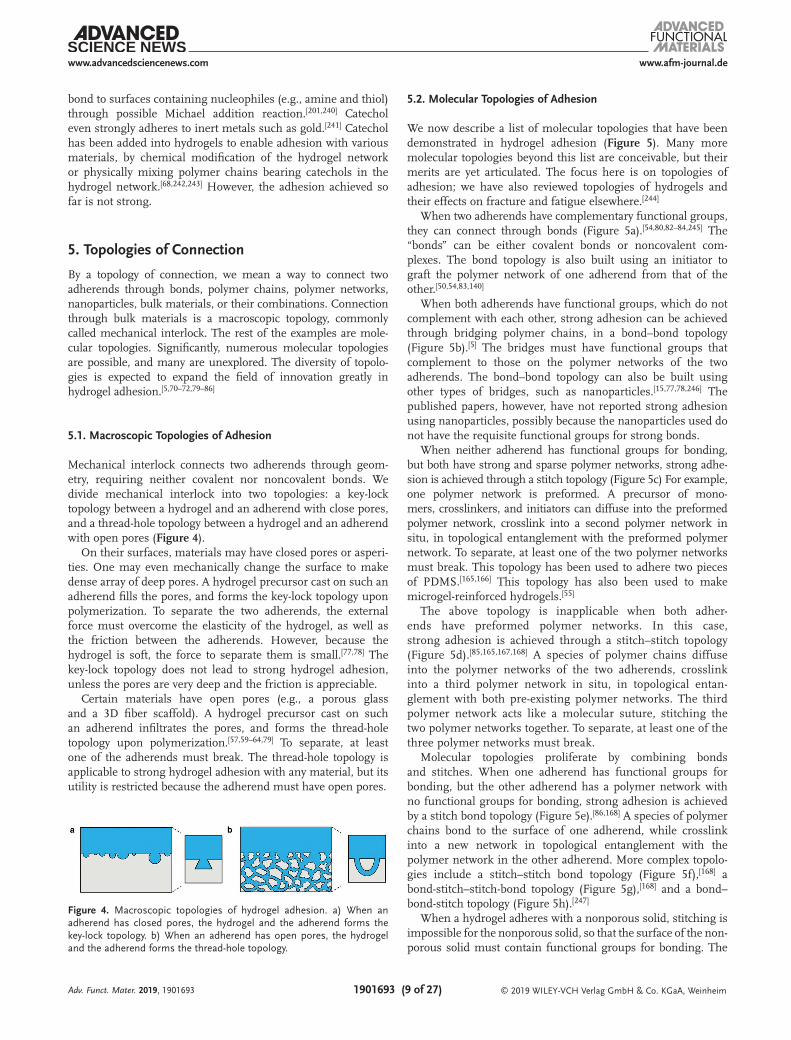
5.1. Macroscopic Topologies of Adhesion
Mechanical interlock connects two adherends through geom-etry, requiring neither covalent nor noncovalent bonds. We divide mechanical interlock into two topologies: a key-lock topology between a hydrogel and an adherend with close pores, and a thread-hole topology between a hydrogel and an adherend with open pores (Figure 4).
On their surfaces, materials may have closed pores or asperi-ties. One may even mechanically change the surface to make dense array of deep pores. A hydrogel precursor cast on such an adherend fills the pores, and forms the key-lock topology upon polymerization. To separate the two adherends, the external force must overcome the elasticity of the hydrogel, as well as the friction between the adherends. However, because the hydrogel is soft, the force to separate them is small.[77,78] The key-lock topology does not lead to strong hydrogel adhesion, unless the pores are very deep and the friction is appreciable.
Certain materials have open pores (e.g., a porous glass and a 3D fiber scaffold). A hydrogel precursor cast on such an adherend infiltrates the pores, and forms the thread-hole topology upon polymerization.[57,59–64,79] To separate, at least one of the adherends must break. The thread-hole topology is applicable to strong hydrogel adhesion with any material, but its utility is restricted because the adherend must have open pores.
5.2. Molecular Topologies of Adhesion
We now describe a list of molecular topologies that have been demonstrated in hydrogel adhesion (Figure 5). Many more molecular topologies beyond this list are conceivable, but their merits are yet articulated. The focus here is on topologies of adhesion; we have also reviewed topologies of hydrogels and their effects on fracture and fatigue elsewhere.[244]
When two adherends have complementary functional groups, they can connect through bonds (Figure 5a).[54,80,82–84,245] The “bonds” can be either covalent bonds or noncovalent com-plexes. The bond topology is also built using an initiator to graft the polymer network of one adherend from that of the other.[50,54,83,140]
When both adherends have functional groups, which do not complement with each other, strong adhesion can be achieved through bridging polymer chains, in a bond–bond topology (Figure 5b).[5] The bridges must have functional groups that complement to those on the polymer networks of the two adherends. The bond–bond topology can also be built using other types of bridges, such as nanoparticles.[15,77,78,246] The published papers, however, have not reported strong adhesion using nanoparticles, possibly because the nanoparticles used do not have the requisite functional groups for strong bonds.
When neither adherend has functional groups for bonding, but both have strong and sparse polymer networks, strong adhe-sion is achieved through a stitch topology (Figure 5c) For example, one polymer network is preformed. A precursor of mono-mers, crosslinkers, and initiators can diffuse into the preformed poly mer network, crosslink into a second polymer network in situ, in topological entanglement with the preformed polymer network. To separate, at least one of the two polymer networks must break. This topology has been used to adhere two pieces of PDMS.[165,166] This topology has also been used to make microgel-reinforced hydrogels.[55]
The above topology is inapplicable when both adher-ends have preformed polymer networks. In this case, strong adhesion is achieved through a stitch–stitch topology (Figure 5d).[85,165,167,168] A species of polymer chains diffuse into the polymer networks of the two adherends, crosslink into a third polymer network in situ, in topological entan-glement with both pre-existing polymer networks. The third polymer network acts like a molecular suture, stitching the two polymer networks together. To separate, at least one of the three polymer networks must break.
Molecular topologies proliferate by combining bonds and stitches. When one adherend has functional groups for bonding, but the other adherend has a polymer network with no functional groups for bonding, strong adhesion is achieved by a stitch bond topology (Figure 5e).[86,168] A species of polymer chains bond to the surface of one adherend, while crosslink into a new network in topological entanglement with the polymer network in the other adherend. More complex topolo-gies include a stitch–stitch bond topology (Figure 5f),[168] a bond-stitch–stitch-bond topology (Figure 5g),[168] and a bond–bond-stitch topology (Figure 5h).[247]
When a hydrogel adheres with a nonporous solid, stitching is impossible for the nonporous solid, so that the surface of the non-porous solid must contain functional groups for bonding. The
Adv. Funct. Mater. 2019, 1901693
Figure 4. Macroscopic topologies of hydrogel adhesion. a) When an adherend has closed pores, the hydrogel and the adherend forms the key-lock topology. b) When an adherend has open pores, the hydrogel and the adherend forms the thread-hole topology.

www.afm-journal.dewww.advancedsciencenews.com
1901693 (10 of 27) © 2019 WILEY-VCH Verlag GmbH & Co. KGaA, Weinheim
hydrogel can adhere with the nonporous solid by direct bonds (Figure 5i),[51,68,72] or through polymer chains (Figure 5j),[248,249] or through a bond-stitch–bond topology (Figure 5k).[168] If the hydrogel has no functional group for bonding, strong adhesion can still be achieved with the nonporous solid through a stitch–bond topology (Figure 5l).[86,168]
6. Mechanics of Dissipation
Adhesion is characterized by the energy needed to advance the separation per unit area. Separating two adherends is a supra-molecular process, pictured here is three cascading events of energy dissipation: bond cleavage, chain retraction, and bulk hysteresis (Figure 6).
6.1. The Griffith Picture of Bond Cleavage
Two brittle solids, such as two crystalline oxides, can adhere through atomic bonds. During separation, a crack cleaves one layer of atomic bonds, while all other atoms inside the mate-rials undergo elastic deformation and do not dissipate energy. Taking the energy of cleaving an atomic bond as 1 eV (10−19 J), and the area of a single bond as 10−19 m2, the adhesion energy is estimated on the order of 1 J m−2. This picture of bond cleavage is due to Griffith, and the process of energy dissipation is confined to atomic scale (Figure 6a).[250]
6.2. The Lake–Thomas Picture of Chain Retraction
Consider a material of a sparse and strong polymer network, adhering to another adherend through covalent interlinks. Each polymer chain has many monomer units. Along the polymer chain, monomer units bind strongly, through cova-lent bonds similar to those in crystalline solids. Between polymer chains, monomer units interact weakly, through physical interactions similar to those in liquids. When an interfacial crack propagates, for a polymer chain at the crack front, every monomer unit carries the same force near the strength of the covalent bond. When a single bond in the polymer chain cleaves, the energy in the entire polymer chain is dissipated. This picture is due to Lake and Thomas, and the process of energy dissipation is confined to the scale
Adv. Funct. Mater. 2019, 1901693
Figure 6. Separating adherends causes cascading events of energy dissi-pation. a) In the Griffith picture of bond cleavage, the separation requires a layer of atomic bonds to cleave. b) In the Lake–Thomas picture of chain scission, the separation causes a layer of stretched chains to retract. c) In the Irwin–Orowan picture of bulk dissipation, the separation causes the bulk of the hydrogel to undergo inelastic deformation.
Figure 5. Molecular topologies of hydrogel adhesion. a–h) Adhesion between two polymer networks. a) A bond topology.[54,80,82–84,245] b) A bond–bond topology through polymer chains.[5] c) A stitch topology.[55] d) A stitch–stitch topology.[85,168] e) A stitch-bond topology.[86,168] f) A stitch–stitch-bond topology.[168] g) A bond-stitch–stitch-bond topology.[168] h) A bond–bond-stitch topology.[247] i–l) Adhesion between a polymer network and a nonporous solid. i) A bond topology.[51,68,72] j) A bond–bond topology.[248,249] k) A bond-stitch–bond topology.[168] l) A stitch-bond topology.[86,168] Left: molecular picture. Right: conceptual picture. In the conceptual pictures, a circle of different color represents a polymer network of different kind, a block represents a non-porous solid, a segment represents a polymer chain, a dot represents a bond inside a polymer network, and a triangle represents a bond at the interface.

www.afm-journal.dewww.advancedsciencenews.com
1901693 (11 of 27) © 2019 WILEY-VCH Verlag GmbH & Co. KGaA, Weinheim
of individual polymer chains (Figure 6b).[251] The adhesion energy is amplified by a factor of n , where n is the number of monomer units in a chain. Taking a typical number of 100–10000 monomer units in the chain, the Lake–Thomas chain retraction dissipates energy about 10–100 times that of the Griffith bond cleavage.
6.3. The Irwin–Orowan Picture of Bulk Hysteresis
When the bulk of a hydrogel contains sacrificial bonds, their rupture further amplifies toughness.[55,89,210,252–257] For the same reason, the large dissipation in the bulk also amplifies adhesion energy.[5,72,83–85] As a crack propagates, the strong polymer chains at the crack front are stretched taut, transmit stress from the crack front into the bulk, and break sacrificial bonds in a large region. The energy dissipated in total is the covalent energy of a layer of polymer chains, and the energy dissipated in the damage zone. This picture of fracture is due to Irwin and Orowan (Figure 6c).[258,259] The size of dissipation zone can be macroscopic, and the adhesion energy is as high as 1000–10000 J m−2. The Irwin–Orowan amplification is essential to tough materials of all types. Well-known examples include ductile metals, tough ceramics, ceramic matrix composites, and rubber-filled plastics.[260–263] Bulk hysteresis results from various inelastic processes, including phase transformation, dislocation motion, and cavitation.
6.4. Mechanisms of Energy Dissipation in Hydrogels
Since the work of Gong et al.,[256] tough hydrogels have been demonstrated with various chemistries of bonds and topolo-gies of networks; their mechanisms of energy dissipation have been extensively reviewed.[244,252,253,255,264] To fix the idea for readers unfamiliar with this development, consider an alginate–polyacrylamide hydrogel (Figure 7).[210] In the hydrogel, polyacrylamide forms a polymer network of covalent crosslinks, and the alginate chains and calcium ions form a polyelectrolyte complex—a polymer network of ionic crosslinks. The two polymer networks entangle topologi-cally. When an applied force drives a crack to advance in the hydrogel, the polyacrylamide network is stretched taut at the crack front, transmits stress into the bulk of the hydrogel, and unzips the calcium alginate complex. That is, the primary network of polyacrylamide, as well as the topological entan-glement of the two networks, elicits large energy dissipa-tion by unzipping the calcium alginate complex. Fracture of a hydrogel is a supramolecular process. The chemistry, topology, and mechanics—in synergy—enable this hydrogel to achieve high toughness.
7. Methods of Strong Hydrogel Adhesion
Recent years have seen transformative advances in achieving strong hydrogel adhesion. Essential to these advances is the synergy of chemistries of bonds, topologies of connection, and mechanics of dissipation. Although the three parts have long
history, their synergy to achieve strong hydrogel adhesion is sur-prisingly recent. Before the work of Yuk et al.,[72] no hydrogel adhesion energy above 1000 J m−2 had ever been reported. After, several other methods have been reported in rapid succession, achieving strong hydrogel adhesion to various materials (hydrogel, tissue, elastomer, plastic, glass, metal, ceramic), in various operations (cast, coat, print, attach, and glue). These recent advances are described in this and the next section.
7.1. Surface Modification
Yuk et al.[72] reported the first method of strong hydrogel adhe-sion by i) eliciting bulk energy dissipation using the alginate-polyacrylamide tough hydrogel, and ii) interlinking the primary network of polyacrylamide to the other adherend using silanes.
Silanes are commonly used to modify surfaces to add reactive groups (e.g., vinyl, epoxy, amine) (Figure 8).[265] When the precursor of a hydrogel is cast on the adherend, the reac-tive groups form covalent interlinks with the polymers in the hydrogel. For example, this method has been used to adhere a polysaccharide hydrogel to PDMS.[80] In this case, even though the interlinks are strong, the adhesion energy is still low because the polysaccharide hydrogel itself is brittle.
In the work of Yuk et al., the adherend is chemisorbed with methacrylate groups. The precursor of the hydrogel is an aqueous solution of acrylamide monomers, initiators, crosslinkers, alginate chains, and calcium ions. The precursor is cast on the surface-modified adherend. During cure, a polyacrylamide network of covalent crosslinks and an alginate network of ionic crosslinks form in topological entanglement. The methacrylate groups chemically graft the polyacrylamide network in situ. When an applied force drives a crack to
Adv. Funct. Mater. 2019, 1901693
Figure 7. Alginate–polyacrylamide hydrogel. Polyacrylamide (gray) forms a network of covalent crosslinks. Alginate chains (black) and calcium ions (red) form a polyelectrolyte complex (i.e., a network of ionic crosslinks). The polyacrylamide network and the calcium-alginate complex entangle topologically.

www.afm-journal.dewww.advancedsciencenews.com
1901693 (12 of 27) © 2019 WILEY-VCH Verlag GmbH & Co. KGaA, Weinheim
separate the hydrogel and the adherend, the crack not only breaks a layer of covalent bonds, but also unzips much of the calcium alginate complex in the bulk. This method achieves adhesion energy comparable to the fracture energy of the tough hydrogel, above 1000 J m−2.
Toxic monomers and multiple steps of chemical reactions may limit the scope of application. Otherwise, this method of strong adhesion is general, applicable to any adherend and a hydrogel of any chemistry of bonds and topology of networks, so long as the primary network of the hydrogel can be strongly interlinked to the adherend. If silanes cannot chemisorb to an adherend, such as gold, interlinks require an alternative surface modification. For example, thiols can form self-assembled monolayer on the surface of gold, and the functional groups on the other end of the thiols can chemically couple with the hydrogel network.[266,267]
7.2. Surface Initiation
Chemisorbing reactive groups are often unsuitable when the adherend is an elastomer. Uncrosslinked polymer chains in the bulk elastomer can migrate to the surface, burying the reactive groups.[268] Furthermore, the elastomer is permeable to oxygen, which inhibits free radical polymerization of the hydrogel.[139,269] Yuk et al.[83] described a method to achieve strong hydrogel–elastomer adhesion by creating covalent inter-links using an initiator. The interlink initiator also scavenges oxygen in the elastomer to alleviate oxygen inhibition.[50,83] A separate initiator is needed for the polymerization inside the hydrogel. The initiators can be thermoactivated or photo-activated, and can be hydrophobic or hydrophilic (Table 4).
The method is described using benzophenone as a hydro-phobic photoinitiator for the interlinks between the hydrogel and elastomer, and Irgacure-2959 as a hydrophilic photoinitiator for the polymerization inside the hydrogel (Figure 9).[83] Ben-zophenone is dissolved in ethanol and poured on the surface of an elastomer. The solution diffuses into a thin layer of the
elastomer.[50,134,270] The precursor of a hydrogel is then cast on the surface of the initiator-treated elastomer. Under UV irra-diation, a benzophenone molecule abstracts a hydrogen atom from a CH bond on a polymer chain and also generates a free radical.[270] Meanwhile, the hydrogel precursor diffuses into the radical-activated surface layer, initiated by both hydrophilic and hydrophobic radicals, polymerizes to a hydrogel network. The elastomer network and the hydrogel network form covalent interlinks, and the two networks are likely in topological entan-glement over some thickness.[50,83] The strong interlinks enable the primary network of polyacrylamide transmits high stress from the crack front into the bulk of the hydrogel, unzipping the calcium-alginate complex in a large volume. This method achieves an adhesion energy above 1000 J m−2.
This method is applicable to various elastomers and various tough hydrogels. The method, however, involves toxic chemicals, multiple steps of reaction, and environment control. Furthermore, the method is inapplicable when the elastomer is not preformed.
7.3. Bulk Modification
The two methods of strong adhesion described above do not apply if the hydrogel is preformed, or if an operation requires the hydrogel and the elastomer to stack in arbitrary sequence (elastomer on hydrogel, hydrogel on elastomer). To overcome these issues, Liu et al.[84] have demonstrated a method of strong hydrogel adhesion by adding chemicals to the bulk of the hydrogel precursor and the bulk of the elastomer precursor. The two precursors are then used in the operation of cast, coat, print, and attach. During cure, covalent bonds link monomers into polymers, crosslink the polymers into networks, and interlink the networks into adhesion.
The method of bulk modification is demonstrated using silane as a coupling agent (Figure 10).[84] In the presence of water, a silane molecule, e.g., triethoxy(vinyl)silane (TOVS), hydrolyzes the alkoxy groups into silanol groups. The silanol groups from two silane molecules condensate to form a siloxane bond. TOVS are first mixed into the precursor of the hydrogel and precursor of the elastomer. During cure, TOVS are copolymerized into the network of the hydrogel and the network of the elastomer. In the hydrogel, the TVOS hydro-lyzes rapidly, but condensates slowly, which allows time to complete an operation before adhesion. After an operation, the silanes condensate, interlinking the two networks by siloxane bonds.
Adv. Funct. Mater. 2019, 1901693
Figure 8. Strong hydrogel–solid adhesion by surface modification. An adherend is oxygen–plasma treated to add hydroxyl groups on the surface. Upon dissolving in water, a silane molecule, e.g., 3-(Trimethoxysilyl)propyl methacrylate (TMSPMA), hydrolyzes to form silanol groups. A silanol group on the silane molecule condensates with the hydroxyl group on the surface of the adherend. The surface so modified has chemisorbed methacrylate groups. When the precursor of a hydrogel is poured on the surface-modified adherend, the methacrylate groups chemically graft the hydrogel network in situ.
Table 4. Examples of initiators.
Hydrophilic initiator Hydrophobic initiator
Photoinitiator Irgacure-2959
α-ketoglutaric acid
Benzophenone
4-benzoylbenzoic acid
Acetophenone
Thermoinitiator Ammonium persulfate
Potassium persulfate
Sodium persulfate
2,2′-Azobisisobutyronitrile
Dicumyl peroxide
Benzoyl peroxide

www.afm-journal.dewww.advancedsciencenews.com
1901693 (13 of 27) © 2019 WILEY-VCH Verlag GmbH & Co. KGaA, Weinheim
Bulk modification has also been used to develop a hydrogel resins. The hydrogel resin consists of uncrosslinked polymer chains bearing randomly distributed alkoxy groups, formed by radical copolymerization of monomers and silanes. The viscosity of the hydrogel resin is tuned by adding chain-transfer agents or by changing water content. The hydrogel resin can be processed into dry substance or powders to suppress saline condensation, prolonging the shelf-life. Whenever adhesion demands, the dry-form of hydrogel resin is hydrated first, and then applied on silane-containing elastomers. Silanes crosslink the hydrogel and bond with the elastomer simultaneously. The hydrogel resins decouple polymerization from the operation of cast, coat, print, and attach. Recall that polymerization involves toxic monomers and a low-oxygen environment.
Possible drawbacks of the method of bulk modification are also noted. Mixing silane into both materials changes their mechanical and chemical properties, although such changes
can be mitigated or made desirable. The method requires time for a postcondensation to reach equilibrium of the adhesion, which is inapplicable for time-sensitive applications.
7.4. Bridging Polymers
The methods of chemical modification are unlikely to be viable for tissue adhesion and mucoadhesion. Rather, they exploit functional groups on tissues and mucosae, such as hydroxyl, amine, and carboxyl. Tissue adhesion and mucoadhesion are typically weak, lacking one or more parts in the synergy of chemistry, topology, or mechanics (Section 2).
Li et al.[5] have described a method of bridging polymers to achieve strong tissue–hydrogel adhesion (Figure 11). Consider the adhesion between an alginate–polyacrylamide hydrogel and a tissue. Both the hydrogel and the tissue have preformed
Adv. Funct. Mater. 2019, 1901693
Figure 9. Strong hydrogel–elastomer adhesion by surface initiation. a) A benzophenone solution diffuses into an elastomer. b) Under UV irradiation, benzophenone generates radicals grafted on the chains of the elastomer and also free radicals. c) A hydrogel precursor cast on the radicalized elastomer forms a polymer network, which is covalently interlinked with the elastomer network.
Figure 10. Strong hydrogel–elastomer adhesion through bulk modification. a) A silane molecule hydrolyzes to silanol groups, and two silanol groups condensate to a siloxane bond. b) Silanes are mixed into precursors of both hydrogel and elastomer. c) During cure, the silanes are copolymerized into the networks and subsequently hydrolyze, but do not condensate. d) After an operation, the silanes condensate, add crosslinks inside each network, and interlink between the two networks. Reproduced under the terms of the Creative Commons Attribution 4.0 International License.[84] Copyright 2018, The Authors Published by Springer Nature Publishing AG.

www.afm-journal.dewww.advancedsciencenews.com
1901693 (14 of 27) © 2019 WILEY-VCH Verlag GmbH & Co. KGaA, Weinheim
polymer networks that bear carboxylic acid groups. A carboxylic acid group and a primary amine group can condensate into an amide bond, in the presence of two reagents, 1-ethyl-3-(3-dimethylaminopropyl)carbodiimide (EDC) and N-hydroxysulfo-succinimide (NHS) (Figure 11a).[271,272] A species of polymers bearing primary amines form amide bonds with both networks, bridging the hydrogel and the tissue. Suitable bridging polymers include chitosan, polyallylamine, and polyethylenimine.
An aqueous solution of a species of bridging polymers (e.g., polyallylamine) and EDC/NHS is spread at the interface. The bridging polymers and the small molecules of EDC/NHS diffuse into both the hydrogel and the tissue, and form amide bonds with both the alginate in the hydrogel and the polymers in the tissue (Figure 11b). The kinetics of adhesion is set by diffusion and reaction. The adhesion energy increases with time, and reaches equilibrium after 1 h. When force is applied to separate the adherends, the bridging polymers elicit the hys-teresis of the adherends. Strong hydrogel adhesion is achieved with skin, cartilage, liver, artery, and heart.
This method of bridging polymers needs to be further developed to address several limitations. i) The adhesion relies on the specific functional groups (e.g., carboxylic acid groups in this case) from the hydrogel, and cannot bond hydrogels without functional groups for chemical coupling. ii) Adhesion takes time, which is undesirable during surgery. iii) the alginate–polyacrylamide hydrogel swells in the body fluid, and alginate suffers biodegradation. iv) the alginate–polyacrylamide hydrogel is relatively stiff compared to most soft tissues, and may restrain the natural movement of certain tissues. v) the method requires the application of pressure during adhesion setup, which might be challenging in tissues that are difficult to access.
We also note a difference in topology between the stitches of chitosan and polyallylamine. Chitosan and polyallylamine both bear primary amines, and can form amide with the hydrogel and the tissue. Chitosan, however, also bears hydroxyl groups.
Consequently, chitosan forms a third network by itself, through amine–hydroxyl hydrogen bonds, in topological entanglement with the preexisting networks of the hydrogel and the tissue. This fact is exploited in the method of topological adhesion (Section 7.5). By contrast, polyallylamine only bears primary amines, and cannot form a network by itself. Rather, it forms amide bonds with alginate chains in the hydrogel and proteins in the tissue. This difference in topology is expected to affect adhesion profoundly, but has not been studied. We note two potentially testable effects. First, because polyallylamine forms covalent bonds to alginate, not to the primary network of poly-acrylamide, the adhesion is expected to weaken significantly at a low crack velocity, when the calcium alginate complex unzips over time. Second, because polyallylamine is incapable of forming a network by itself, a polyallylamine chain contrib-utes to adhesion only when it forms amide bonds with the hydrogel at one end, and with the tissue at the other end. The surfaces of hydrogel and tissue are not smooth at the molecular scale, and their interfaces are likely to have gaps of various dis-tances. Adhesion is expected to be weak if the chain length of the polyallylamine is small compared to the distances of gaps. For example, in a limiting case, adipic acid dihydrazide is a monomer containing two primary amines, and is incapable to adhere two pieces of the alginate–polyacrylamide hydrogels.[273]
7.5. Topological Adhesion
All the methods of strong adhesion described so far rely on functional groups from both the hydrogel and the adherend. This requirement limits the choices of materials. For example, for two hydrogels without any functional groups for binding, how do we achieve strong adhesion? Yang et al.[85] have described a method of topological adhesion, requiring no functional groups from either hydrogels, achieving strong and stretchable adhesion. A species of polymers are spread at the
Adv. Funct. Mater. 2019, 1901693
Figure 11. Strong hydrogel–tissue adhesion through bridging polymers. a) A primary amine and a carboxylic acid condensate into a amide bond in the presence of two reagents, 1-ethyl-3-(3-dimethylaminopropyl)carbodiimide (EDC) and N-hydroxysulfosuccinimide (NHS). b) Bridging polymers bearing primary amines (e.g., polyallylamine) and EDC/NHS molecules diffuse into both polymer networks of the tough hydrogel and the tissue. The bridging polymers form amide bonds with both the alginate in the hydrogel and the polymers in the tissue. The topology of connection is shown on the right, where two horizontal circles represent the hydrogel of topologically entangled alginate network and polyacrylamide network.

www.afm-journal.dewww.advancedsciencenews.com
1901693 (15 of 27) © 2019 WILEY-VCH Verlag GmbH & Co. KGaA, Weinheim
interface of two polymer networks of hydrogels. The polymer chains diffuse into both polymer networks, triggered by an environmental stimuli (e.g., pH, temperature, chemicals, salt, solvent, and light of a specific range of frequency), crosslink into a polymer network, in topological entanglement with both existing polymer networks across the interface. The new polymer network acts as a suture, stitching the two existing networks together at the molecular scale. The crosslinks of the new network act like knots. Such polymers are called stitching polymers, the new polymer network is called stitching network, and this adhesion approach is called topological adhesion, or topohesion for short. Soft materials—elastomers, hydrogels, and tissues—are full of loopholes, waiting to be stitched.
As an example, chitosan chains can serve as stitching polymers, and pH is a trigger. Chitosan is soluble in water below its pKa (≈6.5) (Figure 12a), and form a polymer network crosslinked by hydrogen bonds above its pKa (Figure 12b). Chitosan chains are first dissolved in water as a polyelec-trolyte at pH = 5, and then placed between two pieces of hydrogels, both of pH = 7 (Figure 12c). The chitosan chains at the interface diffuse into both hydrogels, in response to the pH change, crosslink into a third network through hydrogen bonds, in topological entanglement with both polymer net-works of hydrogels (Figure 12d). The chitosan network is flexible enough to retain a soft and stretchable interface, yet strong enough to elicit Irwin–Orowan amplifications. The adhesion energies measured approach the fracture energies of the hydrogels, and can be as high as 1000 J m−2. In addition, using other species of stitching polymers of different pKa, such as cellulose (pKa = 13), poly(4-aminostyrene) (pKa = 4.5), and alginate (pKa = 3.5), strong topohesion is achieved in full range of pH.
Diverse triggers respond to various environmental stimuli. For example, poly(isopropylacrylamide) chains form a complex
in response to elevated temperature, alginate chains form a complex in the presence of calcium ions, and polyacrylamide chains form a complex in an organic solvent. Furthermore, multiple species of polymer chains can be incorporated into one adhesion system to respond to multiple stimuli.
Some disadvantages of topological adhesion are also noted. The preparation of adhesion needs a consistent mechanical compression, which may be impractical for tissues that are difficult to access or to apply compression. The topo-logical adhesion requires a certain period of time to reach equilibrium, which may not be suitable for time-sensitive applications.
7.6. Molecular Staples
Cyanoacrylate is a legendary adhesive routinely used to glue dry materials. In response to a trace amount of water in the envi-ronment, cyanoacrylate polymerizes instantly, forming a stiff, brittle, glassy phase. The polycyanoacrylate strongly adheres dry adherends through densely packed noncovalent interactions. Cyanoacrylate has also been used to glue wet materials (i.e., tissues and hydrogels) (Section 2.2). The glassy poly-cyanoacrylate hardens the interface, and makes the interface translucent or opaque. To resolve these issues, Wirthl et al.[71] have demonstrated transparent, stretchable, strong adhesion using cyanoacrylate diluted in an organic solvent. The mecha-nism of this method of adhesion, however, is yet understood.
Our own study has suggested the following picture (Figure 13).[274] Consider two hydrogels, each having its own polymer network. When a dilute solution of cyanoacrylate is spread at the interface of the two hydrogels, cyanoacrylate monomers diffuse into both hydrogels. In response to water, the cyanoacrylate monomers polymerize into islands of glassy
Adv. Funct. Mater. 2019, 1901693
Figure 12. Topological adhesion. a) Chitosan chains dissolve in water at pH = 5. b) Chitosan chains form a hydrogen-bonded complex in water at pH = 7. c) An aqueous solution of chitosan of pH = 5 is placed between two hydrogels of pH = 7. d) The chitosan chains at the interface diffuse into the two hydrogels, in response to the pH change, crosslink into a network, in topological entanglement with the networks of the two hydrogels. Reproduced with permission.[85] Copyright 2018, Wiley-VCH Verlag GmbH & Co. KGaA, Weinheim.

www.afm-journal.dewww.advancedsciencenews.com
1901693 (16 of 27) © 2019 WILEY-VCH Verlag GmbH & Co. KGaA, Weinheim
phase in situ, in topological entanglement with the polymer networks of the two hydrogels. The glassy islands act as sparse and strong interlinks, binding the two hydrogels as molecular staples. The islands are strong enough so that the advance of a crack requires the scission of polymer chains of the hydrogels, and energy dissipation inside the hydrogels. Even though the polymer chains of the hydrogels locked in the islands are not stretchable, the polymer chains between neighboring islands are stretchable. It is conceivable that the individual islands can be smaller than the wavelength of the visible light, so that the interface retains transparency.
The merits of diluted cyanoacrylate include easy preparation and instant adhesion. The disadvantages are also noted. i) Cyanoacrylate cannot directly bond materials of low surface energy, such as PDMS and polyethylene due to the poor wettability of cyanoacrylate on those surfaces. There-fore, pretreatment of surfaces with primers are usually con-ducted.[275] ii) Cyanoacrylate along with its organic solvent are toxic, which limits its applications inside human body. iii) The polymerization of cyanoacrylate is sensitive to pH, and varia-tion of pH in hydrogels will lead to unsatisfactory adhesion. iv) Cyanoacrylate glues two preformed materials, and cannot allow operations like cast, coat, and print.
The concept of molecular staples is general: all it requires is the formation of solid islands, in topological entanglement with the preexisting networks of the adherends. Potential candidates include precipitates of cellulose, crystallites of poly(vinyl alcohol), and interpolymer complexes of alginate and chitosan. The formation of molecular staples is not limited to the dilute solution, but may also be realized through manual assignment of discrete bonding site,[274] microfluidic systems, or microneedle systems.
The solid islands may concentrate strain in the hydrogel at their edges, which may damage the hydrogel near the edges. To estimate the tolerance of hydrogel against fracture near the edge, the size of the islands should be smaller than the flaw-sensitivity length.[276] For example, the flaw-sensitivity lengths of polyacryla-mide hydrogel and alginate–polyacrylamide hydrogel are 10−3 and 10−2 m, respectively. Therefore, islands below 10−3 and 10−2 m for each hydrogel ensure a stretchable interface.
8. Adhering Hydrogels to Various Materials in Various Operations
Whether a method of hydrogel adhesion is applicable depends on the type of the other adherend, as well as on the type of operation.[84] We now describe hydrogel adhesion in various operations: cast, coat, print, attach, pierce, and glue (Table 5). Also listed is whether an operation is applicable to adhering a hydrogel strongly to a material of various kinds: another hydrogel, a tissue, a hydrophobic elastomer, or a rigid material. As noted in Section 2, tissue adhesion and mucoadhe-sion have been reviewed extensively elsewhere. In this section, we focus on hydrogel adhesion to another hydrogel, a hydro-phobic elastomer, and a rigid solid. Cast, coat, and print start with a precursor of hydrogel, which is an aqueous solution of either monomers or uncrosslinked polymers. Attach, pierce, and glue start with a hydrogel of a preformed polymer network.
8.1. Cast
During cast, the precursor of a hydrogel is placed in a volume enclosed by a mold and an adherend. Both the mold and the adherend are preformed, and are impermeable to all ingredients in the precursor. After cure, the hydrogel forms and adheres to the adherend, but not to the mold. The mold also blocks oxygen from the environment, which is significant if radical polymeri-zation of the hydrogel is needed. Rheology of the precursor is unimportant, and all polymerization techniques are applicable. We next describe examples of cast.
The surface of PDMS is treated with oxygen plasma or UV/ozone to generate a hydroxyl-containing layer of 10–100 nm.[281,282] As a precursor of hydrogel is cast on top and cured, the hydrophilic polymer network of the hydrogel forms hydrogen bonds with the elastomer surface (Figure 14a). The elastomer surface in contact with the hydrogel greatly postpones the hydrophobic recovery,[283] and the adhesion can be maintained for a long time. Mixing a large amount of uncrosslinked hydrophilic polymer chains into the hydrogel increase the amount of hydrogen bonds at the inter-face, and enhance the adhesion energy.[162] This method so far has resulted adhesion energy much below the fracture energy of the hydrogel, but maybe adequate for some applications.
The adherend can be modified with functional groups to form covalent interlinks with the hydrogel. Examples include the surface modification of a nonporous solid with methacrylate (Figure 14b, Section 7.1),[72] the surface modification of an elas-tomer with a hydrophobic initiator (Figure 14c, Section 7.2),[83] and the volume modification of the precursor of a hydrogel and an elastomer (Figure 14d, Section 7.3).[84]
Mechanical interlock achieves strong hydrogel adhesion when the adherend has open pores (Figure 14e, Section 5.1).[79] This mechanism is also valid when an adherend is a mesh, like a cloth (Figure 14f). This method is used to make WMCs.[57–64,66,67]
8.2. Coat
Coat requires no mold, but requires a careful tuning of the rheology of the hydrogel precursor. Rheological modifiers
Adv. Funct. Mater. 2019, 1901693
Figure 13. Molecular staples for strong and stretchable adhesion.

www.afm-journal.dewww.advancedsciencenews.com
1901693 (17 of 27) © 2019 WILEY-VCH Verlag GmbH & Co. KGaA, WeinheimAdv. Funct. Mater. 2019, 1901693
Table 5. Adhering hydrogels to various materials in various operations.
Operations Hydrogel–hydrogel Hydrogel–tissue
Hydrogel–elastomer Hydrogel–bone, glass, ceramic, metal, or plastic
N/Aa) • Surface modification with functional
silanes[80,81]
• Hydrophilic surface modification[162]
• Surface modification with hydrophobic
initiators[83]
• Bulk modification with silanes[84]
• Mechanical interlock[57–62,64,67]
• Surface modification with functional silanes[72]
N/A • Surface modification with functional
silanes[80,81]
• Surface treatment with a primer[51]
• Surface modification with hydrophobic
initiators[83]
• Surface treatment with a primer[51]
N/A • Bulk modification with silanes[39,84]
• Surface modification with hydrophobic
initiators[50]
• Bulk modification of solids with initiators[277]
N/A N/A • Surface modification with functional silanes[74,75]
• Tissue adhesion[1–3]
• Mussel-inspired adhesion[242,278]
N/A • Mussel-inspired adhesion[68]
N/A • Hydrophilic surface modification[245]
• Bulk modification with silanes[84]
• Surface modification with hydrophobic
initiators[53]
• Bulk modification with interlink
initiators[140]
N/A
• Mussel-inspired adhesion[242,243,278]
• Mucoadhesion[23,279]
• Bulk modification with silanes[84]
• Surface modification with hydrophobic
initiators[83]
• Polymer complex[177]
• Interfacial mineralization[70]
• Swellable microneedles[280] N/A N/A
• Nanoparticle solution[15,78]
• Cyanoacrylate[87,125]
• Bridging polymers[5]
• Topological adhesion[85,168]
• Nanoparticle solution[77]
• Cyanoacrylate[71]
• Topological adhesion and covalent
interlink[86,168]
• Polymer complex[177]
• Cyanoacrylate[71]
• Topological adhesion and covalent interlink[86,168]
a)N/A stands for not available.

www.afm-journal.dewww.advancedsciencenews.com
1901693 (18 of 27) © 2019 WILEY-VCH Verlag GmbH & Co. KGaA, Weinheim
include polymers,[53,162,245,284] clays,[115,285] and chain-transfer agents.[84] The viscosity of the precursor is high enough so that the precursor does not flow away, and low enough so that the precursor can spread on an adherend. If the hydrogel is formed by radical polymerization, a chamber is needed to provide an oxygen-free environment, and prevent dehydration of hydro-gels. Methods of coat include brush coat, dip coat, spin coat, and spray coat.
An adherend can be modified with functional groups to form covalent interlinks with the coated hydrogel. Examples include the surface modification of an elastomer with amines (Figure 15a, Section 7.1),[80] the surface modification of an elastomer or a nonporous solid with methacrylate (Figure 15b, Section 7.1),[72,74,75,81] the surface modification of an elastomer with a hydrophobic initiator (Figure 14c, Section 7.2),[50] and the volume modification of the precursors of a hydrogel and an elastomer (Figure 14d, Section 7.3).[84] Moreover, a hydrogel precursor containing dopamine-grafted polymers forms covalent interlinks with a titanium surface.[68]
A method of primer treatment is recently reported to strongly adhere hydrogels on various adherends.[51] An acetone solution of polymers (e.g., poly(vinyl acetate)) and hydrophobic initiators (e.g., benzophenone) is coated on an adherend as a
primer. Upon evaporation of acetone solvent, the primer dries and strongly adheres on the solid. A hydrogel precursor is then brushed onto the primed surface. During cure, covalent bonds link monomers into polymers, crosslink polymers into a network, and interlink the network to the primer (Figure 15c). Although the primer and the adherend interact through nonco-valent bonds, due to high density of the bonds, the adhesion is still appreciable.
Solids (e.g., plastics, ceramics, and metals) can be volume-modified with initiators.[277] Irons are mixed directly in the solids during the synthesis. A hydrogel precursor is applied on iron-loaded solids. The solid releases ferrous ions to the hydrogel pre-cursor, which react with persulfate to generate free radicals, and initiate the polymerization of the hydrogel from the solid sur-face (Figure 15d). The thickness of hydrogel coating ranges from 10 to 1000 µm. The adhesion between hydrogels and solids is weak due to noncovalent interfacial bonds and low bond density.
In the above examples, hydrogels are coated on preformed adherends. Coat elastomer on preformed hydrogels, or coat precursors of hydrogels and elastomers alternatively are needed in many applications. Volume modification of hydrogels and elastomers enables arbitrary stacking sequence.[84]
Adv. Funct. Mater. 2019, 1901693
Figure 14. Adhesion methods in the cast process. a) The surface of an elastomer is hydroxyl-activated, and a hydrogel adheres it by hydrogen bonds. b) The surface of a nonporous solid is modified with methacrylate groups, and a hydrogel adheres it by covalent bonds. c) The surface of an elastomer is modified with hydrophobic initiators, and a hydrogel adheres it by covalent graft and topological entanglement. d) The surface of an elastomer is modified with silanol groups, and a hydrogel containing silanol groups adheres it by siloxane bonds. e) An adherend has open pores, and a hydrogel adheres it by mechanical interlock. f) An adherend is a mesh, and a hydrogel adheres it by mechanical interlock.
Figure 15. Adhesion methods in the coat process. a) The surface of an elastomer is modified with amine groups, and a hydrogel adheres it by amide bonds. b) The surface of an elastomer is modified with methacrylate groups, and a hydrogel adheres it by covalent bonds. c) The surface of a solid is pretreated with a primer that contains hydrophobic initiators. The primer strongly bonds with the solid, and a hydrogel adheres the primer by covalent bonds. d) Initiators are mixed into a solid. A hydrogel precursor is polymerized from the solid surface, and the hydrogel weakly adheres on the solid surface.

www.afm-journal.dewww.advancedsciencenews.com
1901693 (19 of 27) © 2019 WILEY-VCH Verlag GmbH & Co. KGaA, Weinheim
8.3. Print
Extrusion print has been used to print hydrogels on a hydroxyl-activated elastomer (Figure 14a),[245] and on a benzophenone-treated elastomer (Figure 14c).[53] The former achieves modest adhesion, and the latter achieves strong adhesion. Neither approach, however, fulfills a basic requirement: print integrated structures of a hydrogel and an elastomer, in arbitrary sequence, with strong adhesion. This requirement can be realized using volume modification.[84] A recently developed approach cova-lently interlinks the hydrogel network and the elastomer network by adding an interlink initiator in one of the inks (Figure 16).[140] Adhesion energy above 5000 J m−2 is demonstrated.
8.4. Attach
Direct attach generates adhesion when two adherends have com-plementary functional groups, such as grafting silanes onto the polymer networks of a hydrogel and an elastomer (Figure 14d, Section 7.3),[84] and mixing polydopamine chains into hydrogels (Section 4.9).[242,243] Adhesion is also generated by the formation of polymer complexes, such as many cases of mucoadhesion (Section 2.3),[23,187] and polyampholyte adhesion (Figure 17a).[82] Interfacial mineralization is used to adhere a hydrogel with a bone.[70] Calcium–phosphate–hydroxide salt hydroxyapatite (HAp) hybridized layer is created on the surface of hydrogel. When HAp hydrogel attaches on a bone, HAp nanocrystals
inside the hydrogel stimulate the cells on the bone to perform osteogenesis across the interface, generating an interfacial min-eralization (Figure 17b). The HAp hydrogel has been implanted in rabbit femurs, and the adhesion is strong enough to rupture the hydrogel instead of debonding from the bone.
8.5. Pierce
Pierce is a usual operation, but illustrates mechanical inter-lock (Figure 18).[280] An array of microneedles is coated with a dehydrated hydrogel. Once the microneedles are pierced into a soft tissue, the hydrogel imbibes water, swells, and locks the microneedles in the tissue. To unlock, the force must pluck out the microneedles. Because the hydrogel and the tissue are soft, and the friction is typically low, the force is small. Adhesion can be made strong if the hydrogel is stiff and tough, and the pierce is deep.
8.6. Glue
A solution of nanoparticles has been used to glue two soft materials (Figure 19a).[15,77,78,246] Each nanoparticle attaches to multiple polymer chains from both materials, and acts as an interlink. This method so far has demonstrated low adhe-sion energy, on the order of 10 J m−2. It is conceivable that the nano particles can be functionalized to form strong bonds with the polymers in the adherends, much like the method of bridging polymers (Section 7.4).
Strong glues have been demonstrated with cyanoacr-ylate,[71,87,125] bridging polymers,[5] and stitching poly-mers.[85,86,167,168] The stitching polymers can diffuse into a hydrogel, but not into a dry material (e.g., glass, elastomers, plastics, and metals). The dry material, however, may form direct bonds with the stitching polymers (Figure 19b).[86] This bond-stitch topology forms strong adhesion between a hydrogel with no functional group and a dry adherend with functional groups.
9. Permanent, Reversible, Degradable, and On-Demand Detachable Adhesion
The abundance of water in a hydrogel offers opportunities to go beyond achieving strong adhesion. The fluidity and chemistry
Adv. Funct. Mater. 2019, 1901693
Figure 16. An approach to print integrated structures of a hydrogel and an elastomer, in arbitrary sequence, with strong adhesion. a) During print, two inks are extruded in arbitrary sequence, and retain shape against gravity and capillarity. The elastomer ink carries the interlink initiator. b) During cure, covalent crosslinks form within each network, and covalent interlinks form between the two networks. Reproduced with permission.[140] Copyright 2019, Wiley-VCH Verlag GmbH & Co. KGaA, Weinheim.
Figure 17. Adhesion methods in the attach process. a) A polyampholyte hydrogel form a polyelectrolyte complex with another charged surface. Reproduced with permission.[82] Copyright 2015, Wiley-VCH Verlag GmbH & Co. KGaA, Weinheim. b) A HAp hydrogel bonds on a bone through interfacial mineralization. Reproduced with permission.[70] Copyright 2016, Wiley-VCH Verlag GmbH & Co. KGaA, Weinheim.

www.afm-journal.dewww.advancedsciencenews.com
1901693 (20 of 27) © 2019 WILEY-VCH Verlag GmbH & Co. KGaA, Weinheim
of water make the hydrogel an ideal host for many topologies of connection, as well as many chemistries of bonds. In particular, strong adhesion can be made permanent, reversible, degra-dable, or on-demand detachable.
9.1. Permanent Adhesion
Permanent adhesion is required in many applications. Hydrogel ionotronics need to function consistently, water matrix compos-ites need to maintain mechanical properties over a long time, the stretchable seals need to prevent leakage permanently while being repetitively deformed, and some hydrogel implants need to stay inside the body for a lifetime. To build a permanent adhesion, inert chemistry and topology should be used. Chemi-cally inert hydrogels include polyacrylamide, poly(ethylene glycol), poly(isopropylacrylamide), poly(dimethylacrylamide), their double network hydrogels, and their fiber reinforced hydrogels. Inert topologies of connection include a layer of covalent bonds, a layer of chemically inert polymer chains forming covalent interlink, and a covalent stitching network.
9.2. Reversible Adhesion
Reversible adhesion maintains a level of adhesion when two adherends are repetitively separated and adhered. Reversible adhesion is important in practical applications because the adherends or the adhesive layer can be reusable and recyclable, without further treatment. Reversible adhesion can be created using topologies of connection consisting of reversible bonds, including dynamic covalent bonds and noncovalent bonds. Adherends exhibiting functional groups for reversible bonds, or
an adhesive bearing functional groups that can reversibly bond with both adherends are required. For example, ionic bonds,[286] hydrogen bonds,[248] host–guest interaction,[192] van der Waals interaction,[287] and catechol chemistry[288] have been used to achieve reversible adhesion. So far, however, the reported reversible adhesion is not particularly strong. To achieve revers-ible and strong hydrogel adhesion, not only the interlinks, but also the sacrificial bonds for bulk dissipation, must be reversible.
9.3. Degradable Adhesion
Degradable adhesion is especially useful in biomedical appli-cations. Adhesion is desired during a medical procedure, and undesired when the medical procedure is completed. For example, hydrogel adhesives and sealants should strongly adhere to a wound or a tissue defect during healing, while spontaneously degrade or detach after healing. A hydrogel drug vehicle should strongly adhere to a tissue during drug delivery, and degrade after the treatment. Degradable adhesion requires either the hydrogel itself or the topology connection to be degradable. Typical degradable moieties are anhydrides, esters, thioesters, and certain peptide sequences, or certain covalent bonds suffer chemically and mechanically assisted bond breaking.[113,289]
Degradable hydrogel adhesion may use natural polymers (e.g., protein and polysaccharide), and also synthetic polymers such as poly(lactide acid) (PLA), poly(caprolactone) (PCL), and poly (lactic acid-co-glycolic acid) (PLGA). Deg-radation of a hydrogel occurs at the polymer chains or the crosslinks, by hydrolysis or enzyme attack. For example, a synthetic PEG-b-PLA copolymer hydrogel is susceptible to hydrolytic cleavage at the ester bonds of the PLA seg-ments.[290] A mildly oxidized alginate hydrogel hydrolytically degrades through the cleavage of labile bonds.[291] A hydrogel synthesized from the copolymer of PEG and short oligo-peptides undergoes specifically cleavage by collagenase and plasmin proteases.[292]
9.4. On-Demand Detachable Adhesion
Upon completing a function, hydrogel adhesion may be removed. The easy removal of adhesion is important to many
Adv. Funct. Mater. 2019, 1901693
Figure 18. Adhesion method in the pierce process. a) An array of cone-shaped microneedles with a swellable tip. The scale bar is 1 mm. b) Microneedles coated with dehydrated hydrogel are pierced into a soft tissue. The hydrogel swells and locks the microneedles in the tissue. Reproduced with permission.[280] Copyright 2013, Springer Nature.
Figure 19. Adhesion methods in the glue process. a) Nanoparticles interlink two polymeric materials by absorbing polymer chains. Reproduced with permission.[78] Copyright 2014, Springer Nature. b) Chitosan chains chemically anchor on the surface of an elastomer, and topologically entangle with the hydrogel network.[86]

www.afm-journal.dewww.advancedsciencenews.com
1901693 (21 of 27) © 2019 WILEY-VCH Verlag GmbH & Co. KGaA, Weinheim
applications, such as on-demand peel-off of hydrogel sealant, termination of drug delivery, and recycling expensive adherend. Detachable adhesion can be designed wither triggerable bonds, including noncovalent bonds and dynamic covalent bonds.
On-demand detachable adhesion has been demonstrated using topological adhesion.[85,86,168] A stitching polymer net-work forms in response to a trigger, called the adhesion trigger. The adhesion starts to diminish in response to another trigger, called the detach trigger. A single trigger can play both roles. For example, in the chitosan-stitched hydrogels, high pH trig-gers the formation of the chitosan complex, and low pH trig-gers the destruction of the chitosan complex.[85] Two triggers can play two roles separately. For example, in the poly(acrylic acid)-stitched hydrogels, Fe3+ ions are the adhesion trigger for the formation of the Fe3+–poly(acrylic acid) complex, and UV light is the detach trigger to reduce Fe3+ ions to Fe2+ ions (Figure 20).[168]
Triggers applied on hydrogels can deactivate adhesion. For example, an ionically crosslinked alginate hydrogel accelerates the drug release in response to ultrasound, due to the disrup-tion of calcium crosslinks.[293] An alginate–polyacrylamide hydrogel crosslinked by disulfide bonds and ionic bonds degrades by a chelator ethylenediaminetetraacetic acid and a reducing agent glutathione.[114]
10. Concluding Remarks
Adhesion is an ancient art, periodically rejuvenated by new materials, new applications, and new people. Two historical booms in the development of the art of adhesion coincided with the launches of the natural rubber industry and the syn-thetic rubber industry. The development of tissue adhesion and mucoadhesion was initially fueled by the invention of synthetic hydrogels. Strong hydrogel adhesion is only possible after the discovery of strong hydrogels. The development of hydrogel adhesion and its applications adheres disciplines, discovers interlinks, and forges cohesion. This review is a call to action.
Adhesion between hydrogels and various materials seam-lessly merges the natural and artificial, wet and dry, soft and hard. Strong hydrogel adhesion is fundamental to many appli-cations, including wound closure, tissue repair, drug delivery,
human–machine interfaces, soft ionotronics, water matrix composites, and medical instruments (Table 1). Adhesion is a supramolecular process. Strong adhesion is achieved through a supramolecular synergy of chemistry, topology, and mechanics (Figure 1). Both covalent bonds and noncovalent complexes are involved (Table 2). A large number of topologies of connecting adherends have been studied (Figure 5), but many more exist, their merits unarticulated. Separating two adherends dissi-pates energy through cascading events across length scales: bond cleavage, chain retraction, and bulk hysteresis (Figure 6). Strong adhesion has been achieved between hydrogels and various materials, in various operations (Table 5). Despite the transformative advances in recent years, many issues remain open, both fundamental and practical.
Life is a play of covalent–noncovalent symbiosis. For example, covalent bonds link atoms into a strand of DNA, which is stable to preserve genes. During replication, the strand guides the assembly of a complementary strand. The two strands zip into a polymer complex through hydrogen bonds. During transcrip-tion, the two strands unzip to allow the reading of the genes. The covalent–noncovalent symbiosis has led to hydrogels of exceptional toughness and stretchability (Figure 7), as well as photodetachable adhesion (Figure 20). Covalent bonds are strong and mostly static, linking atoms into stable molecules. Noncovalent bonds are weak and mostly dynamic, enabling the molecules to assemble and dissemble dynamically, in response to cues. Even though an individual noncovalent bond is weak, an array of noncovalent bonds forms a complex, which can be as strong as a covalent bond. Supramolecular adhesion through noncovalent bonds can be both strong and dynamic.[177] Cova-lent bonds are all alike; every noncovalent bond is noncova-lent in its own way. This statement—like that of Tolstoy about families—is of course an exaggeration, but does point to an important fact: the diversity of noncovalent bonds, as well as the diversity of topologies, offers an enormous design space for the art of supramolecular adhesion.
Adhesion relies on chemistry, topology, and mechanics, but so far cannot be computed from them. Rather, adhesion is characterized by mechanical tests, four of which are shown in Figure 21. Probe-pull and lap-shear measure adhesion strength (i.e., the maximum force per unit area). Peel and bilayer-stretch measure adhesion toughness (i.e., the amount of energy needed
Adv. Funct. Mater. 2019, 1901693
Figure 20. Photodetachable adhesion. a) An aqueous solution of poly(acrylic acid) chains is spread on the surfaces of two polyacrylamide hydrogels, and the chains diffuse into both hydrogels. b) An aqueous solution of Fe3+ and citric acid is spread on one hydrogel surface, and compress the other hydrogel on top. The Fe3+ ions diffuse into hydrogels and crosslink the chains into a network, in topological entanglement with the two polyacrylamide networks. c) Upon shining a UV light, Fe3+ reduces to Fe2+, which dissociates the network. Reproduced with permission.[168] Copyright 2018, Wiley-VCH Verlag GmbH & Co. KGaA, Weinheim.

www.afm-journal.dewww.advancedsciencenews.com
1901693 (22 of 27) © 2019 WILEY-VCH Verlag GmbH & Co. KGaA, Weinheim
to advance separation per unit area). The four tests measure different adhesion properties, and are often in conflict. In particular, adhesion of high strength may not have high tough-ness. For example, polycyanoacrylate has high strength but low toughness, whereas topological adhesion by chitosan has low strength but high toughness. The reconciliation of the conflict between strength and toughness needs further investigation.
In practice, two adhered materials may contain crack-like flaws at the interface. The size of the flaws varies from sample to sample. Adhesion may, or may not, be sensitive to the flaws. The flaw sensitivity in bulk materials has been extensively studied. A critical length Γ/Wf is defined, where Wf is the work of fracture measured using a sample with no crack, and Γ is the toughness measured using a sample with a long precut crack.[276] When a flaw size is larger than this length, the flaw reduces the strength; when the flaw size is smaller than this length, the flaw has negligible effect on strength. No study on the flaw sensitivity of hydrogel adhesion has been reported.
The enormous diversity of noncovalent bonds has given birth to supramolecular chemistry, but has also led to complex rheology. The rheology of hydrogel adhesion is practically significant, but poorly understood. Adhesion is a supramolecular phenomenon. Both the chemistry of bonds and the topology of connection profoundly affect adhesion. The strength and tough-ness of hydrogel adhesion can vary greatly with loading rate.[85] Adhesion energy is expected to change after an adhered sample is prestretched in the plane of the interface. The topology of connection may be built with a layer of covalent bonds, a layer of noncovalent complexes, a layer of bridging polymers, a cova-lent network, a plastic liquid, and an array of molecular staples. The topological differences of these connections greatly affect the rheology of adhesion.
Of great practical concern is fatigue—the loss of adhesion under cyclic load. Even the load is small and does not cause separation in the first few cycles, it may do so after many cycles. Fatigue is a supramolecular disease, intimately tied to the rheology of noncovalent bonds. Study of fatigue crack growth in bulk hydrogels has only commenced recently.[244,294–301] Progressive separation of hydrogel adhesion under cyclic loads is unexplored.
Polymers in a hydrogel are always immersed in an extremely corrosive environment: water. Stress corrosion cracking has been extensively studied in hard materials, such as silica.[302–304] When silica is under a static load, the siloxane SiOSi bonds at a crack tip are highly strained, readily react with water molecules in the environment, and form two surfaces cov-ered with monolayers of HOSi. Consequently, the static
load causes slow growth of crack. We have recently confirmed that stress corrosion takes place in PDMS, where the polymer chains are built with siloxane bonds.[305] Hydrogel adhesion using siloxane SiOSi bonds (e.g., adhesion through silane coupling agent) is expected to be susceptible to stress corrosion cracking, but no study has been reported. Indeed, stress-assisted cracking should be examined in all polymers suscep-tible to hydrolysis.
In the human body, the mechanical properties of tissues vary greatly. A brain tissue is soft and brittle, whereas a cartilage is strong and tough. Current tissue adhesives mainly concern about biocompatibility, but overlook such mechanical mismatch. In the future design, the mechanical compatibility of a hydrogel adhesive to a tissue should be also considered. Moreover, under physiological conditions, current tough adhesives are unstable, but not fully degradable. The calcium-alginate complex dissoci-ates by ion exchange, but the polyacrylamide network is stable. Fully stable and fully degradable hydrogel adhesion is needed.
In engineering, various hydrogel hybrids bring new func-tions, but rapid prototyping and mass production are hampered by the lack of established methods for fabrication. For example, current fabrications of water matrix composites and hydrogel ionotronic devices mainly rely on manual assembly, which is laborious, time-consuming, and involves inevitable errors. The lack of rapid prototyping becomes a bottleneck for industrial translation. In addition, such manual assembly also prohibits miniaturization. Printing technology offers an opportunity to fabricate highly integrated structures at the resolution of sub-millimeter feature size, and is also low-cost, capable of rapid prototyping and mass production. Recent advances in hydrogel printing manifest strain sensor,[245] chemical sensor,[53] and somatosensitive actuators.[306] For the challenge, printing hydrogels on different materials or printing different materials on hydrogels is generally difficult. Individual design of manu-factures with carefully tuned parameters is required.
Hydrogel adhesion provides a unique platform to study the relation between chemistry and mechanics. The richness of supramolecular chemistry and supramolecular topology has no analog to hard materials. Hydrogels of various chemistries and topologies are readily made in laboratories of modest means. Mechanical tests are versatile probes to study supramolecular chemistry and topology. For example, a cyclic test ascertains the reversibility of noncovalent bonds, and a fracture test estimates the strength of a single polymer chain. Chemistry informs mechanics, mechanics selects chemistry. One example is to select noncovalent bonds to achieve strong and antifatigue hydrogel adhesion. Another example is to design chemistry
Adv. Funct. Mater. 2019, 1901693
Figure 21. Tests to characterize adhesion.

www.afm-journal.dewww.advancedsciencenews.com
1901693 (23 of 27) © 2019 WILEY-VCH Verlag GmbH & Co. KGaA, Weinheim
and topology to make strong hydrogel adhesion permanent, reversible, degradable, or on-demand detachable. Yet another example involves stress corrosion cracking, where the intense stress at a crack front assists hydrolysis. Perhaps what we have just witnessed is merely the beginning of a boom in the devel-opment of the art of adhesion.
AcknowledgementsThis work was supported by NSF MRSEC (DMR-14-20570).
Conflict of InterestThe authors declare no conflict of interest.
Keywordsadhesion, adhesion operations, hydrogels, mechanics, topology
Received: February 25, 2019Revised: March 19, 2019
Published online:
[1] P. J. Bouten, M. Zonjee, J. Bender, S. T. Yauw, H. van Goor, J. C. van Hest, R. Hoogenboom, Prog. Polym. Sci. 2014, 39, 1375.
[2] M. Mehdizadeh, J. Yang, Macromol. Biosci. 2013, 13, 271.[3] N. Annabi, K. Yue, A. Tamayol, A. Khademhosseini, Eur. J. Pharm.
Biopharm. 2015, 95, 27.[4] M. W. Grinstaff, Biomaterials 2007, 28, 5205.[5] J. Li, A. Celiz, J. Yang, Q. Yang, I. Wamala, W. Whyte, B. Seo,
N. Vasilyev, J. Vlassak, Z. Suo, Science 2017, 357, 378.[6] S. Duflo, S. L. Thibeault, W. Li, X. Z. Shu, G. D. Prestwich,
Tissue Eng. 2006, 12, 2171.[7] B. Sharma, S. Fermanian, M. Gibson, S. Unterman, D. A. Herzka,
B. Cascio, J. Coburn, A. Y. Hui, N. Marcus, G. E. Gold, Sci. Transl. Med. 2013, 5, 167ra6.
[8] N. Lang, M. J. Pereira, Y. Lee, I. Friehs, N. V. Vasilyev, E. N. Feins, K. Ablasser, E. D. O’cearbhaill, C. Xu, A. Fabozzo, Sci. Transl. Med. 2014, 6, 218ra6.
[9] D.-A. Wang, S. Varghese, B. Sharma, I. Strehin, S. Fermanian, J. Gorham, D. H. Fairbrother, B. Cascio, J. H. Elisseeff, Nat. Mater. 2007, 6, 385.
[10] N. Annabi, Y.-N. Zhang, A. Assmann, E. S. Sani, G. Cheng, A. D. Lassaletta, A. Vegh, B. Dehghani, G. U. Ruiz-Esparza, X. Wang, Sci. Transl. Med. 2017, 9, eaai7466.
[11] C. A. Cezar, S. M. Kennedy, M. Mehta, J. C. Weaver, L. Gu, H. Vandenburgh, D. J. Mooney, Adv. Healthcare Mater. 2014, 3, 1869.
[12] C. A. Cezar, E. T. Roche, H. H. Vandenburgh, G. N. Duda, C. J. Walsh, D. J. Mooney, Proc. Natl. Acad. Sci. USA 2016, 113, 1534.
[13] N. A. Peppas, J. Z. Hilt, A. Khademhosseini, R. Langer, Adv. Mater. 2006, 18, 1345.
[14] S. Y. Chin, Y. C. Poh, A.-C. Kohler, J. T. Compton, L. L. Hsu, K. M. Lau, S. Kim, B. W. Lee, F. Y. Lee, S. K. Sia, Sci. Rob. 2017, 2, eaah6451.
[15] A. Meddahi-Pellé, A. Legrand, A. Marcellan, L. Louedec, D. Letourneur, L. Leibler, Angew. Chem., Int. Ed. 2014, 53, 6369.
[16] M. R. Prausnitz, R. Langer, Nat. Biotechnol. 2008, 26, 1261.
[17] M. Amjadi, S. Sheykhansari, B. J. Nelson, M. Sitti, Adv. Mater. 2018, 30, 1704530.
[18] J. Li, D. J. Mooney, Nat. Rev. Mater. 2016, 1, 16071.[19] T. R. Hoare, D. S. Kohane, Polymer 2008, 49, 1993.[20] Y. Qiu, K. Park, Adv. Drug Delivery Rev. 2001, 53, 321.[21] A. Ahuja, R. K. Khar, J. Ali, Drug Dev. Ind. Pharm. 1997, 23,
489.[22] J. Smart, I. Kellaway, H. Worthington, J. Pharm. Pharmacol. 1984,
36, 295.[23] V. V. Khutoryanskiy, Macromol. Biosci. 2011, 11, 748.[24] K. Chung, J. Wallace, S.-Y. Kim, S. Kalyanasundaram,
A. S. Andalman, T. J. Davidson, J. J. Mirzabekov, K. A. Zalocusky, J. Mattis, A. K. Denisin, Nature 2013, 497, 332.
[25] F. Chen, P. W. Tillberg, E. S. Boyden, Science 2015, 347, 543.[26] D. Oran, S. G. Rodriques, R. Gao, S. Asano, M. A. Skylar-Scott,
F. Chen, P. W. Tillberg, A. H. Marblestone, E. S. Boyden, Science 2018, 362, 1281.
[27] H. Yuk, B. Lu, X. Zhao, Chem. Soc. Rev. 2019, 48, 1642.[28] H. R. Lee, C. C. Kim, J. Y. Sun, Adv. Mater. 2018, 30, 1704403.[29] C. Yang, Z. Suo, Nat. Rev. Mater. 2018, 3, 125.[30] C. Keplinger, J.-Y. Sun, C. C. Foo, P. Rothemund, G. M. Whitesides,
Z. Suo, Science 2013, 341, 984.[31] B. Chen, Y. Bai, F. Xiang, J. Y. Sun, Y. Mei Chen, H. Wang, J. Zhou,
Z. Suo, J. Polym. Sci., Part B: Polym. Phys. 2014, 52, 1055.[32] C. Xu, B. Li, C. Xu, J. Zheng, J. Mater. Sci.: Mater. Electron. 2015,
26, 9213.[33] E. Acome, S. Mitchell, T. Morrissey, M. Emmett, C. Benjamin,
M. King, M. Radakovitz, C. Keplinger, Science 2018, 359, 61.[34] N. Kellaris, V. G. Venkata, G. M. Smith, S. K. Mitchell, C. Keplinger,
Sci. Rob. 2018, 3, eaar3276.[35] M. S. Sarwar, Y. Dobashi, C. Preston, J. K. Wyss, S. Mirabbasi,
J. D. W. Madden, Sci. Adv. 2017, 3, e1602200.[36] Z. Lei, Q. Wang, S. Sun, W. Zhu, P. Wu, Adv. Mater. 2017, 29,
1700321.[37] J. Y. Sun, C. Keplinger, G. M. Whitesides, Z. Suo, Adv. Mater. 2014,
26, 7608.[38] C. H. Yang, B. Chen, J. J. Lu, J. H. Yang, J. Zhou, Y. M. Chen,
Z. Suo, Extreme Mech. Lett. 2015, 3, 59.[39] P. Le Floch, X. Yao, Q. Liu, Z. Wang, G. Nian, Y. Sun, L. Jia, Z. Suo,
ACS Appl. Mater. Interfaces 2017, 9, 25542.[40] C. H. Yang, B. Chen, J. Zhou, Y. M. Chen, Z. Suo, Adv. Mater. 2016,
28, 4480.[41] J. Wang, C. Yan, G. Cai, M. Cui, A. Lee-Sie Eh, P. See Lee,
Adv. Mater. 2016, 28, 4490.[42] C. Larson, B. Peele, S. Li, S. Robinson, M. Totaro, L. Beccai,
B. Mazzolai, R. Shepherd, Science 2016, 351, 1071.[43] C. H. Yang, S. Zhou, S. Shian, D. R. Clarke, Z. Suo, Mater. Horiz.
2017, 4, 1102.[44] C.-C. Kim, H.-H. Lee, K. H. Oh, J.-Y. Sun, Science 2016, 353, 682.[45] H. R. Lee, J. Woo, S. H. Han, S. M. Lim, S. Lim, Y. W. Kang,
W. J. Song, J. M. Park, T. D. Chung, Y. C. Joo, Adv. Funct. Mater. 2019, 29, 1806909.
[46] X. Pu, M. Liu, X. Chen, J. Sun, C. Du, Y. Zhang, J. Zhai, W. Hu, Z. L. Wang, Sci. Adv. 2017, 3, e1700015.
[47] K. Parida, V. Kumar, W. Jiangxin, V. Bhavanasi, R. Bendi, P. S. Lee, Adv. Mater. 2017, 29, 1702181.
[48] W. Xu, L. B. Huang, M. C. Wong, L. Chen, G. Bai, J. Hao, Adv. Energy Mater. 2017, 7, 1601529.
[49] G. A. Parada, H. Yuk, X. Liu, A. J. Hsieh, X. Zhao, Adv. Healthcare Mater. 2017, 6, 1700520.
[50] Y. Yu, H. Yuk, G. A. Parada, Y. Wu, X. Liu, C. S. Nabzdyk, K. Youcef-Toumi, J. Zang, X. Zhao, Adv. Mater. 2019, 31, 1807101.
[51] R. Takahashi, K. Shimano, H. Okazaki, T. Kurokawa, T. Nakajima, T. Nonoyama, D. R. King, J. P. Gong, Adv. Mater. Interfaces 2018, 5, 1801018.
Adv. Funct. Mater. 2019, 1901693

www.afm-journal.dewww.advancedsciencenews.com
1901693 (24 of 27) © 2019 WILEY-VCH Verlag GmbH & Co. KGaA, Weinheim
[52] X. Liu, T.-C. Tang, E. Tham, H. Yuk, S. Lin, T. K. Lu, X. Zhao, Proc. Natl. Acad. Sci. USA 2017, 114, 2200.
[53] X. Liu, H. Yuk, S. Lin, G. A. Parada, T. C. Tang, E. Tham, C. de la Fuente-Nunez, T. K. Lu, X. Zhao, Adv. Mater. 2018, 30, 1870021.
[54] S. Hu, X. Ren, M. Bachman, C. E. Sims, G. Li, N. L. Allbritton, Anal. Chem. 2004, 76, 1865.
[55] J. Hu, T. Kurokawa, K. Hiwatashi, T. Nakajima, Z. L. Wu, S. M. Liang, J. P. Gong, Macromolecules 2012, 45, 5218.
[56] T. B. Schroeder, A. Guha, A. Lamoureux, G. VanRenterghem, D. Sept, M. Shtein, J. Yang, M. Mayer, Nature 2017, 552, 214.
[57] Y. Huang, D. R. King, T. L. Sun, T. Nonoyama, T. Kurokawa, T. Nakajima, J. P. Gong, Adv. Funct. Mater. 2017, 27, 1605350.
[58] R. Takahashi, T. L. Sun, Y. Saruwatari, T. Kurokawa, D. R. King, J. P. Gong, Adv. Mater. 2018, 30, 1706885.
[59] W. R. Illeperuma, J.-Y. Sun, Z. Suo, J. J. Vlassak, Extreme Mech. Lett. 2014, 1, 90.
[60] Q. He, Z. Wang, Y. Yan, J. Zheng, S. Cai, Extreme Mech. Lett. 2016, 9, 165.
[61] S. Lin, C. Cao, Q. Wang, M. Gonzalez, J. E. Dolbow, X. Zhao, Soft Matter 2014, 10, 7519.
[62] A. Agrawal, N. Rahbar, P. D. Calvert, Acta Biomater. 2013, 9, 5313.[63] D. R. King, T. L. Sun, Y. Huang, T. Kurokawa, T. Nonoyama,
A. J. Crosby, J. P. Gong, Mater. Horiz. 2015, 2, 584.[64] S. E. Bakarich, R. GorkinIII, M. in het Panhuis, G. M. Spinks,
ACS Appl. Mater. Interfaces 2014, 6, 15998.[65] H. Ragelle, M. W. Tibbitt, S.-Y. Wu, M. A. Castillo, G. Z. Cheng,
S. P. Gangadharan, D. G. Anderson, M. J. Cima, R. Langer, Nat. Commun. 2018, 9, 1184.
[66] I.-C. Liao, F. T. Moutos, B. T. Estes, X. Zhao, F. Guilak, Adv. Funct. Mater. 2013, 23, 5833.
[67] P. S. Thayer, A. F. Dimling, D. S. Plessl, M. R. Hahn, S. A. Guelcher, L. A. Dahlgren, A. S. Goldstein, Biomacromolecules 2014, 15, 75.
[68] H. Cheng, K. Yue, M. Kazemzadeh-Narbat, Y. Liu, A. Khalilpour, B. Li, Y. S. Zhang, N. Annabi, A. Khademhosseini, ACS Appl. Mater. Interfaces 2017, 9, 11428.
[69] R. J. LaPorte, Hydrophilic Polymer Coatings for Medical Devices, CRC Press, New York 1997.
[70] T. Nonoyama, S. Wada, R. Kiyama, N. Kitamura, M. T. I. Mredha, X. Zhang, T. Kurokawa, T. Nakajima, Y. Takagi, K. Yasuda, Adv. Mater. 2016, 28, 6740.
[71] D. Wirthl, R. Pichler, M. Drack, G. Kettlguber, R. Moser, R. Gerstmayr, F. Hartmann, E. Bradt, R. Kaltseis, C. M. Siket, Sci. Adv. 2017, 3, e1700053.
[72] H. Yuk, T. Zhang, S. Lin, G. A. Parada, X. Zhao, Nat. Mater. 2016, 15, 190.
[73] S. Lin, H. Yuk, T. Zhang, G. A. Parada, H. Koo, C. Yu, X. Zhao, Adv. Mater. 2016, 28, 4497.
[74] M. Sun, R. Bai, X. Yang, J. Song, M. Qin, Z. Suo, X. He, Adv. Mater. 2018, 30, 1804916.
[75] M. Qin, M. Sun, R. Bai, Y. Mao, X. Qian, D. Sikka, Y. Zhao, H. J. Qi, Z. Suo, X. He, Adv. Mater. 2018, 30, 1800468.
[76] S. Ebnesajjad, A. H. Landrock, Adhesives Technology Handbook, William Andrew, Waltham, MA 2014.
[77] J. Tang, J. Li, J. J. Vlassak, Z. Suo, Soft Matter 2016, 12, 1093.[78] S. Rose, A. Prevoteau, P. Elziere, D. Hourdet, A. Marcellan,
L. Leibler, Nature 2014, 505, 382.[79] T. Kurokawa, H. Furukawa, W. Wang, Y. Tanaka, J. P. Gong,
Acta Biomater. 2010, 6, 1353.[80] C. Cha, E. Antoniadou, M. Lee, J. H. Jeong, W. W. Ahmed,
T. A. Saif, S. A. Boppart, H. Kong, Angew. Chem., Int. Ed. 2013, 52, 6949.
[81] H. Zhang, C. Bian, J. K. Jackson, F. Khademolhosseini, H. M. Burt, M. Chiao, ACS Appl. Mater. Interfaces 2014, 6, 9126.
[82] C. K. Roy, H. L. Guo, T. L. Sun, A. B. Ihsan, T. Kurokawa, M. Takahata, T. Nonoyama, T. Nakajima, J. P. Gong, Adv. Mater. 2015, 27, 7344.
[83] H. Yuk, T. Zhang, G. A. Parada, X. Liu, X. Zhao, Nat. Commun. 2016, 7, 12028.
[84] Q. Liu, G. Nian, C. Yang, S. Qu, Z. Suo, Nat. Commun. 2018, 9, 846.
[85] J. Yang, R. Bai, Z. Suo, Adv. Mater. 2018, 30, 1800671.[86] J. Yang, R. Bai, J. Li, C. Yang, X. Yao, Q. Liu, J. J. Vlassak,
D. J. Mooney, Z. Suo, unpublished 2019.[87] A. J. Singer, J. V. Quinn, J. E. Hollander, Am. J. Emerg. Med. 2008,
26, 490.[88] O. Wichterle, D. Lim, Nature 1960, 185, 117.[89] Y. S. Zhang, A. Khademhosseini, Science 2017, 356, eaaf3627.[90] S. Fenichell, Plastic: the Making of A Synthetic Century,
HarperCollins, New York 1996.[91] A. L. Mescher, Junqueira’s Basic Histology: Text and Atlas,
McGraw-Hill, New York 2013.[92] O. Wichterle, D. Lim, M. Dreifus, Cesk. Oftalmol. 1961, 17, 70.[93] P. C. Nicolson, J. Vogt, Biomaterials 2001, 22, 3273.[94] S. Dubrovskii, M. Afanas′eva, M. Lagutina, K. Kazanskii, Polym.
Bull. 1990, 24, 107.[95] S. R. Caliari, J. A. Burdick, Nat. Methods 2016, 13, 405.[96] M. W. Tibbitt, K. S. Anseth, Biotechnol. Bioeng. 2009, 103, 655.[97] K. Y. Lee, D. J. Mooney, Chem. Rev. 2001, 101, 1869.[98] J. Koffler, W. Zhu, X. Qu, O. Platoshyn, J. N. Dulin, J. Brock,
L. Graham, P. Lu, J. Sakamoto, M. Marsala, Nat. Med. 2019, 25, 263.[99] R. Feiner, T. Dvir, Nat. Rev. Mater. 2017, 3, 17076.
[100] H. Yuk, S. Lin, C. Ma, M. Takaffoli, N. X. Fang, X. Zhao, Nat. Commun. 2017, 8, 14230.
[101] T. Li, G. Li, Y. Liang, T. Cheng, J. Dai, X. Yang, B. Liu, Z. Zeng, Z. Huang, Y. Luo, Sci. Adv. 2017, 3, e1602045.
[102] T. Matsuda, R. Kawakami, R. Namba, T. Nakajima, J. P. Gong, Science 2019, 363, 504.
[103] D. L. Taylor, M. in het Panhuis, Adv. Mater. 2016, 28, 9060.[104] A. Phadke, C. Zhang, B. Arman, C.-C. Hsu, R. A. Mashelkar,
A. K. Lele, M. J. Tauber, G. Arya, S. Varghese, Proc. Natl. Acad. Sci. USA 2012, 109, 4383.
[105] S.-K. Ahn, R. M. Kasi, S.-C. Kim, N. Sharma, Y. Zhou, Soft Matter 2008, 4, 1151.
[106] M. C. Koetting, J. T. Peters, S. D. Steichen, N. A. Peppas, Mater. Sci. Eng., R 2015, 93, 1.
[107] Q. Wang, G. R. Gossweiler, S. L. Craig, X. Zhao, Nat. Commun. 2014, 5, 4899.
[108] L. Ionov, Mater. Today 2014, 17, 494.[109] Z. Hu, X. Zhang, Y. Li, Science 1995, 269, 525.[110] D. J. Beebe, J. S. Moore, J. M. Bauer, Q. Yu, R. H. Liu, C. Devadoss,
B.-H. Jo, Nature 2000, 404, 588.[111] G. Sharifzadeh, H. Hosseinkhani, Adv. Healthcare Mater. 2017, 6,
1700801.[112] J. Kim, J. Yoon, R. C. Hayward, Nat. Mater. 2010, 9, 159.[113] B. Ozcelik, in Biosynthetic Polymers for Medical Applications,
Woodhead Publishing, Waltham, MA 2016, p. 173.[114] J. Liu, Y. Pang, S. Zhang, C. Cleveland, X. Yin, L. Booth, J. Lin,
Y.-A. L. Lee, H. Mazdiyasni, S. Saxton, Nat. Commun. 2017, 8, 124.
[115] S. Hong, D. Sycks, H. F. Chan, S. Lin, G. P. Lopez, F. Guilak, K. W. Leong, X. Zhao, Adv. Mater. 2015, 27, 4035.
[116] D. B. Kolesky, R. L. Truby, A. Gladman, T. A. Busbee, K. A. Homan, J. A. Lewis, Adv. Mater. 2014, 26, 3124.
[117] J. S. Miller, K. R. Stevens, M. T. Yang, B. M. Baker, D.-H. T. Nguyen, D. M. Cohen, E. Toro, A. A. Chen, P. A. Galie, X. Yu, Nat. Mater. 2012, 11, 768.
[118] B. Duan, L. A. Hockaday, K. H. Kang, J. T. Butcher, J. Biomed. Mater. Res., Part A 2013, 101A, 1255.
Adv. Funct. Mater. 2019, 1901693

www.afm-journal.dewww.advancedsciencenews.com
1901693 (25 of 27) © 2019 WILEY-VCH Verlag GmbH & Co. KGaA, Weinheim
[119] L. Hockaday, K. Kang, N. Colangelo, P. Cheung, B. Duan, E. Malone, J. Wu, L. Girardi, L. Bonassar, H. Lipson, Biofabrication 2012, 4, 035005.
[120] J. D. Lloyd, M. J. Marque, R. F. Kacprowicz, Emerg. Med. Clin. North Am. 2007, 25, 73.
[121] A. L. Tajirian, D. J. Goldberg, J. Cosmet. Laser Ther. 2010, 12, 296.
[122] A. Duarte, J. Coelho, J. Bordado, M. Cidade, M. Gil, Prog. Polym. Sci. 2012, 37, 1031.
[123] C. Gokgol, C. Basdogan, D. Canadinc, Med. Eng. Phys. 2012, 34, 882.
[124] B. P. Pereira, P. W. Lucas, T. Swee-Hin, J. Biomech. 1997, 30, 91.[125] D. P. Melody, Polym. Int. 1989, 21, 175.[126] D. R. Robello, T. D. Eldridge, M. T. Swanson, J. Polym. Sci., Part A:
Polym. Chem. 1999, 37, 4570.[127] P. A. Leggat, D. R. Smith, U. Kedjarune, ANZ J. Surg. 2007, 77,
209.[128] R. Bansil, B. S. Turner, Adv. Drug Delivery Rev. 2018, 124, 3.[129] J. Creeth, Br. Med. Bull. 1978, 34, 17.[130] B. M. Boddupalli, Z. N. Mohammed, R. A. Nath, D. Banji, J. Adv.
Pharm. Technol. Res. 2010, 1, 381.[131] C.-M. Lehr, J. A. Bouwstra, F. Spies, J. Onderwater,
J. van het Noordeinde, C. Vermeij-Keers, C. J. van Munsteren, H. E. Junginger, J. Controlled Release 1992, 18, 249.
[132] V. M. Leitner, G. F. Walker, A. Bernkop-Schnürch, Eur. J. Pharm. Biopharm. 2003, 56, 207.
[133] A. Bernkop-Schnürch, Adv. Drug Delivery Rev. 2005, 57, 1569.[134] Y. Wang, H.-H. Lai, M. Bachman, C. E. Sims, G. Li, N. L. Allbritton,
Anal. Chem. 2005, 77, 7539.[135] H. Chun, T. D. Chung, Annu. Rev. Anal. Chem. 2015, 8, 441.[136] J. Leger, M. Berggren, S. Carter, Iontronics: Ionic Carriers in
Organic Electronic Materials and Devices, CRC Press, Boca Raton, FL 2016.
[137] S. Z. Bisri, S. Shimizu, M. Nakano, Y. Iwasa, Adv. Mater. 2017, 29, 1607054.
[138] M. Pharr, J.-Y. Sun, Z. Suo, J. Appl. Phys. 2012, 111, 104114.[139] P. Le Floch, S. Meixuanzi, J. Tang, J. Liu, Z. Suo, ACS Appl. Mater.
Interfaces 2018, 10, 27333.[140] H. Yang, C. Li, M. Yang, Y. Pan, Q. Yin, J. Tang, H. J. Qi, Z. Suo,
Adv. Funct. Mater. 2019, 1901721, in press.[141] H. Furukawa, M. Kawakami, J. Gong, M. Makino, M. H. Kabir,
A. Saito, presented at Nanosensors, Biosensors, and Info-Tech Sensors and Systems 2015, San Diego, California, USA, March 2015.
[142] N. Mills, A. Gilchrist, Accid. Anal. Prev. 1991, 23, 153.[143] J. S. Temenoff, A. G. Mikos, Biomaterials 2000, 21, 431.[144] I. Skeist, Handbook of Adhesives, Chapman & Hall, New York 2012.[145] A. J. Kinloch, Adhesion and Adhesives: Science and Technology,
Chapman & Hall, Cambridge 1987.[146] K. Kendall, Molecular Adhesion and Its Applications: the Sticky
Universe, Kluwer Academic Publishers, New York 2004.[147] U. Gösele, Q.-Y. Tong, Annu. Rev. Mater. Sci. 1998, 28, 215.[148] W. Maszara, J. Electrochem. Soc. 1991, 138, 341.[149] D. Tabor, R. Winterton, Proc. R. Soc. A 1969, 312, 435.[150] K. L. Johnson, K. Kendall, A. Roberts, Proc. R. Soc. A 1971, 324,
301.[151] H. H. Yu, Z. Suo, J. Mech. Phys. Solids 1998, 46, 829.[152] J. F. Lancaster, Metallurgy of Welding, George Allen & Unwin,
London 1980.[153] O. Akselsen, J. Mater. Sci. 1992, 27, 569.[154] M. Ashby, Acta Metall. 1974, 22, 275.[155] F. Swinkels, M. Ashby, Acta Metall. 1981, 29, 259.[156] S. Voyutskii, V. Vakula, J. Appl. Polym. Sci. 1963, 7, 475.[157] P. Cassagnau, V. Bounor-Legaré, F. Fenouillot, Int. Polym. Process.
2007, 22, 218.
[158] M. G. Dodin, J. Adhes. 1981, 12, 99.[159] A. Ahagon, A. Gent, J. Polym. Sci., Polym. Phys. Ed. 1975, 13,
1285.[160] M. K. Chaudhury, G. M. Whitesides, Science 1992, 255, 1230.[161] J. C. McDonald, D. C. Duffy, J. R. Anderson, D. T. Chiu, H. Wu,
O. J. Schueller, G. M. Whitesides, Electrophoresis 2000, 21, 27.[162] K. Tian, J. Bae, Z. Suo, J. J. Vlassak, ACS Appl. Mater. Interfaces
2018, 10, 43252.[163] R. J. Chang, A. Gent, J. Polym. Sci., Polym. Phys. Ed. 1981, 19, 1619.[164] R. J. Chang, A. Gent, J. Polym. Sci., Polym. Phys. Ed. 1981, 19,
1635.[165] A. Gent, R. Tobias, J. Polym. Sci., Polym. Phys. Ed. 1984, 22, 1483.[166] Z. Wang, C. Xiang, X. Yao, P. L. Floch, J. Mendez, Z. Suo,
Proc. Natl. Acad. Sci. USA 2019, 116, 5967.[167] D. Jia, Y. Pang, X. Liang, J. Polym. Sci., Part B: Polym. Phys. 1994,
32, 817.[168] Y. Gao, K. Wu, Z. Suo, Adv. Mater. 2018, 31, 1806948.[169] C. Creton, E. J. Kramer, C. Y. Hui, H. R. Brown, Macromolecules
1992, 25, 3075.[170] H. R. Brown, Macromolecules 1993, 26, 1666.[171] J. Washiyama, E. J. Kramer, C. Y. Hui, Macromolecules 1993, 26,
2928.[172] M. Ellul, A. Gent, J. Polym. Sci., Polym. Phys. Ed. 1984, 22, 1953.[173] C. Jennings, J. Adhes. 1972, 4, 25.[174] D. Satas, Handbook of Pressure Sensitive Adhesive Technology,
Van Nostrand Reinhold, New York 1989.[175] C. Creton, MRS Bull. 2003, 28, 434.[176] X. Hu, M. Vatankhah-Varnoosfaderani, J. Zhou, Q. Li, S. S. Sheiko,
Adv. Mater. 2015, 27, 6899.[177] Y. Wang, K. Jia, C. Xiang, J. Yang, X. Yao, Z. Suo, In preparation.[178] E. A. Bekturov, L. A. Bimendina, in Speciality Polymers, Springer,
Berlin 1981, p. 99.[179] E. A. Bekturov, L. A. Bimendina, J. Macromol. Sci., Part C 1997, 37,
501.[180] T. L. Sun, T. Kurokawa, S. Kuroda, A. B. Ihsan, T. Akasaki, K. Sato,
M. A. Haque, T. Nakajima, J. P. Gong, Nat. Mater. 2013, 12, 932.[181] P. Lin, S. Ma, X. Wang, F. Zhou, Adv. Mater. 2015, 27, 2054.[182] Y. Yang, X. Wang, F. Yang, H. Shen, D. Wu, Adv. Mater. 2016, 28,
7178.[183] T. Aida, E. Meijer, S. Stupp, Science 2012, 335, 813.[184] O. Ikkala, G. ten Brinke, Chem. Commun. 2004, 0, 2131.[185] E. Winfree, F. Liu, L. A. Wenzler, N. C. Seeman, Nature 1998, 394,
539.[186] P. Dubin, J. Bock, R. Davis, D. N. Schulz, C. Thies, Macromolecular
Complexes in Chemistry and Biology, Springer, Berlin 1994.[187] J. D. Smart, Adv. Drug Delivery Rev. 2005, 57, 1556.[188] A. H. Hofman, I. A. van Hees, J. Yang, M. Kamperman, Adv. Mater.
2018, 30, 1704640.[189] C. Heinzmann, C. Weder, L. M. de Espinosa, Chem. Soc. Rev. 2016,
45, 342.[190] X. Liu, Q. Zhang, Z. Gao, R. Hou, G. Gao, ACS Appl. Mater.
Interfaces 2017, 9, 17645.[191] T. Kakuta, Y. Takashima, A. Harada, Macromolecules 2013, 46, 4575.[192] Y. Ahn, Y. Jang, N. Selvapalam, G. Yun, K. Kim, Angew. Chem.
2013, 125, 3222.[193] R. T. Sanderson, Chemical Bonds and Bond Energy, Academic Press,
New York 1976.[194] R. Hołyst, Soft Matter 2005, 1, 329.[195] T. Steiner, Angew. Chem., Int. Ed. 2002, 41, 48.[196] I. Braccini, S. Pérez, Biomacromolecules 2001, 2, 1089.[197] J. M. Berg, J. L. Tymoczko, L. Stryer, J. Berg, J. Tymoczko, L. Stryer,
Biochemistry, WH Freeman, New York 2002.[198] J. Israelachvili, R. Pashley, Nature 1982, 300, 341.[199] W. L. Jorgensen, D. L. Severance, J. Am. Chem. Soc. 1990, 112,
4768.
Adv. Funct. Mater. 2019, 1901693

www.afm-journal.dewww.advancedsciencenews.com
1901693 (26 of 27) © 2019 WILEY-VCH Verlag GmbH & Co. KGaA, Weinheim
[200] K. Houk, A. G. Leach, S. P. Kim, X. Zhang, Angew. Chem., Int. Ed. 2003, 42, 4872.
[201] D. Ruiz-Molina, j. SaizPoseu, F. Busque, F. Nador, J. Mancebo, Angew. Chem., Int. Ed. 2018, 58, 696.
[202] W. Zou, J. Dong, Y. Luo, Q. Zhao, T. Xie, Adv. Mater. 2017, 29, 1606100.
[203] Y. Jin, C. Yu, R. J. Denman, W. Zhang, Chem. Soc. Rev. 2013, 42, 6634.
[204] F. Picchioni, H. Muljana, Gels 2018, 4, 21.[205] G. Deng, F. Li, H. Yu, F. Liu, C. Liu, W. Sun, H. Jiang, Y. Chen,
ACS Macro Lett. 2012, 1, 275.[206] G. Deng, C. Tang, F. Li, H. Jiang, Y. Chen, Macromolecules 2010,
43, 1191.[207] M. C. Roberts, M. C. Hanson, A. P. Massey, E. A. Karren,
P. F. Kiser, Adv. Mater. 2007, 19, 2503.[208] Z. Wei, J. H. Yang, X. J. Du, F. Xu, M. Zrinyi, Y. Osada, F. Li,
Y. M. Chen, Macromol. Rapid Commun. 2013, 34, 1464.[209] K. Y. Lee, D. J. Mooney, Prog. Polym. Sci. 2012, 37, 106.[210] J.-Y. Sun, X. Zhao, W. R. Illeperuma, O. Chaudhuri, K. H. Oh,
D. J. Mooney, J. J. Vlassak, Z. Suo, Nature 2012, 489, 133.[211] C. H. Yang, M. X. Wang, H. Haider, J. H. Yang, J.-Y. Sun,
Y. M. Chen, J. Zhou, Z. Suo, ACS Appl. Mater. Interfaces 2013, 5, 10418.
[212] Q. Chen, X. Yan, L. Zhu, H. Chen, B. Jiang, D. Wei, L. Huang, J. Yang, B. Liu, J. Zheng, Chem. Mater. 2016, 28, 5710.
[213] J. Van der Gucht, E. Spruijt, M. Lemmers, M. A. C. Stuart, J. Colloid Interface Sci. 2011, 361, 407.
[214] B. Philipp, H. Dautzenberg, K.-J. Linow, J. Kötz, W. Dawydoff, Prog. Polym. Sci. 1989, 14, 91.
[215] H. V. Sæther, H. K. Holme, G. Maurstad, O. Smidsrød, B. T. Stokke, Carbohydr. Polym. 2008, 74, 813.
[216] F. Luo, T. L. Sun, T. Nakajima, T. Kurokawa, Y. Zhao, K. Sato, A. B. Ihsan, X. Li, H. Guo, J. P. Gong, Adv. Mater. 2015, 27, 2722.
[217] J. Li, Z. Suo, J. J. Vlassak, J. Mater. Chem. B 2014, 2, 6708.[218] Y. Wang, Z. Wang, K. Wu, J. Wu, G. Meng, Z. Liu, X. Guo,
Carbohydr. Polym. 2017, 168, 112.[219] M. Jiang, M. Li, M. Xiang, H. Zhou, in Polymer Synthesis/
Polymer-Polymer Complexation, Vol. 146, Springer, Berlin 1999, p. 121.
[220] V. V. Khutoryanskiy, Int. J. Pharm. 2007, 334, 15.[221] M. M. Feldstein, E. E. Dormidontova, A. R. Khokhlov, Prog. Polym.
Sci. 2015, 42, 79.[222] X. Liu, Q. Zhang, G. Gao, Adv. Funct. Mater. 2017, 27, 1703132.[223] D. C. Tuncaboylu, A. Argun, M. Sahin, M. Sari, O. Okay, Polymer
2012, 53, 5513.[224] D. C. Tuncaboylu, A. Argun, M. P. Algi, O. Okay, Polymer 2013, 54,
6381.[225] I. Jeon, J. Cui, W. R. Illeperuma, J. Aizenberg, J. J. Vlassak,
Adv. Mater. 2016, 28, 4678.[226] F. Li, Y. Zhu, B. You, D. Zhao, Q. Ruan, Y. Zeng, C. Ding,
Adv. Funct. Mater. 2010, 20, 669.[227] V. Jayawarna, M. Ali, T. A. Jowitt, A. F. Miller, A. Saiani, J. E. Gough,
R. V. Ulijn, Adv. Mater. 2006, 18, 611.[228] A. M. Smith, R. J. Williams, C. Tang, P. Coppo, R. F. Collins,
M. L. Turner, A. Saiani, R. V. Ulijn, Adv. Mater. 2008, 20, 37.[229] G. Henrici-Olivé, S. Olivé, in Chemistry, Vol. 32, Springer, Berlin
1979, p. 123.[230] T. Bai, P. Zhang, Y. Han, Y. Liu, W. Liu, X. Zhao, W. Lu, Soft Matter
2011, 7, 2825.[231] H. Jia, Z. Huang, Z. Fei, P. J. Dyson, Z. Zheng, X. Wang, J. Mater.
Chem. B 2017, 5, 8193.[232] X. Ma, Y. Zhao, Chem. Rev. 2015, 115, 7794.[233] C. Li, M. J. Rowland, Y. Shao, T. Cao, C. Chen, H. Jia, X. Zhou,
Z. Yang, O. A. Scherman, D. Liu, Adv. Mater. 2015, 27, 3298.
[234] Y. Wu, D. U. Shah, C. Liu, Z. Yu, J. Liu, X. Ren, M. J. Rowland, C. Abell, M. H. Ramage, O. A. Scherman, Proc. Natl. Acad. Sci. USA 2017, 114, 8163.
[235] M. Nakahata, Y. Takashima, H. Yamaguchi, A. Harada, Nat. Commun. 2011, 2, 511.
[236] J. H. Waite, J. Exp. Biol. 2017, 220, 517.[237] B. K. Ahn, J. Am. Chem. Soc. 2017, 139, 10166.[238] H. Lee, S. M. Dellatore, W. M. Miller, P. B. Messersmith, Science
2007, 318, 426.[239] B. P. Lee, P. B. Messersmith, J. N. Israelachvili, J. H. Waite,
Annu. Rev. Mater. Res. 2011, 41, 99.[240] H. Lee, N. F. Scherer, P. B. Messersmith, Proc. Natl. Acad. Sci. USA
2006, 103, 12999.[241] A. Rai, C. C. Perry, J. Mater. Chem. 2012, 22, 4790.[242] D. G. Barrett, G. G. Bushnell, P. B. Messersmith, Adv. Healthcare
Mater. 2013, 2, 745.[243] M. Liao, P. Wan, J. Wen, M. Gong, X. Wu, Y. Wang, R. Shi,
L. Zhang, Adv. Funct. Mater. 2017, 27, 1703852.[244] R. Bai, J. Yang, Z. Suo, Eur. J. Mech. - A/Solids 2019, 74, 337.[245] K. Tian, J. Bae, S. E. Bakarich, C. Yang, R. D. Gately, G. M. Spinks,
Z. Suo, J. J. Vlassak, Adv. Mater. 2017, 29, 1604827.[246] K. Shin, J. W. Choi, G. Ko, S. Baik, D. Kim, O. K. Park, K. Lee,
H. R. Cho, S. I. Han, S. H. Lee, Nat. Commun. 2017, 8, 15807.[247] X. Yang, C. Yang, J. Liu, X. Yao, T. Li, W. Yang, Z. Suo,
In preparation.[248] G. Sudre, L. Olanier, Y. Tran, D. Hourdet, C. Creton, Soft Matter
2012, 8, 8184.[249] J. Macron, B. Bresson, Y. Tran, D. Hourdet, C. Creton,
Macromolecules 2018, 51, 7556.[250] A. Griffith, Philos. Trans. R. Soc., A 1921, 221, 163.[251] G. Lake, A. Thomas, Proc. R. Soc. London, Ser. A 1967, 300,
108.[252] C. W. Peak, J. J. Wilker, G. Schmidt, Colloid Polym. Sci. 2013, 291,
2031.[253] X. Zhao, Soft Matter 2014, 10, 672.[254] R. Long, C.-Y. Hui, Extreme Mech. Lett. 2015, 4, 131.[255] C. Creton, Macromolecules 2017, 50, 8297.[256] J. P. Gong, Y. Katsuyama, T. Kurokawa, Y. Osada, Adv. Mater. 2003,
15, 1155.[257] E. Ducrot, Y. Chen, M. Bulters, R. P. Sijbesma, C. Creton, Science
2014, 344, 186.[258] E. Orowan, Rep. Prog. Phys. 1949, 12, 185.[259] G. R. Irwin, “Fracture Dynamics,” Fracturing of Metals, American
Society for Metals, Cleveland, OH 1948, pp. 147–166.[260] A. G. Evans, J. Am. Ceram. Soc. 1990, 73, 187.[261] V. Tvergaard, J. W. Hutchinson, J. Mech. Phys. Solids 1992, 40, 1377.[262] G. Bao, Z. Suo, Appl. Mech. Rev. 1992, 45, 355.[263] J. Du, M. Thouless, A. Yee, Acta Mater. 2000, 48, 3581.[264] J. P. Gong, Soft Matter 2010, 6, 2583.[265] E. P. Plueddemann, in Silane Coupling Agents, Springer, Boston
1991, p. 31.[266] D. P. Nair, M. Podgorski, S. Chatani, T. Gong, W. Xi, C. R. Fenoli,
C. N. Bowman, Chem. Mater. 2014, 26, 724.[267] G. Tan, Y. Liu, L. Zhou, K. Ouyang, Z. Wang, P. Yu, C. Ning,
Macromol. Mater. Eng. 2016, 301, 1423.[268] J. L. Fritz, M. J. Owen, J. Adhes. 1995, 54, 33.[269] G. J. Van Amerongen, J. Appl. Phys. 1946, 17, 972.[270] M. H. Schneider, Y. Tran, P. Tabeling, Langmuir 2011, 27, 1232.[271] N. Nakajima, Y. Ikada, Bioconjugate Chem. 1995, 6, 123.[272] M. A. Gilles, A. Q. Hudson, C. Borders Jr., Anal. Biochem. 1990,
184, 244.[273] J. Yang, Z. Suo, Unpublished.[274] B. Chen, J. Yang, R. Bai, Z. Suo, In preparation.[275] Y. Okamoto, P. T. Klemarczyk, J. Adhes. 1993, 40, 81.[276] C. Chen, Z. Wang, Z. Suo, Extreme Mech. Lett. 2017, 10, 50.
Adv. Funct. Mater. 2019, 1901693

www.afm-journal.dewww.advancedsciencenews.com
1901693 (27 of 27) © 2019 WILEY-VCH Verlag GmbH & Co. KGaA, Weinheim
[277] S. Ma, C. Yan, M. Cai, J. Yang, X. Wang, F. Zhou, W. Liu, Adv. Mater. 2018, 30, 1803371.
[278] M. Rahimnejad, W. Zhong, RSC Adv. 2017, 7, 47380.[279] N. A. Peppas, J. J. Sahlin, Biomaterials 1996, 17, 1553.[280] S. Y. Yang, E. D. O’Cearbhaill, G. C. Sisk, K. M. Park, W. K. Cho,
M. Villiger, B. E. Bouma, B. Pomahac, J. M. Karp, Nat. Commun. 2013, 4, 1702.
[281] S. p. Béfahy, P. Lipnik, T. Pardoen, C. Nascimento, B. Patris, P. Bertrand, S. Yunus, Langmuir 2010, 26, 3372.
[282] K. Mills, X. Zhu, S. Takayama, M. Thouless, J. Mater. Res. 2008, 23, 37.
[283] R. A. Lawton, C. R. Price, A. F. Runge, W. J. Doherty III, S. S. Saavedra, Colloids Surf., A 2005, 253, 213.
[284] W. Liu, Y. S. Zhang, M. A. Heinrich, F. De Ferrari, H. L. Jang, S. M. Bakht, M. M. Alvarez, J. Yang, Y. C. Li, G. Trujillo-de Santiago, A. K. Miri, K. Zhu, P. Khoshakhlagh, G. Prakash, H. Cheng, X. Guan, Z. Zhong, J. Ju, G. H. Zhu, X. Jin, S. R. Shin, M. R. Dokmeci, A. Khademhosseini, Adv. Mater. 2017, 29, 1604630.
[285] A. S. Gladman, E. A. Matsumoto, R. G. Nuzzo, L. Mahadevan, J. A. Lewis, Nat. Mater. 2016, 15, 413.
[286] P. Rao, T. L. Sun, L. Chen, R. Takahashi, G. Shinohara, H. Guo, D. R. King, T. Kurokawa, J. P. Gong, Adv. Mater. 2018, 30, 1801884.
[287] L. C. Bradley, N. D. Bade, L. M. Mariani, K. T. Turner, D. Lee, K. J. Stebe, ACS Appl. Mater. Interfaces 2017, 9, 27409.
[288] H. Lee, B. P. Lee, P. B. Messersmith, Nature 2007, 448, 338.[289] M. K. Beyer, H. Clausen-Schaumann, Chem. Rev. 2005, 105, 2921.
[290] A. Metters, K. Anseth, C. Bowman, Polymer 2000, 41, 3993.[291] T. Boontheekul, H.-J. Kong, D. J. Mooney, Biomaterials 2005, 26,
2455.[292] J. L. West, J. A. Hubbell, Macromolecules 1999, 32, 241.[293] N. Huebsch, C. J. Kearney, X. Zhao, J. Kim, C. A. Cezar, Z. Suo,
D. J. Mooney, Proc. Natl. Acad. Sci. USA 2014, 111, 9762.[294] J. Tang, J. Li, J. J. Vlassak, Z. Suo, Extreme Mech. Lett. 2017, 10, 24.[295] R. Bai, Q. Yang, J. Tang, X. P. Morelle, J. Vlassak, Z. Suo, Extreme
Mech. Lett. 2017, 15, 91.[296] W. Zhang, X. Liu, J. Wang, J. Tang, J. Hu, T. Lu, Z. Suo, Eng. Fract.
Mech. 2018, 187, 74.[297] R. Bai, J. Yang, X. P. Morelle, C. Yang, Z. Suo, ACS Macro Lett.
2018, 7, 312.[298] E. Zhang, R. Bai, X. P. Morelle, Z. Suo, Soft Matter 2018, 14, 3563.[299] W. Zhang, J. Hu, J. Tang, Z. Wang, J. Wang, T. Lu, Z. Suo,
ACS Macro Lett. 2019, 8, 17.[300] R. Bai, J. Yang, X. P. Morelle, Z. Suo, Macromol. Rapid Commun.
2019, 1800883.[301] S. Lin, X. Liu, J. Liu, H. Yuk, H.-C. Loh, G. A. Parada, C. Settens,
J. Song, A. Masic, G. H. McKinley, Sci. Adv. 2019, 5, eaau8528.[302] S. Wiederhorn, J. Am. Ceram. Soc. 1967, 50, 407.[303] S. Wiederhorn, L. Bolz, J. Am. Ceram. Soc. 1970, 53, 543.[304] T. A. Michalske, S. W. Freiman, Nature 1982, 295, 511.[305] X. Yang, J. Yang, L. Chen, Z. Suo, In preparation.[306] R. L. Truby, M. Wehner, A. K. Grosskopf, D. M. Vogt, S. G. Uzel,
R. J. Wood, J. A. Lewis, Adv. Mater. 2018, 30, 1706383.
Adv. Funct. Mater. 2019, 1901693