human lactobacillus strains from the intestine can suppress ige
TRANSCRIPT

microorganisms
Article
Human Lactobacillus Strains from the Intestine canSuppress IgE-Mediated Degranulation of RatBasophilic Leukaemia (RBL-2H3) Cells
Gaku Harata 1, Fang He 1,*, Kyoko Takahashi 2, Akira Hosono 2, Kenji Miyazawa 1,Kazutoyo Yoda 1, Masaru Hiramatsu 1 and Shuichi Kaminogawa 2
1 Technical Research Laboratory, Takanashi Milk Products Co., Ltd., Yokohama 241-0023, Japan;[email protected] (G.H.); [email protected] (K.M.);[email protected] (K.Y.); [email protected] (M.H.)
2 Department of Food Bioscience and Biotechnology, College of Bioresource Sciences, Nihon University,Fujisawa 252-8510, Japan; [email protected] (K.T.); [email protected] (A.H.);[email protected] (S.K.)
* Correspondence: [email protected]; Tel.: +81-45-367-6645
Academic Editors: Haruki Kitazawa and Julio VillenaReceived: 15 August 2016; Accepted: 24 October 2016; Published: 27 October 2016
Abstract: Mast cells play a critical role in immunoglobulin E (IgE)-mediated allergic diseases, and thedegranulation of mast cells is important in the pathogenesis of these diseases. A disturbance ofthe intestinal microflora, especially of endogenous lactic acid bacteria, might be a contributingfactor for IgE-mediated allergic diseases. Additional knowledge regarding the interaction of humanintestinal Lactobacilli with mast cells is still necessary. Twenty-three strains of Lactobacilli, includingcommercial and reference strains and strains from the human intestine, were tested for theirability to regulate degranulation of cells from rat basophilic leukemia RBL-2H3 cells (RBL-2H3)in vitro based on a β-hexosaminidase release assay. Each of the tested Lactobacilli characteristicallysuppressed IgE-mediated degranulation of RBL-2H3 cells, and Lactobacillus GG showed the strongestinhibitory effect on the cells. Furthermore, the bacteria isolated from the human intestine significantlysuppressed degranulation of RBL-2H3 cellsin comparison with the reference strains. These resultssuggest that Lactobacilli, particularly those from the human intestine, can affect the activationof mast cells in a strain-dependent manner. Further study should be conducted to analyse theunderstanding mechanism.
Keywords: degranulation; IgE-mediated allergy; Lactobacilli; mast cell
1. Introduction
Allergic diseases are characterised by enhanced immunoglobulin E (IgE)-mediated responsesto common environmental antigens. The prevalence of these diseases is increasing worldwide,particularly in western industrialised countries. The development of allergic disease is associatedwith lifestyle as well as environmental factors, and the increase of allergic diseases is paralleled toa decrease of exposure to microbial stimuli [1–4]. The evidence presented so far has implicated theintestinal microbiota in maintaining homeostasis and shaping the immune system, and dysbiosisof the intestinal microbiota is associated with the pathology of various allergic diseases and otherautoimmune diseases [5–7]. Therefore, the intestinal microbiota is a possible therapeutic target for themanagement of allergic diseases.
Mast cells are multifunctional regulator cells that are located at normal connective tissue, bloodvessels or nerves, or beneath epithelial surfaces, where these cells are exposed to the environmentvia the respiratory and gastrointestinal tracts. Mast cells comprise 2%–5% of mononuclear cells in
Microorganisms 2016, 4, 40; doi:10.3390/microorganisms4040040 www.mdpi.com/journal/microorganisms

Microorganisms 2016, 4, 40 2 of 10
the lamina propria of the normal gastrointestinal tract. Mast cells are well-known effectors of allergicresponses, such as atopic dermatitis and asthma, and release many mediators such as histamine andproduce pro-inflammatory cytokines [8]. High-affinity IgE receptor, also known as FcεRI, on mastcells plays a key role in the IgE-mediated type I hypersensitivity mediated by allergen cross-linkingof the specific IgE-FcεRI complex. Thus, prevention of IgE binding to FcεRI on these cells is aneffective therapy for allergic disease.Therefore, when developing anti-allergic pharmaceutical drugs,stabilisation of mast cells and suppression of degranulation should be the major targets. Recently, somefood components from tea, fruit, seaweeds and vegetables have been proposed to alleviate symptomsof allergic diseases because these food components have the ability of inhibition for the activation ofmast cells [9–11]. Furthermore, some of the non-pathogenic, commensal, intestinal mucosa-associatedbacteria function as strong direct inhibitors of mast cell degranulation [12–15]. However, whether ornot all the microorganisms have a suppressive effect remains unclear.
In this study, 23 strains of Lactobacilli, including commercial and reference strains and strains fromthe human intestine, were tested for their ability to suppress degranulation of mast cells using a cellline, rat basophilic leukemia RBL-2H3 cells (RBL-2H3), in vitro. Furthermore, the possible mechanismsby which these probiotic strains suppress the activation of mast cells were also explored.
2. Materials and Methods
2.1. Bacterial Preparation
The probiotic strain, Lactobacillus rhamnosus GG (LGG; ATCC 53103), was supplied by Valio Ltd.(Helsinki, Finland). L. gasseri TMC0356 (TMC0356) was isolated from the faeces of a healthy adult andstored at the Technical Research Laboratory of Takanashi Milk Products Co., Ltd. (Yokohama, Japan).
Nine reference strains of Lactobacilli were purchased from Japan Collection of Microorganisms(JCM), The Institute of Physical and Chemical Research (RIKEN; Wako, Japan) as shown inTable 1. Seven commercial strains of Lactobacilli were originally isolated from commercial fermentedmilk/yoghurt.
Table 1. The tested strains of Lactobacilli is olated from the human intestine, commercial andreference strains.
No. Microorganism
1 Lactobacillus brevis JCM1059 T
2 Lactobacillus reuteri JCM1112 T
3 Lactobacillus gasseri JCM1131 T
4 Lactobacillus acidophilus JCM1132 T
5 Lactobacillus casei subsp. Casei JCM1134 T
6 Lactobacillus casei subsp. Rhamnosus JCM1136 T
7 Lactobacillus plantarum JCM1149 T
8 Lactobacillus salivarius subsp. Salivarius JCM1231 T
9 Lactobacillus johnsonii JCM2012 T
10–16 Lactobacilli isolated from fermented milk/yoghurt17–21 Lactobacilli isolated from human fecal
22 Lactobacillus gasseri TMC035623 Lactobacillus rhamnosus GG
T: Type strain.
Lactobacillus sp. strains were previously isolated from five faecal samples obtained from thesubjects who had been administered LGG- and TMC0356-fermented milk in clinical studies conductedin 2006 [16,17]. De Man-Rogosa-Sharpe (MRS) broth (Becton Dickinson, Sparks, MD, USA) wasused to culture Lactobacilli at 37 ◦C for 18 h. After the incubation, cultured bacteria collectedby centrifugation were washed three times with sterile saline, heat-killed at 100 ◦C for 30 min
Microorganisms 2016, 4, 40 3 of 10
and lyophilised. Heat-killed bacteria were re-suspended in Eagle’s minimal essential medium(MEM; Wako, Japan) at a concentration necessary for each experiment.
2.2. Cell Culture
The RBL-2H3 cells(ATTC CRL-2256) were cultured in MEM supplemented with 10%heat-inactivated foetal bovine serum (FBS; Gibco, New York, NY, USA), 100 µg/mL streptomycin,2 mM L-glutamine, 5 × 10⁻⁵ M 2-mercaptoethanol and 100 U/mL penicillin at 37 ◦C in a humidifiedincubator with 5% CO2.
2.3. Analysis of the β-Hexosaminidase Release
The release of β-hexosaminidase was measured for the degranulation of RBL-2H3 cells asdescribed previously [18]. Briefly, the overnight cells (3.0 × 10⁵) with or without Lactobacilli (10 mg/mL)were cultured for 3 h at 37 ◦C and 5% CO2. The cells were washed with MEM and stimulatedwith monoclonal anti-2,4,6-trinitrophenyl (anti-TNP) IgE (clone IgE-3, 40 ng/mL; BD Pharmingen,Tokyo, Japan) for 2 h. the cells were washed with Tyrode’s buffer (126 mM NaCl, 5.6 mM glucose,4.0 mM KCl, 0.6 mM KH2PO4, 10.0 mM 4-(2-hydroxyethyl)-1-piperazineethanesulfonic acid (HEPES),0.6 mM MgCl2/6H2O, 1.0 mM CaCl2 and 0.1% bovine serum albumin (BSA)) and then stimulatedwith TNP-BSA (3 ng/mL; LSL, Tokyo, Japan) for 1 h at 37 ◦C. Culture supernatants were addedto 0.2% Triton X-100 and incubated with 1.3 mg/mL p-nitrophenyl-N-acetyl-β-D-glucopyranoside(Nakarai Tesque, Kyoto, Japan) for 40 min at 37 ◦C. After developing the reaction with 0.2 Mglycine, optical density at 450 nm was measured and the release of granules was calculated as thepercentage of total β-hexosaminidase content determined using cell lysis with 0.2% Triton X-100.The release of β-hexosaminidase in the test sample was calculated using the following equation:Degranulation rate (%) = (T/A)/(C/A) × 100, where C (control) is antigen-induced β-hexosaminidaserelease in the absence of Lactobacillus [TNP-BSA(+) – TNP-BSA(−)], T (test) is the antigen-inducedβ-hexosaminidase of test sample [TNP-BSA(+) – TNP-BSA(−)], and A is total β-hexosaminidasecontent (Triton X-100 extract).
2.4. Binding of IgE
RBL-2H3 cells (5.0 × 10⁵) were seeded in a six-well plate and cultured overnight. The cells werewashed with phosphate buffered saline (PBS), re-suspended in Dulbecco’s modified Eagle’s medium(DMEM) pH 7.2) containing 0.5% flow cytometry (FACS) BSA, 1 mM ethylenediaminetetraacetic acid(EDTA), 10 mM HEPES, 2 mM sodium pyruvate and 0.02% sodium azide and incubated with Alexa488-labelled IgE on ice for 2 h in the presence of various amounts of Lactobacilli (0.01, 0.1 or 1.0 mg/mL).Mouse IgE was labelled with Alexa 488 using a labelling kit purchased from Invitrogen (Carlsbad, CA,USA). The cells were stained with Alexa 488-labelled IgE were analysed using flow cytometry.
2.5. Cytokine Production
RBL-2H3 cells (3.0 × 10⁵) were seeded in a 24-well plate and incubated overnight. After treatmentwith various amounts of Lactobacilli (0.01–1.0 mg/mL) for 3 h and with IgE (0.2 µg/mL) for 2 h,the cells were washed twice with MEM and stimulated with TNP-BSA antigen (30 ng/mL) for 3 h(for TNF-α release) or 6 h (for IL-13 release). Concentrations of TNF-α and IL-13 in the culturesupernatants were determined using ELISA kits (BioSource; Camarillo, CA, USA) according to themanufacturer’s instructions.
2.6. Western Blotting
RBL-2H3 cells (5.0 × 10⁵) were seeded in a six-well plate and cultured overnight. After treatmentwith or without Lactobacilli (1.0 mg/mL) for 3 h, the cells were washed with MEM and stimulatedwith IgE (0.2 µg/mL) for 2 h at 37 ◦C. Next, the cells were washed with MEM and stimulated with
Microorganisms 2016, 4, 40 4 of 10
TNP-BSA antigen (3 ng/mL) for 5 min at 37 ◦C. The cells were then washed with ice-cold PBSand incubated on ice for 10 min in the lysis buffer (20 mM Tris, pH 7.6, 1% Nonidet P-40, 60 mMoctyl-B-glucoside, 50 mM NaF, 1 mM sodium orthovanadate, 2 mM phenylmethylsulphonyl fluoride,10 mg/mL aprotinin, 2 mg/mL leupeptin and 2 mg/mL pepstatin). Proteins were separated by sodiumdodecyl sulphate-polyacrylamide gel electrophoresis (SDS-PAGE) and transferred to polyvinylidenedifluoride membranes for immunoblotting. A primary antibody was used as phosphotyrosine. Ahorseradish peroxidase-conjugated anti-rabbit IgG antibody served as a secondary antibody. Theantibodies were purchased from Cell Signaling Technology (Beverly, MA, USA).
2.7. Statistical Analysis
The statistical significance of the differences between the two groups was calculated usingunpaired Student’s t-test or Welch’s t-test after an F-test.
3. Results
3.1. Inhibition of IgE-Mediated Degranulation by Lactobacilli
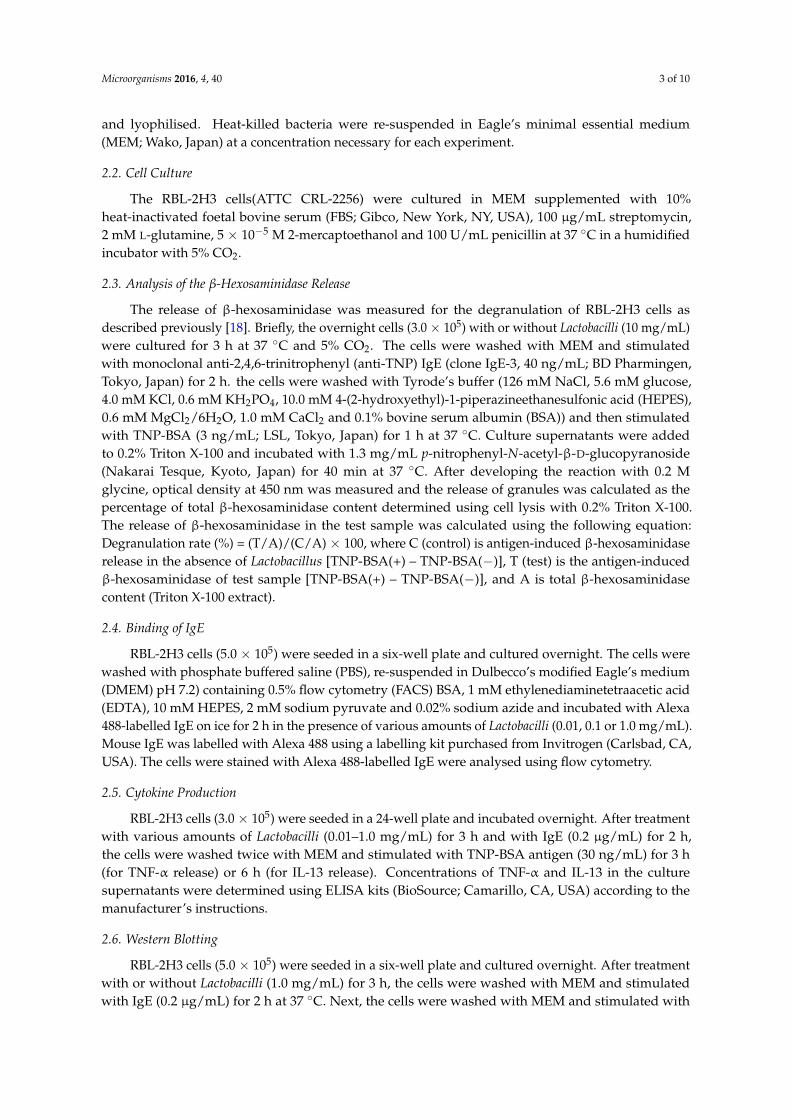
The degranulation of RBL-2H3 cells was detected by measuring the release ofβ-hexosaminidase.The positive control showed 8.94% ± 2.36% (mean ± standard deviation(SD)) in this study and was calculated as 100%. Each of the 23 tested Lactobacilli indicated thatIgE-mediated degranulation of the lysate of the RBL-2H3 cells was released by 35.0%–102.9%, witheach strain showing its own characteristic inhibitory effect (Figure 1a). Of the tested Lactobacilli,LGG (strain No. 23) showed the strongest inhibitory effect on the degranulation of RBL-2H3 cells,whereas L. reuteri JCM 1112 (strain No. 2) did not inhibit the degranulation of RBL-2H3 cells. GroupC contained the Lactobacilli originally isolated from the human intestine; these bacteria significantlysuppressed IgE-mediated degranulation in comparison with the reference strains in Group A(p < 0.05). Furthermore, Group B which contained the commercial strains also tended to suppress thedegranulation in comparison with the reference strains in Group A (p = 0.0513) (Figure 1b).
The inhibitory effects of the live and heat-killed strains LGG on IgE-mediated degranulation ofRBL-2H3 cells were dose-dependent; the heat-killed TMC0356 showed significant inhibition atthedosage of 0.01, 0.1 and 1.0 mg/mL (Figure 1c). There were significant differences between the effectsof heat-killed and live TMC0356 at the dosage of 0.1 mg/mL. Possible toxic effects or damage of thebacteria preparation to tested RBL-2H3 cells were not observed.
3.2. Effect of LGG and TMC0356 on Binding of IgE to the Surface of RBL-2H3 Cells
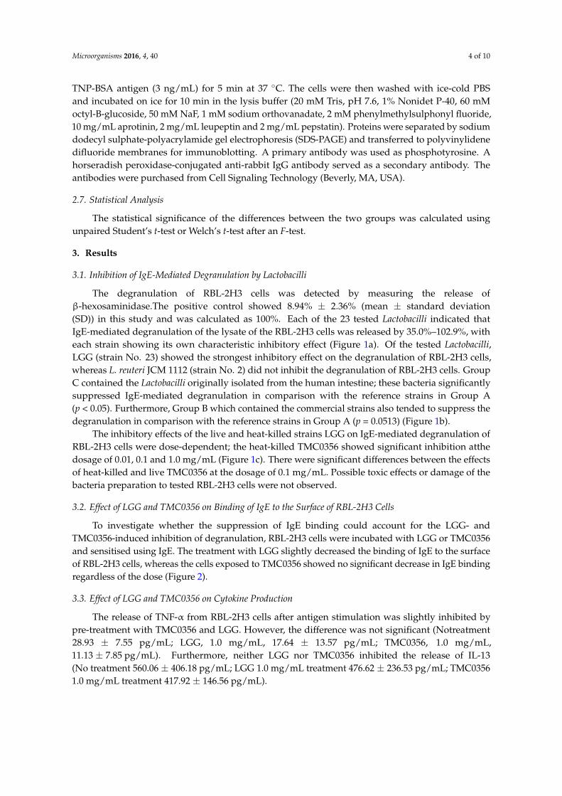
To investigate whether the suppression of IgE binding could account for the LGG- andTMC0356-induced inhibition of degranulation, RBL-2H3 cells were incubated with LGG or TMC0356and sensitised using IgE. The treatment with LGG slightly decreased the binding of IgE to the surfaceof RBL-2H3 cells, whereas the cells exposed to TMC0356 showed no significant decrease in IgE bindingregardless of the dose (Figure 2).
3.3. Effect of LGG and TMC0356 on Cytokine Production
The release of TNF-α from RBL-2H3 cells after antigen stimulation was slightly inhibited bypre-treatment with TMC0356 and LGG. However, the difference was not significant (Notreatment28.93 ± 7.55 pg/mL; LGG, 1.0 mg/mL, 17.64 ± 13.57 pg/mL; TMC0356, 1.0 mg/mL,11.13 ± 7.85 pg/mL). Furthermore, neither LGG nor TMC0356 inhibited the release of IL-13(No treatment 560.06 ± 406.18 pg/mL; LGG 1.0 mg/mL treatment 476.62 ± 236.53 pg/mL; TMC03561.0 mg/mL treatment 417.92 ± 146.56 pg/mL).

Microorganisms 2016, 4, 40 5 of 10
3.4. Effect of LGG and TMC0356 on Intracellular Signalling
The signalling cascade associated with the receptor is initiated when the immunoreceptortyrosine-based activation motif of the β and γ chains are phosphorylated on a tyrosine. This signal isrequired for the activation of mast cells; therefore, tyrosyl transphosphorylation, which occurs duringthe early stages of signal transduction, is considered an important marker of mast cell activation.
To investigate the mechanisms underlying the inhibition of degranulation of RBL-2H3 cells byLGG and TMC0356, RBL-2H3 cells were incubated with LGG or TMC0356, sensitised with IgE andthe antigen, and immunoblotted with anti-phosphotyrosine antibodies to determine the molecularmechanisms by which LGG and TMC0356 inhibit mast cell activation. Total tyrosine phosphorylationpatterns were not significantly affected by LGG or TMC0356 treatment (Figure 3).
Microorganisms 2016, 4, 40 5 of 9
required for the activation of mast cells; therefore, tyrosyl transphosphorylation, which occurs during the early stages of signal transduction, is considered an important marker of mast cell activation.
To investigate the mechanisms underlying the inhibition of degranulation of RBL-2H3 cells by LGG and TMC0356, RBL-2H3 cells were incubated with LGG or TMC0356, sensitised with IgE and the antigen, and immunoblotted with anti-phosphotyrosine antibodies to determine the molecular mechanisms by which LGG and TMC0356 inhibit mast cell activation. Total tyrosine phosphorylation patterns were not significantly affected by LGG or TMC0356 treatment (Figure 3).
(a) (b)
(c)
Figure 1. Degranulation rates of each Lactobacillus according to β-hexosaminidase release.RBL-2H3 cells were pre-incubated with each Lactobacillus strain for 3 h, prior to IgE sensitisation. After stimulation with the antigen, degranulation was detected by measuring the release of β-hexosaminidase. The control is antigen-induced β-hexosaminidase release in the absence of Lactobacillus, calculated as 100%. (a) Strain No. 22 is LGG, and 23 is TMC0356. Group A (strain No. 1–9) consists of the reference strains purchased from JCM, Group B (strain No. 10–16) consists of commercial strains isolated from fermented milk/yoghurt and Group C (strain No. 17–21) consists of Lactobacilli obtained from the human intestine. (b) Comparison of degranulation rates among treatment groups: TMC0356 and LGG. (c) Degranulation rates (dose-response relationship) of live or heat-killed LGG and TMC0356 (0.01, 0.1 or 1.0 mg/mL). The results are presented as mean ± standard deviation (SD) (n = 3). **, p < 0.01, *, p < 0.05 for comparison of the control without Lactobacillus treatment; #, p < 0.05 for comparison to live LGG or TMC0356.
Figure 1. Degranulation rates of each Lactobacillus according to β-hexosaminidase release. RBL-2H3cells were pre-incubated with each Lactobacillus strain for 3 h, prior to IgE sensitisation. After stimulationwith the antigen, degranulation was detected by measuring the release of β-hexosaminidase. Thecontrol is antigen-induced β-hexosaminidase release in the absence of Lactobacillus, calculated as 100%.(a) Strain No. 22 is LGG, and 23 is TMC0356. Group A (strain No. 1–9) consists of the referencestrains purchased from JCM, Group B (strain No. 10–16) consists of commercial strains isolated fromfermented milk/yoghurt and Group C (strain No. 17–21) consists of Lactobacilli obtained from thehuman intestine. (b) Comparison of degranulation rates among treatment groups: TMC0356 and LGG.(c) Degranulation rates (dose-response relationship) of live or heat-killed LGG and TMC0356 (0.01,0.1 or 1.0 mg/mL). The results are presented as mean ± standard deviation (SD) (n = 3). **, p < 0.01,*, p < 0.05 for comparison of the control without Lactobacillus treatment; #, p < 0.05 for comparison tolive LGG or TMC0356.

Microorganisms 2016, 4, 40 6 of 10
Microorganisms 2016, 4, 40 6 of 9
Figure 2. Effect of LGG and TMC0356 on binding of IgE to the surface of RBL-2H3 cells.RBL-2H3 cells were analysed using flow cytometry to evaluate the influence of LGG or TMC0356 (0.01, 0.1 or 1.0 mg/mL). Shadowed areas = no IgE; dotted lines = with IgE but without LGG and TMC0356; bold lines = with IgE and either LGG (a) or TMC0356 (b). The results of one of three independent experiments with similar results are shown.
Figure 3. Effect of LGG and TMC0356 on intracellular signalling.RBL-2H3 cells were incubated with heat-killed LGG or TMC0356 (1.0 mg/mL). After the cells were sensitised with IgE and stimulated with the antigen for 5 min, cell lysates were prepared and immunoblotted with anti-phosphotyrosine antibodies. The results of one of three independent experiments with similar results are shown.
4. Discussion
LGG is a probiotic strain with well-documented anti-allergic effects. This bacterium effectively protects infants with a genetically high risk of allergic diseases caused by the development of atopic diseases [19–21], and it also alleviates atopic eczema-dermatitis syndrome by enhancing interferon-γ responses of peripheral lymphocytes in infants with cow milk allergy or IgE-associated atopic
10 1 10 2 10 3 10 4 10 50
20
40
60
80
100
LGG 0.01 ( μg/ml )10 1 10 2 10 3 10 4 10 5
0
20
40
60
80
100
LGG 0. 110 1 10 2 10 3 10 4 10 5
0
20
40
60
80
100
LGG 1.0
10 1 10 2 10 3 10 4 10 50
20
40
60
80
100
TMC0356 0.01
10 1 10 2 10 3 10 4 10 50
20
40
60
80
100
TMC0356 0. 110 1 10 2 10 3 10 4 10 5
0
20
40
60
80
100
TMC0356 1.0
Cel
l cou
nt (%
)
IgE binding (%)
a
b
Figure 2. Effect of LGG and TMC0356 on binding of IgE to the surface of RBL-2H3 cells.RBL-2H3cells were analysed using flow cytometry to evaluate the influence of LGG or TMC0356 (0.01, 0.1or 1.0 mg/mL). Shadowed areas = no IgE; dotted lines = with IgE but without LGG and TMC0356;bold lines = with IgE and either LGG (a) or TMC0356 (b). The results of one of three independentexperiments with similar results are shown.
Microorganisms 2016, 4, 40 6 of 9
Figure 2. Effect of LGG and TMC0356 on binding of IgE to the surface of RBL-2H3 cells.RBL-2H3 cells were analysed using flow cytometry to evaluate the influence of LGG or TMC0356 (0.01, 0.1 or 1.0 mg/mL). Shadowed areas = no IgE; dotted lines = with IgE but without LGG and TMC0356; bold lines = with IgE and either LGG (a) or TMC0356 (b). The results of one of three independent experiments with similar results are shown.
Figure 3. Effect of LGG and TMC0356 on intracellular signalling.RBL-2H3 cells were incubated with heat-killed LGG or TMC0356 (1.0 mg/mL). After the cells were sensitised with IgE and stimulated with the antigen for 5 min, cell lysates were prepared and immunoblotted with anti-phosphotyrosine antibodies. The results of one of three independent experiments with similar results are shown.
4. Discussion
LGG is a probiotic strain with well-documented anti-allergic effects. This bacterium effectively protects infants with a genetically high risk of allergic diseases caused by the development of atopic diseases [19–21], and it also alleviates atopic eczema-dermatitis syndrome by enhancing interferon-γ responses of peripheral lymphocytes in infants with cow milk allergy or IgE-associated atopic
10 1 10 2 10 3 10 4 10 50
20
40
60
80
100
LGG 0.01 ( μg/ml )10 1 10 2 10 3 10 4 10 5
0
20
40
60
80
100
LGG 0. 110 1 10 2 10 3 10 4 10 5
0
20
40
60
80
100
LGG 1.0
10 1 10 2 10 3 10 4 10 50
20
40
60
80
100
TMC0356 0.01
10 1 10 2 10 3 10 4 10 50
20
40
60
80
100
TMC0356 0. 110 1 10 2 10 3 10 4 10 5
0
20
40
60
80
100
TMC0356 1.0
Cel
l cou
nt (%
)
IgE binding (%)
a
b
Figure 3. Effect of LGG and TMC0356 on intracellular signalling.RBL-2H3 cells were incubated withheat-killed LGG or TMC0356 (1.0 mg/mL). After the cells were sensitised with IgE and stimulatedwith the antigen for 5 min, cell lysates were prepared and immunoblotted with anti-phosphotyrosineantibodies. The results of one of three independent experiments with similar results are shown.

Microorganisms 2016, 4, 40 7 of 10
4. Discussion
LGG is a probiotic strain with well-documented anti-allergic effects. This bacterium effectivelyprotects infants with a genetically high risk of allergic diseases caused by the development of atopicdiseases [19–21], and it also alleviates atopic eczema-dermatitis syndrome by enhancing interferon-γresponses of peripheral lymphocytes in infants with cow milk allergy or IgE-associated atopiceczema-dermatitis syndrome [22,23]. However, LGG does not significantly affect birch pollen allergy,an adult allergic disease [24]. TMC0356 was originally isolated from the intestine of a healthy adult [25];these bacteria generally adhere to human enterocytes and do not enhance inflammatory responses [26].TMC0356 characteristically induces the secretion of pro-inflammatory (IL-12) and anti-inflammatory(IL-10) cytokines by murine macrophages [27]. This bacterium effectively inhibited antigen-augmentedserum IgE in BALB/c mice that was immunised intra-peritoneally with the food antigen, ovalbumin,and it altered the serum IgE concentration of the subjects with high serum IgE levels and perennialallergic rhinitis [28,29].
LGG-TMC0356-fermented milk significantly suppressed ovalbumin (OVA)-induced nasal vascularpermeability and the non-specific IgE level in rats [30], and alleviated OVA-induced nasal blockage inguinea pigs [31]. In 2006, the LGG- andTMC0356-fermented milk was orally administered to patientswith Japanese cedar pollinosis (JCPsis) in a double-blind, placebo-controlled clinical trial during theseason of Japanese cedar pollen. Some clinical improvements were observed after this treatment.However, no significant changes in the levels of serum IgE and other blood biomarkers related to IgEimmunity were observed in this clinical study, although LGG and TMC0356 suppressed IL-4 and IL-5production by peripheral blood mononuclear cells isolated from patients with JCPsis when the cellswere stimulated with both CryJ1 and PHA in vitro [17].
On the other hand, oral administration of LGG- and TMC0356-fermented milk suppressed changesin intestinal microbiota in patients with JCPsis in the same clinical study [16]. Therefore, the oraladministration of LGG and TMC0356 presumably resulted in intestinal colonisation by these bacteriain all patients with JCPsis that were administered the LGG- and TMC0356-fermented milk [17]. Thesestudies indicate that the intestinal microbes that are protected by LGG and TMC0356, which hadcolonised the intestinal tract of the patients with JCPsis, may be involved in the alleviation of theclinical symptoms. However, the analysis of blood conducted in the study was not sufficient to confirmthat notion. Therefore, additional information regarding the effects of LGG, TMC0356 and intestinalmicrobes in patients with JCPsis on immune cells is required to understand the mechanism underlyingthe anti-allergic effect of LGG and TMC0356.
Recently, human intestinal bifidobacteria showed a characteristic inhibitory effect against activemast cells using RBL-2H3 cells. Compared to the infant-specific species, Bifidobacterium bifidum, whichstrongly inhibits degranulation, the adult-specific species, B. adolescentis, showed variation in its abilityto affect IgE-mediated degranulation among different strains [18]. These results indicate that RBH-2H3cells are highly sensitive to different properties of intestinal microbes to implicate the activation ofmast cells.
In the present study, each strain of the tested Lactobacilli characteristically suppressedIgE-mediated degranulation of RBL-2H3 cells. Among the tested bacteria, LGG and TMC0356 showeda stronger inhibitory effect on the degranulation of RBL-2H3 cells than that of other strains. Theseinhibitory effects were dose-dependent and not associated with the viability of these bacteria. Thetested bacteria that were originally isolated from the human intestine, including those isolated fromthe patients with JCPsis, significantly suppressed degranulation of RBL-2H3 cells compared withother bacteria. These results indicate that Lactobacilli, particularly some of the selected strains, maypossess the ability to alter activation of mast cells. Orally administered LGG and TMC0356 cansuccessfully colonise the intestine of patients with JCPsis and stabilise other beneficial intestinalmicrobes, including Lactobacilli and bifidobacteria. The inhibition of degranulation of mast cells maybe a part of the underlying mechanism by which these microbes alleviate the clinical symptoms ofpatients with JCPsis, as reported in the previous study. These results are also supported by another

Microorganisms 2016, 4, 40 8 of 10
study [15] showing that probiotics exert potential anti-allergic effects, at least in part, through directaction on mast cells.
To explore the mechanisms underlying the inhibition of degranulation of RBL-2H3 cells by LGGand TMC0356, LGG and TMC0356 were tested for the ability to affect the tyrosine phosphorylation,TNF-α production and binding of IgE to RBL-2H3 cells. TMC0356 tended to suppress the secretionof TNF-α, whereas LGG slightly inhibited the interaction between IgE and FcεRI. However, thesechanges were not significant and therefore could not sufficiently explain the mechanism underlyingthe inhibitory effects of LGG and TMC0356.LGG can significantly downregulate the expression ofthe genes of high-affinity IgE receptor subtype α (FCER1A) and HRH14, affecting the function ofhuman mast cells, as observed previously by microarray analysis [32]. A Toll-like receptor 2 (TLR2)ligand significantly suppresses the FcεRI-mediated inflammatory responses of mast cells [15]. Furtherstudies will be conducted to test whether LGG and TMC0356 can alter the activation of mast cells bysuppressing expression of the FCER1A and HRH14 genes or by interacting with TLR2.
5. Conclusions
The results obtained from the present study suggest that Lactobacilli, particularly those from thehuman intestine, affect the activation of mast cells in a strain-dependent manner and by differentmechanisms to express the anti-allergic effects.
Acknowledgments: We thank Kazumi Kasakura in the Department of Food Bioscience and Biotechnology, Collegeof Bioresource Sciences, Nihon University, for skilful technical assistance.
Author Contributions: Gaku Harata, Fang He, Kyoko Takahashi, Akira Hosono, and Shuichi Kaminogawaconceived and designed the experiments; Gaku Harata performed the experiments; Gaku Harata,Fang He and Shuichi Kaminogawa analysed the data; Kenji Miyazawa and Kazutoyo Yoda contributedreagents/materials/analysis tools; Gaku Harata and Fang He wrote the paper.
Conflicts of Interest: The authors declare no conflict of interest.
References
1. Strachan, D.P. Hay fever, hygiene, and household size. Br. Med. J. 1989, 299, 1259–1260. [CrossRef]2. Strachan, D.P. Family size, infection and atopy: The first decade of the “hygiene hypothesis”. Thorax 2000,
55, S2–S10. [CrossRef] [PubMed]3. Rautava, S.; Ruuskanen, O.; Salminen, S.; Isolauri, E. The Hygiene Hypothesis of Atopic Disease—An
Extanded Version. J. Pediatr. Gastroenterol. Nutr. 2004, 38, 378–388. [CrossRef] [PubMed]4. Penders, J.; Stobberigh, E.E.; van den Brandt, P.A.; Thijs, C. The role of the intestinal microbiota in the
development of atopic disorders. Allergy 2007, 62, 1223–1236. [CrossRef] [PubMed]5. Björkstén, B.; Naaber, P.; Sepp, E.; Mikelsaar, M. The intestinal microflora in allergic Estonian and Swedish
2-year-old children. Clin. Exp. Allergy 1999, 29, 342–346. [CrossRef] [PubMed]6. Björkstén, B. The intestinal microflora in allergic patients. Biosci. Microflora 2002, 20, 135–140.7. Ouwehand, A.C. Antiallergic effects of probiotics. J. Nutr. 2007, 137, 794–797.8. Abbas, A.K.; Lichtman, A.H. Basic immunology: Functions and disorders of the immune system. Am. J.
Epidemiol. 2001, 2, 185–186.9. Kobayashi, S.; Tanabe, S. Evaluation of the anti-allergic activity of citrus unshiu using rat basophilic leukemia
RBL-2H3 cells as well as basophils of patients with seasonal allergic rhinitis to pollen. Int. J. Molecul. Medical2006, 17, 511–515. [CrossRef]
10. Sugiura, Y.; Takeuchi, Y.; Kakinuma, M.; Amano, H. Infibitory effects of seaweeds on histamine release fromrat basophile leukemia cells (RBL-2H3). Fishers Sci. 2006, 72, 1286–1291. [CrossRef]
11. Kanda, T.; Akiyama, H.; Yanagida, A.; Tanabe, M.; Goda, Y.; Toyoda, M.; Teshima, R.; Saito, Y.Inhibitory effects of apple polyphenol on induced histamine release from RBL-2H3 cells and rat mast cells.Biosci. Biotechnol. Biochem. 1998, 62, 1284–1289. [CrossRef] [PubMed]
12. Chen, X.; Feng, B.S.; Zheng, P.Y.; Liao, X.Q.; Chong, J.; Tang, S.G.; Yang, P.C. Fc gamma receptor signaling inmast cells links microbial stimulation to mucosal immune inflammation in the intestine. Am. J. Pathol. 2008,73, 1647–1656. [CrossRef] [PubMed]

Microorganisms 2016, 4, 40 9 of 10
13. Magerl, M.; Lammel, V.; Siebenhaar, F.; Zuberbier, T.; Metz, M.; Maurer, M. Non-pathogenic commensalEscherichia coli bacteria can inhibit degranulation of mast cells. Exp. Dermatol. 2008, 17, 427–435. [CrossRef][PubMed]
14. Krämer, S.; Sellge, G.; Lorentz, A.; Krueger, D.; Schemann, M.; Feilhauer, K.; Gunzer, F.; Bischoff, S.C.Selective activation of human intestinal mast cells by Escherichia coli hemolysin. J. Immunol. 2008, 181,1438–1445. [CrossRef] [PubMed]
15. Kasakura, K.; Takahashi, K.; Aizawa, T.; Hosono, A.; Kaminogawa, S. A TLR 2 ligand suppresses allergicinflammatory reactions by acting directly on mast cells. Int. Arch. Allergy Immunol. 2009, 150, 359–369.[CrossRef] [PubMed]
16. Kubota, A.; He, F.; Kawase, M.; Harata, G.; Hiramatsu, M.; Salminen, S.; Iino, H. Lactobacillus strains stabilizeintestinal microbiota in Japanese cedar pollinosis patients. Microbiol. Immunol. 2009, 53, 198–205. [CrossRef][PubMed]
17. Kawase, M.; He, F.; Kubota, A.; Hiramatsu, M.; Saito, H.; Ishii, T.; Yasueda, H.; Akiyama, K. Effectof fermented milk prepared with two probiotic strains on Japanese cedar pollinosis in a double-blindplacebo-controlled clinical study. Int. J. Food Microbiol. 2009, 128, 429–434. [CrossRef] [PubMed]
18. Harata, G.; He, F.; Takahashi, K.; Hosono, A.; Kawase, M.; Kubota, A.; Hiramatsu, M.; Kaminogawa, S.Bifidobacterium suppresses IgE-mediated degranulation of rat basophilic leulemia (RBL-2H3) cells.Microbiol. Immunol. 2010, 54, 54–57. [CrossRef] [PubMed]
19. Kalliomäki, M.; Salminen, S.; Arvilommi, H.; Kero, P.; Koskinen, P.; Isolauri, E. Probiotics in primaryprevention of atopic disease: A randomized placebo-controlled trial. Lancet 2001, 357, 1076–1079. [CrossRef]
20. Kalliomaki, M.; Kirjavainen, P.; Eerola, E.; Kero, P.; Salminen, S.; Isolauri, E. Distinct patterns of neonatalgut microflora in infants developing or not developing atopy. J. Allergy Clin. Immunol. 2001, 107, 129–134.[CrossRef] [PubMed]
21. Kalliomäki, M.; Salminen, S.; Poussa, T.; Arvilommi, H.; Isolauri, E. Probiotics and prevention of atopicdisease: 4-Year follow-up of a randomized placebo-controlled trial. Lancet 2003, 361, 1869–1871. [CrossRef]
22. Viljanen, M.; Savilahti, E.; Haahtela, T.; Juntunen-Backman, K.; Korpela, R.; Poussa, T.; Tuure, T.;Kuitunen, M. Probiotics in the treatment of atopic eczema/dermatitis syndrome in infants: A double-blindplacebo-controlled trial. Allergy 2005, 60, 494–500. [CrossRef] [PubMed]
23. Viljanen, M.; Kuitunen, M.; Haahtela, T.; Juntunen-Backman, K.; Korpela, R.; Savilahti, E. Probiotic effects onfaecal inflammatory markers and on faecal IgA in food allergic atopic eczema/dermatitis syndrome infants.Pediatric Allergy Immunol. 2005, 16, 65–71. [CrossRef] [PubMed]
24. Helin, T.; Haahtela, S.; Haahtela, T. No effect of oral treatment with an intestinal bacterial strain, Lactobacillusrhamnosus (ATCC53103), on birch-pollen allergy: A placebo-contralled double-blind study. Allergy 2002, 57,243–246. [CrossRef] [PubMed]
25. Hosoda, M.; Hashimoto, H.; He, F.; Yamazaki, K.; Hosono, A. Inhibitory effects of fecal Lactobacilli andBifidobacteria on the mutagenicities of Trp-P-2 and IQ. Milchwissenschaft 1998, 53, 309–313.
26. Morita, H.; He, F.; Fuse, T.; Ouwehand, A.C.; Hashimoto, H.; Hosoda, M. Adhesion of lactic acid bacteria toCaco-2 cells and their effect on cytokine secretion. Microbiol. Immnol. 2002, 46, 293–297. [CrossRef]
27. Morita, H.; He, F.; Fuse, T.; Ouwehand, A.C.; Hashimoto, H.; Hosoda, M.; Mizumachi, K.; Kurisaki, J.Cytokine production by the murine macrophage cell line J774.1 after exposure to Lactobacilli. Biosci. Biotechnol.Biochem. 2002, 66, 1963–1966. [CrossRef] [PubMed]
28. Kawase, M.; He, F.; Harata, G.; Kubota, A.; Mizumachi, K.; Hiramatsu, M. Characterization of inhibitoryeffects of Lactobacilli against immunoglobulin E production in vitro and in vivo. Int. J. Probiotics Prebiotics2007, 2, 29–38.
29. Morita, H.; He, F.; Kawase, M.; Kubuta, A.; Hiramatsu, M.; Kurisaki, J.; Salminen, S. Preliminary human studyfor possible alteration of serum immunoglobulin production in perennial allergic rhinitis with fermentedmilk prepared with Lactobacillus gasseri TMC0356. Microbiol. Immunol. 2006, 50, 701–706. [CrossRef][PubMed]
30. Kawase, M.; He, F.; Kubota, A.; Hata, J.; Kobayakawa, S.; Hiramatsu, M. Inhibitory effect of Lactobacillusgasseri TMC0356 andLGG on enhanced vascular permeability of nasal mucosa in experimental allergicrhinitis of rats. Biosci. Biotechnol. Biochem. 2006, 70, 3025–3030. [CrossRef] [PubMed]

Microorganisms 2016, 4, 40 10 of 10
31. Kawase, M.; He, F.; Kubota, A.; Harata, G.; Hiramatsu, M. Orally administrated Lactobacillus gasseri TMC0356and Lactobacillus GG alleviated nasal blockage of guinea pig with allergic rhinitis. Microbiol. Immunol. 2007,51, 1109–1114. [CrossRef] [PubMed]
32. Oksaharju, A.; Kankainen, M.; Kekkonen, R.; Lindstedt, K.; Kovanen, P.; Korpela, R.; Miettinen, M. ProbioticLactobacillus rhamnosus downregulates FCER1 and HRH4 expression in human cells. World J. Gastroentero.2011, 17, 750–759. [CrossRef] [PubMed]
© 2016 by the authors; licensee MDPI, Basel, Switzerland. This article is an open accessarticle distributed under the terms and conditions of the Creative Commons Attribution(CC-BY) license (http://creativecommons.org/licenses/by/4.0/).