human immunodeficiency virus type 1-like dna sequences and … · cases in women and prostate...
TRANSCRIPT
CLINICAL AND DIAGNOSTIC LABORATORY IMMUNOLOGY,1071-412X/98/$04.0010
Sept. 1998, p. 645–653 Vol. 5, No. 5
Copyright © 1998, American Society for Microbiology. All Rights Reserved.
Human Immunodeficiency Virus Type 1-Like DNA Sequencesand Immunoreactive Viral Particles with Unique
Association with Breast CancerEVA M. RAKOWICZ-SZULCZYNSKA,1,2,3* BETTY JACKSON,1 ADRIANA M. SZULCZYNSKA,1
AND MCCLURE SMITH1,3
Departments of Obstetrics and Gynecology1 and Biochemistry and Molecular Biology,2 University ofNebraska Medical Center, and NCI-Designated Eppley Cancer Center,3 Omaha, Nebraska
Received 22 December 1997/Returned for modification 1 May 1998/Accepted 27 May 1998
RAK antigens p120, p42, and p25 exhibit molecular and immunological similarity to the proteins encodedby human immunodeficiency virus type 1 (HIV-1) and are expressed by 95% of breast and gynecological cancercases in women and prostate cancer cases in men. The binding of an epitope-specific anti-HIV-1 gp120 mono-clonal antibody (MAb) (amino acids 308 to 322) to cancer RAK antigens has been found to be inhibited by apeptide derived from variable loop V3 of HIV-1. Breast cancer DNAs of 40 patients were PCR amplified withHIV-1 gp41-derived primers, and all of the samples were found to be positive. The DNA fragments amplified inseven blindly selected breast cancer samples were sequenced. The breast cancer DNA sequences showed at least90% homology to the HIV-1 gene for gp41. Antisense oligonucleotides complementary to the HIV-1-like sequencesinhibited reverse transcriptase activity and inhibited the growth of breast cancer cells in vitro. Viral particlesdetected in breast cancer cell lines were strongly immunogold labeled with the anti-HIV-1 gp120 MAb. Theresults obtained strongly suggest that the long-postulated breast cancer virus may, in fact, be related to HIV-1.
Breast cancer affects 1 in every 8 to 10 women in the UnitedStates (10, 19, 32, 35, 36). Approximately 10% of breast cancerpatients exhibit a genetic inheritance pattern, while the over-whelming majority of women develop breast cancer for anunpredictable reason (2, 6, 7, 14, 20, 23, 37, 43). Recent en-thusiasm following the characterization of the BRCA1 andBRCA2 genes, which are associated with some inherited formsof breast and ovarian cancer, has been diminished by the factthat only 1 of 800 women carries the mutated BRCA gene,while at least 80 to 100 of 800 women will develop breastcancer (17, 21, 41, 42).
Involvement of a viral factor in the etiology of human breastcancer has been considered by several laboratories (1, 5, 9,11–13, 18, 39). Special attention was focused on human DNAsequences with homology to mouse mammary tumor virus(MMTV) (1, 38, 40). Retrovirus-like particles with immuno-logical similarity to MMTV proteins were found in humanbreast carcinoma cell lines (13), peripheral blood monocytes ofbreast cancer patients (11), pleural effusion fluids from breastadenocarcinoma patients (39), human breast cancer tissue (5,9, 12, 39), and breast milk (18). No viral agent has been iden-tified as a causative agent of breast cancer in humans.
Except for cervical cancer, which is associated predomi-nantly with human papillomavirus and/or herpes simplex virusinfection (3), the viral etiology of other types of female repro-ductive tract cancer remains unverified. Recent studies onherpesvirus-like DNA sequences in AIDS-associated Kaposi’ssarcoma (4, 22, 25, 33) strongly suggest that the role of retro-viruses in human cancer was underestimated for a long time.
We have recently identified a new class of breast and gyne-cological cancer markers, which have been named Rakowiczmarkers or, briefly, RAK markers (25–30). RAK antigens p120,
p42, and p25 express epitopes in common with envelope pro-tein gp120 of human immunodeficiency virus type 1 (HIV-1)and can be detected either by an epitope-specific anti-HIV-1gp120 monoclonal antibody (MAb) (25–27), or by MAb RAK-BrI, which is directed against breast cancer (28, 29). RAKantigens are absent in normal breast tissue and other normaltissues (28), which suggests the strong diagnostic potential ofthese unique markers. Very recently, RAK markers were alsofound in prostate cancer and in a number of benign hyperpla-sia cases (31).
One of the RAK antigens, p160, corresponding in size toHIV-1 gp160 (precursor of gp120 and gp41), was detected inthe blood of 70% of gynecological cancer patients before sur-gery, in 40 to 50% of breast cancer patients after surgery, in20% of healthy women with a family history of breast cancer,and in 13% of healthy women without a family history of breastor gynecological cancer (29, 30).
The similarity of RAK breast cancer antigens to HIV-1major proteins led to speculation that HIV-1-like DNA se-quences encoding RAK antigens might also be localized incancer DNA. That hypothesis was confirmed by the findingthat HIV-1 gp41-derived primers SK68 and SK69 initiated aPCR with breast cancer DNA but not with normal breast DNA(28). It is noteworthy that the same HIV-1-derived primersamplified prostate cancer DNA but not normal prostate DNA(31). The prostate cancer DNA fragments amplified exhibitedstrong homology to HIV-1 (31). The study described in thisreport revealed strong homology between HIV-1 and breastcancer DNA sequences. Viral particles cross-reactive with theanti-HIV-1 gp120 MAb were also detected in breast cancercells, supporting the hypothesis of a potential viral origin ofbreast cancer.
MATERIALS AND METHODS
Breast cancer tissue. Breast cancer tissue, breast cancer adjacent tissue(NAT), and histologically normal breast tissue samples were provided by theCooperative Human Tissue Network.
* Corresponding author. Mailing address: University of NebraskaMedical Center, Department of Obstetrics and Gynecology, 600 South42nd St., Omaha, NE 68198-3255. Phone: (402) 559-6157. Fax: (402)559-8112. E-mail: [email protected].
645
on March 30, 2020 by guest
http://cvi.asm.org/
Dow
nloaded from

Cell lines. Breast carcinoma cell lines SKBr3 and MCF7 and cervical cancercell line SiHa, obtained from the American Type Culture Collection (ATCC),were grown in Eagle’s minimal essential medium-Leibovitz’s L15 medium (3:4)or other recommended medium supplemented with 10% fetal bovine serum.
MAbs. MAb RAK-BrI, which is directed against breast cancer antigens butalso detects gynecological cancer antigens, was described before (28, 29). Theanti-HIV-1 gp120 MAb (amino acids 308 to 322) was purchased from Du Pont.The ability of that MAb to detect a cancer antigen was reported before (25, 26).Control antibodies, which do not recognize cancer antigens, including anotherepitope-specific anti-HIV-1 gp120 MAb (25, 27) and an anti-HIV-1 p25 MAb,were from Du Pont.
Tissue fractionation. Fractionation of tissue was described before (25–29).Briefly, cancer and normal tissue samples were homogenized in 0.35 M su-crose–10 mM KCl–1.5 mM MgCl2–10 mM Tris-HCl (pH 7.6)–0.12% TritonX-100–12 mM 2-mercaptoethanol and centrifuged at 600 3 g for 10 min. Thesupernatant was defined as the cytoplasmic fraction.
Electrophoresis of proteins. Cytoplasmic and chromatin proteins were ana-lyzed by electrophoresis in a 10% polyacrylamide gel with 0.1% sodium dodecylsulfate (SDS) in a buffer containing 250 mM Tris-HCl (pH 8.3), 195 mM glycine,and 0.1% SDS as described by Laemmli (15).
Western blotting. Blotting of proteins from the polyacrylamide gel to a poly-vinylidene difluoride membrane was performed in 25 mM Tris-HCl (pH 8.6)–192mM glycine buffer containing 10% methanol. Filters were incubated with 1%bovine serum albumin for 16 h at 0°C and then with MAb 5023 (2 mg/ml) or MAbRAK-BrI (0.1 mg/ml), washed with Tris-glycine buffer, and incubated with alka-line phosphatase-conjugated goat anti-mouse immunoglobulin G for 1 h. Afterwashing with TBST, membranes were incubated with 0.1% 1-naphthyl-phos-phate and Fast Red. In some experiments, the anti-HIV-1 gp120 or RAK-BrIMAb (5 mg/ml) was preincubated with peptide RIQRGPGRAFVTIGK,RIQRGPGRKFVTIGK, or RIQRGPGRVVVTGK (18 mg/ml) for 1 h on iceand then incubated with blots.
DNA isolation. Sections of breast cancer tissue, NAT, or normal tissues (ob-tained during standard surgical procedures and stored at 280°C) were lysed in100 mM NaCl–10 mM Tris-HCl (pH 8.0)–25 mM EDTA (pH 8.0)–0.5% SDS.The lysate was digested with proteinase K (0.1 mg/ml), extracted with phenol-chloroform, and immunoprecipitated with ethanol. DNA from HIV-1-infectedcells (positive control) was obtained from Advanced Biotechnology Services, Inc.
PCR. PCR occurred in a solution containing 10 mM KCl; 10 mM (NH4)SO4,20 mM Tris-HCl; 5 mM MgCl2; 0.1% Triton X-100; dATP, dTTP, dCTP, anddGTP at 0.2 mM each; each primer at 0.5 mM; and 2.5 U of Taq polymerase. Theamount of template DNA used was 1.0 mg/50 ml of the reaction mixture. Thereactions ran for 30 cycles in a Perkin Elmer 9600 thermal cycler. The PCR wasdone by using HIV-1-derived primers SK68 (7801-to-7820 region of gp41 Env)[59-AGCAGCAGGAAGCACTATGG-39]) and SK69 (7922-to-7942 region ofgp41 Env) [59-CCAGACTGTGAGTTGCAAGAG-39]). All DNA sampleswhich tested negative with primers SK68 and SK69, as well as approximately 50%of the PCR-positive samples, were tested with a control set of primers derivedfrom the globin gene. Each set of PCRs included DNA isolated from HIV-1-infected lymphocytes (positive control). The PCR mixture was electrophoreti-cally analyzed in a 1.5% agarose gel. DNA was visualized by UV fluorescenceafter staining with ethidium bromide. Each cancer or normal DNA sample wastested by PCR three to seven times in different sets. Samples selected for DNAsequencing were separated in a 4% agarose gel. PCR bands were encoded bynumber and submitted for blind sequencing with primers SK68 and SK69. As apositive control, the amplified HIV-1 DNA fragment was used for sequencing.
Test for RNA-dependent DNA polymerase activity. Cell culture media fromthe cells not exposed (control) or exposed for 4 days to RAK I (100 mg/ml) wereultracentrifuged (160,000 3 g, 60 min). The pellet was resuspended in 137 mMNaCl–3 mM KCl–10 mM phosphate buffer (pH 7.4)–0.6 mM EDTA. The assaymixture contained 5 mM Tris-HCl (pH 8.3); 0.6 mM magnesium acetate; 6 mMNaCl; 2 mM dithiothreitol; 0.08 mM each dATP, dCTP, and dGTP; and 0.001mM [methyl-3H]TTP. A viral pellet sample corresponding to 4 3 106 viralparticles was added to 500 ml of the assay mixture, which was then incubated for45 min at 37°C. The reaction was stopped by adding 5 ml of 20% trichloroaceticacid (34).
Nucleotide sequence accession numbers. The DNA sequences identified inthis study have been assigned accession no. AF073463 to AF073469.
RESULTS
Detection of RAK antigens in breast cancer tissue. Breastcancer tissue samples, NAT, and normal breast tissue wereelectrophoretically separated in a polyacrylamide gel withSDS, transferred onto a polyvinylidene difluoride membrane,and Western blot hybridized with MAb RAK-BrI (Fig. 1) or ananti-HIV-1 MAb (Fig. 2). Of the 125 breast cancer cases test-ed, 95.2% were strongly RAK p120, p42, and p25 positive andthe rest were weakly positive (Table 1). Of 70 NAT samples,only 8 (11.4%) tested moderately RAK positive and an addi-
tional 9 (12.9%) were weakly positive. Of 40 samples obtainedfrom histologically “normal” parts of cancer-affected breasts,only 3 tested positive and 1 was weakly positive (7.5 and 2.5%,respectively). In women with advanced fibrocystic disease, whichqualified them for either partial breast removal or completemastectomy, only 5.7% expressed RAK antigens at a moderatelevel and 2.8% expressed RAK antigens at a low level. Of the10 cases of breast reduction, only 1 tested weakly RAK posi-tive. Breast milk was used as the source of normal breast ep-ithelial cells, and all 10 samples tested RAK negative. Controlsamples of skin and placenta tested negative. Other RAK-neg-ative normal samples were described before (30, 31). The re-sults confirmed a clear association of RAK antigens p120, p42,and p25 with breast cancer.
The expression of all three RAK antigens in several breastand cervical cancer cell lines was described before (25, 27). Inthe current experiments, two cell lines, breast cancer MCF 7and cervical cancer SiHa, were used as models. Expression ofRAK antigens was compared in (i) cells remaining in cellculture for 1 year, (ii) cells which were obtained from ATCC asa frozen stock and grown in the laboratory for 3 to 4 weeks, and(iii) cells which were grown to a monolayer at ATCC facilitiesand immediately processed after receipt at our laboratory. Nodifferences in the expression of RAK antigens was observedbetween groups ii and iii. Expression of RAK antigens in groupi was slightly lower than in groups ii and iii, which is consistentwith the observation that expression of several proteins is low-er in cells remaining in culture for a long time. The last exper-iments also clearly indicated that expression of RAK markersis constitutively associated with MCF 7 and SiHa cells and doesnot represent laboratory contamination.
Mapping of cancer epitopes in common with HIV-1. Theanti-HIV-1 MAb, which recognizes breast and gynecologicalcancer antigens but does not recognize normal cells (Fig. 2A,lanes 1 to 6), was developed against amino acids 308 to 322 (RIQRGPGRAFVTIGK) of the variable loop of HIV-1 gp120,and this MAb binds to the GRAF epitope of HIV-1 gp160 and
FIG. 1. Electrophoretic analysis (10% polyacrylamide gel) and Western blot-ting of cytoplasmic proteins with MAb RAK-BrI. In a blind experiment, bothbreast cancers (CA) (3358 and 3218) tested RAK antigen positive and NATsamples tested RAK antigen negative. No RAK antigens were detected in thefour breast tissue samples obtained during breast reduction (3600, 3696, 3654,and 3696).
646 RAKOWICZ-SZULCZYNSKA ET AL. CLIN. DIAGN. LAB. IMMUNOL.
on March 30, 2020 by guest
http://cvi.asm.org/
Dow
nloaded from

gp120 (8, 16, 24) (Fig. 2A, lanes 7 and 8). To assess the spec-ificity of anti-HIV-1 gp120 MAb binding to the cancer cellepitopes, Western blots were incubated with a MAb which waseither not preincubated or preincubated with the peptide RIQRGPGRAFVTIGK (Fig. 2B). A peptide derived from HIV-1gp120 which contained the consensus sequence GRAF com-petitively blocked the binding of the anti-HIV-1 MAb to allthree cancer antigens. A peptide with the GRAF sequencereplaced with GRVV also competitively blocked the binding ofMAb RAK-BrI to cancer antigens, while a peptide containingthe sequence GRKF did not inhibit MAb RAK-BrI binding tocancer antigens. The results suggest that either the GRAF epi-tope or a very similar epitope is present in cancer antigens.Positively charged lysine (K) destroys the binding site for MAbRAK-BrI. Since the sequence alanine-phenylalanine (AF) mightbe replaced with valine-valine (VV), it is likely that the cancerepitope is conformational and not identical to the HIV epi-tope. The G preceding RAF is critical for cancer antigen bind-ing, since another anti-HIV-1 MAb which recognizes RAF but
forms weak interactions with G does not recognize cancerantigens (25) and has a low affinity for HIV-1 gp120 (8, 16, 24).Another control MAb directed against HIV-1 gp25 did not rec-ognize any of the cancer antigens, which confirmed that thereis limited homology between cancer and HIV-1 proteins, butcancer antigens are very distinct from HIV-1 proteins andcannot originate from HIV-1 contamination.
PCR detection of HIV-1-like DNA sequences in breast can-cer DNA. Primers SK68 and SK69, derived from the HIV-1genome (region gp41), specifically initiated amplification ofbreast cancer DNA (Fig. 3A to C). All 40 of the breast cancercases tested were strongly PCR positive (Table 2). Of 39 sam-ples of DNA isolated from NAT, only 41% tested moderatelyto highly positive and an additional 12.8% tested weakly pos-itive. Of 30 DNA samples obtained from histologically normaltissue of a cancer-affected breast, 26.7% tested moderate-ly positive and 16.7% were weakly positive. Of the 10 DNAsamples obtained from breast reduction, only 1 tested veryweakly positive. DNA extracted from breast milk of healthywomen tested PCR negative. Other control normal tissues alsotested PCR negative (Table 2).
To eliminate the possibility that a negative PCR of normaltissue DNA had been caused by nonspecific inhibition of theamplification reaction, each DNA sample was also amplifiedwith globin primers (Fig. 3D). One of 11 DNA samples ob-tained from normal breast tissue, one DNA sample obtainedfrom breast milk, and one skin DNA sample tested negativewith both globin primers and with the SK68 and SK69 primers.These globin-negative samples were discarded. All of theSK68-SK69-negative samples included in Table 2 were PCRpositive with the globin primers (Fig. 3D, lanes 1 to 3), similarto SK68-SK69-positive cancer DNA (Fig. 3D, lanes 7 to 9).
In a 1.5% agarose gel, a single amplification band was ob-served in HIV-1 and breast cancer DNAs (Fig. 3A and B). Thesize of the amplified DNA region (142 bp) was similar to thesize of the DNA fragment amplified in DNA isolated fromHIV-1-infected cells. For DNA sequencing, seven breast can-cer DNA samples were blindly selected, PCR amplified, andseparated in a 4% agarose gel (Fig. 3C). The PCR band, whichseemed to be homogeneous in a 1.5% agarose, separated intoa dominant band with mobility equal to that of the PCR frag-
FIG. 2. Reactivity of an anti-HIV-1 gp120 MAb with breast and gynecolog-ical cancer cytoplasmic proteins and HIV-1 proteins. (A) Lanes: 1 and 2, normalbreast and normal uterine proteins, respectively; 3 to 5, breast cancer tissuesfrom three different patients; 6, uterine cancer; 7 and 8, HIV-1 gp160 and gp120,respectively; 9, HIV-1 p24 (negative control). The anti-HIV-1 MAb reacted withthree cancer proteins (RAK p120, p42, and p25) but also with HIV-1 gp120 andits precursor gp160. The positions of p120, p42, and p24 are shown on the left.(B) Reactivity of the anti-HIV-1 gp120 MAb with breast cancer proteins ob-tained from two different patients (lanes 1 and 2) before and after preincubationwith the indicated peptides. The peptides containing the consensus sequenceGRAF or GRVV inhibited the binding of the anti-HIV-1 MAb to all threecancer antigens. Positively charged lysine in the peptide GRKF did not allowMAb binding, and the peptide did not affect interaction with cancer antigens. TABLE 1. Expression of RAK antigens in
women with breast cancer
Tissue or cellsNo. of samples tested/
no. (%) positive forRAK antigen
Breast cancer.......................................................... 125/119a 6b (95.2, 4.8)NAT......................................................................... 70/8a 9b (11.4, 12.9)Normal breastc ....................................................... 40/3a 1b (7.5, 2.5)Breast with fibrocystic disease ............................. 35/2a 1b (5.7, 2.8)Breast reductiond ................................................... 10/1b (10)Breast milk ............................................................. 10/0 (0)Skin.......................................................................... 10/0 (0)Placenta................................................................... 2/0 (0)MCF 7, SiHae.........................................................A,a B,a Ca
a Moderate to very high expression of RAK p120, p42, and p25.b Low expression of all three antigens or expression of only two antigens.c The majority of “normal” tissues were obtained from the cancer-affected
breast.d Breast reduction cases are theoretically normal cases. In the United States,
one of every eight women will develop breast cancer during her lifetime; there-fore, some premalignant changes may occur.
e MCF 7 and SiHa are breast and cervical cell lines, respectively, grown underlaboratory conditions for 1 year (A) or 2 to 3 weeks (B) or freshly supplied byATCC.
VOL. 5, 1998 HIV-1-LIKE DNA AND BREAST CANCER 647
on March 30, 2020 by guest
http://cvi.asm.org/
Dow
nloaded from

ment amplified in HIV-1 and an additional band migratingslightly slower (;160 bp).
Both bands were isolated from the gel and sequenced. Eachfragment was sequenced at least four times, twice with primer
SK68 (forward) and twice with the SK69 (reverse) primer. Theoverlapping sequences of the lower band showed a perfectmatch with HIV-1 sequences. Single point mutations were ob-served in DNAs of breast cancer patients, compared to HIV-1sequences. The average similarity to HIV-1 in all of the breastcancer DNAs tested was at least 90% (Fig. 4), which indicateda strong homology of the identified cancer sequences to HIV-1.
The upper PCR band seemed to represent an artifact occur-ring by partial homology of HIV-1 primers to some repetitivesequences of the human genome. No correlation of that frag-ment with HIV-1 or any other sequences has been found.
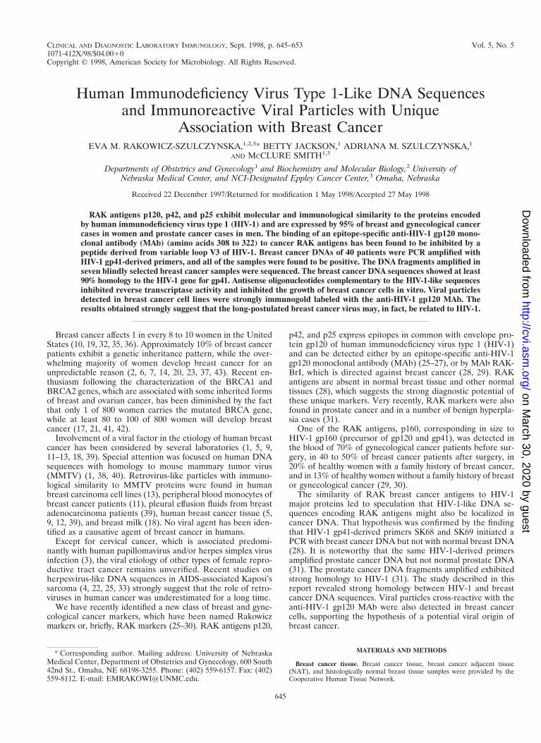
Electron microscopic detection of viral particles. Electronmicroscopic analysis of thin sections of SiHa cervical cancerand MCF 7 breast cancer cells revealed large, membrane-coated vesicles (vacuoles) that were immunogold labeled withthe anti-HIV-1 gp120 MAb and localized in the cytoplasm(Fig. 5A), at the edges of cells (Fig. 5B), and outside of cells(Fig. 5C). Strong immunogold labeling of the villus-like struc-tures on the cell surface, as well as of the intracellular andextracellular particles, was also observed (Fig. 5A).
A cross section (Fig. 5B) of a large cytoplasmic vesicle locat-ed close to the cell surface revealed several oval-shaped struc-tures localized within the vesicle. The virus-like particles werealso “budding” from the extracellular vacuoles (Fig. 5C). Im-munogold staining of the membrane-coated vesicles obtainedby ultracentrifugation (100,000 3 g for 1 h) of SiHa or MCF 7cell culture medium revealed virus-like particles localized in-side the membrane-covered vesicles (Fig. 5D). Negative stain-ing of purified viral particles is shown in Fig. 5E to G. The sizeof the virus-like particles seems to be 120 nm. The tail-and-head structure of the anti-HIV-1 gp120 MAb-labeled viralparticles was frequently visible (Fig. 5E to G). It is noteworthythat labeling of MCF 7 and SiHa cells with the anti-HIV-1gp120 MAb was stronger in cells supplied by ATCC as amonolayer than in those remaining under laboratory condi-tions for 1 year. This observation was consistent with lowerexpression of RAK antigens in the aging cell cultures. BothMCF 7 and SiHa cells were tested by ATCC and found to bemycoplasma negative and tested by Advanced BiotechnologyInc. and found to be HIV-1 negative and mycoplasma and bac-terium free. Thus, the identified particles are likely to belong
FIG. 3. Electrophoretic pattern of PCR products amplified by HIV-1 (gp41Env)-derived primers SK68 and SK69 (A to C) or globin primers (D), separatedin a 1.5% (A and B) or a 4% (C) agarose gel and stained with ethidium bromide.CA, cancer; NL, normal breast tissue. (A) Lanes: 1 and 2, CA and NAT samplesfrom one patient that both tested positive (but the PCR with NAT was weaker);3 and 4, NL sample that tested negative and CA sample from the same patientthat tested positive; 5, 6, and 7, CA samples from different patients that testedpositive; 8, HIV-1-positive control. (B) Lanes 1 and 8, NAT samples from twodifferent patients that tested negative; 2, 3, 6, and 7, CA samples that testedpositive and NAT samples that tested negative; 4 and 5, NL samples fromdifferent patients that tested negative; 9, HIV-1-positive control. (C) PCR am-plification patterns of breast cancer DNAs selected for sequencing. The lowerband (142 bp) corresponded in size to the HIV-1 band. (D) PCR amplificationpatterns obtained with globin primers. Lanes: 1 to 3, normal breast DNA; 4,breast milk DNA; 5, NAT sample DNA; 7 to 9, breast cancer DNA. Sample 6tested globin negative and was discarded. Globin-positive samples 1 to 5 were allnegative with primers SK68 and SK69.
TABLE 2. PCR amplification of breast cancer DNAwith HIV-1-derived primers
Tissue or cellsNo. of samples testedby PCR/no. (%) posi-tive for RAK antigen
Breast cancer ........................................................... 40/40a (100)NAT .......................................................................... 39/16,a 5b (41, 12.8)Normal breastc......................................................... 30/8a 5b (26.7, 16.7)Breast reductiond .................................................... 10/1b (10)Breast milk............................................................... 10/0a (0)MCF 7, SiHae .......................................................... A,a B,a Ca
Cervix........................................................................ 2/0a (0)Ovary ........................................................................ 2/0a (0)Uterus ....................................................................... 2/0a (0)Vagina....................................................................... 2/0a (0)Colon ........................................................................ 3/0a (0)Skin ........................................................................... 6/0a (0)Placenta .................................................................... 2/0a (0)
a See Table 1, footnote a.b See Table 1, footnote b.c See Table 1, footnote c.d See Table 1, footnote d.e See Table 1, footnote e.
648 RAKOWICZ-SZULCZYNSKA ET AL. CLIN. DIAGN. LAB. IMMUNOL.
on March 30, 2020 by guest
http://cvi.asm.org/
Dow
nloaded from

to a virus which expresses epitopes similar to HIV-1 gp120 butcompletely different in structure and function.
Effect of antisense oligonucleotides on cancer cell growthin vitro. To evaluate whether the identified cancer DNA se-quences, which are absent in normal tissues and may belong toa retrovirus, play a role in malignant cell growth, antisenseoligonucleotides have been synthesized and used to treat SiHaand MCF 7 cells in vitro. Antisense oligonucleotide RAK-I(21-mer 59-CCAGACTGTGAGTTGCAACAG-39), comple-mentary to the 39 end of the cancer DNA sequences ampli-fied with HIV-1 Env-derived primers, inhibited the growth ofbreast cancer cells by 70% within 4 days (Fig. 6) and inhibitedreverse transcriptase activity in the cell culture medium by 75%(Table 3). The control antisense oligonucleotide (59-TGTGACATCAGGCTCAAATC-39) neither inhibited cell growth noraffected reverse transcriptase activity. These results suggestthat a cancer antigen(s) encoded by the gene related to HIV-1gp41 is critical for the growth of breast and cervical cancercells. Reverse transcriptase inhibition suggests that the identi-fied DNA sequences may, in fact, belong to a retrovirus.
DISCUSSION
RAK antigens p120, p42, and p25 are expressed in breastand gynecological cancer tissues in women (28, 29) and pros-tate cancer tissue in men (31) but are absent in normal tissuesand in the majority of NATs. Although the nature of theseunique proteins is not fully understood, the unusual associa-tion with cancer strongly suggests a potential role in the etiol-
ogy of cancers of the reproductive system. Why cancers thatdiffer in histological structure and originated from completelydifferent tissues all express these unique proteins remains amedical and biological puzzle. If we assume that RAK antigensare encoded by a virus, then one of the possible mechanisms ishormonal regulation. One of the cancer antigens (RAK anti-gen p160) is expressed in the blood of the majority of breast,cervical, and ovarian cancer patients (29, 30). The molecularweight correlation of blood RAK antigen p160 with the pre-cursor of HIV-1 envelope proteins gp120 and gp41 stronglysupports a viral origin of this marker. Cancer-limited expres-sion of RAK antigens suggests either that normal human genesare selectively transcribed in cancer or that a unique virusaffects reproductive organs, leading to malignancy. Transcrip-tional regulation of human genes is very unlikely in light of thefact that HIV-1 gp41-derived primers PCR amplified breast(28) and prostate (31) cancer DNAs but not normal tissueDNA, including DNA extracted from NAT.
In a low-density agarose gel the electrophoretic migration ofthe breast cancer DNA fragment amplified was indistinguish-able from that of the HIV-1 fragment amplified. High-resolu-tion gel electrophoresis revealed one band (142 bp) commonto HIV-1 and cancer DNAs and another PCR band greater insize (160 bp) that was present only in cancer DNA. In contrastto the smaller band, which exhibited over 90% homology toHIV-1 sequences encoding transmembrane protein gp41, thesequences of the larger band contained long A and T repeatsand did not show homology to any known human gene. It isnoteworthy that the larger band was never amplified in theabsence of the smaller band (in normal tissue DNA). It is likelythat further understanding of the structure of the putative virusand its sites of integration with the human genome will clarifythe role of that 160-bp amplification product.
In 48% of breast cancer patients, HIV-1-like sequences wereabsent in histologically normal tissue of the cancer-affectedbreast, which eliminates the possibility of a random distribu-tion of these sequences in the human genome and excludes thepossibility that the identified sequences were of human origin.Whether the identified HIV-1 gp41-like sequences encodeRAK antigen p42 cannot be established, but it seems veryunlikely that HIV-1-like RAK antigens and HIV-1-like DNAsequences represent two unrelated phenomena, both exclusive-ly associated with cancer.
Breast cancer RAK antigens p120, p42, and p25 exhibit mo-lecular and immunological similarity to the proteins encodedby HIV-1. Moreover, RAK antigens express an amino acid re-gion homologous to variable loop V3 of HIV-1 and cross-reactwith an epitope-specific anti-HIV-1 envelope protein gp120MAb. MAb RAK-BrI, which was developed against RAK an-tigens, also cross-reacts with HIV-1 antigens gp160 and gp120,which confirms the nonaccidental similarity of cancer andHIV-1 proteins. Recent studies indicated that several antibod-ies developed against a nonglycosylated form of HIV-1 gp120recognized RAK p120 in cancer cells, which implies that thehomology of RAK antigen p120 to HIV-1 gp120 is expanded tovarious parts of the molecule but is probably limited to theprimary structure of the protein (31a). Previous studies indi-cated that a MAb raised against HIV-1 gp41 is also able to rec-ognize RAK p42 in breast and cervical cancer cell lines; how-ever, limited studies were done with that MAb (27).
Although RAK antigens exhibit homology to HIV-1 anti-gens, these cancer markers can be easily distinguished fromHIV-1 infection by using (i) any antibody directed against theglycosylated form of HIV-1 gp120, (ii) MAb 5025, or (iii) any
FIG. 4. DNA sequences amplified in breast cancer samples from seven dif-ferent patients with HIV-1 gp41-derived primers SK68 and SK69. Broken linesindicate primer locations. Lines over the sequences indicate variable sequencesthat are different in at least two patients from that of HIV-1.
VOL. 5, 1998 HIV-1-LIKE DNA AND BREAST CANCER 649
on March 30, 2020 by guest
http://cvi.asm.org/
Dow
nloaded from

other anti-HIV-1 MAb which does not cross-react with cancerantigens. Moreover, MAb RAK-BrI binds to RAK antigensp120, p42, and p25 in cancer tissue but only to gp160 and gp120of HIV-1, which automatically eliminates the possibility of in-fection.
The similarity of breast cancer RAK antigens p120, p42, andp25 to HIV-1 major proteins, the fact that all three antigensare usually found together, and the exclusive cancer affiliationof these markers, further supported by the presence of HIV-1-like sequences in cancer DNA, strongly suggest that these
FIG. 5. Transmission electron micrographs of cellular and extracellular vacuoles carrying viral particles in SiHa (A, C, and D) and MCF 7 (B) cells. Viral particleswere obtained by ultracentrifugation (100,000 3 g, 1 h) of cell culture media and negatively stained with uranyl acetate (E to G). In A and D to G, samples were alsoimmunogold labeled with an anti-HIV-1 gp120 MAb. The sizes of the immunogold particles (arrows in A) were 15 (A and D) and 10 (E) nm. V, virus-like particles.Bars, 100 nm. The original magnification was 30,000 (C) or 75,000 (A, B, and D to G).
650 RAKOWICZ-SZULCZYNSKA ET AL. CLIN. DIAGN. LAB. IMMUNOL.
on March 30, 2020 by guest
http://cvi.asm.org/
Dow
nloaded from
antigens belong to a slow retrovirus, a fragment of which hasbeen sequenced. Inhibition of cancer cell growth, in parallelwith inhibition of reverse transcriptase activity in the presenceof antisense oligonucleotides complementary to the HIV-1-
like sequences, supports the viral nature of RAK markers. Themechanism of cancer growth promotion by the putative virusremains to be further investigated. The growth factor-like char-acter of the RAK antigens is suggested by the previously de-
FIG. 5—Continued.
VOL. 5, 1998 HIV-1-LIKE DNA AND BREAST CANCER 651
on March 30, 2020 by guest
http://cvi.asm.org/
Dow
nloaded from

scribed cancer growth activation of the anti-HIV-1 gp120 MAb(27).
A viral etiology of human breast cancer has been consideredby several investigators; however, it has never been confirmed(1, 5, 9, 11–13, 18, 38–40). The assumption that a breast cancervirus would exhibit homology to MMTV was probably mislead-ing, since the human genome contains numerous copies ofgenes with sequence homology to MMTV (1, 38, 40). MMTVsequences were found in both healthy persons and cancer pa-tients, and proteins homologous to those of MMTV were foundin both groups of women as well (1, 12, 38, 40). Our studiessuggest that the breast cancer virus exhibits homology toHIV-1 rather than to MMTV.
Electron microscopic studies revealed some virus-like parti-cles that were immunogold labeled with an anti-HIV-1 MAb.Labeling of breast cancer cells with an anti-HIV-1 gp120 MAbwas also observed when the immunofluorescence techniquewas used (27). Electron microscopic analysis suggests that thevirus buds into the intracytoplasmic vacuoles, which are thensecreted to the cell surface, fuse with the membrane, and re-lease viral particles through exocytosis. Exocytosis of the virus-containing vesicles might be responsible for the formation of
the characteristic “peninsula-like” surface of the tested cancercells, with very long villus-like structures. Alternatively, intactvesicles might be released by cells and the viral particles wouldthus be budding from the surface. Viral particles were ob-served in breast cancer cells by several researchers. It is note-worthy that the tail-and-head structure of the viral particles,with a strong morphological resemblance to MMTV, whichwas reported before by Moore et al. (18) in breast milk studies,was also frequently observed in our study (Fig. 5E to G).However, the particles detected by us in breast cancer cellswere labeled with an anti-HIV-1 gp120 MAb, suggesting ho-mology to HIV-1 rather than to MMTV. The possibility ofcross-reactivity between the anti-HIV-1 gp120 MAb andMMTV was excluded, since MMTV antigens were not recog-nized by this MAb either in a Western blot or in microscopicstudies. The possibility that HIV-1-derived primers SK68 andSK69 amplified MMTV sequences in the human genomemay be also excluded, since (i) these primers did not amplifyMMTV sequences in mouse cells and (ii) cancer DNA se-quences amplified with HIV-1-derived primers exhibited nohomology to MMTV. Further studies are needed to verifywhether the identified RAK markers are expressed by thedetected viral particles.
The presence of HIV in cell cultures or in fresh cancer tissuemay be eliminated since they tested negative for HIV-1 p24.The fact that of the over 1,000 cancer patients tested in this andother studies (28, 29) for the presence of RAK antigens inbreast and gynecological tissue, 95% were RAK positive auto-matically excludes the HIV-1 origin of RAK antigens. Fur-ther studies are needed to fully characterize and classify thevirus.
Independently of the viral or human origin of the novelcancer antigens, the specific association of RAK antigens withgynecological and breast cancer in women and prostate cancerin men and the lack of these markers in normal tissues stronglysuggest that RAK markers have a critical value in the earlydiagnosis of cancers affecting reproductive organs. The diag-nostic value of protein and PCR RAK markers was evaluatedbefore (28, 31). It is suggested that protein RAK markersmight improve the early detection of malignant or premalig-nant changes and would help in more effective evaluation ofcancer margins, leading to a reduction in the number of un-needed mastectomies. PCR markers could be used to deter-mine the predisposition of breast tissue to become malignant.Current studies on the identification of HIV-1-like sequenceswill definitely help to clone the virus and understand theetiology of reproductive tract cancers. If the structure of thenew cancer virus were understood, then completely new ap-proaches to cancer diagnosis, prevention, prognosis, and ther-apy could be developed. Cancer RAK antigen p120 and/orother RAK antigens would definitely play a critical role in theproduction of a breast cancer vaccine.
FIG. 6. Effect of antisense oligonucleotide RAK-I on growth of breast cancercell line MCF 7. Breast cancer MCF 7 cells were grown for 4 days in the absence(A) or presence (B) of antisense oligonucleotide RAK-I (59-CCAGACTGTGAGTTGCAACAG-39), which was added daily to concentrations of 100 mg/ml (day1) and 50 mg/ml (days 2 and 3). The oligonucleotide (59-TGTGACATCAGGCTCAAATC-39) used in control experiments did not affect cell growth (data notshown).
TABLE 3. Effects of antisense oligonucleotide RAK-I and acontrol oligonucleotide on reverse transcriptase activity
Cell line
[3H]TTP incorporation (cpm)a (% inhibition)
Controlb RAK I Controloligonucleotide
MCF 7 10,000 2,550 (74.5) 10,200 (0)SKBr-3 8,300 2,100 (74.7) 8,220 (0)
a Data are means from four experiments. The standard deviations were 5 to15%.
b No oligonucleotide.
652 RAKOWICZ-SZULCZYNSKA ET AL. CLIN. DIAGN. LAB. IMMUNOL.
on March 30, 2020 by guest
http://cvi.asm.org/
Dow
nloaded from

ACKNOWLEDGMENTS
This study was sponsored by the Leland J. and Dorothy H. OlsonFoundation for Women’s Health.
We thank Martin Cane for immunogold labeling of cancer cells,Rick Vaughn for taking photographs of viral particles, and WilliamSnyder for general technical support. We also thank Advanced Bio-technology Inc. for testing cells for contamination with known viruses,bacteria, or mycoplasmas.
REFERENCES
1. Andersson, M. L., P. Medstrand, H. Yin, and J. Blomberg. 1996. Differentialexpression of human endogenous retroviral sequences similar to mousemammary tumor virus in normal peripheral blood mononuclear cells. AIDSRes. Hum. Retroviruses 12:833–840.
2. Biesecker, B. B., M. Boehnke, K. Calzone, D. S. Markel, J. E. Garber, F. S.Collins, and B. L. Weber. 1993. Genetic counseling for families with inher-ited susceptibility to breast and ovarian cancer. JAMA 269:1970–1974.
3. Bouton, A. H., and J. T. Parsons. 1993. Retroviruses and cancer: models forcancer in animals and humans. Cancer Invest. 11:70–79.
4. Chang, Y., E. Cesarman, M. S. Pessin, F. Lee, J. Culpepper, D. M. Knowles,and P. S. Moore. 1994. Identification of herpes virus-like DNA sequences inAIDS-associated Kaposi’s sarcoma. Science 266:1865–1869.
5. Chopra, H. C., and W. F. Feller. 1969. Virus-like particles in human breastcancer. Tex. Rep. Biol. Med. 27:945–954.
6. Claus, E. B., N. Risch, and W. D. Thompson. 1991. Genetic analysis of breastcancer in the cancer and steroid hormone study. Am. J. Hum. Genet. 48:232–242.
7. Coles, C., A. Condie, U. Chetty, C. M. Steel, H. J. Evans, and J. Prosser.1992. p53 mutations in breast cancer. Cancer Res. 52:5291–5298.
8. Durda, P. J., L. Bacheler, P. Clapham, A. M. Jenoski, B. Leece, T. J.Matthews, A. McKnight, R. Pomerantz, M. Rayner, and K. J. Weinhold.1988. HIV-1 neutralizing monoclonal antibodies induced by a synthetic pep-tide. AIDS Res. Hum. Retroviruses 6:1115–1118.
9. Feller, W. F., and H. C. Chopra. 1968. A small virus-like particle observed inhuman breast cancer by means of electron microscopy. J. Natl. Cancer Inst.40:1359–1373.
10. Garfinkel, L., C. C. Boring, and C. W. Heath, Jr. 1994. Changing trends. Anoverview of breast cancer incidence and mortality. Cancer 74:222–227.
11. Kahl, L. P., A. R. Carrol, P. Rhodes, J. Wood, and N. G. Read. 1991. Anevaluation of the putative human mammary tumor retrovirus associated withperipheral blood monocytes. Br. J. Cancer 63:534–540.
12. Keydar, I., C. S. Chou, M. Hareuveni, I. Tsarfaty, E. Sahar, G. Selzer, S.Chaichik, and A. Hizi. 1989. Production and characterization of monoclonalantibodies identifying breast tumor-associated antigens. Proc. Natl. Acad.Sci. USA 86:1362–1366.
13. Keydar, I., T. Ohno, R. Nayak, R. Sweet, F. Simoni, F. Weiss, S. Karby, R.Mesa-Tejada, and S. Spiegelman. 1984. Properties of retrovirus-like parti-cles produced by a human breast carcinoma cell line: immunological rela-tionship with mouse mammary tumor virus proteins. Proc. Natl. Acad. Sci.USA 81:4188–4192.
14. King, M. C., S. Rowell, and S. M. Love. 1993. Inherited breast and ovariancancer. JAMA 269:1975–1980.
15. Laemmli, U. K. 1971. Cleavage of structural proteins during the assembly ofthe head of bacteriophage T4. Nature 227:680–685.
16. Langedijk, J. P. M., N. K. T. Back, P. J. Durda, J. Goudsmit, and R. H.Meloen. 1991. Neutralizing activity of anti-peptide antibodies against theprincipal neutralization domain of human immunodeficiency virus type 1.J. Gen. Virol. 72:2519–2526.
17. Miki, Y., J. Swensen, D. Shattuck-Eidens, P. A. Futreal, K. Harshman, S.Tavtigian, Q. Liu, C. Cochrane, L. M. Bennett, and W. Ding. 1994. A strongcandidate for the breast and ovarian cancer susceptibility gene BRCA1.Science 266:66.
18. Moore, D. H., J. Charney, B. Kramarsky, E. Y. Lasfargues, and N. H.Sarkar. 1971. Search for a human breast cancer virus. Nature 229:611–615.
19. Newcomb, P. A., and P. M. Lantz. 1993. Recent trends in breast cancerincidence, mortality, and mammography. Breast Cancer Res. Treat. 28:97–106.
20. Newman, B., M. A. Austin, M. Lee, and M. C. King. 1988. Inheritance ofhuman breast cancer: for autosomal dominant transmission in high-riskfamilies. Proc. Natl. Acad. Sci. USA 85:3044–3048.
21. Nowak, R. 1994. Breast cancer gene offers surprises. Science 265:1796–1799.
22. O’Leary, J. J., M. M. Kennedy, and J. McGee. 1997. Mol. Pathol. 50:4–8.23. Peles, E., S. S. Bacus, R. A. Koski, H. S. Lu, D. Wen, S. G. Ogden, R. B. Levy,
and Y. Yarden. 1992. Isolation of the Neu/HER-2 stimulatory ligand: a 44 kdglycoprotein that induces differentiation of mammary tumor cells. Cell 69:205–216.
24. Rakowicz-Szulczynska, E., V. Raso, W. Kaczmarski, K. S. Steimer, and P. J.Durda. 1993. Internalization of anti-gp120 monoclonal antibody and humanantibodies by HIV-1-infected T lymphocytes. Antib. Immunoconjug. Radio-pharm. 6:209–219.
25. Rakowicz-Szulczynska, E. M., W. Kaczmarski, K. S. Steimer, and P. J.Durda. 1993. Internalized antibodies as a potential tool against retroviraldisease, p. 180–197. In E. M. Rakowicz-Szulczynska (ed.), Nuclear localiza-tion of growth factors and of monoclonal antibodies. CRC Press, Inc., BocaRaton, Fla.
26. Rakowicz-Szulczynska, E. M., D. G. McIntosh, and M. L. Smith. 1994. Novelfamily of gynecological cancer antigens detected by anti-HIV antibody. In-fect. Dis. Obstet. Gynecol. 2:171–178.
27. Rakowicz-Szulczynska, E. M., D. G. McIntosh, and M. L. Smith. 1995.Mechanisms of cancer growth promotion by HIV-I neutralizing antibodies.Cancer J. 8:143–149.
28. Rakowicz-Szulczynska, E. M., A. Roszak, A. Mackiewicz, J. Markowska, A.Karczewska, W. Snyder, and M. L. Smith. 1997. New protein and PCRmarkers RAK for diagnosis, prognosis and surgery guidance for breast can-cer. Cancer Lett. 112:93–101.
29. Rakowicz-Szulczynska, E. M., A. Roszak, A. Mackiewicz, J. Markowska,D. G. McIntosh, A. Karczewska, W. Snyder, D. Leary, and M. L. Smith. 1996.Diagnostic evaluation of cancer antigens RAK. I. Cervical and ovarian can-cer. Int. J. Oncol. 9:693–699.
30. Rakowicz-Szulczynska, E. M., D. G. McIntosh, M. J. Perry, and M. L. Smith.1995. Antigen RAK: a new breast cancer diagnostic marker. J. TumorMarker Oncol. 10:25–37.
31. Rakowicz-Szulczynska, E. M., B. Jackson, and W. Snyder. 1998. Prostate,breast, and gynecological cancer markers RAK with homology to HIV-1.Cancer Lett. 124:213–223.
31a.Rakowicz-Szulczynska, E. M. Unpublished data.32. Remington, P. L., and P. M. Lantz. 1992. Using a population-based cancer
reporting system to evaluate a breast cancer detection and awareness pro-gram. Cancer 42:367–371.
33. Schalling, M., M. Elkman, E. E. Kaaya, A. Linde, and P. Biberfeld. 1995. Arole for a new herpes virus (KSHV) in different forms of Kaposis’s sarcoma.Nat. Med. 1:705–706.
34. Scolnick, E. M., S. T. Aaronson, G. J. Todaro, and W. P. Parks. 1971. RNAdependent polymerase activity in mammalian cells. Nature 229:318–321.
35. Senie, R. T., M. Lesser, D. W. Kinne, and P. P. Rosen. 1994. Method oftumor detection influences disease-free survival of women with breast car-cinoma. Cancer 73:1666–1672.
36. Silverberg, E., C. C. Boring, and T. S. Squires. 1990. Cancer statistics.Cancer 40:9–15.
37. Slamon, D. J., W. Godolphin, L. A. Jones, J. A. Holt, S. G. Wong, D. E. Keith,W. J. Levin, S. G. Stuart, J. Udove, and A. Ullrich. 1989. Studies of theHER-2/neu proto-oncogene in human breast and ovarian cancer. Science248:707–712.
38. Sorhaug, H., and B. Grinde. 1993. Evolution of mouse mammary tumorvirus-related sequences in the human genome. Virus Res. 30:53–61.
39. Soule, H. R., E. Linder, and T. S. Edgington. 1983. Membrane 126-kilodaltonphosphoglycoprotein associated with human carcinoma identified by a hy-bridoma antibody to mammary carcinoma cells. Proc. Natl. Acad. Sci. USA80:1332–1336.
40. Szakacs, J. G., and L. C. Moscinski. 1991. Sequence homology to deoxyri-bonucleic acid to mouse mammary tumor virus genome in human breasttumors. Ann. Clin. Lab. Sci. 21:402–412.
41. Tonin, P., O. Serova, J. Simard, G. Lenoir, J. Faunterin, K. Morgan, H.Lynch, and S. Narod. 1994. The gene for hereditary breast-ovarian cancer,BRCA1, maps distal to EDH17B2 in chromosome region. Hum. Mol. Genet.3:1679–1682.
42. Wooster, R., S. Neuhausen, J. Mangion, Y. Quirk, D. Ford, D. Collins, K.Nguyen, S. Seal, T. Tran, and D. Averill. 1994. Localization of a breastcancer susceptibility gene, BRCA2, to chromosome 13q12-13. Science 265:2088–2090.
43. Zuppan, P. J., J. M. Hall, M. K. Lee, M. Ponglikitmongkol, and M. C. King.1991. Possible linkage of the estrogen receptor genes to breast cancer in afamily with late-onset disease. Am. J. Hum. Genet. 48:1065–1068.
VOL. 5, 1998 HIV-1-LIKE DNA AND BREAST CANCER 653
on March 30, 2020 by guest
http://cvi.asm.org/
Dow
nloaded from