human cytogenetics - atzlabs.com · cytogenetics laboratories use this method with amniotic fluid,...
TRANSCRIPT

HUMAN CYTOGENETICSPrenatal Diagnostics
Peripheral BloodBone Marrow
Auxiliary Products

Prenatal Diagnostics BIOAMF-1 Basal Medium and Supplement
Optimized Media for Culture and Genetic Analysis of Human Amniotic Fluid Cells and Chorionic Villi (CV) Samples
Chromosome Karyotyping was first developed in the field of Cytogenetics. The basic principle of the method is the preparation of chromosomes for microscopic observation by arresting cell mitosis at metaphase with colchicine and treating the cells with a hypotonic solution. This is followed by regular or fluorescent staining of the chromosomes, which are then tested with the aid of a microscope and computer programs to arrange and identify the chromosomes for the presence of genetic abnormalities. In principle, this method enables the identification of any abnormality -excess chromosomes or chromosome deficiency, broken chromosomes, or excess genetic material (as a result of a recombination process). Clinical cytogenetics laboratories use this method with amniotic fluid, chorionic villi, blood cells, skin cells, and so on, which can be cell cultured to obtain mitotic cells. Most amniotic fluid cells originate from the fetus and include fibroblasts, epithelial cells and amniocytes. The cells suited for genetic analysis are fibroblasts and amniocytes, and chromosome preparation from these cells yields a clear picture of the chromosomes for microscopic observation. Amniocentesis is typically carried out in week 16-20 of pregnancy, when 20-40ml of amniotic fluid is drawn for genetic analysis. The cells can be seeded on a slide or in suitable flasks to obtain colonies or cell cultures. Since the cells divide, chromosome karyotyping can be carried out on them for general genetic testing. To test specific abnormalities, a small number of cells can be taken from the original sample for FISH and/ or QF-PCR testing. The time that elapses until the final results of genetic analysis are obtained is of significant importance both from an emotional point of view – the tension and stress entailed in waiting for the final results - and a practical one - the need to terminate pregnancy if genetic abnormalities are found. Pregnancy termination in the second trimester in effect means performing an abortion; hence the importance of obtaining results as early as possible in order to alleviate the procedure. In the past decade, Biological Industries Ltd. has developed a range of cytogenetics products, including media for culture of amniotic fluid and chorionic villi cells, BIOAMF-1, BIOAMF-2 and BIOAMF-3, which are selling very successfully throughout the world.
Product Name Catalogue Unit No. Size
BIOAMF-1 Basal Medium
01-190-1A 450ml
01-190-1B 90ml
BIOAMF-1 Supplement
01-192-1D 10ml
01-192-1E 50ml
BIOAMF-1 is designed for the primary culture of human amniotic fluid cells and chorionic villi (CV) samples in both open (5% CO2) and closed systems. The medium allows rapid growth of amniocytes or chorionic villi for use in karyotyping. No supplementation with serum or serum-substitutes is necessary. The medium consists of two components: basal medium and frozen supplements.
Instructions for Use For the preparation of 500ml complete medium, use 01-190-1A with 01-192-1E. For the preparation of 100ml complete medium, use 01-190-1B with 01-192-1D. Thaw the BIOAMF-1 Supplement by swirling in a 37ºC water bath, and transfer the contents to the bottle of BIOAMF-1 Basal Medium. Mix the complete medium by swirling the bottle, and add 2mM L-Glutamine (L-Glutamine Solution 200mM, cat. no. 03-020-1). Antibiotics may be added if desired (Pen-Strep, cat. no. 03-031-1).
Storage and Stability BIOAMF-1 Basal Medium is stable for 24 months from production date when stored at 2-8ºC. BIOAMF-1 Supplement is stable for 15 months from production date when stored at -20ºC. The complete medium is stable for 7 days when stored at 2-8ºC. Do not freeze the complete medium. Protect both the basal medium and the complete medium from light.
BIOLOGICAL INDUSTRIES

BIOAMF-2 Complete Medium Product Name Catalogue Unit
No. SizeBIOAMF-2
Complete Medium 01-194-1A 500ml
01-194-1B 100ml
BIOAMF-3 Complete Medium Product Name Catalogue Unit
No. Size BIOAMF-3 Complete Medium
01-196-1A 500ml
01-196-1B 100ml
BIOAMF-2 is a complete medium specifically optimized for the primary culture of human amniotic fluid cells and chorionic villi (CV) samples in both open (5% CO2) and closed systems. No addition of serum is required, and chromosome karyotyping time is greatly reduced compared with the conventional medium. Note: this is a one-bottle formulation, which also contains L-Glutamine and antibiotics. Simply thaw and use!
Figure 1: Comparison of the Percentage of Harvested Plates According To Harvest Day Between BIOAMF-2 By Biological Industries and A Leading Competitor
Leading Competitor BIOAMF-2
Harvest Day
Storage and Stability BIOAMF-2 Medium should be kept frozen at -20°C. After thawing, the medium should be stored at 2-8°C. The medium should be used within 7 days after thawing. Protect the medium from light.
An improved version of complete medium specifically optimized for the primary culture of human amniotic fluid cells and chorionic villi samples used in prenatal diagnostic testing. This medium accelerates the growth of the non-epithelial cells used for chromosome karyotyping. The medium is supplied frozen and contains L-Glutamine and antibiotics
Advantages of BIOAMF-3 • Enhanced buffering capacity both in open (CO2) and closed systems. • Improved banding quality: excellent chromosome morphology and
metaphase structure. • Increased metaphase yield. • Extended stability of the medium at 2-8°C.
Storage and Stability BIOAMF-3 Medium should be kept frozen at -20°C. After thawing, the medium should be stored at 2-8°C. The medium should be used within 14 days after thawing. Protect the medium from light.
BIOLOGICAL INDUSTRIES
50
5 6 7 10 11 12 >12

Blood Lymphocyte Culture
Blood cell karyotyping is an important tool in modern human cytogenetics, providing information about chromosomal abnormalities, their frequency in the population, and the relationship between specif ic chromosoma abnormalities and phenotypic effects Human cytogenetic studies involve the examination of a stimulated lymphocyte after blocking cell division at metaphase with an inhibitor of spindle formation. The nuclear membrane breaks down and chromosome condensation takes place as usual, but the chromosomes fail to organize themselves into a metaphase plate. This gives an appearance quite unlike a natural metaphase, in that the chromosomes are free within the cytoplasm. Subsequent processing and staining allows clear visualization of the chromosomes The chromosomes can be stained either by a technique that gives a fairly uniform intensity, or by a technique that gives differential staining along the length of the chromosome
Biological Industries offers a ready-to-use flat bottom tube which contains 5ml of a complete medium for Peripheral Blood Karyotyping. The medium contains PHA. A slated rack is supplied with each 10 tubes so that the tubes are put into the incubator at the right angle
Advantages • Saves time • Excellent growth promotion • No other supplements required • Slanted rack for convenient incubation
Peripheral Blood Karyotyping Medium Product Name Catalogue Unit
No. Size Peripheral Blood Karyotyping Medium Without Phytohemagglutinin
01-198-1A 500ml
01-198-1B 100ml
Peripheral Blood Karyotyping Medium With Phytohemagglutinin
01-201-1A 500ml
01-201-1B 100ml
01-201-1H 5ml
Peripheral Blood (PB) Karyotyping Medium is specifically optimized for short-term culture of peripheral blood lymphocytes for chromosome analysis. No addition of serum, glutamine or antibiotics is required.The medium is supplied frozen.
Storage and Stability PB Karyotyping Medium should be kept frozen at -20ºC. After thawing, the medium should be stored at 2-8ºC. The medium should be used within 10 days after thawing. Protect the medium from light.
Bone Marrow Culture
Cytogenetic analysis of human hematopoietic cells using bone marrow aspirates is a standard practice in hematology. Cell culture improvements and processing techniques have enabled the identification of a number of recurring abnormalities in solid tumors and hematologic malignant diseases. But even more data are available for leukemias and lymphomas than for solid tumors because of the relative ease of obtaining bone marrow or peripheral blood specimens from leukemia patients. The study of chromosomal abnormalities in leukemia serves two functions: The first is to assist in more accurate diagnosis, thereby providing prognostic information and allowing the more rational selection of therapy for a particular patient. The second is to identify the sites of consistent rearrangements, providing the precise localization required for the isolation and cloning of DNA from these regions. Using molecular techniques the function of the genes can be identified and the mechanisms whereby their altered function is involved in tumorigenesis can be determined. In the past, it was assumed that cytogenetic analysis of hematologic malignant disorders was best performed directly on uncultured bone marrow samples. However, later studies indicate that analysis of cultured samples disclosed a clonal abnormality that would not have been detected if the direct method alone had been used. Thus, for many samples, chromosomal rearrangements were often characterized only after analysis of cultured preparations.
Bone Marrow Karyotyping Medium
Product Name Catalogue Unit No. Size
Bone Marrow Karyotyping Medium Without conditioned medium
01-199-1A 500ml
01-199-1B 100ml
Bone Marrow Karyotyping Medium is intended for use in short-term cultivation of primary bone marrow cells for chromosome evaluation. Bone Marrow Karyotyping Medium is based on RPMI-1640 basal medium supplemented with L-Glutamine, foetal bovine serum, and antibiotics (penicillin and streptomycin). The medium does not contain any mitogens or conditioned medium. Bone Marrow Karyotyping Medium is supplied as frozen medium, which is ready for use after thawing.
BIOLOGICAL INDUSTRIES

Instructions for use The bone marrow karyotyping method was developed to provide information about chromosomal abnormalities. The ready-to-use medium is intended for the culture of bone marrow cells without any mitogens or conditioned medium. After 48-72 hours, a mitotic inhibitor is added to the culture to stop mitosis in the metaphase stage. After treatment by hypotonic solution, fixation and staining, chromosomes can be microscopically observed and evaluated for abnormalities.
Storage and Stability Bone Marrow Karyotyping Medium should be kept frozen at -20ºC. After thawing, the medium should be stored at 2-8ºC. The medium should be used within 10 days after thawing. Protect the medium from light.
Hematopoietic Cell Karyotyping Medium from Bone marrow
After treatment by hypotonic solution, fixation and staining, chromosomes can be microscopically observed and evaluated for abnormalities.
Storage and Stability Hematopoietic Cell Karyotyping Medium should be kept frozen at -20ºC. After thawing, the medium should be stored at 2-8ºC. The medium should be used within 10 days after thawing. Protect the medium from light.
Auxiliary Products
Phytohaemagglutinin M (PHA-M)
Catalogue Unit Size
12-006-1H 5ml
Product Name Catalogue Unit No. Size
Hematopoietic Cell Karyotyping Medium With conditioned medium
01-200-1A 500ml
01-200-1B 100ml
Cytogenetic analysis of human hematopoietic cells using bone marrow aspirates is a standard practice in hematology. Fresh cells or cells grown in short-term cultures often yield an insufficient number of mitotic cells and repeated aspirations are required. Hematopoietic Cell Karyotyping Medium was developed to stimulate the proliferation of human hematopoietic cells from bone marrow as well as peripheral blood. This medium is particularly effective for karyotyping of acute non-lymphocytic leukemias and various stages of chronic myelogenous leukemia, as well as other hematological disorders such as myelodysplastic syndrome and polycythemia vera. Hematopoietic Cell Karyotyping Medium is based on MEM-Alpha basal medium supplemented with L-Glutamine, foetal bovine serum, antibiotics (penicillin and streptomycin) and conditioned medium.
Hematopoietic Cell Karyotyping Medium is supplied as frozen medium, which is ready for use after thawing.
Instructions for use The hematopoietic cell karyotyping method was developed to provide information about chromosomal abnormalities. In the presence of a conditioned medium, acute and chronic nonlymphocytic leukemic cells in bone marrow and peripheral blood cultures are stimulated to enter into mitosis by DNA replication. After 48-72 hours, a mitotic inhibitor is added to the culture to stop mitosis in the metaphase stage.
Phytohaemagglutinin is a lectin extracted from red kidney beans (Phaseolus vulgaris). The protein consists of two molecular species, a leucoagglutinin (PHA-L) and an erythroagglutinin (PHA-E). Each of the proteins contains a family of five isolectins, each being a tetramer held together by noncovalent forces. PHA-M is the mucoprotein form and is a crude extract used for the stimulation of cell proliferation in lymphocyte culture. PHA-M also has a powerful erythroagglutinating property and it was originally used for separating leukocytes from whole blood. PHA-M from Biological Industries is a sterile, freeze-dried preparation of an aqueous extract from selected red kidney beans. Each bottle should be reconstituted by the addition of 5ml sterile distilled water, using a sterile syringe. After reconstitution, each ml will contain 5-10mg of protein. Activity: Each lot is tested and standardized for mitotic stimulation using primary human peripheral blood lymphocytes.
Use: 2-4ml of re-hydrated PHA-M per 100ml of culture medium.
BIOLOGICAL INDUSTRIES
Product Name Phytohaemagglutinin M (PHA-M), Lyophilized
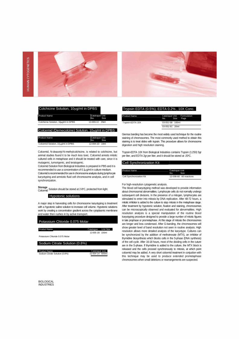
Colchicine Solution, 10 g/ml in DPBS Trypsin EDTA (0.5%), EDTA 0.2% , 10X Conc.
Product Name Catalogue Unit No. Size
Colchicine Solution, 10 g/ml in DPBS 12-003-1C 25ml Colcemid (Demecolcine) Solution, 10 g/ml in DPBS Product Name Catalogue Unit
No. Size Colcemid Solution, 10 g/ml in DPBS 12-004-1D 10ml
Colcemid, N-deacetyl-N-methylcolchicine, is related to colchicine, but animal studies found it to be much less toxic. Colcemid arrests mitotic cultured cells in metaphase and it should be treated with care, since it is mutagenic, tumorigenic, and teratogenic. Colcemid Solution from Biological Industries is prepared in PBS and it is recommended to use a concentration of 0.1 g/ml in culture medium. Colcemid is recommended for use in chromosome analysis during lymphocyte karyotyping and amniotic fluid cell chromosome analysis, and in cell synchronization.
Product Name Catalogue Unit Formulation No. Size Page 03-051-5B 100ml Trypsin-EDTA 10X 03-051-5C 20ml
Giemsa banding has become the most widely used technique for the routine staining of chromosomes. The most commonly used method to obtain this staining is to treat slides with trypsin. This procedure allows for chromosome digestion and high resolution staining.
Trypsin-EDTA 10X from Biological Industries contains Trypsin (1:250) 5gr per liter, and EDTA 2gr per liter, and it should be stored at -20ºC.
Cell Synchronization Kit Product Name Catalogue Unit
No. Size Cell Synchronization Kit 12-008-60 60 reactions
Solution should be stored at 2-8ºC, protected from light.
Hypotonic solutions
A major step in harvesting cells for chromosome karyotyping is treatment with a hypotonic saline solution to increase cell volume. Hypotonic solutions work by creating a concentration gradient across the cytoplasmic membrane and water then rushes in by active transport.
Potassium Chloride 0.075 Molar
Catalogue Unit Size
Potassium Chloride 0.075 Molar
Sodium Citrate Solution (0.8%)
Product Name Sodium Citrate Solution (0.8%)
For high-resolution cytogenetic analysis. The blood cell karyotyping method was developed to provide information about chromosomal abnormalities. Lymphocyte cells do not normally undergo subsequent cell divisions. In the presence of a mitogen, lymphocytes are stimulated to enter into mitosis by DNA replication. After 48-72 hours, a mitotic inhibitor is added to the culture to stop mitosis in the metaphase stage. After treatment by hypotonic solution, fixation and staining, chromosomes can be microscopically observed and evaluated for abnormalities. High resolution analysis is a special manipulation of the routine blood karyotyping procedure designed to provide a large number of mitotic figures in late prophase or prometaphase. At this stage of mitosis the chromosomes are longer and less condensed. After G-banding, the chromosomes will show greater level of band resolution not seen in routine analysis. High resolution allows more detailed analysis of the karyotype. Cultures can be synchronized by the addition of methotrexate (MTX), an inhibitor of thymidine biosynthesis which blocks cells in the S-phase (DNA synthesis) of the cell cycle. After 16-18 hours, most of the dividing cells in the cuture are in the S-phase. If thymidine is added to the culture, the MTX block is released and the cells proceed synchronously to mitosis, at which point colcemid may be added. A very short colcemid treatment in conjuction with this technique may be used to produce extended prometaphase chromosomes when small deletions or rearrangements are suspected.
BIOLOGICAL INDUSTRIES
Storage Colcemid
Product Name 12-005-1B 100ml
Catalogue size No. Size 01-934-1A 500ml

Materials 1. Methotrexate (Amethopterin), 10-5M in HBSS: 4 vials containing 1.5ml each 2. Thymidine, 10-3M in distilled water: 4 vials containing 1.5ml each
Storage and Stability The solutions must be kept frozen and protected from light. If appropriately stored, the solutions are stable for at least 18 months from the date of preparation.
Product Name Catalogue No.
Unit Size
Storage Temp.
Prenatal Diagnostics BIOAMF-1 Basal Medium 450ml 2-8ºC 90ml 2-8ºC BIOAMF-1 Supplement 10ml -20ºC 50ml -20ºC BIOAMF-2 Complete Medium 500ml -20ºC 100ml -20ºC BIOAMF-3 Complete Medium 500ml -20ºC
01-190-1A
01-190-1B
01-192-1D
01-192-1E
01-194-1A
01-194-1B
01-196-1A
01-196-1B 100ml -20ºC Blood Lymphocyte Culture Peripheral Blood Karyotyping Medium Without Phytohemagglutinin
500ml -20ºC
100ml -20ºC Peripheral Blood Karyotyping Medium With Phytohemagglutinin
500ml -20ºC
100ml -20ºC
01-198-1A
01-198-1B 01-201-1A
01-201-1B
01-201-1H
5ml -20ºC Bone Marrow Culture Bone Marrow Karyotyping Medium Without conditioned medium
500ml -20ºC
100ml -20ºC Hematopoietic Cell Karyotyping Medium With conditioned medium
500ml -20ºC
01-199-1A
01-199-1B 01-200-1A
01-200-1B 100ml -20ºC
Auxiliary Products Colchicine Solution, 10 g/ml in DPBS 25ml 2-8ºC Colcemid Solution, 10 g/ml in DPBS
Potassium Chloride, 0.075 Molar 10ml
100ml 2-8ºC
2-8ºC
Phytohemagglutinin-M (PHA-M), Lyophilized 5ml 2-8ºC Cell Synchronization Kit 60 reactions -20ºC Sodium Citrate Solution (0.8%) 500ml AMB Trypsin EDTA (0.5%), EDTA 0.2% 10X Conc* 100ml -20ºC * see formulation page…
12-003-1C
12-004-1D
12-005-1B
12-006-1H
12-008-60
01-934-1A
03-051-5B
03-051-5C 20ml -20ºC