human ca125/muc16 immunoassay · the quantikine® human ca125/muc16 immunoassay is a 4.5 hour...
TRANSCRIPT

Human CA125/MUC16 Immunoassay
Quantikine® ELISA
This package insert must be read in its entirety before using this product. For research use only. Not for use in diagnostic procedures.
Catalog Number DCA125
For the quantitative determination of human Cancer Antigen 125 (CA125) concentrations in cell culture supernates, serum, plasma, saliva, urine, and human milk.

MANUFACTURED AND DISTRIBUTED BY:
USA & Canada | R&D Systems, Inc. 614 McKinley Place NE, Minneapolis, MN 55413, USATEL: (800) 343-7475 (612) 379-2956 FAX: (612) 656-4400E-MAIL: [email protected]
DISTRIBUTED BY:
UK & Europe | R&D Systems Europe, Ltd.19 Barton Lane, Abingdon Science Park, Abingdon OX14 3NB, UKTEL: +44 (0)1235 529449 FAX: +44 (0)1235 533420E-MAIL: [email protected]
China | R&D Systems China Co., Ltd.24A1 Hua Min Empire Plaza, 726 West Yan An Road, Shanghai PRC 200050TEL: +86 (21) 52380373 FAX: +86 (21) 52371001E-MAIL: [email protected]
TABLE OF CONTENTS
SECTION PAGE
INTRODUCTION .....................................................................................................................................................................1
PRINCIPLE OF THE ASSAY ...................................................................................................................................................2
LIMITATIONS OF THE PROCEDURE .................................................................................................................................2
TECHNICAL HINTS .................................................................................................................................................................2
MATERIALS PROVIDED & STORAGE CONDITIONS ...................................................................................................3
OTHER SUPPLIES REQUIRED .............................................................................................................................................3
PRECAUTIONS .........................................................................................................................................................................4
SAMPLE COLLECTION & STORAGE .................................................................................................................................4
SAMPLE PREPARATION........................................................................................................................................................4
REAGENT PREPARATION .....................................................................................................................................................5
ASSAY PROCEDURE .............................................................................................................................................................6
CALCULATION OF RESULTS ...............................................................................................................................................7
TYPICAL DATA .........................................................................................................................................................................7
PRECISION ................................................................................................................................................................................8
RECOVERY.................................................................................................................................................................................8
LINEARITY .................................................................................................................................................................................8
SENSITIVITY .............................................................................................................................................................................9
CALIBRATION ..........................................................................................................................................................................9
SAMPLE VALUES .....................................................................................................................................................................9
SPECIFICITY ........................................................................................................................................................................... 10
REFERENCES ......................................................................................................................................................................... 11
PLATE LAYOUT ..................................................................................................................................................................... 12

www.RnDSystems.com 1
INTRODUCTIONCA125 (cancer antigen 125) was named for an epitope on ovarian cancer cells, and now denotes the member of the mucin protein superfamily encoded by the MUC16 (mucin protein 16) gene (1-3). Expression of isoforms, proteolytic cleavage, and heavy N- and O-linked glycosylation produce type I transmembrane and secreted forms of human CA125 that can vary from 1148 to 22,152 amino acids (aa) in length and 200-5000 kDa in size (1-3). Immunohistochemistry in the mouse demonstrates CA125 in normal epithelia lining the trachea, oral secretory glands, and olfactory gland, cochlear duct surface epithelia, stomach chief cells, mesothelia lining body cavities, and male and female reproductive organs (5). This expression pattern likely occurs also in humans (4). CA125 is cyclically expressed by the human endometrium and decidua (4). Its normal, low concentration in the serum or plasma can decrease post-menopause, but increase with advanced age (4). In the human eye, CA125 and other mucins protect the cornea and keep it hydrated (6, 7).
CA125 is notably overexpressed in ovarian cancers, but also in liver, pancreas, breast, lung, kidney, and other cancers (4, 8-11). Transmembrane CA125 can adhere to mesothelin in the peritoneum and other sites, facilitating invasion, migration, and metastasis of ovarian and pancreatic ductal cancers (11-14). CA125 binding to Galectin-1, Siglec-9, E-Selectin and L-Selectin has also been observed (15-17). CA125 adhesion to NK cells, thought to be mediated by Siglec-9, downregulates CD16, suppresses NK responses, and may promote immune evasion (17-21). CA125 may contribute to immune privilege during pregnancy by a similar mechanism (20, 21). Experimental downregulation of cancer cell CA125 expression inhibits tumor growth and increases apoptosis (22, 23).
In both cancerous and non-cancerous conditions, secreted CA125 in serum or plasma is positively associated with serosal fluid production in the presence of inflammatory cytokines such as TNF-α and IL-6 (24-30). Examples include ascites in the peritoneum and edema fluid surrounding the heart or lungs that is produced in the course of ovarian cancer, nephrotic syndrome, cardiac failure, liver diseases such as cirrhosis, lung cancer, or systemic lupus erythematosus (24-31). High levels of circulating CA125 often correlate with poor prognosis (8, 28-32).
The Quantikine® Human CA125/MUC16 Immunoassay is a 4.5 hour solid-phase ELISA designed to measure human CA125 in cell culture supernates, serum, plasma, saliva, urine, and human milk. It contains CHO cell-expressed recombinant human CA125 and has been shown to accurately quantitate the recombinant factor. Results obtained using natural human CA125 showed linear curves that were parallel to the standard curves obtained using the Quantikine® kit standards. These results indicate that this kit can be used to determine relative mass values for natural human CA125.

For research use only. Not for use in diagnostic procedures.2
PRINCIPLE OF THE ASSAYThis assay employs the quantitative sandwich enzyme immunoassay technique. A monoclonal antibody specific for human CA125 has been pre-coated onto a microplate. Standards and samples are pipetted into the wells and any CA125 present is bound by the immobilized antibody. After washing away any unbound substances, an enzyme-linked polyclonal antibody specific for human CA125 is added to the wells. Following a wash to remove any unbound antibody-enzyme reagent, a substrate solution is added to the wells and color develops in proportion to the amount of CA125 bound in the initial step. The color development is stopped and the intensity of the color is measured.
LIMITATIONS OF THE PROCEDURE• FOR RESEARCH USE ONLY. NOT FOR USE IN DIAGNOSTIC PROCEDURES.
• The kit should not be used beyond the expiration date on the kit label.
• Do not mix or substitute reagents with those from other lots or sources.
• If samples generate values higher than the highest standard, further dilute the samples with calibrator diluent and repeat the assay.
• Any variation in diluent, operator, pipetting technique, washing technique, incubation time or temperature, and kit age can cause variation in binding.
• Variations in sample collection, processing, and storage may cause sample value differences.
• This assay is designed to eliminate interference by other factors present in biological samples. Until all factors have been tested in the Quantikine® Immunoassay, the possibility of interference cannot be excluded.
TECHNICAL HINTS• When mixing or reconstituting protein solutions, always avoid foaming.
• To avoid cross-contamination, change pipette tips between additions of each standard level, between sample additions, and between reagent additions. Also, use separate reservoirs for each reagent.
• To ensure accurate results, proper adhesion of plate sealers during incubation steps is necessary.
• When using an automated plate washer, adding a 30 second soak period following the addition of Wash Buffer, and/or rotating the plate 180 degrees between wash steps may improve assay precision.
• Substrate Solution should remain colorless until added to the plate. Keep Substrate Solution protected from light. Substrate Solution should change from colorless to gradations of blue.
• Stop Solution should be added to the plate in the same order as the Substrate Solution. The color developed in the wells will turn from blue to yellow upon addition of the Stop Solution. Wells that are green in color indicate that the Stop Solution has not mixed thoroughly with the Substrate Solution.

www.RnDSystems.com 3
MATERIALS PROVIDED & STORAGE CONDITIONSStore the unopened kit at 2-8 °C. Do not use past kit expiration date.
PART PART # DESCRIPTIONSTORAGE OF OPENED/ RECONSTITUTED MATERIAL
Human CA125 Microplate
894275 96 well polystyrene microplate (12 strips of 8 wells) coated with a monoclonal antibody specific for human CA125.
Return unused wells to the foil pouch containing the desiccant pack. Reseal along entire edge of the zip-seal. May be stored for up to 1 month at 2-8 °C.*
Human CA125 Conjugate
894276 21 mL of a polyclonal antibody specific for human CA125 conjugated to horseradish peroxidase with preservatives.
May be stored for up to 1 month at 2-8 °C.*
Human CA125 Standard
894277 Recombinant human CA125 in a buffered protein base with preservatives; lyophilized. Refer to the vial label for reconstitution volume.
Assay Diluent RD1X
895121 11 mL of a buffered protein base with preservatives. May contain crystals. Warm to room temperature and mix well to dissolve.
Calibrator Diluent RD5P
895151 2 vials (21 mL/vial) of a buffered protein base with preservative.
Wash Buffer Concentrate
895003 21 mL of a 25-fold concentrated solution of buffered surfactant with preservatives. May turn yellow over time.
Color Reagent A 895000 12 mL of stabilized hydrogen peroxide.
Color Reagent B 895001 12 mL of stabilized chromogen (tetramethylbenzidine).
Stop Solution 895032 6 mL of 2 N sulfuric acid.
Plate Sealers N/A 4 adhesive strips.
* Provided this is within the expiration date of the kit.
OTHER SUPPLIES REQUIRED• Microplate reader capable of measuring absorbance at 450 nm, with the correction
wavelength set at 540 nm or 570 nm.
• Pipettes and pipette tips.
• Deionized or distilled water.
• Squirt bottle, manifold dispenser, or automated microplate washer.
• 500 mL graduated cylinder.
• Horizontal orbital microplate shaker (0.12" orbit) capable of maintaining a speed of 500 ± 50 rpm.
• Test tubes for dilution of standards and samples.
• Human CA125 Controls (optional; R&D Systems®, Catalog # QC56).

For research use only. Not for use in diagnostic procedures.4
PRECAUTIONSHigh concentrations of CA125 are found in saliva. Take necessary precautions to protect kit reagents.
The Stop Solution provided with this kit is an acid solution.
Some components in this kit contain a preservative which may cause an allergic skin reaction. Avoid breathing mist.
Color Reagent B may cause skin, eye, and respiratory irritation. Avoid breathing fumes.
Wear protective gloves, clothing, eye, and face protection. Wash hands thoroughly after handling. Refer to the SDS on our website prior to use.
SAMPLE COLLECTION & STORAGEThe sample collection and storage conditions listed below are intended as general guidelines. Sample stability has not been evaluated.
Cell Culture Supernates - Remove particulates by centrifugation and assay immediately or aliquot and store samples at ≤ -20 °C. Avoid repeated freeze-thaw cycles.
Serum - Use a serum separator tube (SST) and allow samples to clot for 30 minutes at room temperature before centrifugation for 15 minutes at 1000 x g. Remove serum and assay immediately or aliquot and store samples at ≤ -20 °C. Avoid repeated freeze-thaw cycles.
Plasma - Collect plasma using heparin or EDTA as an anticoagulant. Centrifuge for 15 minutes at 1000 x g within 30 minutes of collection. Assay immediately or aliquot and store samples at ≤ -20 °C. Avoid repeated freeze-thaw cycles.
Note: Citrate plasma has not been validated for use in this assay.
Saliva - Collect saliva in a tube and centrifuge for 5 minutes at 10,000 x g. Collect the aqueous layer and assay immediately, or aliquot and store samples at ≤ -20 °C. Avoid repeated freeze-thaw cycles.
Urine - Aseptically collect the first urine of the day (mid-stream), voided directly into a sterile container. Centrifuge to remove particulate matter, and assay immediately or aliquot and store at ≤ -20 °C. Avoid repeated freeze-thaw cycles.
Human Milk - Centrifuge for 15 minutes at 1000 x g at 2-8 °C. Collect the aqueous fraction and centrifuge twice more for a total of 3 times. Assay immediately or aliquot and store samples at ≤ -20 °C. Avoid repeated freeze-thaw cycles.
SAMPLE PREPARATIONSerum and plasma samples require at least a 2-fold dilution. A suggested 2-fold dilution is 150 μL of sample + 150 μL of Calibrator Diluent RD5P.
Saliva samples require at least a 60-fold dilution. A suggested 60-fold dilution is 10 μL of sample + 590 μL of Calibrator Diluent RD5P.
Human milk samples require at least a 40-fold dilution. A suggested 40-fold dilution is 10 μL of sample + 390 μL of Calibrator Diluent RD5P.

www.RnDSystems.com 5
REAGENT PREPARATIONBring all reagents to room temperature before use.
Note: High concentrations of CA125 are found in saliva. Take necessary precautions to protect kit reagents.
Wash Buff er - If crystals have formed in the concentrate, warm to room temperature and mix gently until the crystals have completely dissolved. Add 20 mL of Wash Buff er Concentrate to 480 mL of deionized or distilled water to prepare 500 mL of Wash Buff er.
Substrate Solution - Color Reagents A and B should be mixed together in equal volumes within 15 minutes of use. Protect from light. 200 μL of the resultant mixture is required per well.
Human CA125 Standard - Refer to the vial label for reconstitution volume. Reconstitute the Human CA125 Standard with deionized or distilled water. This reconstitution produces a stock solution of 320 U/mL. Mix the standard to ensure complete reconstitution and allow the standard to sit for a minimum of 15 minutes. Mix well prior to making dilutions.
Pipette 900 μL of Calibrator Diluent RD5P into the 32 U/mL tube. Pipette 500 μL into the remaining tubes. Use the stock solution to produce a dilution series (below). Mix each tube thoroughly before the next transfer. The 32 U/mL standard serves as the high standard. Calibrator Diluent RD5P serves as the zero standard (0 U/mL).
100 µL Std.
320 U/mL 32 U/mL 16 U/mL 8 U/mL 4 U/mL 2 U/mL 1 U/mL 0.5 U/mL
500 µL 500 µL 500 µL 500 µL 500 µL 500 µL

For research use only. Not for use in diagnostic procedures.6
ASSAY PROCEDURE Bring all reagents and samples to room temperature before use. It is recommended that all standards, controls, and samples be assayed in duplicate.
Note: High concentrations of CA125 are found in saliva. Take necessary precautions to protect kit reagents.
1. Prepare all reagents, working standards, and samples as directed in the previous sections.
2. Remove excess microplate strips from the plate frame, return them to the foil pouch containing the desiccant pack, and reseal.
3. Add 100 μL of Assay Diluent RD1X to each well. Assay Diluent RD1X may contain crystals. Warm to room temperature and mix well to dissolve before use.
4. Add 100 μL of standard, control, or sample* per well. Cover with the adhesive strip provided. Incubate for 2 hours at room temperature on a horizontal orbital microplate shaker (0.12" orbit) set at 500 ± 50 rpm. A plate layout is provided to record standards and samples assayed.
5. Aspirate each well and wash, repeating the process three times for a total of four washes. Wash by filling each well with Wash Buffer (400 μL) using a squirt bottle, manifold dispenser, or autowasher. Complete removal of liquid at each step is essential to good performance. After the last wash, remove any remaining Wash Buffer by aspirating or decanting. Invert the plate and blot it against clean paper towels.
6. Add 200 μL of Human CA125 Conjugate to each well. Cover with a new adhesive strip. Incubate for 2 hours at room temperature on the shaker.
7. Repeat the aspiration/wash as in step 5.
8. Add 200 μL of Substrate Solution to each well. Incubate for 30 minutes at room temperature on the benchtop. Protect from light.
9. Add 50 μL of Stop Solution to each well. The color in the wells should change from blue to yellow. If the color in the wells is green or the color change does not appear uniform, gently tap the plate to ensure thorough mixing.
10. Determine the optical density of each well within 30 minutes, using a microplate reader set to 450 nm. If wavelength correction is available, set to 540 nm or 570 nm. If wavelength correction is not available, subtract readings at 540 nm or 570 nm from the readings at 450 nm. This subtraction will correct for optical imperfections in the plate. Readings made directly at 450 nm without correction may be higher and less accurate.
*Samples may require dilution. See Sample Preparation section.

www.RnDSystems.com 7
CALCULATION OF RESULTSAverage the duplicate readings for each standard, control, and sample and subtract the average zero standard optical density (O.D.).
Create a standard curve by reducing the data using computer software capable of generating a log/log curve-fit. As an alternative, construct a standard curve by plotting the mean absorbance for each standard on the y-axis against the concentration on the x-axis and draw a best fit curve through the points on a log/log graph. The data may be linearized by plotting the log of the human CA125 concentrations versus the log of the O.D. on a linear scale, and the best fit line can be determined by regression analysis.
If the samples have been diluted, the concentration read from the standard curve must be multiplied by the dilution factor.
TYPICAL DATAThis standard curve is provided for demonstration only. A standard curve should be generated for each set of samples assayed.
(U/mL) O.D. Average Corrected0 0.013 0.013 —
0.0130.5 0.063 0.064 0.051
0.0641 0.106 0.111 0.098
0.1152 0.214 0.216 0.203
0.2184 0.374 0.394 0.381
0.4148 0.760 0.763 0.750
0.76516 1.400 1.412 1.399
1.42332 2.480 2.492 2.479
2.503

For research use only. Not for use in diagnostic procedures.8
PRECISIONIntra-assay Precision (Precision within an assay) Three samples of known concentration were tested twenty times on one plate to assess intra-assay precision.
Inter-assay Precision (Precision between assays) Three samples of known concentration were tested in twenty separate assays to assess inter-assay precision. Assays were performed by at least three technicians using two lots of components.
Intra-Assay Precision Inter-Assay Precision
Sample 1 2 3 1 2 3
n 20 20 20 20 20 20
Mean (U/mL) 4.26 10.0 19.0 4.22 9.74 18.7
Standard deviation 0.066 0.101 0.266 0.313 0.527 0.666
CV (%) 1.5 1.0 1.4 7.4 5.4 3.6
RECOVERYThe recovery of human CA125 spiked to levels throughout the range of the assay in various matrices was evaluated.
Sample Type Average % Recovery Range
Cell culture media (n=4) 99 95-102%
Serum* (n=4) 97 90-101%
EDTA plasma* (n=4) 97 93-101%
Heparin plasma* (n=4) 97 92-100%
*Samples were diluted prior to assay.
LINEARITYTo assess the linearity of the assay, samples containing high concentrations of human CA125 were serially diluted with calibrator diluent to produce samples with values within the dynamic range of the assay.
Cell culture supernates* (n=4)
Serum* (n=4)
EDTA plasma* (n=4)
Heparin plasma* (n=4)
Saliva* (n=4)
Urine (n=4)
Human milk* (n=4)
1:2Average % of Expected 107 109 107 107 105 104 105
Range (%) 103-109 107-112 106-108 104-110 103-108 103-106 102-107
1:4Average % of Expected 107 112 110 110 105 102 106
Range (%) 102-109 108-115 107-113 106-114 103-108 97-106 102-108
1:8Average % of Expected 104 111 110 110 102 100 103
Range (%) 97-107 106-116 106-112 107-114 100-105 98-102 99-107
1:16Average % of Expected 100 107 108 108 98 ____ 99
Range (%) 93-104 102-112 104-113 104-110 95-102 ____ 92-104
*Samples were diluted prior to assay.

www.RnDSystems.com 9
SENSITIVITYTwenty-six assays were evaluated and the minimum detectable dose (MDD) of human CA125 ranged from 0.007-0.099 U/mL. The mean MDD was 0.035 U/mL.
The MDD was determined by adding two standard deviations to the mean O.D. value of twenty zero standard replicates and calculating the corresponding concentration.
CALIBRATIONThis immunoassay is calibrated against a highly purified CHO cell-expressed recombinant human CA125/MUC16 produced at R&D Systems®. Unit values were assigned by methods outlined in Klug, T.L. et al. (1984) Cancer Res. 44:1048.
SAMPLE VALUESSerum/Plasma/Saliva/Urine/Human Milk - Samples from apparently healthy volunteers were evaluated for the presence of human CA125 in this assay. No medical histories were available for the donors used in this study.
Sample Type Mean (U/mL) Range (U/mL) Standard Deviation (U/mL)
Serum (n=36) 8.95 1.81-34.8 7.32
EDTA plasma (n=36) 8.54 1.72-32.1 6.84
Heparin plasma (n=36) 8.52 1.70-32.9 6.92
Human milk (n=18) 234 35.7-1071 252
Saliva (n=10) 396 64.6-1073 312
Sample Type Mean of Detectable (U/mL) % Detectable Range (U/mL)
Urine (n=12) 5.97 42 ND-17.4
ND=Non-detectable
Cell Culture Supernates - OVCAR-3 human ovarian carcinoma cells were cultured in RPMI and supplemented with 20% fetal bovine serum, 10 μg/mL bovine insulin, 10 mM HEPES, 1 mM sodium pyruvate, 4.5 g/L glucose, and 1.5 g/L sodium bicarbonate until confluent. An aliquot of the cell culture supernate was removed, assayed for human CA125, and measured 1565 U/mL.
For research use only. Not for use in diagnostic procedures.10
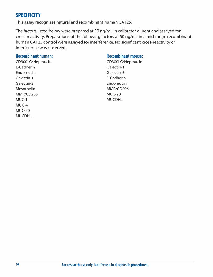
SPECIFICITYThis assay recognizes natural and recombinant human CA125.
The factors listed below were prepared at 50 ng/mL in calibrator diluent and assayed for cross-reactivity. Preparations of the following factors at 50 ng/mL in a mid-range recombinant human CA125 control were assayed for interference. No significant cross-reactivity or interference was observed.
Recombinant human:CD300LG/NepmucinE-CadherinEndomucinGalectin-1Galectin-3MesothelinMMR/CD206MUC-1MUC-4MUC-20MUCDHL
Recombinant mouse:CD300LG/NepmucinGalectin-1Galectin-3E-CadherinEndomucinMMR/CD206MUC-20MUCDHL

www.RnDSystems.com 11
REFERENCES1. Bast, R.C. et al. (1981) J. Clin. Invest. 68:1331.
2. Yin, B.W.T. and K.O. Lloyd (2001) J. Biol. Chem. 276:27371.
3. Yin, B.W.T. et al. (2002) Int. J. Cancer 98:737.
4. Sikaris, K.A. (2011) Heart Lung Circ. 20:634.
5. Wang, Y. et al. (2008) Differentiation 76:1081.
6. Blalock, T.D. et al. (2007) Invest. Ophthalmol. Vis. Sci. 48:4509.
7. Mantelli, F. and P. Argueso (2008) Curr. Opin. Allergy Clin. Immunol. 8:477.
8. Haridas, D. et al. (2011) PLoS ONE 6:e26839.
9. Higashi, M. et al. (2012) Pathobiology 79:101.
10. Hoshino, M. et al. (2010) J. Cancer Res. Clin. Oncol. 136:457.
11. Kaneko, O. et al. (2009) J. Biol. Chem. 284:3739.
12. Rump, A. et al. (2004) J. Biol. Chem. 279:9190.
13. Shimizu, A. et al. (2012) Cancer Sci. 103:739.
14. Gubbels, J.A.A. et al. (2006) Mol. Cancer 5:50.
15. Seelenmeyer, C. et al. (2003) J. Cell Sci. 116:1305.
16. Chen, S.H. et al. (2012) FASEB J. 26:1349.
17. Belisle, J.A. et al. (2010) Mol. Cancer 9:118.
18. Patankar, M.S. et al. (2005) Gynecol. Oncol. 99:704.
19. Gubbels, J.A.A. et al. (2010) Mol. Cancer 9:11.
20. Belisle, J.A. et al. (2007) Immunology 122:418.
21. Tyler, C. et al. (2012) Am. J. Reprod. Immunol. 68:28.
22. Reinartz, S. et al. (2012) Eur. J. Cancer 48:1558.
23. Lakshmanan, I. et al. (2012) Oncogene 31:805.
24. Topalak, O. et al. (2002) Gynecol. Oncol. 85:108.
25. Yang, Z. et al. (2012) Clin. Exp. Rheumatol. 30:93.
26. Peng, T. et al. (2012) Clin. Lab. 58:113.
27. Huang, F. et al. (2012) Med. Hypotheses 79:381.
28. D’Aloia, A. et al. (2003) J. Am. Coll. Cardiol. 41:1805.
29. Kouris, N.T. et al. (2006) Hellenic J. Cardiol. 47:269.
30. Kosar, F. et al. (2006) Eur. J. Heart Fail. 8:270.
31. Karaca, O. et al. (2012) Congest. Heart Fail. 18:144.
32. Prochazka, V. et al. (2012) Int. J. Hematol. 96:58.

For research use only. Not for use in diagnostic procedures.12
PLATE LAYOUTUse this plate layout to record standards and samples assayed.

www.RnDSystems.com 13
NOTES

For research use only. Not for use in diagnostic procedures.14
08.12 753010.3 4/18
©2018 R&D Systems®, Inc.
NOTES
All trademarks and registered trademarks are the property of their respective owners.