human biochemistry dp chemistry option b r. slider
TRANSCRIPT
Human BiochemistryDP Chemistry Option BR. Slider
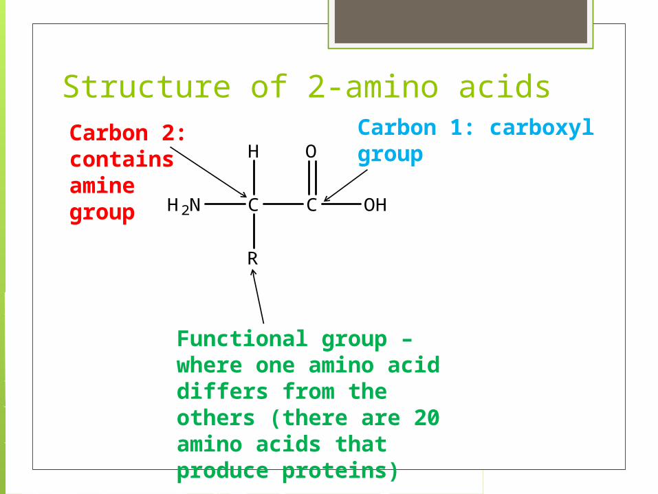
Structure of 2-amino acids
H2N C C
R
OH
OHCarbon 1: carboxyl group
Carbon 2: contains amine group
Functional group – where one amino acid differs from the others (there are 20 amino acids that produce proteins)

Properties of amino acids Formation of a zwitterion Isoelectric point Buffering action

Formation of a Zwitterion
Amino acids can form a zwitterion , which is a substance that contains both a positive and a negative charge and is also known as a dipolar ion. This occurs due to an internal acid-base reaction where a H+ ion is transferred from the acid portion of the molecule to the amino group.
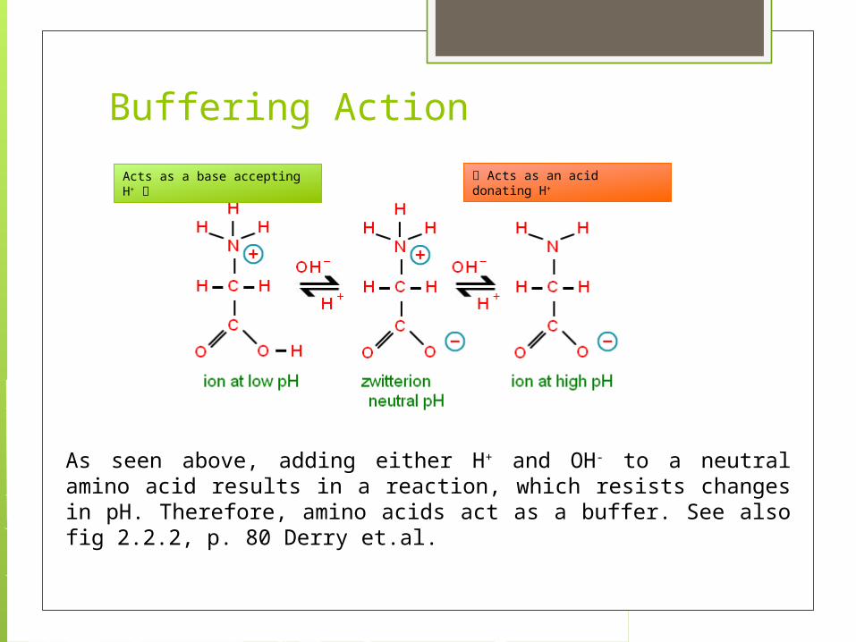
Buffering Action
As seen above, adding either H+ and OH- to a neutral amino acid results in a reaction, which resists changes in pH. Therefore, amino acids act as a buffer. See also fig 2.2.2, p. 80 Derry et.al.
Acts as an acid donating H+Acts as a base accepting H+

Primary Structures of Proteins
A polypeptide containing >50 amino acids is called a protein
The primary structure of a protein is the sequence of amino acids in the peptide chain covalently linked together.
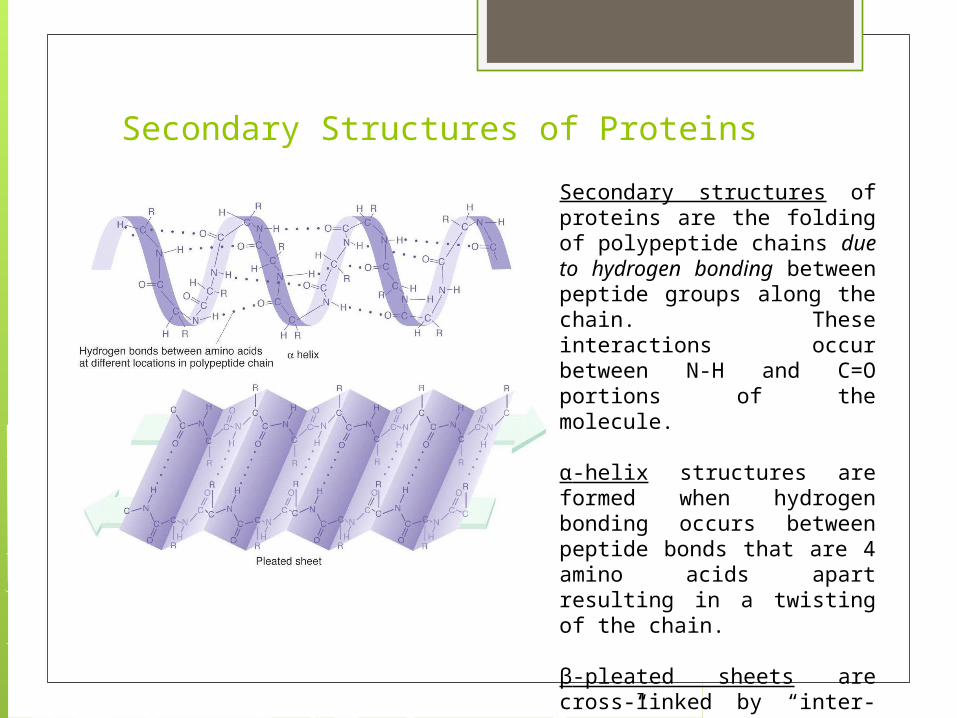
Secondary Structures of ProteinsSecondary structures of proteins are the folding of polypeptide chains due to hydrogen bonding between peptide groups along the chain. These interactions occur between N-H and C=O portions of the molecule.
α-helix structures are formed when hydrogen bonding occurs between peptide bonds that are 4 amino acids apart resulting in a twisting of the chain.
β-pleated sheets are cross-linked by “inter-chain” hydrogen bonds forming corrugated sheets.
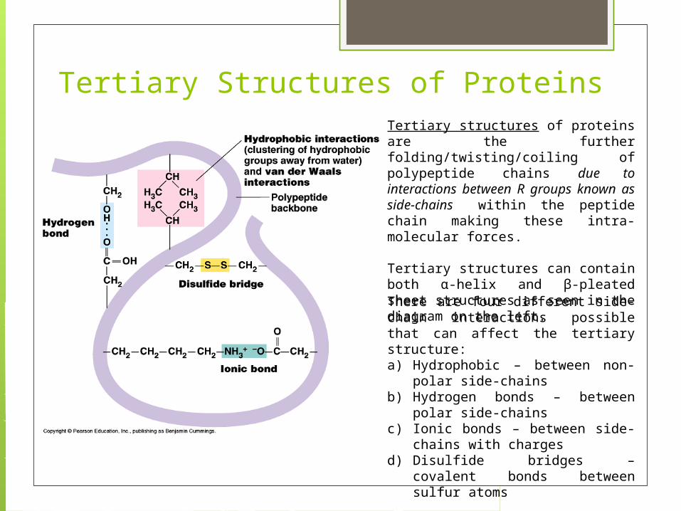
Tertiary Structures of ProteinsTertiary structures of proteins are the further folding/twisting/coiling of polypeptide chains due to interactions between R groups known as side-chains within the peptide chain making these intra-molecular forces.
Tertiary structures can contain both α-helix and β-pleated sheet structures as seen in the diagram on the left.
There are four different side-chain interactions possible that can affect the tertiary structure:a) Hydrophobic – between non-polar
side-chainsb) Hydrogen bonds – between polar
side-chainsc) Ionic bonds – between side-chains
with chargesd) Disulfide bridges – covalent bonds
between sulfur atoms
Quaternary Structures of Proteins
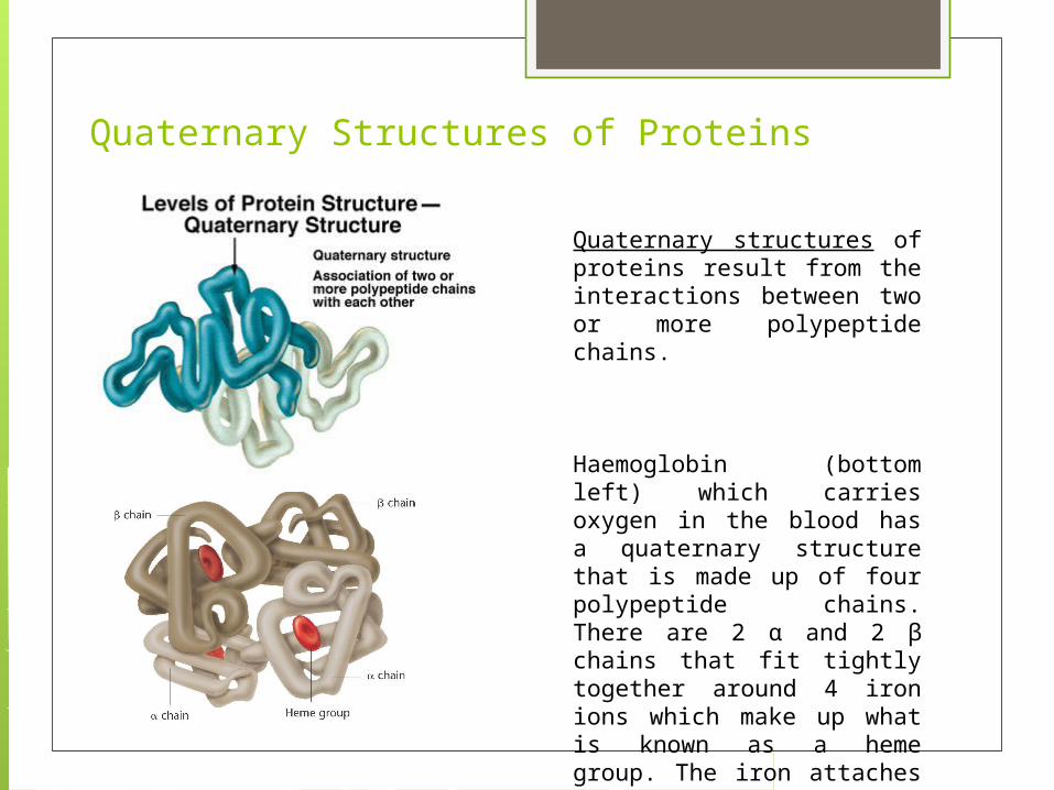
Quaternary structures of proteins result from the interactions between two or more polypeptide chains.
Haemoglobin (bottom left) which carries oxygen in the blood has a quaternary structure that is made up of four polypeptide chains. There are 2 α and 2 β chains that fit tightly together around 4 iron ions which make up what is known as a heme group. The iron attaches to the oxygen.
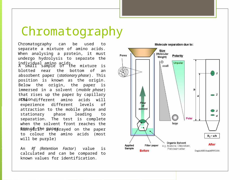
ChromatographyChromatography can be used to separate a mixture of amino acids. When analysing a protein, it must undergo hydrolysis to separate the individual amino acids.
A small sample of the mixture is blotted near the bottom of an absorbent paper (stationary phase). This position is known as the origin. Below the origin, the paper is immersed in a solvent (mobile phase) that rises up the paper by capillary action.
The different amino acids will experience different levels of attraction to the mobile phase and stationary phase leading to separation. The test is complete when the solvent front reaches the top of the paper.
An Rf (Retention Factor) value is calculated and can be compared to known values for identification.
Ninhydrin is sprayed on the paper to colour the amino acids (most will be purple)
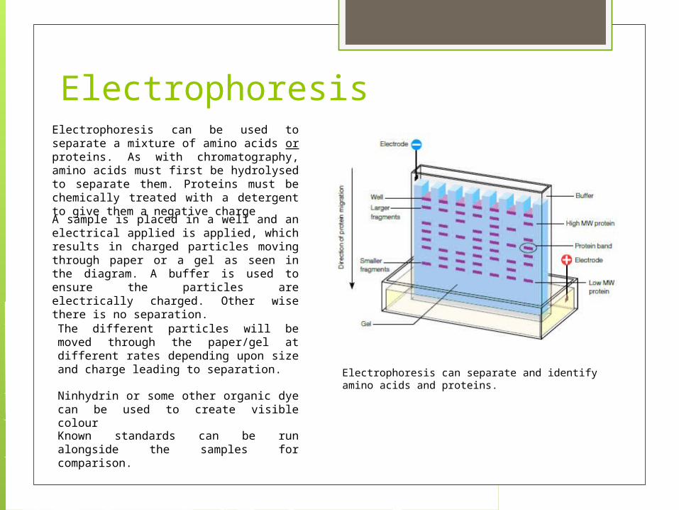
ElectrophoresisElectrophoresis can be used to separate a mixture of amino acids or proteins. As with chromatography, amino acids must first be hydrolysed to separate them. Proteins must be chemically treated with a detergent to give them a negative charge
A sample is placed in a well and an electrical applied is applied, which results in charged particles moving through paper or a gel as seen in the diagram. A buffer is used to ensure the particles are electrically charged. Other wise there is no separation.
The different particles will be moved through the paper/gel at different rates depending upon size and charge leading to separation.
Known standards can be run alongside the samples for comparison.
Ninhydrin or some other organic dye can be used to create visible colour
Electrophoresis can separate and identify amino acids and proteins.
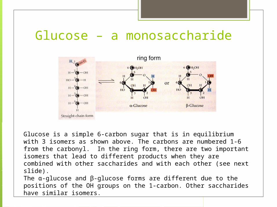
Glucose – a monosaccharide
Glucose is a simple 6-carbon sugar that is in equilibrium with 3 isomers as shown above. The carbons are numbered 1-6 from the carbonyl. In the ring form, there are two important isomers that lead to different products when they are combined with other saccharides and with each other (see next slide).The α-glucose and β-glucose forms are different due to the positions of the OH groups on the 1-carbon. Other saccharides have similar isomers.

Monosaccharides Disaccharides
Monosaccharides (single sugar unit) are the simplest carbohydrate and can be combined in a condensation reaction to form disaccharides releasing a water molecule. This results in a glycosidic link between monosaccharides that is designated by the carbons that are involved in the bonding (i.e. 1-4 or 1-2 above).

Monosaccharides Disaccharides
You try: Lactose is made from -glucose and -galactose. Draw this condensation reaction
β - glucose β - galactose
Monosaccharides Polysaccharides
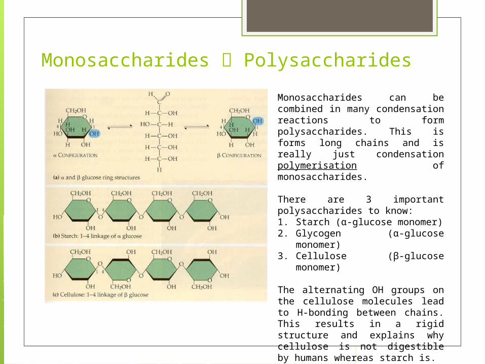
Monosaccharides can be combined in many condensation reactions to form polysaccharides. This is forms long chains and is really just condensation polymerisation of monosaccharides.
There are 3 important polysaccharides to know:1. Starch (α-glucose monomer)2. Glycogen (α-glucose
monomer)3. Cellulose (β-glucose monomer)
The alternating OH groups on the cellulose molecules lead to H-bonding between chains. This results in a rigid structure and explains why cellulose is not digestible by humans whereas starch is.