human adipose derived stroma/stem cells grow in serum-free medium
TRANSCRIPT
Research Article
Human adipose derived stroma/stem cells grow in serum-freemedium as floating spheres
C. Dromarda,b, P. Bourinc, M. Andréa,b, S. De Barrosa,b, L. Casteillaa,b, V. Planat-Benarda,b,⁎
aUniversité de Toulouse, UPS, UMR5241 Métabolisme, Plasticité et Mitochondrie, BP 84 225, F-31 432 Toulouse, FrancebCNRS, UMR5241 Métabolisme, Plasticité et Mitochondrie, BP 84 225, F-31 432 Toulouse, FrancecEtablissement Français du Sang, Pyrénées-Méditerranée, Toulouse, France
A R T I C L E I N F O R M A T I O N A B S T R A C T
Article Chronology:Received 25 June 2010Revised version received19 November 2010Accepted 3 January 2011Available online 19 January 2011
With the goal of obtaining clinically safe human adipose-derived stroma/stem cells (ASC) andeliminating the use of serum, we have developed a new culture system that allows the expansionof ASC as spheres in a definedmedium. These spheres can be passaged several times. They are notonly aggregated cells but rather originate from single cells as clonal spheres can be obtained afterseeding at very low density and reform clonal spheres after dissociation. These spheres can alsorevert to monolayer growth when plated in medium containing human plasma and evengenerate fibroblast-like colonies (CFU-f). Under several differentiation-specific media, spheres-derived ASC maintain their capacity to differentiate into osteoblasts, endothelial cells andadipocytes. These results indicate that human ASC can be maintained in a serum-free 3D culturesystem, which is of great interest for the expansion in bioreactors of autologous ASC and their usein clinical trials.
© 2011 Elsevier Inc. All rights reserved.
Keywords:Human adipose stem cellsSerum-freeNon-adherentSpheres
Introduction
Adipose tissuehas longbeen knownas themain energy sourceof theorganism. The discovery of its ability to produce and secreteadipokines such as leptin and adiponectin has revealed its endocrinefunction and its pivotal role in metabolism regulation andphysiological homeostasis [1,2]. More recently, certain cells havebeen isolated from the stromal vascular fraction of adipose tissuethat are capable of multilineage differentiation even at the clonallevel [3–10]. These cells are termed ASC for adipose derived stroma/stem cells. Although different, they share numerous features withmesenchymal stem cells isolated from bone marrow (MSC) andumbilical blood cord [11–14] and represent an alternative source of
adult stem cells. Indeed, access to subcutaneous deposit is a routinesurgical procedure that can be performed under local anaesthesiawithminimumdiscomfort for thepatient;moreover, ASC isolation isa simple enzyme-based protocol, making this tissue a moreattractive source of stem cells. ASC isolated from human adiposetissue have the potential to differentiate into bone, cartilage, fat andendothelial cells when cultivated under lineage specific condi-tions [4,9,15,16]. Engineering of these mesenchymal tissues is ofmajor interest in human diseases such as inherited, traumatic ordegenerative tissue defects or after tumour surgery [17]. The criticalpoint related to clinical use is that in vitro culture of ASC is stilldependent of fetal calf serum or human plasma [8,18]. Plasma andserum contain undefined factors, which vary in composition from
E X P E R I M E N T A L C E L L R E S E A R C H 3 1 7 ( 2 0 1 1 ) 7 7 0 – 7 8 0
⁎ Corresponding author at: UPS/CNRS - UMR5241 Métabolisme, Plasticité et Mitochondrie, BP 84 225 – 31 432 Toulouse Cedex, France. Fax: +33562170905.
E-mail address: [email protected] (V. Planat-Benard).
0014-4827/$ – see front matter © 2011 Elsevier Inc. All rights reserved.doi:10.1016/j.yexcr.2011.01.001
ava i l ab l e a t www.sc i enced i r ec t . com
www.e l sev i e r . com/ loca te /yexc r

individual to individual andmay activate or inhibit cell proliferationand differentiation. Moreover, their stock depends on donors andeach batch must be tested for innocuousness (infectious pathogenssuch as prions). The above combined with the fact that differentialgene expression was revealed between ASC cultures in human orfetal bovine serum [18], highlight the need for a substitutive definedcellmedium formulation and independent of interest in a serum freemedium, to understand the specific effect of any compound.
Multiple approaches employ sphere cluster technology tostudy stem cells [19]. In contrast to conventional monolayer cellculture, in which cells grow only in two dimensions on a flatsurface of a plastic dish, suspension cultures allow cell growth inall three dimensions. Stem cells have been grown as spheres innumerous studies using different protocols (non-adherent plates,methylcellulose semi-solid media, hanging drops and round-bottom 96-well plates) [19,20]. The most studied of these modelsare embryoid bodies, defined as spherical clusters of bothpluripotent and committed stem cells that can organize in adevelopmental-specific manner and give rise to mature cells fromany differentiation lineage. The primary goal of growing cells asspheres has been to reveal the basic principles of a normal tissueorganization that gives cells the opportunity to arrange in a threedimensional context similar to tissues [19,21]. This spheretechnology is now largely used in cancer research to isolate cancerinitiating cells, as tumour spheres are very similar to solid tumoursin situ [22]. They are also used as molecular assay systems intoxicology and pharmacology [23]. Additionally, embryonic stemcells can be driven to generate insulin-producing cell clusters, thatundergo rapid vascularization when injected into diabetic miceand display an organization and functionality similar to pancreaticislets [24]. This has led us to investigate a 3D culture system thatenables maintenance of ASC in serum-free conditions.
Materials and methods
Adipose tissue cell isolation
Subcutaneous adipose tissue was obtained from patients under-going elective abdominal dermolipectomy. No objection certifi-cates were obtained according to bioethics law no. 2004-800 ofAugust 6, 2004. Cells were isolated as previously described [25].Briefly, adipose tissue (AT) was digested in DMEM-F12 medium(Invitrogen, Carlsbad, USA) supplemented with 2 mg/ml collage-nase A (Roche Diagnostic, Indianapolis, IN) and bovine serumalbumin (BSA) (2%) for 45 min at 37 °C under agitation. Thesuspension was filtered through 100 μm and 25 μm nylonmembrane and centrifuged at 600 g for 10 min to separate floatingmature adipocytes from the stromal-vascular fraction (SVF). SVFwas incubated in erythrocyte lysis buffer (ammonium chloridesolution, StemCell Technologies, Vancouver, Canada) for 5 min at4 °C and washed in PBS (phosphate-buffered saline, Sigma, StQuentin Fallavier, France). SVF cells were counted and eithercultured in vitro or used for flow cytometry analysis.
Cell culture
For ASC monolayer culture, SVF cells were seeded at 4000 cell/cm2
in flasks treated for cell culture (TPP, Dominique Dutscher,Brumath, France) in ASC expansion medium which consisted of
alpha-MEM (Invitrogen) supplemented with 2% human plasmaenriched with human platelet growth factors (PGFEP), heparinChoay (1 U/ml, Sigma), amphotericin (0.25 μg/ml), streptomycin(100 μg/ml) and penicillin (100 U/ml) (Invitrogen). Cells wereincubated at 37 °C under 5% CO2 and the medium was changedtwice a week. For sphere culture, SVF cells were seeded at 60,000cell/cm2 in ultra low adherent flasks (Corning, Avon, France) indefined culture medium which consisted of GMEM supplementedwith L-glutamine (2 mM), non essential amino acids (1×), B27(1×) (Invitrogen), glucose (0.6%, Sigma), human bFGF (10 ng/ml),human EGF (20 ng/ml), human thrombin (1 U/ml) (PeproTech,Rocky Hill, NJ) and ciprofloxacin (2 μg/ml, Bayer, France). Cellswere incubated at 37 °C under 5% CO2 and half of the mediumwaschanged once a week.
To assess the presence of proliferating cells in defined culturemedium, BrdU (10 μM)was added for 3 days on the third day afterseeding. To test if sphere containing cells can revert to monolayergrowth, spheres were dissociated with trypsin-EDTA (0.05%,Invitrogen) and plated in flasks treated for cell culture (TPP) inASC expansion medium.
Flow cytometry
Dissociated spheres were incubated with PBS (phosphate-bufferedsaline) supplemented with 0.5% new calf serum and FcR Blockreagent (StemCell Technologies, Vancouver). Sextuplet stainingswere performed by incubating cells for 30 min at 4 °C with thefollowing conjugated primary antibodies or appropriate IgG isotypecontrols: CD45-APC-Cy7, CD90-FITC, CD73-PE, CD34-PerCP, CD44-FITC, CD29-APC, CMH1-APC, CD117-PE-Cy7, CD15-FITC, CD56-PE,CXCR4-PE, Lin-FITC, CD38-PE-Cy7, CMH2-FITC, CD3-PE-Cy7, CD11b-APC, CD19-PerCP, CD14-PE-Cy7 (BD Biosciences, Le Pont de Claix,France), CD105-APC, CD146-FITC, CD31-APC (eBiosciences, Paris,France),VEGFR2-PE (R&D Systems, Lille, France), and CD133-APC(Miltenyi, Paris, France). Cells were analyzed on a FACS Canto II (BDBiosciences). Data acquisition and analysis were performed withFACS Diva software (BD Biosciences). Only viable cells that excludedthe LIVE/DEAD dye (Molecular Probes, Interchim, Montluçon,France) were considered.
Cell differentiation
Spheres cultured for 7 days were dissociated with trypsin/EDTA(0.05%, Invitrogen) and seeded on gelatin-coated plates (0.1% inPBS, Sigma) for differentiation. The day of plating was consideredas day 0. Differentiation was assessed after 24 h (noted as day 1)and after 10 days or 21 days depending on differentiation lineage.
Adipogenic differentiationOn day 0, cells from dissociated spheres were plated at 65,800cell/cm2 in 12-well tissue-culture plates (Falcon, DominiqueDutscher, Brumath, France) and cultured for 3 days in adipogenicdifferentiation medium which consisted in ASC expansion medi-um supplemented with dexamethasone (1 μM), IBMX (450 μM),and indomethacin (60 μM) (Sigma). Subsequently, IBMX wasremoved from the medium and differentiation was extendedduring 19 days. The medium was changed every 3 days. The extentof differentiation was noted by observation of multilocularrefringent droplets in the induced cells and by staining of neutrallipids by Oil red-O as previously described [26]. Cellular triglyceride
771E X P E R I M E N T A L C E L L R E S E A R C H 3 1 7 ( 2 0 1 1 ) 7 7 0 – 7 8 0

(TG) content was measured with a commercial test (TriglyceridesEnzymatique PAP 150, Biomerieux). The protein content wasdetermined using the DC Protein Assay Kit (BioRad, Marne laCoquette, France). Differentiation quantification was evaluated bycalculating the ratio of TG content per μg of protein.
Osteogenic differentiationOn day 0, cells from dissociated spheres were plated at 15,200cell/cm2 in 12-well tissue-culture plates (Falcon) and cultured for21 days in osteogenic differentiation medium which consisted inASC expansion medium supplemented with dexamethasone(0.1 μM), ascorbic acid (250 μM), and NaH2PO4 (3 mM). Themedium was changed every 4 days. Mineralization was revealedby staining calcium-rich deposits with Alizarin red [6]. Alizarin redquantity (μg) was assessed at day 1 and day 21 by addition of 10%acetic acid to the stained culture dishes and measurement of theoptical density at 405 nm with a spectrophotometer.
Angiogenic differentiationOn day 0, cells from dissociated spheres were plated at 100,000cell/cm2 in 48-well tissue-culture plates (Falcon) and cultured forten days in angiogenic differentiation medium which consisted inASC expansionmedium supplementedwith VEGF (10 ng/ml, Sigma).The medium was replaced every 3 days. Angiogenic differentiationwas assessed by CD31 immunostaining. CD31-positive extensionlengths were measured using the Elements AR 3.0 image analyzersoftware (Nikon, Champigny sur Marne, France).
Colony forming unit-fibroblast assay
To evaluate the frequency of mesenchymal-like progenitorsamong cells growing in defined medium, spheres were dissociatedand cells seeded in 25 cm2 flasks (TPP) at a final concentration of8 cells/cm2 in ASC expansion medium as previously described. Themedium was renewed every 2 or 3 days. After 12 days, cells werewashed with PBS and fixed with methanol for 15 min. The colonyforming unit-fibroblasts (CFU-f) were stained with Giemsa (6%)for 30 min and scored under an optical microscope. Colonies wereconsidered as clusters of more than 50 cells.
Immunostaining
Adherent cells were fixed for 15 min with 4% paraformaldehyde. Toavoid non specific binding, cellswere first incubated for 1 h in PBS andBSA (2%), then for 2 h with the mouse anti human CD31 antibody(1/10, Dako, Trappes, France) ormouse IgG1 control isotype (Dako).After washing, the secondary antibody goat anti-mouse conjugatedto Alexa 488 was added for 1 h (1/200, Invitrogen). Nuclei werestained with DAPI (1/10,000). Cell staining was observed with afluorescence microscope (DMRB Leica, Gennevilliers, France) andanalyzed with the image analyzer software.
For BrdU detection, whole spheres and freshly dissociatedspheres were plated by centrifugation (10 min, 1000 rpm) onpoly-L-ornithine-coated coverslips before fixation. They weretreated with 2 N HCl for 30 min, washed with 0.1 M borate buffer,and incubated in TBS (Tris buffer saline) triton (0.1%) and goatserum (5%) for 1 h and stained with the mouse anti human BrdUantibody conjugated to FITC (1/20, e-biosciences) or mouse IgG1control isotype; nuclei were counterstained with Hoechst 33242(5 μg/ml, Sigma).
For paraffin sections, spheres were successively fixed withparaformaldehyde (4%, 1 h), ethanol (70% 15 min–95% 15 min–100% 3× 1 h), toluene substitute (Microclearing, 1 h–30 min) andembedded in paraffin. Sections of 4 μm thickness were depar-affined and stained with hematoxylin/eosin solution.
Real time PCR
Cell total RNA was isolated using RNAeasy minikit (Qiagen,Courtaboeuf, France) according to themanufacturer's recommenda-tions. RNA (1 μg) was reverse-transcribed using the High-CapacitycDNA Reverse Transcription kit. All amplification reactions wereperformed in duplicate from 25 ng cDNA in 50 μl Power SYBR®Green PCR Master Mix containing 0.2 μM of each specific primers(for control, Platinum® Taq DNA polymerase was omitted):
hPUM1 Fp: GCCATGTTGTCGGAGTGAAA/Rp: ACACATGCAACGCT-CATTCC,hOCT4 Fp: CCCCTGGTGCCGTGAAG/Rp: GCAAATTGCTC-GAGTTCTTTCTG,hNANOG Fp: AGAACTCTCCAACATCCTGAACCT/Rp: ATTCTTCGGC-CAGTTGTTTTTC.hPPARγ Fp: GATACACTGTCTGCAAACATATCAC/RP: CCACGGAGCT-GATCCCAAhLPL Fp: GGTCGAAGCATTGGAATCCAG/RP: TAGGGCATCTGA-GAACGAGTChRUNX 2 Fp: ATTCCTGTAGATCCGAGCACC/Rp: GCTCACGTCGCT-CATTTTGChOsteoprotegerin Fp: AGCACCCTGTAGAAAACACAC/Rp: ACAC-TAAGCCAGTTAGGCGTAA.
Amplification and detection were performedwith the ABI PRISM7500 Sequence Detection System as follows: initial denaturation at94 °C for 2 min, followed by 40 cycles of denaturation at 94 °C for30 s, annealing at 60 °C for 30 s and extension at 72 °C for 1 min. Toevaluate the specificity of the amplifications a dissociation curvewas analyzed (95 °C (15 s), 60 °C (20 s), and 95 °C (15 s)). All PCRamplificationswere analyzed induplicates and includedpositive andnegative (H2O) control samples. Relative quantification of specificgenes mRNA levels was determined using the 2−ΔΔCT method.Results were analyzed with the GeneAmp 7500 software, and genesmRNA expression level was first normalized by PUM taken asreference gene and then to a calibrator consisting of human ASC
Table 1 – Serum-free culture conditions tested for sphereformation. Serum free supplements B27 and N2 as well asglucose and the mitotic cell toxin ARA-C were tested inseveral combinations with the following basic medium:GMEM medium supplemented with L-glutamine, nonessential amino acids, human bFGF, human EGF and humanthrombin. All these media formulations were tested on cellsplated in culture dishes treated for adherent cell culture orin ultra low adherent supports.
A B C D I J K L
B27 (1×) + + + + − − − −N2 (1×) − − − − + + + +Glucose (0.6%) − + − + − + − +ARAC (5–100 μM) − − + + − − + +
772 E X P E R I M E N T A L C E L L R E S E A R C H 3 1 7 ( 2 0 1 1 ) 7 7 0 – 7 8 0

control. All PCR products and analyzerwere purchased fromAppliedBiosystems, Villebon, France.
Data analysis
Results were expressed as means±SEM. Data from at least threeindependent preparations of ASC grown as spheroids or as amonolayer were statistically processed with the Prism 4 software(GraphPad, San Diego, CA). Significance was defined as *p≤0.05,**p≤0.01 and ***p≤0.001.
Results
Serum-free culture conditions allowing for sphere formation
We have evaluated the capacity of several enzymatic digestionconditions, medium formulations and culture dishes to supportthe growth of human ASC as spheres without the use of serum.Two collagenases were tested for digestion of adipose tissue:collagenase A type II or VIII, during 45, 60 or 90 min. These
Fig. 1 – Characterization of spheres. (A) Morphology of spheres 7 days after seeding at 190 cells/μl (scale bars 200 μm, 100 μm and20 μm). (B) Spheres display heterogeneous sizes after 7 days of culture. (C) Hematoxylin and eosin staining on 4 μm-thick paraffinsections of spheres (scale bar 20 μm). (D) BrdU immunodetection (green) on spheres grown for 7 days in presence of BrdU during3 days. Nuclei are stained with Hoechst (blue) (scale bar 20 μm). (E) Clonal spheres obtained after 20 days of culture from primarysphere cells dissociated and seeded at 0.5 cell/μl (scale bars 200 μm and 20 μm).
773E X P E R I M E N T A L C E L L R E S E A R C H 3 1 7 ( 2 0 1 1 ) 7 7 0 – 7 8 0

conditions were associated with several products combinationslisted in Table 1, in culture dishes treated for adherent cell culture orin ultra low adherent supports and with the following mediumformulation: GMEM medium supplemented with L-glutamine(2 mM), non essential amino acids (1×), human bFGF (10 ng/ml),
human EGF (20 ng/ml), human thrombin (1 U/ml) and ciproflox-acine (2 μg/ml).
Only collagenase A type II allowed the formation of regularspheres and 45 min of digestion appeared to be sufficient. Usingthis collagenase, clonal spheres were only obtained in conditions
Fig. 2 – Flow cytometry analysis of cells composing the spheres. The vast majority of cells composing the spheres are CD34+ anddisplay a mesenchymal phenotype CD105+, CD90+, CD73+, and CD45−. They are CD29+, CD44+, and CMH1+ but CD133−, CD117−,CD15−, CD31−, VEGFR2−, CD146−, CD56−, CXCR4−, Lin−, CD38−, CMH2−, CD3−, CD11b−, CD19− and CD14−.
774 E X P E R I M E N T A L C E L L R E S E A R C H 3 1 7 ( 2 0 1 1 ) 7 7 0 – 7 8 0
A, B and J and among them, sphere structure appeared to be mostregular under condition B. In flasks treated for cell culture, somecells adhered to the bottom of the flask while spheres formed inthe supernatant. In ultra low adherent flasks, no cell adhered tothe bottom and only spheres were obtained in the supernatant.Thus, in order to better evaluate the potential of the cells thatcomposed the spheres, subsequent studies were performed usingcollagenase A type II for 45 min for tissue digestion, and culture inmedium formulation B in ultra low adherent flasks. These optimumconditions allowing sphere formation in defined medium aregathered in the Materials and methods section.
Characterization of spheres
Spheres were roundwith defined borders and varied in size 7 daysafter their first emergence (Figs. 1A and B). As shown in Fig. 1B,most spheres (80%) were small in diameter (<100 μm), 18% weremedium (100–200 μm) and the remaining spheres were large(>200 μm).
To determine their structure, spheres were sectioned andstained with hematoxylin and eosin. The cells that composed thespheres appeared homogeneous in size andmorphology. No cavitywas observed (Fig. 1C). Secretion of extracellular matrix proteinswas evaluated on spheres sections by immunocytochemistry ordye staining. Neither collagen, fibronectin, hyaluronic acid norelastic fibers were detected (data not shown).
Evidence of a proliferation rate was observed as few cellswere KI67 positive (data not shown) and 3 days after addition ofBrdU in the medium, around 15% of the cells that composed thespheres appeared to have incorporated BrdU (Fig. 1D). A very
limited amount of dead cells was assessed by TUNEL assay (datanot shown). Spheres could be passaged at least three times, asenzymatically dissociated spheres could reform new spheres atclonal densities as low as 0.5 cells per μl (Fig. 1E). These data suggestthat a sphere can be formed at clonal level through proliferation.However, a very small amount of cells can be considered as sphereinitiating cells, as upon dissociation, only 0.8% of the cells thatcompose a sphere could form secondary spheres.
FACS analysis revealed that, similar to ASC cultured directlyfrom SVF in ASC expansion medium, most of the cells thatcomposed the spheres expressed simultaneously CD34 andclassical mesenchymal cell-associated markers such as CD105,CD90, and CD73, and were negative for CD45 (Fig. 2A). Evaluationof the expression of other markers revealed that the majority ofcells contained in the spheres expressed CD29, CD44, and CMH1but not CD133, CD117, CD15, CD31, VEGFR2, CD146, CD56, CXCR4,Lin, CD38, CMH2, CD3, CD11b, CD19 nor CD14 (Fig. 2B). No dif-ference in marker expression profiles was observed betweenprimary and secondary spheres (spheres formed initially andpassaged once respectively) except for CD34 positive cells whichrepresent 80% (+/−10) and 43% (+/−11) of the cells in primaryand secondary spheres respectively.
After enzymatic dissociation and culture in ASC expansionmedium containing fetal calf serum, spheres could revert tomonolayers in flasks treated for adherent cell culture. They re-adhered and morphologically resembled primary cultured ASCobtained from SVF plating and continued to proliferate.
Taken together, these results suggest that ASC can grow inserum-free medium in suspension as clonal spheres, maintaintheir mesenchymal phenotype and revert to monolayer growth.
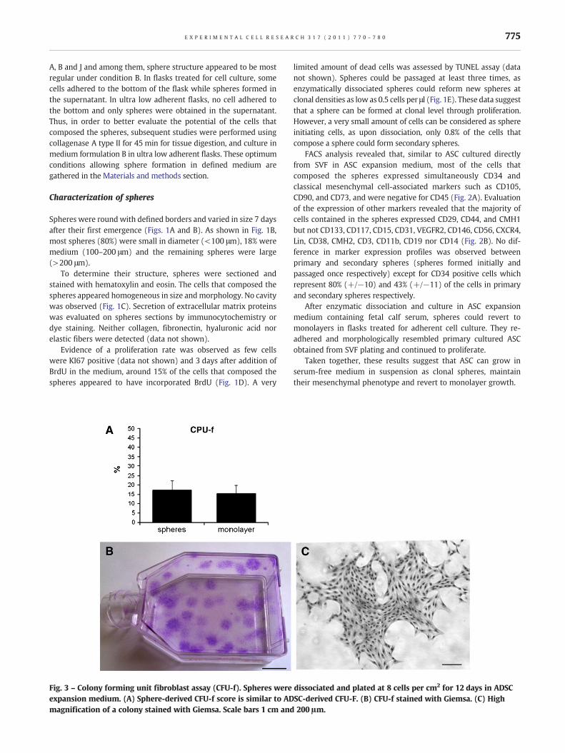
Fig. 3 – Colony forming unit fibroblast assay (CFU-f). Spheres were dissociated and plated at 8 cells per cm2 for 12 days in ADSCexpansion medium. (A) Sphere-derived CFU-f score is similar to ADSC-derived CFU-F. (B) CFU-f stained with Giemsa. (C) Highmagnification of a colony stained with Giemsa. Scale bars 1 cm and 200 μm.
775E X P E R I M E N T A L C E L L R E S E A R C H 3 1 7 ( 2 0 1 1 ) 7 7 0 – 7 8 0

ASC maintain their multipotentiality when cultured asspheres in defined medium
In order to determine whether cells that composed the spheresexpressed pluripotency-associated markers, the expression ofOct4 and Nanog was evaluated by RT-PCR. Both markers wereexpressed but no significant difference was observed compared to
the expression of these markers in ASC cultured directly from SVFin ASC expansion medium (data not shown).
The progenitor content of spheres was evaluated by the CFU-ftest. This revealed that the proportion of true progenitorscontained in spheres (17±5%) was similar to that in ASC cellpopulation cultured under conventional conditions (15±4%)(Fig. 3).
Fig. 4 – Adipogenic differentiation. SVF-derived cells were cultured for 7 days as spheres in defined medium before adipogenicdifferentiation. After 21 days in differentiation medium, cells display numerous lipidic droplets (B) stained by Oil red-O (D) whichwere rare at day 1 (A and C) (scale bars 50 μmand 20 μM). (E) PPARγ gene expression is upregulated after 21 days of differentiation.(F) LPL gene expression is upregulated after 21 days of differentiation. (E) Triglyceride content is significantly increased on 21 daysrelative to day 1 of differentiation. *p≤0.05.
776 E X P E R I M E N T A L C E L L R E S E A R C H 3 1 7 ( 2 0 1 1 ) 7 7 0 – 7 8 0

To confirm that the differentiation capacity of sphere-derivedASC was maintained 7 days, spheres were dissociated andincubated in lineage specific differentiation media. Early differen-tiation (noted day 1) was assessed 24 h after plating, whereas latedifferentiation was assessed 10 days after plating for angiogenicdifferentiation or 21 days after plating for adipogenic andosteogenic differentiation. Following 21 days in adipogenic differ-entiation medium, sphere-derived ASC displayed intracellularaccumulation of lipid droplets detected by Oil red-O staining(Figs. 4A–D). The peroxisome proliferator-activated receptorgamma (PPARγ) and the lipoprotein lipase (LPL) genes, whichplay a key role in lipid metabolism, were upregulated (Figs. 4Eand F) and the triglyceride content was 3 fold higher after 21 daysof adipogenic differentiation relative to day 1 (Fig. 4G). It isnoteworthy that lipid accumulation is an early and spontaneousevent as sphere-derived ASC displayed some Oil red-O positivedroplets as soon as day 1 in differentiating medium. Sphere-derivedASC cultured for 10 days under angiogenic conditions showed atypical endothelial-like organization in culture as they expressedCD31 (PECAM-1) and formed highly branched networks (Figs. 5Aand B). Process length of cells differentiated for 10 days were 625+/−22 μmwhereas theywere only 109+/−6 μmatday 1 (Fig. 5C).When induced towards osteogenic lineage, dissociated sphereswerecapable of forming mineralized nodules visualized by Von Kossa(Figs. 6A and B) and Alizarin red staining (Figs. 6C and D). The Runt-related transcription factor 2 (Runx 2) and osteoprotegerin genes,which play a central role in bone metabolism, were upregulated(Figs. 6E and F) and Alizarin red accumulation was highly increased
after 21 days of differentiation relative to day 1 (Fig. 6G). Thus, ourresults show that sphere-derived ASC display in vitro multipotency.
Discussion
Our data provide the first evidence thatmesenchymal cells derivedfrom human adipose tissue can be grown in serum-free medium insuspension as floating spheres through several passages andmaintain their multilineage differentiation capacities.
Similarly, it has been reported that mesenchymal cells derivedfrom other tissues like umbilical cord [27] or skeletal muscles [28]can grow in serum free medium as spheres and maintainmultipotency. Studies that aimed to use serum-free media toculture mesenchymal cells from adipose tissue were all restrictedto monolayer cultures [29–31] and the ones that derivedaggregates from ASC all required a first step of monolayer culturein 10% fetal bovine serum containingmedia [32–34]. In the presentwork, we plated cells from freshly dissociated tissue directly innon-adherent conditions without any preliminary monolayerculture and only in serum-free media. Our culture process inducedthe loss of the vast majority of the harvested primary SVF cells,while some of the growth factor-responsive cells survived,divided, and generated spheres. The amount of cells initiatingthe spheres was reflected by the number of secondary spheresformed after the dissociation of primary spheres and reseeding atclonal density. It represented 0.8%, revealing that only rare cellsowned the property to develop as spheres. In comparison, 17% of
Fig. 5 – Angiogenic differentiation. SVF-derived cells were cultured for 7 days as spheres in defined medium before angiogenicdifferentiation. (A–B) After 10 days of differentiation, cells expressed CD31 (green) and formed highly branched networks. Nucleiwere stained with DAPI (blue). Scale bars 50 μm. (C) Process length (μm). ***p≤0.0001.
777E X P E R I M E N T A L C E L L R E S E A R C H 3 1 7 ( 2 0 1 1 ) 7 7 0 – 7 8 0

cells obtained from dissociated spheres could form CFU-f,suggesting that cells forming CFU-f are not necessarily able toinitiate a new sphere or at least have distinct requirements inculture. Therefore, cells initiating spheres could be considered as anon-adherent stem cell population. Indeed, they behold capacitiesto proliferate in non-adherent condition, to renew medium sizespheres (100 μm diameter) at clonal level, to maintain in vitro
through several passages (i.e. to be dissociated and re-plated toform secondary then tertiary spheres) and to differentiate into thethree primarymesenchymal phenotypes— adipocytes, osteoblastsand endothelial cells, demonstrating the key in vitro properties of aprogenitor/stem cell — self-renewal at clonal level in culture andmultilineage differentiation. In the same way, Westerman et al.who derived myospheres from skeletal muscles in serum-free
Fig. 6 – Osteogenic differentiation. SVF-derived cells were cultured for 7 days as spheres in defined medium before osteogenicdifferentiation. After 21 days, differentiated cells formed calcic deposits. (A–B) Von Kossa staining. (C–D) Alizarin red staining (scalebars 50 μm). (E) Runx2 gene expression is upregulated after 21 days of differentiation. (F) Osteoprotegerin gene expression isupregulated after 21 days of differentiation. (G) Quantification of Alizarin red staining (μg/cm2). *p≤0.05 and ***p≤0.0001.
778 E X P E R I M E N T A L C E L L R E S E A R C H 3 1 7 ( 2 0 1 1 ) 7 7 0 – 7 8 0

media, demonstrated the pre-myogenic nature of the myosphereinitiating cells [28]. However, they were not able to generateclonal myospheres. To demonstrate the clonal nature of spheres,individual cells can be plated in single well of 96-well plates.However, their propagation under these conditions is difficultbecause it prevents paracrine communications between the cells.For these reasons we have chosen to derive clonal spheres throughplating at very low cell density (0.5 cell/μl). Indeed, Coles-Takabeet al. demonstrated the clonal nature of spheres through mixingexperiments, in which cells were labeled with two differentmarkers (yellow and red fluorescent proteins) and then mixedtogether [35]. These experiments showed that from platingdensity of 1 cell/μl, almost all the spheres generated after mixingcontained only one of the two fluorescent labels. Moreover, we leftthe cells undisturbed during sphere formation to prevent any fluidmotion that could increase the probability of aggregation and thuschimeric sphere formation.
In addition, the sphere culture of ASC may enhance theirbiological activity as the administration of human ASC increasedthe rate of diabetic wound healing when ASC were derived fromaggregates, but not when delivered ASC derived from monolayerculture [34]. Indeed, in this study, ASC formulated as three-dimensional aggregates by the hanging droplet method producedsignificantly more extracellular matrix proteins and secretedsoluble factors compared to monolayer culture. In our sphereculture process that differs by the absence of serum we could notfind the presence of the extracellular matrix proteins collagen,fibronectin, hyaluronic acid and elastin (data not shown).
This new non-adherent and serum-free method presented hereis a first step towards developing improved protocols for stem cellculture. First, it appears necessary to extend the analyze of geneexpression central for cell cycle progression and cell differentia-tion in sphere-derived ASC, as differential gene expression wasestablished between human serum and fetal bovine serumcultured ASC [18]. The next steps will involve the optimization ofmedium formulation for higher expansion rate, followed by thedevelopment of serum free differentiation media as described forbone-marrow mesenchymal stem cells [36]. The differentiationpotential of sphere-derived ASC needs to be further analyzed as wehave only searched for the differentiation potential of themesenchymal lineage. For example, ASC derived from rhesusadipose tissue cultured asmonolayer in proliferationmediumwith10% fetal calf serum could be committed to neuronal differenti-ation after a step of sphere culture [37]. Finally, there is a growinginterest in designing bioreactor systems, that provide safe andcontrolled high-density cell culture in production.
In this context, our results represent an important step in ourability to isolate and maintain ASC in culture and extend thepossibilities of cell-based therapies.
Acknowledgments
This work was supported by the Etablissement Français du Sang –
Pyrénées Méditerranée. S. De Barros was financially supported bythe Fondation pour la Recherche Medicale (DCV20070409252,programme Vieillissement Cardiovasculaire Normal et Pathologi-que). We thank Pascale Guillou for technical assistance and ArielleEstival for paraffin sections and hematoxylin–eosin staining of thespheres that were performed at the Platform of Experimental
Histopathology of the IFR-BMT/150 (Toulouse - Midi-Pyrénéesgénopole), Toulouse, France.
R E F E R E N C E S
[1] P. Trayhurn, J.H. Beattie, Physiological role of adipose tissue:white adipose tissue as an endocrine and secretory organ, Proc.Nutr. Soc. 60 (2001) 329–339.
[2] M.H. Fonseca-Alaniz, J. Takada, M.I. Alonso-Vale, F.B. Lima,Adipose tissue as an endocrine organ: from theory to practice,J. Pediatr. (Rio J) 83 (2007) S192–S203.
[3] J. Gimble, F. Guilak, Adipose-derived adult stem cells: isolation,characterization, and differentiation potential, Cytotherapy 5(2003) 362–369.
[4] V. Planat-Benard, J.S. Silvestre, B. Cousin, M. Andre, M. Nibbelink,R. Tamarat, M. Clergue, C. Manneville, C. Saillan-Barreau, M.Duriez, A. Tedgui, B. Levy, L. Penicaud, L. Casteilla, Plasticity ofhuman adipose lineage cells toward endothelial cells:physiological and therapeutic perspectives, Circulation 109(2004) 656–663.
[5] G.R. Erickson, J.M. Gimble, D.M. Franklin, H.E. Rice, H. Awad, F.Guilak, Chondrogenic potential of adipose tissue-derived stromalcells in vitro and in vivo, Biochem. Biophys. Res. Commun. 290(2002) 763–769.
[6] Y.D. Halvorsen, D. Franklin, A.L. Bond, D.C. Hitt, C. Auchter, A.L.Boskey, E.P. Paschalis, W.O. Wilkison, J.M. Gimble, Extracellularmatrix mineralization and osteoblast gene expression by humanadipose tissue-derived stromal cells, Tissue Eng. 7 (2001)729–741.
[7] A. Miranville, C. Heeschen, C. Sengenes, C.A. Curat, R. Busse, A.Bouloumie, Improvement of postnatal neovascularization byhuman adipose tissue-derived stem cells, Circulation 110 (2004)349–355.
[8] A. Schaffler, C. Buchler, Concise review: adipose tissue-derivedstromal cells—basic and clinical implications for novel cell-basedtherapies, Stem Cells 25 (2007) 818–827.
[9] P.A. Zuk, M. Zhu, H. Mizuno, J. Huang, J.W. Futrell, A.J. Katz, P.Benhaim, H.P. Lorenz, M.H. Hedrick, Multilineage cells fromhuman adipose tissue: implications for cell-based therapies,Tissue Eng. 7 (2001) 211–228.
[10] F. Guilak, K.E. Lott, H.A. Awad, Q. Cao, K.C. Hicok, B. Fermor, J.M.Gimble, Clonal analysis of the differentiation potential of humanadipose-derived adult stem cells, J. Cell. Physiol. 206 (2006)229–237.
[11] T.M. Liu, M. Martina, D.W. Hutmacher, J.H. Hui, E.H. Lee, B. Lim,Identification of common pathways mediating differentiation ofbone marrow- and adipose tissue-derived human mesenchymalstem cells into three mesenchymal lineages, Stem Cells 25 (2007)750–760.
[12] S. Kern, H. Eichler, J. Stoeve, H. Kluter, K. Bieback, Comparativeanalysis of mesenchymal stem cells from bone marrow, umbilicalcord blood, or adipose tissue, Stem Cells 24 (2006) 1294–1301.
[13] R. Izadpanah, C. Trygg, B. Patel, C. Kriedt, J. Dufour, J.M. Gimble, B.A.Bunnell, Biologic properties of mesenchymal stem cells derivedfrom bone marrow and adipose tissue, J. Cell. Biochem. 99 (2006)1285–1297.
[14] S. Roche, B. Delorme, R.A. Oostendorp, R. Barbet, D. Caton, D. Noel,K. Boumediene, H.A. Papadaki, B. Cousin, C. Crozet, O. Milhavet, L.Casteilla, J. Hatzfeld, C. Jorgensen, P. Charbord, S. Lehmann,Comparative proteomic analysis of human mesenchymal andembryonic stem cells: towards the definition of a mesenchymalstem cell proteomic signature, Proteomics 9 (2009) 223–232.
[15] P.A. Zuk, M. Zhu, P. Ashjian, D.A. De Ugarte, J.I. Huang, H. Mizuno,Z.C. Alfonso, J.K. Fraser, P. Benhaim, M.H. Hedrick, Human adiposetissue is a source of multipotent stem cells, Mol. Biol. Cell 13(2002) 4279–4295.
779E X P E R I M E N T A L C E L L R E S E A R C H 3 1 7 ( 2 0 1 1 ) 7 7 0 – 7 8 0

[16] A. Dicker, K. Le Blanc, G. Astrom, V. van Harmelen,C. Gotherstrom, L. Blomqvist, P. Arner, M. Ryden, Functionalstudies of mesenchymal stem cells derived fromadult human adipose tissue, Exp. Cell Res. 308 (2005)283–290.
[17] J.M. Gimble, A.J. Katz, B.A. Bunnell, Adipose-derived stem cells forregenerative medicine, Circ. Res. 100 (2007) 1249–1260.
[18] B. Lindroos, K.L. Aho, H. Kuokkanen, S. Raty, H. Huhtala, R.Lemponen, O. Yli-Harja, R. Suuronen, S. Miettinen, Differentialgene expression in adipose stem cells cultured in allogeneichuman serum versus fetal bovine serum, Tissue Eng. A 16 (2010)2281–2294.
[19] P.G. Layer, A. Robitzki, A. Rothermel, E. Willbold, Of layers andspheres: the reaggregate approach in tissue engineering, TrendsNeurosci. 25 (2002) 131–134.
[20] H. Kurosawa, Methods for inducing embryoid body formation: invitro differentiation system of embryonic stem cells, J. Biosci.Bioeng. 103 (2007) 389–398.
[21] W. Mueller-Klieser, Three-dimensional cell cultures: frommolecular mechanisms to clinical applications, Am. J. Physiol. 273(1997) C1109–C1123.
[22] M.T. Santini, G. Rainaldi, Three-dimensional spheroid model intumor biology, Pathobiology 67 (1999) 148–157.
[23] P.G. Layer, T. Weikert, E. Willbold, Chicken retinospheroids asdevelopmental and pharmacological in vitro models:acetylcholinesterase is regulated by its own and bybutyrylcholinesterase activity, Cell Tissue Res. 268 (1992)409–418.
[24] N. Lumelsky, O. Blondel, P. Laeng, I. Velasco, R. Ravin, R. McKay,Differentiation of embryonic stem cells to insulin-secretingstructures similar to pancreatic islets, Science 292 (2001)1389–1394.
[25] C. Saillan-Barreau, B. Cousin, M. Andre, P. Villena, L. Casteilla, L.Penicaud, Human adipose cells as candidates in defense andtissue remodeling phenomena, Biochem. Biophys. Res. Commun.309 (2003) 502–505.
[26] P. Laharrague, D. Larrouy, A.M. Fontanilles, N. Truel, A. Campfield,R. Tenenbaum, J. Galitzky, J.X. Corberand, L. Penicaud, L. Casteilla,High expression of leptin by human bone marrow adipocytes inprimary culture, FASEB J. 12 (1998) 747–752.
[27] F. Zaibak, P. Bello, J. Kozlovski, D. Crombie, H. Ang, M. Dottori,R. Williamson, Unrestricted somatic stem cells from human
umbilical cord blood grow in serum-free medium as spheres,BMC Biotechnol. 9 (2009) 101.
[28] K.A. Westerman, A. Penvose, Z. Yang, P.D. Allen, C.A. Vacanti,Adult muscle ‘stem’ cells can be sustained in culture asfree-floating myospheres, Exp. Cell Res. 316 (2010) 1966–1976.
[29] P. Lund, L. Pilgaard, M. Duroux, T. Fink, V. Zachar, Effect of growthmedia and serum replacements on the proliferation anddifferentiation of adipose-derived stem cells, Cytotherapy 11(2009) 189–197.
[30] K. Rajala, B. Lindroos, S.M. Hussein, R.S. Lappalainen, M.Pekkanen-Mattila, J. Inzunza, B. Rozell, S. Miettinen, S. Narkilahti,E. Kerkela, K. Aalto-Setala, T. Otonkoski, R. Suuronen, O. Hovatta,H. Skottman, A defined and xeno-free culture method enablingthe establishment of clinical-grade human embryonic, inducedpluripotent and adipose stem cells, PLoS ONE 5 (2010) e10246.
[31] A.M. Parker, H. Shang, M. Khurgel, A.J. Katz, Low serum andserum-free culture of multipotential human adipose stem cells,Cytotherapy 9 (2007) 637–646.
[32] F. De Francesco, V. Tirino, V. Desiderio, G. Ferraro, F. D'Andrea, M.Giuliano, G. Libondi, G. Pirozzi, A. De Rosa, G. Papaccio, HumanCD34/CD90 ASCs are capable of growing as sphere clusters,producing high levels of VEGF and forming capillaries, PLoS ONE 4(2009) e6537.
[33] O.M. Martinez-Estrada, Y. Munoz-Santos, J. Julve, M. Reina, S.Vilaro, Human adipose tissue as a source of Flk-1+ cells: newmethod of differentiation and expansion, Cardiovasc. Res. 65(2005) 328–333.
[34] P.J. Amos, S.K. Kapur, P.C. Stapor, H. Shang, S. Bekiranov, M.Khurgel, G.T. Rodeheaver, S.M. Peirce, A.J. Katz, Humanadipose-derived stromal cells accelerate diabetic wound healing:impact of cell formulation and delivery, Tissue Eng. A 16 (2010)1595–1606.
[35] B.L. Coles-Takabe, I. Brain, K.A. Purpura, P. Karpowicz, P.W.Zandstra, C.M. Morshead, D. van der Kooy, Don't look: growingclonal versus nonclonal neural stem cell colonies, Stem Cells 26(2008) 2938–2944.
[36] T. Felka, R. Schafer, P. De Zwart, W.K. Aicher, Animal serum-freeexpansion and differentiation of human mesenchymal stromalcells, Cytotherapy 12 (2010) 143–153.
[37] S.K. Kang, L.A. Putnam, J. Ylostalo, I.R. Popescu, J. Dufour, A.Belousov, B.A. Bunnell, Neurogenesis of Rhesus adipose stromalcells, J. Cell Sci. 117 (2004) 4289–4299.
780 E X P E R I M E N T A L C E L L R E S E A R C H 3 1 7 ( 2 0 1 1 ) 7 7 0 – 7 8 0