how to get published in a science family journalsep/files/pdfs/goughhowtogetpublished.pdf · how to...
TRANSCRIPT

How to Get Published in a Science Family Journal

Mission: Advance science for the benefit of all people
Visit www.aaas.org for details
AAAS: Non-profit journal publisher and so much more
ENHANCING GLOBAL POLICY & PUBLIC SUPPORTING
EDUCATION OUTREACH ADVOCACY ENGAGEMENT CAREERS

Specialty journals
Science Signaling
Science Translational Medicine
Science Immunology
Science Robotics (academic)
General science journals
Science
Science Advances
(open access, academic)
The Science family of journals

A Word about Access Immediate access: Research
authors receive a link to their
article immediately after
publication that can be put on a
web page to allow free access to
the article.
6 months after publication:
Accepted version of peer-
reviewed content can be posted in authorized public repositories
(such as PubMed Central).
12 months after publication:
Research content is freely
available at the journal’s website.
Science
Robotics &
Science
Immunology
too!

Sister journals
Impact factor
Acceptance (or rejection) rate
Press and promotion
Where to submit?
Journal scope and audience
Review process and criteria
Article types and format
Editorial board
Access

Science
Is your finding a big step forward
with broad implications?
Is your paper cross disciplinary?
Did you apply a new technique to
investigate difficult scientific
questions?
Is your research in the biological,
physical, or social sciences?
Is your study self-contained and
suitable for the Science format?
Science Advances
Does your funding agency require
publication in an open access
journal?
Was your paper recommended from
a Science journal?
Did a member of the Academic
Board encourage you to submit?

Was your paper referred from another journal in the family?
Is your research within the scope of one of the specialty journals?
Were you approached by an editor of one of the journals at a
conference?
Did you receive an email from the journal encouraging you to
submit?
Have you reviewed for the journal?
Science family specialty journal

Do you study cellular or organismal regulation:
with implications for understanding physiology and
pathophysiology?
with implications for treating disease?
with mechanistic insight? (for regulation of cellular
processes)
with computational or modeling analysis leading to
experimentally tested predictions?
Was your paper recommended from Science or
Science Translational Medicine?
Science Signaling
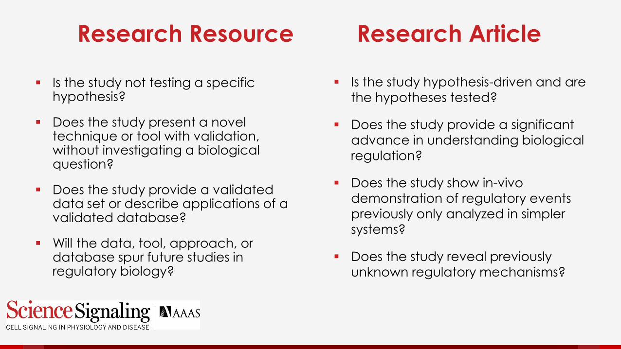
Is the study not testing a specific hypothesis?
Does the study present a novel technique or tool with validation, without investigating a biological question?
Does the study provide a validated data set or describe applications of a validated database?
Will the data, tool, approach, or database spur future studies in regulatory biology?
Research Resource Research Article
Is the study hypothesis-driven and are
the hypotheses tested?
Does the study provide a significant
advance in understanding biological regulation?
Does the study show in-vivo
demonstration of regulatory events previously only analyzed in simpler
systems?
Does the study reveal previously
unknown regulatory mechanisms?


Practical tips for preparing your manuscript

• Know your audience
• Write clearly
• Write concisely
• Write accurately
• Follow instructions
Five golden rules
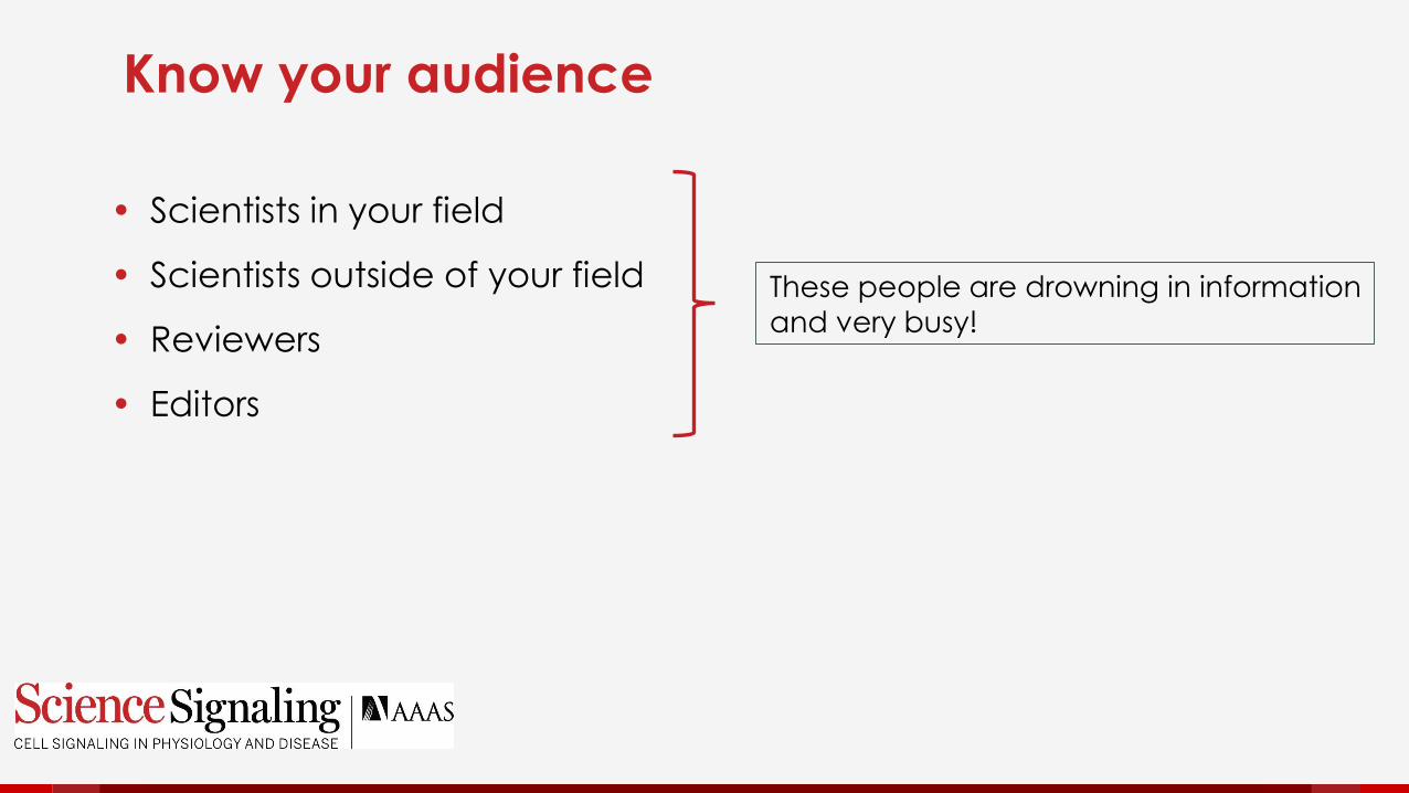
• Scientists in your field
• Scientists outside of your field
• Reviewers
• Editors
Know your audience
These people are drowning in information
and very busy!

May not be an expert in your field
Has 40 active papers
Fields multiple enquiries from authors
a day
Attends meetings (internal & external)
Wants to find the best papers for the
journal
First stop - The editor

Are there good reasons to proceed
with this paper?
Are there good reasons to reject this
paper?
Is the paper within the journal’s
scope?
Will this be one of our best papers?
Can I convince my fellow editors of
the value of this paper?
Will ask

Is a specialist in your field
Is very busy running a lab and writing
their own papers
Wants the review process to be as
efficient as possible
The reviewer

Is this correct?
Is it interesting enough for a Sciencejournal?
If I saw this paper in Science or one of the sister journals would I say
“Cool!”
“This changes my entire way of thinking!”
“I can’t wait to share this with my lab!”
“What were those idiot editors thinking?”
Will ask

How can you maximize your paper’s chance for success?
• Nothing substitutes for good content.
• To get into a top-tier journal, you will need an important
result that advances the field.
• Your conclusions must be well supported by experiments.
• The statistics must be good.
• The experiments must be properly controlled.
How can you ensure that your paper meets these criteria?

Is the result important?
Do the conclusions advance new
concepts?
Is the approach original?
Are the data reliable and
reproducible (how many n)?
Are the data properly quantified and
statistically evaluated?
Are experiments properly controlled?
Before you start writing – start your own review process

Assemble your figures into a presentation that matches the paper
Organize the introduction the way you plan to present it in the paper
Assemble your colleagues: Some within and some outside your lab
Ask them to give you feedback
What controls are missing?
What data are being overinterpreted?
Where are there gaps in logic?
What data are presented out of order?
Why do they think the study is important?
Invite critique from your colleagues

Incorporate the feedback from your
colleagues
Go back to the lab and do more
experiments, if necessary
Make sure the controls are presented
with the experiments
Make sure the flow of the figures and
data are logical
Make sure you’ve set the stage for
your study adequately
You’re almost ready to write

Present your data effectively
Restate the data, which should be readily discerned from the figure or table
State results without setting the experimental context, don’t make the reader guess
Present the study as a story in chronological order of experiments or refer to data shown later
Use lab jargon or shorthand in the text or figures
Use unnecessary or atypical abbreviations
Set the stage for the reader so that they can interpret the data
Limit the results to a description of the data and their indication; use the Introduction to set the necessary background and the Conclusions to put the study into broader context
Present the data in logical order, so that all results needed to make an interpretation are presented before the interpretation is drawn
Be consistent in labeling and define any abbreviations used in the figures that differ from those used in the text
Use only those abbreviations that are standard in your field and be sure to define them the first time they are used
DON’T DO

Lab jargon
Labeling Western blots with the name of the antibody, instead of the epitope or protein
Inconsistent labels
Do not have 3 different ways of labeling the same stable cell line, use one set of abbreviations consistently throughout
Common data presentation mistakes

A close-up of the MS0015203 binding mode showing the direct H-bond interactions between the ligand and GPR171 is shown in Fig. 1B.
Option 1: Add method information and specifics of interaction to guide reader through the result
Molecular docking analysis predicted that the ligand MS0015203 formed H-bonds with residues XX, XX, and XX in XX extracellular loops and XX in the XX transmembrane domain of the receptor GPR171 (Fig. 1B).
Option 2: If the figure is well labeled and very clear and method has already been described:
The ligand MS0015203 was predicted to form multiple H-bonds in residues in several regions of the receptor GPR171 (Fig. 1B).
The analysis predicted that the ligand MS0015203 formed H-bonds with residues in extracellular loops and a transmembrane domain of the receptor GPR171 (Fig. 1B).
Wordy, yet vague

Avoid imprecise words
Regulates
Alters
Influence
Avoid words with multiple meanings
Levels
Elevates
Significant
Avoid lab jargon
Write clearly
Precise
Stimulates the activity
Increases the abundance
Represses the gene’s expression
Unambiguous
Amount, abundance, or concentration
Increase
Substantial or important
Genes, RNAs, and proteins
Use italics for genes and transcripts
Use plain text for proteins and active RNA molecules

Avoid convoluted sentences with multiple clauses
Avoid long complicated qualifying adjectives
Avoid presenting published results as a historical review
Write concisely
Use simple declarative sentences;
divide into two sentences if
necessary
If published work is not in dispute,
present it as a fact

Avoid claims of novelty
Avoid speculation
Avoid superlatives
Correlations ≠ cause and effect
Written ≠ spoken language
Don’t anthropomorphize
Cells don’t have feelings and proteins are not people!
Write accurately
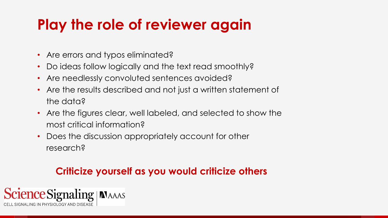
• Are errors and typos eliminated?
• Do ideas follow logically and the text read smoothly?
• Are needlessly convoluted sentences avoided?
• Are the results described and not just a written statement of
the data?
• Are the figures clear, well labeled, and selected to show the
most critical information?
• Does the discussion appropriately account for other
research?
Criticize yourself as you would criticize others
Play the role of reviewer again

Invite critique: Round 2
A scientist in your own specialty
A scientist in an unrelated specialty
A good editor for the English language

Think like the editor again for a minute:
What will he see first?
The cover letter
The title
The abstract
Ready, set, submit– But wait!

Cover letter – outline the conclusions
in plain and honest language,
without exaggeration, but do say
why you think the work is exciting.
Is the title reflective of the main
findings of the study?
Does the abstract convey the main
findings and importance.
First impressions

Practice while you read
As you read for Journal Club, consider these best practices
and ‘pencil’-edit the papers.
As you read background materials for your research, find the
errors and think about how the writing could be improved.
Help each other. Read each other’s manuscripts.
Don’t be afraid of the red pen!


The Science Signaling editorial team
Nancy Gough
All areas
Computational & systems biology
Channels and biophysics
John Foley
Immunology
Biochemistry
G protein-coupled receptor signaling
Wei Wong
Vascular biology
Cell biology
Physiology
Leslie Ferrarelli
Cancer
Neuroscience
Annalisa VanHook
Podcasts
Developmental biology
Microbial signaling
Plant signaling

Before and after samples

Title
Original: Targeting Poly (ADP-Ribose) Polymerase and the c-Myb-TopBP1-ATR-Chk1 Signaling Pathway in Castration-Resistant
Prostate Cancer
Edited: Targeting Poly (ADP-Ribose) Polymerase and the c-Myb -
Regulated DNA Damage Response Pathway in Castration-
Resistant Prostate Cancer

Since we previously found that GPR171 is activated by a relatively long (16 amino acid) peptide (6), we sought to identify the minimal length required to activate the receptor. For this we used N-terminally truncated b-LEN-derived peptides and found that the C-terminal four amino acids (LLPP, present in both the mouse and rat b-LEN sequences) were necessary and sufficient to displace b-LEN from the receptor (fig. S1A).
Problem 1: Stage is not set properly for understanding the experiment.
Problem 2: Authors did not test for necessity because none of the tested peptides lack these residues. They only tested for sufficiency. Data are overinterpreted.
Problem 3: Since should only be used when there is a temporal component. Most often it should be replaced with Because.
Problem 4: Why was it important to see if a smaller peptide could affect receptor function?
Because we previously found that GPR171 is activated by a relatively long (16 amino acid) peptide (6), we sought to identify the minimal peptide required to activate the receptor. We used N-terminally truncated b-LEN-derived peptides to evaluate the minimal sequence that displaced radiolabeled b-LEN binding. The C-terminal four amino acids (LLPP, present in both the mouse and rat b-LEN sequences) were sufficient to displace b-LEN from the receptor (fig. S1A). Because this sequence is so small, small molecule ligands may exist that can affect receptor activity.
Results

Abstract unedited
Androgen deprivation is the standard systemic treatment for advanced prostate cancer (PCa), but
most patients ultimately develop castration-resistance. We show here that MYB is transcriptionally
activated by androgen deprivation or impairment of androgen receptor (AR) signaling. MYB gene
silencing significantly inhibited PCa growth in vitro and in vivo. Microarray data revealed that c-
Myb shares a substantial subset of DNA damage response (DDR) target genes with AR, suggesting
that c-Myb may replace AR for the dominant role in the regulation of their common DDR target
genes in AR inhibition-resistant or AR-negative PCa. Gene signatures comprising AR, MYB, and their
common DDR target genes are significantly correlated with metastasis, castration-resistance,
recurrence, and shorter overall survival in PCa patients. We demonstrated in vitro that silencing of
MYB, BRCA1 or TOPBP1 synergized with poly (ADP-ribose) polymerase (PARP) inhibitor olaparib (OLA)
to increase cytotoxicity to PCa cells. We further demonstrated that targeting the c-Myb-TopBP1-
ATR-Chk1 pathway by using the Chk1 inhibitor AZD7762 synergizes with OLA to increase PCa
cytotoxicity. Our results reveal new mechanism-based therapeutic approaches for PCa by
targeting PARP and the c-Myb-TopBP1-ATR-Chk1 pathway

Abstract Edited
Androgen deprivation is the standard treatment for advanced prostate cancer (PCa), but
most patients ultimately develop resistance and tumor recurrence. We found that MYB is
transcriptionally activated by androgen deprivation therapy or genetic silencing of the
androgen receptor (AR). MYB silencing inhibited PCa growth in culture and xenografts in mice.
Microarray data revealed that c-Myb and AR shared a subset of target genes that encode
DNA damage response (DDR) proteins, suggesting that c-Myb may supplant AR as the
dominant regulator of their common DDR target genes in AR inhibition–resistant or AR-negative
PCa. Gene signatures including AR, MYB, and their common DDR-associated target genes
positively correlated with metastasis, castration resistance, tumor recurrence, and decreased
survival in PCa patients. In culture and in xenograft-bearing mice, a combination strategy
involving the knockdown of MYB, BRCA1, or TOPBP1 or the abrogation of cell cycle checkpoint
arrest with AZD7762, an inhibitor of the checkpoint kinase Chk1, increased the cytotoxicity of
the poly[adenosine 5′-diphosphate (ADP)–ribose] polymerase (PARP) inhibitor olaparib in PCa
cells. Our results reveal new mechanism-based therapeutic approaches for PCa by targeting
PARP and the DDR pathway involving c-Myb, TopBP1, ataxia telangiectasia mutated– and
Rad3-related (ATR), and Chk1.

Research Article Abstract: Before (224 words)
Cells derived from ataxia telangiectasia (A-T) patients exhibit defective cell cycle checkpoints following
ionizing radiation (IR), profound radiosensitivity and high levels of chromosome aberrations. We have
shown that transient ATM kinase inhibition from +15 to +75 min following IR is sufficient to radiosensitize
cells and accumulate persistent chromosome aberrations. We show here that DNA-PK kinase inhibition
from +15 to + 75 min is also sufficient to radiosensitize cells and accumulate persistent chromosome
aberrations. The ATM kinase-dependent mechanisms that ensure cell survival and suppress
chromosome aberrations during this interval are independent of DNA-PK kinase activity. Neither the
activation nor the recovery of the IR-induced G2/M cell cycle checkpoint are affected by ATM kinase
inhibition from +15 to +75 min, indicating that 15 min of ATM kinase signaling is sufficient to induce this
cell cycle checkpoint. Surprisingly, ATM kinase inhibition from +15 to +75 min abrogates IR-induced sister
chromatid exchange (SCE), a phenotype attributed to the repair of damaged replication forks. Further,
ATM kinase inhibition using either KU55933 or KU60019 is sufficient to disrupt camptothecin-induced SCE.
Since DNA damage-induced SCE is maintained A-T cells that express no ATM protein, and the ATM
kinase inhibitors have no effect on DNA damage-induced SCE in A-T cells, these data reveal that the
consequences of acute ATM kinase inhibition and adaptation to ATM protein disruption are distinct in S-
phase cells.

ProblemsCells derived from ataxia telangiectasia (A-T) patients exhibit
defective cell cycle checkpoints following ionizing
radiation (IR), profound radiosensitivity and high levels of
chromosome aberrations.
We have shown that transient ATM kinase inhibition from
+15 to +75 min following IR is sufficient to radiosensitize cells
and accumulate persistent chromosome aberrations.
We show here that DNA-PK kinase inhibition from +15 to +
75 min is also sufficient to radiosensitize cells and
accumulate persistent chromosome aberrations.
The ATM kinase-dependent mechanisms that ensure cell
survival and suppress chromosome aberrations during this
interval are independent of DNA-PK kinase activity.
What is radiosensitivity?
What is the relationship of
radiosensitivity to chromosome
aberrations?
ATM kinase = The kinase that
phosphorylates ATM? NO
What are ATM and DNA-PK?
DNA-PK kinase = The kinase that
phosphorylates DNA-PK? NO
ATM kinase-dependent = Mechanisms
that rely on phosphorylation of ATM?
NO
How does cell survival relate to
radiosensitivity?

What is the IR-induced G2/M checkpoint?
Why is this surprising? What do damaged
replication forks have to do with IR-induced
damage?
Too much experimental detail.
What is camptothecin?
Neither the activation nor the recovery of the IR-
induced G2/M cell cycle checkpoint are affected
by ATM kinase inhibition from +15 to +75 min,
indicating that 15 min of ATM kinase signaling is
sufficient to induce this cell cycle checkpoint.
Surprisingly, ATM kinase inhibition from +15 to +75
min abrogates IR-induced sister chromatid
exchange (SCE), a phenotype attributed to the
repair of damaged replication forks.
Further, ATM kinase inhibition using either KU55933
or KU60019 is sufficient to disrupt camptothecin-
induced SCE.
More problems

Since should be because.
Genes are expressed, not proteins.
The information about S-phase is out of context.
Since DNA damage-induced SCE is maintained A-T
cells that express no ATM protein, and the ATM
kinase inhibitors have no effect on DNA damage-
induced SCE in A-T cells, these data reveal that the
consequences of acute ATM kinase inhibition and
adaptation to ATM protein disruption are distinct in
S-phase cells.
And more problems

Lack of context
Too much methodological detail
Imprecise language
Too many undefined terms
Did your eyes glaze over?

Clean edited version with editorial queries (182 words)

Abstract: Original 265 words!Depolarization of resting membrane potential in select cells in Xenopus larvae induces normal
melanocytes to undergo a conversion to a metastatic phenotype. Here, we show that this non-
cell-autonomous process is mediated by cAMP, CREB, and the transcription factors Sox10 and
Slug, which have been previously shown to be implicated in various cancers, including
melanoma. Our microarray analysis reveals specific transcripts responsive to Vmem levels
within a few hours of depolarization, and a set of 517 transcripts whose expression remains
altered during the full hyperpigmented phenotype over a week later, linking instructor cell-
depolarization to a range of developmental processes and disease states. We also show that
voltage-dependent conversion of melanocytes involves the MSH-secreting melanotrope cells of
the pituitary, and formulate a model for the molecular pathway linking the bioelectric properties
of melanocyte cells’ microenvironment in vivo to the genetic and cellular changes induced in
this melanoma-like phenotype. Remarkably, the phenotype is all-or-none: each individual
animal either undergoes melanocyte conversion or not, as a whole. This group decision is
stochastic, resulting in varying percentages of hyperpigmented individuals for a given
experimental treatment. To explain the observed stochasticity as an inherent dynamic property
of this complex signaling system, we developed a novel computational method that reverse-
engineered a dynamic regulatory network that quantitatively explained our complex dataset, and
made correct predictions for new experiments. Taken together, these data (1) reveal new
molecular details about a novel trigger of metastatic cell behavior in vivo, (2) suggest new
targets for biomedical intervention, and (3) demonstrate proof-of-principle of a computational
method for understanding stochastic decision-making by cells during development and cancer.
Problems:
• Vague terminology: “select
cells”
• Confusing connection
between frogs and cancer:
Hyperpigmented frogs have
cancer?
• Undefined abbreviations:
cAMP, MSH, CREB
• Using whose to refer to
transcripts
• Too long (PubMed truncates
at 250)
• Methodological context
unclear
• Mixing background and
results
• What did they do in this study
versus what was already
known?

Experimentally induced depolarization of resting membrane potential in “instructor cells” in Xenopus laevis embryos causes
hyperpigmentation in an all-or-none fashion in some tadpoles due to excess proliferation and migration of melanocytes. We
showed that this stochastic process involved serotonin signaling, adenosine 3’,5’-monophosphate (cAMP), the transcription
factors cAMP response element binding protein (CREB), Sox10, and Slug. Transcriptional microarray analysis of embryos
taken at stage 15 (early neurula) and stage 45 (free-swimming tadpole) revealed changes in the abundance of 45 transcripts
and 517 transcripts, respectively, between control embryos and embryos exposed to the instructor cell-depolarizing agent
ivermectin. Bioinformatic analysis revealed that the human homologs of some of the differentially regulated genes were
associated with cancer, consistent with the induced arborization and invasive behavior of converted melanocytes. We
identified a physiological circuit that utilizes serotonergic signaling between instructor cells, melanotrope cells of the pituitary,
and melanocytes to control the proliferation, cell shape, and migration properties of the pigment cell pool. To understand the
stochasticity and properties of this multiscale signaling system, we applied a computational machine-learning method that
iteratively explored network models to reverse engineer a stochastic dynamic model that recapitulated the frequency of the all-
or-none hyperpigmentation phenotype produced in response to various pharmacological and molecular-genetic manipulations.
This computational approach may provide insight into stochastic cellular decision-making that occurs during normal
development and pathological conditions, such as cancer.
Abstract: After 225 words

Although most PD cases are sporadic, at least seven genes have been reported to be implicated in the
pathogenesis of familial PD (1).
(hint: too many words)
In vitro studies indicated that several pathogenic mutations in LRRK2 caused an increase in the kinase activity,
such as mutations R1441C in ROC GTPase domain and G2019S in kinase domain (4-6).
(hint: multiple problems, including a misplaced clause)
While the physiological function of LRRK2 remains largely unknown, recent studies indicated a dispensable role
of the intrinsic kinase activity of LRRK2 in neuron survival and its protective activity against neurotoxin (10-12).
(hint: multiple problems, especially temporal words)
The current paper reports for the first time a sex reversal in transsexual people in the interstitial nucleus of the
anterior hypothalamus (INAH) 3, a sexually dimorphic hypothalamic nucleus that was previously shown to be
related to sexual orientation (citation 1, citation 2).
Practice