how streptococci make isoprenoids - beilstein-institut
TRANSCRIPT

����������������
How Streptococci Make Isoprenoids
Scott T. Lefurgy*
and Thomas S. Leyh
Department of Microbiology & Immunology,The Albert Einstein College of Medicine,
1300 Morris Park Ave., Bronx, NY 10461, U.S.A.
E-Mail: *[email protected]
Received: 16th June 2010 / Published: 14th September 2010
Abstract
Isoprenoids are the set of ~ 25,000 unique compounds based on the
ubiquitous C5 donor isopentenyl diphosphate (IPP), including qui-
nones, steroid hormones, bile acids, protein membrane anchors and
secondary metabolites. Streptococci and other gram-positive bacteria
produce IPP via the mevalonate pathway, whose function is required
for the respiratory pathogen Streptococcus pneumoniae to survive in
lung and serum. With the discovery of potent selective feedback in-
hibition by the metabolite diphosphomevalonate (DPM), our laboratory
has positioned the pneumococcal mevalonate pathway as a novel target
for clinical intervention against an organism that claims the lives of
over 4000 people daily. Our studies have revealed unique features of
each of each of the three GHMP family kinases that comprise the
pathway, including potent allosteric inhibition, a catalytic switch, and
a concerted elimination mechanism-informing the design of antibiotics
that can simultaneously inhibit multiple steps in a single pathway.
Introduction
Streptococcus pneumoniae kills over 1 million people each year worldwide, mostly children
and the elderly, and is the primary bacterial cause of pneumonia, meningitis and otitis media
[1, 2]. Antibiotic resistance remains a major problem in treating infections, and multiple-
drug resistance rates as high as 95% are seen in some countries [3]. Despite the successful
introduction in 2000 of a vaccine covering seven of the most prevalent and infectious of the
45
http://www.beilstein-institut.de/ESCEC2009/Proceedings/Lefurgy/Lefurgy.pdf
Experimental Standard Conditions of Enzyme Characterizations,
September 13th – 16th, 2009, Rudesheim/Rhein, Germany
> 100 subtypes of pneumcoccus [5], nonvaccinated subtypes are rapidly filling the biological
niche created by the vaccine and becoming more virulent [6]. There is an unequivocal need
for new strategies to fight this pernicious bacterium.
Isoprenoid biosynthesis has recently emerged as a new target for antibiotic development.
The isoprenoids are a class of ~ 25,000 unique compounds composed of a single building-
block, isopentenyl diphosphate (IPP). The C5 isoprene units of IPP are concatenated and
then converted either to universal essential cofactors, vitamins, steroids and a host of
secondary metabolites, or attached to other biomolecules, such tRNAs or proteins-facilitat-
ing association of the latter with membranes. Most eubacteria synthesize IPP starting from
glyceraldehyde-3-phosphate and pyruvate using the methylerythritol phosphate pathway [7].
Gram-positive bacteria, including S. pneumoniae, archaebacteria and eukaryotes make IPP
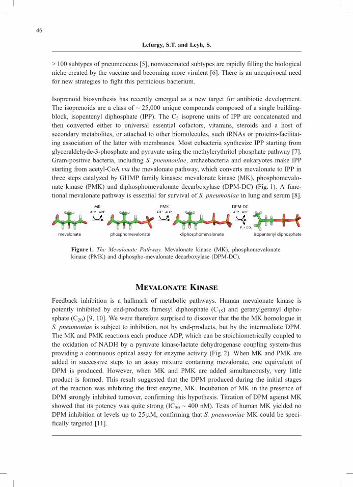
starting from acetyl-CoA via the mevalonate pathway, which converts mevalonate to IPP in
three steps catalyzed by GHMP family kinases: mevalonate kinase (MK), phosphomevalo-
nate kinase (PMK) and diphosphomevalonate decarboxylase (DPM-DC) (Fig. 1). A func-
tional mevalonate pathway is essential for survival of S. pneumoniae in lung and serum [8].
Figure 1. The Mevalonate Pathway. Mevalonate kinase (MK), phosphomevalonate
kinase (PMK) and diphospho-mevalonate decarboxylase (DPM-DC).
Mevalonate Kinase
Feedback inhibition is a hallmark of metabolic pathways. Human mevalonate kinase is
potently inhibited by end-products farnesyl diphosphate (C15) and geranylgeranyl dipho-
sphate (C20) [9, 10]. We were therefore surprised to discover that the the MK homologue in
S. pneumoniae is subject to inhibition, not by end-products, but by the intermediate DPM.
The MK and PMK reactions each produce ADP, which can be stoichiometrically coupled to
the oxidation of NADH by a pyruvate kinase/lactate dehydrogenase coupling system-thus
providing a continuous optical assay for enzyme activity (Fig. 2). When MK and PMK are
added in successive steps to an assay mixture containing mevalonate, one equivalent of
DPM is produced. However, when MK and PMK are added simultaneously, very little
product is formed. This result suggested that the DPM produced during the initial stages
of the reaction was inhibiting the first enzyme, MK. Incubation of MK in the presence of
DPM strongly inhibited turnover, confirming this hypothesis. Titration of DPM against MK
showed that its potency was quite strong (IC50 ~ 400 nM). Tests of human MK yielded no
DPM inhibition at levels up to 25mM, confirming that S. pneumoniae MK could be speci-
fically targeted [11].
46
Lefurgy, S.T. and Leyh, S.

Figure 2. Discovery of MK Inhibition by DPM. Reaction progress curves for reactions
in which enzymes were added sequentially or simultaneously. ADP produced by MK
or PMK is stoichiometrically coupled to oxidation of NADH by a pyruvate kinase/
lactate dehydrogenase coupling system, resulting in a decrease in absorbance at
386 nm.
An important consideration when developing an antibiotic that targets a metabolic pathway
is the mechanism of inhibition. An allosteric mechanism of pathway inhibition – in which
the inhibitory ligand binds distal to active site – has an advantage over simple occlusion of
the active site, in that inhibition cannot be overcome by the thermodynamic push that
accompanies a buildup of the metabolite (i. e., the substrate) just upstream of the inhibited
step. MK inhibition by DPM was investigated using initial-rate experiments that simulate
this condition and can thus distinguish between mechanisms: the maximal reaction velocity
at (theoretically) infinite substrate concentration was determined as a function of inhibitor
concentration. If DPM binds to the active site, its inhibitory effects (at any concentration of
inhibitor) will be completely irrelevant at infinite substrate concentration and have no impact
on the maximal velocity. We observed that DPM reduced this maximal velocity – with equal
potency versus both mevalonate and ATP – indicating that DPM must bind to an allosteric
site. This result was confirmed by the observation that DPM had an identical affinity for MK
both in the presence and absence of saturating concentrations of mevalonate and AMPPNP
(a non-hydrolyzable ATP analogue) in an equilibrium binding study. If DPM bound at the
active site, its apparent affinity should have been altered by the presence of competing
ligands. The stoichiometry of DPM binding to the MK dimer was shown to be 1:1, suggest-
ing that the allosteric site was symmetrically disposed to both subunits, and was perhaps
located at the dimer interface.
The X-ray crystal structure of MK in the presence of mevalonate, AMPPNP and DPM
showed that two molecules of DPM are bound, one to each subunit, in the mevalonate
binding pocket (Fig. 3A) [12]. This result is perhaps not surprising, given that DPM is a
47
How Streptococci Make Isoprenoids

partial bisubstrate analogue whose pyrophosphoryl moiety could take the place of the beta-
and gamma-phosphates of ATP; however, the result stands at odds with the functional data
described above and may represent a crystallographic artifact. The structure also revealed a
pore at the dimer interface having excellent charge- and shape-complementarity to DPM;
this pore could be the allosteric site. (Fig. 3B). We are currently pursuing the solution-phase
structure of the fully-liganded MK (bound to mevalonate, AMPPNP and DPM) by NMR, to
identify this site and study structural changes to the enzyme that occur upon allosteric
binding.
Figure 3. Locating the DPM Allosteric Site. (A) Crystal structure of S. pneumoniae
MK with DPM (magenta sticks) and Mg2+ (green spheres) bound (PDB: 2OI2).
(B) MK model with DPM positioned at a pore in the subunit interface. Vacuum
electrostatics near the pore are displayed as colored surfaces (blue, positive; red,
negative).
Phosphomevalonate Kinase
The X-ray structure of phosphomevalonate kinase presents a classic GHMP kinase scaffold.
Comparison of the apo and ternary-complex forms of the enzyme (Fig. 4) reveals that four
regions undergo significant conformational changes as a result of ligand binding [13, 14].
48
Lefurgy, S.T. and Leyh, S.

Figure 4. Comparison of the Apo and Ternary-Complex Structure of PMK from
S. pneumoniae. Regions that do not change noticeably upon ligand binding are grey,
the four responsive elements (L1, L2, L3 and aH) are colored in blue, red, cyan and
green, respectively – the more intense colors are associated with the ternary complex.
These regions (L1, L2 L3 and aH) are color-coded, and the more intense colors are
associated with the ternary complex. L1 (blue) is disordered in the absence of ligand, and
reorganizes upon binding of nucleotide (AMPPNP, purple) with the result that residues that
would otherwise obstruct binding are withdrawn from the binding pocket, and hydrogen
bonds to the adenine ring are established. L2 (red) undergoes a considerable structural
change in which a small helical element unravels to deliver the ammonium group of
Lys101 into direct contact with the b,g-bridging atom of AMPPNP (Fig. 5), where it will
stabilize the negative charge expected to develop at this position during bond cleavage in a
dissociative reaction. In the apo structure, Lys101 forms a salt bridge to the carboxylate of
Glu98, and is positioned to obstruct the binding of nucleotide. Upon binding, the salt bridge
is broken, the helix unwinds, ‘‘swinging’’ Lys101 past Lys100, which hydrogen bonds to the
amide backbone of Lys208. The structural change resemble a ‘‘lysine switch’’ that when
thrown, activates catalysis. It is interesting to note that while other GHMP kinases also
feature a lysine switch, the catalytic lysine has migrated to a different position in the active
site.
49
How Streptococci Make Isoprenoids

Figure 5. A Di-lysine Catalytic Switch. The L2 regions of the apo- (purple) and
ternary-complex (cyan) forms of PMK are overlain. The b- and g- phosphoryl groupsof AMPPNP are shown.
The structure of the PMK ternary complex suggested that the substrates are positioned in a
non-reactive orientation. The phosphoryl-group of phosphomevalonate is nearly orthogonal
to what is expected for in-line nucleophillic attack at the g-phosphate of ATP (Fig. 6A).
Comparison of the PMK arrangement to that of six other GHMP-kinase ternary complexes
revealed that of the seven total structures, three exhibited the non-reactive arrangement, three
exhibited what appeared to be an excellent positioning for in-line attack (e. g., Fig. 5B), and
one, erythritol kinase, exhibited both conformations. Clearly, these active sites can bind
substrates in either conformation, suggesting that these forms might interconvert during
the catalytic cycle. To assess the likelihood that the PMK active site might accommodate
interconversion, the substrate configurations were aligned by superposing the Ca-traces of
the ternary complex of PMK with that of Gal-NAc kinase – a good example of a reactive
complex. The alignment suggested that the only large-scale movement was the migration of
the divalent cation between ‘‘walls’’ of the active site, and that the active site was indeed
capacious enough to accommodate such migration.
50
Lefurgy, S.T. and Leyh, S.
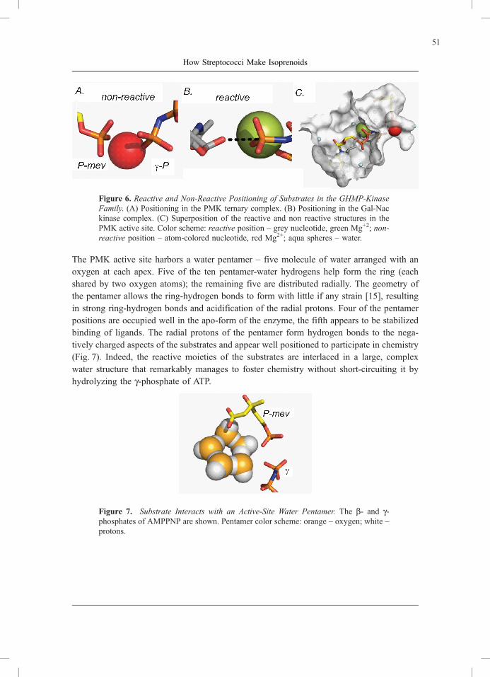
Figure 6. Reactive and Non-Reactive Positioning of Substrates in the GHMP-Kinase
Family. (A) Positioning in the PMK ternary complex. (B) Positioning in the Gal-Nac
kinase complex. (C) Superposition of the reactive and non reactive structures in the
PMK active site. Color scheme: reactive position – grey nucleotide, green Mg+2; non-
reactive position – atom-colored nucleotide, red Mg2+; aqua spheres – water.
The PMK active site harbors a water pentamer – five molecule of water arranged with an
oxygen at each apex. Five of the ten pentamer-water hydrogens help form the ring (each
shared by two oxygen atoms); the remaining five are distributed radially. The geometry of
the pentamer allows the ring-hydrogen bonds to form with little if any strain [15], resulting
in strong ring-hydrogen bonds and acidification of the radial protons. Four of the pentamer
positions are occupied well in the apo-form of the enzyme, the fifth appears to be stabilized
binding of ligands. The radial protons of the pentamer form hydrogen bonds to the nega-
tively charged aspects of the substrates and appear well positioned to participate in chemistry
(Fig. 7). Indeed, the reactive moieties of the substrates are interlaced in a large, complex
water structure that remarkably manages to foster chemistry without short-circuiting it by
hydrolyzing the g-phosphate of ATP.
Figure 7. Substrate Interacts with an Active-Site Water Pentamer. The b- and g-phosphates of AMPPNP are shown. Pentamer color scheme: orange – oxygen; white –
protons.
51
How Streptococci Make Isoprenoids
Diphosphomevalonate Decarboxylase
The final step in the mevalonate pathway is carried out by diphosphomevalonate decarboxy-
lase (DPM-DC), which phosphorylates the C3-hydroxyl of DPM and subsequently elimi-
nates phosphate and CO2. This reaction is thought to proceed in a stepwise manner, with
phosphate departing first, resulting in the formation of a carbocation. Abeles and coworkers
provided evidence that a carbocation transiently forms by showing that substitution of the
C3-methyl group with a hydrogen or a fluoromethyl group causes the reaction to halt after
phosphorylation, implying that the electron-donating methyl group is required to stabilize
the carbocation inductively [16]. We sought to exploit carbocation formation to inactivate
DPM-DC through use of DPM analogues in which the C3-methyl group was replaced by
substituents that were able, by resonance with the carbocation, to generate strongly electro-
philic species that could become covalently bound to the enzyme (Fig. 8) [3]. One of these
analogues contained a cyclopropyl substituent that was proposed to undergo ring opening
upon ionization, forming a homoallyl cation that is stabilized by the loss of 27 kcal/mol of
ring strain [17, 18]. If this rearrangement occurred, we anticipated that a nucleophile on the
enzyme surface could attack the carbocationic intermediate, forming a covalent bond and
inactivating the enzyme. However, inactivation did not occur over the course of > 1000
turnovers, suggesting that no such adduct had formed. We therefore monitored the fate of the
analogue during turnover using the unique NMR signal of the cyclopropyl protons. If the
ring opened and formed a primary alcohol (by quenching with water), the proton chemical
shifts associated with the ring would move significantly downfield [19]. Instead, we ob-
served only a very small change (0.3 ppm) in the cyclopropyl proton chemical shifts that
was consistent with an intact cyclopropyl group adjacent to a carbon-carbon double bond;
this structure was subsequently confirmed by 2-D NMR. These data rule out a ring-opened
product, and suggest that the analogue undergoes a decarboxylation that resembles that of
the native substrate, DPM.
52
Lefurgy, S.T. and Leyh, S.

Figure 8. Probing the Mechanism of DPM-DC with Cyclopropyl-DPM. An analogue
of DPM with a cyclopropyl substitution at C3 is phosphorylated by the enzyme. If a
carbocation forms during the reaction (Dissociative branch), it can be delocalized into
the cyclopropyl ring, causing the ring to open, forming a strong electrophile. This
ring-opened carbocation can be quenched by a nucleophile on the enzyme surface or
water. Alternatively, a concerted mechanism produces only decarboxylated products.
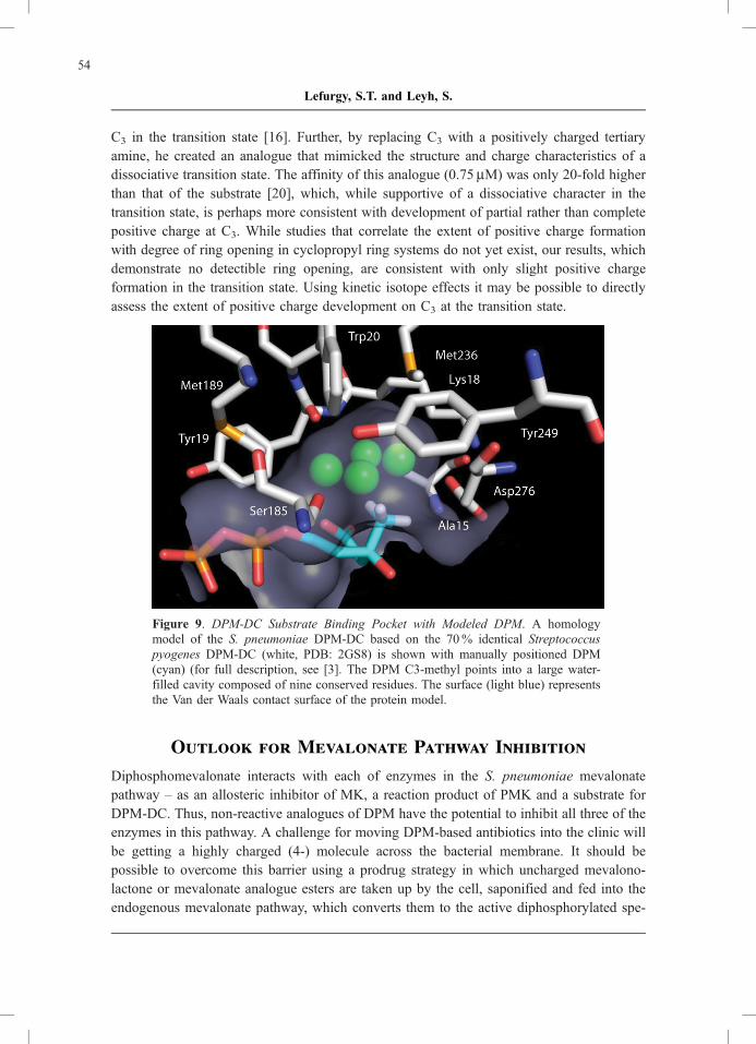
These results call into question the extent of carbocation formation in the DPM-DC transi-
tion state. If a full carbocation formed, we expected to observe rearrangement of the
cyclopropyl group. It is conceivable that a carbocation could form but not react, provided
that no nucleophile is in close proximity to the carbocation (the protein surface or water);
however, the presence of four crystallographic water molecules in the large pocket adjacent
to C3 and the proximity of the Asp276, Lys18, Ser185, and Met189 side chains and the
Tyr19 carbonyl as potential nucleophiles argue against this possibility (Fig. 9). Alternatively,
carbocation formation may be minimal or absent, in which case elimination of the carbox-
ylate and the phosphate is concerted rather than dissociative. Abeles’ work on the effects of
altered electron induction at C3 with the mammalian enzyme shows clearly that DPM-DC
chemistry is sensitive to such changes and supports the development of a positive charge at
53
How Streptococci Make Isoprenoids
C3 in the transition state [16]. Further, by replacing C3 with a positively charged tertiary
amine, he created an analogue that mimicked the structure and charge characteristics of a
dissociative transition state. The affinity of this analogue (0.75 mM) was only 20-fold higher
than that of the substrate [20], which, while supportive of a dissociative character in the
transition state, is perhaps more consistent with development of partial rather than complete
positive charge at C3. While studies that correlate the extent of positive charge formation
with degree of ring opening in cyclopropyl ring systems do not yet exist, our results, which
demonstrate no detectible ring opening, are consistent with only slight positive charge
formation in the transition state. Using kinetic isotope effects it may be possible to directly
assess the extent of positive charge development on C3 at the transition state.
Figure 9. DPM-DC Substrate Binding Pocket with Modeled DPM. A homology
model of the S. pneumoniae DPM-DC based on the 70% identical Streptococcus
pyogenes DPM-DC (white, PDB: 2GS8) is shown with manually positioned DPM
(cyan) (for full description, see [3]. The DPM C3-methyl points into a large water-
filled cavity composed of nine conserved residues. The surface (light blue) represents
the Van der Waals contact surface of the protein model.
Outlook for Mevalonate Pathway Inhibition
Diphosphomevalonate interacts with each of enzymes in the S. pneumoniae mevalonate
pathway – as an allosteric inhibitor of MK, a reaction product of PMK and a substrate for
DPM-DC. Thus, non-reactive analogues of DPM have the potential to inhibit all three of the
enzymes in this pathway. A challenge for moving DPM-based antibiotics into the clinic will
be getting a highly charged (4-) molecule across the bacterial membrane. It should be
possible to overcome this barrier using a prodrug strategy in which uncharged mevalono-
lactone or mevalonate analogue esters are taken up by the cell, saponified and fed into the
endogenous mevalonate pathway, which converts them to the active diphosphorylated spe-
54
Lefurgy, S.T. and Leyh, S.

cies in situ [21, 22]. It must also be considered that, while DPM selectively inhibits
pneumococcal MK, DPM analogues may cross-react with the human homologues of PMK
and DPM-DC. However, inhibitors of the human mevalonate pathway, such as the widely-
used cholesterol-lowering statins and anti-proliferative bisphosphonates, are generally very
well tolerated and have shown the potential to have a clinical impact on asthma [23, 24],
wound-healing [25], hepatitis [26, 27] and HIV [28], suggesting that even nonselective
inhibitors have potential therapeutic benefits.
References
[1] Obaro, S., and Adegbola, R. (2002) The pneumococcus: carriage, disease and con-
jugate vaccines. J. Med. Microbiol. 51:98–104.
[2] Schuchat, A., Robinson, K., Wenger, J.D., Harrison, L.H., Farley, M., Reingold, A.L.,
Lefkowitz, L., and Perkins, B.A. (1997) Bacterial meningitis in the United States in
1995. Active Surveillance Team. N. Engl. J. Med. 337:970–976.
[3] Lefurgy, S.T., Rodriguez, S.B., Park, C.S., Cahill, S., Silverman, R.B., and Leyh, T.S.
Probing ligand-binding pockets of the mevalonate pathway enzymes from Strepto-
coccus pneumoniae. J. Biol. Chem. in press.
[4] Van Bambeke, F., Reinert, R.R., Appelbaum, P.C., Tulkens, P.M., and Peeter-
mans, W.E. (2007) Multidrug-resistant Streptococcus pneumoniae infections: current
and future therapeutic options. Drugs 67:2355–2382.
doi: http://dx.doi.org/10.2165/00003495-200767160-00005.
[5] Kyaw, M.H., Lynfield, R., Schaffner, W., Craig, A.S., Hadler, J., Reingold, A.,
Thomas, A.R., Harrison, L.H., Bennett, N.M., Farley, M.M., Facklam, R.R., Jorgen-
sen, J.H., Besser, J., Zell, E.R., Schuchat, A., and Whitney, C.G. (2006) Effect of
introduction of the pneumococcal conjugate vaccine on drug-resistant Streptococcus
pneumoniae. N. Engl. J. Med. 354:1455–1463.
doi: http://dx.doi.org/10.1056/NEJMoa051642.
[6] Huang, S.S., Hinrichsen, V.L., Stevenson, A.E., Rifas-Shiman, S.L., Kleinman, K.,
Pelton, S.I., Lipsitch, M., Hanage, W.P., Lee, G.M., and Finkelstein, J.A. (2009)
Continued impact of pneumococcal conjugate vaccine on carriage in young children.
Pediatrics 124:e1–11.
doi: http://dx.doi.org/10.1542/peds.2008-3099
[7] Lange, B.M., Rujan, T., Martin, W., and Croteau, R. (2000) Isoprenoid biosynthesis:
The evolution of two ancient and distinct pathways across genome. Proc. Natl. Acad.
Sci. U.S.A. 97:13172–13177.
doi: http://dx.doi.org/10.1073/pnas.240454797.
55
How Streptococci Make Isoprenoids

[8] Wilding, E.I., Brown, J.R., Bryant, A.P., Chalker, A.F., Holmes, D.J., Ingraham, K.A.,
Iordanescu, S., So, C.Y., Rosenberg, M., and Gwynn, M.N. (2000) Identification,
evolution, and essentiality of the mevalonate pathway for isopentenyl diphosphate
biosynthesis in gram-positive cocci. J. Bacteriol. 182:4319–4327.
doi: http://dx.doi.org/10.1128/JB.182.15.4319-4327.2000.
[9] Hinson, D.D., Chambliss, K.L., Toth, M.J., Tanaka, R.D., and Gibson, K.M. (1997)
Post-translational regulation of mevalonate kinase by intermediates of the cholesterol
and nonsterol isoprene biosynthetic pathways. J. Lipid Res. 38:2216–2223.
[10] Dorsey, J.K., and Porter, J.W. (1968) The inhibition of mevalonic kinase by geranyl
and farnesyl pyrophosphates. J. Biol. Chem. 243:4667–4670.
[11] Andreassi, J.L., 2nd, Dabovic, K., and Leyh, T.S. (2004) Streptococcus pneumoniae
isoprenoid biosynthesis is downregulated by diphosphomevalonate: an antimicrobial
target. Biochemistry 43:16461–16466.
doi: http://dx.doi.org/10.1021/bi048075t.
[12] Andreassi, J.L., 2nd, Bilder, P.W., Vetting, M.W., Roderick, S.L., and Leyh, T.S.
(2007) Crystal structure of the Streptococcus pneumoniae mevalonate kinase in
complex with diphosphomevalonate. Protein Sci. 16:983–989.
doi: http://dx.doi.org/10.1110/ps.072755707.
[13] Andreassi, J.L., 2nd, Vetting, M.W., Bilder, P.W., Roderick, S.L., and Leyh, T.S.
(2009) Structure of the ternary complex of phosphomevalonate kinase: the enzyme
and its family. Biochemistry 48:6461–6468.
doi: http://dx.doi.org/10.1021/bi900537u.
[14] Romanowski, M.J., Bonanno, J.B., and Burley, S.K. (2002) Crystal structure of the
Streptococcus pneumoniae phosphomevalonate kinase, a member of the GHMP
kinase superfamily. Proteins 47:568–571.
doi: http://dx.doi.org/10.1002/prot.10118.
[15] Harker, H.A., Viant, M.R., Keutsch, F.N., Michael, E.A., McLaughlin, R.P., and
Saykally, R.J. (2005) Water pentamer: characterization of the torsional-puckering
manifold by terahertz VRT spectroscopy. J. Phys. Chem. A 109:6483–6497.
doi: http://dx.doi.org/10.1021/jp051504s.
[16] Dhe-Paganon, S., Magrath, J., and Abeles, R.H. (1994) Mechanism of mevalonate
pyrophosphate decarboxylase: evidence for a carbocationic transition state. Biochem-
istry 33:13355–13362.
doi: http://dx.doi.org/10.1021/bi00249a023.
[17] Hart, H., and Sandri, J.M. (1959) The Solvolysis of p-Nitrobenzoates of Certain
Cyclopropylcarbinols. J. Am. Chem. Soc. 81:320–326.
doi: http://dx.doi.org/10.1021/ja01511a016.
56
Lefurgy, S.T. and Leyh, S.
[18] Liebman, J.F., and Greenberg, A. (1976) A Survey of Strained Organic Molecules.
Chem. Rev. 76:311–365.
doi: http://dx.doi.org/10.1021/cr60301a002.
[19] Gunther, H. (1997) NMR Spectroscopy: Basic Principles, Concepts, and Applications
in Chemistry. John Wiley Wolfenden, R. (1999) Conformational aspects of inhibitor
design: enzyme-substrate interactions in the transition state. Bioorg. Med. Chem.
7:647–652.
doi: http://dx.doi.org/10.1016/S0968-0896(98)00247-8.
[21] Cuthbert, J.A., and Lipsky, P.E. (1990) Inhibition by 6-fluoromevalonate demon-
strates that mevalonate or one of the mevalonate phosphates is necessary for lym-
phocyte proliferation. J. Biol. Chem. 265:18568–18575.
[22] Cuthbert, J.A., and Lipsky, P.E. (1995) Suppression of the proliferation of Ras-
transformed cells by fluoromevalonate, an inhibitor of mevalonate metabolism. Can-
cer Res. 55:1732–1740.
[23] Camoretti-Mercado, B. (2009) Targeting the airway smooth muscle for asthma treat-
ment, Transl. Res. 154:165–174.
doi: http://dx.doi.org/10.1016/j.trsl.2009.06.008.
[24] Zeki, A.A., Franzi, L., Last, J., and Kenyon, N.J. (2009) Simvastatin inhibits airway
hyperreactivity: implications for the mevalonate pathway and beyond. Am. J. Respir.
Crit. Care Med. 180:731–740.
doi: http://dx.doi.org/10.1164/rccm.200901-0018OC.
[25] Vukelic, S., Stojadinovic, O., Pastar, I., Vouthounis, C., Krzyzanowska, A., Das, S.,
Samuels, H.H., and Tomic-Canic, M. (2009) Farnesyl pyrophosphate inhibits epithe-
lialization and wound healing through the glucocorticoid receptor. J. Biol. Chem.
285:1980–1988.
doi: http://dx.doi.org/10.1074/jbc.M109.016741.
[26] Lyn, R.K., Kennedy, D.C., Sagan, S.M., Blais, D.R., Rouleau, Y., Pegoraro, A.F.,
Xie, X. S., Stolow, A., and Pezacki, J.P. (2009) Direct imaging of the disruption of
hepatitis C virus replication complexes by inhibitors of lipid metabolism. Virology
394:130–142.
doi: http://dx.doi.org/10.1016/j.virol.2009.08.022.
[27] Ye, J., Wang, C., Sumpter, R., Jr., Brown, M.S., Goldstein, J.L., and Gale, M., Jr.
(2003) Disruption of hepatitis C virus RNA replication through inhibition of host
protein geranylgeranylation. Proc. Natl. Acad. Sci. U.S.A. 100:15865–15870.
doi: http://dx.doi.org/10.1073/pnas.2237238100.
57
How Streptococci Make Isoprenoids

[28] del Real, G., Jimenez-Baranda, S., Mira, E., Lacalle, R.A., Lucas, P., Gomez-Mou-
ton, C., Alegret, M., Pena, J.M., Rodriguez-Zapata, M., Alvarez-Mon, M., Martinez,
A.C., and Manes, S. (2004) Statins inhibit HIV-1 infection by down-regulating Rho
activity. J. Exp. Med. 200:541–547.
doi: http://dx.doi.org/10.1084/jem.20040061
58
Lefurgy, S.T. and Leyh, S.