how do amino acids transport electrons through peptides?edoc.unibas.ch/754/1/dissb_8284.pdf · how...
TRANSCRIPT

How Do Amino Acids Transport Electrons
Through Peptides?
Inaugural Dissertation
zur
Erlangung der Würde eines Doktors der Philosophie
vorgelegt der
Philosophisch-Naturwissenschaftlichen Fakultät
der Universität Basel
von
Meike Cordes
aus Mülheim an der Ruhr (Deutschland)
Basel 2008

Genehmigt von der Philosophisch-Naturwissenschaftlichen Fakultät der Universität Basel
auf Antrag von
Prof. Dr. Bernd Giese
Prof. Dr. Helma Wennemers
Basel, den 25.03.2008
Prof. Dr. Hans-Peter Hauri
Dekan
Cover Picture: Cordes, M., Köttgen, A., Jasper, C. Jacques, O., Boudebous, H., Giese, B.: Influence of Amino Acid Side
Chains on Long-Distance Electron Transfer in Peptides: Electron Hopping via "Stepping Stones", Angew. Chem. Int. Ed..
2008, 47, 3461.; Der Einfluss von Aminosäureseitenketten auf weitreichenden Elektronentransfer in Peptiden:
Elektronenhopping mit Zwischenstationen, Angew. Chem. 2008, 120, 3511., Copyright Wiley-VCH Verlag GmbH & Co.
KGaA. Reproduced with permission.
The work presented here was initiated and supervised by Prof. Bernd Giese at the Chemistry
Departement of the University of Basel, during the time period October 2004 to Februar 2008.
Excerpts from this work are published in:
Cordes, M., Jacques, O., Köttgen, A., Jasper, C., Boudebous, H., Giese, B.: Development of a Model
System for the Study of Long Distance Electron Transfer in Peptides, Adv. Synth. Cat., in press.
Cordes, M., Köttgen, A., Jasper, C. Jacques, O., Boudebous, H., Giese, B.: Influence of Amino Acid
Side Chains on Long-Distance Electron Transfer in Peptides: Electron Hopping via "Stepping
Stones", Angew. Chem. Int. Ed.. 2008, 47, 3461.; Der Einfluss von Aminosäureseitenketten auf
weitreichenden Elektronentransfer in Peptiden: Elektronenhopping mit Zwischenstationen, Angew. Chem. 2008, 120, 3511.
The work was supported by:
Stiftung Stipendien Fonds, Verband der chemischen Industrie e.V.


Diese Arbeit ist meinem Freund Christian Wetter gewidmet.
einander zudrehen
einanderzudrehen und
aufeinandereinstellen
ineinandergreifen und
einandermitteilen
miteinanderdrehen und
voneinanderlösen
auseinanderkreisen und
einanderzudrehen
aufeinandereinstellen und
ineinandergreifen
einandermitteilen und
miteinanderdrehen
voneinanderlösen und
auseinanderkreisen
einanderzudrehen und
(Eugen Gomringer)
Ein Gleichnis
Wie wenn da einer, und er hielte
ein frühgereiftes Kind, das schielte,
hoch in den Himmel und er bäte:
„Du hörst jetzt auf den Namen Käthe!“ —
Wär dieser nicht dem Elch vergleichbar,
der tief im Sumpf und unerreichbar
nach Wurzeln, Halmen, Stauden sucht
und dabei stumm den Tag verflucht,
an dem er dieser Erde Licht …
Nein? Nicht vergleichbar? Na, dann nicht!
(Robert Gernhardt)

Acknowledgements
I want to thank my supervisor Prof. Dr. Bernd Giese for providing me with a fascinating research topic
and creating a work climate, which was governed by confidence, optimism and scientific curiosity. I
would also like to thank Prof. Dr. Helma Wennemers for helpful discussions and for co-refereeing this
thesis.
I am grateful to all former and present members of the Giese group, who contributed to this work: Dr.
Christian Jasper and Dr. Agnieszka Köttgen were a tremendous help in the development of the
syntheses of the building blocks. So was Dr. Olivier Jacques, who also had a patient ear and a helping
hand for all technical and synthetical questions. Dr. Hassen Boudebous introduced me to the field of
laser flash photolysis.
I would also like to thank Michael Kümin from the Wennemers group for his assistance with the
peptide synthesizer and the CD spectrometer.
I want to thank the technical staff at the Departement of Chemistry, especially Dr. Klaus Kulicke for
NMR-analysis, Dr. Heinz Nadig, who recorded the EI and FAB mass spectra, Werner Kirsch, who
measured the elemental analyses and Dr. Sigmund Gunzenhauser for the tedious maintainenance of
the ESI mass spectrometer. I would like to thank the complete team of "Werkstatt" and
"Materialausgabe" and also the secretaries, for furnishing the infrastrucure of the departement.
My thanks go to the past members of the Giese group for leaving not only a tremendous amount of
chemicals, but also of scientific knowledge behind and building up a friendly and cooperative tradition
- and to the present members of the Giese group for keeping this tradition alive. It was a great pleasure
to work with you.
I am especially grateful to my lab colleagues Stephan Buergi and Michael Graber who made lab work
enjoyable and solved any upcoming problem, no matter if it concerned synthesis, office work or just
motivation.
To Christian Wetter:
not only the correct angles in my schemes would be missing in this work, if you had not been there to
help and support me during the last 3.5 years. Thank you.

Table of Contents
1
Table of Contents
1 Introduction ..................................................................................................................................... 3
2 Electron Transfer............................................................................................................................. 4
2.1 Marcus Theory....................................................................................................................... 4
2.2 Distance Dependence of Electron Transfer............................................................................ 5
2.3 Electron Transfer through Peptides........................................................................................ 6
2.3.1 Superexchange................................................................................................................... 6
2.3.2 Hopping ........................................................................................................................... 10
3 Research Project ............................................................................................................................ 17
4 Spectroscopic Sensors for the Observation of Transients in ET through peptides........................ 19
4.1 Introduction.......................................................................................................................... 19
4.2 Laser Flash Photolysis ......................................................................................................... 21
4.3 Amino Acids as Spectroscopic Sensors ............................................................................... 22
4.3.1 Synthesis and Properties of Methoxysubstituted Phenylalanine-Derivatives.................. 26
5 Stability of Radical Cations........................................................................................................... 31
6 Occurence of Oxidized Intermediates in ET through peptides...................................................... 37
6.1 Introduction.......................................................................................................................... 37
6.2 Synthesis of Enantiopure Non-natural Amino Acids........................................................... 38
6.3 Synthesis of Peptides ........................................................................................................... 39
6.4 Data Collection and Analysis............................................................................................... 42
6.4.1 Limitations....................................................................................................................... 43
6.4.2 Analysis of the Data ........................................................................................................ 46
6.5 Results and Discussion ........................................................................................................ 48
7 Improving Charge Injection .......................................................................................................... 57
7.1 Synthesis of a New Charge Injector..................................................................................... 58
7.2 Synthesis of Peptides ........................................................................................................... 60
7.3 Results and Discussion ........................................................................................................ 63
8 Influence of Amino-Acid Side Chains on ET ............................................................................... 67
8.1 Introduction.......................................................................................................................... 67
8.2 Structure............................................................................................................................... 70
8.3 Results and Discussion ........................................................................................................ 72
8.3.1 Mechanism of Electron Transfer ..................................................................................... 72
8.3.2 Electron Transfer Rates ................................................................................................... 79
9 The Phenylalanine Effect .............................................................................................................. 83
10 Summary and Outlook ............................................................................................................. 87
11 Experimental Part ..................................................................................................................... 89

Table of Contents
2
11.1 Conditions of Measurements ............................................................................................... 89
11.2 Synthesis .............................................................................................................................. 91
11.2.1 Materials, Solvents and Reagents................................................................................ 91
11.2.2 Notation....................................................................................................................... 92
11.2.3 General Procedures (GP) ............................................................................................ 93
11.2.4 Characterisation of Peptides........................................................................................ 95
11.3 Compounds .......................................................................................................................... 96
11.4 List of LFP Experiments.................................................................................................... 135
12 Abbreviations ......................................................................................................................... 140
13 References .............................................................................................................................. 144
Introduction
3
Introduction
Long range electron transfer (ET), i.e. the transfer of electrons across distances of 10 Å and more, is
essential in all biological systems. In 1941 Szent-Györgyi reported a transfer of electrons between
enzymes in the oxidation system.[1] Yet regarding the insulating properties of the peptide matrix
described by Evans and Gergely,[2] with an energy gap between filled and empty bands in a
polypeptide structure far too large for semiconductive properties at physiological temperatures, no
simple explanation for the ability of living organisms to translocate electrons through enzymes could
be found. Twenty years later, the importance of biological long range ET for the metabolism was
confirmed by Mitchell, who proposed in his chemiosmotic hypothesis that during photosynthesis and
respiration a flow of electrons is directed across the membrane dielectric, spanning a range of 35 Å.[3]
All living cells are therefore powered by the translocation of electrons - "the flow of electrons on
oxidation-reduction reactions is responsible directly or indirectly for all of the work done in living
organisms".[4]
By now many examples for ET not only in enzymes involved in photosynthesis and respiration, e.g.
photosystem II[5] (PSII) and fumarate reductase,[6] but also in metabolic catalysis, like prostaglandin H
synthase[7] and ribonucleotide reductase,[8] are known and have been investigated in detail. Yet many
questions remain to be answered regarding the factors that govern fast and selective distal ET in
peptides.[9] There is an ongoing discussion about the relative importance of superexchange and
sequential mechanisms in biological ET. And while some information about the identity of the
"stepping stones" in sequential ET could be obtained from molecular biology experiments, there is still
not much known about the specific abilities of the 20 different natural amino acids to mediate protein
ET.

Electron Transfer
4
1 Electron Transfer
1.1 Marcus Theory
A first theoretical model for the reaction of an electron donor D with an electron acceptor A was
established by Marcus in the late 1950`s.[10, 11] In this model, the factors that control the ET reaction
are the driving force -∆G0, arising from the difference in the oxidation potentials of D and A, and the
reorganization energy λ needed for the nuclear rearrangements that accompany ET. The ET rate kET
can then be estimated as:
λ
∆+λ−=
RT4
)G(exp)0(kk
20
ETET
Equation 1: classical Marcus theory, ET transfer rate kET as a function of reorganization energy λ and driving
force ∆G0.
The factor kET(0) represents the rate for activationless ET (-∆G0 = λ) A graphical outline of the
Marcus theory is given in Figure 1, showing a rate increase with increasing driving force in the
Marcus normal region. A maximum rate is reached at -∆G0 = λ. This contains the central lesson of the
Marcus theory: for an ET event to occur in an efficient manner, available driving force and necessary
reorganization energy have to be balanced. The energy needed to bring the nuclei from the equilibrium
position of the reactants to the equilibrium position of the products (λ) has to be compensated by the
driving force.
Figure 1: Dependence of ET rates on driving force and reorganisation energy according to the Marcus theory.

Electron Transfer
5
In addition to this intuitive coherency, the Marcus theory also predicts an "inverted region". For
-∆G0 > λ, the rate decreases with increasing driving force. Excess free energy has to be dissipated in
order to allow ET.
1.2 Distance Dependence of Electron Transfer
The Marcus theory explains ET in cases, where electron donor D and electron acceptor A are in close
contact. For cases, in which D and A are separated by a distance r, the occurence of non-adiabatic ET
can be explained by electronic coupling of D and A. An additional factor, the electronic coupling
matrix element HAD, was introduced into the Marcus theory by Levich.[12] This changes Equation 1 to
the following expression:
( )
λ∆+λ−
λ
π=
RT4
GexpH
Tkh
4k
202AD
2
1
B2
3
ET
Equation 2: Marcus-Levich equation for non-adiabatic ET, describing the rate of electron transfer kET as a
function of reorganisation energy λ, driving force ∆G0 and electronic coupling between donor and acceptor
HAD.
with the square of the electronic coupling HAD2 giving the probability that an electron tunnels through
the potential barrier between D and A. Since the height of the barrier is dependent on D/A-distance,
the electronic coupling matrix element introduces a distance dependence into ET theory. The strength
of electronic coupling of D and A decays exponentially with increasing D/A separation (Equation 3):
)rr(0ADAD
0DAeHH −β−=
Equation 3: Distance dependence of the electronic coupling element HAD, with r0 representing the close-contact
donor/acceptor distance.
The importance of the separating medium for the strength of electronic coupling is expressed by the
exponential factor β in Equation 3. When D and A are separated by a matrix, the ability of this matrix
to mediate the electronic coupling influences the efficiency of ET. The importance of the exponential
factor β increases with increasing D/A-distance.
Electron Transfer through Peptides
6
1.3 Electron Transfer through Peptides
As could be seen in paragraph 1.2, the characteristics of a matrix separating electron donor (D) and
electron acceptor (A) are of great importance for the efficiency of ET processes between D and A. The
distance decay factor β in the extended Marcus theory for non-adiabatic ET is a measure for the ability
of the matrix to mediate electronic coupling between D and A. In vacuum β is estimated to 3 – 5 Å-1,
in water to 1.65 Å-1.[13] With β-values of this order, the tunneling of electrons across distances of 10 Å
would take several million years, according to the classical theory. Since electrons are transferred
through active enzymes across distances of 20 Å and more with rates in the millisecond range,[14] it
can be concluded that the peptide matrix is able to support ET between distal redox-partners in a
highly efficient manner. Two major models are discussed to explain the specific ET properties of
peptides: the superexchange and the hopping model.[15]
1.3.1 Superexchange
Already in 1949 Evans and Gergely[2] pointed out that semiconductivity can be excluded as an
explanation for ET through peptides since the energy gap is far too large to be overcome at
physiological temperatures. An alternative explanation for efficient ET through enzymes was
suggested by DeVault and Chance, who measured the temperature dependence of cytochrome
oxidation in Chromatium vinosum,[16] and found the reaction rates to show a very weak temperature
dependence. They proposed a mechanism based on electron tunneling. A model for the kinetics of ET
between proteins by a thermally activated tunneling process was then described by Hopfield,[17] who
predicted a distance decay constant β of 1.44 Å-1.
The superexchange - or tunneling - model is based on the idea that the orbitals of the bridge molecules
are mixed with the donor and acceptor orbitals to increase electronic coupling between donor and
acceptor. A simple model for ET across a bridge consisting of n identical repeat units was developed
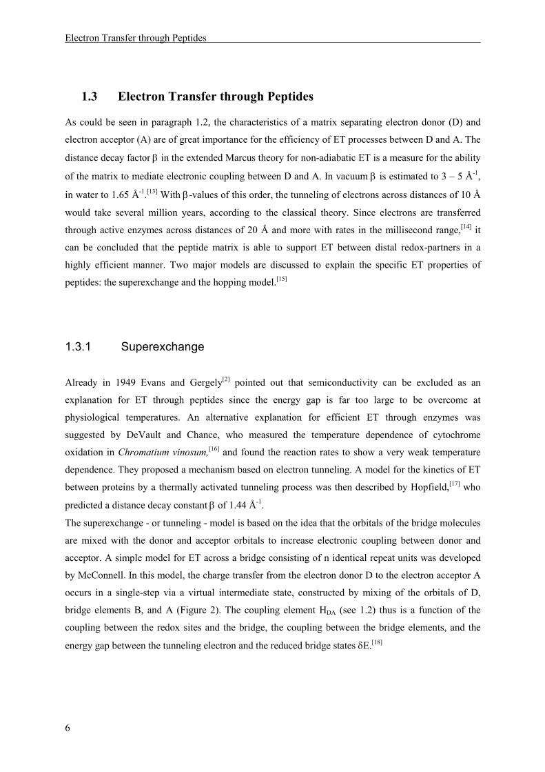
by McConnell. In this model, the charge transfer from the electron donor D to the electron acceptor A
occurs in a single-step via a virtual intermediate state, constructed by mixing of the orbitals of D,
bridge elements B, and A (Figure 2). The coupling element HDA (see 1.2) thus is a function of the
coupling between the redox sites and the bridge, the coupling between the bridge elements, and the
energy gap between the tunneling electron and the reduced bridge states δE.[18]

Electron Transfer through Peptides
7
Mixing of all available electronic states lowers the potential barrier and thereby increases the tunneling
probability HAD2. The ET rate is increased, as compared to the transfer rate in vacuum, by an improved
electronic coupling, which is expressed by a lowered value of the distance-decay factor β (see
Equation 3).
kET
D
A
δE
B1 B2 Bn
Figure 2: energy diagram for superexchange ET across a bridge of n identical units Bn (McConnell)
Since peptides can be built up from 20 different amino acids, the simple McConnell model cannot be
applied in this case. A semiempirical approach by Dutton is based on the observation that ET rates
through different types of proteins (reaction centres of different photosynthetic bacteria and several
synthetic and semisynthetic proteins) are almost identical for similar D/A-distances. Dutton therefore
concludes that the influence of peptide sequence is small and that ET pathways are optimized only in
terms of distance, not in terms of peptide structure. In consequence, he regards the peptide matrix as a
uniform barrier and estimates the β-value to 1.4 Å-1.[19, 20] A rough estimate of structural differences
has been taken up into a refined version of Duttons uniform barrier model, which includes the packing
density of the protein matrix as an additional factor.[21] Yet bioinformatic investigations revealed no
major difference in protein packing density between long-distance and short-distance electron
transfers. This leads back to the central hypothesis of Dutton`s model: the belief, that in the course of
evolution large effects of structural changes in ET proteins would have been highly unfavourable and
that nature therefore preferred ET-proteins to be robust when it comes to structural and sequence
changes. A a consequence of this demand, the peptide matrix is equipped with a mainly uniform ET
behaviour, controlled by cofactor distances, and not optimized with respect to ET properties in special
enzymes.[22]

Electron Transfer through Peptides
8
This view has been challenged by Gray and Winkler, who measured ET rates in modified
proteins.[14, 23] They used naturally occuring metalloproteins with copper (azurin)[24] or iron (cyto-
chrome)[25] cofactors and with known crystal structures and attached a ruthenium-complex to specific
histidine residues at defined distances. ET was then induced by photoexcitation of the ruthenium
chromophore, and subsequent rapid oxidation by an external acceptor (flash-quench-technique). The
rates of ET from the metal cofactor to the oxidized ruthenium complex were measured by absorption
spectroscopy of the transient metal complexes. The original oxidation state was then recovered by a
slow reaction with the reduced quencher (Figure 3). This method spans a wide time frame for the
measurement of ET rates ranging from sub-microseconds to seconds.[13]
Mn+
His Ru2+
Mn+
His *Ru2+
Mn+
His Ru3+
M[n+1]+
His Ru2+
hνννν
Q
Q
ET
Q
Q
Figure 3: Flash-quench technique for the induction and observation of ET in Ru-modified metalloproteins
The method was used to gain a large collection of experimental data from different classes of modified
metalloproteins. From the azurin series, a timetable for ET through proteins that proceeds according to
the tunneling model, with rates in the microsecond range for ET across 15-20 Å and a β-value of
1.1 Å-1 for a β-strand, was derived.[13] Nevertheless, in several cases, ET velocities cannot be
explained by D/A distances alone: ET rates for similar D/A distances can differ by several orders of
magnitude and similar rates can be observed for distances that differ by as much as 5 Å.[26] Gray and
Winkler therefore concluded, that the structure of the peptide matrix is the main factor controlling ET
rates.

Electron Transfer through Peptides
9
Based on the experimental data, a more complex theoretical model, which takes protein secondary
structure into account, has been proposed by Beratan and Onuchic.[27-29] It is built on the assumption,
that pathways of optimized electronic coupling exist, which are composed of covalent bonds,
H-bridges and through-space contacts. For each of these links a specific coupling decay (εc, εH, εS) is
assigned and the electronic coupling element HAD can be determined as the product of all couplings in
a specific pathway (Equation 4).
SHCADH εΠεΠεΠ∝
Equation 4:The electronic coupling element in the pathway model is given as the product of coupling through
covalent bonds εC, H-bonds εH and through space contacts εS.
Modern computational techniques in combination with available protein structure data allow a search
of all possible pathways to identify the optimum pathway. Using this method, a good agreement
between theory and the experimental data for the ET rates observed in modified metalloproteins can
be achieved.[30] The model also allows predictions concerning the ET properties of secondary structure
motifs. In recent work, the pathway model has been further refined by taking the effects of protein
dynamics into account.[14, 31-33] The key determinant of biological ET in the pathway model, as
opposed to the uniform-barrier model, is the protein fold.
Electrochemical measurements of oligopeptidic systems with α-aminoisobutyric acid (Aib) as the
peptide matrix , a phtalimid as N-terminal electron donor and peroxide as C-terminal electron acceptor
(Scheme 1), carried out by Maran and coworkers, support the pathway theory.[34] They found
increasing rates with increasing number n of spacer amino acids and attributed this unexpected effect
to an increase in intramolecular H-bonding, leading to an improved electronic coupling. Following
their argumentation, an increase in n does not only lead to an increased donor/acceptor separation, but
also to a concomitant increase in the number of intramolecular H-bonds in the 310-helix formed by
Aib-oligomers. [35]
NNH
O
O
HN O
O
nn = 0 to 6
O
O
Scheme 1: Oligopeptidic model system designed for electrochemical measurement of ET rates by Maran et al.
In summary, in superexchange ET, migration of the charge from the donor to the acceptor is enhanced
by bridge-mediated mixing of donor and acceptor wave functions, and occurs in a single step. The
Electron Transfer through Peptides
10
exponential decrease of ET rates with increasing D/A distance is characteristic of the superexchange
mechanism, with the magnitude of the exponential decay constant β depending on the ability of the
bridging matrix to mediate electronic coupling. The coupling strength of peptides is either regarded as
the result of a network of covalent bonds, H-bridges and through-space contacts (pathway model) or as
a structure-independent feature of these organic supramolecules (uniform barrier).
1.3.2 Hopping
Since the driving forces in naturally ocurring ET are relatively low, a practical upper distance-limit for
the superexchange mechanism exists. Dutton regards a distance of 14 Å as the upper limit of efficient
ET by superexchange.[20] Gray and Winkler set the maximum tunneling distance to 20Å.[13] Dutton
proposes the alignement of metal-containing redox-cofactors, especially Fe-S-clusters and hemes, in
enzymes involved in ET cascades, as a possible solution to allow transfer steps of more then 14 Å
overall distance.[20] If the distances between these cofactors stay below the limit of superexchange ET,
a sequence of tunneling steps can be used to transfer the electron. Such a stepwise processs is known
as multistep tunneling or electron hopping.
The hopping model for ET has first been developed for DNA. It stems from the observation that DNA
is a very efficient charge-carrier and that the efficiency of charge transfer scales with the number of
guanine base pairs. The experimental data on ET through DNA double strands, collected by Barton
and coworkers,[36] Giese and coworkers,[37, 38] and Lewis and Schuster and coworkers,[39, 40] could not
be explained by the superexchange theory. The electron coupling properties of DNA are in a range
expected for organic matrices, with β-values from 0.6 to 1.2 Å-1 and cannot rationalize the highly
efficient distal ET. However, the guanine base pairs with their low oxidation potential, offer the
possibility to form oxidized chemical intermediates in the ET process. ET between D and A can thus
proceed in a multistep process with the DNA-bases serving as stepping stones, which allows short
distances (and thus high transfer rates) for each step. This results in overall ET rates for which the
exponential D/A-distance dependency, predicted from the superexchange theory, does not apply.
Instead, the rate scales algebraically with distance and is a function of the number of intervening base
pairs n. The theoretical treatment of Bixon and Jortner:[41-43]
η−∝ nkk ibET
Equation 5: Hopping model for DNA. The rate of electron transfer kET as a function of the number of
intervening base pairs n.
Electron Transfer through Peptides
11
with kib giving the rate of interbase ET and η taking values between 1 and 2, has been confirmed
experimentally by work of Giese and coworkers.[44]
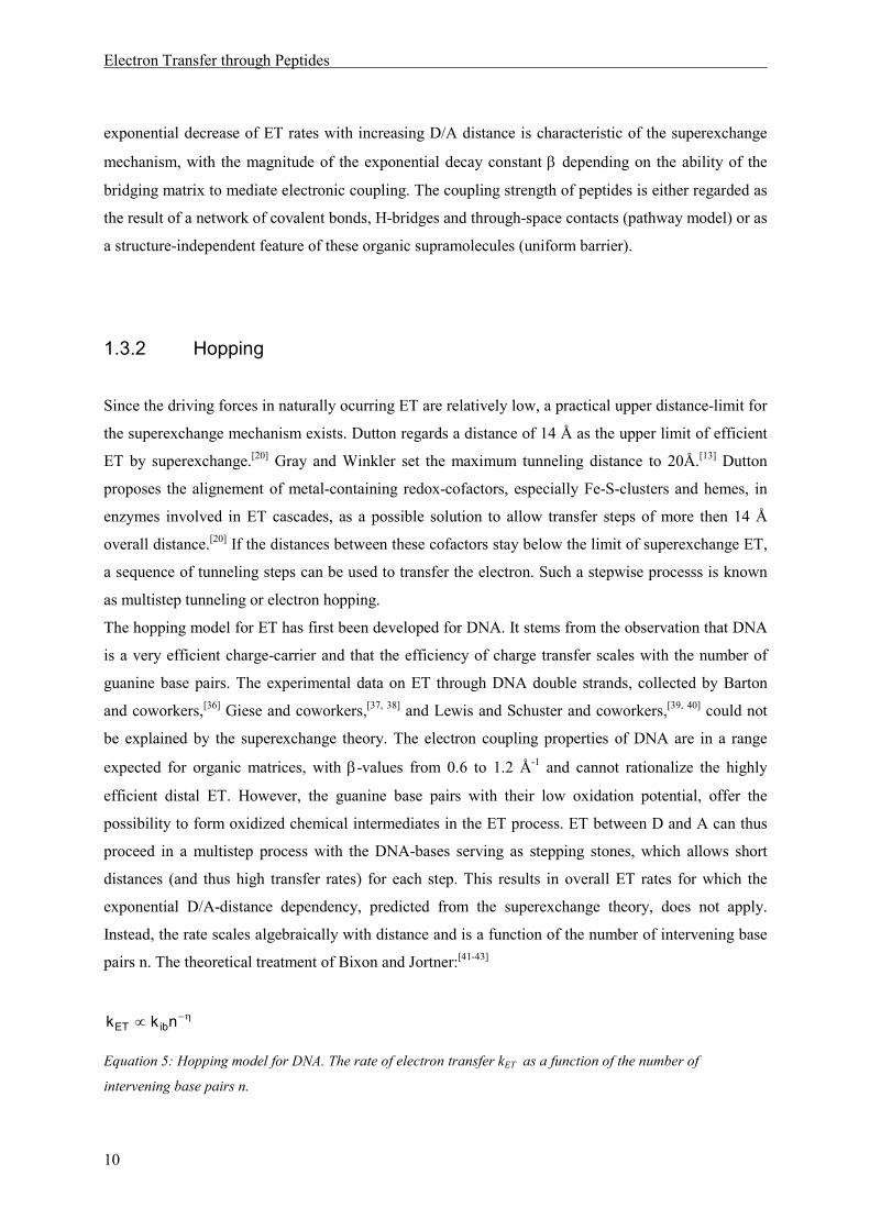
The main characteristic of sequential electron transfer (hopping) is the occurence of chemical
intermediates, oxidized (or reduced) relays R. The charge is transferred from the donor D to a
localized relay site R on the bridge, then passed through the bridge by a number of adjacent
oxidation/reduction reactions, before it finally reaches the acceptor A. An important characteristic of
this model is the possibility of endergonic transfer steps (Figure 4).[20, 45]
D
A
δE
R1 R2 Rn
Figure 4: Energy diagram for sequential ET with relays R that allow the occurence of oxidized intermediates.
A sequential ET mechanism is now generally accepted for electron transport during photosynthesis,
with the electron hopping from the reaction-centre (P680) to chlorophyll and subsequently to
pheophytin. Similar sequential transfer chains, using redox cofactors as relays, have been proposed for
other ET pathways in biology, [21] e.g. in E. coli fumarate reductase electrons are relayed over more
than 30 Å, presumably using a chain of Fe-S clusters.[6]
But in a number of proteins, electrons have to be transferred across large distances without the
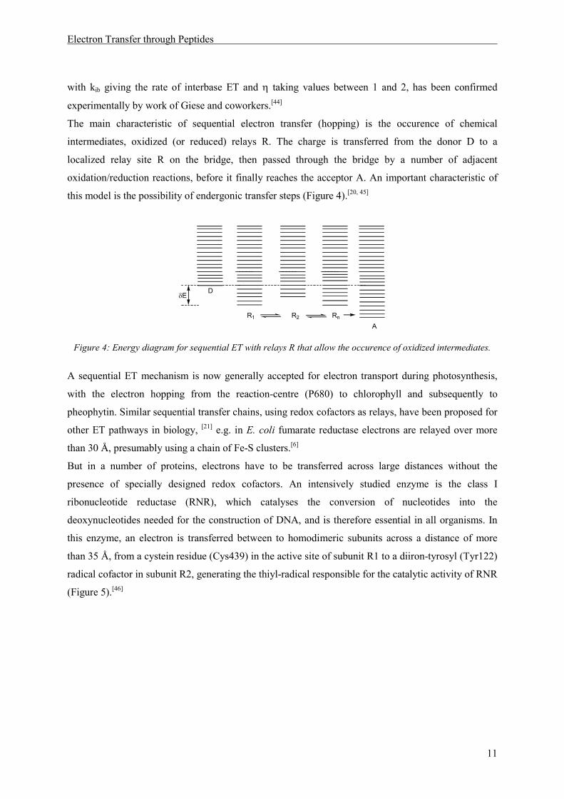
presence of specially designed redox cofactors. An intensively studied enzyme is the class I
ribonucleotide reductase (RNR), which catalyses the conversion of nucleotides into the
deoxynucleotides needed for the construction of DNA, and is therefore essential in all organisms. In
this enzyme, an electron is transferred between to homodimeric subunits across a distance of more
than 35 Å, from a cystein residue (Cys439) in the active site of subunit R1 to a diiron-tyrosyl (Tyr122)
radical cofactor in subunit R2, generating the thiyl-radical responsible for the catalytic activity of RNR
(Figure 5).[46]
Electron Transfer through Peptides
12
A transfer of the electron by a single-step superexchange mechanism can be excluded, since it would
be slow and limit the rate of nucleotide-conversion to 10-4 – 10-9 s-1, whereas the observed turnover
number for class I RNRs are measured to 2-10 s-1.[46] An ET hopping pathway, using aromatic amino
acids as relays, has been proposed in this enzyme.[47, 48]
Tyr122
O
O
Asp84
-O
N
HN
118His
NH
Fe Fe
HO
Tyr731
Tyr730HS
Cys439
Trp84Tyr356
R1
R2
7.6 Å
25 Å
3.3 Å
3.4 Å
4.2 Å
HO
Figure 5: Aromatic residues possibly involved in ET through RNR.
In vitro and in vivo mutagenensis studies have proven the aromatic amino acids depicted in Figure 5 to
be essential for enzyme function.[49] Substitution of Tyr356 by fluorinated tyrosine residues of higher
oxidation potential led to a loss of enzyme function.[50] This is a strong argument in favor of a
sequential mechanism, since the energy needed to form oxidized intermediates in the bridge is the
main factor determining the relative magnitude of superexchange versus hopping mechanism.[15, 45]
The enzyme DNA photolyase is another example, in which aromatic amino acid side chains are
thought to operate as "stepping stones" in electron hopping.[51, 52]

Electron Transfer through Peptides
13
In short-time spectroscopic studies of E. coli type I photolyase, the tryptophane residues Trp382,
Trp359 and Trp306 have shown to be oxidized during electron transfer from the flavine adenine
nucleotide (FAD) cofactor in the enzyme active site to a second redox active cofactor located close to
the protein surface (Figure 6).[53]
HN
NH
NH
Trp382
Trp359
Trp306FAD
4.2 Å
3.9 Å5.2 Å
Figure 6:Tryptophane residues participating in ET through typeI DNA photolyase
From this experimental data it can be concluded that amino acid side chains possibly operate as relays
in sequential ET and that, in consequence, not only secondary structure but also amino acid sequence
has to be considered in the explanation of protein ET. Aromatic amino acids as oxidized intermediates
might thus expand the theory of specifically designed sequential redox-chains[20] to enzymes that lack
the redox-active metal containing cofactors present in photosynthesis and respiration. Even in
enzymes with a high cofactor-densitiy, like PSII, aromatic amino acid residues are discussed as
additional intermediates in ET processes.[54] From the relay stations assumed in the DNA photolyase
(Figure 6), it can moreover be deduced that sequential ET might already come into play at distances
that are in principle short enough to be overcome by monostep tunneling.
Whether electron hopping does also occur in peptides without aromatic residues as relays, is a
question still under discussion. Peptide bonds have been proposed as possible relay sites by Schlag. He
developed a model for stepwise ET through peptide bonds, in which the bond-angles, i.e. peptide
dynamics, are of central relevance.[55, 56]
NH
CαC
HN
O
RΦ Ψ
Figure 7: Ramachandran angles in a peptide
He observed facile migration of a positive charge, generated by photosensitization of an aromatic
chromophore, through small peptides in the gas phase. The distance and direction of charge migration
depended strongly on the ionization potentials of the individual amino acids in the peptide chain – not

Electron Transfer through Peptides
14
on the overall ionization potential of the supramolecule. Based on these observations, Schlag
concluded that the charge is transferred via a stepwise mechanism. He calculated the energy barriers
for ET between adjacent amino acid residues and found them to be strongly dependent on the torsional
angles of the peptide bond, Φ and Ψ, the Ramachandran angles (Figure 7). According to his theory,
ET through a peptide chain is preceeded by the arrangement of adjacent peptide bonds into a favorable
angle, which minimizes the distance between neighbouring CO groups to 2.8 Å and allows a
subsequent fast oxidation reaction (firing).[55, 57]
The experiments on oligopeptides synthesized by Isied and coworkers also point towards the
possibility of electron hopping, even without the presence of oxidizable aromatic side chains. They
have developed an experimental system to measure the distance dependence of ET rates in
oligoprolines, in which ET in a Creutz-Taube analogon containing two ruthenium centres, is induced
by pulse radiolysis A or flash photolysis B (Scheme 2). The ET reaction can be monitored by
absorption spectroscopy of the transient metal complexes. From the measured rate differences upon
variation of spacer length, Isied concluded, that a mechanistic transition from superexchange
(exponential distance dependence) to hopping (linear distance dependence) takes places, starting from
a length of 5 proline residues.[58]
O
n
eaq + RuIIL-Pron-(apy)RuIII
RuIIL-Pron-(apy)RuII
RuII(L -)-Pron-(apy)RuIII
RuIIL*-Pron-(apy)RuIII
RuIIL-Pron-(apy)RuIII
RuIIIL-Pron-(apy)RuIIhν
A
B
N
HN
O NRu(NH3)5
(bpy)2Ru
Scheme 2: Oligoproline model synthesized by Isied for distance dependent ET measurements
A linear distance dependence for ET has also been observed by Kraatz and coworkers in oligoglycine
self-assembled monolayers, in which ET rates between ferrocene and a gold-electrode were measured
electrochemically. [59, 60] Yet though there is, in addition, spectroscopical data available, that points
towards amide participation in ET through peptides,[61] the occurence of oxidized peptide bonds as
intermediates seems to be unfavourable, due to the high activation energy needed to permit such an
oxidation reaction.[13]

Electron Transfer through Peptides
15
An explanation for the observation of unexpected ET behaviour upon peptide-spacer elongation, is
through-space electron transfer. In contrast to the superexchange theory discussed in paragraph 1.3.1,
the peptide bridge orbitals are not involved in mediating ET in this model.[62] Instead, ET occurs
directly between D and A, either by direct D/A orbital overlap, or by participation of solvent
molecules. Through-space ET has been discussed especially in the context of experiments on small
peptides in solution. Peptide models for the measurement of ET based on oligoproline units as spacers
have been developed among others by Meyer[63, 64] and Giese.[65] The weak dependencies of ET rates
on the number of separating proline units observed in those systems, point to through-space ET as the
dominant pathway.
In summary, the sequential or hopping mechanism for ET is characterized by the temporary
localization of the electron on the bridge, i.e. it requires the existence of oxidized chemical inter-
mediates. ET rates exhibit only a weak (linear) distance dependence and are strongly dependent on the
oxidation potentials of the bridging units (relays). Endergonic steps are possible in sequential ET.
Traveling of the electron between the possible redox-sites can occur via through-bond or through-
space steps. Metal-cofactors are known to assist in long-range peptide electron transfer through
enzymes involved in photosynthesis and respiration. Long-range ET is, however, also observed in
enzymes that do not possess suitable cofactor-arrangement. A hypothesis currently studied is the
function of aromatic amino acid side chains as hopping relays in such enzymes.


Research Project
17
2 Research Project
The aim of this work was to contribute to the investigation of sequential ET via oxidizable amino acid
side chains. Information about the role of individual amino acid side chains in ET through peptides
should be gained. The advantage of a small model systems in this context is the modular design, that
allows the fast synthesis of peptides which differ in parameters relevant for ET, like distances and
driving forces. And, in comparison with molecular biology approaches, the possibility to replace
individual amino acids without completely shutting down ET activity. Moreover, the spectroscopic
observation of intermediates is less restricted by a smaller matrix.
For these reasons, an oligopeptidic model should be designed, that allows the site selective injection of
a charge into a peptide, generating an electron acceptor site A, and the subsequent observation of
charge migration to an electron donor site D. An amino acid, suitable to serve as a relay for stepwise
ET should be placed between D and A (Figure 8). So far, the differentiation between superexchange
and hopping mechanism in model peptides has been achieved by measurement of distance
dependencies, which are expected to be exponential for superexchange and linear for sequential ET.
Yet the structural flexibility of small peptides in solution is a limitation of this approach, since D/A
edge-to-edge distances vary significantly for different peptide conformers. A straightforward approach
to overcome this problem is the incorporation of an additional spectroscopic sensor into the model,
that allows the detection of chemical intermediates and thus provides direct proof of the hopping
mechanism.
HN
O
Peptide PeptideD A
R
Figure 8: Schematic design of a peptide model for the observation of sequential ET via the relay R
We designed an aromatic amino acid R, to function as a central redox relay and spectroscopic sensor.
Using this approach, a peptide should be developed, in which stepwise intramolecular ET can be
induced and subsequently observed time-dependently, using orthogonal spectroscopic properties of D,
R and A. This design results in a number of experimental restrictions concerning the electrochemical
and spectroscopical properties of D, A and R, that will be discussed in detail in chapter 3.3. Another
difficulty stems from the fact that the concentration of the intermediate depends on the relative rates of
the tranfer steps D→R and R→A. The synthesis of peptides with different D/R and R/A distances was
therefore likely to be necessary in the search for a system with appropriate rate ratios to observe the

Research Project
18
oxidized intermediate. Replacement of R by side chains, which can not serve as relays and/or do not
produce observable intermediates, should then yield information about the importance of the relay for
ET. This leads to additional restrictions concerning the structure of the peptides. Structural uniformity
among a peptide series with different relays R has to be guaranteed to allow comparability of the
results. In other words: replacing R should show only minor effects on the overall structure, yet allow
the observation of effects on intramolecular ET efficiency. A system, in which those conditions can be
met, allows the investigation of the role of amino acid side chains in ET through peptides. In addition,
quantitative information about the individual rates might be derived from the spectroscopic
measurements.
In summary, a peptide model should be developed, in which a) sequential ET can be directly observed,
b) the role of individual amino acid side chains in this transfer can be investigated, c) information
about ET rates can be gained.

Spectroscopic Sensors for the Observation of Transients in ET through peptides
19
3 Spectroscopic Sensors for the Observation of Transients in
ET through peptides
3.1 Introduction
A peptide model 1 that allows the site selective generation of an oxidized aromatic amino acid side
chain at the C-terminus of an oligopeptide and the subsequent time-resolved spectroscopical
observation of ET from an N-terminal electron donating amino acid, was synthesized in the Giese
group by Napp (Scheme 3).[65, 66]
AcHNN
HN
O
O
O
O
O
OOPO(OPh)2
HO
OMe
MeO
OMe
n
1
Scheme 3: Napp-system for the observation of ET through peptides
In this peptide, a charge injection system 2, similar to the one developed in the Giese group to study
the transfer of a positive charge through DNA,[67, 68] was placed at the C-terminus of the oligopeptide
via ester-linkage. Irradiation of 2 with a laser flash of 308 nm leads to Norrish-type-I photocleavage of
the ketone functionality. The resulting radical 3 subsequently undergoes heterolytic β-elimination of
the phosphate group and yields radical cation 4 (Scheme 4). A major advantage of this injection
system is the generation of radicals in the electronic ground state. ET reactions can thus be observed
without side reactions stemming from photoexcited intermediates.
O
HO
O
t-Bu
OPO(OPh)2
- t-Bu- CO
O
OPO(OPh)2
O- (PhO)2PO2
-hν (λ = 308 nm)
2 3 4
HO HO
Scheme 4: Photoinduced radical cation generation
The mechanism of the injection reaction leads to several requests regarding the reaction conditions for
laser flash photolysis (LFP) measurements. They have to be carried out in an oxygen-free
environment, because oxygen addition to radical 3 is a fast side reaction, leading to formation of the
peroxide and quenching the intended radical cation formation. Moreover polar solvents are necessary,

Spectroscopic Sensors for the Observation of Transients in ET through peptides
20
to ensure a fast dissociation of the phosphate leaving group form the remaining radical cation 4.[69, 70]
A 3:1 mixture of acetonitrile and water has been found to offer optimum reaction conditions with
respect to injector function and peptide solubility.
With an oxidation potential of estimated 1.29 – 1.44 V vs. NHE,[71] radical cation 4 is able to oxidize
the aromatic side chain of the adjacent amino acid in peptide 1 and thus generate an electron acceptor
in the oligopeptide, yielding the transient 5. Radical cation 5 can be observed by transient absorption
spectroscopy, with an absorption maximum at 550 nm. This signal vanishes by ET from tyrosine,
which deprotonates upon oxidation and yields a tyrosyl radical 6, with an absorption maximum around
410 nm[72] (Scheme 5).
hν (308 nm)
- H+
AcHNN
HN
O
O
O
O
O
HO
MeO
OMe
OMe
AcHNN
HN
O
O
O
O
O
O
MeO
OMe
OMe
nn1
5 6
ET
Scheme 5: Electron transfer through an oligoproline chain in the Napp-model
Napp noticed, that the introduction of non redox-active amino acid residues between the
trimethoxysubstituted phenylalanine and the injection moiety leads to a severe drop in signal
intensities. This can be explained by a side reaction of radical cation 4, the water-addition reaction,
which comes into play when ET between 4 and the aromatic ring is slower than 8·106 s-1. A short
linkage between the oxidizable amino acid and the injector is therefore needed to ensure fast injection
and prevent side reactions of 4.[66] Peptide model 1 was used for time-resolved measurements of ET
5 → 6 in molecules with different oligoproline spacers (n = 0, 1, 3, 5). As a result, the rates for
intramolecular ET, extracted from the decay of the transient absorption at 550 nm (5), depended only
weakly on the spacer length, with rates ranging from 8.6·106 for direct D/A linkage (n = 0) to 2.7·105
for n = 5. This led to the conclusion, that ET occurs in a through-space mechanism, not via through-
bond tunneling (cf. paragraph 1.3, page15). The small effect of spacer-elongation on the rate might be
rationalized by the flexibility of the peptide structure, due to rotational freedom around the Cα-CH2-
bond of the aromatic residues and the resulting existence of peptide conformers with short D/A-
distances. Molecular modeling experiments for 1 with n = 5, with the peptide backbone conformation
fixed to the all-trans polyproline II structure, showed distance variations for the aromatic residues of
more than 5 Å.[66]
In summary, peptide model 1 can be used for the direct observation of ET through a peptide matrix.
ET can be assumed to occur in a through-space mechanism.

Spectroscopic Sensors for the Observation of Transients in ET through peptides
21
3.2 Laser Flash Photolysis
The method of laser flash photolysis (LFP) offers several advantages for the analysis of
photogenerated ET reactions through peptides. Since peptides with specific absorption properties can
be designed by the choice of amino acid side chains, the rates of ET can be derived directly from the
transient spectra. Neither photoproduct analysis nor the comparison of observed rates with competing
side reactions is necessary.[73] ET processes were induced by irradiation of peptide solutions with a
laser flash of 308 nm wavelength, 20 ns pulse duration and an energy of 100 – 130 mJ. The flash was
generated by a XeCl-excimer laser and directed to a UV-cuvette (Figure 9), that allowed for degassing
of the sample and storage under vacuum. Orthogonal to the laser a pulsed Xe-lamp provided the light
needed to record a UV-Vis spectrum of the sample.
cuvette
laser
Xe-lamp lense
mirror
monochromator
photomultiplier
oscilloscope
computerCCD-camera
x
shutter
Figure 9: Experimental setup for laser flash photolysis.
Two detection systems could be used: a photomultiplier allowed time-resolved measurements of
absorption at a defined wavelength. Processes with rates up to 3·107 s-1 could be resolved. With a
CCD-camera and a fast electronic shutter the complete transient absorption spectrum between 300 and
650 nm could be measured after a defined time-delay.[74-76]
The concentration of the sample may not exceed 8 mM because the optical density (OD), i.e. the UV-
absorption of the peptide sample at the wavelength of laser-irradiation, should be below 0.5 to
guarantee homogeneous photoreaction properties. Because of the low effiency of Norrish-
Spectroscopic Sensors for the Observation of Transients in ET through peptides
22
photocleavage of the tert-butylketone injection moiety, the signal intensities dropped below detection
limits at sample-concentration of less than 1 mM. Therefore, the experiments discussed in this work
were carried out at concentrations of 1 – 8 mM.
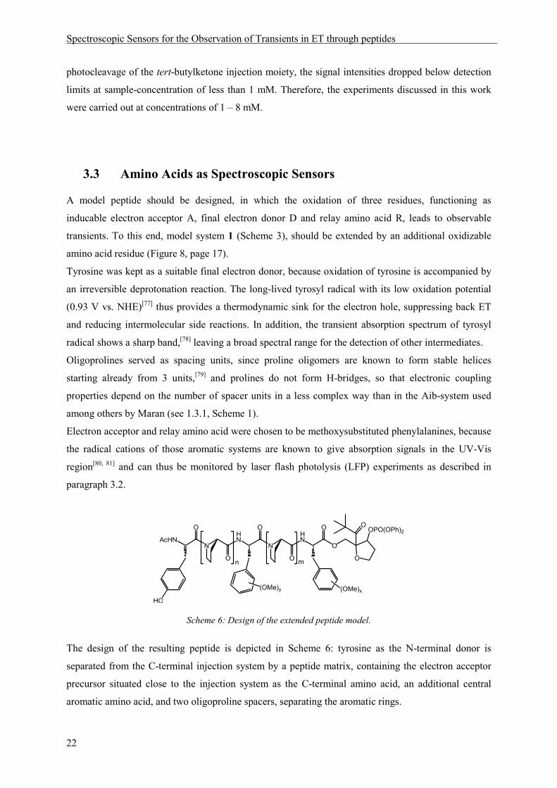
3.3 Amino Acids as Spectroscopic Sensors
A model peptide should be designed, in which the oxidation of three residues, functioning as
inducable electron acceptor A, final electron donor D and relay amino acid R, leads to observable
transients. To this end, model system 1 (Scheme 3), should be extended by an additional oxidizable
amino acid residue (Figure 8, page 17).
Tyrosine was kept as a suitable final electron donor, because oxidation of tyrosine is accompanied by
an irreversible deprotonation reaction. The long-lived tyrosyl radical with its low oxidation potential
(0.93 V vs. NHE)[77] thus provides a thermodynamic sink for the electron hole, suppressing back ET
and reducing intermolecular side reactions. In addition, the transient absorption spectrum of tyrosyl
radical shows a sharp band,[78] leaving a broad spectral range for the detection of other intermediates.
Oligoprolines served as spacing units, since proline oligomers are known to form stable helices
starting already from 3 units,[79] and prolines do not form H-bridges, so that electronic coupling
properties depend on the number of spacer units in a less complex way than in the Aib-system used
among others by Maran (see 1.3.1, Scheme 1).
Electron acceptor and relay amino acid were chosen to be methoxysubstituted phenylalanines, because
the radical cations of those aromatic systems are known to give absorption signals in the UV-Vis
region[80, 81] and can thus be monitored by laser flash photolysis (LFP) experiments as described in
paragraph 3.2.
HN
N
HN
O
O
O
O
O
O
OOPO(OPh)2
N
O
AcHN
HO
(OMe)x
n m
(OMe)y
Scheme 6: Design of the extended peptide model.
The design of the resulting peptide is depicted in Scheme 6: tyrosine as the N-terminal donor is
separated from the C-terminal injection system by a peptide matrix, containing the electron acceptor
precursor situated close to the injection system as the C-terminal amino acid, an additional central
aromatic amino acid, and two oligoproline spacers, separating the aromatic rings.

Spectroscopic Sensors for the Observation of Transients in ET through peptides
23
To choose the right substitution pattern for the non-natural methoxysubstituted phenylalanines, several
conditions have to be taken into account:
1. the redox potentials of the aromatic rings should be between 1.3 V vs. NHE (injector radical
cation 4)[71] and 0.93 V vs. NHE (Tyr).[77] Though endergonic steps are in principle possible in
electron hopping,[20, 45, 51] the transfer chain should be designed as a sequence of downhill steps. Thus,
attention had to be paid to the relative oxidation potentials of acceptor and relay as well.
2. The transient absorption maxima of the generated oxidized species should be located at different
wavelengths, to allow the simultaneous observation of all oxidized intermediates. In addition, the
extinction coefficients of acceptor and relay radical cation as well as the tyrosyl radical should be of
comparable magnitude, so that good resolution of all three signals is possible.
3. The amino acids used in the construction of the peptide model should be transparent at the
wavelength of laser irradiation, 308 nm, to avoid a loss in photocleavage efficiency as well as possible
side reactions of photoexcited intermediates.
2,4,6-Trimethoxyphenylalanine has already served as a sensor in ET through peptides and is thus
known to meet conditions 1 and 3. Dimethoxybenzenes with 1,3 and 1,4 substitution pattern were
examined on their suitability to supplement the tyrosine/trimethoxyphenylalanine system, with focus
on the conditions described above.
ad 1: The redox potentials of methoxysubstituted benzenes could be extracted from the literature. The
oxidation potential of 1,3,5-trimethoxybenzene, the side chain of the amino acid used by Napp in the
model system 1, is given with 1.46 - 1.49 V vs. SCE;[82-84] 1,3-dimethoxybenzene with 1.49 V - 1.6 V
vs. SCE[82, 85, 86] and 1,4-dimethoxybenzene with values ranging from 1.34 to 1.51 vs. SCE.[82, 83, 85, 86]
So the oxidation potentials of all possible candidates fall in a range suitable to serve as relays in ET
between the injector radical cation 4 (1.54 vs. SCE) and tyrosine (1.17 vs. SCE).
ad2:
In addition to the literature values available for the absorption maxima of the radical cations
(1,3-dimethoxybenzene: 477 - 505 nm, 1,4-dimethoxybenzene: 443 - 475 nm),[82] photosensitization
experiments were carried out to allow direct comparison of the radical cation absorptions. For this
purpose, 1 mM concentrations of 1,3-dimethoxybenzene and 1,4-dimethoxybenzene were irradiated
with dicyanonaphthalene (DCN) as a photosensitizer and biphenyl (BP) as a codonor in air saturated
acetonitrile. Electron rich aromatic compounds can be photooxidized under these conditions.[81, 87]
The reaction starts with the excitation of DCN. Subsequent fast ET from BP generates the BP radical
cation, which is able to oxidize electron rich aromatic compounds (Ar). DCN is then regenerated by

Spectroscopic Sensors for the Observation of Transients in ET through peptides
24
reaction with oxygen (Scheme 7). The transient absorption spectra of the formed aromatic radical
cations were measured after a delay of 500 ns. (Figure 10).
DCN BP BP
Ar BP Ar
DCN O2 DCN O2
DCN+-
BP+ +
- -
hν
308 nm
CN
CN
DCN BP
Scheme 7: Photosensitization to genererate aromatic radical cations Ar·+ from the respective aromates.
While the absorption maximum of the para substituted dimethoxybenzene radical cation (grey line)
stretches from 430 nm to 460 nm, the meta substituted derivative (black line) has its maximum
absorption between 445 and 475 nm.
Figure 10: Transient absorption spectra recorded after oxidation of 1,3-dimethoxybenzene (black) and 1,4-
dimethoxybenzene (grey) by photosensitized electron transfer with 1,4-dicyanonaphthalene as a sensitizer and
biphenyl as a codonor.
Both radical cations yield a transient absorption spectrum that can be differentiated from the spectrum
of the tyrosyl radical (λmax = 410 nm) and the trimethoxyphenylalanine radical cation (λmax = 550 nm).
OMe
OMe
OMe
OMe

Spectroscopic Sensors for the Observation of Transients in ET through peptides
25
ad3: Since peptide concentrations in the LFP measurements have to be in the millimolar range, due to
the low absorption coefficient of the injector chromophore, the transparency of the chosen amino acid
residues at the wavelength of laser irradiation is of crucial importance. UV-spectra of 1,3-
dimethoxybenzene and 1,4-dimethoxybenzene were therefore recorded at 8 mM concentrations in
acetonitrile.
Figure 11: UV-spectra of 1,3-dimethoxybenzene (black) and 1,4-dimethoxybenzene (grey) 8 mM in acetonitrile.
The vertical line in Figure 11 indicates 308 nm. It is obvious from the graphs that the para substituted
dimethoxybenzene has to be excluded as a candidate for one of the non-natural amino acids needed to
construct a peptide model containing three oxidizable spectroscopic sensors (Scheme 6).
1,3-dimethoxybenzene was therefore chosen together with the already established 1,3,5-
trimethoxybenzene as an appropriate aromatic side chain for the acceptor resp. relay amino acid.
Spectroscopic Sensors for the Observation of Transients in ET through peptides
26
3.3.1 Synthesis and Properties of Methoxysubstituted Phenylalanine-
Derivatives
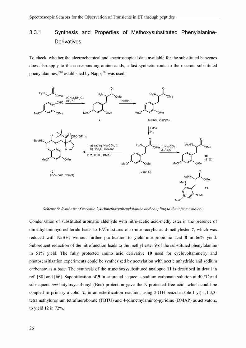
To check, whether the electrochemical and spectroscopical data available for the substituted benzenes
does also apply to the corresponding amino acids, a fast synthetic route to the racemic substituted
phenylalanines,[88] established by Napp,[66] was used.
O
OMeO2N
CHO
O2N
O
OMeO2N
O
OMe
H2N
O
OMeAcHN
O
OMe
(CH3)2NH2Cl,KF, ∆ NaBH4
Pd/C,H2
1. a) sat aq. Na2CO3, ∆ b) Boc2O, dioxane
1. Na2CO32. Ac2O
OMeMeO OMeMeO OMeMeO
OMeMeO OMeMeO
2. 2, TBTU, DMAP
BocHNO
O
AcHN
O
OMe
OMe
MeO
MeO
7 8 (66%, 2 steps)
9 (51%)12 (72% calc. from 9)
10(81%)
11
MeO OMe
O
OPO(OPh)2
O
Scheme 8: Synthesis of racemic 2,4-dimethoxyphenylalanine and coupling to the injector moiety.
Condensation of substituted aromatic aldehyde with nitro-acetic acid-methylester in the presence of
dimethylaminhydrochloride leads to E/Z-mixtures of α-nitro-acrylic acid-methylester 7, which was
reduced with NaBH4 without further purification to yield nitropropionic acid 8 in 66% yield.
Subsequent reduction of the nitrofunction leads to the methyl ester 9 of the substituted phenylalanine
in 51% yield. The fully protected amino acid derivative 10 used for cyclovoltammetry and
photosensitization experiments could be synthesized by acetylation with acetic anhydride and sodium
carbonate as a base. The synthesis of the trimethoxysubstituted analogue 11 is described in detail in
ref. [88] and [66]. Saponification of 9 in saturated aequeous sodium carbonate solution at 40 °C and
subsequent tert-butyloxycarbonyl (Boc) protection gave the N-protected free acid, which could be
coupled to primary alcohol 2, in an esterification reaction, using 2-(1H-benzotriazole-1-yl)-1,1,3,3-
tetramethyluronium tetrafluoroborate (TBTU) and 4-(dimethylamino)-pyridine (DMAP) as activators,
to yield 12 in 72%.

Spectroscopic Sensors for the Observation of Transients in ET through peptides
27
The injector moiety 2 was prepared in 7 steps starting from pivaldehyde with a 1,3 dipolar
cycloaddition of a nitrileoxide to furane as the key step, according to synthesis protocols developed in
the Giese group.[66, 89, 90]
The redox potentials of the aromatic amino acids were measured by differential pulse
cyclovoltammetry, using acetonitrile as a solvent and ferrocene as internal standard (Figure 12).
Figure 12: Differential pulse cyclovoltammetric measurement of Ac-2,4-dimethoxyphenylalaninemethylester 10
(black) and Ac-2,4,6-trimethoxyphenylalaninemethylester 11 (grey) 2mM in acetonitrile with 100 mM
tetrabutylammonium hexafluorophosphate (TBAPF6) as electrolyte and ferrocene as internal standard.
The potentials referring to ferrocene are 0.95 V for 2,4-dimethoxyphenylalanine 10 and 0.94 V for
2,4,6-trimethoxyphenylalanine 11. In comparison, we measured the potential of N-Acetyl-tyrosine-
methylester to amount to 0.85 V vs. the ferrocene redox couple. This equals the value determined by
Harriman in water (pH 7) of 0.93 V vs. NHE.[77]
Spectroscopic Sensors for the Observation of Transients in ET through peptides
28
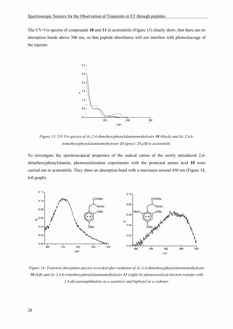
The UV-Vis spectra of compounds 10 and 11 in acetonitrile (Figure 13) clearly show, that there are no
absorption bands above 300 nm, so that peptide absorbance will not interfere with photocleavage of
the injector.
Figure 13: UV-Vis spectra of Ac-2,4-dimethoxyphenylalaninemethylester 10 (black) and Ac-2,4,6-
trimethoxyphenylalaninemethylester 11 (grey); 20 µM in acetonitrile.
To investigate the spectroscopical properties of the radical cation of the newly introduced 2,4-
dimethoxyphenylalanine, photosensitization experiments with the protected amino acid 10 were
carried out in acetonitrile. They show an absorption band with a maximum around 450 nm (Figure 14,
left graph).
Figure 14: Transient absorption spectra recorded after oxidation of Ac-1,4-dimethoxyphenylalaninemethylester
10 (left) and Ac-2,4,6-trimethoxyphenylalaninemethylester 11 (right) by photosensitized electron transfer with
1,4-dicyanonaphthalene as a sensitizer and biphenyl as a codonor.
OMe
COOMe
NHAc
OMe
OMe
COOMe
NHAc
OMeMeO
Spectroscopic Sensors for the Observation of Transients in ET through peptides
29
For comparison, the spectrum of the trimethoxyphenylalanine 11 used in previous experiments (see
also ref. [66] and [65]) was also recorded, showing a maximum at 550 nm (Figure 14, right graph).
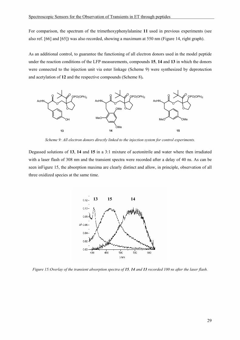
As an additional control, to guarantee the functioning of all electron donors used in the model peptide
under the reaction conditions of the LFP measurements, compounds 15, 14 and 13 in which the donors
were connected to the injection unit via ester linkage (Scheme 9) were synthesized by deprotection
and acetylation of 12 and the respective compounds (Scheme 8).
AcHNO
O
O
OOPO(OPh)2
AcHNO
O
O
OOPO(OPh)2
AcHNO
O
O
OOPO(OPh)2
13 14 15
OH
OMe
MeO
OMe
MeO OMe
Scheme 9: All electron donors directly linked to the injection system for control experiments.
Degassed solutions of 13, 14 and 15 in a 3:1 mixture of acetonitrile and water where then irradiated
with a laser flash of 308 nm and the transient spectra were recorded after a delay of 40 ns. As can be
seen inFigure 15, the absorption maxima are clearly distinct and allow, in principle, observation of all
three oxidized species at the same time.
Figure 15:Overlay of the transient absorption spectra of 15, 14 and 13 recorded 100 ns after the laser flash.
13 15 14

Spectroscopic Sensors for the Observation of Transients in ET through peptides
30
The spectroscopical and electrochemical data collected on the racemic amino acids as well as a first
test in LFP experiments confirmed the suitability of the chosen phenylalanine derivatives to function
as charge carriers and spectroscopic sensors in ET through peptides. From the oxidation potentials
cited in the literature for the aromatic systems, no explicit trend could be derived with respect to the
order of the side chains preferred in ET. But the pulsed cyclovoltammetry measurements carried out
on the amino acids, indicated a slightly higher potential for the dimethoxysubstituted ring.

Stability of Radical Cations
31
4 Stability of Radical Cations
The small difference in oxidation potential between the two non-natural aromatic amino acids led to
the question, if 2,4-dimethoxyphenylalanine radical cation can oxidize 2,4,6-trimethoxyphenylalanine
in the absence of tyrosine, which provides the driving force for ET in a peptide model containing all
three redox sites. Therefore, quenching experiments were carried out with the small model 15. Time-
resolved measurements of the absorption intensity at 450 nm, the maximum absorption of 2,4-
dimethoxyphenylalanine radical cation (see above), were carried out with 15 alone, and after addition
of equimolar amounts of Ac-2,4,6-trimethoxyphenylalaninemethylester (11), resp. Ac-tyrosine-
methylester. The curves depicted in Figure 16 show a considerable quenching effect upon addition of
trimethoxyphenylalanine (red line), demonstrating that ET from the trimethoxysubstituted aromatic
ring to the oxidized dimethoxy derivative is possible in an intermolecular reaction. As expected, a
large radical cation quenching effect is observed in the presence of tyrosine (green line).
Figure 16: Time-resolved LFP measurements of the absorption at 450 nm of small model 15 alone (black line)
and upon addition of Ac-2,4,6-trimethoxyphenylalanine-methylester (red line) resp. Ac-tyrosine-methylester(11)
(green line) as a quencher.
MeO
OMe
OMe
COOMe
NHAc
OH
COOMe
NHAc
AcHNO
O
O
MeO
OMe
+
+
11

Stability of Radical Cations
32
In a next experiment, the question should be answered, to which extent ET between electron acceptor
and relay amino acid occurs, when those two are assembled in one peptide (for a detailed description
of peptide synthesis protocols see chapter 5.3.). In peptide 16, only the two methoxysubstituted
phenylalanine derivatives, separated by one proline unit, are present; no driving force is induced by a
final irreversible ET step (Scheme 10).
AcHNN
HN
O
O
O
O
O
OOPO(OPh)2
OMe
OMe
16
OMe
MeO
MeO
Scheme 10: Peptide 16 contains electron acceptor and relay amino acid in close proximity but no final donor
functioning as a thermodynamic sink.
The transient absorption spectrum, recorded 40 ns after laser irradiation of 16, clearly shows the
simultaneous presence of both radical cations with two absorption bands at 450 nm (2,4-
dimethoxyphenylalanine radical cation) and 550 nm (2,4,6-trimethoxyphenylalanine radical cation).
Thus, in a peptide containing aromatic residues of similar oxidation potentials, a distribution of the
charge across the aromatic rings is observed.
Figure 17: Transient absorption spectrum recorded 40 ns after laser irradiation of 16.
For comparison, the spectrum of peptide 17 containing the final electron donor tyrosine at the N-
terminal position, separated from 2,4-dimethoxyphenylalanine as electron acceptor precursor by one
proline unit, was recorded.

Stability of Radical Cations
33
In this peptide, the electron transfer step can be assumed to be irreversible, due to deprotonation of
tyrosine upon ET. The transient absorption spectrum, recorded 40 ns after the laser flash, clearly
shows, that the charge has almost completely been transferred to the final electron donor
(λmax = 410 nm) within this delay, with just a small residual absorption intensity around 450 nm
(Figure 18).
Figure 18: Transient absorption spectrum recorded 40 ns after laser irradiation of 17.
This result is in accordance with the difference in driving forces for ET in peptides 16 and 17,
resulting from the oxidation potentials of the participating redox partners. While a contribution of the
charge between dimethoxy and trimethoxybenzene in 16 might be achieved in an equilibrium reaction,
due to their similar oxidation potentials, tyrosine is deprotonated upon oxidation to the tyrosyl radical
in an irreversible reaction in 17.
HN
O
O
O
OOPO(OPh)2
N
O
AcHN
HO
OMe
OMe
O
17

Stability of Radical Cations
34
In an additional quenching experiment, the stability of the electron acceptor in peptide 16 was
investigated. Surprising results were obtained from time-resolved measurements of 450 nm absorption
in this peptide. A slower decay of the dimethoxyphenylalanine radical cation (black line, Figure 19) is
observed, compared to the respective decay in small model 15, in which no second amino acid is
present (black line, Figure 16, page 31). This points to an increase in stability, possibly induced by the
formation of a charge transfer complex between the two aromatic sites. Upon addition of equimolar
amounts of trimethoxyphenylalanine derivative 11 as a quencher to the solution of 16, no substantial
effect on the radical cation decay could be measured (red line). Addition of tyrosine, however, led to
the expected acceleration in signal decay (green line).
Figure 19: Time-resolved LFP measurements of the absorption at 450 nm of peptide 16 alone (black line) and
upon addition of Ac-2,4,6-trimethoxyphenylalaninemethylester (grey line) resp. Ac-tyrosinemethylester (light
grey line) as a quencher.
MeO
OMe
OMe
COOMe
NHAc
OH
COOMe
NHAc
AcHNN
HN
O
O
O
O
OOMe
OMe
OMeMeO
MeO
+
+
11
Stability of Radical Cations
35
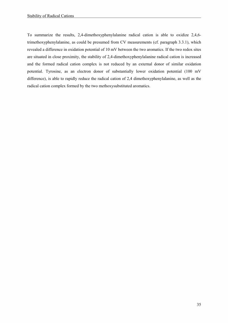
To summarize the results, 2,4-dimethoxyphenylalanine radical cation is able to oxidize 2,4,6-
trimethoxyphenylalanine, as could be presumed from CV measurements (cf. paragraph 3.3.1), which
revealed a difference in oxidation potential of 10 mV between the two aromatics. If the two redox sites
are situated in close proximity, the stability of 2,4-dimethoxyphenylalanine radical cation is increased
and the formed radical cation complex is not reduced by an external donor of similar oxidation
potential. Tyrosine, as an electron donor of substantially lower oxidation potential (100 mV
difference), is able to rapidly reduce the radical cation of 2,4 dimethoxyphenylalanine, as well as the
radical cation complex formed by the two methoxysubstituted aromatics.


Occurence of Oxidized Intermediates in ET through peptides
37
5 Occurence of Oxidized Intermediates in ET through peptides
5.1 Introduction
A peptide model should be constructed, in which sequential ET through a peptide chain containing
three aromatic amino acid side chains as redox sites, was possible and could be observed by transient
absorption spectroscopy. The natural amino acid tyrosine as an irreversible electron donor should be
situated at the N-terminus. Two methoxysubstituted phenylalanines, 2,4-dimethoxyphenylalanine and
2,4,6-trimethoxyphenylalanin were chosen as additional redox sites, because of their suitable
electrochemical and spectroscopic properties. The information regarding the possibility of ET between
2,4,6-trimethoxyphenylalanine and 2,4-dimethoxyphenylalanine radical cation, obtained by
cyclovoltammetry measurements and quenching experiments, lead to the decision to place the
dimethoxy phenylalanine derivative at the acceptor and the trimethoxy derivative at the relay position.
The resulting peptide model is depicted in Scheme 11.
HN
N
HN
O
O
O
O
O
O
N
O
AcHN
HO
OMe
OMe
1.02 V1.01 V0.93 V
450 nm550 nm410 nm
n m
t-Bu OOPO(OPh)2
18MeO
OMe
MeO
Scheme 11: Peptides suitable for the observation of intermediates in a sequential ET process with the oxidation
potentials (vs. NHE) and the wavelengths of maximum absorption of the oxidized side chains indicated below the
respective aromatic rings.
In peptides of type 18 an electron hole can be generated by laser irradiation, according to the reaction
depicted in Scheme 4 (page 9). Oxidation of the C-terminal amino acid, generating an electron
acceptor, will lead to a transient absorption signal at 450 nm. Subsequent ET from the N-terminal
electron donor tyrosine will be observable by an absorption at 410 nm. If ET ocurrs in a sequential
mechanism via oxidation of the relay amino acid, an additional signal with a maximum at 550 nm
should be visible in the absorption spectrum (see also Scheme 16).

Occurence of Oxidized Intermediates in ET through peptides
38
5.2 Synthesis of Enantiopure Non-natural Amino Acids
To guarantee a higher degree of structural homogeneity in the investigated peptide models, and to
synthesize peptides similar to naturally ocurring structures, the non-natural amino acids in peptides 18
should be synthesized as enantiopure molecules.
For this purpose, a synthetic route to the amino acids was developed, based on an Erlenmeyer
azlactone procedure,[91, 92] with an asymmetric hydrogenation using a commercially available Rhodium
Deguphos[93] catalyst that has been used before in the enantioselective hydrogenation of acrylates,[94]
as the key step (Scheme 12).
CO2Me
HN
O
Ph
Rh*/H2 (50bar)
HCl, Ac2O
OMe
R
CO2H
NH2
OMe
R
O
NaOAc, Ac2O
OMe
R
N
OO
Ph
Na2CO3
OMe
OMe
R
OMe OMe
OMe
hippuric acid
R = H R = OMe
19: R = H (72%)20: R = OMe (50%)
21: R = H (76%)22: R = OMe (85%)
25: R = H (73%)26: R = OMe (70%)
CO2Me
HN
O
Ph
OMe
OMe
R
23: R = H (96%)24: R = OMe (95%)
Rh N
P
P
MeOH
H2O, ∆
Scheme 12: Synthesis of 2,4-dimethoxyphenylalanine and 2,4,6-trimethoxyphenylalanine.

Occurence of Oxidized Intermediates in ET through peptides
39
Condensation of the commercially available methoxy substituted benzaldehydes with the adequate
substitution pattern, led to the corresponding azlactones 19 and 20. This reaction, als well as the basic
opening of the lactones to the acrylic acid derivatives 21 and 22 could be carried out on a multigram
scale in moderate to good yields. Subsequent asymmetric hydrogenation at 50 bar hydrogen pressure
with 3R,4R-(+)-bis(diphenylphosphino)-1-benzylpyrrolidine(1,5-cyclooctadiene) rhodium(I) as a
catalyst gave the (S)-configured amino acid derivatives 23 and 24 in more than 90% yield and
>95% ee. Deprotection under strongly acidic conditions and subsequent purification via ion exchange
chromatography yielded free amino acids 25 and 26 (70%).
5.3 Synthesis of Peptides
For the assembly of peptides 18, we chose to work in solution, using the Boc-strategy, to avoid the use
of high excess of our non-natural amino acids. The peptides were synthesized in a convergent strategy
from the fragments indicated in Scheme 13.
HN
N
HN
O
O
O
O
O
O
OOPO(OPh)2
N
O
AcHN
HO
OMe
OMe
n m
H3N+
O
O
O
OOPO(OPh)2
O
N
O
AcHN
BnO
OH
O
N
O
H3N+
MeO
OtBu
OMe
MeOn m
18
27 28 29
MeOOMe
MeO
OMeMeO
Cl- Cl-
Scheme 13: Synthetic strategy for the assembly of peptide models of type 18.
To synthesize the fragments 28 and 29, conversion of the non-natural amino acids 26 and 25 to the
respective Boc-protected residues 30 and 31 was possiblein good yields using di-tert-butyldicarbonate
(Boc2O) and carbonate bases (Scheme 14, A). Esterification of Boc-2,4-dimethoxyphenylalanine 30
with injection system 2 (cf. also Scheme 8, page26) was then followed by acidic deprotection with
HCl in dioxane to give the amine hydrochloride 29 of the C-terminal electron acceptor precursor

Occurence of Oxidized Intermediates in ET through peptides
40
(Scheme 14, B). Fragment 28, containing the relay amino acid, was obtained by peptide coupling in
solution, of amino acid derivative 31 to an oligoproline unit of the requested length, with 2-(1H-
benzotriazole-1-yl)-1,1,3,3-tetramethyluronium tetrafluoroborate (TBTU) or 2-(6-chloro-1-H-
benzotriazole-1-yl)-1,1,3,3-tetramethyluronium hexafluorophosphate (HCTU) as coupling reagents
and N,N'-diisopropylethylamine (DIPEA) as base, and subsequent acidic Boc-deprotection (Scheme
14, C). Commercially available benzyl-protected Boc-tyrosine was used as starting material in the
analogous synthesis of the N-terminal electron-donor fragment 27, depicted in Scheme 14, D.
OMe
R
CO2H
NH2
OMe
OMe
R
COOH
NHBoc
OMe
OBn
CO2H
NHBoc1. H-Pron-Ot-Bu HCTU or TBTU, DIPEA2. HCl/dioxane
O
N
O
AcHN
BnO
OH
1. H-Pron-Ot-Bu HCTU or TBTU, DIPEA2. HCl/dioxane O
N
O
H3N+
MeO
OtBu
OMe
MeOm
n
NaHCO3, MeOH
25: R = H26: R = OMe
30: R = H (89%)31: R = OMe (86%)
1. 2, TBTU, DMAP, DIPEA
H3N+
O
O
O
OOPO(OPh)2
2. HCl/dioxane
Cl-
3. Ac2O, DIPEA4. TFA/CH2Cl2
30
A
B
D
C
29 (67%)
31
28
27
OMeMeO
Cl-
Boc2O
sat aq Na2CO3/dioxaneor
Scheme 14: Synthesis of the peptide fragments needed for the assembly of model peptide 18.
For the proline spacers, commercially available N-Boc protected proline, the free amine of proline-
tert-butylester and N-Boc protected diproline were used as building blocks. Peptide couplings (not
depicted) were then carried out in solution with HCTU or TBTU as coupling reagents and DIPEA as
Deprotection of the Boc group was achieved under acidic conditions (HCl in dioxane).
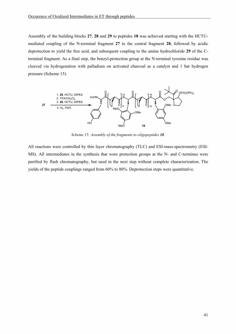
Occurence of Oxidized Intermediates in ET through peptides
41
Assembly of the building blocks 27, 28 and 29 to peptides 18 was achieved starting with the HCTU-
mediated coupling of the N-terminal fragment 27 to the central fragment 28, followed by acidic
deprotection to yield the free acid, and subsequent coupling to the amine hydrochloride 29 of the C-
terminal fragment. As a final step, the benzyl-protection group at the N-terminal tyrosine residue was
cleaved via hydrogenation with palladium on activated charcoal as a catalyst and 1 bar hydrogen
pressure (Scheme 15).
1. 28, HCTU, DIPEA2. TFA/CH2Cl23. 29, HCTU, DIPEA
HN
N
HN
O
O
O
O
O
O
OOPO(OPh)2
N
O
AcHN
HO
OMe
OMe
n m274. H2, Pd/C MeO
MeO
OMe
18
Scheme 15: Assembly of the fragments to oligopeptides 18.
All reactions were controlled by thin layer chromatography (TLC) and ESI-mass-spectrometry (ESI-
MS). All intermediates in the synthesis that wore protection groups at the N- and C-terminus were
purified by flash chromatography, but used in the next step without complete characterization. The
yields of the peptide couplings ranged from 60% to 80%. Deprotection steps were quantitative.

Occurence of Oxidized Intermediates in ET through peptides
42
5.4 Data Collection and Analysis
The experimental setup for Laser Flash Photolysis (LFP) experiments is described in chapter 3.2. LFP
should be used to detect the appearance of oxidized intermediates during ET through peptides 18
(Scheme 16). Induced by the laser flash, an electron acceptor A was generated at the C-terminus,
giving peptide 32 which absorbs around 450 nm. Subsequent transfer of electrons from the N-terminal
electron donor tyrosine (D), resulting in the formation of a tyrosyl radical (D•) 34 leads to the build-up
of a second absorption maximum at 410 nm.
HN
N
HN
O
O
O
O
O
O
N
O
AcHN
HO
OMe
OMe
18hν (308 nm)
n m
D R A
HN
N
HN
O
O
O
O
O
O
N
O
AcHN
O
OMe
OMe
n m
ET
- H+
D R A
via:
33
32
34
MeOOMe
MeO
MeOOMe
MeO
NH
O
MeO
OMe
MeO
Scheme 16: ET from D to A through the peptide chain might occur via oxidation of the relay amino acid R.
Special attention should be payed to the role of the aromatic amino acid R, separating the proline
matrix into two spacers with defined numbers of residues n and m, which is in principle able to serve
as a relay in sequential ET between D and A, yielding an oxidized intermediate 33 (R•+), with an
absorption maximum at 550 nm.
Occurence of Oxidized Intermediates in ET through peptides
43
5.4.1 Limitations
Some limitations regarding the LFP measurements result from the design of the model.
Time-resolved measurements
While the signals of all three oxidized intermediates can be clearly distinguished in the transient
absorption spectrum (see 3.3.1), rate measurements at the respective maxima will suffer from errors
due to significant signal-overlap (cf. Figure 15). Moreover, a reliable rate detection at 410 nm
(wavelength of tyrosyl radical absorption), is complicated by a strong fluorescence band at that
wavelength, emitted from the pivaloyl chromophore. This negative absorption saturates the
photomultiplier, which takes 200 – 300 ns to recover. Detailed analysis of the kinetic data is, in
addition, only possible if assumptions are made about the mechanism of radical cation decay. We
therefore decided not to rely on time-resolved measurements at a specific wavelength, but to use the
complete transient absorption spectra to gain information about the course of the reaction depicted in
Scheme 16. The CCD-camera used to record the transient absorption spectra is not hampered by the
fluorescence, since it can be triggered to a 40 ns delay after irradiation and the fluorescence signal
itself decays within 20 ns, as could be estimated by single photon counting experiments.
Intermolecular ET
Since photocleavage of the injector moiety is based on a quantum-mechanically forbidden n-π*
transition, the extinction coefficient of the respective band at the wavelength of laser irradiation is low
(ε (308 nm) approx 30 – 40 M-1cm-1).[95] Therefore, LFP experiments have to be carried out in
millimolar concentrations of peptide, to gain sufficient signal intensities. At concentrations in this
range, intermolecular reactions can already play a role. As a rough estimation, from the rate
coefficient for diffusion controlled reactions in acetonitrile (109 s-1), rates of intermolecular ET at
millimolar peptide concentrations can be expected to be in the range of 106 s-1. Dilution measurements
performed by Napp on peptide models of type 1 (Scheme 3, page 19), allow a more precise
determination of intermolecular ET rates. He measured a rate coefficient of 3·108 s-1 for intermolecular
ET between the C-terminal 2,4,6-trimethoxyphenylalanine radical cation and an N-terminal tyrosine in
peptides with 5 and 7 amino acid residues.[65] This value results in intermolecular ET rates of
1 - 2.5·106 s-1 for the 3 - 8 mM concentrations used in LFP. Thus, intensity changes of around
4% - 10% after a time delay of 40 ns can be expected to be caused by intermolecular contributions,
whereas the error caused by intermolecular reactions reaches already 50% after 500 ns.
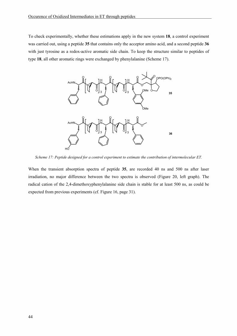
Occurence of Oxidized Intermediates in ET through peptides
44
To check experimentally, whether these estimations apply in the new system 18, a control experiment
was carried out, using a peptide 35 that contains only the acceptor amino acid, and a second peptide 36
with just tyrosine as a redox-active aromatic side chain. To keep the structure similar to peptides of
type 18, all other aromatic rings were exchanged by phenylalanine (Scheme 17).
HN
N
HN
O
O
O
O
O
O
OOPO(OPh)2
N
O
AcHN
OMe
OMe
3 3
HN
N
HN
O
O
O
O
O
N
O
AcHN
HO
3 3
35
36
Scheme 17: Peptide designed for a control experiment to estimate the contribution of intermolecular ET.
When the transient absorption spectra of peptide 35, are recorded 40 ns and 500 ns after laser
irradiation, no major difference between the two spectra is observed (Figure 20, left graph). The
radical cation of the 2,4-dimethoxyphenylalanine side chain is stable for at least 500 ns, as could be
expected from previous experiments (cf. Figure 16, page 31).

Occurence of Oxidized Intermediates in ET through peptides
45
When peptide 36 is added in equimolar amounts as an intermolecular electron donor (Figure 20, right
graph), the spectrum changes from almost entirely electron acceptor (35•+) 40 ns after the flash (black
line) to a significant amount of tyrosyl radical (36•) formed already 500 ns after the flash (red line,
λmax[tyr•] = 410 nm). This result is in accordance with the considerations described above, and with
quenching experiments performed on small peptides (cf. chapter 4).
Figure 20: left graph: Transient absorption spectra of peptide 35 recorded 40 ns (black line) and 500 ns (red
line) after laser irradiation.; right graph: Transient absorption spectra of an equimolar solution of peptides 35
and 36 recorded 40 ns (black line) and 500 ns (red line) after laser irradiation.
As a consequence of these considerations, it was decided, that the transient absorption spectra of
peptide models of type 18, gained 40 ns after laser irradiation, should be used for analysis. In these
spectra, the amount of intermolecular ET is low (≤10%) and thus does not hamper the observation of
intramolecular processes. Shorter delays are not useful, since the rate for the ß-fragmentation of the
injector radical 3 to give the charged intermediate 4 that initiates the ET cascade (see Scheme 4, page
19), has been determined by competition experiments to 2x10⁸ s-1,[66] resulting in a half-life time of
approximately 20 ns for electron acceptor generation.

Occurence of Oxidized Intermediates in ET through peptides
46
5.4.2 Analysis of the Data
A first, qualitative analysis of the obtained spectra reveals directly, which aromatic side chains are
present in the oxidized form, since the three spectroscopic sensors, indicating the electron acceptor,
the oxidized relay and the oxidized donor, yield different absorption spectra. For a quantification of
the analysis, reference curves obtained from measurements of the respective side chains coupled
directly to the injector (Figure 14, page 28), were used, representative of the transient species 32, 33
and 34. Fitting of these curves, so that the sum of the spectrum of the tyrosyl radical D• (blue,
λmax = 410 nm), the 2,4-dimethoxyradical cation A (red, λmax = 450 nm), and the 2,4,6-trimethoxy-
phenylalanine radical cation R•+ (green, λmax = 550 nm) yields the measured overall absorption,
allowed a graphical analysis of the contributions of the respective transients. An example is depicted
in Figure 21.

Occurence of Oxidized Intermediates in ET through peptides
47
The absorption values extracted from this analysis were then corrected by the extinction coefficients of
the radical cations at their maximum absorption. The extinction coefficient of the phenoxyl radical is
known to amount to 3000 M-1cm-1,[78], while 1,3-dimethoxybenzene radical cation and 1,3,5-
trimethoxybenzene radical cation have extinction coefficients of approx. 4000 M-1cm-1.[80, 81] Thus, the
values determined for tyrosine from the graphical analysis were multiplied with a factor of 1.33.
•
Figure 21: Decomposition of a recorded transient absorption spectrum (black) to reveal the contributions of
tyrosyl radical (blue), 2,4-dimethoxyphenylalanine radical cation (red) and 2,4,6-trimethoxyphenylalanine
radical cation (green).
Since the laser energy varies slightly between two flashes and significantly between different days of
measurements, due to variations in the gas mixture, the absolute absorption values could not be used
for the comparison of different molecules. The obtained values were therefore scaled to the sum of all
absorption maxima, set to 100%.
D• (40%) A (50%) R
•+(10%)
AcHNO
O
O
AcHNO
O
O
AcHNO
O
O
D
R
A
OMeMeO
OMe
MeO
OMe
O

Occurence of Oxidized Intermediates in ET through peptides
48
5.5 Results and Discussion
As a first molecule, peptide 37 in which electron acceptor, relay amino acid and electron donor are
assembled in close proximity, was synthesized and ET in this peptide was examined by LFP.
Figure 22: Transient absorption spectrum of 37 40 ns after laser irradiation.
It is obvious from the resulting spectrum recorded 40 ns after the laser flash (Figure 22), that the main
part of the charge has been transferred to the final electron donor tyrosine already within this short
delay, yielding tyrosyl radical 34 (D•, λmax = 410 nm). Around 550 nm, the wavelength of absorption
of the relay radical cation 33 (R•+), a small absorption band is observed. Analysis of the spectrum by
graphical decomposition of the curve (cf. Figure 21) reveals a distribution of oxidized species of 85%
tyrosyl radical 34 (D•), 9% remaining electron acceptor 32 (A), and 6% oxidized relay 33 (R•+). Since
intermolecular donor to acceptor ET has been estimated to cause effects of less than 10%, it can be
concluded, that very fast intramolecular ET from tyrosine to 2,4-dimethoxyphenylalanine radical
cation takes place in peptide 37.
D• (85%) A (9%) R
•+ (6%)
HN
N
HN
O
O
O
O
O
O
OOPO(OPh)2
N
O
AcHN
HO
OMe
OMe
37
MeOOMe
MeO
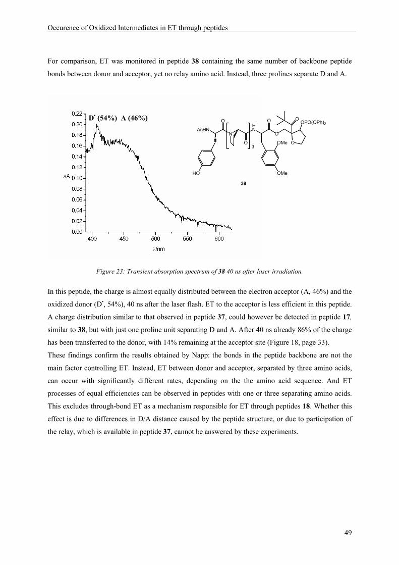
Occurence of Oxidized Intermediates in ET through peptides
49
For comparison, ET was monitored in peptide 38 containing the same number of backbone peptide
bonds between donor and acceptor, yet no relay amino acid. Instead, three prolines separate D and A.
Figure 23: Transient absorption spectrum of 38 40 ns after laser irradiation.
In this peptide, the charge is almost equally distributed between the electron acceptor (A, 46%) and the
oxidized donor (D•, 54%), 40 ns after the laser flash. ET to the acceptor is less efficient in this peptide.
A charge distribution similar to that observed in peptide 37, could however be detected in peptide 17,
similar to 38, but with just one proline unit separating D and A. After 40 ns already 86% of the charge
has been transferred to the donor, with 14% remaining at the acceptor site (Figure 18, page 33).
These findings confirm the results obtained by Napp: the bonds in the peptide backbone are not the
main factor controlling ET. Instead, ET between donor and acceptor, separated by three amino acids,
can occur with significantly different rates, depending on the the amino acid sequence. And ET
processes of equal efficiencies can be observed in peptides with one or three separating amino acids.
This excludes through-bond ET as a mechanism responsible for ET through peptides 18. Whether this
effect is due to differences in D/A distance caused by the peptide structure, or due to participation of
the relay, which is available in peptide 37, cannot be answered by these experiments.
D• (54%) A (46%)
HN
O
O
O
OOPO(OPh)2
N
O
AcHN
HO
OMe
OMe
O3
38
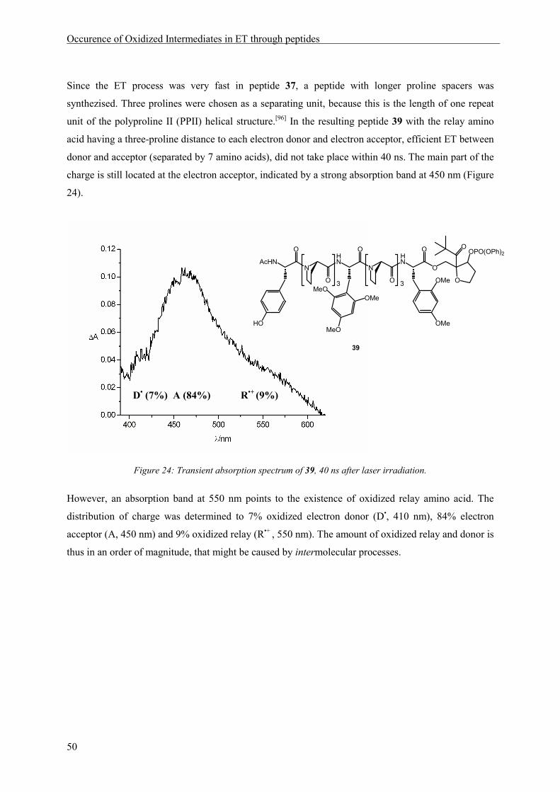
Occurence of Oxidized Intermediates in ET through peptides
50
Since the ET process was very fast in peptide 37, a peptide with longer proline spacers was
synthezised. Three prolines were chosen as a separating unit, because this is the length of one repeat
unit of the polyproline II (PPII) helical structure.[96] In the resulting peptide 39 with the relay amino
acid having a three-proline distance to each electron donor and electron acceptor, efficient ET between
donor and acceptor (separated by 7 amino acids), did not take place within 40 ns. The main part of the
charge is still located at the electron acceptor, indicated by a strong absorption band at 450 nm (Figure
24).
Figure 24: Transient absorption spectrum of 39, 40 ns after laser irradiation.
However, an absorption band at 550 nm points to the existence of oxidized relay amino acid. The
distribution of charge was determined to 7% oxidized electron donor (D•, 410 nm), 84% electron
acceptor (A, 450 nm) and 9% oxidized relay (R•+ , 550 nm). The amount of oxidized relay and donor is
thus in an order of magnitude, that might be caused by intermolecular processes.
D• (7%) A (84%) R
•+ (9%)
HN
N
HN
O
O
O
O
O
O
OOPO(OPh)2
N
O
AcHN
HO
OMe
OMe
3 3
39
MeOOMe
MeO

Occurence of Oxidized Intermediates in ET through peptides
51
To answer the question, whether intramolecular ET takes place in peptide 39, a control experiment
was carried out. In addition to peptides 35 and 36, used to estimate the effect of intermolecular
transfer between donor and acceptor, a third peptide 40 was synthesized, containing only the relay
amino acid as redox-active residue.
HN
N
HN
O
O
O
O
O
O
OOPO(OPh)2
N
O
AcHN
OMe
OMe
3 3
HN
N
HN
O
O
O
O
O
N
O
AcHN
3 3
HN
N
HN
O
O
O
O
O
N
O
AcHN
HO
3 3
35
40
36
MeOOMe
MeO
Scheme 18: Peptides used to estimate the role of intermolecular ET.
An equimolar mixture of 35, 36 and 40 was investigated by LFP, and the spectrum obtained after
40 ns (Figure 25, black), was compared to the transient absorption spectrum of 39 (Figure 25, red).
While the absorption band at 550 nm, stemming from the oxidized relay (33, R•+), is clearly visible in
peptide 39 (red line), no significant amount of oxidized relay is formed in the control experiment
(black line).
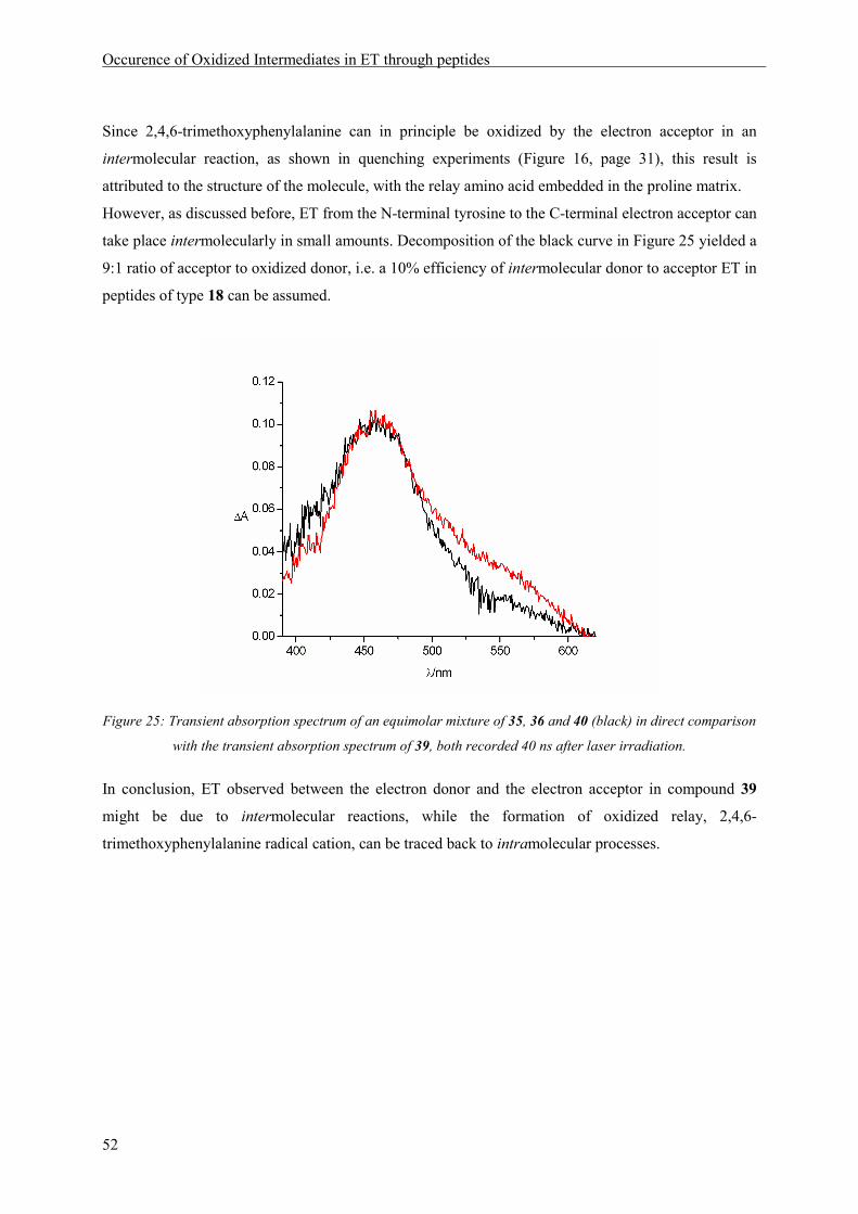
Occurence of Oxidized Intermediates in ET through peptides
52
Since 2,4,6-trimethoxyphenylalanine can in principle be oxidized by the electron acceptor in an
intermolecular reaction, as shown in quenching experiments (Figure 16, page 31), this result is
attributed to the structure of the molecule, with the relay amino acid embedded in the proline matrix.
However, as discussed before, ET from the N-terminal tyrosine to the C-terminal electron acceptor can
take place intermolecularly in small amounts. Decomposition of the black curve in Figure 25 yielded a
9:1 ratio of acceptor to oxidized donor, i.e. a 10% efficiency of intermolecular donor to acceptor ET in
peptides of type 18 can be assumed.
Figure 25: Transient absorption spectrum of an equimolar mixture of 35, 36 and 40 (black) in direct comparison
with the transient absorption spectrum of 39, both recorded 40 ns after laser irradiation.
In conclusion, ET observed between the electron donor and the electron acceptor in compound 39
might be due to intermolecular reactions, while the formation of oxidized relay, 2,4,6-
trimethoxyphenylalanine radical cation, can be traced back to intramolecular processes.

Occurence of Oxidized Intermediates in ET through peptides
53
While in the nonapeptide 39, containing two triproline spacers, intramolecular donor to acceptor
charge transfer does not occur within 40 ns, the amount of tyrosyl radical formed is significantly
increased upon spacer-shortening. In peptide 41, relay and acceptor are only separated by one proline,
while the relay/donor distance is kept to three prolines (Figure 26). The ratio of oxidized species
formed 40 ns after irradiation of 41, amounts to 40% tyrosyl radical (D•), 50% remaining electron
acceptor (A), and 10% oxidized relay (R•+). So the contribution of oxidized relay to the overall
absorption does not differ from the value measured upon LFP of molecule 39 (around 10%, see
above). But donor to acceptor electron transfer has increased and can no longer be explained by
intermolecular reactions.
Figure 26: Transient absorption spectrum of 41, 40 ns after laser irradiation.
Intramolecular donor to acceptor and relay to acceptor ET takes place in peptide 41.
D• (40%) A (50%) R
•+ (10%)
HN
N
HN
O
O
O
O
O
O
OOPO(OPh)2
N
O
AcHN
OMe
OMe
3
OH
41
MeOOMe
MeO
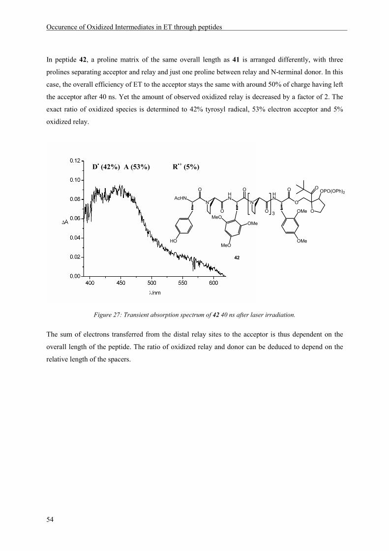
Occurence of Oxidized Intermediates in ET through peptides
54
In peptide 42, a proline matrix of the same overall length as 41 is arranged differently, with three
prolines separating acceptor and relay and just one proline between relay and N-terminal donor. In this
case, the overall efficiency of ET to the acceptor stays the same with around 50% of charge having left
the acceptor after 40 ns. Yet the amount of observed oxidized relay is decreased by a factor of 2. The
exact ratio of oxidized species is determined to 42% tyrosyl radical, 53% electron acceptor and 5%
oxidized relay.
Figure 27: Transient absorption spectrum of 42 40 ns after laser irradiation.
The sum of electrons transferred from the distal relay sites to the acceptor is thus dependent on the
overall length of the peptide. The ratio of oxidized relay and donor can be deduced to depend on the
relative length of the spacers.
D• (42%) A (53%) R
•+ (5%)
HN
N
HN
O
O
O
O
O
O
OOPO(OPh)2
N
O
AcHN
OMe
OMe
3
HO
42
MeOOMe
MeO

Occurence of Oxidized Intermediates in ET through peptides
55
Upon elongation of the separating matrix to a total of seven prolines with one proline between
acceptor and relay and six prolines between relay and donor, as in peptide 43, the amount of oxidized
relay observed after 40 ns is increased to 35% of overall absorption. Around 50% of the charge has
already left the electron acceptor after that delay. The graphical decomposition leads to a distribution
of 19% oxidized tyrosine, 46% oxidized electron acceptor and 35% oxidized relay. However, the
quality of the measured spectrum is low, due to a lower signal intensity.
Figure 28: Transient absorption spectrum of 43 40 ns after laser irradiation.
To summarize the results obtained in LFP of peptides of type 18, signals of oxidized relay amino acids
are observed in all experiments. A control experiment showed, that intermolecular reactions can be
excluded as an origin of these oxidations. Therefore, intramolecular ET processes between relay
amino acids and electron acceptors occur in peptides 18. While a small amount of tyrosyl radical can
be explained by intermolecular oxidation reactions, the efficient donor to acceptor ET observed in
peptides 37, 41 and 42, can only be explained by intramolecular processes. This can be deduced from
considerations referring to diffusion controlled reaction rates, as well as from a control experiment and
from the consideration that diffusion controlled processes between the respective ends of two peptide
chains should not depend on the length and structure of the peptide strong enough to explain the
remarkable differences in ET efficiency in peptides 18 noted above. In conclusion, intramolecular ET
between donor, relay and acceptor occurs in peptides of type 18. The distribution of the charge across
the three available redox sites depends on the length of the spacers separating those sites.
D• (19%) A (46%) R
•+ (35%)
HN
N
HN
O
O
O
O
O
O
OOPO(OPh)2
N
O
AcHN
OMe
OMe
1
HO
6
43
MeOOMe
MeO

Occurence of Oxidized Intermediates in ET through peptides
56
These experimental results leads to the interpretation, that ET in peptides 18 possibly occurs in a
stepwise manner with ET efficiencies controlled by distance (dependent on the spacer length) and
driving force of each, the donor to relay as well as the relay to acceptor transfer step. However, these
conclusions have to be taken with care, because though all investigated peptides have been constructed
from the same building blocks, with units of 1 proline resp. 3 or 6 prolines (one or two repeat units of
the PPII-helix), not much is known about the structure of the peptides in solution and the resulting
actual distances between the redox sites. An additional uncertainty exists concerning the mechanism of
ET. It could be proven that ET occurs intramolecularly, and that oxidized intermediates of the relay
site can be observed. But it is not clear, whether the electrons are passed successively through the
available redox sites in those peptides, where all three transients are observed upon irradiation. A
direct transfer of the charge from the injector radical cation 4 to the relay or donor site should be
inefficient, due to competition with water addition, as could be proven by experiments of Napp (cf.
paragraph 3.1), but could not be excluded completely so far. Additional experiments performed to
check this possibility are described in the following chapter. It is moreover conceivable, that different
members of the ensemble of peptide conformers in solution either allow oxidation of the final electron
donor or of the relay amino acid and that the distribution of conformers is the crucial factor controlling
the observed ET efficiencies. Since efficient donor to acceptor ET only takes place in peptides
containing at least one short (monoproline) spacer, presumably resulting in significant structural
flexibility, the discussed systems are not suited as a starting point for the replacement of the amino
acid at the relay position. To solve the possible problem of direct injection into distal sites and to
design a model suitable for the investigation of side-chain relevance, a new injection system was
designed, as described in chapter 6.
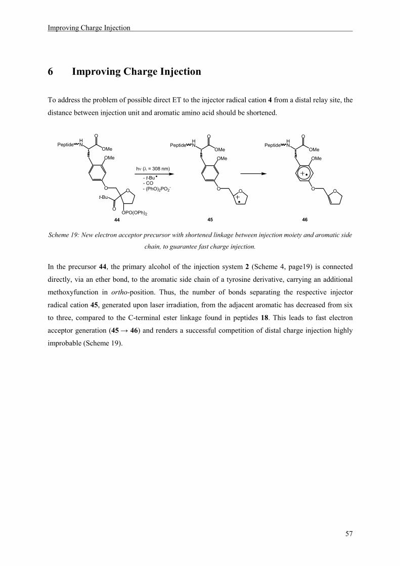
Improving Charge Injection
57
6 Improving Charge Injection
To address the problem of possible direct ET to the injector radical cation 4 from a distal relay site, the
distance between injection unit and aromatic amino acid should be shortened.
HN
OMe
O
OMe
O O
O
t-Bu
OPO(OPh)2
Peptide
44
HN
OMe
O
OMe
O O
Peptide
45
HN
OMe
O
OMe
O O
Peptide
46
- t-Bu- CO- (PhO)2PO2
-
hν (λ = 308 nm)
Scheme 19: New electron acceptor precursor with shortened linkage between injection moiety and aromatic side
chain, to guarantee fast charge injection.
In the precursor 44, the primary alcohol of the injection system 2 (Scheme 4, page19) is connected
directly, via an ether bond, to the aromatic side chain of a tyrosine derivative, carrying an additional
methoxyfunction in ortho-position. Thus, the number of bonds separating the respective injector
radical cation 45, generated upon laser irradiation, from the adjacent aromatic has decreased from six
to three, compared to the C-terminal ester linkage found in peptides 18. This leads to fast electron
acceptor generation (45 → 46) and renders a successful competition of distal charge injection highly
improbable (Scheme 19).

Improving Charge Injection
58
6.1 Synthesis of a New Charge Injector
The synthesis of non-natural amino acid 50 (Scheme 20) was possible analogous to the synthesis of
the methoxysubstituted phenylalanines depicted in Scheme 12, starting from commercially available
4-hydroxy-2-methoxybenzaldehyde applying an Erlenmeyer azlactone procedure.[25, 26] Basic opening
of lactone 47 and asymmetric hydrogenation of acrylic acid 48 with a commercially available
Rhodium Deguphos[27] catalyst gave the (S)-configured amino acid derivative 49 in very good yield
and >95% ee. Deprotection under strongly acidic conditions and purification via ion exchange
chromatography yielded free amino acid 50 (78%), which could be converted to the N-Boc protected
methyl ester 51 using standard conditions. However, a migration of the Boc-protection group to the
hydroxy function of the aromatic ring lowered the yield in this reaction to 35%. Due to the instability
of compound 51, coupling to the injection moiety was carried out directly after the isolation.
CO2Me
HN
O
Ph
Rh*/H2 (50bar)
HCl conc., H2O, ∆
OH
CO2H
NH2
OH
O
hippuric acid
OAc
N
OO
Ph
Na2CO3 OMe
OH
OMe OMe
OMe1. SOCl2, MeOH
OH
CO2Me
NHBoc
OMe
2. Boc2O, Et3N
45 46
4849
CO2Me
HN
O
Ph
OMe
OH
47(77 %) (85%) (96%)
(78%)(35% for 2 steps)
Rh N
P
P
Rh*:
NaOAc, Ac2O MeOH
ion exchange
CH2Cl2
Scheme 20: Synthesis of enantiopure 4-hydroxy-2-methoxyphenylalanine
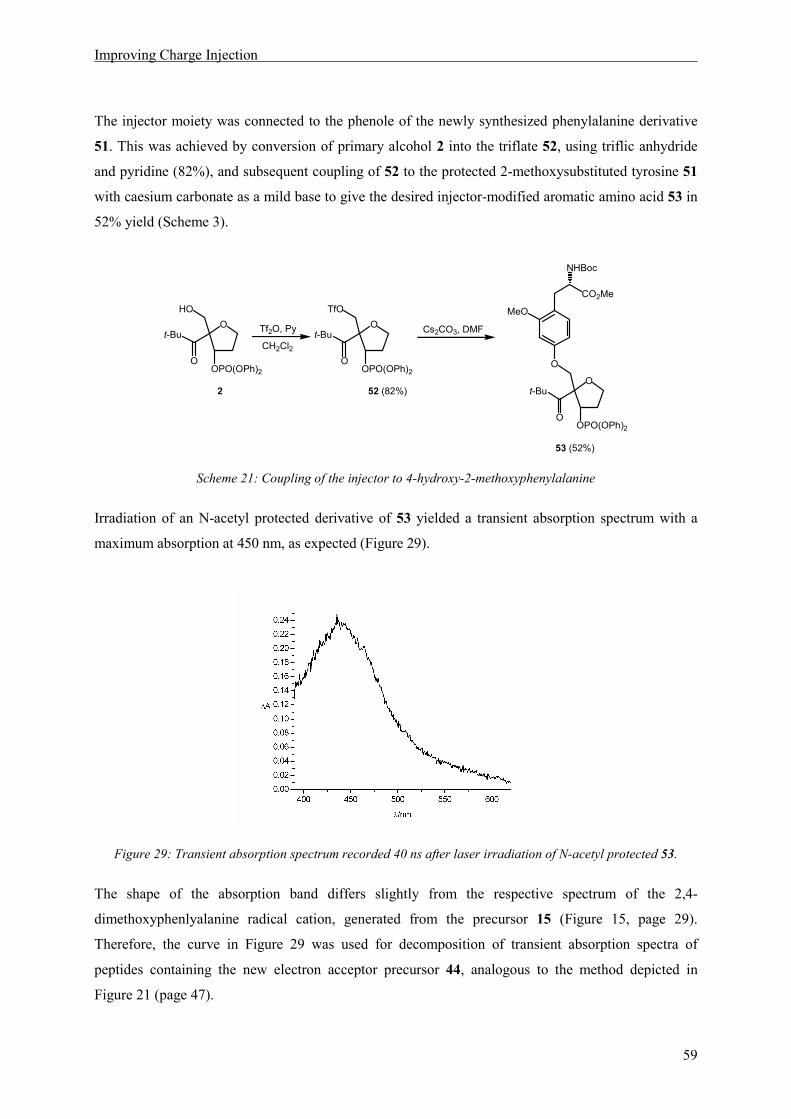
Improving Charge Injection
59
The injector moiety was connected to the phenole of the newly synthesized phenylalanine derivative
51. This was achieved by conversion of primary alcohol 2 into the triflate 52, using triflic anhydride
and pyridine (82%), and subsequent coupling of 52 to the protected 2-methoxysubstituted tyrosine 51
with caesium carbonate as a mild base to give the desired injector-modified aromatic amino acid 53 in
52% yield (Scheme 3).
O
TfO
O
t-Bu
OPO(OPh)2
O
HO
O
t-Bu
OPO(OPh)2O
MeO
CO2Me
NHBoc
O
O
t-Bu
OPO(OPh)2
Tf2O, Py
CH2Cl2
Cs2CO3, DMF
2 52 (82%)
53 (52%)
Scheme 21: Coupling of the injector to 4-hydroxy-2-methoxyphenylalanine
Irradiation of an N-acetyl protected derivative of 53 yielded a transient absorption spectrum with a
maximum absorption at 450 nm, as expected (Figure 29).
Figure 29: Transient absorption spectrum recorded 40 ns after laser irradiation of N-acetyl protected 53.
The shape of the absorption band differs slightly from the respective spectrum of the 2,4-
dimethoxyphenlyalanine radical cation, generated from the precursor 15 (Figure 15, page 29).
Therefore, the curve in Figure 29 was used for decomposition of transient absorption spectra of
peptides containing the new electron acceptor precursor 44, analogous to the method depicted in
Figure 21 (page 47).

Improving Charge Injection
60
6.2 Synthesis of Peptides
Peptides containing the new electron acceptor precursor were assembled using two different strategies,
dependent on the nature of the relay amino acid.
1.) If the relay amino acid, situated inside the proline matrix and separating it into two fragments of n
and m units, was a non-natural amino acid (in this case 2,4,6-trimethoxyphenylalanine) peptides 54
were assembled by solution synthesis using the Boc-protection strategy, analogous to the peptide
models of type 18 (cf. Scheme 13, page 39). The fragments used are indicated in Scheme 22.
Synthesis of the respective fragments 27 and 28 is depicted in Scheme 14 (page 40). Fragment 55 can
be obtained by acidic deprotection of 53, using HCl in dioxane.
HN
N
HN
OMe
O
O
O
O
N
O
AcHN
HO
OMe
O
3 3
O
O
t-Bu
OPO(OPh)2
O
N
O
AcHN
BnO
OH
O
N
O
H3N+
MeO
OtBu
OMe
R3 3
OMe
O
H3N+Cl−
OMe
O O
O
t-Bu
OPO(OPh)2
Cl−
54
27 28 55
MeO
MeO
OMe
Scheme 22: Synthesis of peptides containing the new injection unit and a non-natural amino acid as relay.
Assembly of the peptide fragments is achieved analogous to the procedure depicted in Scheme 15
(page 41), by HCTU-mediated coupling and subsequent reductive benzyl-deprotection of the tyrosine
hydroxy function with hydrogen and Pd on activated charcoal as a last step, yielding peptides 54.

Improving Charge Injection
61
2.) If the central amino acid was a natural amino acid, in this case phenylalanine, peptides 56 were
assembled from two fragments, one consisting of the N-deprotected electron acceptor precursor 55.
The second fragment, the free acid of the N-terminal peptide part 57, was synthesized using solid
phase peptide synthesis (SPPS) (Scheme 23).
HN
N
HN
OMe
O
O
O
O
N
O
AcHN
HO
OMe
O
n m
O
O
t-Bu
OPO(OPh)2
OMe
O
H3N+Cl−
OMe
O O
O
t-Bu
OPO(OPh)2
HN
NOH
O
O
O
N
O
AcHN
t-BuO
57
n m
56
55
Scheme 23: Fragments used for the synthesis of peptides containing the natura amino acid phenylalanine at the
relay position.
The course of the SPPS is depicted in Scheme 24. It was carried out on a commercially available
chlorotrityl-resin, charged with proline, using Fmoc strategy in a fully automatized peptide
synthesizer. Cleavage of the resulting oligopeptide with trifluoroethanol and acetic acid yielded the
free acid 57, which was coupled to the deprotected electron acceptor precursor 55 in solution. The tert-
butylether group protecting the tyrosine hydroxy function was subsequently cleaved with
trifluoroacetic acid (TFA) in CH2Cl2 (Scheme 24).
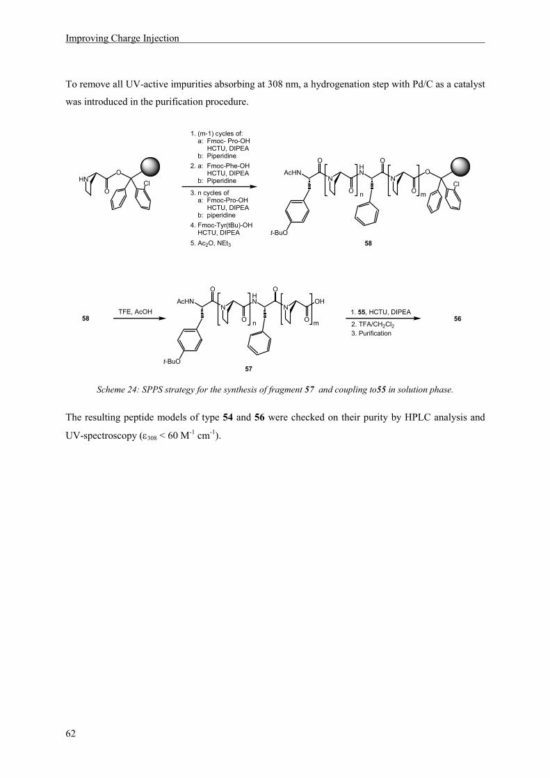
Improving Charge Injection
62
To remove all UV-active impurities absorbing at 308 nm, a hydrogenation step with Pd/C as a catalyst
was introduced in the purification procedure.
1. (m-1) cycles of: a: Fmoc- Pro-OH HCTU, DIPEA b: Piperidine
5. Ac2O, NEt3
TFE, AcOH
HN
NOH
O
O
O
N
O
AcHN
57
n m
2. a: Fmoc-Phe-OH HCTU, DIPEA b: Piperidine
3. n cycles of a: Fmoc-Pro-OH HCTU, DIPEA b: piperidine
4. Fmoc-Tyr(tBu)-OH HCTU, DIPEA
1. 55, HCTU, DIPEA
2. TFA/CH2Cl256
58
3. Purification
O
ClO
HN
t-BuO
HN
NO
O
O
O
N
O
AcHN
n m
t-BuO
Cl
58
Scheme 24: SPPS strategy for the synthesis of fragment 57 and coupling to55 in solution phase.
The resulting peptide models of type 54 and 56 were checked on their purity by HPLC analysis and
UV-spectroscopy (ε308 < 60 M-1 cm-1).
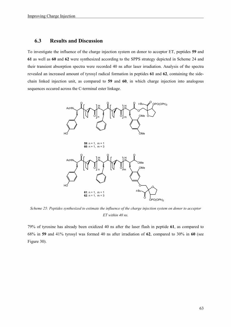
Improving Charge Injection
63
6.3 Results and Discussion
To investigate the influence of the charge injection system on donor to acceptor ET, peptides 59 and
61 as well as 60 and 62 were synthesized according to the SPPS strategy depicted in Scheme 24 and
their transient absorption spectra were recorded 40 ns after laser irradiation. Analysis of the spectra
revealed an increased amount of tyrosyl radical formation in peptides 61 and 62, containing the side-
chain linked injection unit, as compared to 59 and 60, in which charge injection into analogous
sequences occured across the C-terminal ester linkage.
HN
N
HN
OMe
O
O
O
O
N
O
AcHN
HO
OMe
O
n m
O
O
t-Bu
OPO(OPh)2
HN
N
HN
O
O
O
O
O
O
OOPO(OPh)2
N
O
AcHN
HO
OMe
OMe
n m
t-Bu
59: n = 1, m = 160: n = 1, m = 3
61: n = 1, m = 162: n = 1, m = 3
Scheme 25: Peptides synthesized to estimate the influence of the charge injection system on donor to acceptor
ET within 40 ns.
79% of tyrosine has already been oxidized 40 ns after the laser flash in peptide 61, as compared to
68% in 59 and 41% tyrosyl was formed 40 ns after irradiation of 62, compared to 30% in 60 (see
Figure 30).
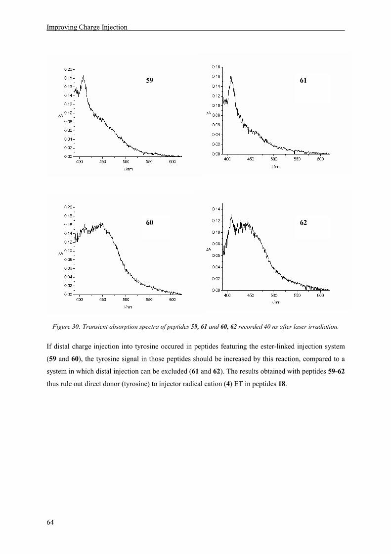
Improving Charge Injection
64
Figure 30: Transient absorption spectra of peptides 59, 61 and 60, 62 recorded 40 ns after laser irradiation.
If distal charge injection into tyrosine occured in peptides featuring the ester-linked injection system
(59 and 60), the tyrosine signal in those peptides should be increased by this reaction, compared to a
system in which distal injection can be excluded (61 and 62). The results obtained with peptides 59-62
thus rule out direct donor (tyrosine) to injector radical cation (4) ET in peptides 18.
59 61
60 62

Improving Charge Injection
65
To investigate the possibility of distal injection into the relay amino acid, the transient absorption
spectra of peptides 42 and 63, as well as 39 and 64 were compared.
HN
N
HN
OMe
O
O
O
O
N
O
AcHN
HO
OMe
O
n m
O
O
t-Bu
OPO(OPh)2
HN
N
HN
O
O
O
O
O
O
OOPO(OPh)2
N
O
AcHN
HO
OMe
OMe
n m
t-Bu
42 n = 1, m = 339 n = 3, m = 3
63 n = 1, m = 364 n = 3, m = 3
MeOOMe
MeO
OMe
MeO
MeO
Scheme 26: Peptides synthesized to estimate the influence of the charge injection system on relay to acceptor ET
within 40 ns.
As a result of the measurement, the intensity of the relay amino acid signal is higher in 63, containing
the directly linked injection system (11% oxidized relay), than in 42 (5% oxidized relay). Distal
charge injection into the relay amino acid in peptides 18, containing the ester-linked C-terminal
injector, can thus also be excluded.
Laser flash photolysis of peptides 39 and 64, showed a similar yield of oxidized relay (9% and 10%
respectively), yet a significant difference in the amount of donor to acceptor ET during 40 ns. While in
39 almost no tyrosine oxidation occured within the time frame of the measurement, in 64 the tyrosyl
radical amounts to 36% of overall oxidized species within that delay.
The increased donor to acceptor ET efficiency within a 40 ns delay, observed upon introduction of the
directly linked injector, points to an increase in the injection rate. In this context, the observation of
oxidized donor 40 ns after irradiation in the nonapeptide 64 is of special interest, since the ester-linked
injection system did not allow the observation of donor to acceptor ET within 40 ns in a peptide of
analogous sequence (39). The newly developed electron acceptor precursor 44 therefore also
contributes to the construction of a suitable model for the investigation of side-chain influence on ET
through peptides, as will be discussed in detail below.

Influence of Amino-Acid Side Chains on ET
67
7 Influence of Amino-Acid Side Chains on ET
7.1 Introduction
In chapter 6.3 it could be shown, that electron acceptor generation in peptide models of type 54,
containing the electron acceptor precursor 44 (Scheme 19, page 57), can be assumed to proceed faster
than in the respective peptides of type 18, discussed in chapter 5. The distance between the injector
radical cation generated upon irradiation, and the C-terminal aromatic ring is substantially smaller in
peptides 54 than in peptides 18. As a consequence, one advantage of the electron acceptor precursor
44 is its lower susceptibility for side reactions of the respective precursor radical 45. Much more
important, it widens the number of possible candidates to observe intramolecular ET: The limitations
of our model have been discussed extensively in chapter 5.4.1. One important point is the small time
window available for the observation of intramolecular donor to acceptor ET, due to high peptide
concentrations and the resulting high rates for bimolecular reactions. Significant results, yielding
information about intramolecular ET, can be obtained by analyzing spectra recorded only 40 ns after
the laser flash. This limits the possible distances between electron donor and acceptor, i.e. the length
of the proline spacers. On the other hand, systems of high structural rigidity, and thus long proline
spacers, are needed, if the influence of amino acid side chains on ET shall be investigated, because
only if structural uniformity can be guaranteed, the ET-mediation properties of an individual amino
acid can be observed as the key factor controlling the overall ET behaviour of the peptide. During the
investigation of peptide models 18 no suitable system could be found. However, the faster charge
injection in peptides 54, caused by shorter injector/acceptor distance, allows longer spacers without
blocking intramolecular ET in the available time window. Special care has to be taken to the
observation of an oxidized intermediate during ET, because this proof is needed to confirm a
sequential mechanism. The amount of intermediate formed in a consecutive reaction depends on the
relative rates of the reaction steps. Therefore the structure of a peptide suited for the observation of
oxidized intermediates has to be optimized also in this respect.

Influence of Amino-Acid Side Chains on ET
68
For these reasons, the finding, that in peptide 64, with two triproline spacers separating the central
2,4,6-trimethoxyphenylalanine from C-terminal electron acceptor and N-terminal electron donor each,
all three oxidized aromatic side chains can be detected after 40 ns (Figure 31), was highly encouraging
to us.
The N-terminal donor getting oxidized within 40 ns, indicated by the maximum at 410 nm, points to
intramolecular, donor to acceptor ET, as discussed extensively above.
Figure 31: Transient absorption spectrum of 64, 40 ns after laser irradiation.
Moreover, the absorption detected at 550 nm results from 2,4,6-trimethoxyphenylalanine radical
cation, and thus indicates the occurence of oxidized relay amino acid. Therefore, the distances in
peptide 64 render it suitable as a sensor for intramolecular ET between all three aromatic side chains.
In addition, several hints were available, that triproline spacers guarantee a defined structure. First, in
peptide 39, containing the ester-linked C-terminal injector, the distance between electron acceptor and
electron donor was too large to allow the intramolecular reaction, leading to the conclusion that no
conformers with short through-space distances are present. Second, it can be derived from the
literature, that the onset of secondary structure in oligoprolines can be expected starting from three
consecutive prolines.[79] Moreover, the preferred conformation of oligoprolines in polar solvents, the
polyproline II helix, has a repeat unit of three prolines,[96] so that an alignement of residues in a proline
matrix using triproline spacers, can be assumed to result in a regular pattern.
HN
N
HN
OMe
O
O
O
O
N
O
AcHN
HO
OMe
O
3 3
O
O
t-Bu
OPO(OPh)264
MeOOMe
MeO
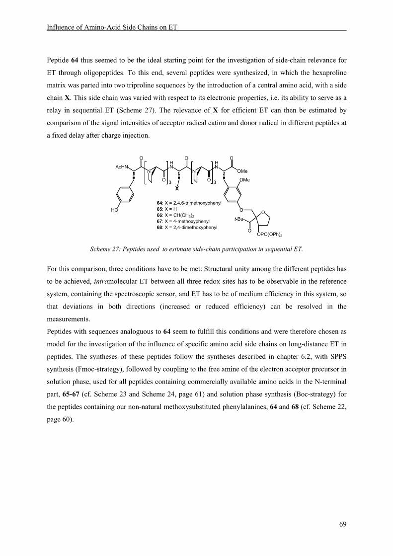
Influence of Amino-Acid Side Chains on ET
69
Peptide 64 thus seemed to be the ideal starting point for the investigation of side-chain relevance for
ET through oligopeptides. To this end, several peptides were synthesized, in which the hexaproline
matrix was parted into two triproline sequences by the introduction of a central amino acid, with a side
chain X. This side chain was varied with respect to its electronic properties, i.e. its ability to serve as a
relay in sequential ET (Scheme 27). The relevance of X for efficient ET can then be estimated by
comparison of the signal intensities of acceptor radical cation and donor radical in different peptides at
a fixed delay after charge injection.
HN
N
HN
OMe
O
O
O
O
N
O
AcHN
HO
X
OMe
O O
O
t-Bu
OPO(OPh)2
64: X = 2,4,6-trimethoxyphenyl65: X = H66: X = CH(CH3)2
67: X = 4-methoxyphenyl68: X = 2,4-dimethoxyphenyl
3 3
Scheme 27: Peptides used to estimate side-chain participation in sequential ET.
For this comparison, three conditions have to be met: Structural unity among the different peptides has
to be achieved, intramolecular ET between all three redox sites has to be observable in the reference
system, containing the spectroscopic sensor, and ET has to be of medium efficiency in this system, so
that deviations in both directions (increased or reduced efficiency) can be resolved in the
measurements.
Peptides with sequences analoguous to 64 seem to fulfill this conditions and were therefore chosen as
model for the investigation of the influence of specific amino acid side chains on long-distance ET in
peptides. The syntheses of these peptides follow the syntheses described in chapter 6.2, with SPPS
synthesis (Fmoc-strategy), followed by coupling to the free amine of the electron acceptor precursor in
solution phase, used for all peptides containing commercially available amino acids in the N-terminal
part, 65-67 (cf. Scheme 23 and Scheme 24, page 61) and solution phase synthesis (Boc-strategy) for
the peptides containing our non-natural methoxysubstituted phenylalanines, 64 and 68 (cf. Scheme 22,
page 60).

Influence of Amino-Acid Side Chains on ET
70
7.2 Structure
To gain more information about the structure of peptides 64 - 68, circular dichroism (CD) spectra of
these peptides were recorded (Figure 32). The solvent system needed for LFP experiments, a 3:1
mixture of acetonitrile and water, was used for CD spectroscopy. Due to the high sensitivity of this
analytical method, the peptides had to be dissolved in low concentrations of approximately
20 µg mL-1.
Figure 32: CD spectra of peptides 64 - 68 in acetonitrile/water (3:1) at 25 °C, with concentrations of
approximately 20 µg mL-1.
The spectra show the characteristic shape expected for a polyproline II (PPII) helix, with a strong
negative band around 206 nm and a weaker positive band at 229 nm.[96]
The PPII helix is the typical structure adopted by proline oligomers in polar solvents, especially
water,[97, 98] and an important secondary structure in natural enzymes.[99, 100] All peptide bonds are
trans-configured, with Ramachandran angles (cf. Figure 7, page 13) of -78° (Φ) and +146°
(Ψ), respectively.[101] The repeat unit of the left-handed helix consists of three prolines, with a lateral
transition given with 3.12 Å per residue.[96, 102]

Influence of Amino-Acid Side Chains on ET
71
Results from Förster resonance energy (FRET) measurements have been used to estimate end-to-end
distances in proline oligomers. While long peptides, containing 20-40 residues have been shown to
exhibit unexpectedly fast and efficient fluoresence quenching, pointing towards significant bending of
the molecule and thus shortened end-to-end distances,[103] proline oligomers of up to 12 residues can
be approximated by a rigid rod structure[104] and are thus suited to serve as spectroscopic rulers,
providing spacers of defined length.
The stability of the helix conformation of peptides 64 - 68 in the solvent system was checked by a
melting experiment, in which the peptide solution was heated up to 80 °C and the absorption at the
maximum of the CD spectrum was monitored. Just a modest linear decrease, no phase transition, could
be observed within a temperature range from 10 °C to 80 °C. This points to a high stability of the helix
conformation and it can thus be concluded, that a large majority of the peptides in the LFP
experiments is present in PPII conformation.[105] Since only a small fraction of the peptides undergoes
photocleavage, resulting in electron acceptor formation, the observed signals in the transient
absorption spectra with very high probability stem from the major conformer, exhibiting a uniform
structure throughout the series of varying side chains X.
HN
N
HN
OMe
O
O
O
O
N
O
AcHN
HO
X
OMe
O
20 Å
10 Å10 Å
3 3
O
Figure 33: Distances between the relay sites arranged with triproline spacers.
Thus, the peptide series 64 - 68 is suited to serve as a model for the influence of the side chain on ET,
with the properties of the side chain representing the significant factor controlling differences in this
series. The distances between the relay sites can be estimated from the known properties of the PPII
helix, resulting in an overall donor to acceptor distance of approx. 20 Å, i.e. a distance of approx. 10 Å
between the relay and both, electron donor and acceptor (Figure 33). The aromatic residues can be
assumed to be aligned in 120° angles to each other.

Influence of Amino-Acid Side Chains on ET
72
7.3 Results and Discussion
7.3.1 Mechanism of Electron Transfer
As a starting point for the investigation of side-chain particicpation in ET through peptides, peptide 64
has been chosen. In this peptide, signals of oxidized electron acceptor A (2,4-dialkoxyphenylalanine
radical cation, red, λmax = 450 nm), oxidized relay amino acid R•+ (2,4,6-trimethoxyphenylalanine
radical cation, green, λmax = 550 nm) and oxidized electron donor D• (tyrosyl radical, blue,
λmax = 410 nm) could be observed simultaneously 40 ns after laser irradiation. The effiency of ET to
the acceptor during this time could be determined to 46%, with 36% of the electrons provided from the
final donor tyrosine and 10% of the charge residing on the relay amino acid.
Figure 34: Graphical decomposition of the transient absorption spectrum of 64 recorded 40 ns after laser
irradiation.
The simultanoeus occurence of the three transients proofs, that the formation of oxidized relays is in
principle possible in our peptides. Now, the question should be answered, whether the possibility to
form chemical intermediates during the ET process is of relevance for donor to acceptor ET.
Therefore, a peptide was synthezised, which resembles 64 in overall structure as well as electron
donor and acceptor properties, yet does not feature oxidized intermediates. To this end, the aromatic
side chain occupying the central position in 64 was replaced by an aliphatic residue, placing alanine at
the position of the relay amino acid. The transient absorption spectrum recorded 40 ns after laser
irradiation of the resulting peptide 65 is shown in Figure 35.
D• A R
•+
(36%) (54%) (10%)
HN
N
HN
OMe
O
O
O
O
N
O
AcHN
HO
OMe
O
3 3
O
O
t-Bu
OPO(OPh)264
MeOOMe
MeO

Influence of Amino-Acid Side Chains on ET
73
Obviously, no absorption is visible around 550 nm. The main part of the charge is located at the
electron acceptor, leading to a strong 450 nm absorption. A small shoulder is visible at lower
wavelength, indicating the formation of a small amount of tyrosyl radical. The graphical
decomposition yielded a distribution of 93% oxidized electron acceptor to 7% oxidized donor. Since
donor to acceptor distance in peptide 65 amounts to approximately 20 Å (see 7.2), the occurence of
intramolecular ET seemed highly improbable within the short time delay of the measurement. This led
to the question, whether the small amount of tyrosyl radical formed can be attributed to intermolecular
reactions.
Figure 35: Transient absorption spectrum recorded 40 ns after laser irradiation of 65.
A control experiment was carried out, using two different peptides 69 and 36, each containing only
one of the redox active aromatic side chains (Scheme 28).
D• (7%) A (93%)
HN
N
HN
OMe
O
O
O
O
N
O
AcHN
HO
OMe
O
3 3
O
O
t-Bu
OPO(OPh)2
65
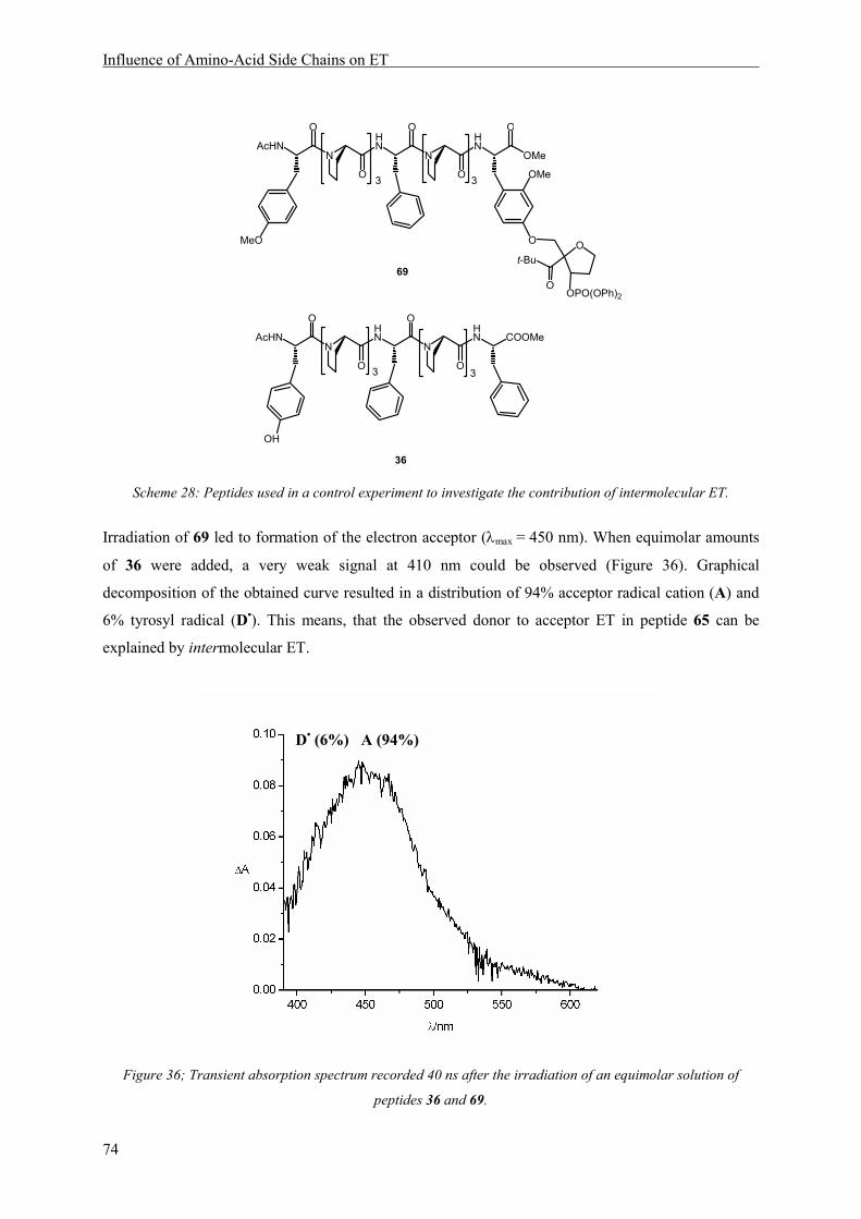
Influence of Amino-Acid Side Chains on ET
74
HN
N
HN
O
O
O
N
O
AcHN
3 3
COOMe
OH
36
HN
N
HN
OMe
O
O
O
O
N
O
AcHN
MeO
OMe
O
3 3
O
O
t-Bu
OPO(OPh)2
69
Scheme 28: Peptides used in a control experiment to investigate the contribution of intermolecular ET.
Irradiation of 69 led to formation of the electron acceptor (λmax = 450 nm). When equimolar amounts
of 36 were added, a very weak signal at 410 nm could be observed (Figure 36). Graphical
decomposition of the obtained curve resulted in a distribution of 94% acceptor radical cation (A) and
6% tyrosyl radical (D•). This means, that the observed donor to acceptor ET in peptide 65 can be
explained by intermolecular ET.
Figure 36; Transient absorption spectrum recorded 40 ns after the irradiation of an equimolar solution of
peptides 36 and 69.
D• (6%) A (94%)

Influence of Amino-Acid Side Chains on ET
75
Thus, the replacement of the redox active aromatic relay 2,4,6-trimethoxyphenylalanine by a non-
redox active, aliphatic central amino acid results in a severe drop in intramolecular ET efficiency from
46% in peptide 64 to <1% in peptide 65. This effect can be rationalized by the shutdown of a possible
sequential ET pathway, consisting of two 10 Å steps, upon exchanging the aromatic side chain in
peptide 64 by an aliphatic side chain in peptide 65. While in peptide 64, the overall distance of 20 Å
can be overcome in an intramolecular reaction within less than 40 ns, using the relay as a stepping
stone; in peptide 65, in contrast, tunneling of the electron over a 20 Å spacer is required to occur in a
single step. Yet superexchange ET across that distance does not take place efficiently within 40 ns.
A second example for the effect an aliphatic side chain exhibits, was provided by peptide 66,
containing a leucine as central residue. The transient spectrum clearly shows the main absorption at
450 nm (94%), the wavelength of acceptor radical cation absorption. Only a minor fraction (6%) of
electrons has already been transferred from tyrosine (Figure 37). This amount can be explained by
intermolecular ET, as discussed above.
Figure 37:Transient absorption spectrum of peptide 66, 40 ns after laser irradiation.
Thus, it was proven in two cases, that 20 Å ET across a hexaproline matrix, separated in half by an
aliphatic amino acid, is not possible intramolecularly with fast rates. For a quantification of the
observed effects, see below in paragraph 7.3.2.
D• (6%) A (94%)
HN
N
HN
OMe
O
O
O
O
N
O
AcHN
HO
OMe
O
3 3
O
O
t-Bu
OPO(OPh)264
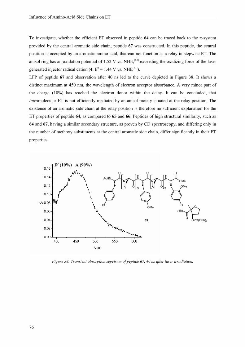
Influence of Amino-Acid Side Chains on ET
76
To investigate, whether the efficient ET observed in peptide 64 can be traced back to the π-system
provided by the central aromatic side chain, peptide 67 was constructed. In this peptide, the central
position is occupied by an aromatic amino acid, that can not function as a relay in stepwise ET. The
anisol ring has an oxidation potential of 1.52 V vs. NHE,[83] exceeding the oxidizing force of the laser
generated injector radical cation (4, E0 = 1.44 V vs. NHE[71]).
LFP of peptide 67 and observation after 40 ns led to the curve depicted in Figure 38. It shows a
distinct maximum at 450 nm, the wavelength of electron acceptor absorbance. A very minor part of
the charge (10%) has reached the electron donor within the delay. It can be concluded, that
intramolecular ET is not efficiently mediated by an anisol moiety situated at the relay position. The
existence of an aromatic side chain at the relay position is therefore no sufficient explanation for the
ET properties of peptide 64, as compared to 65 and 66. Peptides of high structural similarity, such as
64 and 67, having a similar secondary structure, as proven by CD spectroscopy, and differing only in
the number of methoxy substituents at the central aromatic side chain, differ significantly in their ET
properties.
Figure 38: Transient absorption sepctrum of peptide 67, 40 ns after laser irradiation.
D• (10%) A (90%)
HN
N
HN
OMe
O
O
O
O
N
O
AcHN
HO
OMe
O
3 3
O
O
t-Bu
OPO(OPh)2
OMe
65

Influence of Amino-Acid Side Chains on ET
77
The hypothesis, that the redox potential of the central amino acid, i.e. its suitability to function as an
electron relay, is the main factor controlling ET efficiency, should be confirmed by investigation of
the ET properties of an additional peptide 68. This peptide contains 2,4-dimethoxyphenylalanine as
central amino acid. The oxidation potential of this residue can be assumed to be similar to that of the
electron acceptor, since both feature a dialkoxy substitution of the same pattern. In this respect, the
design of peptide 68 accords to the situation in DNA, where electrons are transferred over large
distances, using stepping stones of similar redox properties - the DNA bases.
In contrast to peptide 64, containing three orthogonal spectroscopic sensors, ET through system 68
does only lead to two different absorption maxima. The transient absorption spectrum of the oxidized
relay cannot be distinguished from the spectrum of the electron acceptor, and the absorption band
around 450 nm represents the sum of oxidized electron acceptor and relay (Figure 39). It amounts to
63%. This means, that. 27% of the charge has already migrated to the final donor tyrosine, causing an
additional observable absorption maximum at 410 nm.
Figure 39: Transient absorption spectrum of peptide 68, 40 ns after laser irradiation.
In summary, with peptides 64 and 68, two examples are available for efficient ET across a 20 Å
distance through a peptide matrix. Both peptides contain an oxidizable aromatic amino acid at the
central position, cutting the 20 Å spacer in half. In peptide 64, the oxidized relay generates a
characteristic absorbance, different from the absorption of electron acceptor and electron donor. Using
this peptide, the simultaneous detection of the oxidized forms of three redox sites involved in ET, was
possible. In three peptides 65, 66 and 67, non-oxidizable aliphatic or aromatic side chains are present
at the central position. In these peptides, no efficient intramolecular ET was observed. This leads to
D• (27%) [A+R
•+] (63%)
HN
N
HN
OMe
O
O
O
O
N
O
AcHN
HO
OMe
O
3 3
O
O
t-Bu
OPO(OPh)2
OMe
OMe
66

Influence of Amino-Acid Side Chains on ET
78
the conclusion that efficient intramolecular long range ET is made possible by the presence of redox
relays. These relays function as stepping stones in the ET process, with the charge possesing a lifetime
at this position, generating a chemical intermediate. It can be concluded, that aromatic amino acid side
chains can assist in carrying a charge over large distances, featuring electron hopping. Sequential ET
pathways, using naturally occuring aromatic amino acids, have been proposed in several enzymes, e.g.
ribonucleotide reductase or DNA photolyase. The importance of the respective residues has been
proven by molecular biology studies, yet so far no spectroscopic evidence for the course of a
consecutive reaction is available in these systems. We have now developed a peptide model, which
allows the determination of sequential ET. An oxidizable relay in the matrix renders efficient donor to
acceptor ET across 20 Å (or rather 2 x 10 Å) possible within short reaction times. Without this relay,
ET has to occur via a single-step superexchange mechanism, which cannot be observed with high
efficiency during the time frame of our experiment (Scheme 29). Thus our system allows the
investigation of individual amino acids with respect to their ability to serve as stepping stones in
electron hopping.
HN
N
HN
OMe
O
O
O
O
N
O
AcHN
HO
XOMe
O
3 3
for: X =
MeO
OMe
OMe
OMe
OMe
HN
N
HN
OMe
O
O
O
O
N
O
AcHN
HO
XOMe
O
3 3
for: X = H
OMe
(57) (65)
(62) (63) (64)
O
O
Scheme 29: The deciding factor controlling ET mechanism (and efficiency) in the oligopeptide models is the
character of the side chain X. Electron transfer by hopping (on top) occurs, when oxidizable aromatic side
chains are present in the peptide matrix. ET by single step superexchange (at the bottom) does not take place
within the time frame of the experiment.

Influence of Amino-Acid Side Chains on ET
79
7.3.2 Electron Transfer Rates
To gain information about the ET rates achieved in peptides 64 - 68, an interpretation of the
quantitative data obtained from the graphical analyis was done. The data is summarized in the second
column of Table 1. As a key to the interpretation, the competition between intramolecular and
intermolecular ET in the respective peptides was utilized. From a control experiment, the amount of
tyrosyl radical formed intermolecularly under the conditions of the LFP experiments was estimated to
6%. Substraction of this value from the observed tyrosyl radical absorption leads to the values given in
the last column of Table 1, representing the contribution of the intramolecular reaction.
Table 1: Tyrosyl radicals yielded within 40 ns after laser flash photolysis of 64 - 68 (relative error for all values:
15%), without and with correction for the contribution of intermolecular ET. The sum of oxidized aromatic side
chains is set to 100%. The amount of tyrosyl radical formed by intermolecular reaction of 69 and 36 is given for
comparison.
Tyrosyl-Radical (detected) Tyrosyl-Radical (corrected)
69+36 6.1% -
65 6.9% < 1%
66 6.1% < 1%
67 10% 3%
68 27% 21%
64 36% 30%
Intermolecular reaction rates have been determined by dilution experiments carried out by Napp on
peptides of type 1 (Scheme 3, page 19), in which the ET rates were determined as a function of
peptide concentration.[65, 66]Since only a small fraction of the peptides (approx. 0.1%) is cleaved by the
laser flash, the peptide concentration remains constant during the experiment (pseudo first oder
reaction), and the rate of the bimolecular quenching reaction depends linearly on the peptide
concentration employed in the experiment.
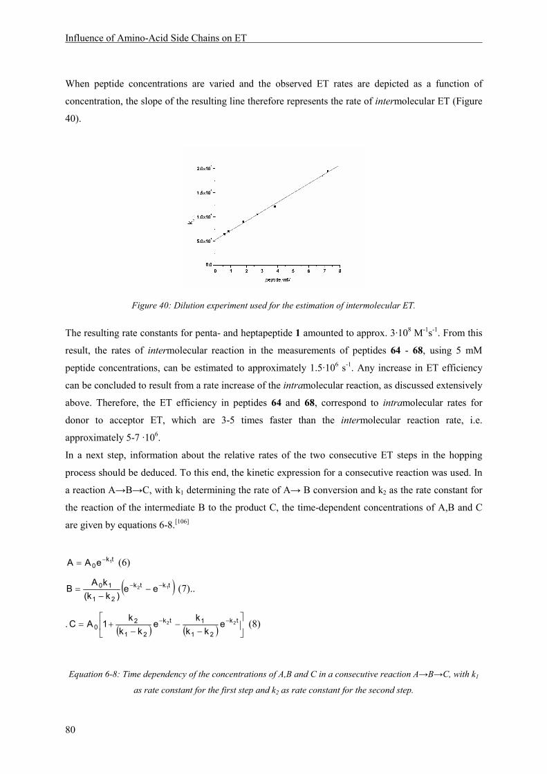
Influence of Amino-Acid Side Chains on ET
80
When peptide concentrations are varied and the observed ET rates are depicted as a function of
concentration, the slope of the resulting line therefore represents the rate of intermolecular ET (Figure
40).
Figure 40: Dilution experiment used for the estimation of intermolecular ET.
The resulting rate constants for penta- and heptapeptide 1 amounted to approx. 3·108 M-1s-1. From this
result, the rates of intermolecular reaction in the measurements of peptides 64 - 68, using 5 mM
peptide concentrations, can be estimated to approximately 1.5·106 s-1. Any increase in ET efficiency
can be concluded to result from a rate increase of the intramolecular reaction, as discussed extensively
above. Therefore, the ET efficiency in peptides 64 and 68, correspond to intramolecular rates for
donor to acceptor ET, which are 3-5 times faster than the intermolecular reaction rate, i.e.
approximately 5-7 ·106.
In a next step, information about the relative rates of the two consecutive ET steps in the hopping
process should be deduced. To this end, the kinetic expression for a consecutive reaction was used. In
a reaction A→B→C, with k1 determining the rate of A→ B conversion and k2 as the rate constant for
the reaction of the intermediate B to the product C, the time-dependent concentrations of A,B and C
are given by equations 6-8.[106]
tk0
1eAA −= (6)
( )tktk
21
10 12 ee)kk(
kAB −− −
−= (7)..
. ( ) ( )
−−
−+= −− tk
21
1tk
21
20
22 ekk
ke
kk
k1AC (8)
Equation 6-8: Time dependency of the concentrations of A,B and C in a consecutive reaction A→B→C, with k1
as rate constant for the first step and k2 as rate constant for the second step.

Influence of Amino-Acid Side Chains on ET
81
The concentrations of starting material, product and intermediate at a defined time thus are a function
of the rate constants k1 and k2 of the reaction. From the transient absorption spectrum upon LFP of
peptide 64, the relative concentrations of electron acceptor (A), oxidized relay (B) and oxidized
electron donor (C) are available. While a substantial amount of C (30%, if corrected for the
intermolecular contribution) has already been formed, when A has been reduced to 54%, the
intermediate B is present in detectable concentrations of 10%. A rate ratio of 5:1 for k2/k1 leads to such
a distribution of A, B and C, according to equations 6-8. A graphical depiction of this case is shown in
Figure 41, with the observed concentration ratio indicated by a vertical line.
Figure 41: Time-dependent concentrations of A, B and C in a consecutive reaction A→B→C, with a 1:5 ratio of
rate constants. The concentration ratio derived from LFP of peptide 64 is indicated by a vertical line.
From these considerations it can be concluded, that ET between tyrosine and the oxidized central relay
amino acid 2,4,6-trimethoxyphenylalanine radical cation is five times faster than ET from the relay to
the electron acceptor (Scheme 30). This result is in accordance with the oxidation potentials of the
engaged redox sites. While acceptor and relay amino acid differ in oxidation potentials by only 10 mV
(0.95 resp. 0.94 V vs. the ferrocene redox couple), the difference between the donor and the relay
amino acid is significantly larger, with tyrosine showing a potential of 0.85 V vs. the ferrocene redox
couple.
A B Ck1 k2
k2 = 5 k1

Influence of Amino-Acid Side Chains on ET
82
HN
N
HN
OMe
O
O
O
O
N
O
AcHN
HO
OMe
O
3 3
k2 k1
ET ET
MeOOMe
MeO
k2 = 5 k1
O
Scheme 30: relative rates for sequential ET through peptide 64.
In summary, quantitative analysis of the data obtained from LFP experiments, yielded information
about ET rates in peptides 64 and 68. Those peptides contain central amino acids with aromatic side
chains that can participate in sequential ET and thereby allow ET to occur with 20-30 times higher
rates than the single-step superexchange ET in peptides 65 and 66. Intramolecular electron hopping
between the N-terminal electron donor tyrosine and the C-terminal electron acceptor, separated by a
peptide matrix of 20 Å length, occurs with rates of approximately 5-7 ·106 in peptides 64 and 68. From
the relative amount of transients observed in irradiated peptide 64, 40 ns after the flash, the rate ratio
for relay to acceptor versus donor to relay ET was estimated to 1:5.

The Phenylalanine Effect
83
8 The Phenylalanine Effect
A surprising result was obtained, when phenylalanine was introduced as a central amino acid. The
benzene ring has an oxidation potential of 2.2 V vs. NHE,[84] which is significantly higher than the
potential of the injector radical cation 4 (1.44 vs. NHE[71]). Thus, phenylalanine cannot function as a
relay in stepwiseET through our model peptides and the result of LFP was expected to resemble the
case of peptide 67, containing an anisole moiety at the central position, in which no efficient ET
between donor and acceptor occurs within 40 ns (cf. Figure 38). However, the transient absorption
spectrum recorded 40 ns after irradiation of 70, shows a significant amount of tyrosyl radical,
indicated by the 410 nm absorption.
Figure 42:Transient absorption spectrum of peptide 70 40 ns after irradiation.
The relative amount of oxidized species determined by graphical curve decomposition is 30% tyrosine
and 70% electron acceptor. This amount of tyrosyl radical formed within 40 ns, can only be explained
by intramolecular ET. The strucuture of peptide 70, determined by CD-spectroscopy, is similar to the
structures of peptides 64 - 68, i.e. the peptide forms a PPII helix. The unexpectedly high ET efficiency
can thus not be traced back to a different overall structure of the peptide backbone.
HN
N
HN
OMe
O
O
O
O
N
O
AcHN
HO
OMe
O
3 3
O
O
t-Bu
OPO(OPh)268

The Phenylalanine Effect
84
Comparable observations with respect to the ET properties of phenylalanine-containing peptides were
made in peptides 71 and 72, as well as 63 and 62. ET efficiencies do not drop significantly upon
replacement of the redox active trimethoxysubstituted benzene by a simple benzene moiety. In peptide
74, containing phenylalanine as a central amino acid, ET efficiency within 40 ns even increased
slightly as compared to 73, containing the redox active methoxysubstituted relay (Scheme 31, Table
2).
HN
N
HN
OMe
O
O
O
O
N
O
AcHN
HO
OMe
O
n m
O
O
t-Bu
OPO(OPh)269: R = OMe70: R = H
n = 1, m = 2
n = 1, m = 3
n = 2, m = 271: R = OMe72: R = H
57: R = OMe62: R = H
n = 3, m = 358: R = OMe68: R = H
R
RR
Scheme 31: Peptides used to investigate the role of phenylalanine in ET.

The Phenylalanine Effect
85
The results of the comparison of phenylalanine-containing with 2,4,6-trimethoxyphenylalanine-
containing peptides are summarized in Table 2. The relative amounts of oxidized species formed
within 40 ns after the laser flash are derived by graphical decomposition of the respective transient
absorption spectra (see Figure 21, page 47). All spectra not depicted in the text are reproduced in the
Experimental Part.
Table 2: relative amounts of transients formed 40 ns after irradiation of peptides
oxidized electron donor
(%) oxidized relay
(%) electron acceptor
(%)
71 63 7 30
72 70 - 30
63 35 11 54
62 41 - 59
73 53 8 39
74 76 - 24
64 36 10 54
70 41 - 59
From these results, the placement of phenylalanine inside the peptide matrix seems to exhibit an
influence on the donor to acceptor ET rates, which is comparable to the influence of 2,4,6-
trimethoxyphenylalanine. These findings could not be rationalized so far.

Summary and Outlook
87
9 Summary and Outlook
A peptide model was designed, which allows the investigation of amino acid side chain participation
in ET through peptides. Aromatic amino acids function as oxidizable spectroscopic sensors for the
direct observation of charged intermediates during the ET process. Tyrosine as electron donor, situated
at the N-terminus of the peptide, provides driving force for the ET process and is irreversibly oxidized
to a long-lived phenoxyl radical with a sharp absorption band. Two methoxysubstituted phenylalanine
derivatives were chosen as additional spectroscopic sensors, yielding oxidized transients with different
absorption spectra. They were synthesized in their enantiopure form and investigated with respect to
their electrochemical and spectroscopical properties. In the peptide model, they function as C-terminal
electron acceptor precursor and central relay, separated from each other and the donor by a proline
matrix. The electron acceptor can be generated by laser irradiation of an injection unit, containing a
tbutyl ketone as chromophore. The half-life time of acceptor generation is estimated to 20 ns and
intermolecular ET started to gain importance from approximately 100 ns after irradition. Thus,
transient absorption spectra recorded 40 ns after the laser flash were used for the examination of
intramolecular ET efficiencies between the redox sites. In a first study, the injection unit was
connected to the peptide C-terminus via an ester bond and peptides with a total length of 5-10 residues
were synthesized. In this case, ET efficiency was observed to depend on the construction of the
separating matrix. Efficient intramolecular ET to the electron acceptor within 40 ns after irradiation
was observed in peptides, in which either donor and relay or relay and acceptor were arranged in close
proximity (separated by one proline). Signals of oxidized relay were observed in all cases, proving that
the occurence of oxidized intermediates in donor to acceptor ET can be detected in this model.
Efficient intramolecular ET through the whole chain, i.e. from the N-terminal donor to the electron
acceptor, was only observed in peptides containing a total of up to four prolines in the matrix. The
synthesis of a new electron acceptor precursor, in which the injection unit was connected directly, via
an ether bond, to a phenoxyl function at the aromatic side chain of the C-terminal amino acid, then
allowed the observation of intramolecular ET from the N-terminus to the C-terminus of a nonapeptide,
which consisted of two triproline spacers and the three spectroscopic sensors. This peptide showed a
well-defined polyproline II helical structure, leading to a donor/acceptor separation of 20 Å. The
aromatic relay, functioning as spectroscopic sensor for the detection of oxidized intermediates in
donor to acceptor ET, could be exchanged by a number of different amino acids, without changing the
overall structure of the peptide. Thus, the peptide model allowed us to determine the influence of the
central amino acid - separated from both, donor and acceptor, by 10 Å - on ET. Efficient ET was
observed in those two cases, where the central amino acid was able to function as an oxidizable relay
in ET. The spectrum of the peptide containing the three distinguishable spectroscopic sensors, showed
the simultaneous occurence of electron acceptor, oxidized donor and oxidized relay and thereby
88
proved that the charge resides on the relay site during ET. In two peptides containing aliphatic central
residues, intramolecular donor to acceptor ET did not take place efficiently within the time frame of
our experiments – a 20 to 30 fold decrease in donor oxidation was observed. A peptide containing an
anisol ring as central aromatic side chain, with an oxidation potential far above the electron acceptor,
did also not render efficient intramolecular ET possible. In conclusion, efficient ET across the 20 Å
distance in our model peptides was only possible within 40 ns, if an oxidizable amino acid side chain
was present inside the matrix. This side chain functioned as a stepping stone in ET, with the charge
residing on it, forming a chemical intermediate. Thus, electron hopping via aromatic amino acid side
chains was proven to allow distal (20 Å) ET transfer in peptides within 40 ns whereas single-step
superexchange ET did not take place within 40 ns in peptides of similar structure. From considerations
regarding the competition of intermolecular and intramolecular ET, the rate of intramolacular ET
between electron acceptor and electron donor by a two-step electron hopping process, with 10 Å
distance for each step, was determined to 5 - 7 ·106 s-1. In the peptide containing three spectroscopic
sensors, the relative intensities of all signals could be used to conclude on the ratio of rate constants in
the consecutive ET reaction, leading to a five times faster rate constant for donor to relay, than for
relay to acceptor ET. This is consistent with the differences in driving force for the two reactions.
Our model provides an experimental approach to determine the suitability of individual amino acids to
function as relays in electron hopping. Future experiments could be carried out with the naturally
occuring aromatic amino acids tryptophane and histidine, as well as with cysteine, as an additional
oxidizable amino acid. The introduction of protection groups, to avoid deprotonation upon oxidation,
which renders the ET irreversible (as in the case of tyrosine) might be necessary. First experiments
lead to the assumption, that phenylalanine plays an unexpected role in the mediation of long-range ET.
Whether this is due to unknown local structural features, not visible in the CD spectrum, or to
electronic effects is not yet clear. This might be further investigated by the introduction of electron-
donating or electron-withdrawing groups to the benzene ring.
Experimental Part
89
10 Experimental Part
10.1 Conditions of Measurements
NMR-Spectrometry
Nuclear magnetic resonance (NMR) spectra were recorded on Bruker spectrometers (Avance400: 400
MHz for 1H and 101 MHz for 13C; Avance DRX500: 500 MHz for 1H and 125 MHz for 13C) at
ambient temperature in the solvents indicated, and referenced to trimethylsilate (TMS). Chemical
shifts δ are given in ppm. Multiplets are assigned with s (singlett), s,br (broad singlet), d (doublet), t
(triplet), q (quartet) and m (multiplet). Assignements of the signals are given in italics.
Melting Points (M.p.)
Melting points were measured on a Büchi 530 and are not corrected.
Mass Spectrometry (MS)
Electron ionizaition (EI) and fast atom bombardment (FAB) mass spectra were recorded by Dr. H.
Nadig (University of Basel) on a VG70-250 (EI) and a MAT 312, using 3-nitrobenzyl alcohol as a
matrix (FAB). Electron Spray Ionisation (ESI) spectra were recorded in methanol on a Finnigan MAT
LCQ spectrometer. High Resolution Mass Spectrometry (HR-MS) were performed by the mass
service at the university of Fribourg (CH) (Fredy Nydegger, "Département de Chimie Université de
Fribourg"). Mass signals are given in mass units per charge. The fragments and intensities are given in
brackets.
Elemental Analysis (EA)
Elemental analyses were carried out on a Leco CHN-900 device by W. Kirsch (University of Basel).
The values are given in mass percent.
Enantiomeric Purity
Enantiomeric excess (ee) was determined by chiral HPLC (Chiracel columns from Daicel,
heptane/iPrOH 70:30, 0.7 mL/min, 20 °C). Optical rotations were measured on a Perkin Elmer
polarimeter 241. The concentrations are given in g/100 mL.
Experimental Part
90
UV-Vis Spectroscopy
UV-Vis spectra were recorded on a Perkin Elmer spectrometer (Lambda Bio 40) in MeCN or
MeCN/water 3:1. The extinction coefficients at 308 nm (ε308) of peptides were determined using 5-8
mM concentrations.
High Performance Liquid Chromatography (HPLC)
HPLC chromatograms were recorded on a Water Alliance 2690 Separation Module with a 2680 Dual
Mode Detector adjusted to 260 nm and 308 nm. Columns for reversed–phase HPLC were Merck
LiChroSpher 100 (RP–18e, 5 µm, 125 × 4mm). Eluents were acetonitrile and nanopure water, using a
gradient program reaching from 0% water to 100% in 40 min at a flow rate of 1.0 mL·min−1. Retention
times (tR) are given in minutes.
Circular Dichroism (CD) Spectroscopy
CD spectra were recorded using a Chirascan spectrometer with a spectral bandwidth of 1 nm at 25 °C
with a time constant of 3 s and a step resolution of 1 nm. CD data are given as mean residual molar
ellipticities (ΘMRW in deg cm2 dmol-1). A Quartz cell with a path length of 1 cm was used with
solutions containing approximately 70 µgml-1 (6x10⁻⁴ M per residue) peptide solutions.
Differential Pulse Cyclovoltammetry
Pulsed cyclovoltammetric measurements were performed using a BAS 100 B/W electrochemical
workstation and a three-electrode cell, consisting of a silver-wire as pseudo-reference, a glassy carbon
disk as working and a platinum wire as counter electrode. The amino acids were dissolved in degassed
acetonitrile at 2 mM concentrations. Tetrabutylammoniumhexafluorophosphate was added as an
electrolyte (100 mM) and ferrocene as an internal standard. The measurements were carried out under
an argon blanket.
Laser Flash Photolysis (LFP)
The laser experiments were carried out using a COMPex 205 XeCl-Excimer Laser (Lambda Physik)
with a pulse energy of 100-150 mJ and a pulse width of 20 ns for irradiation. A pulsed Xe-lamp served
as a second light source for read-out. (cf. Figure 9). The peptides were dissolved in acetonitrile/water
3:1 in concentrations of 5-8 mM, degassed in three freeze/thaw cycles and stored under vacuum in a
quartz cuvette (4.5 cm x 1 cm x 1 cm). Transient absorption spectra were recorded with an iStar ICCD
detector (Andor). The signals of 5 laser flashes were accumulated. Time-resolved measurements were
carried out using a 1P28 photomultipier. Photosensitization experiments were carried out in oxygen-
saturated acetonitrile with 1 mM concentrations of compound, 0.1 mM dicyanonapthtalene and 20
mM biphenyl. The data was processed using MacFitFlash and Origin.
Experimental Part
91
10.2 Synthesis
10.2.1 Materials, Solvents and Reagents
Technical grade solvents used for extraction and column chromatography were distilled prior to use.
Absolute solvents were purchased from Fluka or Aldrich in septum sealed bottles. Deuterated solvents
were purchased from Cambridge Isotope Laboratories, Inc.. Nanopure water, used in LFP, CD
spectroscopy and HPLC chromatography, was filtered over a Barnstead ultrapure water system.
Amino acids, coupling reagents and resins for SPPS were ordered from Novabiochem or Bachem and
used without further processing. The chiral Rh-catalyst was ordered from Strem and stored under
argon-atmosphere at r.t.. Other materials and reagents were purchased from Fluka, Aldrich and Acros
in the highest available grade and used without further purification. Ion exchange chromatography was
performed on Dowex 400 resin activated by washing with 2 M aq. sodium hydroxide and subsequently
2 M acetic acid. Silica gel 60 (Fluka) was used for flash chromatography. TLC plates from Merck
were used for reaction control (silica gel 60 on aluminium sheets) and chromatographic analysis (silica
gel 60 on glass). CAM-Dip (10 g cerium(IV)sulfate-tetrahydrate, 25 g ammonium-heptamolybdate-
tetrahydrate, 100 mL sulfuric acid (96%), 900 mL water) was used for staining of the TLC plates.

Experimental Part
92
10.2.2 Notation
Peptide sequences are noted from the N-terminus to the C-terminus. Amino acids are denoted in the
three letter code, with N-terminal and C-terminal protection groups separated by hyphens and side-
chain protection groups given in brackets, e.g Ac-Tyr(OBn)-OH for N-acetyl-O-benzyl-tyrosine. All
non-racemic amino acids are used as L-enantiomers. The non-natural amino acids are abbreviated as
follows:
DMP: 2,4,6-trimethoxyphenylalanine
TMP: 2,4-dimethoxyphenylalanine
DAP (dialkoxyphenylalanine): 3-{4-[2-(2,2-dimethylpropionyl)-3-(diphenoxyphosphoryl-oxy)tetra-
hydrofuran-2-ylmethoxy]-2-methoxyphenyl}propionic acid methylester (electron acceptor precursor
44 )
In peptides containing the injection unit 2, linked to the C-terminus via an ester bond (peptides 18),
this unit will be denoted as: Inj. Two examples for the resulting nomenclature for peptides of type 18
resp. 54 are given in Figure 43.
HN
N
HN
O
O
O
O
O
O
OOPO(OPh)2
N
O
AcHN
HO
OMe
OMe
OMeMeO
OMe
3
Ac-Tyr-Pro-TMP-(Pro)3-DMP-Inj
HN
N
HN
OMe
O
O
O
O
N
O
AcHN
HO
OMe
O
3 3
O
O
t-Bu
OPO(OPh)2
OMeMeO
MeO
Ac-Tyr-(Pro)3-TMP-(Pro)3-DAP-OMe
Figure 43: Nomenclature for oligopeptides.

Experimental Part
93
10.2.3 General Procedures (GP)
Synthetic steps following a general procedure are indicated by roman numbering, according to the
following section.
I. Deprotection of tert-Butylcarbamates – Boc Deprotection (GP I)
To remove the tert-butyloxycarbonyl (Boc) protection group from the amine function of an amino acid
(or peptide), the respective compound was dissolved in a cold hydrogen chloride solution (4 M in 1,4-
dioxane) and stirred at r.t. for 30 min. After coevaporation with toluene under reduced pressure, the
dry, solid amine hydrochloride could be obtained as a foam by addition of small amounts of CH2Cl2
and evaporation of all solvent traces under high vacuum. Completeness of the deprotection reaction
was checked by ESI-MS, before subjecting the amine hydrochloride to the next coupling step.
II. Deprotection of tert-Butylesters (GP II)
To remove the tert-butylester group from the carbonic acid function of an amino acid (or peptide), the
respective compound was dissolved in a 1:1 (v/v) mixture of trifluoroacetic acid and CH2Cl2 and
stirred for 45-90 minutes at r.t.. Progress of the reaction was checked by TLC (CH2Cl2, 4% MeOH,
1% AcOH). Evaporation of the solvents under reduced pressure and coevaporation with toluene
yielded the free acid as a yellowish solid.
III. Deprotection of Aromatic Benzylethers [Tyr(OBn)] (GP III)
The benzylether protection group at the side-chain of tyrosine residues was removed by a
hydrogenation procedure, using palladium on activated charcoal (10%) as a catalyst and 1 bar
hydrogen pressure. The reaction was carried out in methanol and reaction progress was checked by
TLC (CH2Cl2, 4-15% MeOH). Acetic acid was added to enhance catalytic activity, if necessary. After
complete conversion of the starting material (2-8 hs), the reaction mixture was filtered over celite, the
filtrate was washed thoroughly with methanol, and the solvents were removed under reduced pressure
before further purification of the crude product.
IV. Deprotection of Aromatic tert-Butylethers [Tyr(OtBu)] (GP IV)
The benzylether protection group at the side-chain of tyrosine residues was removed by dissolving the
compound in a 3:2 (v/v) mixture of CH2Cl2 and trifluoroacetic acid and stirring at r.t. for 15 min. After
coevaporation with toluene under reduced pressure, the deprotected peptide could be obtained as a
solid by addition of small amounts of CH2Cl2 and drying under high vacuuum. Completeness of the
deprotection was checked by ESI-MS, before subjecting the peptide to the purification procedure (GP
VII).
Experimental Part
94
V. Peptide Couplings in Solution
Peptide couplings were carried out under inert gas atmosphere. The carbonic acid was dissolved in
DMF/CH2Cl2 (1:3, v/v) with 2 eq. of coupling reagent (2-(1H-benzotriazole-1-yl)-1,1,3,3-
tetramethyluronium tetrafluoroborate (TBTU) or 2-(6-chloro-1-H-benzotriazole-1-yl)-1,1,3,3-
tetramethyluronium hexafluorophosphate (HCTU)). The amine (1 eq.) was added in CH2Cl2 with 4 eq.
of N,N'-diisopropylethylamine (DIPEA). The reaction mixture was stirred for 3-6 hs, then poured into
a separatory funnel, containing a biphasic system of sat. aq. NH4Cl-solution and EtOAc. After
washing with sat. aq. NH4Cl-solution (2x) and sat. aq. NHCO3-solution (2x), each followed by brine,
the organic phase was dried (MgSO4) and concentrated under reduced pressure. Crude products were
filtered over silica (CH2Cl2, 4%-15% methanol) and submitted to the next reaction step or the
purification procedure (GP VII) after ESI-MS control. Yields of peptide couplings varied between 60
and 80%.
VI. Acetylation in Solution
To acetylate the N-terminus of a peptide, the peptide was dissolved in CH2Cl2 and 10 eq. of acetic
anhydride as well as 10 eq. of N,N'-diisopropylethylamine (DIPEA) were added, before stirring for 30
min at r.t.. Completeness of the reaction was checked by TLC (CH2Cl2, 4%-15% methanol) or ESI-
MS. After removal of the solvents under reduced pressure, the residue was dissolved in EtOAc and
washed with aq. NH4Cl-solution and sat. aq. NHCO3-solution, each followed by brine. The organic
phase was dried (MgSO4) and concentrated under reduced pressure, before crude products were
filtered over silica (CH2Cl2, 4%-15% methanol) and submitted to the next reaction step or the
purification procedure (GP VII).
VII. Peptide Purification
Peptides used in LFP measurements were purified by column chromatography, using a gradient of
methanol in CH2Cl2, starting from 4% (1% for very short peptides) and reaching to 10-20%,
depending on the polarity of the respective peptides. For gradients of >10% methanol, the resulting
product was dissolved in CH2Cl2 (containing up to 4% methanol to increase solubility) and filtered, to
remove silica particles. Purity of the peptides was checked by HPLC chromatography and UV-
spectroscopy. The extinction coefficient at 308 nm, stemming from the pivaloyl chromophore, should
be below 60 (below 100 in some larger peptides). Hydrogenation with Pd/C and subsequent column
chromatography has been proven to be an efficient method to remove UV-active impurities.
Therefore, some peptides without benzyl-ether protection groups were subjected to this procedure, if
stirring with activated charcoal was not sufficient for the removal of yellowish impurities. Peptides
were dried by addition of small amounts of CH2Cl2 and subsequent removal of all solvent traces at the
high vacuum line, to yield white foams.

Experimental Part
95
VIII. Solid Phase Peptide Synthesis (SPPS)
a) Procedure
Solid phase peptide synthesis was carried out employing a Syro I Peptide Synthesizer (MultiSynTech
GmbH, Witten, Germany), with plastic syringes as reaction vessels. 2-Chlorotrityl resin, preloaded
with proline (Novabiochem) was used as the solid matrix. Fmoc-protected amino acids and coupling
reagents were purchased from Novabiochem and Bachem. 2-(6-chloro-1-H-benzotriazole-1-yl)-
1,1,3,3-tetramethyluronium hexafluorophosphate (HCTU) was used as a coupling reagent. For the
peptide couplings, the Fmoc-protected amino acid (4 eq., 0.5 M in DMF), HCTU (4 eq. 0.5 M in
DMF) and N,N'-diisopropylethylamine (DIPEA, 12 eq., 3 M in NMP) were added to the resin. The
mixture was agitated for 1 h and subsequently washed with DMF (3x). For Fmoc-deprotection, a
mixture of piperidine and DMF (1:5, v/v) was added to the resin. The reaction mixture was agitated
for 5 min and for another 10 min after addition of fresh piperidine solution, and then washed with
DMF (5x).
b) Acetylation
After the last deprotection step, the resin was thoroughly washed. Then 30 eq. of acetic anhydride and
30 eq. of triethylamine were added in CH2Cl2 (3 mL total volume). After shaking for 30 min, the resin
was washed with CH2Cl2 and subjected to the cleavage procdure.
c) Cleavage of the Peptide
The resin was covered with a 1:1:8 (v/v/v) mixture of trifluoroethanol, acetic acid and CH2Cl2 and
shaked for 30 min, then washed with CH2Cl2, collecting all solvents. The procedure was repeated. The
combined solutions were then concentrated under reduced pressure, cooled to 0 °C and cold Et2O was
added. The precipitated peptide was washed with cold Et2O (3x), then dried in high vacuum.
10.2.4 Characterisation of Peptides
A complete NMR-spectroscopical analysis was carried out for the small peptides 16 and 17, as well as
for the molecules 15 to 13, to confirm that the building blocks are stable to the reaction conditions
used in peptide synthesis. Based on these peptides, a protocol for peptide characterisation was
established, which consists of HPLC and ESI-MS analysis, as well as UV-spectroscopy to determine
the purity of the peptide (cf. GP VII).
Experimental Part
96
10.3 Compounds
2 (2R,3S)-2-tert-Butylcarbonyl-3-diphenoxyphosphoryloxy-2-hydroxymethyl-
tetrahydrofurane
Alcohol 2 was synthesized according to procedures established in the Giese group, starting from
pivaldehyde in 7 steps with 3% overall yield.[65, 66]
The NMR and mass spectroscopical data are given here. For full characterisation see the indicated
references.
1H-NMR (CDCl3, 400 MHz): δ = 7.37-7.31 (m, 4H; HAr), 7.24-7.17 (m, 6H; HAr), 5.36-5.28 (m, 1H;
CΗ−ΟP), 4.17-4.07 (m, 2H; CH2CH2), 3.60-3.53 (m, 2H; CH2OH), 2.72 (br, 1H OH), 2.33-2.24 (m,
2H; OCH2CH2), 1.16 (s, 9H; C(CH3)3).
13C-NMR (101 MHz, CDCl3 ): δ = 215.7 (s, C=O, COtBu), 150.6 (d, JC-P = 6.9 Hz, CAr-OP), 150.5 (d,
JC-P = 7.2 Hz, CAr-OP), 130.0 (d, 2 p-CHAr(PhOP)), 125.7 (d, 2 m-CHAr(PhOP)), 125.6 (d, 2 m-
CHAr(PhOP)), 120.4 (d, JC-P = 4.9 Hz, 2 o-CHAr(PhOP)) 120.3 (d, JC-P = 5.3 Hz, 2 o-CHAr(PhOP)), 94.5
(d, JC-P = 9.1 Hz, CCH-OP), 83.7 (d, JC-P = 5.0 Hz, CHCH2), 68.2. 67.2 (2t, OCH2CH2, CH2OH), 45.5
(s, C(CH3)3), 33.1 (t, OCH2CH2), 25.9 (q, C(CH3)3).
MS (ESI, MeOH, m/z): 891 ([2M+Na]+, 100), 457 ([M+Na]+, 40).
8 3-(2,4-dimethoxy-phenyl)-2-nitro-propionic acid methyl ester
1,2-Dimethoxybenzaldehyde (3.0 g, 18 mmol), methylnitroacetate (2.0 mL, 22 mmol), dimethylamine
hydrochloride (3.0 g, 57 mmol) and KF (157 mg, 2.71 mmol) were refluxed in toluene (110 mL) for
12 h, using a Dean-Stark water trap. The solvent was removed under reduced pressure and the
remaining orange solid was dissolved in CH2Cl2 and washed three times with water. The organic
phase was dried (MgSO4) and concentrated under vacuum to give α-nitro-acrylic acid-methylester 7 as
an orange oil, which was used in the following reaction without further purification. The raw product
of 7 was dissolved in chloroform (226 mL) and isopropanol (67 mL) and Merck Silica gel (36 g) was
added. NaBH4 (3.4 g, 90 mmol) was added portionwise to the vigorously stirred suspension during 5
min. After additional 2hof stirring, acetic acid (10 mL) was added carefully and the solid components
were removed by filtration. The remaining solution was concentrated under reduced pressure and the
crude product was dissolved in CH2Cl2/water (5:1). After extraction of the aq. phase with CH2Cl2, the
Experimental Part
97
combined organic phases were dried (MgSO4) and concentrated under vacuum. The residue was
purified by column chromatography (EtOAc/hexane 1:9) to give 8 as a yellow solid; yield: 3.2 g
(60%).
1H-NMR (400 MHz, CDCl3, 25°C, TMS): δ = δ = 7.00 (d, J = 8.1 Hz, 1H; o-HAr), 6.44-6.38 (m, 2H;
2m-HAr), 5.51 (dd, , J = 9.3, 5.8 Hz, 1H; CH-(CH2CAr)), 3.82, 3.80, (2s, 6H; 2 CAr-OCH3), 3.78 (s, 3H;
CO2CH3), 3.46 (dd, J = 14.1, 5.8 Hz, 1H; CHaHb-CAr), 3.38 (dd, J = 14.1, 9.3 Hz, 1H; CHaHb-CAr).
13C-NMR (101 MHz, CDCl3, 25°C): δ = 165.2 (s, C=O, CO2CH3), 161.0 (s, o-CAr-OCH3), 158.6 (s, p-
CAr-OCH3), 131.9 (d, o-CHAr), 114.5 (s, CAr-CH2), 104.4, 98.8 (d, 2 m-CHAr), 87.4 (d, CH-(CH2CAr)),
55.58, 55.57 (q, 2 CAr-OCH3), 53.7 (q, CO2CH3), 31.9 (t, CH2-CAr).
MS (ESI, MeOH, m/z): 292 ( [M+Na]+, 100).
EA: calc. for C12H15NO6: C: 53.53, H: 5.62, N: 5.20; found C: 53.44, H: 5.57, N: 5.18.
9 (rac)-2,4-dimethoxyphenylalanine-methylester [(rac)-DMP-OMe]
Arylproprionate 8 (3 g, 11 mmol) was stirred in dry methanol (50 mL) with palladium on activated
charcoal (300 mg, 10%). Ammoniumformiate (3.5 g, 56 mmol) was added in a single portion. After
stirring for 72 hs, the reaction mixture was filtered over celite and the filtrate was washed thoroughly
with methanol. After concentration of the solution under reduced pressure, water and CH2Cl2 was
added to the crude product. The aqueos phase was acidified with acetic acid and extracted with CH2Cl2
(3x). The combined organic phases were dried (MgSO4), concentrated under vacuum and the resulting
crude product was purified by column chromatography (CH2Cl2 → 4% MeOH in CH2Cl2) to yield 9 as
a white foam (1.5 g, 56%).
1H-NMR (400 MHz, CDCl3/MeOH (30:1), 25°C, TMS): δ = 7.00 (d, J = 8.1 Hz, 1H; o-HAr), 6.43-6.39
(m, 2H; 2m-HAr), 3.77 (s, 6H, 2 CAr-COCH3), 3.68 (s, 3H, CO2CH3), 3.74 (dd, , J = 8.1, 5.6 Hz, 1H;
CH-(CH2CAr)), 3.03 (dd, J = 13.5, 5.6 Hz, 1H; CHaHb-CAr), 2.76 (dd, J = 13.4, 8.1 Hz, 1H; CHaHb-
CAr).

Experimental Part
98
13C-NMR (101 MHz, CDCl3, 25°C): δ = 175.9 (s, C=O, CO2CH3), 160.1 (s, o-CAr-OCH3), 158.7 (s, p-
CAr-OCH3), 131.7 (d, o-CHAr), 117.9 (s, CAr-CH2), 104.1, 98.7 (d, 2 m-CHAr), 55.5 (d, CH-(CH2CAr)),
54.6, 52.1 (q, 2 CAr-OCH3), 35.6 (t, CH2-CAr).
MS (ESI, MeOH m/z): 240 ( [M+H]+, 100).
10 N-acetyl-(rac)-2,4-dimethoxyphenylalanine-methylester [(rac)- Ac-DMP-OMe]
(rac)-2,4-Dimethoxyphenylalanine-methylester 9 (200 mg, 1.25 mmol) was stirred in a 1:1 mixture of
sat. aq. Na2CO3-solution and dioxane (6 mL) with acetic anhydride (0.53 mL, 5.6 mmol) for 2hat r.t..
Extraction with CH2Cl2, concentration of the dried (MgSO4) organic phases under reduced pressure
and purification of the resulting crude product by column chromatography (1% MeOH in CH2Cl2 →
2% MeOH in CH2Cl2) yielded 10 as a white foam (285 mg, 81%).
M.p. = 103-105 °C.
1H-NMR (400 MHz, MeOD, 25°C, TMS): δ = 6.96 (d, J = 8.1 Hz, 2H; HAr-6), 6.44-6.41 (m, 2H; HAr-
3, 5), 4.69 (dd, J = 13.6, 6.3 Hz, 1H; CαH ), 3.81, 3.79, 3.71 (s, 9H; 3CH3-O), 3.03 (d, J = 6.3 Hz, 2H;
CαHCH2), 1.93 (CH3CO).
13C-NMR (101 MHz, MeOH): δ = 172.9 (s, C=O, CO2Me), 170.1 (s, C=O, COCH3), 160.6 (s, oCAr-
OCH3), 158.8 (s, pCAr-OCH3), 131.9 (d, CHAr), 131.6 (d, CHAr), 117.2 (s, CAr) , 104.8 (d, CHAr), 99.0
(d, CHAr), 55.8, 55.7, 52.5 (q, 3CH3-O), 53.7 (d, CαH), 32.1 (t, CH2), 23.5 (s, COCH3).
MS (FAB, NBA, m/z): 151 (86), 180 (24), 222 (62), 282 (100, [M]+) .
MS (ESI, MeOH m/z): 585 (100, [2M+Na]+), 304 (40, [M+Na]+).
EA: calc. for C14H18NO5: C: 59.99, H: 6.47, N: 5.00; found C: 60.03, H: 6.59, N: 5.11.
11 N-acetyl-(rac)-2,4,6-trimethoxyphenylalanine-methylester [(rac)-Ac-TMP-OMe]
(rac)-2,4,6-Trimethoxyphenylalanine-methylester[88] (337 mg, 1.25 mmol) was stirred in a 1:1 mixture
of sat. aq. Na2CO3-solution and dioxane (6 mL) with acetic anhydride (0.53 mL, 5.6 mmol) for 2hat
r.t.. Extraction with CH2Cl2, concentration of the dried (MgSO4) organic phases under reduced

Experimental Part
99
pressure and purification of the resulting crude product by column chromatography (1% MeOH in
CH2Cl2 → 2% MeOH in CH2Cl2) yielded 11 as a white foam (311 mg, 81%).
M.p. = 131-132 °C.
1H-NMR (400 MHz, MeOD, 25°C, TMS): δ = 6.18 (s, 2H; HAr), 4.44 (t, J = 7.6 Hz, 1H; CαH ), 3.78,
3.77, 3.63 (s, 12H; 4 x CH3-O), 3.05 (dd, J = 13.2, 7.6 Hz, 1H; CαHCHH), 2.93 (dd, J = 13.2, 7.6 Hz,
1H; CαHCHH), 1.89 (CH3CO).
13C-NMR (101 MHz, MeOH): δ = 174.3 (s, C=O, CO2Me), 173.0 (s, C=O, COCH3), 162.0 (s, oCAr-
OCH3), 160.5 (s, pCAr-OCH3), 106.0 (s, CAr), 91.4 (d, 2CHAr), 56.1, 55.7, 52.4(q, 4CH3-O), 54.0 (d,
CαH), 25.8 (t, CH2), 22.3 (s, COCH3).
MS (ESI, MeOH m/z): 645.0 (100, [2M+Na]+), 334.3 (24, [M+Na]+).
EA: calc. for C15H21NO6: C: 57.87, H: 6.80, N: 4.50; found C: 57.76, H: 6.64, N: 4.32.
12 2-tert-butoxycarbonyl-(rac)-3-(2,4-dimethoxy-phenyl)-propionic acid 2-(2,2-dimethyl-
propionyl)-3-(diphenoxy-phosphoryloxy)-tetrahydro-furan-2-ylmethyl ester [(rac)-Boc-
DMP-Inj]
2,4-Dimethoxyphenylalanine-methylester 9 (400 mg, 1.67 mmol) was suspended in sat. aq. Na2CO3-
solution (54 mL) using sonication and stirred at 40 °C for 18 h. After cooling to r.t., di-tert-
butyldicarbonate (730 mg, 3.34 mmol) was added in dioxane (18 mL). The mixture was stirred another
60 min, then cooled to 0 °C and acidified to pH 2 by careful addition of HCl (37%). Extraction with
EtOAc, followed by extraction of the organic phase with 1 M NaOH, washing of the resulting aq.
phase with EtOAc and subsequent addition of 1 M HCl at 0 °C, led to precipitation of pure Boc-2,4-
dimethoxyphenylalanine as a white solid in 80% yield, which was filtered and dried under high
vaccuum (for analytical data see 30).
Boc-2,4-dimethoxyphenylalanine (107 mg, 330 µmol), TBTU (212 mg, 660 µmol) and DMAP (81
mg, 660 µmol) was dissolved in DMF (3 mL) and CH2Cl2 (6 mL) under an argon-atmosphere. Alcohol
2 (143 mg, 330 µmol) was added, dissolved in CH2Cl2 with DIPEA (226 µL, 1.32 mmol). The
solution was stirred for 3hat r.t.. The reaction was quenched by pouring it onto sat. aq. NH4CO3-
solution (15 mL) covered with a layer of EtOAc. After washing with brine, then sat. aq. NaHCO3-
solution and again brine, the organic phase was dried (MgSO4), concentrated under reduced pressure
Experimental Part
100
and the residue was purified by colum chromatography (hexane/EtOAc 4:1 → 1:1) and dried under
high vacuum to yield 12 as a white foam; yield 220 mg (90%).
1H-NMR (400 MHz, CDCl3, 25°C, TMS): δ = 7.37-7.31 (m, 4H; HAr(PhOP)), 7.24-7.18 (m, 6H;
HAr(PhOP)), 6.97-6.92 (m, 1H; o-HAr-(Phe-(OCH3)2)), 6.44-6.40 (m, 2H; m-HAr-(Phe-(OCH3)2)), 5.18-
5.12 (m, 2H; NH, CΗ−ΟP), 4.47-4.37 (m, 1H; CαHCH2,), 4.29-4.09 (m, 4H; COOCH2, OCH2CH2),
3.79, 3.78 (2s, 6H; 2CAr-OCH3), 3.04-2.87 (m, 2H; CαHCH2), 2.30-2.17 (m, 2H; OCH2CH2), 1.37 (s,
9H, CO2C(CH3)3), 1.18, 1.16 (2s, 9H, C(CH3)3).
13C-NMR (125 MHz, CDCl3 ): δ = 212.6 (s, C=O; COtBu), 172.0 (s, C=O, CHαCO2), 160.2 (s, p-
CArOCH3), 158.5 (s, o-CAr-OCH3), 150.4 (s, JC-P = 7.1 Hz, CAr-OP), 150.3 (s, JC-P = 7.3 Hz, CAr-OP),
131.5 (d, o-CHAr(Ph(OCH3)2)), 129.8 (d, 4 m-CHAr(PhOP)), 125.6, 125.4 (2s, 2 p-CHAr(PhOP)), 120.2
(d, JC-P = 4.9 Hz, 2 o-CHAr(PhOP)), 120.1 (d, JC-P = 4.9 Hz, 2 o-CHAr(PhOP)), 116.6 (s, CArCH2CHα),
104.3 (d, m-CHAr(Ph(OCH3)2)), 98.6 (d, m-CHAr(Ph(OCH3)2)), 95.0 (s, OCCHCH2), 84.0 (d, JC-P = 5.5
Hz, CHOP), 79.6 (s, CO2C(CH3)3), 68.1, 68.2 (2t, OCH2CH2, COOCH2), 55.3, 55.4 (2q, 2 CArOCH3),
54.3, 54.1 (d, CHα), 45.3 (s, C(CH3)3), 33.2 (t, CH2CH2), 32.2 (t, CArCH2CHα), 28.3 (q, CO2C(CH3)3)
25.8 (q, C(CH3)3).
MS (ESI, MeOH m/z): 764 ([M+Na]+, (100)), 1505 ([2M+Na]+, (15)).
EA: calc. for C38H48NO12P: C: 61.53, H: 6.52, N: 1.89; found C: 61.47, H: 6.60, N: 1.80.
13 2-Acetylamino-3-(4-hydroxy-phenyl)-propionic acid 2-(2,2-dimethyl-propionyl)-3-
(diphenoxy-phosphoryloxy)-tetrahydro-furan-2-ylmethyl ester [Ac-Tyr-Inj]
Boc-tyrosinebenzylether (112 mg, 300 µmol), TBTU (192 mg, 600 µmol) and DMAP (74 mg, 600
µmol) were dissolved in DMF (3 mL) and CH2Cl2 (6 mL) under an Ar-atmosphere. Alcohol 2 (130
mg, 300 µmol) was added in CH2Cl2 (2 mL) with DIPEA (0.20 mL, 1.2 mmol). The solution was
stirred for 2.5hat r.t.. The reaction was quenched by pouring it onto sat. aq. NH4CO3-solution (15 mL)
covered with a layer of EtOAc. After washing with brine, then sat. aq. NaHCO3-solution and again
brine, the organic phase was dried (MgSO4), concentrated under reduced pressure and the residue was
filtered over silica (hexane/EtOAc 1:1) and dried under high vacuum to yield a white foam. Boc-
deprotection was achieved according to GP I then CH2Cl2 (6 mL), DIPEA (308 µL, 1.8 mmol) and
acetic anhydride (84 µL, 0.90 mmol) were added and the solution was stirred for 60 min at r.t..
Evaporation of CH2Cl2 was followed by an aq. workup procedure, using EtOAc as organic phase and
washing with sat. aq. NH4Cl-solution and sat. aq. NaHCO3-solution (1x) each followed by brine.
Experimental Part
101
Deprotection of the benzyl-group was achieved according to GP III . Purification of the crude product
by column chromatography (hexane/EtOAc 1:1) yielded 13; yield: 167 mg (87%).
1H-NMR (400 MHz, CDCl3, 25°C, TMS): δ = 7.40-7.33 (m, 4H; HAr(PhOP)), 7.25-7.18 (m, 6H;
HAr(PhOP)), 6.97, 6.90 (2d, J = 8.56, 8.34 Hz, 2H, o-HAr(Tyr)), 6.81, 6.74 (2d, J = 8.34, 8.56 Hz, 2H,
m-HAr(Tyr)), 6.10-5.89 (m, 1H; NH), 5.04-4.81 (m, 1H; CΗ−ΟP), 4.72-4.57 (m, 1H; CαHCH2,), 4.27-
3.85 (m, 4H; COOCH2, OCH2CH2), 3.13-2.73 (m, 2H; CαHCH2), 2.11-1.75 (m, 2H; OCH2CH2), 1.61
(s, 3H, COCH3), 1.57, 1.11 (2s, 9H, C(CH3)3).
13C-NMR (100 MHz, CDCl3 ): δ = 212.8 (s, C=O; COtBu), 171.8 (s, C=O, CO2), 170.4 (s, C=O,
COCH3), 156.1 (s, p-CAr-OH), 150.4 (s, JC-P = 7.3 Hz, CAr-OP), 150.2 (s, JC-P = 6.9 Hz, CAr-OP), 130.4,
130.1 (d, o-CHAr(Tyr), m-CHAr(PhOP)), 127,3 (s, CArCH2), 126.0 (d, p-CHAr(PhOP)), 120.4, 120.3 (d,
JC-P = 5.0 Hz, 2 o-CHAr(PhOP)), 116.8 (d, m-CHAr(Tyr)), 94.7 (s, OCCHCH2), 84.8, 84.0 (d, JC-P = 5.0
Hz, CHOP), 68.8, 68,5, 68.2, 68.1 (t, OCH2CH2, COOCH2), 54.5 (d, CHα), 45.6 (s, C(CH3)3), 38.1 (t,
CH2CHα), 33.2 (t, CH2CH2), 25.9 (q, C(CH3)3), 23.1 (q, COCH3).
MS (ESI, MeOH m/z): 662 ([M+Na]+, (100)), 1300 ([2M+Na]+, (30)).
ε308 = 35 M-1cm-1.
HPLC (Merck LiChroSpher 100-5, RP-18e; acetonitrile/water 0% to 100% in 40 min): tR 29.9'.
14 2-Acetylamino-(rac)-3-(2,4,6-trimethoxy-phenyl)-propionic acid 2-(2,2-dimethyl-
propionyl)-3-(diphenoxy-phosphoryloxy)-tetrahydro-furan-2-ylmethyl ester [(rac)-Ac-TMP-
Inj]
Boc-2,4,6-dimethoxyphenylalanine[66, 88] (cf. also 31) (117 mg, 330 µmol), TBTU (212 mg, 660 µmol)
and DMAP (81 mg, 660 µmol) were dissolved in DMF (3 mL) and CH2Cl2 (6 mL) under an Ar-
atmosphere. Alcohol 2 (143 mg, 330 µmol) was added in CH2Cl2 (2 mL) with DIPEA (226 µL, 1.32
mmol). The solution was stirred for 3.5hat r.t.. The reaction was quenched by pouring it onto sat. aq.
NH4CO3-solution (15 mL) covered with a layer of EtOAc. After washing with brine, then sat. aq.
NaHCO3-solution and again brine, the organic phase was dried (MgSO4), concentrated under reduced
pressure and the residue was filtered over silica (hexane/EtOAc 1:1) and dried under high vacuum to
yield a yellowish foam. Boc-deprotection was achieved according to GPI then acetylation was carried

Experimental Part
102
out (GP VI). Yellow impurities were removed by stirring in methanol with Pd/C, filtration and
column chromatography (hexane/EtOAc 1:1) to give pure 14; yield: 173 mg (68%).
1H-NMR (400 MHz, CDCl3, 25°C, TMS): δ = 7.37-7.31 (m, 4H; HAr(PhOP)), 7.25-7.16 (m, 6H;
HAr(PhOP)), 6.35-6.30 (m, 1H; NH), 6.12 (s, 2H; m-HAr-(Phe-(OCH3)3)), 5.26-5.19 (m, 1H; CΗ−ΟP),
4.53-4.43 (m, 1H; CαHCH2,), 4.35-4.04 (m, 4H; COOCH2, OCH2CH2), 3.80, 3.79, 3.78 (3s, 9H; 3CAr-
OCH3), 3.13-3.04 (m, 1H; CαHCHaHb), 2.95-2.88 (m, 1H; CαHCHaHb), 2.40-2.15 (m, 2H; OCH2CH2),
1.87 (s, 3H, COCH3), 1.19, 1.17 (2s, 9H, C(CH3)3).
13C-NMR (100 MHz, CDCl3 ): δ = 213.1 (s, C=O; COtBu), 172.3 (s, C=O, CO2), 170.3 (s, C=O,
COCH3), 160.9 (s, p-CArOH3), 159.3 (s, 2 o-CAr-OCH3), 150.8, 150.7 (2s, JC-P = 7.2 Hz, CAr-OP), 130.2
(d, m-CHAr(PhOP)), 125.9, 125.8 (s, 2 p-CHAr(PhOP)), 120.5 (d, JC-P = 5.0 Hz, o-CHAr(PhOP)), 105.2
(s, CArCH2CHα), 95.6, 95.5 (s, OCCHCH2), 91.0 (d, m-CHAr(Ph(OCH3)2)), 84.9, 84.6 (d, JC-P = 5.3 Hz,
CHOP), 68.7, 68.5 (2t, OCH2CH2, COOCH2), 56.0, 55.7 (2q, 3 CArOCH3), 53.8, 53.6 (d, CHα), 45.7
(s, C(CH3)3), 33.7, 33.5 (2t, CH2CH2, CArCH2CHα), 26.1 (q, C(CH3)3), 23.3 (q, COCH3).
MS (ESI, MeOH m/z): 736 ([M+Na]+, (100)), 1449 ([2M+Na]+, (75)).
ε308 = 39 M-1cm-1.
HPLC (Merck LiChroSpher 100-5, RP-18e; acetonitrile/water 0% to 100% in 40 min): tR 34.6'.
15 2-Acetylamino-(rac)-3-(2,4-dimethoxy-phenyl)-propionic acid 2-(2,2-dimethyl-propionyl)-
3-(diphenoxy-phosphoryloxy)-tetrahydro-furan-2-ylmethyl ester [(rac)-Ac-DMP-Inj]
Boc-deprotection of 12 (220 mg, 297 µmol) was carried out according to GP I then CH2Cl2 (6 mL),
DIPEA (305 µL, 1.78 mmol) and acetic anhydride (84 µL, 0.89 mmol) were added and the solution
was stirred for 30 min at r.t.. Evaporation of CH2Cl2 was followed by an aqueos workup procedure,
using EtOAc as organic phase and washing with sat. aq. NH4Cl-solution and sat. aq. NaHCO3-solution
(1x) each followed by brine. The crude product obtained after drying of the organic phase and removal
of the solvent under reduced pressure was purified by column chromatography (hexane/EtOAc 1:1) to
give 15 (yield: 163 mg, 80%).
Experimental Part
103
1H-NMR (400 MHz, CDCl3, 25°C, TMS): δ = 7.37-7.32 (m, 4H; HAr(PhOP)), 7.24-7.18 (m, 6H;
HAr(PhOP)), 6.97-6.90 (m, 1H; o-HAr-(Phe-(OCH3)2)), 6.45-6.40 (m, 2H; m-HAr-(Phe-(OCH3)2)), 6.21
(br, 1H; NH), 5.21-5.15 (m, 1H; CΗ−ΟP), 4.71-4.60 (m, 1H; CαHCH2,), 4.31-4.05 (m, 4H; COOCH2,
OCH2CH2), 3.79, 3.78 (2s, 6H; 2CAr-OCH3), 3.07-2.88 (m, 2H; CαHCH2), 2.29-2.15 (m, 2H;
OCH2CH2), 1.91 (s, 3H, COCH3), 1.18, 1.16, 1.15 (3s, 9H, C(CH3)3).
13C-NMR (125 MHz, CDCl3 ): δ = 212.6 (s, C=O; COtBu), 171.5 (s, C=O, CO2), 169.7 (s, C=O,
COCH3), 160.3 (s, p-CArOCH3), 158.4 (s, o-CAr-OCH3), 150.4 (s, JC-P = 7.3 Hz, CAr-OP), 150.2 (s, JC-P
= 7.3 Hz, CAr-OP), 131.6 (d, o-CHAr(Ph(OCH3)2)), 129.8 (d, 2 m-CHAr(PhOP)), 129.7 (d, 2 m-
CHAr(PhOP)), 125.5 (s, 2 p-CHAr(PhOP)), 120.2 (d, JC-P = 5.0 Hz, 2 o-CHAr(PhOP)), 120.1 (d, JC-P =
5.0 Hz, 2 o-CHAr(PhOP)), 116.4 (s, CArCH2CHα), 104.5 (d, m-CHAr(Ph(OCH3)2)), 98.7 (d, m-
CHAr(Ph(OCH3)2)), 95.0, 94.8 (s, OCCHCH2), 84.2, 83.8 (d, JC-P = 5.4 Hz, CHOP), 68.2 (2t,
OCH2CH2, COOCH2), 55.4 (2q, 2 CArOCH3), 53.4 (d, CHα), 45.3 (s, C(CH3)3), 33.2 (t, CH2CH2), 31.8
(t, CArCH2CHα), 25.9 (q, C(CH3)3), 23.0 (q, COCH3).
MS (ESI, MeOH m/z): 706 ([M+Na]+, (100)), 1390 ([2M+Na]+, (45)).
EA: calc. for C35H42NO11P: C: 61.49, H: 6.19, N: 2.05; found C: 60.02, H: 6.09, N: 2.03.
ε308 = 55 M-1cm-1.
HPLC (Merck LiChroSpher 100-5, RP-18e; acetonitrile/water 0% to 100% in 40 min): tR 33.7'.
16 Ac-(rac)TMP-Pro-(rac)DMP-Inj
Boc-TMP-OH (Boc-2,4,6-dimethoxyphenylalanine[66, 88] (cf. also 31)) was coupled to H-Pro-OtBu
according to GP V, then deprotected (GP I) and acetylated (GP VI). Deprotection of the C-terminus
(GP II) yielded the free acid (Ac-(rac)TMP-Pro-OH), which was coupled to deprotected 12 (GP I)
using GP V. Purification by column chromatography (GP VII) yielded 16.
Experimental Part
104
1H-NMR (400 MHz, CDCl3, 25°C, TMS): δ = 7.35-7.29 (m, 4H; HAr(PhOP)), 7.23-7.17 (m, 6H;
HAr(PhOP)), 7.09-6.84 (m, 1H; o-HAr-(Phe-(OCH3)2), 6.83-6.46 (m, 2H; m-HAr-(Tyr)), 6.42-6.32,
6.14-5.99 (2 m, 4H; m-HAr-(Phe-(OCH3)x)), 5.32-5.07 (m, 1H; CΗ−ΟP), 4.89-4.48 (m, 2H; 2
CαH(Phe(OCH3)x)), 4.29-3.91 (m, 5H; COOCH2, OCH2CH2, CHα(Pro)), 3.38-3.69 (m, 15H; 5CAr-
OCH3), 3.60-2.76 (m, 6H; H-5 (Pro), CαHCH2), 2.48-1.64 (m, 9H; OCH2CH2, H-3 (Pro), H-4 (Pro),
COCH3), 1.15-1.13 (m, 9H, C(CH3)3).
13C-NMR (100 MHz, CDCl3 ): (to abstract from the diastereoisomers present in the solution, only
signal which differ by more than 1 ppm are given) δ = 213 (s, C=O; COtBu), 172, 171, 170 (s, 4 C=O,
CO2, 2 CO-NH, CO-N), 160 (s, 2 p-CArOCH3), 159 (s, 3 o-CAr-OCH3), 150 (s, CAr-OP), 132 (d, o-
CHAr(Ph(OCH3)2)), 129 (d, m-CHAr(PhOP)), 126 (d, p-CHAr(PhOP)), 120 (d, 2 o-CHAr(PhOP)), 118,
117 (s, CArCH2CHα), 105, 104, 98, 91 (d, m-CHAr(Ph(OCH3)x)), 95 (s, OCCHCH2), 84 (d, CHOP), 68
(t, OCH2CH2, COOCH2), 60 (d, CHα(Pro)), 56, 55 (q, CArOCH3), 53, 52 (d, CHα(Phe(OCH3)x)), 47, 46
(t, CH2(Pro)), 45 (s, C(CH3)3), 33 (t, OCH2CH2), 32 (t, CαHCH2(Phe(OCH3)x), 25 (q, C(CH3)3), 23 (q,
COCH3).
MS (ESI, MeOH m/z): 1040 ([M+Na]+, (100)).
ε308 = 42 M-1cm-1.
HPLC (Merck LiChroSpher 100-5, RP-18e; acetonitrile/water 0% to 100% in 40 min): tR 36.0'.
17 Ac-Tyr-Pro-(rac)DMP-Inj
Boc-Tyr(OBn)-OH was coupled to H-Pro-OtBu according to GP V, then deprotected (GP I) and
acetylated (GP VI). Deprotection of the C-terminus (GP II) yielded the free acid (Ac-(rac)DMP-Pro-
OH), which was coupled to deprotected 12 (GP I) using GP V. Benzyl deprotection (GP III) followed
by column chromatography (GP VII) yielded 17.
1H-NMR (400 MHz, CDCl3, 25°C, TMS): δ = 7.35-7.31 (m, 4H; HAr(PhOP)), 7.23-7.16 (m, 6H;
HAr(PhOP)), 7.01-6.90 (m, 3H; o-HAr-(Phe-(OCH3)2), 2 o-HAr-(Tyr)), 6.70-6.57 (m, 2H; m-HAr-(Tyr)),
6.42-6.37 (m, 2H; m-HAr-(Phe-(OCH3)2)), 5.33-5.14 (m, 1H; CΗ−ΟP), 4.92-4.83 (m, 1H; CαH(Tyr)),
4.72-4.60 (m, 1H; CαH(Phe(OCH3)2)), 4.46-4.00 (m, 5H; COOCH2, OCH2CH2, CHα(Pro)), 3.77 - 3.69
(m, 6H; 2CAr-OCH3), 3.58-3.46 (m, 2H; H-5 (Pro)), 3.11-2.76 (m, 4H, CαHCH2), 2.35-1.83 (m, 6H;
OCH2CH2, H-3 (Pro), H-4 (Pro)), 1.91 (s, 3H, COCH3), 1.16 (s, 9H, C(CH3)3).

Experimental Part
105
13C-NMR (125 MHz, CDCl3 ): (to abstract from the diastereoisomers present in the solution, only
signal which differ by more than 1 ppm are given) δ = 212 (s, C=O; COtBu), 172, 171, 170 (s, 4 C=O,
CO2, 2 CO-NH, CO-N), 160 (s, p-CArOCH3), 158 (s, o-CAr-OCH3), 156 (q, CAr-OH), 150 (s, CAr-OP),
132 (d, o-CHAr(Ph(OCH3)2)), 130 (d, o-CHAr(Tyr)), 129 (d, m-CHAr(PhOP)), 127 (s,
CArCH2CHα(Tyr)), 125 (d, 2 p-CHAr(PhOP)), 120 (d, 2 o-CHAr(PhOP)), 116 (s, CArCH2CHα), 115 (d,
m-CHAr(Tyr)), 104 (d, m-CHAr(Ph(OCH3)2)), 98 (d, m-CHAr(Ph(OCH3)2)), 95 (s, OCCHCH2), 84 (d,
CHOP), 68 (t, OCH2CH2, COOCH2), 60 (d, CHα(Pro)), 55 (q, CArOCH3), 53 (d, CHα(Phe(OCH3)2)),
52 (d, CHα(Tyr)), 47, 46 (t, CH2(Pro)), 45 (s, C(CH3)3), 38 (t, CαHCH2(Tyr)), 33 (t, OCH2CH2), 32 (t,
CαHCH2(Phe(OCH3)2), 26 (q, C(CH3)3), 23 (q, COCH3).
MS (ESI, MeOH m/z): 966 ([M+Na]+, (100+)).
ε308 = 42 M-1cm-1.
HPLC (Merck LiChroSpher 100-5, RP-18e; acetonitrile/water 0% to 100% in 40 min): tR 32.3'.
19 4-[1-(2,4-Dimethoxy-phenyl)-meth-(Z)-ylidene]-2-phenyl-4H-oxazol-5-one
2,4-Dimethoxybenzaldehyde (10 g, 60 mmol), hippuric acid (11 g, 63 mmol) and NaOAc (4.9 g, 60
mmol) were stirred in acetic anhydride (30 mL) at 85°C for 3 h. The mixture was cooled to 0 °C and
cold EtOH was added. The formed orange precipitate was filtered and washed with cold EtOH and hot
water to yield oxazolone 19 as a yellow solid; yield: 13.3 g (72%).
M.p. = 161-163 °C.
1H-NMR (MeOD, 400 MHz): δ = 8.90 (d, J = 8.8 Hz, 1H; HAr-6), 8.17-8.14 (m, 2H; HAr-2`, 6`), 7.81
(s, 1H; CH), 7.61-7.47 (m, 3H; HAr-3`, 4`, 5`), 6.66 (dd, J = 8.8, 2.3 Hz, 1H; HAr-5), 6.45 (d, J = 2.3
Hz, 1H; HAr-3), 3.89 (s, 6H; 2CH3-O).
13C-NMR (MeOH, 101 MHz): δ = 168.7 (s, C=O), 164.5 (s, C=N), 161.5 (s, pCAr-OCH3), 161.0 (s,
oCAr-OCH3), 134.1 (s, CAr), 133.1 (d, CHAr), 130.6 (d, CHAr), 129.2 (d, 2CHAr), 128.4 (d, 2CHAr),
126.7 (d, CH=C), 123.2 (s, CH=C), 116.6 (s, CAr), 106.7 (d, CHAr), 98.1 (d, CHAr), 56.0, 56.1 (q,
2CH3-O).
MS (EI, 70 eV, m/z): 309 ([M]+, 30), 206 (64), 105 (100), 77 (25).

Experimental Part
106
EA: calc. for C20H21NO6: C: 69.89, H: 4.89, N: 4.53; found C: 69.63, H: 4.92, N: 4.40.
20 2-Phenyl-4-[1-(2,4,6-trimethoxy-phenyl)-meth-(Z)-ylidene]-4H-oxazol-5-one.
2,4,6-Trimethoxybenzaldehyde (15 g, 76 mmol), hippuric acid (14.4, 80 mmol) and sodium acetate
(6.3 g, 76 mmol) were stirred in acetic anhydride (40 mL) at 85°C for 2 h. The mixture was cooled to
0 °C and cold EtOH was added. The mixture was left in the fridge for 1 h. The resulting yellow
precipitate was filtered and washed with cold EtOH and hot H2O to yield oxazolone 20 as a yellow
solid; yield: 12.9 g (50%).
M.p. = 150-152 °C.
1H-NMR (400 MHz, CDCl3, 25°C, TMS): δ = 8.06 (d, J = 7.6 Hz, 2H; HAr-2`, 6`), 7.57-7.43 (m, 3H;
HAr-3`, 4`, 5`), 7.47 (s, 1H; CH), 6.15 (s, 2H; HAr-3, 5), 3.87, 3.86, 3.84 (s, 9H; 3CH3-O).
13C-NMR (101 MHz, CDCl3): δ = 168.6 (s, C=O), 164.7 (s, p-CAr-OCH3), 161.2 (s o-2CAr-OCH3),
161.0 (s, C=N), 132.9 (d, CHAr), 132.8 (s, C=CH), 129.1 (d, 2CHAr), 128.3 (d, 2CHAr), 126.9 (d,
C=CH), 105.9 (s, CAr) , 91.3 (d, 2CHAr), 56.3, 55.9 (q, 3CH3-O).
MS (EI, 70 eV, m/z): 339 (36, [M]+), 206 (64), 105 (100), 77 (24).
HR-MS (ESI): m/z=362.0997, calcd. for C19H17NO5 (M+Na): 362.1107.
21 (Z)-2-Benzoylamino-3-(2,4-dimethoxy-phenyl)-acrylic acid methyl ester
Oxazolone 19 (13.3 g, 43 mmol) and Na2CO3 (13.7 g, 129 mmol) were suspended in MeOH (150 mL)
and then refluxed for 2 h. The mixture was filtered hot and the remaining solution was concentrated
under reduced pressure to 10% of the original volume. Crystallisation at 4°C over night and
subsequent washing with methanol/water 1:1 yielded 21 as white crystalline needles; yield: 11.1 g
(76%).
M.p. = 136-138 °C.
1H-NMR (CDCl3, 400 MHz): δ = 8.07 (s, 1H; NH), 7.85 (d, J = 7.4 Hz, 2H; HAr-2`, 6`), 7.57 (s, 1H;
CH), 7.51 (d, J = 7.6 Hz, 1H; HAr-6), 7.48-7.40 (m, 3H; HAr-3`, 4`, 5`), 6.45-6.40 (m, 2H; HAr-3, 5),
3.87, 3.84, 3.79 (s, 9H; 3CH3-O).
Experimental Part
107
13C-NMR (CDCl3, 101 MHz): δ = 166.5 (s, C=O, CO2Me), 165.8 (s, C=O, COPh), 162.4 (s, pCAr-
OCH3), 158.9 (s, oCAr-OCH3), 134.3 (s, CAr), 132.4 (d, CHAr), 131.5 (d, CHAr), 129.1 (d, 2CHAr),
127.8 (d, 2CHAr), 126.5 (d, CH=C), 123.9 (s, CH=C), 116.2 (s, CAr) , 105.7 (d, CHAr), 99.1 (d, CHAr),
56.3, 55.8, 53.0 (q, 3CH3-O).
MS (ESI, m/z): 364 ( [M+Na]+, 100).
EA: calc. for C20H21NO6: C: 66.85, H: 5.61, N: 4.10; found C: 66.95, H: 5.53, N: 4.03.
22 (Z)-2-Benzoylamino-3-(2,4,6-trimethoxy-phenyl)-acrylic acid methyl ester
Oxazolone 20 (14 g, 41 mmol) and sodium carbonate (11.4 g, 107 mmol) were suspended in MeOH
(140 mL) and refluxed for 1 h. The mixture was filtered hot and the remaining solution was
concentrated under reduced pressure to 10% of the original volume. Recrystallisation of the resulting
solid from hot MeOH yielded 22 as large white crystals; yield: 12.9 g (85%).
M.p. 141-143°C.
1H-NMR (CDCl3, 400 MHz): δ = 8.54 (s, 1H; NH), 7.80 (d, J = 7.6 Hz, 2H; HAr-2`, 6`), 7.51-7.40 (m,
3H; HAr-3`, 4`, 5`), 7.32 (s, 1H; CH), 6.15 (s, 2H; HAr-3, 5), 3.86 (s, 6H; 2CH3-O, COOCH3), 3.81 (s,
3H; CH3-O).
13C-NMR (MeOH, 101 MHz): δ = 166.1 (s, C=O, CO2Me), 165.3 (s, C=O, COPh), 162.7 (s, p-CAr-
OCH3), 159.0 (s, o-2CAr-OCH3), 134.6 (s, CAr), 132.1 (d, CHAr), 128.9 (d, 2CHAr), 127.7 (d, 2CHAr),
126.7 (s, C=CH), 121.7 (d, C=CH), 105.6 (s, CAr) , 91.5 (d, 2CHAr), 56.6, 55.8, 52.8 (q, 4CH3-O).
MS (ESI, m/z): 394 ([M+Na]+, 100).
EA: calc. for C20H21NO6: C: 64.68, H: 5.70, N: 3.77; found C: 64.67, H: 5.57, N: 3.72.
23 2-(S)-Benzoylamino-3-(2,4-dimethoxy-phenyl)-propionic acid methyl ester
Acrylic acid methyl ester 21 (1.0 g, 2.9 mmol) was suspended in MeOH (15 mL). After the addition of
3R,4R-(+)-bis(diphenylphosphino)-1-benzylpyrrolidine(1,5-cyclooctadiene) rhodium(I) (11 mg, ca.
0.45 mol%), the mixture was stirred for 15 minutes at r.t. The reaction suspension was then enclosed
in an autoclave and hydrogenated (50 bar H2, 15 h). The formed brown solution was concentrated
Experimental Part
108
under reduced pressure and the residue purified by column chromatography (EtOAc/hexane 2:1) to
yield 23 as a white solid; yield: 970 mg (96%, ee > 95%).
M.p. 97-99 °C., [α]D20 = +17.5 cm3g-1dm-1 (c = 1.38 in CH2Cl2).
1H-NMR (MeOD, 400 MHz): δ = 7.70 (d, J = 6.8 Hz, 2H; HAr-2`, 6`), 7.52-7.45 (m, 1H; HAr-4`),
7.42-7.37 (m, 2H; HAr-3`, 5`), 7.03 (d, J = 8.1 Hz, 2H; HAr-6), 6.47-6.40 (m, 2H; HAr-3, 5), 4.85 (dd, J
= 13.1, 6.3 Hz, 1H; CαH ), 3.80, 3.78, 3.74 (s, 9H; 3CH3-O), 3.17 (d, J = 6.3 Hz, 2H; CαHCH2).
13C-NMR (MeOH, 101 MHz): δ = 172.4 (s, C=O, CO2Me), 167.0 (s, C=O, COPh), 160.2 (s, oCAr-
OCH3), 158.3 (s, pCAr-OCH3), 134.1 (s, CAr), 131.7 (d, CHAr), 131.6 (d, CHAr), 128.5 (d, 2CHAr),
127.0 (d, 2CHAr), 116.9 (s, CAr), 104.6 (d, CHAr), 98.7 (d, CHAr), 55.5, 55.4, 52.2 (q, 3CH3-O), 54.1 (d,
CαH), 31.6 (t, CH2).
MS (ESI, m/z): 366 ([M+Na]+, 100).
EA: calc. for C20H21NO6: C: 66.46, H: 6.16, N: 4.08; found C: 66.65, H: 5.95, N: 4.03.
24 2-(S)-Benzoylamino-3-(2,4,6-trimethoxy-phenyl)-propionic acid methyl ester
Acrylic acid methyl ester 22 (1.0 g, 2.7 mmol) was suspended in 15 mL of MeOH and after the
addition of 3R,4R-(+)-bis(diphenylphosphino)-1-benzylpyrrolidine(1,5-cyclooctadiene) rhodium(I)
(11 mg, ca. 0.5 mol%), the mixture was stirred for 15 minutes at r.t. The reaction suspension was then
enclosed in an autoclave and hydrogenated (50 bar H2, 15 h). The formed brown solution was
concentrated under reduced pressure and the residue purified by column chromatography
(EtOAc/hexane 2:1) to yield 24 as a white solid; yield: 955 mg (95%, ee > 95%).
M. p. 147-149 °C; [α]D20 = - 38.7 cm3g-1dm-1 (c = 0.99 in H2O).
1H-NMR (MeOD, 400 MHz): δ = 6.15 (d, J = 8.6 Hz, 2H; HAr-2`, 6`), 5.99-5.87 (m, 3H; HAr-3`, 4`,
5`), 4.64 (s, 2H; HAr-3, 5), 3.06 (dd, J = 8.6 Hz, J = 5.8 Hz, 1H; CαH), 2.24, 2.21, 2.15 (s, 12H; 4CH3-
O), 1.65 ( dd, J = 13.4 Hz, J = 5.8 Hz, 1H; CαHCHH), 1.54 ( dd, J = 13.4 Hz, J = 8.6 Hz, 1H;
CαHCHH).
13C-NMR (MeOH, 101 MHz): δ = 171.7 (s, C=O, CO2Me), 167.3 (s, C=O, COPh), 159.6 (s CAr-
OCH3), 157.9 (s, 2CAr-OCH3), 132.6 (s, CAr), 130.4 (d, CHAr), 127.1 (d, 2CHAr), 125.6 (d, 2CHAr),
103.6 (s, CAr), 89.0 (d, 2CHAr), 53.7, 53.2, 50.1 (q, 4CH3-O), 52.4 (d, CαH), 22.9 (t, CH2).

Experimental Part
109
MS (ESI, m/z): 396 ([M+Na]+, 100).
HR-MS (ESI): m/z=396.1413, calcd. for C20H23NO6 (M+Na): 396.1525.
25 2-(S)-Amino-3-(2,4-dimethoxy-phenyl)-propionic acid (21) ["2,4-Dimethoxyphenyl-
alanine", DMP]
Propionic acid methyl ester 23 was dissolved in H2O (15 mL) and acetic anhydride (15 mL). HCl
(32%, 30 mL) was added carefully and the solution was stirred for 2 h at 110°C. After cooling and
concentration under reduced pressure, water (15 mL) was added and the mixture was extracted with
CH2Cl2 (3 x 5 mL). The aqueous phase was concentrated under vacuum, the residue was subjected to
ion exchange chromatography and amino acid 25 was obtained as a white solid; yield: 470 mg (73%).
M.p. 228-230 °C. [α]D20 = -36.5 cm3g-1dm-1 (c=1.02 in MeOH).
1H-NMR (MeOD, 400 MHz): δ = 5.53 (d, J = 8.1 Hz, 1H; HAr-6), 4.96 (s, 1 HAr-3), 4.89 (dd, J = 8.1,
2.0 Hz, 1H; HAr-5), 2.26 (s, 3H; O-CH3), 2.25-2.18 (m, 1H; CαH), 2.19 (s, 3H; CH3-O), 1.28 (dd, J =
13.6, 4.52 Hz, 2H; CαHCH2).
13C-NMR (MeOD, 101 MHz): δ = 171.5 (s, C=O, CO2H), 159.6 (s, p-CAr-OCH3), 157.5 (s, o-CAr-
OCH3), 130.3 (d, CHAr-6), 114.9 (s, CAr-CH2), 103.3 (d, CHAr-5), 97.0 (d, CHAr-3), 54.5 (d, CαH),
53.2(q, 2 CH3-O), 30.2 (t, CαHCH2).
MS (FAB, NBA m/z): 226 ([M+1]+, 100), 209 (18), 180 (22), 151 (43).
HR-MS (ESI): m/z=226.1074, calcd. for C11H15NO4 (M+1): 226.1001.
26 2-(S)-Amino-3-(2,4,6-trimethoxy-phenyl)-propionic acid (22). ["2,4,6-Trimethoxyphenyl-
alanine", TMP]
Propionic acid methyl ester 24 was dissolved in H2O (15 mL) and acetic anhydride (15 mL). HCl
(32%, 30 mL) was added carefully and the solution was stirred for 2 h at 110°C. After cooling and
concentration under reduced pressure, water (15 mL) was added and the mixture was extracted with
CH2Cl2 (3 x 5 mL). The aqueous phase was concentrated under vacuum, the residue was subjected to
ion exchange chromatography and amino acid 26 was obtained as a white solid; yield: 479 mg (70%).
Experimental Part
110
M.p. 219-221 °C; [α]D20 = -14.1 cm3g-1dm-1 (c=0.6 in MeOH).
1H-NMR (MeOD/D2O, 400 MHz): δ = 4.86 (s, 2H; HAr-3, 5), 2.43 (s, 9H; 3O-CH3), 2.30 (dd, J =
10.1 Hz, J = 3.8 Hz, 1H; CαH), 1.86 (dd, J = 13.9 Hz, J = 3.8 Hz, 1H; CαHCHH), 1.60 (dd, J = 13.9
Hz, J = 10.1 Hz, 1H; CαHCHH).
13C-NMR (MeOH, 101 MHz): δ = 172.4 (s, C=O, CO2H), 159.7 (s, CAr-OCH3), 158.2 (s, 2CAr-
OCH3), 103.0 (s, CAr), 89.4 (d, 2CHAr), 54.7 (d, CαH), 54.0, 53.8 (q, 3CH3-O), 23.4 (t, CH2).
MS (FAB, NBA): 256 ([M]+, 100), 239 (21), 210 (24), 181 (52).
HR-MS (ESI): m/z=256.1185, calcd. for C12H17NO5 (M+1): 256.1107.
29 HCl·H2N-DMP-Inj
Boc-DMP-OH 30 (65 mg, 0.20 mmol), TBTU (64 mg, 0.20 mmol) and DMAP (24 mg, 0.20 mmol)
were dissolved in DMF (3 mL) and CH2Cl2 (6 mL) under an Ar-atmosphere. Alcohol 2 (143 mg, 330
µmol) was added dissolved in CH2Cl2 with DIPEA (140 µL, 800 µmol). The solution was stirred for
2hat r.t., then TBTU (64 mg, 0.20 mmol) was added, before stirring for another 2 hs. The reaction was
quenched by pouring it onto sat. aq. NH4CO3-solution (15 mL) covered with a layer of EtOAc. After
washing with brine, then sat. aq. NaHCO3-solution and again brine, the organic phase was dried
(MgSO4), concentrated under reduced pressure and the residue was by colum chromatography
(hexane/EtOAc 2:1 → 1:2) and dried under high vacuum to yield Boc-DMP-Inj as a white foam (99
mg, 67%). (Not converted 2 could be regained during column chromatography. The reaction could not
be driven to complete conversion by addition of more or different (HCTU, HATU) coupling reagents.)
1H-NMR (400 MHz, CDCl3, 25°C, TMS): δ = 7.37-7.31 (m, 4H; HAr(PhOP)), 7.24-7.18 (m, 6H;
HAr(PhOP)), 6.97-6.92 (m, 1H; o-HAr-(Phe-(OCH3)2)), 6.44-6.40 (m, 2H; m-HAr-(Phe-(OCH3)2)), 5.18-
5.12 (m, 2H; NH, CΗ−ΟP), 4.47-4.37 (m, 1H; CαHCH2,), 4.29-4.09 (m, 4H; COOCH2, OCH2CH2),
3.79, 3.78 (2s, 6H; 2CAr-OCH3), 3.04-2.87 (m, 2H; CαHCH2), 2.30-2.17 (m, 2H; OCH2CH2), 1.37 (s,
9H, CO2C(CH3)3), 1.18, 1.16 (2s, 9H, C(CH3)3).

Experimental Part
111
13C-NMR (125 MHz, CDCl3 ): δ = 212.6 (s, C=O; COtBu), 172.0 (s, C=O, CHαCO2), 160.2 (s, p-
CArOCH3), 158.5 (s, o-CAr-OCH3), 150.4 (s, JC-P = 7.1 Hz, CAr-OP), 150.3 (s, JC-P = 7.3 Hz, CAr-OP),
131.5 (d, o-CHAr(Ph(OCH3)2)), 129.8 (d, 4 m-CHAr(PhOP)), 125.6, 125.4 (2s, 2 p-CHAr(PhOP)), 120.2
(d, JC-P = 4.9 Hz, 2 o-CHAr(PhOP)), 120.1 (d, JC-P = 4.9 Hz, 2 o-CHAr(PhOP)), 116.6 (s, CArCH2CHα),
104.3 (d, m-CHAr(Ph(OCH3)2)), 98.6 (d, m-CHAr(Ph(OCH3)2)), 95.0 (s, OCCHCH2), 84.0 (d, JC-P = 5.5
Hz, CHOP), 79.6 (s, CO2C(CH3)3), 68.1, 68.2 (2t, OCH2CH2, COOCH2), 55.3, 55.4 (2q, 2 CArOCH3),
54.3, 54.1 (d, CHα), 45.3 (s, C(CH3)3), 33.2 (t, CH2CH2), 32.2 (t, CArCH2CHα), 28.3 (q, CO2C(CH3)3)
25.8 (q, C(CH3)3).
MS (ESI, MeOH m/z): 764 ([M+Na]+, (100)), 1505 ([2M+Na]+, (15)).
Boc-DMP-Inj was then converted to the amine hydrochloride 29 used in subsequent peptide couplings
by GP I. Conversion of this deprotection reaction was controlled by ESI-MS spectroscopy.
30 Boc-DMP-OH
2,4-dimethoxyphenylalanine 25 (163 mg, 720 µmol) was suspended in methanol with NaHCO3 (180
mg, 2.14 mmol) using sonication and di-tert-butyldicarbonate (158 mg, 720 µmol) was added.
Reaction progress was followed by TLC (CH2Cl2 4% methanol 1% AcOH) and additional di-tert-
butyldicarbonate was added in small portions, to drive the reaction to completion. After complete
conversion of the starting material (2-4 hs), the reaction mixture was filtered and evaporated to
dryness. The residue was dissolved in water and the product was precipitated as a white voluminous
solid by acidification with sat. aq. KHSO3-solution, and dried in high vacuum (208 mg, 89%).
1H-NMR (400 MHz, MeOD, 25°C, TMS): δ = 7.02 (d, 1H, J = 8.34 Hz, HAr), 6.44-6.42 (m, 2H, HAr),
5.44-5.27 (m, 1H, NH), 4.45-4.33 (m, 1H; CαH ), 3.79 (s, 6H; 3 O-CH3), 3.11-3.01 (m, 2H, CαHCH2),
1.39 (s,br C(CH3)3).
13C-NMR (101 MHz, MeOH): δ = 177.7 (s, C=O, CO2H), 160.3, 158.7 (s, CAr-OCH3), 155.8 (s,
CO2tBu), 131.8 (d, o-CArH), 116.8 (s, CArCH2), 104.4 (d, m-CHAr), 98.6 (d, m-CHAr), 80.0 (s,
C(CH3)3), 55.5, 55.3 (q, 2OCH3), 54.3 (d, CαH), 31.9 (t, CHαCH2), 28.4 (q, C(CH3)3).
MS (ESI, MeOH m/z): 324 (100, [M-H]-).
EA: calc. for C16H23NO6: C: 59.07, H: 7.12, N: 4.31; found C: 59.02, H: 6.95, N: 4.27.

Experimental Part
112
31 Boc-TMP-OH
2,4,6-trimethoxyphenylalanine 26 (150 mg, 590 µmol) was dissolved in a 1:1 (v/v) mixture of sat. aq.
Na2CO3-solution and dioxane (6 mL) using sonication and di-tert-butyldicarbonate (256 mg, 1.17
mmol) was added.. After 2hof stirring the reaction mixture was cooled to 0 °C and acidified to pH2 by
careful addition of HCl (37%). Extraction with EtOAc, followed by extraction of the organic phase
with 1 M NaOH, washing of the resulting aq. phase with EtOAc and subsequent addition of 1 M HCl
at 0 °C, led to precipitation of Boc-2,4,6-trimethoxyphenylalanine 31 as a yellowish solid. Residual
water could be removed by dissolving the product in CH2Cl2 and coevaporating it with toluene; yield:
180 mg (86%).
[α]D20 = - 16.9° (c = 0.49 in CHCl3).
1H-NMR (400 MHz, CDCl3, 25°C, TMS): δ = 6.12 (s, 2H; HAr), 5.49-5.40 (m, 1H, NH), 4.39-4.26 (m,
1H; CαH ), 3.80 (s,br 9H; 3 O-CH3), 3.15 (dd, J = 13.6, 4.4 Hz, 1H; CαHCHH), 2.99 (dd, J = 13.6, 9.2
Hz, 1H; CαHCHH), 1.38 (s,br C(CH3)3).
13C-NMR (101 MHz, CDCl3): δ = 177.8 (s, C=O, CO2H), 160.3, 159.1 (s, CAr-OCH3), 156.0 (s,
CO2tBu),105.0 (s, CArCH2), 90.4 (d, 2CHAr), 79.7 (s, C(CH3)3), 55.5, 55.3 (q, 3OCH3), 54.1 (d, CαH),
28.2 (q, C(CH3)3), 24.5 (t, CHαCH2).
MS (ESI, MeOH m/z): 354 (100, [M-H]-).
35 Ac-Phe-(Pro)3-Phe-(Pro)3-DMP-Inj
Boc-Phe-OH was coupled to HCl·HN-(Pro)3-OtBu according to GP V, then deprotected with HCl in
dioxane (GP I). A part of the resulting amine hydrochloride was acetylated (GP VI) and the C-
terminal protection group was removed (GP II) to yield the free acid Ac-Phe-(Pro)3-OH, which was
coupled to the remaining amine hydrochloride using GP V. Deprotection of the resulting octapeptide
Ac-Phe-(Pro)3-Phe-(Pro)3-OtBu was then followed by coupling to 29 (GP V). Purification by column
chromatography (GP VII) yielded 35.
MS (ESI, MeOH m/z): 1583 ([M+Na]+, (100)), 803 ([M+2Na]2+, (15)).
ε308 = 100 M-1cm-1.

Experimental Part
113
HPLC (Merck LiChroSpher 100-5, RP-18e; acetonitrile/water 0% to 100% in 40 min): tR 32.4'.
36 Ac-Tyr-(Pro)3-Phe-(Pro)3-Phe-OMe
Ac-Tyr(OtBu)-(Pro)3-Phe-(Pro)3-OH was prepared by SPPS on proline-preloaded chlorotrityl resin
(0.67 mmol/g) on a 400 µmol scale, following GP VIII. The cleaved petide (0.12 mmol) was coupled
to H-Phe-OMe in solution phase according to GP V, then deprotected (GP IV) and purified (GP VII)
to give 36; yield: 64 mg (48%).
MS (ESI, MeOH m/z): 1136 ([M+Na]+, (100)).
ε308 = 10 M-1cm-1.
HPLC (Merck LiChroSpher 100-5, RP-18e; acetonitrile/water 0% to 100% in 40 min): tR 20.6'.
37 Ac-Tyr-Pro-TMP-Pro-DMP-Inj
Boc-Tyr(OBn)-OH was coupled to HN-Pro-OtBu according to GP V, then deprotected with HCl in
dioxane (GP I), acetylated (GP VI) deprotected (GP II) to yield the free acid Ac-Tyr(OBn)-Pro-OH,
which was coupled to HCl·H2N-TMP-Pro-OtBu [synthesized by coupling of 31 to H-Pro-OtBu (GP
V) and Boc-deprotection (GP I)] using GP V. Deprotection of the resulting tetrapeptide Ac-
Tyr(OBn)-Pro-TMP-Pro-OtBu was then followed by coupling to 29 (GP V). Reductive cleavage of
the benzyl ether and subsequent purification of the peptide by column chromatography (GP VII)
yielded 37.
MS (ESI, MeOH m/z): 1300 ([M+Na]+, (100)).
ε308 = 43 M-1cm-1.
HPLC (Merck LiChroSpher 100-5, RP-18e; acetonitrile/water 0% to 100% in 40 min): tR 34.3'.

Experimental Part
114
38 Ac-Tyr-(Pro)3-DMP-Inj
Boc-Tyr(OBn)-OH was coupled to H-(Pro)3-OtBu according to GP V, then deprotected (GP I) and
acetylated (GP VI). Deprotection of the C-terminus (GP II) yielded the free acid (Ac-Tyr(OBn)-
(Pro)3-OH), which was coupled to deprotected 12 (GP I , cf. also 14) using GP V. Purification by
column chromatography (GP VII) yielded 38.
MS (ESI, MeOH m/z): 1160 ([M+Na]+, (100)).
ε308 = 41 M-1cm-1.
HPLC (Merck LiChroSpher 100-5, RP-18e; acetonitrile/water 0% to 100% in 40 min): tR 30.5'.
39 Ac-Tyr-(Pro)3-TMP-(Pro)3-DMP-Inj
Boc-Tyr(OBn)-OH was coupled to HCl·HN-(Pro)3-OtBu according to GP V, then deprotected with
HCl in dioxane (GP I), acetylated (GP VI) and deprotected (GP II) to yield the free acid Ac-
Tyr(OBn)-(Pro)3-OH. Boc-TMP-OH 31 was coupled to HCl·HN-(Pro)3-OtBu (GP V) and deprotected
to the amine hydrochloride (GP I) HCl·H2N-TMP-(Pro)3-OtBu. Coupling of the free acid to the amine
gave the octapeptide Ac-Tyr(OBn)-(Pro)3-TMP-(Pro)3-OtBu, which was deprotected (GP II) and
coupled to 29 (GP V). Reductive cleavage of the benzyl ether and subsequent purification of the
peptide by column chromatography (GP VII) yielded 39.
MS (ESI, MeOH m/z): 1689 ([M+Na]+, (100)).
ε308 = 65 M-1cm-1.
HPLC (Merck LiChroSpher 100-5, RP-18e; acetonitrile/water 0% to 100% in 40 min): tR 30.6'.
Experimental Part
115
40 Ac-Phe-(Pro)3-TMP-(Pro)3-Phe-OMe
Boc-Phe-OH was coupled to HCl·HN-(Pro)3-OtBu according to GP V, then deprotected with HCl in
dioxane (GP I). The resulting amine hydrochloride was acetylated (GP VI) and the C-terminal
protection group was removed (GP II) to yield the free acid Ac-Phe-(Pro)3-OH. Boc-TMP-OH 31 was
coupled to HCl·HN-(Pro)3-OtBu (GP V) and deprotected to the amine hydrochloride (GP I) HCl·H2N-
TMP-(Pro)3-OtBu. Coupling of the free acid to the amine gave the octapeptide Ac-Phe-(Pro)3-TMP-
(Pro)3-OtBu, which was deprotected (GP II) and coupled to H-Phe-OMe. Purification of the resulting
peptide 40 by carrying out a hydrogenation procedure (Pd/C) and subsequent column chromatography
(GP VII) yielded 40.
MS (ESI, MeOH m/z): 1211 ([M+Na]+, (100)).
ε308 = 7 M-1cm-1.
HPLC (Merck LiChroSpher 100-5, RP-18e; acetonitrile/water 0% to 100% in 40 min): tR 23.0'.
41 Ac-Tyr-(Pro)3-TMP-Pro-DMP-Inj
Boc-Tyr(OBn)-OH was coupled to HCl·HN-(Pro)3-OtBu according to GP V, then deprotected with
HCl in dioxane (GP I), acetylated (GP VI) and deprotected (GP II) to yield the free acid Ac-
Tyr(OBn)-(Pro)3-OH. Boc-TMP-OH 31 was coupled to HCl·HN-Pro-OtBu (GP V) and deprotected to
the amine hydrochloride (GP I) HCl·H2N-TMP-Pro-OtBu. Coupling of the free acid to the amine gave
the hexapeptide Ac-Tyr(OBn)-(Pro)3-TMP-Pro-OtBu, which was deprotected (GP II) and coupled to
29 (GP V). Reductive cleavage of the benzyl ether and subsequent purification of the peptide by
column chromatography (GP VII) yielded 41.
MS (ESI, MeOH m/z): 1495 ([M+Na]+, (100)).
ε308 = 58 M-1cm-1.
HPLC (Merck LiChroSpher 100-5, RP-18e; acetonitrile/water 0% to 100% in 40 min): tR 33.0'.

Experimental Part
116
42 Ac-Tyr-Pro-TMP-(Pro)3-DMP-Inj
Boc-Tyr(OBn)-OH was coupled to HN-Pro-OtBu according to GP V, then deprotected with HCl in
dioxane (GP I), acetylated (GP VI) and deprotected (GP II) to yield the free acid Ac-Tyr(OBn)-Pro-
OH. Boc-TMP-OH 31 was coupled to HCl·HN-(Pro)3-OtBu (GP V) and deprotected (GP I) to the
amine hydrochloride HCl·H2N-TMP-(Pro)3-OtBu. Coupling of the free acid to the amine gave the
hexapeptide Ac-Tyr(OBn)-Pro-TMP-(Pro)3-OtBu, which was deprotected (GP II) and coupled to 29
(GP V). Reductive cleavage of the benzyl ether and subsequent purification of the peptide by column
chromatography (GP VII) yielded 42.
MS (ESI, MeOH m/z): 1495 ([M+Na]+, (100)).
ε308 = 45 M-1cm-1.
HPLC (Merck LiChroSpher 100-5, RP-18e; acetonitrile/water 0% to 100% in 40 min): tR 32.2'.
43 Ac-Tyr-(Pro)6-TMP-Pro-DMP-Inj
Boc-Tyr(OBn)-OH was coupled to HCl·HN-(Pro)3-OtBu according to GP V, then deprotected with
HCl in dioxane (GP I), acetylated (GP VI) and deprotected (GP II) to yield the free acid Ac-
Tyr(OBn)-(Pro)3-OH, which was coupled to the triproline fragment HCl·HN-(Pro)3-OtBu (GP V) and
deprotected (GP II) to yield the free acid Ac-Tyr(OBn)-(Pro)6-OH. Boc-TMP-OH 31 was coupled to
HN-Pro-OtBu (GP V) and deprotected to the amine hydrochloride (GP I) HCl·H2N-TMP-Pro-OtBu.
Coupling of the free acid to the amine gave the nonapeptide Ac-Tyr(OBn)-(Pro)6-TMP-Pro-OtBu,
which was deprotected (GP II) and coupled to 29 (GP V). Reductive cleavage of the benzyl ether and
subsequent purification of the peptide by column chromatography (GP VII) yielded 43.
MS (ESI, MeOH m/z): 1786 ([M+Na]+, (100)), 905 ([M+2Na]2+, (30)).
ε308 = 76 M-1cm-1.
HPLC (Merck LiChroSpher 100-5, RP-18e; acetonitrile/water 0% to 100% in 40 min): tR 31.3'.
Experimental Part
117
47 4-(2´-Methoxy-4´-acetoxybenzylidene)-2-phenyloxazol-5(4H)-one
4-Hydroxy-2-methoxybenzaldehyd (4.04 g, 26.6 mmol), hippuric acid (4.80 g, 26.8 mmol), sodium
acetate (2.24 g, 27.3 mmol) and acetic anhydride (6.0 mL, 6.5 g, 64 mmol) were placed in a 100 mL
round bottom flask, liquefied with a heat gun and then heated in an oil bath at 100°C for 2 h. The
resulting solid mixture was cooled to r.t. before H2O (30 mL) was added. The resulting suspension was
filtrated, washed with aqueous Na2CO3 and dried in vacuo. The desired azlactone 47 was then
obtained after recrystallization from acetone/H2O (2:1) as a yellow solid; yield: 6.89 g (77%).
M.p. 188-189°C.
1H-NMR (CDCl3, 400 MHz): δ = 8.90 (d, J = 8.6 Hz, 1H; HAr-6(Tyr-OCH3)), 8.19-8.12 (m, 2H; HAr-
2`, 6`), 7.77 (s, 1H; CH), 7.62-7.56 (m, 1H; HAr-4`), 7.55-7.48 (m, 2H; HAr-3`, 5`), 6.85 (dd, J = 8.6,
2.0 Hz, 1H; HAr-5(Tyr-OCH3)), 6.70 (d, J = 2.0 Hz, 1H; HAr-3(Tyr-OCH3)), 3.89 (s, 3H; CH3CO2),
2.33 (s, 3H; CAr-OCH3).
13C-NMR (CDCl3, 101 MHz): δ = 168.9 (s, C=O, COCN), 167.8 (s, C=O, CH3CO2), 163.0 (s, C=N),
160.1 (s, o-CAr-OCH3), 154.2 (s, p-CAr-OAc), 133.8 (d, CH=C), 133.1 (d, p-CHAr), 132.3 (s, CAr),
128.9 (d, 2o-CHAr), 128.2 (d, 2m-CHAr), 125.7 (s, CH=C), 124.9 (d, CHAr-6(Tyr-OCH3)), 120.3 (s,
CAr-CH), 114.2 (d, CHAr-5(Tyr-OCH3)), 104.7 (d, CHAr-3(Tyr-OCH3)), 55.8 (q, CH3-O), 21.2 (q,
CH3CO2).
MS (EI, 70 eV, 200 °C): m/z (%) = 337 (13), 295 (35), 105 (100), 77 (26).
EA: calc. for C19H15NO5·1/3 H2O: C: 66.47, H: 4.60, N: 4.08; found C: 66.54, H: 4.50, N: 4.12.
48 2-Benzoylamino-3-(4-hydroxy-2-methoxyphenyl)-acrylic acid methyl ester
A 100 mL round bottom flask was charged with 4-acetoxy-2-methoxyazlactone 47 (6.71 g, 19.9
mmol), a 1:1 mixture of CH2Cl2 and MeOH (80 mL) and Na2CO3 (1.0 g, 9.4 mmol). The mixture was
stirred for 16 h at r.t., filtrated over celite and the resulting solution was concentrated under reduced
until 48 precipitated as a white solid. yield: 5.53 g (85%).
M.p. 207-208°C.
1H-NMR (CDCl3/DMSO = 30:1, 400 MHz): δ = 8.82 (s, 1H; NH), 7.97-7.89 (m, 2H; 2o-
HAr(CONHPh)), 7.67 (s, 1H; CH=C), 7.57-7.51 (m, 1H; p-HAr(CONHPh)), 7.50-7.42 (m, 3H; 2m-
HAr(CONHPh), HAr-6(Tyr-OCH3)), 6.43 (d, J = 2.3 Hz, 1H; HAr-3(Tyr-OCH3)), 6.39 (dd, J = 8.6, 2.3
Hz, 1H; HAr-5(Tyr-OCH3)), 3.84 (s, 3H; CO2CH3), 3.81 (s, 3H; CAr-OCH3).
Experimental Part
118
13C-NMR (CDCl3/DMSO = 30:1, 101 MHz): δ = 165.9 (s, C=O, CO2CH3), 165.7 (s, C=O, CONHPh),
160.1 (s, CAr-OCH3), 158.6 (s, CAr-OH), 133.4 (s, CAr-CONHPh), 131.3, 130.5 (d, CHAr-6(Tyr-OCH3),
p-CHAr(CONHPh)), 128.0 (d, 2m-CHAr(CONHPh)), 127.6 (d, C=CH), 127.2 (d, 2o-CHAr(CONHPh)),
122.4 (s, C=CH), 113.4 (s, CAr-CH), 107.7 (d, CHAr-5(Tyr-OCH3)), 98.7 (d, CHAr-3(Tyr-OCH3)), 55.2
(q, CAr-OCH3), 51.8 (q, CO2CH3).
MS (EI, 70 eV, 250 °C): m/z (%) = 327 (47), 190 (21), 163 (8), 148 (8), 105 (100), 77 (33).
EA: calc. for C18H17NO5: C: 66.05, H: 5.23, N: 4.28; found C: 65.86, H: 5.23, N: 4.30.
49 2-(S)-Benzoylamino-3-(4-hydroxy-2-methoxyphenyl)-propionic acid methyl ester
Acrylic acid methyl ester 48 (411 mg, 1.26 mmol) was suspended in 15 mL of MeOH and after the
addition of 3R,4R-(+)-bis(diphenylphosphino)-1-benzylpyrrolidine(1,5-cyclooctadiene) rhodium(I)
(4.0 mg, ca. 0.5 mol%), the mixture was stirred for 15 minutes at r.t. The reaction suspension was then
enclosed in an autoclave and hydrogenated (50 bar H2, 24 h). The formed brown solution was
concentrated under reduced pressure and the residue purified by column chromatography
(EtOAc/hexane 3:1) to yield 49 as a white solid; yield: 400 mg, (96%, ee>95%).
M.p. 206-208°C. [α]D20 = +21.2 cm3g-1dm-1 (c = 0.73 in CHCl3).
1H-NMR (CDCl3, 400 MHz): δ = 7.74-7.67 (m, 2H; 2o-HAr(CONHPh)), 7.52-7.45 (m, 1H; p-
HAr(CONHPh)), 7.43-7.36 (m, 2H; 2m-HAr(CONHPh)), 7.12 (d, J = 7.1 Hz, 1H; NH), 6.90 (d, J = 8.1
Hz, 1H; HAr-6(Tyr-OCH3)), 6.36 (d, J = 2.3 Hz, 1H; HAr-3(Tyr-OCH3)), 6.32 (dd, J = 8.1, 2.3 Hz, 1H;
HAr-5(Tyr-OCH3)), 4.87 (dd, J = 7.0, 6.2 Hz, 1H; CαH), 3.74 (s, 3H; CO2CH3), 3.72 (s, 3H; CAr-
OCH3), 3.16-3.11 (m, 2H; CαHCH2 ).
13C-NMR (CDCl3, 101 MHz): δ = 172.5 (s, C=O, CO2CH3), 167.4 (s, C=O, CONHPh), 158.3 (s, CAr-
OCH3), 156.8 (s, CAr-OH), 133.8 (s, CAr-CONHPh), 131.73, 131.66 (d, CHAr-6(Tyr-OCH3), p-
CHAr(CONHPh)), 128.5 (d, 2m-CHAr(CONHPh)), 127.0 (d, 2o-CHAr(CONHPh)), 115.9 (s, CAr-CH),
107.5 (d, CHAr-5(Tyr-OCH3)), 99.2 (d, CHAr-3(Tyr-OCH3)), 55.4 (q, CAr-OCH3), 54.2 (d, CαH), 52.3
(q, CO2CH3), 31.7 (t, CH2).
MS (FAB, NBA): m/z (%): 330 ([M+1]+, 100), 270 (24), 208 (30), 105 (37).
EA: calc. for C18H19NO5: C: 65.64, H: 5.81, N: 4.25; found C: 65.33, H: 5.93, N: 4.27.

Experimental Part
119
50 2-(S)-Amino-3-(4-hydroxy-2-methoxyphenyl)-propionic acid (13). ["4-Hydroxy-2-
phenylalanine"]
Propionic acid methyl ester 49 (400 mg, 1.21 mmol) was dissolved in HCl (32%)/H2O (10 mL, 3:2
v/v) and stirred for 5 h at 110°C. After cooling and concentration under reduced pressure, H2O was
added (15 mL) and the mixture was extracted with CH2Cl2 (3 x 5 mL). The aqueous phase was
concentrated under vacuum, the residue was subjected to ion exchange chromatography and amino
acid 50 was obtained as a white solid; yield: 200 mg (78%).
M.p. 166-168°C. [α]D20 = - 32.4 cm3g-1dm-1 (c = 0.11 in H2O).
1H-NMR (D2O, 400 MHz): δ = 7.04 (d, J = 8.2 Hz, 1H; HAr-6(Tyr-OCH3)), 6.53 (d, J = 2.3 Hz, 1H;
HAr-3(Tyr-OCH3)), 6.45 (dd, J = 8.2, 2.3 Hz, 1H; HAr-5(Tyr-OCH3)), 3.92 (dd, J = 8.0, 4.7 Hz, 1H;
CαH), 3.80 (s, 3H; OCH3), 3.23 (dd, J = 14.5, 4.7 Hz, 1H; CαHCHH), 2.92 (dd, J = 14.5, 8.0 Hz, 1H;
CαHCHH).
13C-NMR (D2O/MeOHtrace, 101 MHz): δ = 174.8 (s, C=O, CO2H), 159.2 (s CAr-OCH3), 157.1 (s, CAr-
OH), 132.8 (d, CHAr-6(Tyr-OCH3)), 115.7 (s, CAr-CH2), 108.0 (d, CHAr-5(Tyr-OCH3)), 99.9 (d, CHAr-
3(Tyr-OCH3)), 56.1 (d, CαH), 55.9 (q, OCH3), 31.4 (t, CH2); MS (FAB, NBA): m/z (%): 365
([M+NBA+H]+, 5), 212 ([M+H]+, 100), 166 (22), 137 (43).
MS (EI, 70 eV, 200 °C): m/z (%): 211 ([M]+, 1), 137 ([2-OMe,4-OH-PhCH2]+, 100), 107 (34), 77 (9).
EA: calc. for C10H13NO4·1/3 H2O: C: 55.29, H: 6.34, N: 6.45; found C: 55.21, H: 6.64, N: 6.39.
51 2-tert-Butoxycarbonylamino-3-(4-hydroxy-2-methoxy-phenyl)-propionic acid methyl ester
1.) To a suspension of amino acid 50 (30 mg, 0.14 mmol) in MeOH (30 mL), SOCl2 (0.36 mL, 0.71
mmol) was added dropwise. After 2 days of stirring at r.t., the mixture was concentrated under vacuum
to give 2-amino-3-(4-hydroxy-2-methoxy-phenyl)-propionic acid methyl ester as a yellow solid in
quantitative yield.
2.) To a suspension of 2-amino-3-(4-hydroxy-2-methoxy-phenyl)-propionic acid methyl ester (70 mg,
0.31 mmol) in CH2Cl2 (3 mL) was added Et3N (60 µL, 0.47 mmol) at r.t.. After 10 minutes of stirring,
Boc2O (75 mg, 0.34 mmol) in CH2Cl2 (2 mL) was added dropwise. 30 minutes later, sat. aq. NH4Cl-
solution (5 mL) was added and the mixture was extracted with CH2Cl2 (3 x 3 mL). The combined
organic phases were dried, concentrated under vacuum and the residue was purified by column
Experimental Part
120
chromatography (EtOAc/hexane 3:1, quick, the product slowly decomposes on SiO2!) to give 51 as a
white solid; yield: 40 mg (35%).
1. 2-Amino-3-(4-hydroxy-2-methoxy-phenyl)-propionic acid methyl ester
M.p. 128-130°C.
1H-NMR (MeOD, 400 MHz): δ = 6.92 (d, J = 8.4 Hz, 1H; HAr-6(Tyr-OCH3)), 6.46 (d, J = 2.3 Hz, 1H;
HAr-3(Tyr-OCH3)), 6.36 (dd, J = 8.4, 2.3 Hz, 1H; HAr-5(Tyr-OCH3)), 4.21 (dd, J = 7.3, 5.6 Hz, 1H;
CαH), 3.81 (s, 3H; CO2CH3), 3.78 (s, 3H; CAr-OCH3), 3.21 (dd, J = 14.4, 5.6 Hz, 1H; CαHCHH), 2.99
(dd, J = 14.4, 7.3 Hz, 1H; CαHCHH).
13C-NMR (101 MHz, MeOD, 25°C): δ = 168.1 (s, C=O, CO2CH3), 157.7, 157.3 (2s, CAr-OCH3, CAr-
OH), 130.3 (d, CHAr-6(Tyr-OCH3)), 111.8 (s, CAr-CH2), 105.7 (d, CHAr-5(Tyr-OCH3)), 97.5 (d, CHAr-
3(Tyr-OCH3)), 53.2 (q, CAr-OCH3), 51.9 (d, CαH), 50.9 (q, CO2CH3), 29.6 (t, CαHCH2).
MS (FAB, NBA): m/z (%): 226 ([M]+, 100), 212 (62), 137 (32).
2.
M.p. 103-105°C.
1H-NMR (CDCl3, 400 MHz): δ = 6.90 (d, J = 7.8 Hz, 1H; HAr-6(Tyr-OCH3)), 6.37 (d, J = 2.3 Hz, 1H;
HAr-3(Tyr-OCH3)), 6.33 (dd, J = 7.8, 2.3 Hz, 1H; HAr-5(Tyr-OCH3)), 5.18 (d, J = 11.4, 1H; NH), 4.44
(m, 1H; CαH), 3.77 (s, 3H; CO2CH3), 3.70 (s, 3H; CAr-OCH3), 2.97 (m, 2H; CαHCH2), 1.39 (s, 9H;
CO2C(CH3)3).
13C-NMR (101 MHz, CDCl3, 25°C): δ = 172.1 (s, C=O, CO2CH3), 159.8 (s, CAr-OCH3), 155.5 (s, CAr-
OH ), 151.7 (s, C=O, CO2C(CH3)3), 131.6 (d, CHAr-6(Tyr-OCH3)), 120.5 (s, CAr-CH2), 112.5 (d,
CHAr-5(Tyr-OCH3)), 108.4 (d, CHAr-3(Tyr-OCH3)), 84.1 (s, C(CH3)3), 55.8 (q, CAr-OCH3 ), 54.0 (d,
CαH), 52.7 (q, CO2CH3), 32.2 (t, CαHCH2 ), 28.6 (q, C(CH3)3).
MS (ESI, MeOH): m/z (%): 348 ([M+Na]+, 24), 673 ([2M+Na]+, 9), 683 (30), 741 (100).

Experimental Part
121
52 Trifluoro-methanesulfonic acid 2-(2,2-dimethylpropionyl)-3-(diphenoxyphosphoryloxy)
tetrahydrofuran-2-yl methyl ester
To a solution of alcohol 2 (188 mg, 0.433 mmol) in CH2Cl2 (2 mL), pyridine (0.11 mL, 1.4 mmol) was
added at 0 °C. After 10 min of stirring, Tf2O (0.14 mL, 0.82 mmol) was added dropwise and the
reaction mixture was warmed to r.t.. After the addition of sat. aq. NH4Cl-solution (2 mL) and phase
separation, the aq. phase was extracted with CH2Cl2 (3 x 2 mL). The combined organic phases were
dried (MgSO4) and concentrated under vacuum to give triflate 52 as a white solid; yield: 251 mg
(82%).
M.p. 96-97°C.
1H-NMR (CDCl3, 400 MHz): δ = 7.39-7.32 (m, 4H; HAr), 7.27-7.17 (m, 6H; HAr), 5.17-5.12 (m, 1H;
CHCH2), 4.71 (d, J = 11.4 Hz, 1H; CHHOTf), 4.32 (d, J = 11.4 Hz, 1H; CHHOTf), 4.28-4.15 (m, 2H;
OCH2CH2), 2.30-2.20 (m, 2H; OCH2CH2), 1.18 (s, 9H; C(CH3)3).
13C-NMR (101 MHz, CDCl3 ): δ = 212.3 (s, C=O, COtBu), 150.6 (d, JC-P = 6.9 Hz, CAr-OP), 150.5 (d,
JC-P = 7.3 Hz, CAr-OP), 130.3 (d, 2 p-CHAr(PhOP)), 126.2 (d, 2 m-CHAr(PhOP)), 126.0 (d, 2 m-
CHAr(PhOP)), 120.5 (d, JC-P = 5.0 Hz, 2 o-CHAr(PhOP)) 120.4 (d, JC-P = 5.0 Hz, 2 o-CHAr(PhOP)),
118.9 (q, J = 320 Hz, CF3), 94.5 (d, JC-P = 10.0 Hz, CCH-OP), 83.5 (dd, JC-P = 5.0 Hz, CHCH2), 78.3
(t, CH2OSO2CF3), 69.1 (t, OCH2CH2), 45.6 (s, C(CH3)3), 33.7 (t, OCH2CH2), 26.0 (q, C(CH3)3).
MS (FAB, NBA): m/z (%): 367 ([M+1]+, 52), 251 (100), 77 (18), 57 (70).
53 2-tert-Butoxycarbonylamino-3-{4-[2-(2,2-dimethylpropionyl)-3-(diphenoxyphosphoryl-
oxy)tetrahydrofuran-2-ylmethoxy]-2-methoxyphenyl}propionic acid methyl ester [Boc-DAP-
OMe]
To a solution of 51 (70 mg, 0.22 mmol) in DMF (3 mL) was added Cs2CO3 (101 mg, 0.236 mmol) at
r.t.. After 10 min of stirring, a solution of triflate 52 (218 mg, 0.215 mmol) in DMF (2 mL) was added
dropwise and stirring was continued for 3 h. Then, sat. aq. NH4Cl-solution (2 mL) was added and the
mixture was extracted with EtOAc (3 x 3 mL). The combined organic phases were washed with water
(3 x 3 mL), dried (MgSO4) and concentrated under vacuum. Column chromatography (hexane/EtOAc
2:1) gave 9 as a white solid; yield: 83 mg (52%).
M.p. 47-49°C.

Experimental Part
122
1H-NMR (CDCl3, 400 MHz): δ = 7.39-7.31 (m, 4H; HAr(PhOP)), 7.27-7.16 (m, 6H; HAr(PhOP)), 6.95
(d, J = 7.8 Hz, 1H; HAr-6(Tyr-OCH3)), 6.36-6.31 (m, 2H; HAr-3(Tyr-OCH3), HAr-5(Tyr-OCH3)), 5.35-
5.31 (m, 1H; CH-OP), 5.19-5.11 (br, 1H; NH), 4.49-4.38 (m, 1H; CαHCH2,), 4.31-4.09 (m, 4H;
OCH2CO, OCH2CH2), 3.78, (s, 3H; CAr-OCH3), 3.68 (s, 3H; CO2CH3), 3.03-2.84 (m, 2H; CαHCH2),
2.50-2.38 (m, 1H; OCH2CHH), 2.28-2.20 (m, 1H; OCH2CHH), 1.39, 1.22 (2s, 2 x 9H, 2C(CH3)3).
13C-NMR (101 MHz, CDCl3 ): δ = 213.0 (s, C=O; COtBu), 172.1 (s, C=O, CO2CH3), 159.2 (s, CAr-
OCH3), 158.9 (s, C=O, NHCO2tBu), 155.6 (s, CAr-OCH2), 150.8 (d, JC-P = 7.3 Hz, CAr-OP), 150.7 (d,
JC-P = 7.3 Hz, CAr-OP), 131.9 (d, CHAr-6(Tyr-OCH3)), 130.2 (d, 2 p-CHAr(PhOP)), 126.2 (d, 2 m-
CHAr(PhOP)), 126.0 (d, 2 m-CHAr(PhOP)), 120.6 (d, JC-P = 5.0 Hz, 2 o-CHAr(PhOP)) 120.5 (d, JC-P =
5.0 Hz, 2 o-CHAr(PhOP)), 118.1 (s, CAr-CH2), 105.3 (d, CHAr-6(Tyr-OCH3)), 99.2 (s, CHAr-3(Tyr-
OCH3)), 96.8, 96.7 (s, OCCHCH2), 84.8 (dd, JC-P = 5.7 Hz, CCHCH2), 79.9 (s, OC(CH3)3), 73.4 (t,
OCH2CH2), 68.4 (t, CH2OCAr), 55.8 (q, CAr-OCH3), 54.5 (d, CH), 52.4 (q, CO2CH3), 45.8 (s,
C(CH3)3), 34.2 (t, CαHCH2 ), 32.6 (t, OCH2CH2), 28.7 (q, C(CH3)3), 26.0 (q, C(CH3)3).
MS (ESI, MeOH m/z): 764 ([M+Na]+, 100); MS (FAB, NBA): m/z (%): 780 ([M_H+K]+, 11), 642
(23), 553 (15), 251 (23), 167 (19), 57 (100).
EA: calc. for C38H48NO12P: C: 61.53, H: 6.52, N: 1.89; found C: 61.37, H: 6.51, N: 1.60.
59 Ac-Tyr-Pro-Phe-Pro-DMP-Inj
Boc-Tyr(OBn)-OH was coupled to HN-Pro-OtBu according to GP V, then deprotected with HCl in
dioxane (GP I), acetylated (GP VI) and deprotected (GP II) to yield the free acid Ac-Tyr(OBn)-(Pro-
OH. Boc-Phe-OH was coupled to HN-Pro-OtBu (GP V) and deprotected to the amine hydrochloride
(GP I) HCl·H2N-Phe-Pro-OtBu. Coupling of the free acid to the amine gave the tetrapeptide Ac-
Tyr(OBn)-Pro-Phe-Pro-OtBu, which was deprotected (GP II) and coupled to 29, according to (GP V).
Reductive cleavage of the benzyl ether and subsequent purification of the peptide by column
chromatography (GP VII) yielded 59.
MS (ESI, MeOH m/z): 1211 ([M+Na]+, (100)).
ε308 = 40 M-1cm-1.
HPLC (Merck LiChroSpher 100-5, RP-18e; acetonitrile/water 0% to 100% in 40 min): tR 33.3'.
Experimental Part
123
60 Ac-Tyr-Pro-Phe-(Pro)3-DMP-Inj
Ac-Tyr(OtBu)-Pro-Phe-(Pro)3-OH was prepared by SPPS on proline-preloaded chlorotrityl resin (0.67
mmol/g) on a 670 µmol scale, following GP VIII. The cleaved peptide (117 mg, 0.143 mmol) was
coupled to 29 (0.11 mmol) in solution phase according to GP V, then deprotected (GP IV) and
purified (GP VII), including a hydrogenation procedure (Pd/C), to give 60; yield: 95 mg (62% rel. to
29).
MS (ESI, MeOH m/z): 1404 ([M+Na]+, (100)).
ε308 = 42 M-1cm-1.
HPLC (Merck LiChroSpher 100-5, RP-18e; acetonitrile/water 0% to 100% in 40 min): tR 31.7'.
61 Ac-Tyr-Pro-Phe-Pro-DAP
Ac-Tyr(OtBu)-Pro-Phe-Pro-OH was prepared by SPPS on proline-preloaded chlorotrityl resin (0.67
mmol/g) on a 400 µmol scale, following GP VIII.The cleaved peptide (126 mg, 200 µmol) was
coupled to deprotected 53 (190 µmol, GP I), according to (GP V). Cleavage of the tbutyl ether (GP
IV) and subsequent purification of the peptide by applying a hydrogenation procedure (GP III, 2 hs)
and subsequent column chromatography (GP VII) yielded 61 (190 mg, 85%).
MS (ESI, MeOH m/z): 1211 ([M+Na]+, (100)).
ε308 = 66 M-1cm-1.
HPLC (Merck LiChroSpher 100-5, RP-18e; acetonitrile/water 0% to 100% in 40 min): tR 33.1'.
62 Ac-Tyr-Pro-Phe-(Pro)3-DAP
Ac-Tyr(OtBu)-Pro-Phe-(Pro)3-OH was prepared by SPPS on proline-preloaded chlorotrityl resin (0.67
mmol/g) on a 670 µmol scale, following GP VIII. The cleaved peptide (106 mg, 130 µmol) was
coupled to deprotected 53 (100 µmol, GP I), according to (GP V). Cleavage of the tbutyl ether (GP
IV) and subsequent purification of the peptide by applying a hydrogenation procedure (GP III, 1 h)
and subsequent column chromatography (GP VII) yielded 62 (86 mg, 62%).
Experimental Part
124
MS (ESI, MeOH m/z): 1404 ([M+Na]+, (100)).
ε308 = 47 M-1cm-1.
HPLC (Merck LiChroSpher 100-5, RP-18e; acetonitrile/water 0% to 100% in 40 min): tR 30.9'.
63 Ac-Tyr-Pro-TMP-(Pro)3-DAP
Boc-Tyr(OBn)-OH was coupled to HN-Pro-OtBu according to GP V, then deprotected with HCl in
dioxane (GP I), acetylated (GP VI) and deprotected (GP II) to yield the free acid Ac-Tyr(OBn)-Pro-
OH. Boc-TMP-OH 31 was coupled to HCl·HN-(Pro)3-OtBu (GP V) and deprotected (GP I) to the
amine hydrochloride HCl·H2N-TMP-(Pro)3-OtBu. Coupling of the free acid to the amine gave the
hexapeptide Ac-Tyr(OBn)-Pro-TMP-(Pro)3-OtBu, which was deprotected (GP II) and coupled to
deprotected 53 (GP I), according to GP V. Reductive cleavage of the benzyl ether and subsequent
purification of the peptide by column chromatography (GP VII) yielded 63.
MS (ESI, MeOH m/z): 1495 ([M+Na]+, (100)).
ε308 = 60 M-1cm-1.
HPLC (Merck LiChroSpher 100-5, RP-18e; acetonitrile/water 0% to 100% in 40 min): tR 31.0'.
64 Ac-Tyr-(Pro)3-TMP-(Pro)3-DAP
Boc-Tyr(OBn)-OH was coupled to HCl·HN-(Pro)3-OtBu according to GP V, then deprotected with
HCl in dioxane (GP I), acetylated (GP VI) and deprotected (GP II) to yield the free acid Ac-
Tyr(OBn)-(Pro)3-OH. Boc-TMP-OH 31 was coupled to HCl·HN-(Pro)3-OtBu (GP V) and deprotected
to the amine hydrochloride (GP I) HCl·H2N-TMP-(Pro)3-OtBu. Coupling of the free acid to the amine
gave the octapeptide Ac-Tyr(OBn)-(Pro)3-TMP-(Pro)3-OtBu, which was deprotected (GP II) and
coupled to deprotected 53 (GP I), according to GP V. Reductive cleavage of the benzyl ether and
subsequent purification of the peptide by column chromatography (GP VII) yielded 64.
MS (ESI, MeOH m/z): 1689 ([M+Na]+, (100)).
ε308 = 58 M-1cm-1.

Experimental Part
125
HPLC (Merck LiChroSpher 100-5, RP-18e; acetonitrile/water 0% to 100% in 40 min): tR 30.6'.
65 Ac-Tyr-(Pro)3-Ala-(Pro)3-DAP
Ac-Tyr(OtBu)-(Pro)3-Ala-(Pro)3-OH was prepared by SPPS on proline-preloaded chlorotrityl resin
(0.67 mmol/g) on a 400 µmol scale, following GP VIII. The cleaved peptide (93 mg, 0.10 mmol) was
coupled to deprotected 53 (90 µmol, GP I), according to GP V. Cleavage of the tbutyl ether (GP IV)
and subsequent purification of the peptide by column chromatography (GP VII) yielded 65 (93 mg,
69%).
MS (ESI, MeOH m/z): 1523 ([M+Na]+, (100)).
ε308 = 74 M-1cm-1.
HPLC (Merck LiChroSpher 100-5, RP-18e; acetonitrile/water 0% to 100% in 40 min): tR 27.9'.
66 Ac-Tyr-(Pro)3-Leu-(Pro)3-DAP
Ac-Tyr(OtBu)-(Pro)3-Leu-(Pro)3-OH was prepared by SPPS on proline-preloaded chlorotrityl resin
(0.69 mmol/g) on a 414 µmol scale, following GP VIII. The cleaved peptide (98 mg, 0.10 mmol) was
coupled to deprotected 53 (100 µmol, GP I), according to (GP V), but without aq. workup, instead the
reaction mixture was evaporated to dryness, by coevaporation with toluene and filtered over silica.
Cleavage of the tbutyl ether (GP IV) and subsequent purification of the peptide by column
chromatography (GP VII) yielded 66 (56 mg, 36%).
MS (ESI, MeOH m/z): 1565 ([M+Na]+, (100)), 794 ([M+2Na]2+, (20)).
ε308 = 81 M-1cm-1.
HPLC (Merck LiChroSpher 100-5, RP-18e; acetonitrile/water 0% to 100% in 40 min): tR 29.4'.

Experimental Part
126
67 Ac-Tyr-(Pro)3-Tyr(OMe)-(Pro)3-DAP
Ac-Tyr(OtBu)-(Pro)3-Tyr(OMe)-(Pro)3-OH was prepared by SPPS on proline-preloaded chlorotrityl
resin (0.69 mmol/g) on a 414 µmol scale, following GP VIII. The cleaved peptide (214 mg, 220
µmol) was coupled to deprotected 53 (212 µmol, GP I), according to GP V. Cleavage of the tert-
butylether (GP IV) and subsequent purification of the peptide by column chromatography (GP VII)
yielded 62 (80 mg, 23%).
MS (ESI, MeOH m/z): 1629 ([M+Na]+, (100)), 826 ([M+2Na]2+, (20)).
ε308 (MeCN/water 3:1) = 86 M-1cm-1.
HPLC (Merck LiChroSpher 100-5, RP-18e; acetonitrile/water 0% to 100% in 40 min): tR 30.2'.
68 Ac-Tyr-(Pro)3-DMP-(Pro)3-DAP
Boc-Tyr(OBn)-OH was coupled to HCl·HN-(Pro)3-OtBu according to GP V, then deprotected with
HCl in dioxane (GP I), acetylated (GP VI) and deprotected (GP II) to yield the free acid Ac-
Tyr(OBn)-(Pro)3-OH. Boc-DMP-OH 30 was coupled to HCl·HN-(Pro)3-OtBu (GP V) and deprotected
(GP I) to the amine hydrochloride HCl·H2N-DMP-(Pro)3-OtBu. Coupling of the free acid to the amine
gave the octapeptide Ac-Tyr(OBn)-(Pro)3-DMP-(Pro)3-OtBu, which was deprotected (GP II) and
coupled to deprotected 53 (GP I), according to GP V. Reductive cleavage of the benzyl ether and
subsequent purification of the peptide by column chromatography (GP VII) yielded 64.
MS (ESI, MeOH m/z): 1659 ([M+Na]+, (100)).
ε308 = 100 M-1cm-1
HPLC (Merck LiChroSpher 100-5, RP-18e; acetonitrile/water 0% to 100% in 40 min): tR 30.1'.
69 Ac-Tyr(OMe)-(Pro)3-Phe-(Pro)3-DAP
Ac-Tyr(OMe)-(Pro)3-Phe-(Pro)3-OH was prepared by SPPS on proline-preloaded chlorotrityl resin
(0.67 mmol/g) on a 400 µmol scale, following GP VIII. The cleaved peptide (106 mg, 110 µmol) was
coupled to deprotected 53 (100 µmol, GP I), according to GP V. The resulting peptide 69 was
Experimental Part
127
purified by stirring with activated charcoal in methanol and subsequent column chromatography (GP
VII) (yield: 105 mg, 66%).
MS (ESI, MeOH m/z): 1612 ([M+Na]+, (100)).
ε308 = 90 M-1cm-1.
HPLC (Merck LiChroSpher 100-5, RP-18e; acetonitrile/water 0% to 100% in 40 min): tR 31.2'.
70 Ac-Tyr-(Pro)3-Phe-(Pro)3-DAP
Ac-Tyr(OtBu)-(Pro)3-Phe-(Pro)3-OH was prepared by SPPS on proline-preloaded chlorotrityl resin
(0.67 mmol/g) on a 400 µmol scale, following GP VIII. The cleaved peptide (156 mg, 155 µmol) was
coupled to deprotected 53 (150 µmol, GP I), according to GP V, but without aq. workup, instead the
reaction mixture was evaporated to dryness by coevaporation with toluene and filtered over silica.
Cleavage of the tbutyl ether (GP IV) and subsequent purification of the peptide by column
chromatography (GP VII) yielded 70 (131 mg, 55%).
MS (ESI, MeOH m/z): 1599 ([M+Na]+, (100)).
ε308 = 69 M-1cm-1.
HPLC (Merck LiChroSpher 100-5, RP-18e; acetonitrile/water 0% to 100% in 40 min): tR 29.4'.
71 Ac-Tyr-(Pro)-TMP-(Pro)2-DAP
Boc-Tyr(OBn)-OH was coupled to HN-Pro-OtBu according to GP V, then deprotected with HCl in
dioxane (GP I), acetylated (GP VI) and deprotected (GP II) to yield the free acid Ac-Tyr(OBn)-Pro-
OH. Boc-TMP-OH 31 was coupled to HCl·HN-(Pro)2-OtBu (GP V) and deprotected (GP I) to the
amine hydrochloride HCl·H2N-TMP-(Pro)2-OtBu. Coupling of the free acid to the amine gave the
pentapeptide Ac-Tyr(OBn)-Pro-TMP-(Pro)2-OtBu, which was deprotected (GP II) and coupled to
deprotected 53 (GP I), according to GP V. Reductive cleavage of the benzyl ether and subsequent
purification of the peptide by column chromatography (GP VII) yielded 71.

Experimental Part
128
MS (ESI, MeOH m/z): 1398 ([M+Na]+, (100)).
ε308 = 74 M-1cm-1
HPLC (Merck LiChroSpher 100-5, RP-18e; acetonitrile/water 0% to 100% in 40 min): tR 32.5'.
72 Ac-Tyr-Pro-Phe-(Pro)2-DAP
Ac-Tyr(OtBu)-Pro-Phe-(Pro)2-OH was prepared by SPPS on proline-preloaded chlorotrityl resin (0.67
mmol/g) on a 400 µmol scale, following GP VIII. The cleaved peptide (122 mg, 170 µmol) was
coupled to deprotected 53 (150 µmol, GP I), according to GP V. Cleavage of the tbutyl ether (GP IV)
and subsequent purification of the peptide by column chromatography (GP VII) yielded 72 (144 mg,
75%).
MS (ESI, MeOH m/z): 1307 ([M+Na]+, (100)).
ε308 = 61 M-1cm-1.
HPLC (Merck LiChroSpher 100-5, RP-18e; acetonitrile/water 0% to 100% in 40 min): tR 32.4'.
73 Ac-Tyr-(Pro)2-TMP-(Pro)2-DAP
Boc-Tyr(OBn)-OH was coupled to HCl·HN-(Pro)2-OtBu according to GP V, then deprotected with
HCl in dioxane (GP I), acetylated (GP VI) and deprotected (GP II) to yield the free acid Ac-
Tyr(OBn)-(Pro)2-OH. Boc-TMP-OH 31 was coupled to HCl·HN-(Pro)2-OtBu (GP V) and deprotected
to the amine hydrochloride (GP I) HCl·H2N-TMP-(Pro)2-OtBu. Coupling of the free acid to the amine
gave the hexapeptide Ac-Tyr(OBn)-(Pro)2-TMP-(Pro)2-OtBu, which was deprotected (GP II) and
coupled to deprotected 53 (GP I), according to (GP V). Reductive cleavage of the benzyl ether and
subsequent purification of the peptide by column chromatography (GP VII) yielded 73.
MS (ESI, MeOH m/z): 1495 ([M+Na]+, (100)), 759 ([M+2Na]2+, (7)).
ε308 = 100 M-1cm-1.
Experimental Part
129
HPLC (Merck LiChroSpher 100-5, RP-18e; acetonitrile/water 0% to 100% in 40 min): tR 32.0'.
74 Ac-Tyr-(Pro)2-Phe-(Pro)2-DAP
Ac-Tyr(OtBu)-(Pro)2-Phe-(Pro)2-OH was prepared by SPPS on proline-preloaded chlorotrityl resin
(0.67 mmol/g) on a 400 µmol scale, following GP VIII. The cleaved peptide (138 mg, 170 µmol) was
coupled to deprotected 53 (150 µmol, GP I), according to GP V. Cleavage of the tbutyl ether (GP IV)
and subsequent purification of the peptide by column chromatography (GP VII) yielded 74 (126 mg,
52%).
MS (ESI, MeOH m/z): 1404 ([M+Na]+, (100)).
ε308 = 69 M-1cm-1.
HPLC (Merck LiChroSpher 100-5, RP-18e; acetonitrile/water 0% to 100% in 40 min): tR 31.4'.

Experimental Part
130
10.4 List of LFP Experiments
2-Acetylamino-3-(4-hydroxy-phenyl)-propionic acid 2-(2,2-dimethyl-propionyl)-3-(diphenoxy-
phosphoryloxy)-tetrahydro-furan-2-ylmethyl ester [Ac-Tyr-Inj] (13)
Figure 15, page 29.
2-Acetylamino-(rac)-3-(2,4,6-trimethoxy-phenyl)-propionic acid 2-(2,2-dimethyl-propionyl)-3-
(diphenoxy-phosphoryloxy)-tetrahydro-furan-2-ylmethyl ester [(rac)-Ac-TMP-Inj] (14)
Figure 15, page 29.
2-Acetylamino-(rac)-3-(2,4-dimethoxy-phenyl)-propionic acid 2-(2,2-dimethyl-propionyl)-3-
(diphenoxy-phosphoryloxy)-tetrahydro-furan-2-ylmethyl ester [(rac)-Ac-DMP-Inj] (15)
Figure 15, page 29.
Ac-(rac)TMP-Pro-(rac)DMP-Inj (16)
Figure 17, page 32.
Ac-Tyr-Pro-(rac)DMP-Inj (17)
Figure 18, page 33.
Ac-Phe-(Pro)3-Phe-(Pro)3-DMP-Inj (35)
Figure 20, page 45; Figure 25, page 52.
Ac-Tyr-(Pro)3-Phe-(Pro)3-Phe-OMe (36)
Figure 20, page 45; Figure 25, page 52; Figure 36, page 74.
Ac-Tyr-Pro-TMP-Pro-DMP-Inj (37)
Figure 22, page 48.

Experimental Part
131
Ac-Tyr-(Pro)3-DMP-Inj (38)
Figure 23, page 49.
Ac-Tyr-(Pro)3-TMP-(Pro)3-DMP-Inj (39)
Figure 24, page 50.
Ac-Phe-(Pro)3-TMP-(Pro)3-Phe-OMe (40)
Figure 25, page 52.
Ac-Tyr-(Pro)3-TMP-Pro-DMP-Inj (41)
Figure 26, page 53.
Ac-Tyr-Pro-TMP-(Pro)3-DMP-Inj (42)
Figure 27, page 54.
Ac-Tyr-(Pro)6-TMP-Pro-DMP-Inj (43)
Figure 28, page 55.
Ac-Tyr-Pro-Phe-Pro-DMP-Inj (59)
Figure 30 (upper left), page 64
Ac-Tyr-Pro-Phe-(Pro)3-DMP-Inj (60)
Figure 30 (lower left), page 64
Ac-Tyr-Pro-Phe-Pro-DAP (61)
Figure 30 (upper right), page 64
Ac-Tyr-Pro-Phe-(Pro)3-DAP (62)
Figure 30 (lower right), page 64

Experimental Part
132
Ac-Tyr-Pro-TMP-(Pro)3-DAP (63)
Figure 44: Transient absorption spectrum of peptide 63, 40 ns after laser irradiation.
Ac-Tyr-(Pro)3-TMP-(Pro)3-DAP (64)
Figure 31, page 68; Figure 34, page 72.
Ac-Tyr-(Pro)3-Ala-(Pro)3-DAP (65)
Figure 35, page 73.
Ac-Tyr-(Pro)3-Leu-(Pro)3-DAP (66)
Figure 37, page 75.
Ac-Tyr-(Pro)3-Tyr(OMe)-(Pro)3-DAP (67)
Figure 38, page 76.
Ac-Tyr-(Pro)3-DMP-(Pro)3-DAP (68)
Figure 39, page 77.
Ac-Tyr(OMe)-(Pro)3-Phe-(Pro)3-DAP (69)
Figure 36, page 74.
Ac-Tyr-(Pro)3-Phe-(Pro)3-DAP (70)
Figure 42, page 83.

Experimental Part
133
Ac-Tyr-(Pro)-TMP-(Pro)2-DAP (71)
Figure 45: Transient absorption spectrum of peptide 71, 40 ns after irradiation.
Ac-Tyr-Pro-Phe-(Pro)2-DAP (72)
Figure 46: Transient absorption spectrum of peptide 72, 40 ns after irradiation.

Experimental Part
134
Ac-Tyr-(Pro)2-TMP-(Pro)2-DAP (73)
Figure 47: Transient absorption spectrum of peptide 59, 40 ns after irradiation.
Ac-Tyr-(Pro)2-Phe-(Pro)2-DAP (74)
Figure 48: Transient absorption spectrum of peptide 74, 40 ns after irradiation.

Abbreviations
135
11 Abbreviations
A accceptor (electron acceptor)
Å Ångström
abs. absolute
Ac acetyl
Ac2O acetic anhydride
approx. approximately
aq. aqueous
Ar aromatic (aryl)
Asp aspartate
Bn benzyl
Boc tert-butyloxycarbonyl
Boc2O di-tert-butyldicarbonate
BP biphenyl
br broad
C activated charcoal
°C Celsius degree
calc. calculated
CCD charge coupled device
CD circular dichroism
cf. confer
cm centimetre
Cys cysteine
D donor (electron donor)
δ chemical shift (NMR)
∆A transient absorption
DAP 3-{4-[2-(2,2-dimethylpropionyl)-3-(diphenoxyphosphoryl-oxy)tetrahydrofuran-2-
ylmethoxy]-2-methoxyphenyl}propionic acid methylester
DCN dicyanonaphtalene
DIPEA N,N'-diisopropylethylamine
DMAP 4-(dimethylamino)-pyridine
DMF dimethylformamide
DMP 2,4-dimethoxyphenylalanine
DMSO dimethylsulfoxide
Abbreviations
136
DNA 2'-deoxyribonucleic acid
ε extinction coefficient
EA elemental analysis
ee enantiomeric excess
e.g. for example
e.i. that is
EI electron ionization
eq. equivalent
ESI-MS electron spray ionisation mass spectrometry
ET electron transfer
Et ethyl
EtOAc ethylacetate
FAB fast atom bombardement
Fmoc 9H-fluoren-9-ylmethoxycarbonyl
g gram
GP general procedure
h hour(s)
HCTU 2-(6-chloro-1-H-benzotriazole-1-yl)-1,1,3,3-tetramethyl-uronium
hexafluoro-phosphate
His histidine
hν light
HOAc acetic acid
HPLC high performance liquid chromatography
HRMS high resolution mass spectrometry
i.e. that is
J coupling constant
k rate constant
λ wavelength
LFP laser flash photolysis
M molar (mol/L)
µ micro
M.p. melting point
max maximum
Me methyl
MeOH methanol
mg milligram

Abbreviations
137
MHz megahertz
min minute
mL milliliter
mM millimolar
MS mass spectrometry
NBA 3-nitrobenzyl alcohol
NHE normal hydrogen electrode
nm nanometre
NMR nuclear magnetic resonance
ns nanosecond
OD optical density
Ph phenyl
Phe phenylalanine
PPII polyproline II
ppm parts per million
Pro Proline
PSII photosystem II
Py pyridine
R relay
r.t. room temperature
rac racemic
resp. respectively
RNR ribonucleotide reductase
RP reverse phase
s second
sat. saturated
SCE standard calomel electrode
SPPS solid phase peptide synthesis
t or tert tertiary
TBAPF6 tetrabutylammonium hexafluorophosphate
TBTU 2-(1H-benzotriazole-1-yl)-1,1,3,3-tetramethyluronium tetrafluoroborate
t-Bu tert-butyl
Tf triflate (trifluoromethanesulfonate)
Tf2O triflic anhydride
TFA trifluoroacetic acid
TLC thin layer chromatography

Abbreviations
138
TMP 2,4,6-trimethoxyphenylalanine
TMS tetramethylsilate
tR retention time
Trp tryptophane
Tyr tyrosine
UV ultraviolett
UV-Vis ultraviolett and visible
V volt
v volume
vs. versus

References
139
12 References
[1] A. Szent-Györgyi, Science 1941, 93, 609. [2] M. G. Evans, J. Gergely, Biochim. Biophys. Act. 1949, 3, 188. [3] P. Mitchell, Nature 1961, 144. [4] A. L. Lehninger, D. L. Nelson, M. M. Cox, Priciples of Biochemistry, Worth Publishers, New
York, 1998. [5] J. H. A. Nugent, Eur. J. Biochem 1996, 237, 519. [6] J. M. Hudson, K. Heffron, V. Kotlyar, Y. Sher, E. Maklashina, G. Cecchini, F. A. Armstrong,
J. Am. Chem. Soc. 2005, 127, 6977. [7] W. L. Smith, T. E. Eling, R. J. Kulmacz, L. J. Marnett, A.-L. Tsai, Biochemistry 1992, 31, 3. [8] J. Stubbe, Curr. Opin. Chem. Biol. 2003, 7, 183. [9] Y.-T. Long, E. Abu-Irhayem, H.-B. Kraatz, Chem. Eur. J. 2005, 11, 5186. [10] R. A. Marcus, Angew. Chem. Int. Ed. Engl. 1993, 32, 1111. [11] R. A. Marcus, N. Sutin, Biochim. Biophys. Acta 1985, 811, 265. [12] V. G. Levich, Adv. Electrochem. Sci. Eng. 1966, 4, 249. [13] H. B. Gray, J. R. Winkler, Q. Rev. Biophys. 2003, 36, 341. [14] J. R. Winkler, H. B. Gray, T. R. Prytkova, I. V. Kurnikov, D. N. Beratan, in Bioelectronics
(Eds.: I. Willner, E. Katz), Wiley-VCH, Weinheim, 2005, pp. 15. [15] B. P. Paulson, J. R. Miller, W.-X. Gan, G. Closs, J. Am. Chem. Soc. 2005, 127, 4860. [16] D. DeVault, B. Chance, Biophys. J. 1966, 6, 825. [17] J. J. Hopfield, Proc. Natl. Acad. Sci. U. S. A. 1974, 71, 3640. [18] H. M. McConnell, J. Chem. Phys. 1961, 35, 508. [19] C. C. Moser, J. M. Keske, K. Warncke, R. S. Farid, P. L. Dutton, Nature 1992, 355, 796. [20] C. C. Page, C. C. Moser, X. X. Chen, P. L. Dutton, Nature 1999, 402, 47. [21] C. C. Page, C. C. Moser, P. L. Dutton, Curr. Opin. Chem. Biol. 2003, 7, 551. [22] C. C. Moser, C. C. Page, P. L. Dutton, Phil. Trans. R. Soc. B 2006, 361, 1295. [23] R. Langen, J. L. Colón, D. R. Casimiro, T. B. Karpishin, J. R. Winkler, H. B. Gray, J. Biol.
Inorg. Chem. 1996, 1, 221. [24] N. M. Kostic, R. Margalit, C.-M. Che, H. B. Gray, J. Am. Chem. Soc. 1983, 105, 7765. [25] I.-J. Chang, H. B. Gray, J. R. Winkler, J. Am. Chem. Soc. 1991, 113, 7056. [26] H. B. Gray, J. R. Winkler, Proc. Natl. Acad. Sci. U. S. A. 2005, 102, 3534. [27] D. N. Beratan, J. N. Onuchic, Photosynth. Res. 1989, 22, 173. [28] D. N. Beratan, J. N. Onuchic, Photosynth. Res. 1992, 22, 173. [29] D. N. Beratan, J. N. Onuchic, J. J. Hopfield, J. Phys. Chem. 1987, 86, 4488. [30] J. N. Onuchic, D. N. Beratan, J. R. Winkler, H. B. Gray, Science 1992, 258, 1740. [31] T. R. Prytkova, I. V. Kurnikov, D. N. Beratan, Science 2007, 315, 622. [32] S. S. Skourtis, I. A. Balabin, T. Kawatsu, D. N. Beratan, Proc. Natl. Acad. Sci. U. S. A. 2005,
102, 3552. [33] A. A. Stuchebrukhov, Theor. Chem. Acc. 2003, 110, 291. [34] F. Polo, S. Antonello, F. Formaggio, C. Tonioli, F. Maran, J. Am. Chem. Soc. 2005, 127, 492. [35] S. Antonello, F. Formaggio, A. Moretto, C. Tonioli, F. Maran, J. Am. Chem. Soc. 2003, 125,
2874. [36] J. K. Barton, M. E. Nunez, Curr. Opin. Chem. Biol. 2000, 4, 199. [37] B. Giese, Top. Curr. Chem. 2004, 236, 27. [38] B. Giese, Curr. Opin. Chem. Biol. 2002, 6, 612. [39] G. B. Schuster, Acc. Chem. Res. 2000, 33, 253. [40] F. D. Lewis, R. L. Letsinger, M. R. Wasielewski, Acc. Chem. Res. 2001, 34, 159. [41] M. Bixon, J. Jortner, J. Am. Chem. Soc. 2001, 123, 12556. [42] M. Bixon, J. Jortner, J. Chem. Phys. 2002, 281, 393.

References
140
[43] J. Jortner, M. Bixon, T. Langenbacher, M. E. Michel-Beyerle, Proc. Natl. Acad. Sci. U. S. A. 1998, 95, 12759.
[44] B. Giese, M. Spichty, Chem. Phys. Chem. 2000, 1, 195. [45] E. Sim, J. Phys. Chem. B 2005, 109, 11829. [46] J. Stubbe, D. G. Nocera, C. S. Yee, C. Y. Chang, Chem. Rev. 2003, 103, 2167. [47] M. Ekberg, S. Potsch, E. Sandin, M. Thunissen, P. Nordlund, M. Sahlin, B. M. Sjöberg, J.
Biol. Chem. 1998, 273, 21003. [48] U. Rova, A. Adrait, S. Potsch, A. Graslund, L. Thelander, J. Biol. Chem. 1999, 274, 23746. [49] M. C. Y. Chang, C. S. Yee, D. G. Nocera, J. Stubbe, J. Am. Chem. Soc. 2004, 126, 16702. [50] M. R. Seyedsayamdost, C. S. Yee, S. Y. Reece, D. G. Nocera, J. Stubbe, J. Am. Chem. Soc.
2006, 128, 1562. [51] C. Aubert, M. H. Vos, P. Mathis, A. P. M. Eker, K. Brettel, Nature 2000, 405, 586. [52] Y. F. Li, P. F. Heelis, A. Sancar, Biochemistry 1991, 30, 6322. [53] T. Carell, L. T. Burgdorf, L. M. Kundu, M. Cichon, Curr. Opin. Chem. Biol. 2001, 5, 491. [54] J. H. A. Nugent, Eur. J. Biochem. 1996, 237, 519. [55] E. W. Schlag, D.-Y. Yang, S.-Y. Sheu, H. L. Selzle, S. H. Lin, P. M. Rentzepis, Proc. Natl.
Acad. Sci. 2000, 97, 9849. [56] S.-Y. Sheu, E. W. Schlag, D.-Y. Yang, H. L. Selzle, J. Phys. Chem. 2001, 105, 6353. [57] E. W. Schlag, S.-Y. Sheu, D.-Y. Yang, H. L. Selzle, S. H. Lin, Angew. Chem. 2007, 119,
3258. [58] R. A. Malak, Z. Gao, J. F. Wishart, S. S. Isied, J. Am. Chem. Soc. 2004, 126, 13888. [59] H.-B. Kraatz, I. Bediako-Amoa, S. H. Gyepi-Garbrah, T. C. Sutherland, J. Phys. Chem. B
2004, 108, 20164. [60] I. Bediako-Amoa, T. C. Sutherland, C.-Z. Li, R. Silerova, H.-B. Kraatz, J. Phys. Chem. B
2004, 108, 704. [61] I. Pujols-Ayala, C. A. Sacksteder, B. A. Barry, J. Am. Chem. Soc. 2003, 125, 7536. [62] S. S. Isied, M. Y. Ogawa, J. F. Wishart, Chem. Rev. 1992, 92, 381. [63] D. R. Striplin, S. Y. Reece, D. G. McCafferty, C. G. Wall, D. A. Friesen, B. W. Erickson, T. J.
Meyer, J. Am. Chem. Soc. 2004, 126, 5282. [64] S. A. Serron, W. S. Aldridge III, C. N. Fleming, R. M. Danell, M.-H. Baik, M. Sykora, D. M.
Dattelbaum, T. J. Meyer, J. Am. Chem. Soc. 2004, 126, 14506. [65] B. Giese, M. Napp, O. Jacques, H. Boudebous, A. M. Taylor, J. Wirz, Angew. Chem. Int. Ed.
2005, 44, 4073. [66] M. Napp, Dissertation thesis, University of Basel (Basel), 2004. [67] E. Meggers, M. E. Michel-Beyerle, B. Giese, J. Am. Chem. Soc. 1998, 120, 12950. [68] E. Meggers, D. Kusch, M. Spichty, U. Wille, B. Giese, Angew. Chem. Int. Ed. 1998, 37, 460. [69] J. H. Horner, E. Taxil, M. Newcomb, J. Am. Chem. Soc. 2002, 124, 5402. [70] E. Taxil, L. Bagnol, J. H. Horner, M. Newcomb, Org. Lett. 2003, 5, 827. [71] K. Bernhard, J. Geimer, M. Canle-Lopez, J. Reynisson, D. Beckert, R. Gleiter, S. Steenken,
Chem. Eur. J. 2001, 7, 4640. [72] M. Faraggi, M. R. Defelippis, M. H. Klapper, J. Am. Chem. Soc. 1989, 111, 5141. [73] E. Meggers, A. Dussy, T. Schäfer, B. Giese, Chem. Eur. J. 2000, 6, 485. [74] E. Hasler, A. Hörmann, G. Persy, H. Pletsch, J. Wirz, J. Am. Chem. Soc. 1993, 115, 5400. [75] J. Wirz, Pure Appl. Chem. 1984, 56, 1289. [76] E. Leyva, M. S. Platz, G. Persy, J. Wirz, J. Am. Chem. Soc. 1986, 108, 3783. [77] A. Harriman, J. Phys. Chem. 1987, 91, 6102. [78] R. H. Schuler, P. Neta, H. Zemel, R. W. Fessenden, J. Am. Chem. Soc. 1976, 98, 3826. [79] A. Vassilian, J. F. Wishart, B. Vanhemelryck, H. Schwarz, S. S. Isied, J. Am. Chem. Soc.
1990, 112, 7278. [80] P. O`Neill, S. Steenken, D. Schulte-Frohlinde, J. Phys. Chem. 1975, 79, 2773. [81] N. Ichinose, T. Tanaka, S. Kawanishi, T. Suzuki, J. Phys. Chem. A 1999, 103, 7923. [82] D. Chiapperino, S. McIlroy, D. E. Falvey, J. Am. Chem. Soc. 2002, 124, 3567. [83] A. Zweig, W. G. Hodgson, W. H. Jura, J. Am. Chem. Soc. 1964, 86, 4124. [84] S. Dileesh, K. R. Gopidas, Chem. Phys. Let. 2000, 330.
References
141
[85] P. Jacques, X. Allonas, M. von Raumer, P. Suppan, E. Haselbach, J. Photochem. Photobiol. A 1997, 111, 41.
[86] S. Yasui, M. Tsujimoto, K. Itoh, A. Ohno, J. Org. Chem. 2000, 65, 4715. [87] T. A. Gadosy, D. Shukla, L. J. Johnston, J. Phys. Chem. A 1999, 103, 8834. [88] D. Dauzonne, R. Royer, Synthesis 1987, 399. [89] R. Glatthaar, M. Spichty, A. Gugger, R. Batra, W. Damm, M. Mohr, H. Zipse, B. Giese,
Tetrahedron 2000, 56, 4117. [90] A. Marx, P. Erdmann, M. Senn, S. Körner, T. Jungo, M. Petretta, P. Imwinkelried, A. Dussy,
K. J. Kulicke, L. Macko, M. Zehnder, B. Giese, Helv. Chim. Acta 1996, 79, 1980. [91] K. Kim, K. Parang, O. D. Lau, P. A. Cole, Bioorg. Med. Chem. 2000, 8, 1263. [92] F. M. Bautista, J. M. Campelo, A. García, D. Luna, J. M. Marinas, A. A. Romero, J. Chem.
Soc. Perkin Trans. 2 2002, 227. [93] U. Nagel, Angew. Chem. 1984, 96, 425. [94] M. Tagaki, K. Yamamoto, Tetrahedron 1991, 47, 8869. [95] A. Gugger, Dissertation thesis, University of Basel 2000. [96] S. Kakinoki, Y. Hirano, M. Oka, Polym. Bull. 2005, 53, 109. [97] I. Z. Steinberg, W. F. Harrington, A. Berger, M. Sela, E. Katchalsky, J. Am. Chem. Soc. 1960,
82, 5263. [98] J. Engel, Biopolymers 1966, 4, 440. [99] B. J. Stapley, T. P. Creamer, Protein Sci. 1999, 8, 587. [100] Z. Shi, K. Chen, Z. Liu, N. R. Kallenbach, Chem. Rev. 2006, 106, 1877. [101] A. Berger, J. Kurtz, E. Katchalsky, J. Am. Chem. Soc. 1954, 76. [102] P. M. Cowan, S. McGavin, Nature 1955, 176, 501. [103] B. Schuler, E. A. Lipman, P. J. Steinbach, W. A. Eaton, Proc. Natl. Acad. Sci. 2005, 102,
2754. [104] L. Stryer, R. P. Haugland, Proc. Natl. Acad. Sci. U. S. A. 1967, 58, 719. [105] D. G. McCafferty, D. A. Friesen, E. Danielson, C. G. Wall, M. J. Saderholm, B. W. Erickson,
T. J. Meyer, Proc. Natl. Acad. Sci. U. S. A. 1996, 93, 8200. [106] R. Huisgen, in Methoden der Organischen Chemie (Houben-Weyl), Vol. 3 (Ed.: E. Müller),
Georg Thieme Verlag, Stuttgart, 1955, pp. 139.

Curriculum Vitae
MEIKE CORDES
PERSONAL INFORMATION
Date/Place of birth April 19, 1980 in D-Mülheim/Ruhr
Nationality German
EDUCATION
10/2004 – 04/2008 PhD work at the University of Basel (CH), Departement of Chemistry, How Do Amino Acids Transport Electrons Through Peptides?, supervisor: Prof. B. Giese
11/2005 – 11/2007 Distance learning: Specialised Journalism (Deutsche Fachjournalisten-Schule)
08/2004 Diploma (Chemistry)
01/2004 – 08/2004 Diploma thesis, supervisor: Prof. M. Bröring (Philipps Universität Marburg, D), Mn(IV)corrols with σ-Aryl-Substituents
08/2002 – 01/2003 Exchange student at Stockholms Universitet, Sweden
10/2001 Vordiplom
08/1999 – 08/2004 Chemistry studies at Philipps-Universität Marburg
18.05.1999 Abitur (Gymnasium Broich, Mülheim/Ruhr, D)
SCHOLARSHIPS
03/2005 – 03/2007 Kekulé-grant for PhD students (Stiftung Stipendien-Fonds, Verband der Chemischen Industrie)
04/2002 – 08/2004 Online-grant e-fellows.net (current status: Alumnus)
TEACHING
10/2006 – 01/2008 Lecture assistance: basic lectures in organic chemistry
04/2005 – 07/2006 Laboratory teaching assistent in organic chemistry for 2nd year biology and pharmacy students; participation in the reorganisation of the course due to increasing number of students
04/2004 – 07/2004 Laboratory teaching assistant for 1st year biology students
10/2001 – 06/2002 Laboratory teaching assistant for 1st year medicine students
PUBLICATIONS
Bröring M, Cordes M, Köhler S: Manganese(IV) Corroles with σσσσ-Aryl Ligands. Z. Anorg. Allg. Chem. 2008, 125.
Bröring M, Link S, Cordes M, Brandt C: Oxidative Pyrrole Exchange and meso Pyrrylation -
Unexpected Reactions of Open-Chain Tetrapyrrolic 2,2`-Bidipyrrins. Z. Anorg. Allg. Chem. 2007, 211.
Cordes, M., Köttgen, A., Jasper, C., Jacques, O., Boudebous, H., Giese, B.: Development of a Model
System for the Study of Long Distance Electron Transfer in Peptides, Adv. Synth. Cat., in press.
Cordes, M., Köttgen, A., Jasper, C. Jacques, O., Boudebous, H., Giese, B.: Influence of Amino Acid Side
Chains on Long-Distance Electron Transfer in Peptides: Electron Hopping via "Stepping Stones", Angew. Chem. Int. Ed.. 2008, 47, 3461.

Eidesstattliche Erklärung
Ich erkläre, dass ich die Dissertation "How Do Amino Acids Transport Electrons Through Peptides?"
nur mit der darin angegebenen Hilfe verfasst und bei keiner anderen Universität und keiner anderen
Fakultät der Universität Basel eingereicht habe.
Basel, 11.03.2008 Meike Cordes