host-pathogen dynamics of squirrelpox virus infection in … · web viewat 8 dpc, mild...
TRANSCRIPT

Host-pathogen dynamics of squirrelpox virus infection in red squirrels (Sciurus vulgaris)
C. Fiegnaa, M.P. Dagleisha, L. Coultera, E. Milneb, A. Meredithb, J. Finlaysona, A. Di Nardoc,d & C.J. McInnesa,*
a Moredun Research Institute, Pentlands Science Park, Penicuik, Edinburgh, EH26 0PZ, Scotland, UK
b The Royal (Dick) School of Veterinary Studies and The Roslin Institute, University of Edinburgh, Easter Bush Campus, Roslin, Midlothian, EH25 9RG, Scotland, UK
c The Pirbright Institute, Pirbright, Woking, Surrey, GU24 0NF, UK
d Institute of Biodiversity, Animal Health and Comparative Medicine, College of Medical, Veterinary and Life Sciences, University of Glasgow, Glasgow, G12 8QQ, Scotland, UK
* Corresponding author and contact details: C.J. McInnes, Moredun Research Institute, Pentlands Science Park, Penicuik, Edinburgh, Scotland, UK. E-mail address: [email protected]; Tel: 0131 445 5111
1
1
23
45
67
8
910
111213
14

Abstract
To improve our understanding of squirrelpox virus (SQPV) infection in the susceptible host, three
red squirrels were challenged with wild-type SQPV via scarification of the hind-limb skin. All
squirrels seroconverted to the infection by the end of the experiment (17 days post-challenge).
Challenged animals suffered disease characterised by the development of multiple skin and oral
lesions with rapid progression of skin lesions at the infection site by day 10 post-challenge. No
internal pathological changes were found at post-mortem examination. A novel SQPV Taqman®
Real-time PCR detected viral DNA from multiple organs, with the largest amounts consistently
associated with the primary and secondary skin and oral lesions where viral replication was most
likely occurring. Immunohistochemistry clearly detected viral antigen in the stratified squamous
epithelium of the epidermis, tongue and the oropharyngeal mucosa-associated lymphoid tissue and
was consistently associated with histological changes resulting from viral replication. The lack of
internal pathological changes and the detection of relatively low levels of viral DNA when
compared with primary and secondary skin lesions argue against systemic disease, although
systemic spread of the virus cannot be ruled out. This study allowed a comprehensive investigation
of the clinical manifestation and progression of SQPV infection with a quantitative and qualitative
analysis of virus dissemination and shedding. These findings suggest two separate routes of SQPV
transmission under natural conditions, with both skin and saliva playing key roles in infected red
squirrels.
Keywords:
squirrelpox virus, SQPV, red squirrel, experimental infection, viral shedding, pathogenesis
2
15
16
17
18
19
20
21
22
23
24
25
26
27
28
29
30
31
32
33
34
35
36
37
38

1. Introduction
The native red squirrel (Sciurus vulgaris) populations of the UK and Eire have declined
dramatically over the last 100 years and are now heading towards extinction with just a few isolated
populations remaining (Bosch and Lurz, 2012). In contrast, the grey squirrel (Sciurus carolinensis),
first introduced from North America in 1876, has greatly expanded its range. The reasons for the
decline in the red squirrel population can be attributed to many variables (Bosch and Lurz, 2012),
and although an earlier report suggested disease introduced with the grey squirrel (Middleton,
1930), the connection between epidemic disease in red squirrels and the presence of the imported
grey squirrels was debated throughout the 20th century (Edwards, 1962; Keymer, 1974; Vizoso,
1968). It was only latterly that a viral agent (initially described as parapoxvirus virus) responsible
for disease outbreaks in red squirrels was identified by transmission electron microscopy (TEM) of
the eyelid skin from a diseased red squirrel (Scott et al., 1981). Even then it took several more
years before research established the role of the grey squirrel as a reservoir species for the virus
(Reynolds, 1985; Sainsbury et al., 2000) and emphasised the crucial importance of this epizootic
disease in the wider context of disease-mediated competition with grey squirrels (Rushton et al.,
2006, 2000; Tompkins et al., 2003). Today, conservation strategies for the red squirrel take account
of squirrelpox virus (SQPV) as a major contributing factor in the threat to red squirrels from the
grey squirrel.
Squirrelpox virus is now classified as the sole member of an unclassified genus within the
Poxviridae family (Thomas et al., 2003; McInnes et al., 2006; Darby et al., 2014). The origin of
SQPV is still unknown, although it is thought that it was imported with grey squirrels from North
America. While the virus has never been described in the USA, serological samples collected from
grey squirrels in North America have tested positive for anti-SQPV antibodies (McInnes et al.,
2006). Fatal cases of disease resulting from SQPV infection have been described solely in red
squirrels in the UK and the Ireland (Sainsbury and Ward, 1996; Tompkins et al., 2002; Thomas et
al., 2003; Sainsbury et al., 2008; McInnes et al., 2009; LaRose et al., 2010; McInnes et al., 2013;
3
39
40
41
42
43
44
45
46
47
48
49
50
51
52
53
54
55
56
57
58
59
60
61
62
63
64

Naulty et al., 2013) with the exception of one report of a grey squirrel showing clinical signs of
SQPV disease which was confirmed by TEM (Duff et al., 1996). Otherwise, grey squirrels are
considered to be asymptomatic or sub-clinically affected by the virus (Atkin et al., 2010; Tompkins
et al., 2002). In red squirrels, the infection causes multifocal skin lesions characterised by
erythematous and ulcerative dermatitis which progress to haemorrhagic scabs and the disease
causes significant mortality (Carroll et al., 2009; Chantrey et al., 2014; Duff et al., 2010; LaRose et
al., 2010; McInnes et al., 2013, 2009; Sainsbury and Gurnell, 1995; Sainsbury and Ward, 1996;
Tompkins et al., 2002). To confirm that the disease is clinically asymptomatic in the grey squirrel,
an experimental infection with SQPV was performed (Tompkins et al., 2002). Grey and red
squirrels were inoculated simultaneously via skin scarification and subcutaneous routes. All the red
squirrels developed typical, severe SQPV-associated dermatitis and their health deteriorated rapidly
in contrast to grey squirrels which showed no clinical signs of infection and remained healthy
throughout.
Despite the disease and the potential carrier role of the grey squirrel being recognised for the last 15
years, remarkably little has been discovered about the pathogenesis or the transmission route(s) of
the virus either within red or grey squirrels or between the species. To date, there have been no
detailed studies on the temporal development of SQPV infection in red squirrels focusing on the
incubation period, the occurrence and development of lesions or which tissues or organs support
viral replication and, potentially, shedding. Furthermore, what is known has been derived primarily
from epidemiological studies of field cases. As part of a wider study to investigate the feasibility of
producing a vaccine against SQPV it was necessary first to establish, under experimental
conditions, an infection of red squirrels resembling SQPV infection in the wild. To investigate this
and provide additional information on the pathogenesis of SQPV we developed a specific and
sensitive SQPV Taqman® qPCR assay to compare the viral load from a broad range of tissues from
SQPV positive red squirrels both naturally and experimentally infected. We have correlated these
results with those from a variety of diagnostic techniques such as post mortem examination,
4
65
66
67
68
69
70
71
72
73
74
75
76
77
78
79
80
81
82
83
84
85
86
87
88
89
90

histopathology and a novel SQPV-specific immunohistochemical method (IHC) to determine which
tissues harbour the virus and which are most likely permissive for viral replication and are therefore
likely to be involved in virus transmission and shedding of virus.
2. Materials and methods
2.1 Experimentally-infected animals and related procedures
All experimental protocols involving infection of red squirrels were approved by the
Moredun Research Institute Animal Experiments & Ethical Review Committee and adhered strictly
to the requirements of the UK Animals (Scientific Procedures) Act 1986. Three adult red squirrels
(two male, sq. 01/12 and sq. 06/12; one female, sq. 04/12) were individually housed in 1 m × 0.75
m × 0.75 m cages which were furnished with a nest box with a removable lid. Food (mixed nuts,
sunflower seeds, whole corn and fruit) and water were provided ad libitum. All squirrels were
allowed to adjust to their environment for 14 days before the start of the procedures. All handling
procedures were performed under general anaesthesia. Anaesthesia was induced with gaseous 5%
isoflurane (IsoFlo, Abbott Animal Health, UK) in oxygen administered within an anaesthetic
chamber and maintained by 1.5-4% isoflurane in oxygen via a face mask.
2.2 Preparation of inocula, experimental infection and clinical observations
Dry skin lesions and scabs were collected from dead free-ranging red squirrels in the UK
found with clinical signs typical of SQPV disease and confirmed positive by either SQPV qPCR or
TEM. Scabs were ground with sterile sand in sterile PBS, approximately 6% v/v
penicillin/streptomycin solution (100 units/mL and 100 mg/mL, respectively) was added and the
inoculum clarified by centrifugation at 2,000 x g for 5 min. The inoculum was dispensed into
multiple aliquots and stored at -70 °C. SQPV DNA concentration was quantified by qPCR and
found to be approximately 2.5×10¹⁰ virus genome equivalents/mL. The presence of intact SQPV
virion particles was confirmed by TEM (courtesy of David Everest, Animal and Plant Health
Agency, Weybridge, UK). Mock inoculum was prepared using the antibiotic solutions added to
5
91
92
93
94
95
96
97
98
99
100
101
102
103
104
105
106
107
108
109
110
111
112
113
114
115
116

PBS.
Inocula were applied topically onto a previously shaved and scarified 2 × 2 cm area of skin on the
lateral aspect of the squirrel thighs. Scarification was with the tip of a 16G needle in a cross hatched
pattern with scratches approximately 0.5 cm apart. Each squirrel was challenged with 100 μl of
SQPV inoculum on the right thigh and 100 μl of mock inoculum on the left thigh. Animals were
monitored daily for clinical signs of disease with the skin lesions distant from the challenge sites
being fully assessed and recorded at the time of post-mortem examination (PM). The weight of each
squirrel was measured at the time of the virus challenge and estimated every day thereafter by
weighing the squirrels within their nest boxes. Clinical scores were recorded daily using a
modification of that used previously to assess the impact of squirrelpox disease on red squirrels
(Tompkins et al., 2002). A total clinical score of 6 on three consecutive days was the designated
humane end-point at which an individual animal would be removed from the experiment. All three
animals were euthanised, while under general anaesthetic, by intracardiac injection of
pentobarbitone sodium B.P. (approx. 200 mg/kg) 17 days post challenge (DPC) in line with this
clinical scoring procedure.
2.3 Post-mortem examination and collection of samples from experimental animals
The distribution of SQPV within 34 different tissues, blood, faecal and urine samples was
determined by qPCR analyses. Sterile nylon-flocked swabs (Thermo Scientific, Sterilin, Newport,
UK), pre-wetted or not with sterile PBS as appropriate, were used to collect samples of oral and
ocular secretions, samples from the skin surfaces associated with challenge sites and secondary
lesions and the lids of the nest boxes. Immediately post-euthanasia, 3-5 mL of blood was collected
by cardiac puncture using a 21G hypodermic needle. Approximately 2.5 mL was allowed to clot for
serology to test for the presence of antibodies against SQPV using the enzyme-linked
immunosorbent assay (ELISA) previously described (Sainsbury et al., 2000). The remainder was
placed into paediatric EDTA tubes for extraction of nucleic acids. Individual sterile surgical
instruments were used to harvest each tissue sample to avoid possible SQPV cross-contamination
6
117
118
119
120
121
122
123
124
125
126
127
128
129
130
131
132
133
134
135
136
137
138
139
140
141
142

between tissues. Scarified skin samples were collected first followed by other representative skin
samples (eyelid, lip, chin, nose, ear, axilla, anterior and posterior digital and mock scarified skin).
Harderian glands, submandibular lymph nodes (SM LN), submandibular salivary glands (SM SG)
and mucosa-associated lymphoid tissue (MALT) present bilaterally between the palatoglossal and
palatopharyngeal arches on the lateral walls of the oropharynx (corresponding to the anatomical
location of palatine tonsils in other mammals) were collected in sequence followed by tongue and
parotid salivary gland samples. Tissue samples were collected in duplicate with one stored at -70 °C
and the other fixed in 10% v/v neutral buffered formalin solution. Due to their small size, both right
and left popliteal lymph nodes were collected for molecular analyses only, whereas MALT samples
from both sides of the oropharyngeal mucosa from squirrel 01/12 were collected for histopathology
only. All major internal body organs were examined for evidence of macroscopic abnormalities
followed, whenever possible, by an additional ~0.5 cm thick tissue sample processed as above. The
brain was removed whole, fixed and processed as above after a small section of the frontal cortex
was collected and frozen for molecular studies. Urine samples were usually collected by
cystocentesis during the PM and faecal samples were collected from the descending colon and
rectum. A full list of tissues sampled appears in Table 1.
2.4 Suspected naturally-infected red squirrels
Wild red squirrel carcasses (n=13) found by red squirrel conservation organizations, ranger
services and members of the public were submitted to the Royal (Dick) School of Veterinary
Studies or to the Moredun Research Institute. Where the condition of the carcasses permitted, a full
diagnostic PM, including histology, was undertaken to establish the cause of death, and sera (or
fluid from the body cavity) tested for the presence of SQPV antibodies (Sainsbury et al., 2000).
SQPV qPCR analyses were performed on a panel of different tissues as indicated in Table 1. Where
no skin lesions suspicious of SQPV disease were present, tissue samples were generally confined to
eyelid, digital and lip skin. A skin lesion or scab sample was also taken from suspected SQPV-
infected animals for confirmation of SQPV by TEM (data not shown).
7
143
144
145
146
147
148
149
150
151
152
153
154
155
156
157
158
159
160
161
162
163
164
165
166
167
168
2.5 Histology and immunohistochemistry
Tissue samples were processed routinely prior to embedding in paraffin wax. Sections (5 μm
thick) were stained with hematoxylin and eosin (HE) for histology. For SQPV
immunohistochemistry (IHC), sections (5 μm) were mounted on Superfrost™ slides (Menzel-
Gläser, Braunschweig, Germany), dewaxed in xylene and taken through graded alcohols to 95%
prior to quenching endogenous tissue peroxidase activity with 3% hydrogen peroxide in methanol
(v/v) for 20 minutes at room temperature (~18-22 °C). Slides were washed in water for 5 minutes
then transferred to PBS containing 0.05% v/v Tween20 (PBS-T). Antigen retrieval was performed by
immersing sections in a solution of 0.01M Trizma® base, 0.001M EDTA, 0.05% Tween20 at pH 9.0
(all Sigma-Aldrich Co., Dorset, UK) for 10 minutes at 95C. Non-specific antigen binding was
blocked by incubation with 25% normal rabbit serum (NRS), diluted in PBS-T, for 30 minutes at
room temperature. The IgG fraction was purified from pooled sera from grey squirrels previously
testing positive in the SQPV ELISA (Sainsbury et al., 2000). Approximately 0.5 ml of pooled sera
was diluted to 5ml in PBS containing 0.02% sodium azide and applied to a 1ml Protein A/G
cartridge (ThermoFisher Scientific Loughborough UK) prewashed with 10ml PBS.
Chromatography was carried out on an AKTA FPLC (GE Healthcare Life Sciences, Little Chalfont
UK). Bound IgG was washed with 20ml PBS, then eluted with Pierce IgG Elution Buffer
(ThermoFisher Scientific, UK) and 1ml fractions collected into tubes containing 100µl 1M Tris pH
8.4. The majority of IgG, estimated by monitoring the OD280, was eluted in a 2ml volume which
was dialysed overnight against a 2 L volume of PBS at 4oC. Purified IgG was adjusted to 0.5mg/ml
prior to use. It was then further diluted 1/500 in 25% NRS/PBS-T (final total IgG concentration
24μg/ml), applied to sections and the slides incubated overnight at 4°C prior to being washed in
PBS-T. Primary antibodies were visualised using a rabbit anti-squirrel IgG: horse radish peroxidase
conjugate (kindly provided by David Deane, Moredun Research Institute) diluted 1/200 in 25%
NRS/PBS-T and applied to sections for one hour at room temperature. Sections were washed in
PBS-T, Nova red chromogen (Vector Laboratories, Peterborough, UK) added for 10 minutes,
8
169
170
171
172
173
174
175
176
177
178
179
180
181
182
183
184
185
186
187
188
189
190
191
192
193
194

washed in tap water and counterstained with hematoxylin Z (Cellpath plc., Newtown, Powys, UK),
blued-up with Scot’s tap water substitute. Negative control slides were prepared by substituting the
primary antibody, at the same concentration, with purified squirrel IgG from the sera of SQPV
antibody negative grey squirrels.
2.6 SQPV Taqman® RealTime PCR
The SQPV Taqman® Real-Time PCR (qPCR) assay was based on the amplification of part
of the SQPV_003 gene (Accession number: HE601899) (Darby et al., 2014; McInnes et al., 2006;
Thomas et al., 2003) which encodes a protein predicted to contain an immunoglobulin-like domain.
Primers: forward 5'-TCCTGCAGTCATCCATCGAA-3', reverse
5'TCGCTGATGTTGTAGATGAAGTTG-3' and probe 5'Fam-CTCCGATCCCCGTCGCAACCT-
3'Tam were designed to amplify a 142 bp fragment of the SQPV gene. Reaction conditions were
established for the ABI PRISM® 7000 Sequence Detection System (Applied Biosystems,
Warrington, UK) in a total reaction volume of 25 μl. The assay mixture contained 12.5 μl of 2 ×
Taqman® Universal PCR Master Mix (Applied Biosystems, UK), 2.5 μl of 3 μM forward primer,
2.5 μl of 9 μM reverse primer and 2.5 μl of 2 μM probe and 5 μl RNase/DNase-free water (for the
no-template control; NTC), 5 μl of DNA template (40ng/ μl) or 5 μl SQPV uncut SQPV cosmid
standard dilutions (40 ng/μl), as appropriate. All samples were tested in quadruplicate. For positive
controls, squirrelpox viral genomic DNA was used. Total DNA extracted from each tissue was
quantified by spectrophotometry at 260/280 nm and adjusted to a final concentration of 40 ng/μl. As
blood, urine and swab samples generally contained lower concentrations of genomic DNA qPCR
reactions were performed with 5 μl of purified DNA without adjusting the concentration. Thermal
reaction conditions consisted of 2 minutes at 50 °C, 10 minutes at 95 °C and 45 cycles each
consisting of 95 °C for 15 seconds and 60 °C for 1 minute. The mean cycle threshold (Ct) value for
each sample was determined for all reactions. When at least one of the quadruplicate NTC gave a
positive reaction, the assay was considered to be invalid and therefore repeated. Unknown samples
with all four replicates negative were considered SQPV-negative. Viral DNA concentrations were
9
195
196
197
198
199
200
201
202
203
204
205
206
207
208
209
210
211
212
213
214
215
216
217
218
219
220

calculated for all those samples with 4/4 positive replicates and a Ct score ≤36.5. If the standard
deviation of the resultant mean was >1, the sample was retested. Samples with Ct scores ≤36.5 but
with less than 4/4 positive replicates were classified as borderline positive as were those with at
least 1/4 positive replicates and a mean Ct score ≥36.5.
2.7 qPCR validation and relative SQPV quantification in samples
Ten-fold dilutions of a cosmid containing the SQPV_003 gene (McInnes et al., 2006),
ranging from 3 to 3×10⁷ viral genome equivalents/reaction, were used to generate 23 consecutive
and independent standard curves. Standard curves were generated by plotting the resulting Ct values
versus the log10 of the cosmid dilutions . To mimic the concentration and composition of clinical
samples, 200 ng of squirrel genomic DNA (containing no SQPV DNA) were added to each standard
dilution. Values obtained from the 23 standard curves were employed to construct a reference grand
mean calibration curve [i.e. the Grand Mean Standard curve (GMS)] for determining the amount of
SQPV DNA in each clinical sample. Viral load was inferred from viral genome equivalent/μg of
total DNA extracted (V/μg) for all tissue and faecal samples, viral genome equivalent/mL (V/mL)
for urine and blood samples and viral genome equivalent/swab (V/s) for swab samples.
DNA extraction was performed using appropriate commercial kits in accordance with the sample
type and followed as per manufacturer’s instructions. Briefly, DNeasy Blood and Tissue Kit
(Qiagen, Crawley, UK) was used for DNA extraction from 100 μl of EDTA blood, 30 μl SQPV
inoculum and from solid tissues, where 15-25 mg of samples were homogenised in ATL buffer
using Lysing Matrix D beads (MP Biomedical, UK) prior DNA extraction. Homogenates were
incubated overnight at 56 °C with 40 μl proteinase K solution (Qiagen, Crawley, UK). QIAamp
DNA Mini kit (Qiagen, Crawley, UK) was used for swab samples and DNA extractions from 180-
220 mg of frozen faecal pellet was performed using the QIAamp DNA stool Mini Kit (Qiagen,
Crawley, UK). QIAamp Viral RNA Minikit (Qiagen, Crawley, UK) was used for DNA extraction
from 140 μl of urine. To confirm that the latter protocol was suitable for DNA extraction from urine
samples an aliquot of squirrel urine was spiked with two different amounts of SQPV DNA as an
10
221
222
223
224
225
226
227
228
229
230
231
232
233
234
235
236
237
238
239
240
241
242
243
244
245
246

internal positive control for nucleic acid extraction.
3. Results
3.1 Disease progression in experimentally infected red squirrels and gross pathology at post-
mortem examination
All three SQPV-challenged red squirrels developed extensive lesions typical of SQPV
infection at the inoculation site and one or more skin lesions distal from this. Over the course of the
experiment there was moderate loss of body weight (<20% ). A significant correlation between the
clinical scores (derived from assessing appetite, depression, size of the primary pox lesion and
presence of suspected secondary pox lesions), and the time course of the experiment was observed
(z = 12.2, p = 0.000), with all animals showing a consistent increase in the clinical score from 8
DPC (z = 3.9, p = 0.000).
The site of SQPV challenge was visibly red by 1 DPC similar to the contra-lateral mock-infected
site. However, in contrast to the mock infection site where the initial insult regressed, the skin at the
site of SQPV inoculation became progressively more swollen and markedly erythematous between
2 and 4 DPC, and by 10 DPC the primary skin lesions measured at least 3 mm wide in all three
squirrels. At 8 DPC, mild perifollicular erythema associated with focal alopecia and mild cutaneous
swelling was observed on the upper lip of squirrel 01/12. Similarly, mild bilateral eyelid skin
oedema and focal alopecia was observed in squirrel 06/12 by11 DPC, however, at this time-point no
macroscopic ulcerative pox-like secondary skin lesions were noted in any of the squirrels. By 14
DPC the ill-defined area observed on the upper lip of squirrel 01/12 had a mild sero-sanguineous
exudate. Between 10-17 DPC the progression of the SQPV skin lesions at the challenge site evolved
markedly in all three squirrels and were characterised by irregular, coalescing and centrally
depressed skin ulcers almost entirely coated with dark red/brown sero-sanguineous or sero-purulent
plaque (scab) and surrounded by oedematous and erythematous skin margins (Figure 1).
Post-mortem examinations (17 DPC) confirmed the presence of additional multifocal secondary
skin lesions, mainly confined to the skin of the face and oral mucosa. The lesions at the site of
11
247
248
249
250
251
252
253
254
255
256
257
258
259
260
261
262
263
264
265
266
267
268
269
270
271
272

SQPV challenge measured approximately 20-25 mm in diameter whereas the scarified skin of the
mock-challenge appeared grossly normal with early hair re-growth. The secondary lesion initially
suspected 8 DPC on the upper lip skin of squirrel 01/12 included most of the muzzle skinand was
covered with a semi-moist red crust. Similarly, squirrel 06/12 had a large secondary skin lesion
affecting part of the muzzle which was severely ulcerated with a thick scab. Multiple additional
secondary cutaneous lesions were also present randomly over the body including one vesicular
lesion on the left pinna of sq. 06/12. Two squirrels had focal umbilicated crusty lesions, one close to
the margin of the muco-cutaneous junction of the lower lip (sq. 04/12), the other on the left eyelid
(sq. 06/12). In addition to the skin lesions, two squirrels (sq. 01/12 and sq.06/12) had an ulcerative
lesion on the dorsal mucosal surface of the tongue. No gross pathological changes were observed in
any internal organs of the three squirrels except mild to moderate enlargement of the sub-
mandibular lymph nodes. All three squirrels had moderate amounts of food in their stomachs and/or
intestines.
3.2 Histopathology and immunohistochemistry of experimentally infected red squirrels
At 17 DPC microscopic skin lesions were similar to those previously reported in naturally-
infected red squirrels (Atkin et al., 2010; Duff, 2012; McInnes et al., 2013, 2009; Naulty et al.,
2013) and changes characteristic of different stages of lesions development were simultaneously
and multifocally present in the same animal. Early infection was characterised by epidermal and
follicular hyperplasia (acanthosis) with associated mild hyperkeratosis and multifocal to focally
extensive ballooning degeneration of epidermal cells. Swollen cells contained the occasional
intracytoplasmic (2-5 µm) palely eosinophilic viral inclusion bodies (IB) and multiple foci of
degenerate cells coalesced resulting in intraepidermal vesiculopustular formations (Figure 2 A). As
the lesions progressed into the chronic stage, a severe ulcerative necrotizing dermatitis/cellulitis,
often associated with numerous intraepidermal pustular lesions, was observed. The lesions were
covered by a thick serocellular crust formed by layers of degenerated and necrotic keratinocytes,
polymorphonuclear leukocytes (PMN) admixed with serous proteinaceous fluid and numerous
12
273
274
275
276
277
278
279
280
281
282
283
284
285
286
287
288
289
290
291
292
293
294
295
296
297
298

clusters of cocci-shaped bacteria (Figure 2 B). The underlying dermis was expanded by a dense
perivascular to diffuse, mixed inflammatory cell infiltrate composed of variable numbers of viable
and degenerated PMN admixed with macrophages, lymphocytes and plasma cells according to the
degree of epidermal/dermal ulceration, necrosis and the extent of secondary bacterial infection. The
microscopic changes associated with an early stage of SQPV infection were present primarily at the
margins of the gross lesions or in some skin samples (e.g. eyelid skin, mock scarified skin, ear skin)
that lacked typical ulcerative and crusty lesions characteristic of SQPV disease.
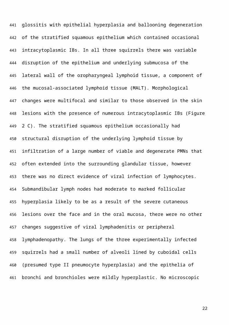
In addition to the skin, microscopic changes were also present in the oral mucosa. In 2/3 squirrels
(sq. 01/12 and sq. 06/12) the tongue was affected by multifocal necrotising or ulcerative glossitis
with epithelial hyperplasia and ballooning degeneration of the stratified squamous epithelium which
contained occasional intracytoplasmic IBs. In all three squirrels there was variable disruption of the
epithelium and underlying submucosa of the lateral wall of the oropharyngeal lymphoid tissue, a
component of the mucosal-associated lymphoid tissue (MALT). Morphological changes were
multifocal and similar to those observed in the skin lesions with the presence of numerous
intracytoplasmic IBs (Figure 2 C). The stratified squamous epithelium occasionally had structural
disruption of the underlying lymphoid tissue by infiltration of a large number of viable and
degenerate PMNs that often extended into the surrounding glandular tissue, however there was no
direct evidence of viral infection of lymphocytes.
Submandibular lymph nodes had moderate to marked follicular hyperplasia likely to be as a result
of the severe cutaneous lesions over the face and in the oral mucosa, there were no other changes
suggestive of viral lymphadenitis or peripheral lymphadenopathy. The lungs of the three
experimentally infected squirrels had a small number of alveoli lined by cuboidal cells (presumed
type II pneumocyte hyperplasia) and the epithelia of bronchi and bronchioles were mildly
hyperplastic. No microscopic changes indicative of SQPV infection were detected in any of the
other tissues examined.
13
299
300
301
302
303
304
305
306
307
308
309
310
311
312
313
314
315
316
317
318
319
320
321
322
323
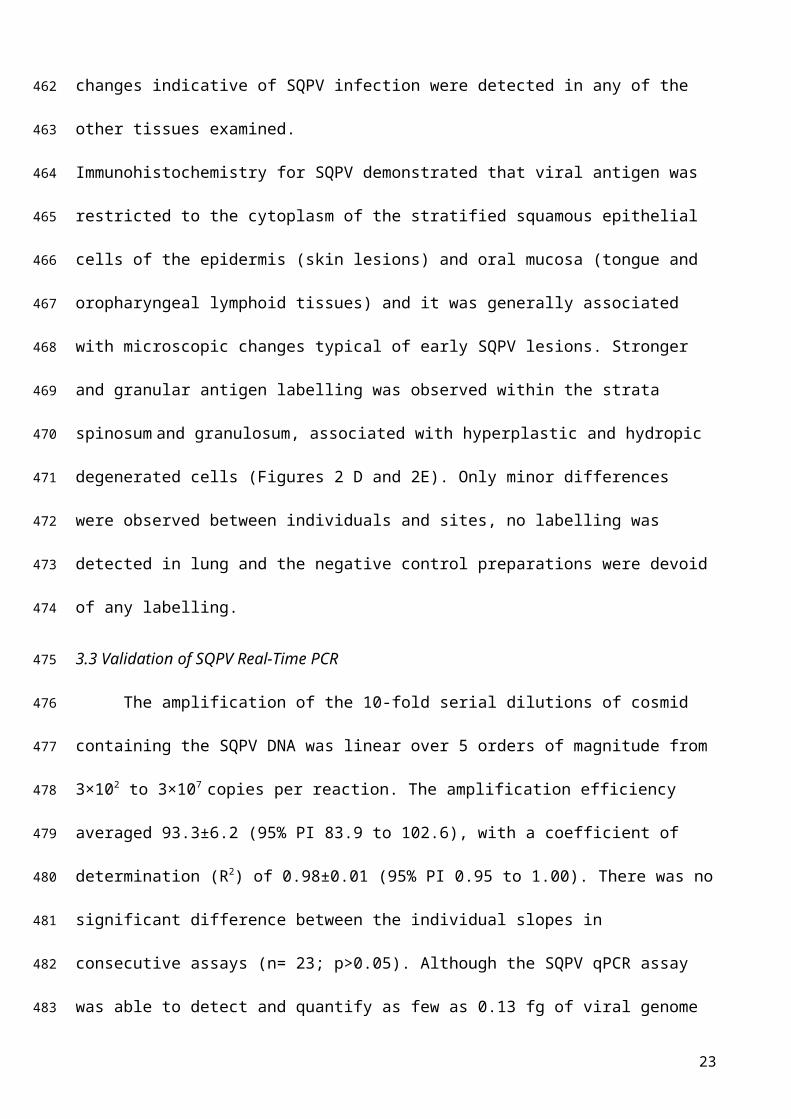
Immunohistochemistry for SQPV demonstrated that viral antigen was restricted to the cytoplasm of
the stratified squamous epithelial cells of the epidermis (skin lesions) and oral mucosa (tongue and
oropharyngeal lymphoid tissues) and it was generally associated with microscopic changes typical
of early SQPV lesions. Stronger and granular antigen labelling was observed within the strata
spinosum and granulosum, associated with hyperplastic and hydropic degenerated cells (Figures 2
D and 2E). Only minor differences were observed between individuals and sites, no labelling was
detected in lung and the negative control preparations were devoid of any labelling.
3.3 Validation of SQPV Real-Time PCR
The amplification of the 10-fold serial dilutions of cosmid containing the SQPV DNA was
linear over 5 orders of magnitude from 3×10² to 3×10⁷ copies per reaction. The amplification
efficiency averaged 93.3±6.2 (95% PI 83.9 to 102.6), with a coefficient of determination (R2) of
0.98±0.01 (95% PI 0.95 to 1.00). There was no significant difference between the individual slopes
in consecutive assays (n= 23; p>0.05). Although the SQPV qPCR assay was able to detect and
quantify as few as 0.13 fg of viral genome equivalent, which represents 3 copies of the cosmid per
reaction (equivalent to 15 V/μg DNA and 600 V/mL), the probability of detection was 15% and
58% for 3 and 30 viral genome equivalents, respectively. At >30 genome equivalents (>150 V/μg
DNA) the detection probability was 100%. The estimated intra-assay coefficient of variation (CV)
ranged between 0.5% and 6.5%, with samples containing lower virus copies consistently producing
higher variability. The inter-assay CV estimated from the linear regressions computed for each of
the 23 standard curves was, on average, 4.3%±1.72 (95% PI 1.9 to 7.9). The resulting GMS curve
constructed from the 23 standard curves is shown in Figure 3. The average efficiency was 95.9%
(95% CI 91.8 to 100.5%) with a slope equivalent to -3.42 (95%CI -3.31 to -3.54) and coefficient of
determination (R2) of 0.95. The variability within the Ct data used for estimating the GMS curve
was expressed by a CV of 5.7%. SQPV DNA load of each tested sample was subsequently
predicted from the GMS curve regression equation as Ct = 41.36 + (-3.42×log10 SQPV DNA copy).
The optimal cut-off for reliable quantification was arbitrarily set at a Ct value of 36.5. No
14
324
325
326
327
328
329
330
331
332
333
334
335
336
337
338
339
340
341
342
343
344
345
346
347
348
349
significant variation in the viral load (p>0.05), expressed as log10 of viral copies/μg DNA, was
reported between the values obtained from each sample run with the relative standard curve and the
average loads derived from the GMS curve, where 97.9% of the values lay within the 95% limits of
agreement.
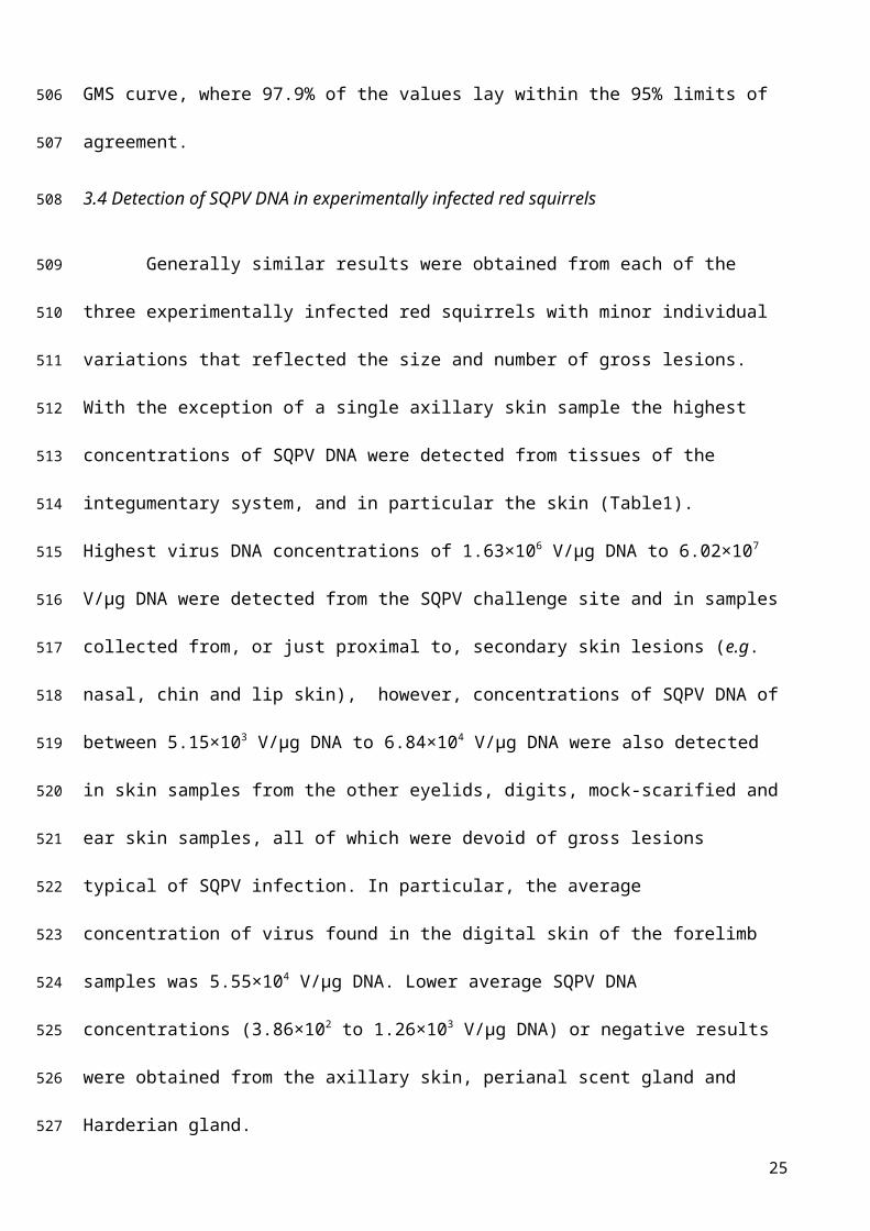
3.4 Detection of SQPV DNA in experimentally infected red squirrels
Generally similar results were obtained from each of the three experimentally infected red
squirrels with minor individual variations that reflected the size and number of gross lesions. With
the exception of a single axillary skin sample the highest concentrations of SQPV DNA were
detected from tissues of the integumentary system, and in particular the skin (Table1). Highest
virus DNA concentrations of 1.63×10⁶ V/μg DNA to 6.02×10⁷ V/μg DNA were detected from the
SQPV challenge site and in samples collected from, or just proximal to, secondary skin lesions (e.g.
nasal, chin and lip skin), however, concentrations of SQPV DNA of between 5.15×10³ V/μg DNA
to 6.84×10⁴ V/μg DNA were also detected in skin samples from the other eyelids, digits, mock-
scarified and ear skin samples, all of which were devoid of gross lesions typical of SQPV infection.
In particular, the average concentration of virus found in the digital skin of the forelimb samples
was 5.55×10⁴ V/μg DNA. Lower average SQPV DNA concentrations (3.86×10² to 1.26×10³ V/μg
DNA) or negative results were obtained from the axillary skin, perianal scent gland and Harderian
gland.
Of the lymphoid organs, the highest SQPV DNA concentration was detected in the oropharyngeal
MALT (n=2, average 2.55×10⁷ V/μg DNA). Despite no gross lesions, the viral load was comparable
to those found in secondary skin lesions and scabs (Table1). The amount of SQPV DNA detected in
selected lymph nodes was approximately 3 to 5 orders of magnitude less compared with
oropharyngeal MALT samples and the average viral load in the two right popliteal lymph nodes,
draining the challenge site, was only slightly higher compared to the contra-lateral popliteal lymph
node (1.34×10⁴ and 3.21×10² V/μg DNA, respectively). SQPV concentration in one spleen sample
15
350
351
352
353
354
355
356
357
358
359
360
361
362
363
364
365
366
367
368
369
370
371
372
373
374
(sq. 01/12) was 1.61×10² V/μg DNA, where as the spleens from the other two squirrels were either
negative or borderline positive (as defined above).
In the alimentary tract (Table 1) decreasing amounts of SQPV DNA were detected in the tongue,
faeces, stomach, parotid salivary gland, SI, SM SG, caecum, rectum, LI and liver. The amount of
SQPV DNA detected in faecal samples (average of 1.16×10⁵ V/μg DNA) was approximately
comparable with those in the tongue or stomach tissue samples of squirrel 01/12. Lower
concentrations of viral DNA (≤1.81×10² V/μg DNA) were detected in all other tissues of the
digestive system that were examined. Virus DNA was detected in the liver of all three squirrels but
at very low levels (<150 V/μg DNA). Similarly virus DNA was detected unreliably or at
concentrations never exceeding 4.05×10³ V/μg DNA in the lungs, kidneys, gonads, heart and brain,
suggesting that virus replication was unlikely in these tissues (Table 1).
Relatively high amounts of viral DNA were detected in the oral and ocular swabs as well as skin
swabs from the challenge site and the mock-scarified skin (Table 1). The average amount of viral
DNA detected in the oral swabs was higher (5.15×10⁷ V/s) than that obtained from either the tongue
or oropharyngeal MALT and in particular the viral load obtained from the oral swab of the only
squirrel (sq. 04/12) which lacked gross lesions on the tongue was similar to the other two squirrels
which had oral lesions. A swab of the vesiculopustular lesion on the left pinna of squirrel 06/12 also
contained high levels of viral DNA (3.72×10⁷ V/s), confirming the lesion was associated with
SQPV replication. Swabs of the metal surfaces of the nest boxes were also found to contain
relativity high amounts (average of 1.28×10⁵ V/s) of viral DNA. SQPV DNA was detected in the
urine of only one squirrel (4.50×10³ V/mL) and that same squirrel was positive for SQPV DNA
(2.83×10⁴ V/mL) in its blood (Table 1).
3.5 Detection of SQPV DNA from naturally infected red squirrel samples
Of the 13 suspected cases of squirrelpox in naturally infected red squirrels, none of the
tissue samples from one of them (R86/07) gave qPCR positive results. It was subsequently regarded
as not having been infected with SQPV and was used as an additional negative control. Among
16
375
376
377
378
379
380
381
382
383
384
385
386
387
388
389
390
391
392
393
394
395
396
397
398
399
400

samples from the other 12 red squirrels, with the exception of a single eyelid sample (squirrel
R05/08), the greatest amounts of SQPV DNA (2.94×10⁷ to 6.78×10⁷ V/μg DNA) were detected
consistently in skin samples(Table 1).
The tissues that contained the next largest amount of viral DNA (7.13×10⁴ to 4.52×10⁵ V/μg DNA)
were stomach, tongue and SM LN, although 48% of these samples were either qPCR negative or
borderline positive. Decreasing viral DNA concentrations ( 4.54×10³ to 2.26×10⁴ V/μg DNA) were
present in the rectum, SI, LI, caecum, brain, heart and lung samples, with a large number of samples
negative or borderline positive (Table 1). The overall lowest average viral DNA concentrations
detected, in the order of 4 to 5 magnitudes lower compared to skin samples, were obtained from
liver, spleen and kidney samples many of which were considered negative.
3.6 Serology
Of the naturally-infected red squirrels 6/12 (50%) were positive for anti-SQPV antibodies,
whilst all three (100%) of the experimental animals were positive at 17 DPC.
4. Discussion
This study successfully reproduced SQPV infection in all three experimentally-challenged
red squirrels via epidermal scarification. Clinical signs and PM findings confirmed that secondary
cutaneous lesions were found consistently and yet no virus-associated pathological changes were
present in any of the internal organs. This is in agreement with a single previous description of
SQPV experimental infection in red squirrels (Tompkins et al., 2002) and many reports of naturally
acquired SQPV infection (Atkin et al., 2010; Duff et al., 2010; McInnes et al., 2009; Naulty et al.,
2013; Sainsbury and Ward, 1996). In our study all three squirrels (100%) developed both gross and
histological lesions. Although one animal was less severely affected with regard to the size, number
and severity of skin lesions (sq. 04/12) it was not possible to determine if this animal might have
survived the infection as is found in some 8-10% of naturally-infected animals (Chantrey et al.,
2014; Sainsbury et al., 2008). The incubation period of SQPV in our study, inferred from the
observation of suspected secondary macroscopic skin lesions, was estimated to be ~8-10 days
17
401
402
403
404
405
406
407
408
409
410
411
412
413
414
415
416
417
418
419
420
421
422
423
424
425
426

which agrees with field observations suggesting an incubation time of less than 15 days (Carroll et
al., 2009).
The novel SQPV Taqman® qPCR assay was capable of detecting as few as 3 copies of the virus
gene, as sensitive as other reported poxvirus qPCR assays (Balamurugan et al., 2009; Sofi Ibrahim
et al., 2003). It uses an imported external standard curve (i.e. GMS curve) removing the necessity to
perform one for each run, saving both time and reagents. Overall, it confirmed the relative lack of
virus in the internal organs, but also pointed to a potential source of virus in the saliva.
Virus dissemination within the tissues of the experimentally infected squirrels was similar to the
distribution and relative quantities of viral DNA found in naturally infected animals and, in
agreement with previous reports (Atkin et al., 2010; Collins et al., 2014), the highest average viral
DNA concentration was consistently detected in skin, oral mucosa, scabs and faeces. Although the
amount of viral DNA detected generally correlated with the severity and extent of the skin lesions
significant amounts of viral DNA, with quantities comparable to the primary skin lesions, were
detected in the oropharyngeal mucosa (ie. MALT) of experimentally challenged squirrels despite
the absence of macroscopic lesions. The average amount of viral DNA detected in the oral swabs
was even higher, again even in one squirrel (sq. 04/12) which lacked lesions on the tongue.
Although SQPV-associated lesions were reported previously on the dorsal surface of the tongue
(McInnes et al., 2009) and viral DNA has also been detected previously in saliva (Collins et al.,
2014), this is the first time that significant amounts of SQPV DNA have been associated with viral
replication, as suggested by the IHC, and potential shedding from the stratified squamous
epithelium of the oropharyngeal MALT. Infection of the oropharyngeal epithelial mucosa may have
resulted from persistent ingestion of virus particles during feeding (e.g. direct contact with lip skin
lesions) and self-grooming, followed by the virus entry via oral epithelial micro-abrasions.
Combined IHC, histology and qPCR analyses indicate that oral epithelium and epidermis are highly
permissive for SQPV replication and could suggest that the relatively high viral DNA loads
detected in the gastro-intestinal tract and faeces are as a result of repeated ingestion of virus
18
427
428
429
430
431
432
433
434
435
436
437
438
439
440
441
442
443
444
445
446
447
448
449
450
451
452

particles generated in the oral mucosa. Consequentially, faecal-oral transmission may represent a
potential route of infection as suggested previously (Atkin et al., 2010; Collins et al., 2014). Indeed,
Gledhill (1962) demonstrated that after oral exposure with ectromelia virus (ECTV), infected mice
can sustain infection with virus shed persistently in the faeces of apparently healthy mice. However,
because of the lack of permissive cells for culturing the wild type SQPV used in these studies we do
not know if the viral DNA detected in the faeces represents viable virus or if the gastric acids and
digestive enzymes are able to inactivate the virus.
The results of this study also confirm that cutaneous lesions contain large amounts of virus. These
lesions were most commonly found around the head and face and again are likely to be as a result,
of virus entry through minor skin abrasions. During squirrelpox outbreaks, the majority of diseased
red squirrels are found usually to have more abundant skin lesions compared with our experimental
group. Some of these may be associated with more the numerous traumatic skin abrasions which are
likely to occur in the natural environment.. It is therefore likely that social interactions (e.g.
interactions at feeders, drey sharing and fighting), scent marking (face-wiping behaviour on
branches and bark), self- and allo-grooming may play an important part in the transmission and
dissemination of naturally acquired disease (Rushton et al., 2000; Bruemmer et al., 2010). In
addition, given the very high concentration of virus in skin and scab samples, and also
vesiculopustular lesions, the contribution of mechanical transmission by ectoparasites between
individuals (e.g. fleas in shared dreys) cannot be excluded, as previously suggested (Atkin et al.,
2010; Collins et al., 2014; Rushton et al., 2000), although the epidemiological role of arthropods in
sustaining disease is less clear.
It has been hypothesised that the epidemiological characteristics of SQPV infection resemble those
of acute disease in systemic poxvirus infections, especially ECTV (Atkin et al., 2010; Carroll et al.,
2009). In our study, the detection of SQPV DNA in many organs suggests that systemic spread of
the virus had occurred. However, it seems unlikely that the lymph nodes, thymus, spleen, liver,
heart, brain, lung, kidney or gastro-intestinal tract act as sites of virus replication given that SQPV
19
453
454
455
456
457
458
459
460
461
462
463
464
465
466
467
468
469
470
471
472
473
474
475
476
477
478

DNA was not consistently detected in these tissues and the concentration was relatively low
compared with skin samples. The absence of both gross and histopathological abnormalities in these
organs was also taken as evidence of the lack of systemic disease, although not necessarily lack of
systemic dissemination of the virus via the lymphatic ? haematogenous system. The presence of
SQPV DNA in the blood as an indication of systemic disease, however, remains unclear. Although
we detected SQPV DNA in the blood of experimentally infected squirrels, similar to what had been
found previously in cases of naturally acquired infection (Atkin et al., 2010; Collins et al., 2014),
results were inconsistent between the three animals. This may reflect the fact that we were only able
to sample the blood once, at 17 DPC, and therefore missed the main viraemia. However, similar to
the pathogenesis of vesicular exanthema of swine virus in experimentally infected pigs (Gelberg
and Lewis, 1982), we speculate that SQPV may initially enter the blood via virus-laden infected
leukocytes from the drainage area of the primary lesion and subsequently, attracted by chemotactic
gradients, migrate and be released in distal sites of previously injured epithelial cells (e.g. skin and
oral mucosa).
5. Conclusion
This study has helped describe the possible routes of SQPV transmission and tissue
locations of viral shedding. Firstly, it confirms that viral entry via damaged skin is an effective route
of infection. Secondly, our results suggest that the oral mucosa and consequently saliva may have
important roles in SQPV epidemiology where apparently healthy animals without gross skin lesions
may be a source of infection by contaminating food at feeders, through bite wounds or mutual
grooming. Rapid identification of infectious animals is a key component of surveillance strategies
and the qPCR assay described here could be a useful addition to the diagnostic repertoire as it can
detect low copy number of virus particles at an early stage of infection. In addition to skin samples,
oral and skin swabs are probably useful and easy alternatives for sampling live red squirrels during
SQPV outbreaks and routine screening. Further research should therefore be undertaken to confirm
20
479
480
481
482
483
484
485
486
487
488
489
490
491
492
493
494
495
496
497
498
499
500
501
502
503

whether primary SQPV infection can be contracted via the oral route and through environmental
contamination.
Conflict of interest statement
None
Acknowledgment
The authors would like to thank Ann R. Wood, Jackie Thomson and all MRI BioServices
staff for animal husbandry and monitoring, Kim Willoughby for advice on qPCR assay design,
Janice Gilray and David Dean (MRI) for providing serology results and SQPV antibodies and
David Everest (APHA) for providing TEM. Thanks also to all field workers, conservation
organizations and ranger services for providing red squirrels carcasses. This study was funded by
Wildlife Ark Trust and the Scottish Government Rural and Environment Science and Analytical
Services Division.
References
Atkin, J.W., Radford, A.D., Coyne, K.P., Stavisky, J., Chantrey, J., 2010. Detection of squirrel poxvirus by nested and real-time PCR from red (Sciurus vulgaris) and grey (Sciurus carolinensis) squirrels. BMC Vet. Res. 6, 33.
Balamurugan, V., Jayappa, K.D., Hosamani, M., Bhanuprakash, V., Venkatesan, G., Singh, R.K., 2009. Comparative Efficacy of Conventional and TaqMan Polymerase Chain Reaction Assays in the Detection of Capripoxviruses from Clinical Samples. J. Vet. Diagnostic Investig. 21, 225–231.
Bosch, S., Lurz, P.W.W., 2012. The Eurasian Red Squirrel Sciurus vulgaris. Westarp Wissenschaften Verlagsgesellschaft mbH, Germany.
Bruemmer, C.M, Rushton, S.P., Gurnell, J., Lurz, P.W.W., Nettleton, P., Sainsbury, A.W., Duff, J.P., Gilray,J., McInnes, C.J., 2010. Epidemiology of squirrelpox virus in grey squirrels in the UK. Epidemiol. Infect. 138, 941–950.
Carroll, B., Russell, P., Gurnell, J., Nettleton, P., Sainsbury, A W., 2009. Epidemics of squirrelpox virus disease in red squirrels (Sciurus vulgaris): temporal and serological findings. Epidemiol. Infect. 137, 257–265.
Chantrey, J., Dale, T.D., Read, J.M., White, S., Whitfield, F., Jones, D., McInnes, C.J., Begon, M., 2014. European red squirrel population dynamics driven by squirrelpox at a gray squirrel invasion interface. Ecol Evol 4, 3788–3799.
Collins, L.M., Warnock, N.D., Tosh, D.G., McInnes, C., Everest, D., Montgomery, W.I., Scantlebury, M., Marks, N., Dick, J.T., Reid, N., 2014. Squirrelpox virus: assessing prevalence, transmission and environmental degradation. PLoS One 9, e89521.
21
504
505
506
507
508
509
510
511
512
513
514
515
516
517518519
520521522
523524
525526527
528529530
531532533
534535536
Darby, A.C., McInnes, C.J., Kjaer, K.H., Wood, A.R., Hughes, M., Martensen, P.M., Radford, A.D., Hall, N., Chantrey, J., 2014. Novel host-related virulence factors are encoded by squirrelpox virus, the main causative agent of epidemic disease in red squirrels in the UK. PLoS One 9, e96439.
Duff, J.P., 2012. Squirrelpox. In: Gavier-Widèn, D., Meredith, A., Duff, J.P. (Eds.). Infectious Diseases of Wild Mammals and Birds in Europe. Oxford, UK: Wiley-Blackwell, pp. 196-199.
Duff, J.P., Haley, P., Wood, R., Higgins, R.J., 2010. Causes of red squirrel (Sciurus vulgaris) mortality in England. Vet. Rec. 167, 461.
Duff, J.P., Scott, A., Keymer, I.F., 1996. Parapox virus infection of the grey squirrel. Vet Rec 138, 527.
Edwards, F.B., 1962. Red squirrel disease. Vet. Rec. 74, 739–741.
Gelberg, H.B., Lewis, R.M., 1982. The pathogenesis of vesicular exanthema of swine virus and San Miguel sea lion virus in swine. Vet Pathol 19, 424–443.
Gledhill, A.W., 1962. Latent Ectromelia. Nature 196, 298–298.
Keymer, I.F., 1974. Report of the pathologist, 1971 and 1972. The zoological Society of London, Scientific report 1971-1973. J. Zool. 173, 55.
LaRose, J.P., Meredith, A.L., Everest, D.J., Fiegna, C., McInnes, C.J., Shaw, D.J., Milne, E.M., 2010. Epidemiological and postmortem findings in 262 red squirrels (Sciurus vulgaris) in Scotland, 2005 to 2009. Vet Rec 167, 297–302.
McInnes, C.J., Coulter, L., Dagleish, M.P., Deane, D., Gilray, J., Percival, A., Willoughby, K., Scantlebury, M., Marks, N., Graham, D., Everest, D.J., McGoldrick, M., Rochford, J., McKay, F., Sainsbury, A.W., 2013. The emergence of squirrelpox in Ireland. Anim. Conserv. 16, 51–59.
McInnes, C.J., Coulter, L., Dagleish, M.P., Fiegna, C., Gilray, J., Willoughby, K., Cole, M., Milne, E., Meredith, A., Everest, D.J., Macmaster, A.-M., 2009. First cases of squirrelpox in red squirrels (Sciurus vulgaris) in Scotland. Vet. Rec. 164, 528–531.
McInnes, C.J., Wood, A.R., Thomas, K., Sainsbury, A.W., Gurnell, J., Dein, F.J., Nettleton, P.F., 2006. Genomic characterization of a novel poxvirus contributing to the decline of the red squirrel (Sciurus vulgaris) in the UK. J. Gen. Virol. 87, 2115–2125.
Middleton, A.D., 1930. The Ecology of the American Grey Squirrel (Sciurus carolinensis Gmelin) in the British Isles. Proc. Zool. Soc. London 2, 809–843.
Naulty, F., Everest, D., Warnock, N.D., Phelan, K., Callanan, J.J., 2013. Squirrelpox virus in red squirrels (Sciurus vulgaris) in the Republic of Ireland. J Wildl Dis 49, 1070–1073.
Reynolds, J.C., 1985. Details of the demographic replacement of the red squirrel (Sciurus vulgaris) by the grey squirrel (Sciurus carolinensis) in Eastern England. J. Anim. Ecol. 54, 149–162.
Rushton, S.P., Lurz, P.W.W., Gurnell, J., Fuller, R., 2000. Modelling the spatial dynamics of parapoxvirus disease in red and grey squirrels: a possible cause of the decline in the red squirrel in the UK? J. Appl. Ecol. 37, 997–1012.
Rushton, S.P., Lurz, P.W.W., Gurnell, J., Nettleton, P., Bruemmer, C., Shirley, M.D.F., Sainsbury, A.W., 2006. Disease threats posed by alien species: the role of a poxvirus in the decline of the native red squirrel in Britain. Epidemiol. Infect. 134, 521–533.
22
537538539
540541
542543
544
545
546547
548
549550
551552553
554555556
557558559
560561562
563564
565566
567568
569570571
572573574
Sainsbury, A.W., Deaville, R., Lawson, B., Cooley, W.A., Farelly, S.S., Stack, M.J., Duff, P., McInnes, C.J., Gurnell, J., Russell, P.H., Rushton, S.P., Pfeiffer, D.U., Nettleton, P., Lurz, P.W., 2008. Poxviral disease in red squirrels Sciurus vulgaris in the UK: spatial and temporal trends of an emerging threat. Ecohealth 5, 305–316.
Sainsbury, A.W., Gurnell, J., 1995. An investigation into the health and welfare of red squirrels, Sciurus vulgaris, involved in reintroduction studies. Vet Rec 137, 367–370.
Sainsbury, A.W., Nettleton, P., Gilray, J., Gurnell, J., 2000. Grey squirrels have high seroprevalence to a parapoxvirus associated with deaths in red squirrels. Anim. Conserv. 3, 229–233.
Sainsbury, T., Ward, L., 1996. Parapoxvirus infection in red squirrels. Vet Rec 138, 400.
Scott, A.C., Keymer, I.F., Labram, J., 1981. Parapoxvirus infection of the red squirrel (Sciurus vulgaris). Vet Rec 109, 202.
Sofi Ibrahim, M., Kulesh, D.A., Saleh, S.S., Damon, I.K., Esposito, J.J., Schmaljohn, A.L., Jahrling, P.B., 2003. Real-time PCR assay to detect smallpox virus. J. Clin. Microbiol. 41, 3835–9.
Thomas, K., Tompkins, D.M., Sainsbury, A.W., Wood, A.R., Dalziel, R., Nettleton, P.F., McInnes, C.J., 2003. A novel poxvirus lethal to red squirrels (Sciurus vulgaris). J. Gen. Virol. 84, 3337–3341.
Tompkins, D.M., Sainsbury, A.W., Nettleton, P., Buxton, D., Gurnell, J., 2002. Parapoxvirus causes a deleterious disease in red squirrels associated with UK population declines. Proc. R. Soc. London. Ser. B Biol. Sci. 269, 529–533.
Tompkins, D.M., White, a. R., Boots, M., 2003. Ecological replacement of native red squirrels by invasive greys driven by disease. Ecol. Lett. 6, 189–196.
Vizoso, A.D., 1968. A red squirrel disease. Symp. Zool. Soc. London 24, 29–38.
23
575576577578
579580
581582
583
584585
586587
588589
590591592
593594
595
596
597

Figures and Table captions
Figure 1. Gross appearance of SQPV-scarified skin at 17 days post-challenge
Right lateral thigh - Squirrel 06/12. Skin lesion at inoculation site. Ssevere erythematous, exudative
and ulcerative dermatitis with scab formation.
Figure 2. Histopathology and immunohistochemistry of experimentally infected red squirrels
at 17 days post-challenge with SQPV
(A) Section of nasal skin at the edge of a macroscopic ulcerative lesion - Squirrel 06/12. Marked
epidermal hyperplasia and ballooning degeneration of epidermal and follicular epithelial cells
(asterisks) which leads to intraepidermal pustule formation (arrows). Bar = 200 μm. (B) Section of
SQPV scarified skin at the inoculation site. - Squirrel 06/12. Ulcerative and necrotising dermatitis
covered by scab formation (asterisks) typical of an advanced SQPV skin lesion. Bar = 500 μm. (C)
Section of the mucosal-associated lymphoid tissue (MALT) of the lateral wall of the oropharynx -
Squirrel 04/12. There is marked acanthosis of surface epithelium associated with ballooning
degeneration of the stratum spinosum. Individual swollen cells often have intracytoplasmic, palely
eosinophilic viral inclusion body (arrowheads). The underlying submucosa is expanded by a dense
inflammatory cell infiltrate composed mostly of viable and degenerated neutrophils. Bar = 100 μm.
(D) Lip skin - Squirrel 01/12. Immunohistochemical labelling (brick red colour) of SQPV antigen is
restricted to the epidermal and follicular epithelial cells. Mild to strong, diffuse (arrowhead) to
granular (arrows) labelling is present within the cytoplasm of hyperplastic, spongiotic and
ballooning degenerated epithelial cells . Bar = 500 μm. (E) A mucosal fold (asterisk) of the
oropharyngeal MALT - Squirrel 06/12. Focally extensive, moderate (arrowhead) to strong (arrow)
immunolabelling of intracytoplasmic SQPV antigen which is restricted to the stratified squamous
epithelium and is associated with hyperplastic and hydropic degenerated cells. Bar = 200 μm.
Inserts within D and E represent negative control preparations.
Figure 3. SQPV qPCR Grand Mean Standard calibration curve (GMS)
24
598
599
600
601
602
603
604
605
606
607
608
609
610
611
612
613
614
615
616
617
618
619
620
621
622
Grand Mean Standard (GMS) calibration curve (red line) obtained by combining the data resulting
from 23 independent and consecutive standard curves (grey palettes) generated by plotting the log10
of the SQPV DNA cosmid serial dilution (x-axis) against the cycle threshold (Ct) values (y-axis).
Coloured areas indicate the extent of the 95% coefficient interval (CI) range, where grey dots
correspond to the actual values of the qPCR reads. Each standard SQPV cosmid dilution was
performed in quadruplicate reactions with concentrations ranging from 0.13 pg/reaction (3 viral
equivalent) to 1.3 ng/reaction (3×10⁷ viral equivalent). The relative slope, y-axis intercept and
correlation coefficient (R2) for the GMS curve are displayed on the bottom left.
Table 1. qPCR detection of SQPV DNA in samples from red squirrels experimentally and naturally infected.
Viral DNA detection, concentrations and relative average from different samples of 3 red squirrels
(01/12, 04/2 and 06/12) experimentally infected with SQPV at 17 DPC and of 12 red squirrels
naturally infected with SQPV (R21/07, R23/07, R52/07, R69/97, R05/08, R06/08, R27/08, R811,
R812, R23/09, R24/09, R31/09). Virus loads were determined from the GMS curve and expressed
as: V/ μg of DNA extracted from lymphoid organs, integumentary system, alimentary tract and
other organs samples; V/S for swab samples; V/mL for blood and urine samples. For squirrel 06/12
the average viral DNA relative to eyelid skin and ocular swabs was obtained using the average of
both samples (right and left). (-) negative qPCR result; (+) borderline positive results; (*) ELISA
positive squirrels; (empty cells) no samples tested.
25
623
624
625
626
627
628
629
630
631632
633
634
635
636
637
638
639
640
641
642