hospital for special surgery: what’s the diagnosis – case 63
DESCRIPTION
What’s the Diagnosis? is a means for you to test your orthopaedic, rheumatologic and radiology/imaging knowledge. Monthly, new cases will be presented as unknowns. The answers will be available and indexed so that should you want to search on cases representative of a specific topic, you can do so. The cases are from the records of HSS and the teaching files of the Department of Radiology and Imaging. The cases are intended to be representative and informative demonstrating the comprehensive care of Orthopaedics, Rheumatology, Radiology and Imaging and related services at HSS. We know you like to be challenged and hope this section meets your expectations.TRANSCRIPT

What’s the Diagnosis – Case 63
What’s the Diagnosis – Case 63 1
What’s the Diagnosis – Case 63
What’s the Diagnosis – Case 63 2

What’s the Diagnosis – Case 63
What’s the Diagnosis – Case 63 3

What’s the Diagnosis – Case 63
What’s the Diagnosis – Case 63 4
What’s the Diagnosis – Case 63
What’s the Diagnosis – Case 63 5
What’s the Diagnosis – Case 63
What’s the Diagnosis – Case 63 6
What’s the Diagnosis – Case 63
What’s the Diagnosis – Case 63 7

What’s the Diagnosis – Case 63
What’s the Diagnosis – Case 63 8

What’s the Diagnosis – Case 63
What’s the Diagnosis – Case 63 9
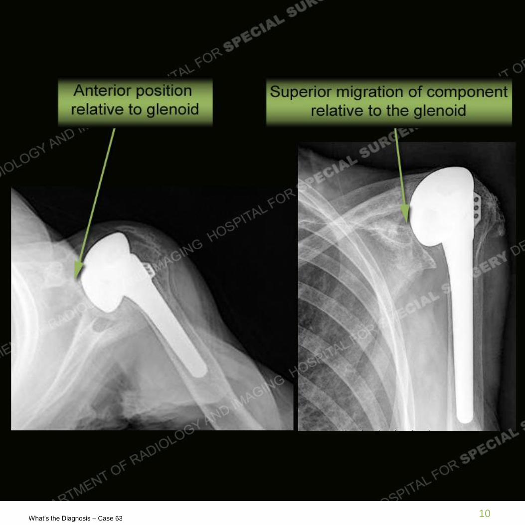
Findings
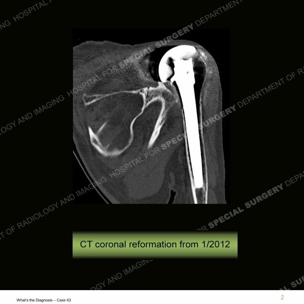
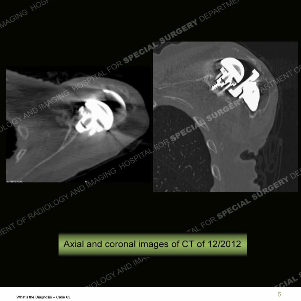
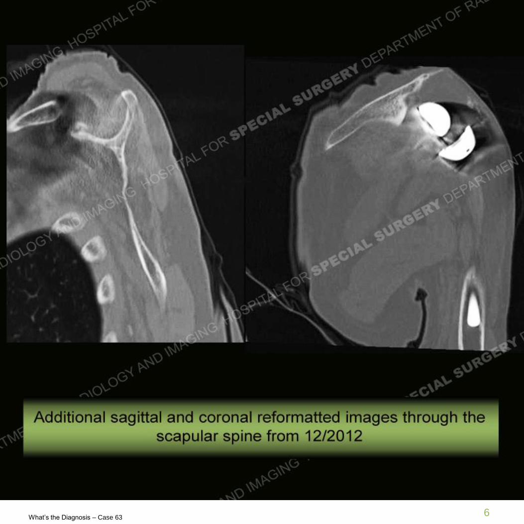
• Initial studies demonstrate a left shoulder hemiarthroplasty with superior and
anterior positioning of the component. CT study also demonstrates
disruption through the base of the coracoid. Subsequent radiographs
demonstrate a reverse type total shoulder arthroplasty which shows
anatomic positioning on CT, particularly as relates to the metalglene or base
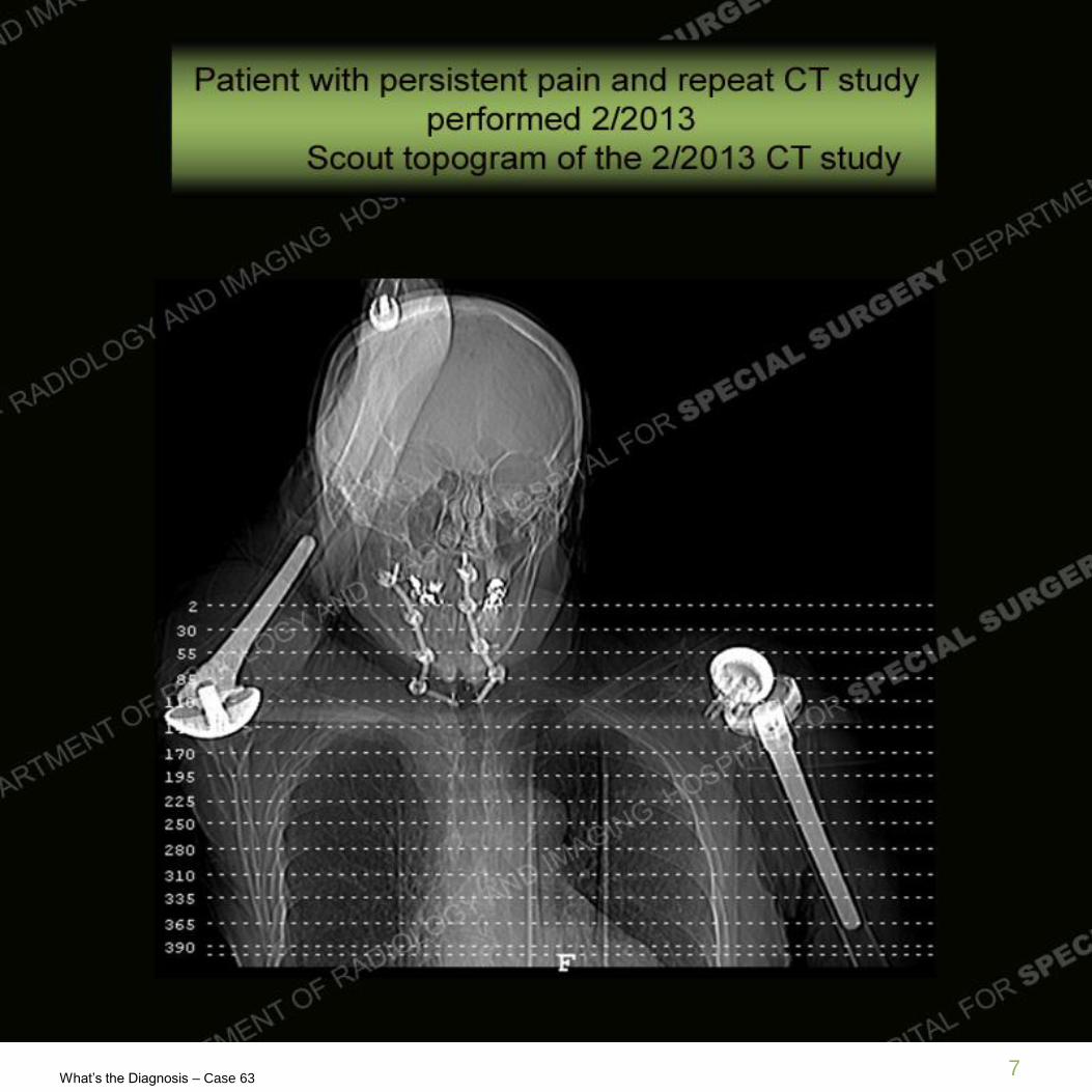
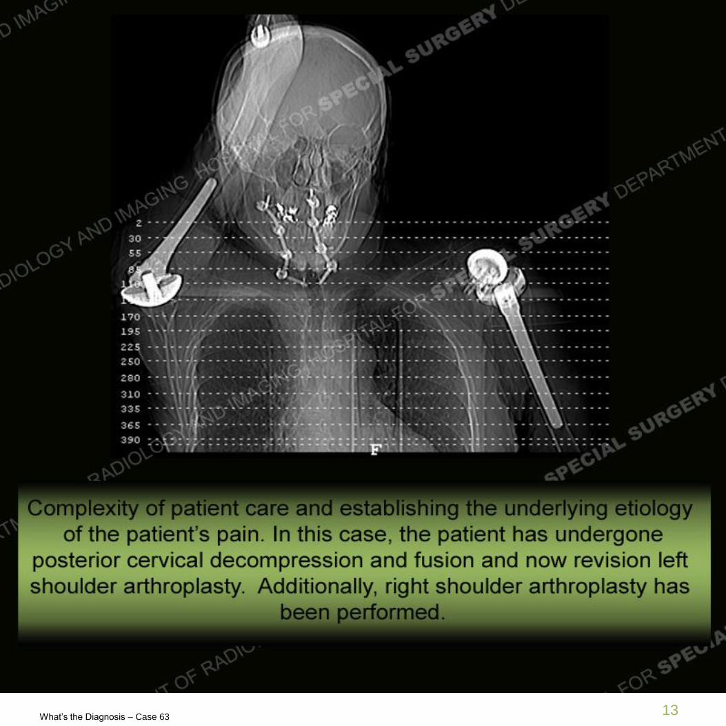
plate to the native glenoid. Repeat CT demonstrates on the scout topogram,
the often very complex nature of these cases with multiple sites of pathology.
The repeat CT demonstrates a very subtle defect of the scapular spine.
What’s the Diagnosis – Case 63
What’s the Diagnosis – Case 63 10

What’s the Diagnosis – Case 63
What’s the Diagnosis – Case 63 11

What’s the Diagnosis – Case 63
What’s the Diagnosis – Case 63 12
What’s the Diagnosis – Case 63
What’s the Diagnosis – Case 63 13

What’s the Diagnosis – Case 63
What’s the Diagnosis – Case 63 14

What’s the Diagnosis – Case 63
What’s the Diagnosis – Case 63 15
Diagnosis: Complications of shoulder arthroplasty with multiple
periprosthetic fractures
• This case demonstrates only a couple of the complications that can come
in the setting of joint arthroplasty and especially in this case shoulder
arthroplasty. Hemiarthroplasties are known to be at risk for progression of
instability often related to progressed degeneration and tearing of the
rotator cuff as seen in this case. In addition, patients often suffer from
propagation of arthritis necessitating total shoulder arthroplasty as seen in
this case. One other complication related to progressive disease of the
cuff is a loss of the acromiohumeral interval yielding increased stress on
the coracoacromial arch and precipiating fractures as in this case of the
coracoid.
• Total shoulder arthroplasties and in particular in this case, reverse total
shoulder arthroplasties, can have complications. Scapular notching,
difficulty in seating the base plate, dissociation, infection, and loosening
are well known complications. In addition, particularly in the setting of the
reverse TSA, acromial and scapular spine fractures are becoming more
recognized. These fractures may of course be painful but may necessitate
additional fixation as well.
What’s the Diagnosis – Case 63
What’s the Diagnosis – Case 63 16
Resources
• Crosby LA, Hamilton A, Twiss T (2011) Scapula fractures after reverse
total shoulder arthroplasty: classification and treatment. Clin Orthop Relat
Res 469(9):2544–2549.
• Levine WN, Fischer CR, Nguyen D, Flatow EL, Ahmad CS, Bigliani LU.
Long-term follow-up of shoulder hemiarthroplasty for glenohumeral
osteoarthritis. J Bone Joint Surg Am. 2012 Nov 21;94.
• Scarlat MM. Complications with reverse total shoulder arthroplasty and
recent evolutions. Int Orthop. 2013 Mar 3.
• Special thanks to Frank Cordasco, MD and Larry Gulotta, MD for their
insight and assistance on this case.