hormones lecture h02
TRANSCRIPT

HormonesG Protein’s Signaling
Dr. Aga Syed SameerCSIR Lecturer (Demonstrator)Department of Biochemistry,Medical College,Sher-I-Kashmir Institute of Medical Sciences, Bemina, Srinagar, Kashmir, 190010. India.
First MBBS
Lecture No: H 02
Time : 10:00am
Dated: 07/03/2015

The 2012 Nobel Prize in Chemistry was awarded jointly to
Robert J. Lefkowitz (l) and Brian K. Kobilka
for
studies of G-protein-coupled receptors
G Protein Coupled Receptor’s (GPCR’s)
• They are the receptor named just because they interact with G Protein’s.
• Also referred to as “Serpentine Receptor’s” as they contain seven trans-membrane helices (7TM)
• Largest known receptor family – Constitutes > 1% of the human genome.
• Comprises receptors for a diverse array of molecules: neurotransmitters, odorants, lipids, neuropeptides, large glycoprotein hormones
G Protein Coupled Receptor’s (GPCR’s)

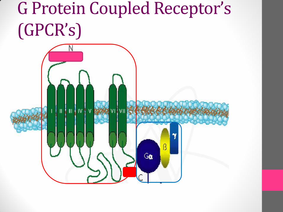
• Their Structure consists of:
Amino- Terminal: Present on the outside of the cell
Seven α helices: traversing the plasma membrane & connected by loops at varying length
Carboxyl-Terminal: Present on Inside of the cell
• Ligand binding site: three loops that are on outer surface of the cell
• Docking site: three loops that are on inner surface/ cytoplasmic side of the cell; provide site for binding of intracellular protein – G protein’s
G Protein Coupled Receptor’s (GPCR’s)
G Protein Coupled Receptor’s (GPCR’s)
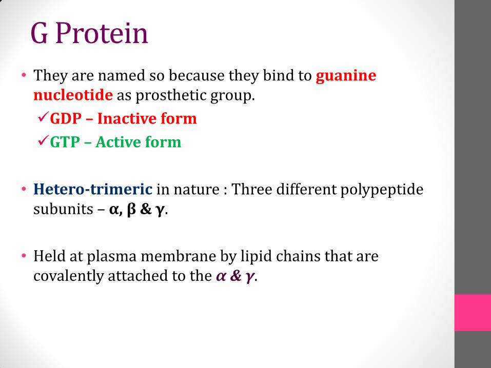
• They are named so because they bind to guanine nucleotide as prosthetic group.
GDP – Inactive form
GTP – Active form
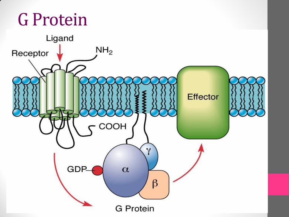
• Hetero-trimeric in nature : Three different polypeptide subunits – α, β & γ.
• Held at plasma membrane by lipid chains that are covalently attached to the α & γ.
G Protein
• The guanine nucleotide binding site is present on Gαsubunit:
In GDP bound conformation (Inactive, b): Gα subunit has high affinity for the Gβγ; hence they remain together as trimer on cell surface
In GTP bound conformation (Active, a): : Gα subunit has low affinity for the Gβγ; leading to its dissociation from complex
• The two conformations are inter-convertible via activation by GPCR’s; which cause GDP-GTP switching on Gα subunit of trimer.
• Each dissociated Gα subunit in turn is free to activate an effector protein – Like “Adenylyl Cyclase” etc.
G Protein
G Protein
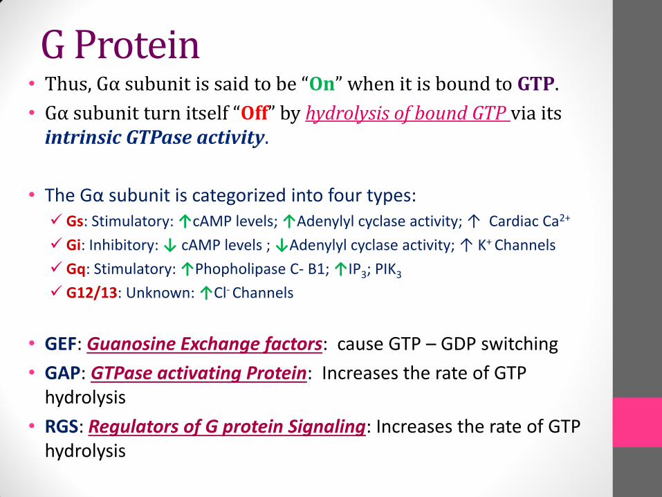
• Thus, Gα subunit is said to be “On” when it is bound to GTP.
• Gα subunit turn itself “Off” by hydrolysis of bound GTP via its intrinsic GTPase activity.
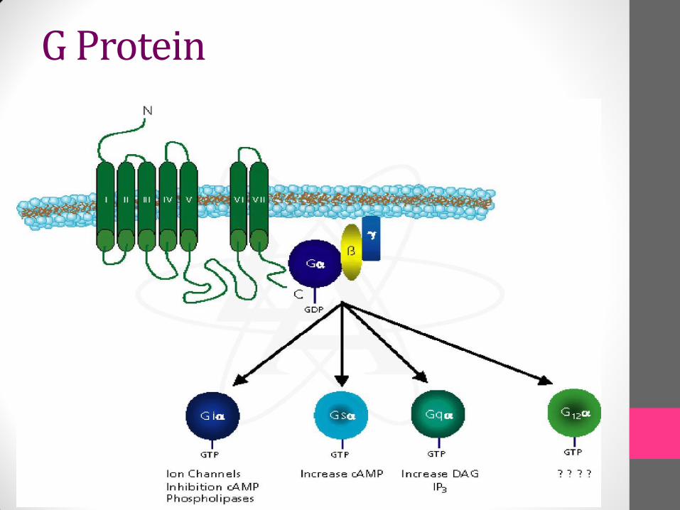
• The Gα subunit is categorized into four types: Gs: Stimulatory: ↑cAMP levels; ↑Adenylyl cyclase activity; ↑ Cardiac Ca2+
Gi: Inhibitory: ↓ cAMP levels ; ↓Adenylyl cyclase activity; ↑ K+ Channels
Gq: Stimulatory: ↑Phopholipase C- B1; ↑IP3; PIK3
G12/13: Unknown: ↑Cl- Channels
• GEF: Guanosine Exchange factors: cause GTP – GDP switching
• GAP: GTPase activating Protein: Increases the rate of GTP hydrolysis
• RGS: Regulators of G protein Signaling: Increases the rate of GTP hydrolysis
G Protein
G Protein
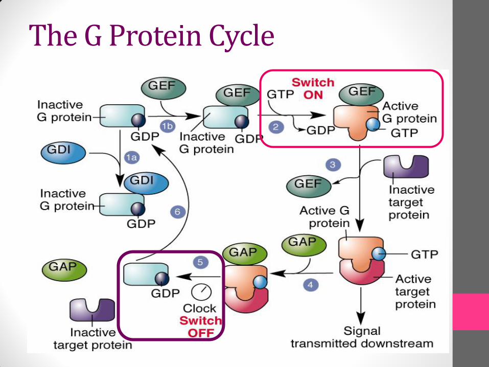
The G Protein Cycle
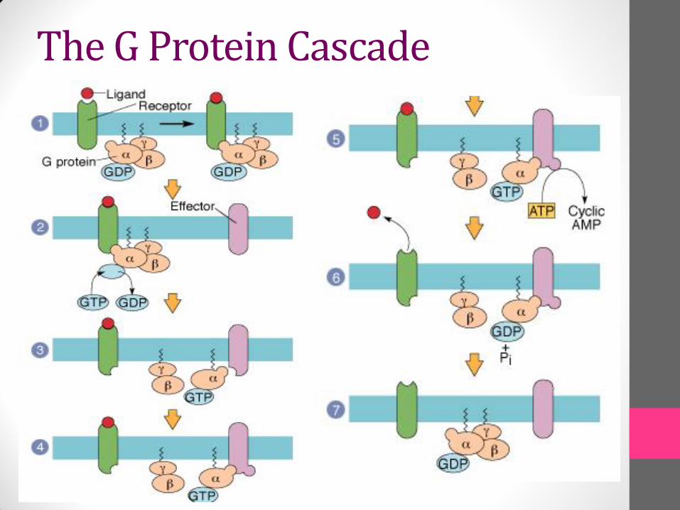
The G Protein Cascade

• Cyclic AMP
• Cyclic GMP
• Inositol Tri Phosphate - IP3
• Diacyl glycerol - DAG
• Ca/Calmodulin
• Smad
Second Messengers

• Are small easily diffusible chemicals/intermediates
• Have very short half life
• They relay signals received at the receptors on the cell surface
• Serve to enhance the strength of the signal
• Affect more than one pathway and/ or protein
• Cause divergence of the signal
Second Messengers
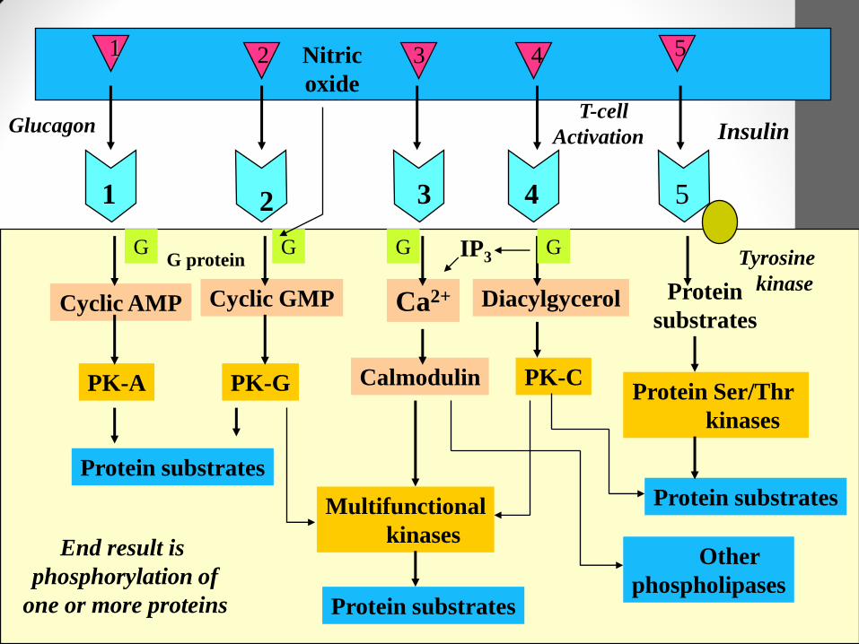
Cyclic AMP Cyclic GMP Ca2+ Diacylgycerol Protein
substrates
PK-A PK-G Calmodulin PK-CProtein Ser/Thr
kinases
Protein substrates
Protein substrates
Protein substratesMultifunctional
kinasesOther
phospholipases
1 2 3 4 5
1 2 3 4 5
Tyrosine
kinase
IP3G G G G
InsulinGlucagonT-cell
Activation
Nitric
oxide
G protein
End result is
phosphorylation of
one or more proteins
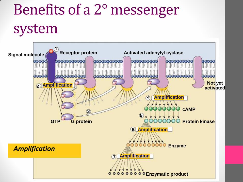
Benefits of a 2° messenger system
Amplification
Signal molecule Receptor protein Activated adenylyl cyclase
Amplification
Amplification
Amplification
Amplification
GTP G protein
2
1
3
4
5
6
7
Enzymatic product
Enzyme
Protein kinase
cAMP
Not yetactivated
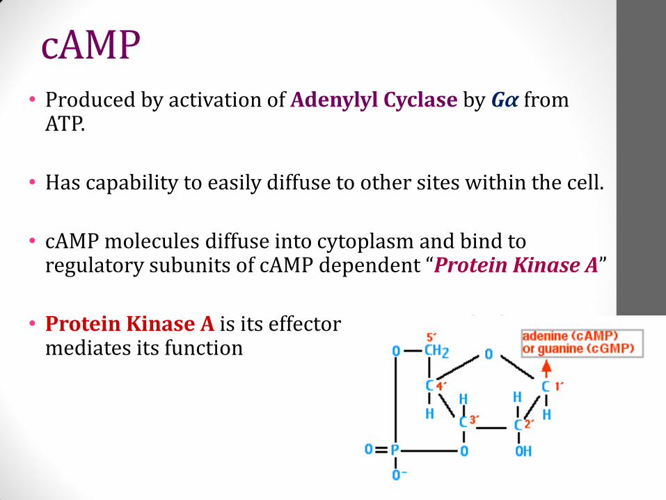
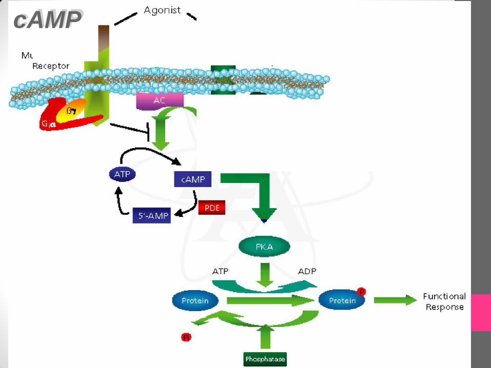
• Produced by activation of Adenylyl Cyclase by Gα from ATP.
• Has capability to easily diffuse to other sites within the cell.
• cAMP molecules diffuse into cytoplasm and bind to regulatory subunits of cAMP dependent “Protein Kinase A”
• Protein Kinase A is its effector protein via which cAMPmediates its function
cAMP
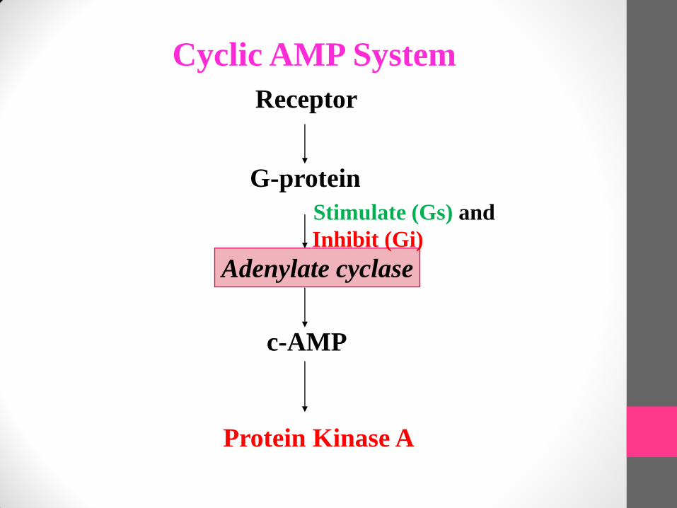
Cyclic AMP System
Receptor
Adenylate cyclase
G-protein
Protein Kinase A
c-AMP
Stimulate (Gs) and
Inhibit (Gi)
cAMP
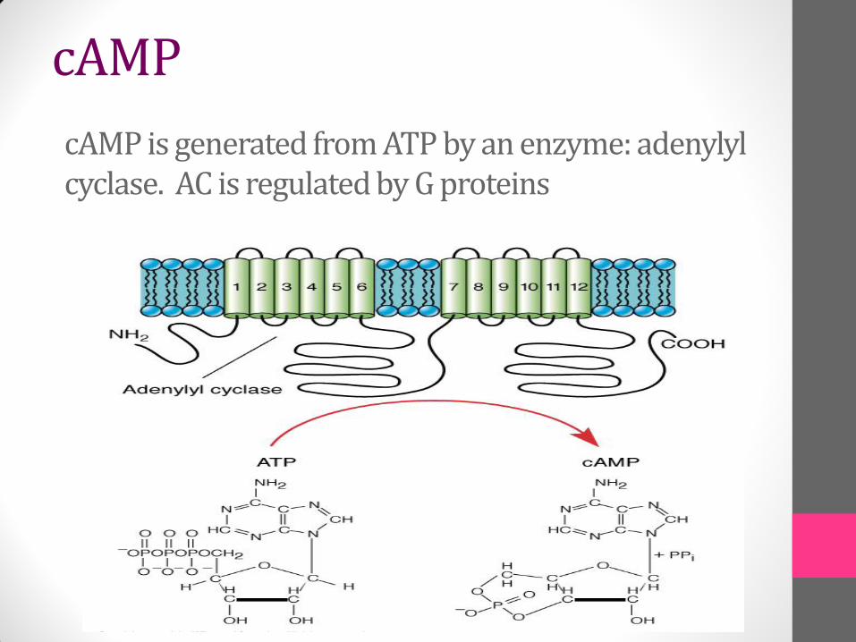
cAMP is generated from ATP by an enzyme: adenylyl cyclase. AC is regulated by G proteins
cAMP
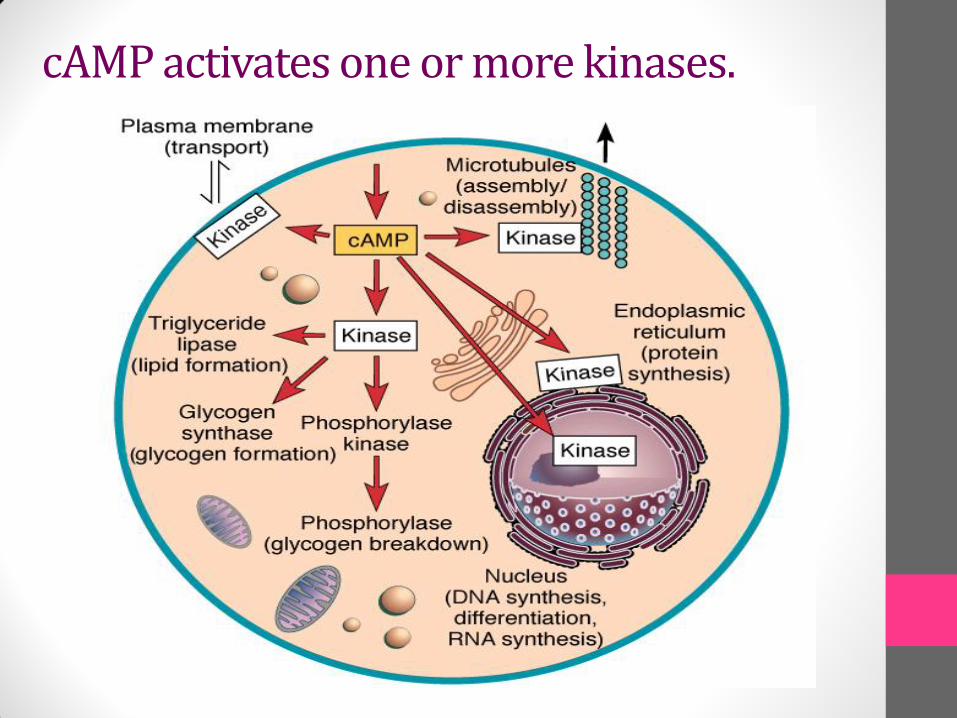
cAMP activates one or more kinases.
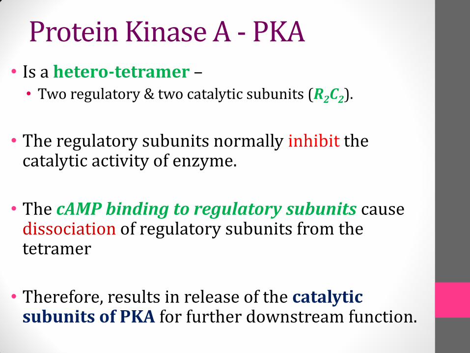
• Is a hetero-tetramer –• Two regulatory & two catalytic subunits (R2C2).
• The regulatory subunits normally inhibit the catalytic activity of enzyme.
• The cAMP binding to regulatory subunits causedissociation of regulatory subunits from the tetramer
• Therefore, results in release of the catalytic subunits of PKA for further downstream function.
Protein Kinase A - PKA
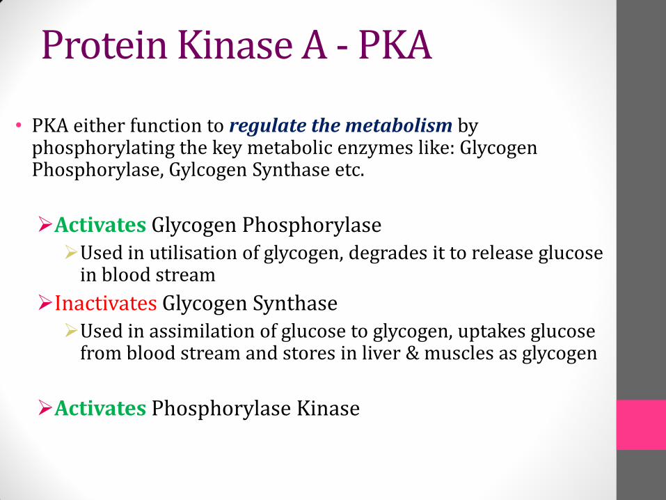
• PKA either function to regulate the metabolism by phosphorylating the key metabolic enzymes like: Glycogen Phosphorylase, Gylcogen Synthase etc.
Activates Glycogen PhosphorylaseUsed in utilisation of glycogen, degrades it to release glucose
in blood stream
Inactivates Glycogen SynthaseUsed in assimilation of glucose to glycogen, uptakes glucose
from blood stream and stores in liver & muscles as glycogen
Activates Phosphorylase Kinase
Protein Kinase A - PKA

• Or some of it translocates to the nucleus
• In nucleus it phosphorylates key nuclear proteins which function as transcription factor called CREB (cAMP Response element Binding- Protein)
• CREB forms dimer and then binds to the DNA at particular sequences within the promoter/regulatory region 5-TGACGTCA known as CRE (cAMP Response Element)
Protein Kinase A - PKA
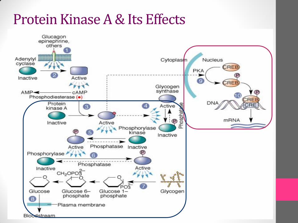
Protein Kinase A & Its Effects
Cholera toxin catalyzes covalent modification of Gs.
• ADP-ribose is transferred from NAD+ to an arginine residue at the GTPase active site of Gs.
• ADP-ribosylation prevents GTP hydrolysis by Gs.
• The stimulatory G-protein is permanently activated.
Pertussis toxin (whooping cough disease) catalyzes ADP-ribosylation at a cysteine residue of the inhibitory Gi, making it incapable of exchanging GDP for GTP.
• The inhibitory pathway is blocked.
ADP-ribosylation is a general mechanism by which activity of many proteins is regulated, in eukaryotes (including mammals) as well as in prokaryotes.
Toxic Effects

Questions