hormone biosynthesis and metabolism
TRANSCRIPT

Hormone Biosynthesis and
Metabolism
Hoon Kim, MD, PhD, NCMP Associate Professor Department of OB & GYN Seoul Nat’l Univ College of Medicine

Introduction
• “Hormones”
Substances that provide means of communication
Chemical regulatory and signaling agents
Local sites: cellular communication necessary
Paracrine
• Intercellular communication, local diffusion
Autocrine
• Intracellular communication, production of regulating
substances
Intracrine
• Intracellular communication, unsecreted substances bind to
intracelllular receptors

Structure of Steroid Hormone
• Basically similar structure with relatively minor chemical
differences
• Basic structure: per-hydro-cyclo-pentane-phenanthrene
molecule
• Composed of 6-carbon rings and one 5-carbon ring
• One 6-carbon ring is benzene, two of the 6-carbon rings
are naphthalene, and three 6-carbon rings are
phenanthrene

Structure of Steroid Hormone
• phenanthrene (three 6-carbon rings) + cyclopentane (5-
carbon ring)
• Four rings named A,B,C and D
A,B,C : cyclohexanes (hexagon)
D: cyclopentane (pentagon)
Direction of numbering: from A to D

21 Carbons - Pregnane
19 Carbons - Androstane
18 Carbons - Estrane

Steroid Hormone Nomenclature
• Number of carbon atoms for basic name: pregnane, androstane or
estrane
• Position indicated by number of carbon attachment
• Double bonds: -ene (1), -diene (2), -triene (3)
• Hydroxyl groups: -ol (1), -diol (2), -triol (3)
• Ketone groups: -one (1), -dione (2), -trione (3)
• Special designations
Elimination: dehydro (OH), deoxy (O2), nor (carbon)
Delta or ∆: location of double bond
Example> Progesterone
⇒ 4-Pregnene-3,20-dione

Isomers of Sex Steroid
• Trans-fused cyclohexane rings are more stable than cis-
fused
One hydrogen down
One hydrogen up
both hydrogens up

Configurational Isomers of Steroids
• Fusion points between rings
3 fusion points x 2= 6
26 isomers = 64

Major sites of steroid hormone biosynthesis
• Adrenal cortex
Zona fascicularis and reticularis
• Minor mineralocorticoids, Glucocorticoids, Adrenal androgens
Zona glomerulosa
• Aldosterone
• Ovaries
Theca cells
• DHEA
Granulosa cells and developing follicles
• Estrogen
Corpus luteum
• Progesterone

Reactions during Steroidogenesis
Cleavage of a side chain (catalyze the formation or destruction of carbon-carbon bonds)
Desmolase rxn
Conversion of hydroxyl groups into ketones or ketones into hydroxyl groups
Dehydrogenase rxn
Addition of OH group Hydroxylation rxn
Creation of double bonds Removal of hydrogen
Addition of hydrogen to reduce double bonds saturation
CYPC19A1

[Ovary]
► Absence of 21-hydroxylase & 11β-
hydroxylase
► glucocorticoid, mineralocorticoid
생산 불가

Steroidogenesis Pathway

CAH
• 21-hydroxylase def.
Simple virilizing, salt-wasting and late-onset (non-classic)
Markedly elevated level of 17-OHP
• 3β-HSD def.
Increased DHEA, DHEA-S
• 11-hydroxylase def.
Virilization, hypertension
Classic form: elevated DOC, DHEA, DHEA-S, ADD

Cholesterol → Pregnenolone
• P450scc in the mitochondria
by effect of tropic hormone stimulation by ant. Pituitary
Side chain cleavage at carbon 20 and 22 position
= 20,22-desmolase

Rate-limiting Step in Steroidogenesis
• Transfer of cholesterol from outer mitochondrial
membrane to inner mitochondrial membrane
• Regulator proteins of acute intracellular cholesterol
transfer
Sterol carrier protein 2 (SCP2)
Steroidogenesis activator polypeptide (SAP)
Peripheral benzodiazepine receptor (PBR)
Steroidogenic acute regulator (StAR) protein

Steroidogenic Acute Regulator (StAR) Protein
• Increases steroid production
• Imported and localized in mitochondria
• Congenital Lipoid Adrenal Hyperplasia (AR disorder)
Mutation in StAR gene → premature stop codons
Failure in adrenal, gonadal steroidogenesis
Low level of steroidogenesis possible → feminization at puberty
Accumulation of intracellular lipid deposits → destroys
steroidogenic capability
• StAR required for adrenal and gonadal steroidogenesis, necessary for
normal male sexual differentiation

Pregnenolone → Progesterone
• Pregnenolone → progesterone
: two pathways!
- By 3β-hydroxysteroid dehydrogenase (HSD)
- By ∆4-5 isomerase reaction
3β-H
SD
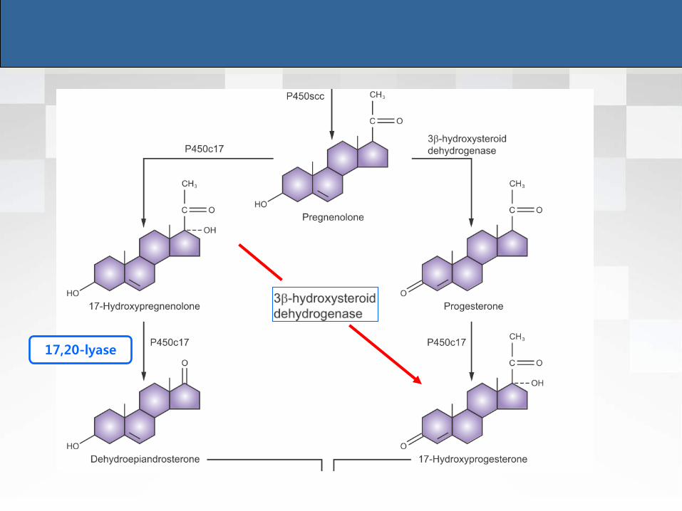
17,20-lyase

• P450c17
17-hydroxylase and 17,20-lyase
Adrenal gland
• Very little effect of 17,20-lyase
Theca cell and Leydig cell
• Both activities
• Principal pathway via DHEA
Corpus luteum
• Principal pathway via progesterone


The Two-Cell System
• First proposed by Falck in 1959
1. FSH receptors are present on granulosa cells 2. FSH receptors are induced by FSH itself 3. LH receptors are present on theca cells and initially absent on granulosa cells → FSH induces appearance of LH receptors on granulosa cells with follicle growth 4. FSH induces aromatase enzyme activity in granulosa cells

Primordial Follicle
• 1st visible sign of follicular development
Increase in the size of oocyte
Cuboidal granulosa cells rather than squamous
Maturation rather than growth
• Initiation of follicular growth: independent of FSH or LH
Primordial follicles in anencephalic fetus
Baker & Scrimgeour. J Reprod Fertil 1980;60(1):193-9.

Preantral Follicle
• Preantral follicle
Enlargement of oocyte
Surrounded by zona pellucida
Multilayer of G cells
and organization of theca layer
Dependent on gonadotropin
Correlated with increasing production of estrogen
FSH
Granulosa cell growth Aromatase
Estrogen

Preantral Follicle
• Estrogen production
Limited by FSH Rc contents
FSH: raise the concentration of its own receptor on G cells
1,500 receptors / G cell

Two Cell, Two-Gonadotropin Theory
• Preantral follicle & antral follicle
LH receptors: only on the theca cells (20,000 Rc / cell)
FSH receptors: only on the granulosa cells

Antral Follicle
• Dominant substance of FF in antral follicle
FSH & E2
• LH
Not normally present in FF until the midcycle
Premature elevation
Decreased mitotic activity in G cells
Degenerative change
Increased level of androgen

Two Cell, Two-Gonadotropin Theory
• Interaction btw granulosa and theca cells
Not fully functional until later in antral development
• G cells in small antral follicle
Tendency to convert androgen to 5α-reduced form
• G cells in large antral follicle
Preferentially metabolize androgen to estrogen

Two Cell, Two-Gonadotropin Theory
• P450c17
Expression in the theca cells
Conversion of 21-carbon substrate to androgens
17α-hydroxylase / 17,20-lyase
P450scc
17α-hydroxylase
17,20-lyase
P450c17
(theca)
P450arom
(G cell) androstenediol Testosterone

Androgen in Preantral Follicle
• Substrate for FSH-induced aromatization
+
Low conc. High conc.
Enhance aromatase activity Conversion to more potent 5α- reduced
androgens rather than estrogen
- Cannot be converted to estrogen
- Inhibition of aromatase activity
- Inhibition of FSH induction of LH Rc
formation
Androgen-rich follicle Atresia

Androgen in Preantral Follicle
Success of a follicle
androgen-dominated microenvironment
estrogen-dominated microenvironment

Blood Transport of Steroids
• Estradiol and testosterone circulating in blood
Mostly bound to a protein carrier, “SHBG”
30% loosely bound to albumin
1% unbound and free
Very small percentage bound to corticosteroid-binding globulin
• Hyperthyroidism, pregnancy, estrogen ⇒ SHBG ↑
• Corticoids, androgens, progestins, GH, insulin, IGF-1 ⇒ SHBG ↓
• Biologic effects of major sex steroids: determined by unbound portion
“free hormone” “active hormone”
• Routine assays mostly determine „total‟ hormone concentrations

SHBG
• localized to short arm (p12-13) of chromosome 17
• Circulating level of SHBG: inversely related to Bwt
Weight gain → SHBG ↓ → changes in unbound levels of sex
steroids
• Insulin resistance, hyperinsulinemia → decreases SHBG levels
• Android fat (abdominal wall, visceral-mesenteric locations)
→ asso. with hyperinsulinemia, hyperandrogenism, decreased SHBG
• SHBG conc. ⇒ marker for hyperinsulinemic insulin resistance
• Low SHBG ⇒ predictor for type 2 DM

Estrogen Metabolism
• Estradiol: major estrogen product of ovary, two pathways
Testosterone → (P450 AROM) → Estradiol
Androstenedione → via estrone (secreted in significant amounts) → Estradiol

Estrogen Metabolism
• E2 production: 100-300ug/day
• ADD production: 3 mg/ day Significant
contribution

Progesterone Metabolism
• Progesterone production rate
No Pph. conversion
Combination of secretion from adrenal gland and ovaries
Luteal phase: 20-30mg/day
10-20% excreted as pregnanediol
• Blood levels of progesterone
After ovulation: 3~15ng/mL
CAH
• serum progesterone: as high as 50 times above normal
• Serum 17-OHP: 10-400 times normal

Androgen Metabolism
• Origin of circulating androgens in female
ADD
• Adrenal (50%), ovary (50%)
DHEA
• Adrenal (50%), ovary (25%), pph. Conversion (25%)
Testosterone
• Adrenal (25%), ovary (25%), pph. Conversion (50%)

Androgen Metabolism
• In men..
Majority of circulating DHT is derived from testosterone ,
converted by 5α reductase to DHT
• In women..
Production rate: ADD > T
Blood DHT primarily derived from ADD and partly from DHEA-S
• DHT
Intracrine hormone (formed and act within target tissues)
Blood DHT only about one-tenth the level of circulating
testosterone

Androgen Metabolism
Testosterone converted to DHT by 5 α-reductase DHT further reduced to androstenediol (inactive) by 3 α-keto-reductase Wolffian duct structures - epididymis, vas deference, seminal
vesicle - Dependent on testosterone
Urogenital sinus /tubercle - Male external genitalia, urethra,
prostate - Requires conversion to DHT

Recommended Readings


CAH

Steroid Hormone Nomenclature
• Usage of „Nor‟ terminology

Preantral Follicle
• Synergistic effect of FSH and estrogen
Rapid accumulation of FSH Rc
Follicular response to relatively low conc. of FSH
Autocrine function for E2 within the follicle
FSH E2