hlt41812 - hltpat410d-hltpat411d specialised testing
DESCRIPTION
pTRANSCRIPT

HLT41812 Certificate IV in Pathology
HLTPAT410D Collect Pathology specimens other than blood for specialized testing
HLTPAT411D Perform blood collection for specialized testing

1. Prepare for specimen collection. 2. Perform procedure for specialised test 3. Perform post collection procedures. 4. Label and store specimens for
transportation.
HLTPAT410D - Collect pathology specimens other than blood for specialised testing

1. Prepare for blood collection 2. Perform collection for specialised test 3. Perform post-blood collection
procedures. 4. Label and store specimens for
transportation
HLTPAT411D Perform blood collection for specialised testing

As a part of these units you need to have some understanding of basic anatomy and physiology and any conditions that require specialized tests to be performed in the pathology collection rooms.
There are many disease processes which you will come in contact with however the following are some of interest.
Specialised tests Circulatory and cardiac systems


Haemophilia
Haemophilia belongs to a group of inheritable blood disorders that includes haemophilia A, haemophilia B (Christmas disease) and Von Willebrand's disease.
In haemophilia, the blood's ability to clot is severely reduced because an essential clotting factor is partly or completely missing. This means that people bleed for longer than normal.

Haemophilia A
Haemophilia A is a deficiency of an essential clotting factor called factor VIII, which is normally produced in the liver. The faulty gene is found on the X chromosome (X-linked). It's five times more common than haemophilia B.

Every person has two sex chromosomes. Women have two X-chromosomes while a man has one X and one Y chromosome. Both factor VIII and IX proteins are located on the X chromosome.
A fault in the Factor VIII or IX gene on the X chromosome will result in Haemophilia A or B in males because there is not a normal X chromosome to balance the abnormality.
Females carrying the haemophilia gene, the normal X chromosome compensates for the abnormal X chromosome.
Hereditary factor

An affected male cannot pass the disorder on to his sons but all his daughters will carry the haemophilia gene.
Females only rarely have symptoms of the disorder, they may carry the haemophilia gene and may pass on the disorder to their sons.
Sons of women with the haemophilia gene have a one in two chance of being affected and daughters will have a one in two chance of carrying the haemophilia gene.
Hereditary factor

Symptoms range from easy bruising to prolonged bleeding.
Bleeds can occur spontaneously (without an external cause) or as a result of injury.
Symptoms and long-term problems

How easily or badly a person bleeds depends on the severity of their deficiency. Minor cuts and grazes don't usually cause any problems, but internal bleeding can be life threatening, while repeated bleeding in the joints typically leads to arthritis or long-term joint damage.
Symptoms and long-term problems
Mobility problems may also result from these spontaneous bleeds. Haemorrhages into the brain are particularly difficult to manage and can be fatal.
Other long-term problems include the risk of infection from blood products.
Education and employment may also be disrupted
Symptoms and long-term problems

Diagnosis
Carriers of the gene can be identified with a blood test.
Pregnant women can be assessed using ultrasound to determine the sex of the baby and so estimate the risk of the disease.
They can then decide whether to have more invasive tests - amniocentesis and CVS - which can detect the relevant gene in the baby.

There's no cure for haemophilia and, although patients are treated with injections of the missing clotting factor, there's no permanent way of replacing or increasing its level.
Treatment

Haemophiliacs may receive medication, such as desmopressin, to try to raise the levels of the missing clotting factors, especially prior to planned surgery or dentistry, or they may receive factor concentrate.
Management of acute bleeding remains an essential part of their management if long-term health is to be sustained.
Severely affected children often receive regular injections to prevent bleeding
Treatment

Haemostatic Disorders
THROMBOCYTOPENIA HAEMOPHILIA Some tests you will deal with: Fibrin degradation tests (FDP) Prothrombin Time (PT) Partial Thromboplastin Time/activated
partial Prothoplastin time (PTT/APTT) Factor Assays

Fibrin degradation tests (FDP)
Prothrombin Time (PT)
Partial Thromboplastin Time/activated partial Prothoplastin time (PTT/APTT)
Factor Assays
Look up your book: What tubes would
you use? How much Blood? What department
do you send the sample?
Are there any special requirements?
Tests

Blood Types
ANTIBODY Any body or substance soluble or cellular
which is invoked by the stimulus provided by the introduction of antigen and which reacts specifically with the antigen in some way

Blood Types
ANTIGEN Any substance that as a result of coming
in contact with appropriate cells induces a state of sensitivity and /or immune response in some demonstrable way

The site of the bleeding must be found and the bleeding arrested. Transfusion may be necessary.
Anemia

Laboratory findings Positive or negative DAT (Direct Antiglobulin
Test) red cell abnormalities Specimen Blood – serum significance: Corticosteroid therapy eg. Prednisolone ↓ Haptoglobins: Haemolysis due to auto
immune disease, transfusion reaction, mechanical trauma (heart valve) ineffective red cell production due to severe Vit B12 or folic acid deficiency. Overactive spleen, liver disease.
Tests for the presence of Haemolysis

Tests for causes of Haemolysis
Further tests to identify the cause of haemolysis: Direct Antiglobulin Test /DAT Direct Coombs Test / DCT
Used to separate haemolytic anaemia due to an immune reaction from non-immune causes a positive test detects the presence of antibodies and therefore indicates an immune cause. Symptoms: Anaemia, jaundice, tea coloured urine

Anti bodies consists of chains of protein attached to a sugar molecule
One part binds to antigens One part interacts with elements of the
immune system such as neutrophils and macrophages which have Fc receptors on their surfaces
There are five classes of immuniglobulins:IgG IgA IgM IgD
Immunoglobulins

Tests for causes of Haemolysis
Other laboratory results Bilirubin ↓ Haptoglobins Specimen Blood: Haematology Method: tests for the presence of anti red-cell IgG, IgA Antibodies or complement attached to RBC, which cause RBCs to clump together.

Cold aggluntins
A cold aggluntins is an antibody that attaches to the red blood cell and causes them to clump together or agglutinate at a temperature below body temperature
Cold aggluntins are present in the serum of person with mycoplasma pneumoniae
(an atypical pneumonia) glandular fever, syphilis and certain blood diseases such as haemolytic anaemia

Cold aggluntins
To prevent the antibody from attaching to the RBCs, a cold agglutinin specimen must be collected in a tube pre-warmed to 37˚ and kept at this temperature until the serum is separated form the cells (look up special instructions).

Excessive bleeding may result from: Inability to arrest blood loss (↓ Platelet
count or platelets that do not function properly).
Failure to form a permanent clot (defect in the clotting process). Overactive of the system that breaks
down clots (excessive fibrinolysis). Easy breakage in the smallest blood
vessels (fragile capillaries).
Coagulation Studies

There are two alternative pathways: Intrinsic - stimulated by contact with a
foreign surface or material Extrinsic - activated by tissue damage.
The final two reactions is the conversion of prothrombin to thrombin and fibrinogen to fibrin.
Coagulation Studies
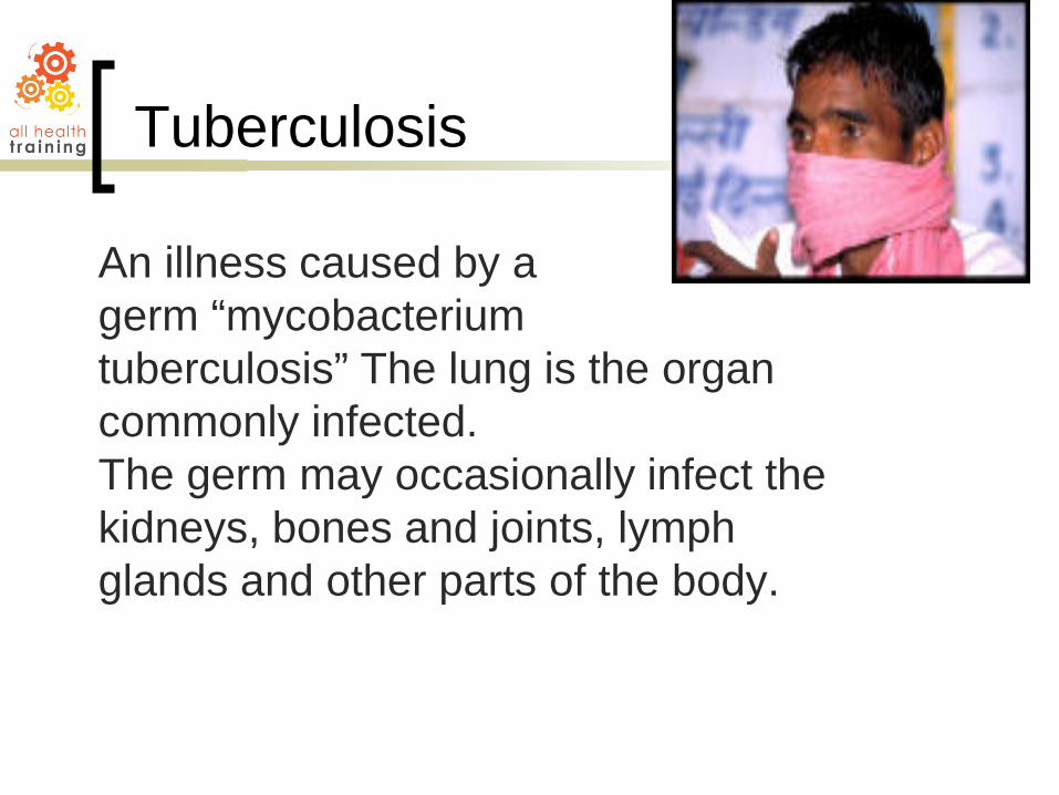
An illness caused by a germ “mycobacterium tuberculosis” The lung is the organ commonly infected. The germ may occasionally infect the kidneys, bones and joints, lymph glands and other parts of the body.
Tuberculosis

About 800 cases are found and treated in Australia each year
Newly arrived immigrant who caught the disease prior to arrival are at risk
More likely to affect people with low resistance to infection, mal-nourished etc.
Tuberculosis

Only 5 – 10 % of people who become infected with TB get sick.
TB can lie dormant for many years and when the immune system is weakened the chance of getting sick increases.
Dissemination of tuberculosis is when the infection spreads from the lungs to other organs.
Tuberculosis

SYMPTOMS Fever Cough Loss of energy
tiredness Nocturnal sweats Weight loss Phlegm may
sometimes have the presence of blood
TREATMENT 3-4 antibiotics
taken together over many months to be certain of a cure
Time will vary according to many factors
Follow up regularly
Tuberculosis

Insert Digestive in here

AFB (shown in red) are tubercle bacilli Also known as: TB culture and sensitivity Formal name: Acid-fast bacillus smear and culture and sensitivity
AFB (shown in red) are tubercle bacilli
AFB Smear

Tuberculosis

Tuberculosis

Tubercular Lesion

The patient initially improved with anti-tuberculosis treatment but then developed new fever and cough. Sputum cultures were positive for Klebsiella pneumoniae.
Klebsiella Pneumoniae

A 50 year old female presented with four weeks of cough

The most useful diagnosis methods are: Chest x-ray Sputum cultures Tuberculin skin
test Bronchoscopy Open lung biopsy
Diagnosis

QuantiFERON – TB Gold Blood Collection
There are three tubes: 1. Red Cap = TB Antigen 2. Grey Cap = Nil – negative control 3. Purple cap = Mitogen control
1. Collect 1ml exactly into each tube Blood tube MUST be filled to 1ml for
optional test performance ie to INDICATOR line
Tubes may fill slowly, keep tube on needle until blood flow stops to ensure correct volume
For butterfly needle always use a PURGE tube
Collection of blood

2. Shake tubes for 5 seconds to resuspend ingredients on the tube wall
3. Do NOT refrigerate. Leave at room temperature (maximum 16 hours)
Collection of blood

BCG Vaccination

Contains soluble growth products derived from the tubercle bacillus. When administered intra-dermally either by injection or by means of a puncture devise, a hypersensitivity reaction, manifesting as induration as erythema will appear in sensitive individuals.
Tuberculin PPD

Inject intradermally 0.1ml of a solution containing 1.00IU per ml (ie 10IU per dose of 0.1ml) into the ventral surface of the upper part of the forearm.
The reaction commences within 24 hours and reach maximum size in 48- to 72 hours.
The result should be read in 72 hours but could be read from 48 hours to the fifth day.
Mantoux testing procedure using Tuberculin PPD

Mantoux testing procedure using Tuberculin PPD

A positive reaction is an indication that the patient has/had at some time a tuberculosis infection. A positive test does not indicate an active infection, but indicates that further investigation should be done.
Mantoux testing procedure using Tuberculin PPD

Tuberculin skin test involves injection of PPD ( a purified protein derivative) into an area like the forearm (pictured right). If there is a reaction, TB is present
Mantoux testing procedure using Tuberculin PPD

It is the oedema and induration that is important and can usually be detected with a finger rather than the eye.
he diameter of the area of induration or oedema is measures in millimeters and recorded.
Erythema without oedema or induration should be disregarded.
Mantoux testing procedure using Tuberculin PPD

Mantoux testing procedure using Tuberculin PPD

Read reaction 48-72 hours after injection Measure only induration
Record reaction in millimeters
Reading the Tuberculin skin test

The National Tuberculosis Advisory Council (Canberra) has suggested the following degrees of reaction to the 10U dose Mantoux Test: Negative: less than 5mm diameter Weak positive : 5-9mm diameter Intermediate positive: 10-14 diameter. Strong positive : 15mm diameter or more
vesiculation
Mantoux testing procedure using Tuberculin PPD

Routine BCG vaccination occurred in Victoria from the early 1950’s until 1984
In the US routine skin testing is done during baby check-ups
Anyone who has been exposed to TB should have a tuberculin skin test
Infected people should stay home from work or school to prevent spreading TB
Prevention and treatment

TB can be treated using drugs like: o Isoniazid (INH) o Rifampin o Pyrazinamide o Ethambutol o Streptomycin
People with TB take a combination of drugs. MDR (Multi-drug-resistant) TB develops
when people do not follow instructions about medication.
Prevention and treatment

TB is starting to become more common again
HIV is speeding up the spread of TB TB accounts for about 15% of deaths of
HIV sufferers worldwide Greater movement of people around the
world is helping to spread TB In the US, nearly 40% of TB cases are
among foreign born people
Tuberculosis Facts

Untreated TB can spread quickly through refugee camps
Up to 50% of the worlds refugees may be infected with tuberculosis
In 1995 about 30% of San Francisco’s homeless were infected with TB compared to an overall 7% of the US population
Tuberculosis Facts

Kills more people in the world than any other infectious disease
Has probably been around for more than 2000 years
Has been known by names such as: o Phthisis o Consumption o White plague
Tuberculosis Facts

In Victoria the Western Metropolitan area has the largest incidence
This is primarily due to the number of immigrants from Asia, Africa, Europe and the Middle East
Of immigrants, males 25 – 29 are the highest risk group
Changes to Visa procedures for overseas students now require them to undergo a medical examination before approval
Tuberculosis Facts

Only people with pulmonary (lung) TB are infectious
An untreated person with infective TB will infect 10 – 15 people each year
A person only needs to breathe in a few bacteria to become infected
Tuberculosis Facts

A chest x-ray shows lungs, heart and diaphragm. A chest x-ray can be ordered for any chest complaint,
Sputum cultures identify bacteria in a sample of sputum from the lungs
A Bronchoscopy is when a camera is sent down the throat into the lungs so the doctor can see what is happening inside the lungs
Tuberculosis Diagnosis

Addison's disease is the result of an under active adrenal gland. An under active adrenal gland produces insufficient amounts of corticosteroid hormones. Four in every 100,000 people have Addison's disease.
Underactive adrenal glands/ Addison’s Disease

Most of the time, the cause of the disease is unknown. About one-third of Addison's disease cases are caused by the actual destruction of the adrenal glands through cancer, infection, or other diseases. Other causes may include:
Use of corticosteroids as a treatment (such as prednisone) causes a slow down in production of natural corticosteroids by the adrenal glands.
Certain drugs used to treat fungal infections may block production of corticosteroids in the adrenal glands.
Causes

weakness fatigue dizziness dark skin black
freckles weight loss
dehydration lack of appetite muscle aches nausea vomiting diarrhea intolerance to
cold
Signs and symptoms

Signs and symptoms
bluish-black discoloration around the nipples, mouth, rectum, scrotum,
or vagina

In addition to a complete medical history and medical examination, diagnostic procedures for Addison's disease may include: blood tests to measure corticosteroid
hormone levels kidney function tests to determine if urine
is concentrated
Diagnosis

Since Addison's disease can be life threatening, treatment often begins with administration of corticosteroids. Corticosteroids, such as prednisone, may be taken orally or intravenously, depending on the patient's condition. Usually the patient has to continue taking the corticosteroid the rest of his/her life. Treatment may also include taking fludrocortisone, a drug that helps restore the body's level of sodium and potassium.
Treatment


Cushing's syndrome is the result of the excessive production of corticosteroids by the adrenal glands. An overproduction of corticotropin -- the hormone that controls the adrenal gland -- by the pituitary gland, which stimulates the adrenal glands to produce corticosteroids, may be one cause. In addition, certain lung cancers and other tumors outside the pituitary gland may produce corticotropins.
Overactive adrenal glands/ Cushing’s Syndrome

Cushing’s Syndrome
upper body obesity round face increased fat around
neck thinning arms and
legs fragile and thin skin

stretch marks on abdomen, thighs, buttocks, arms, and breasts
bone and muscle weakness
severe fatigue high blood pressure high blood sugar
Cushing’s Syndrome

irritability and anxiety
excess hair growth in women
irregular or stopped menstrual cycles in women
reduced sex drive and fertility in men
Cushing’s Syndrome

In addition to a complete medical history and medical examination, diagnostic procedures for Cushing's syndrome may include:
X-rays to locate any tumors 24-hour urinary test to measure for corticosteroid
hormones Computed tomography (CT or CAT scan) - a non-
invasive procedure that takes cross-sectional images of the brain or other internal organs; to detect any abnormalities that may not show up on an ordinary x-ray
Magnetic resonance imaging (MRI) - a non-invasive procedure that produces two-dimensional views of an internal organ or structure, especially the brain or spinal cord
Diagnosis

Dexamethasone suppression test - to differentiate whether the excess production of corticotropins are from the pituitary gland or tumors elsewhere
corticotropin-releasing hormone (CRH) stimulation test - to differentiate whether the cause is a pituitary tumor or an adrenal tumor
Other laboratory tests
Diagnosis

Synthetic ACTH, known as synacthen, is used to stimulate the adrenal cortex to produce cortisol METHOD: No preparation is necessary. Blood sample taken to measure baseline cortisiol. An injection of synacthen is administered Blood samples are taken at 30 minutes and 60
minutes SIGNIFICANCE: Failure to raise cortisol above test baseline indicates adrenal insufficiency
Synacthen Stimulation Test

Dexamethasone, a corticosteroid, is similar to a natural hormone produced by your adrenal glands. It often is used to replace this chemical when your body does not make enough of it.
Relieves inflammation (swelling, heat, redness, and pain)
Used to treat certain forms of arthritis; skin, blood, kidney, eye, thyroid, and intestinal disorders (e.g., colitis); severe allergies; and asthma.
Dexamethasone is also used to treat certain types of cancer.
Dexamethasone

In Cushings disease and ACTH producing cancers (eg certain lung cancers or other tumours) the raised cortisol level is not suppressed by Dexamethasone. Measurement of cortisol forms the basis of both this and the Synacthen Stimulation test Specimen: Blood
Dexamethasone Suppression test

METHOD: Dexamethasone is taken by tablet at midnight : Cortisol measures from a blood sample collected 8 hours later (8am) NORMAL VALUES: Check with laboratory, normally 200-650 nmol/L cortisol
Dexamethasone Suppression test

SIGNIFICANCE: Non suppression of cortisol with a dose of Dexamethasone - Cushing’s Disease High dose Dexamethasone non suppression – ACTH-producing lung or stomach cancer, adrenal tumour or cancer (high doses of Dexamethasone will usually suppress the pituitary tumour production of ACTH in Cushing’s disease) FURTHER TESTS: CT, MRL, Insulin Hypoglycaemic Stimulation Test, Synacthen Stimulation Test.
Dexamethasone Suppression test

Treatment for Cushing's syndrome depends on its cause.
Surgery may be needed to remove tumors or the adrenal glands.
Other treatment may include radiation, chemotherapy, and use of certain hormone-inhibiting drugs.
Treatment

There are three main types of diabetes that require clinical care by a physician or other healthcare professional: 1. type 1 diabetes 2. type 2 diabetes 3. gestational diabetes
Diabetes


Type 1 diabetes is also known as diabetes mellitus, insulin-dependent diabetes mellitus (IDDM), juvenile diabetes, brittle diabetes, or sugar diabetes. There are two forms of type 1 diabetes: idiopathic type 1 - refers to rare forms of the
disease with no known cause. immune-mediated diabetes - an autoimmune
disorder in which the body's immune system destroys, or attempts to destroy, the cells in the pancreas that produce insulin. Immune-mediated diabetes is the most common form of type 1 diabetes
Type I Diabetes

The cause of type 1 diabetes is unknown, but it is believed that people inherit a tendency to develop diabetes, and that viruses may be involved.
Causes

This auto-immune disease results from the body's failure to produce insulin, the hormone that allows glucose to enter the cells of the body to provide fuel.
This is the result of an autoimmune process in which the body's immune system attacks and destroys the insulin producing cells of the pancreas.
Causes

When glucose cannot enter the cells, it builds up in the blood and the body's cells literally starve to death.
People with type 1 diabetes must take daily insulin injections and regularly monitor their blood sugar levels
Causes

high levels of sugar in the blood when tested
high levels of sugar in the urine when tested
unusual thirst frequent urination extreme hunger but loss of weight blurred vision nausea and vomiting extreme weakness and tiredness irritability and mood changes
Signs and Symptoms

Fasting plasma glucose of greater than or equal to 126 mg/dl with symptoms of diabetes.
Casual plasma glucose (taken at any time of the day) of greater than or equal to 200 mg/dl with the symptoms of diabetes.
Oral glucose tolerance test (GTT) value of greater than or equal to 200 mg/dl measured at a two-hour interval. The GTT is given over a two -hour time span.
Diagnosis

People with type 1 diabetes must have daily injections of insulin to keep the blood sugar level within normal ranges. Other parts of the treatment protocol may include: appropriate foods to manage blood sugar
level. exercise to lower and help the body use
blood sugar. regular blood testing for blood-sugar levels. regular urine testing for ketone levels.
Treatment

Type 2 Diabetes This type is far more common than type 1, affecting between 90 percent and 95 percent of people with diabetes over age 20. It occurs when your body is resistant to the effects of insulin or your pancreas produces some, but not enough, insulin to maintain a normal glucose level.
Type 2 Diabetes

Test used to measure glucose sugar in the blood. It is oxidised be cells to liberate energy for metabolic function. The brain an nervous system rely almost entirely on glucose for energy.
Specimen – Oxolate. Specimen may be ‘random’ (taken approx. 2 hours after a meal_ or ‘fasting’ (taken at least 8 hours after a meal)
Blood glucose Blood sugar levels (BSL)

↑ Blood Glucose Common – Diabetes
mellitus Uncommon - Excess
thyroid Hormone, acute or chronic pancreatitis, mumps, cystic fibrosis, cancer of the pancreas, stroke, heart attack, chronic liver disease
↓ Blood Sugar Common - low blood
sugar (hypoglycaemia) due to fasting missed meals carbohydrate/ insulin reactions
Uncommon – tumours of the islets cells. Glucagon defeciency, cancer of the adrenal glands, liver damage due to poisoning decrease pituitary or thyroid hormones
Significance Results should be interpreted in conjunction with blood glucose results

Used to confirm Diabetes Mellitus Booked test - required to stay in the
rooms for 2 hours Fasting test – 8 hours Dietary Requirements – 3 days prior to
test patient’s diet should include approx. 15G of carbohydrate
Test – Oxalate tubes . A standard dose of glucose is given in a drink and blood glucose levels are checked over the next 2 hours
Glucose Tolerance Test (GTT)

Used to screen pregnant women for gestational Diabetes: Specimen- Oxalate tube Dietary requirements – nil Fasting – no Test - is performed at 26-28 weeks gestation Women are given a glucose drink and blood specimen is collected 1 hour later
Glucose Challenge Test (GCT)


TESTING: Urinary Drug
screen Alcohol levels Paternity testing Arsenic testing
REQUIREMENTS: Identify in the
usual manner with referral
Pre-label sample container
Chain of custody

Accompany client to the toilet Explain the procedure with emphasis that
it is urinary drug screen Eg. “I am required to see you pass urine
under the conditions of the screen” Two personelle at the branch Do not put yourself in a position of being
compromised
Urinary drug screen

Chain of custody form filled out partly before starting then signed on completion of sample being obtained
If the client is unable to sign a designated person to sign Guard or police officer to verify signature .
Sample collection taken Just an ordinary sample. A urinary drug
screen is not a MSU
Urinary drug screen

After collection: In the presence of the client, seal the jar
with red tape or appropriate sealer with the signature over the seal
Form is signed by the client or designated signatory and verified
Specimen sent in a tamper proof biohazard bag
If the laboratory does not receive it intact, the specimen needs to be recollected
Urinary drug screen

Urine Catheter collection
C.S.U= catheter specimen of urine. Samples of urine collected from the bladder catheter. Collected by attending medical staff. Transported and stored a 4° centigrade