histopathology(mtsts)
TRANSCRIPT

HISTOPATHOLOGY
Histopathology
Refers to the microscopic examination of tissue in order to study the manifestations of disease. Specifically, in clinical medicine, histopathology refers to the examination of a biopsy or surgical specimen by a pathologist, after the specimen has been processed and histological sections have been placed onto glass slides.
In contrast, cytopathology examines free
cells or tissue fragments.

Collection of tissues (Histopathological
examination of tissues starts with:
surgery, biopsy, or autopsy.)
NOTE: The tissue is removed from the body, and then placed in
a fixative which stabilizes the tissues to prevent decay. The most common fixative is formalin (10% formaldehyde in water).
The tissue is then prepared for viewing under a microscope using either chemical fixation or frozen section

Biopsy
is a medical test commonly performed by a surgeon, interventional
radiologist, or an interventional cardiologist involving
sampling of cells or tissues for examination.
Biopsies are most commonly performed for insight into possible
cancerous and inflammatory conditions.
Conditions identified with biopsies
Cancer
Precancerous
conditions
Inflammatory
conditions
Cancer
Pathologic examination of a biopsy can determine whether a lesion is benign or malignant, and can help differentiate between different types of cancer.
Precancerous
conditions
Easily detected and accessed sites, any suspicious lesions may be assessed. Originally, this was skin or superficial masses. X-ray, then later CT, MRI, and ultrasound along with endoscopy extended the range.
Inflammatory conditions
A biopsy of the temporal arteries is often performed for suspected vasculitis. In inflammatory bowel disease (Crohn's disease and
ulcerative colitis), frequent biopsies are taken to assess the activity of disease and to assess changes that precede malignancy.

Biopsied sites
Location Description
BONE MARROW
Since cells are formed in the bone marrow , a bone marrow biopsy is employed in the diagnosis of abnormalities of blood cells when the diagnosis cannot be made from the
peripheral blood alone. In malignancies of blood cells ( and ) a bone marrow biopsy is used in staging the disease. The
procedure involves taking a core of using a trabecular bone , using a trephine and then aspirating material.
LUNGS Biopsies of the lungs can be performed in a variety of ways
depending on the location.
PROSTATE Forms of prostate biopsy include transrectal
biopsy and transurethral biopsy
NERVOUS SYSTEM
Forms include brain biopsy, nerve biopsy, and meningeal biopsy
Urogenital system
Forms include renal biopsy, endometrial biopsy and cervical conization

GASTROINTESTINAL TRACT
Flexible enables access to the upper and lower , such that biopsy of the , and via the mouth and the , and terminal are commonplace. A variety of biopsy instruments may be introduced through the endoscope and the visualized site biopsied. Until recently, the majority of the small intestine could not be visualized for biopsy. The double-balloon “push-pull” technique allows visualization and biopsy of the entire gastrointestinal tract.Needle core biopsies or aspirates of the pancreas may be made through the duodenum or stomach.
Other Other sites include breast biopsy, lymph
node biopsy, muscle biopsy, and skin biopsy
Autopsy
a clinical or academic autopsy is performed to find the medical cause of death and is used in cases of unknown or uncertain
death, or for research purposes.
also known as a post-mortem examination, necropsy (particularly as to non-human bodies), autopsia cadaverum, or
obduction
is a highly specialized surgical procedure that consists of a thorough examination of a corpse to determine the cause and manner of death and to evaluate any disease or injury that may be present.
It is usually performed by a specialized medical doctor called a pathologist.
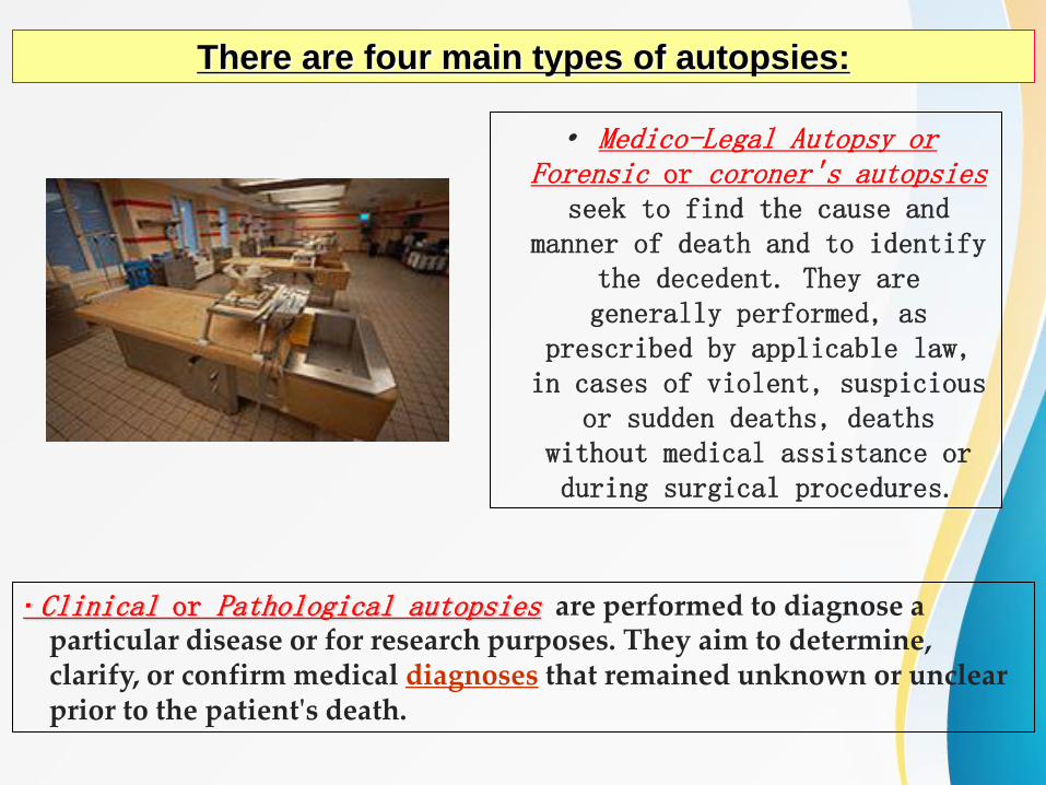
There are four main types of autopsies:
· Medico-Legal Autopsy or Forensic or coroner's autopsies
seek to find the cause and manner of death and to identify
the decedent. They are generally performed, as
prescribed by applicable law, in cases of violent, suspicious
or sudden deaths, deaths without medical assistance or during surgical procedures.
· Clinical or Pathological autopsies are performed to diagnose a particular disease or for research purposes. They aim to determine, clarify, or confirm medical diagnoses that remained unknown or unclear prior to the patient's death.

· Anatomical or academic autopsies are performed by
students of anatomy for study purpose
only.
Virtual or medical imaging autopsies are performed utilizing
imaging technology only, primarily magnetic
resonance imaging (MRI) and computed tomography
(CT).
is an ancient medical specialty that uses operative manual and instrumental techniques on a patient to investigate and/or treat a pathological condition such as disease or injury
Surgery
≈
help improve bodily function or appearance or to repair unwanted ruptured areas.
A surgeon is a person who practises surgery.
A surgery can last from minutes to hours, but is typically not an ongoing or periodic type of treatment.
The term surgery can also refer to the place where surgery is performed, or simply the office of a physician, dentist, or veterinarian.

Histology Sample Preparation
Prepares tissue specimens for sectioning, staining and diagnosis. The standard paraffin process (tissue processing) moves specimens through a
series of steps so the soft tissue is supported in a medium that allows sectioning.
The standard steps are:
Embedding that allows orientation of the
specimen in a “block” that can be sectioned
and is easy to store and handle
Sectioning using a microtome to produce
very thin sections that are placed on a
microscope slide ready for staining.
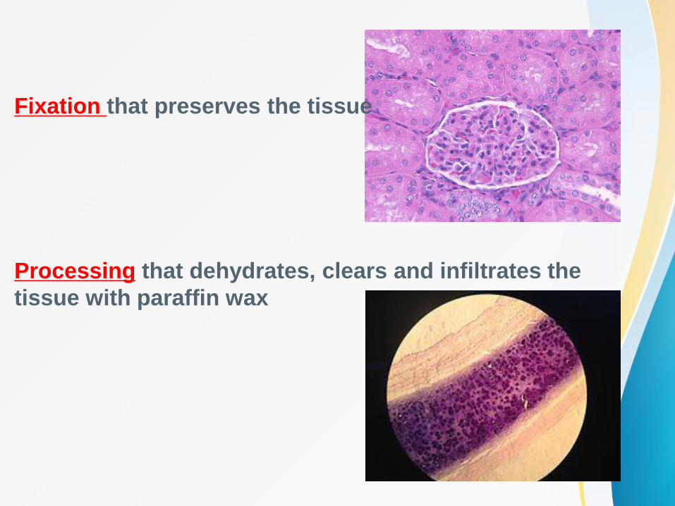
Fixation that preserves the tissue
Processing that dehydrates, clears and infiltrates the
tissue with paraffin wax

History of Histopathology
(compound of the Greek words: ἱστός histos "tissue", and -λογία -logia
"science")
the study of the microscopic anatomy of cells and tissues of plants and
animals.
It is commonly performed by examining cells and tissues by sectioning and
staining, followed by examination under a light microscope or electron
microscope
In the 19th century, histology was an academic
discipline in its own right. The 1906 Nobel Prize in
Physiology or Medicine was awarded to
histologistsCamillo Golgi and Santiago Ramon y
Cajal. They had dueling interpretations of the neural
structure of the brain based in differing
interpretations of the same images. Cajal won the
prize for his correct theory and Golgi for the staining
technique he invented to make it possible.
Santiago Ramón y Cajal
in his laboratory

Instruments in HISTOPATHOLOGY (principles)
A microtome (from the Greek mikros, meaning "small", and
temnein, meaning "to cut") is a tool used to cut extremely thin
slices of material, known as sections. Important in science,
microtomes are used in microscopy, allowing for the preparation of samples for
observation under transmitted light or electron radiation.
Microtomes use steel, glass, or diamond blades depending upon the specimen being sliced and the desired thickness of the
sections being cut.

A freezing microtome is an instrument used in the
laboratory to section small delicate materials. Sectioning is done on a platform which is frozen by snowing CO2. The
microtome knife is also snowed with CO2. In this
type of microtome, sectioning is done by the microtome knife moving towards the material to
be sectioned. For materials that have
alcohol, it should first be removed before being
sectioned.
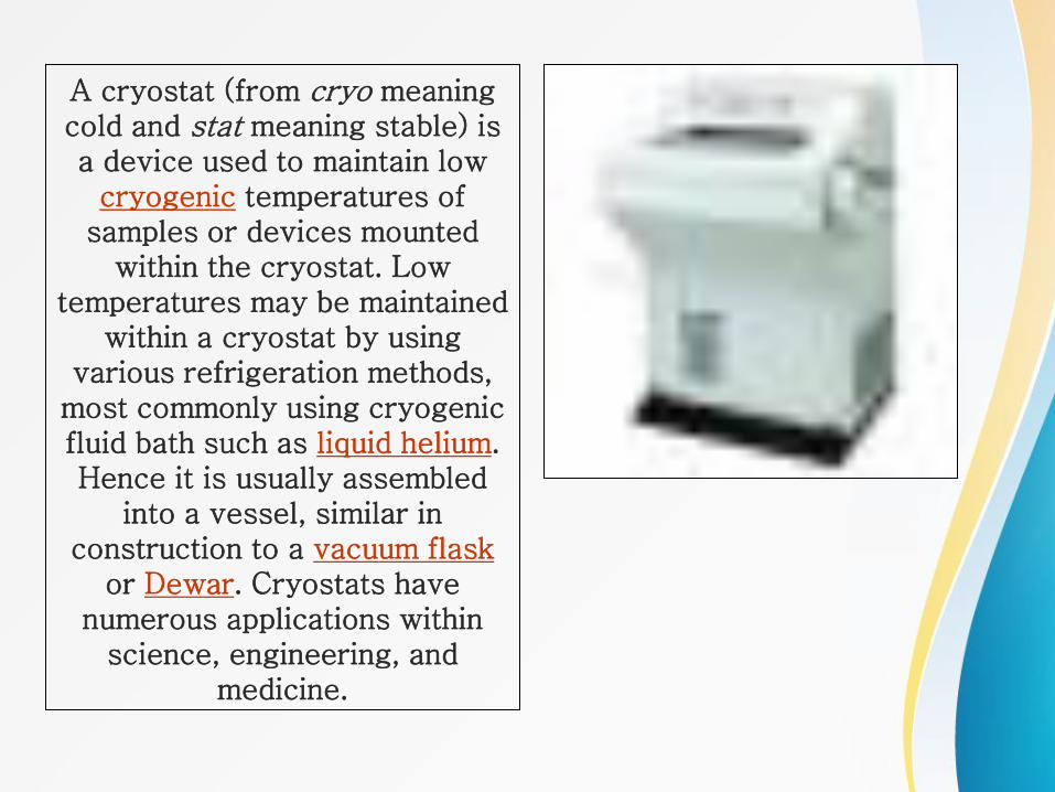
A cryostat (from cryo meaning cold and stat meaning stable) is a device used to maintain low
cryogenic temperatures of samples or devices mounted
within the cryostat. Low temperatures may be maintained
within a cryostat by using various refrigeration methods,
most commonly using cryogenic fluid bath such as liquid helium. Hence it is usually assembled
into a vessel, similar in construction to a vacuum flask
or Dewar. Cryostats have numerous applications within
science, engineering, and medicine.

The paraffin Embedding System allows a convenient, quick and careful paraffin embedding of tissue specimens. The modular structure and the standard height of the working area permit to combine the two modules of the embedding Station so as to meet the needs of the user.

vibrating microtomes (vibratomes) let you accurately cut tissue without freezing or
embedding.