histopathology: vascular pathology - rated medicine heal by organization. the image is of a coronary...
TRANSCRIPT

These presentations are to help you identify basic histopathological features.They do not contain the additional factual information that you need to learn
about these topics, or necessarily all the images from resource sessions.Before viewing this presentation you are advised to review relevant histology,
relevant sections of a pathology textbook (atherosclerosis, hypertension,Marfan’s syndrome and vasculitis (the latter an overview only),
histopathology atlas and relevant lecture notes.Copyright University of Adelaide 2011
(The histopathology of blood vessels is introduced in semester 1, year 1)Vasculitis and cystic medial degeneration aren’t specifically covered in years 1-3 and are presented for
general information.
Histopathology: Vascular pathology

With age the following arterial changes may occur:• Arterioles: hyaline thickening (arteriolar hyalinosis, hyaline
arteriolosclerosis) -> narrowing• Small arteries: medial hypertrophy, thickened intima with increased
connective tissue• Muscular arteries: medical calcification• Aorta: fragmentation of elastin and increase in collagen in media,
thickened intima with increased connective tissue-> increased wall stiffness, slight dilatation
The changes in arterioles and small arteries are exacerbated by systemichypertension.

Age and hypertension related intimal fibrosis in a small artery

Age and hypertension related hyaline change in arterioles (black arrows) in the kidney. Red star: capillary.Yellow star: normal arteriole. Blue star: interstitial connective tissue. Blue arrow: fibroblast nucleus

Accelerated hypertension. Loose myxomatous fibrous intimal proliferation (I) can be seen in a small artery.From Stevens et al: Core Pathology 3rd ed. Copyright 2009 by Mosby, an imprint of Elsevier, Ltd.

Accelerated hypertension: small artery. There is concentric thickening of the wall (hyperplasticarteriolosclerosis or "onion skin" endarteritis) and thrombosis (black arrow) of the lumen.

Accelerated hypertension. Fibrinoid necrosis of arterioles, visible as replacement of the wall by materialstained bright red (F), is seen in the wall of a renal arteriole in malignant hypertension. From Stevens et al:Core Pathology 3rd ed. Copyright 2009 by Mosby, an imprint of Elsevier, Ltd.
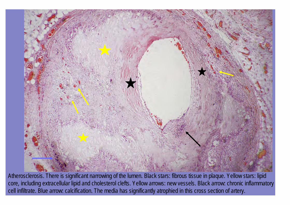
Atherosclerosis. There is significant narrowing of the lumen. Black stars: fibrous tissue in plaque. Yellow stars: lipidcore, including extracellular lipid and cholesterol clefts. Yellow arrows: new vessels. Black arrow: chronic inflammatorycell infiltrate. Blue arrow: calcification. The media has significantly atrophied in this cross section of artery.

Atherosclerosis. There is significant narrowing with thrombosis of the lumen (yellow star). Black stars: fibrous tissue inplaque.Blue arrows (dark material): calcification.Red star: lipid core
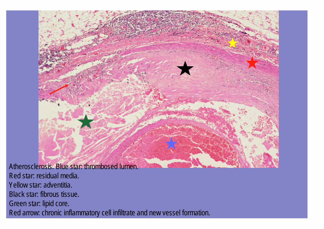
Atherosclerosis. Blue star: thrombosed lumen.Red star: residual media.Yellow star: adventitia.Black star: fibrous tissue.Green star: lipid core.Red arrow: chronic inflammatory cell infiltrate and new vessel formation.

Atherosclerosis. Black star: fibrous tissue.Red star: lipid core.Black arrows: chronic inflammatory cell infiltrate and new vessel formation.Yellow star: residual media

Atherosclerosis.Red star: lumen.Black star: fibrous cap.Yellow arrows: spaces left by cholesterol crystals.Red arrows: calcification
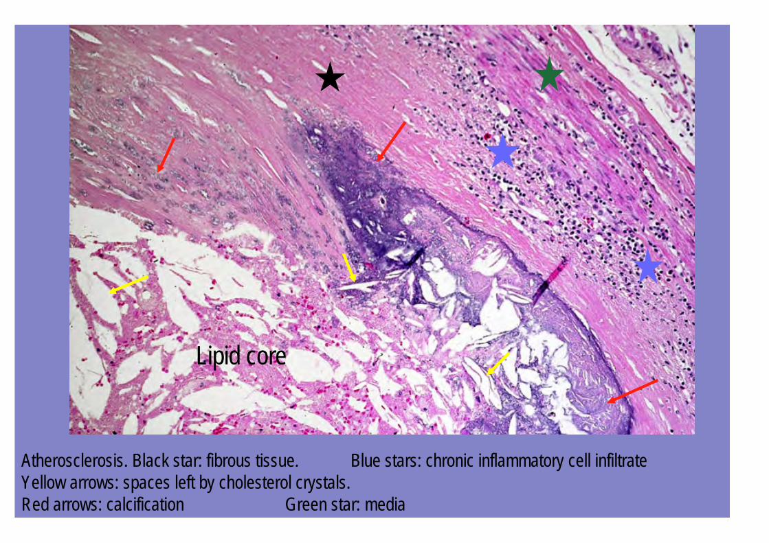
Atherosclerosis. Black star: fibrous tissue. Blue stars: chronic inflammatory cell infiltrateYellow arrows: spaces left by cholesterol crystals.Red arrows: calcification Green star: media
Lipid core

Atherosclerosis. Black arrows: foam cells (foamy macrophages).

Thrombi heal by organization. The image is of a coronary artery. An occlusive thrombus in the lumen hasbeen replaced with loose connective tissue (black star). There is recanalization (formation of new bloodvessels through the blocked lumen e.g. black arrows) but the vessels are small and will not carry sufficientblood through the blockage to supply distal tissues (which have infarcted long before anyway). Residualfibrous atherosclerotic plaque is also noted (red stars).

Histologic view of an aortic dissection demonstrating an intramural haematoma (asterisk). Aortic elastic layers stainblack and blood red in this section, stained with Movat stain. The intima is at the top of the section. Note that thedissection with blood is in the outer media. (From Robbins & Cotran Pathologic Basis of Disease 6E CopyrightElsevier).

Vasculitis can show a variety of patterns depending on the type. In temporal (giant cell) arteritis an inflammatory cellinfiltrate, including giant cells (arrow), is present within the wall. The giant cells are located in the the region of theinternal elastic lamina and an elastic stain would show its disruption. There is fibrotic thickening of the intima (blackstars). (Adapted from Robbins & Cotran Pathologic Basis of Disease 7E Copyright Elsevier).

Changes of cystic medial degeneration in the aorta in Marfan’s syndrome. In this special stain, the elastic tissue is black and collagenred. The intimal surface is indicated by the black arrow and the adventitia, containing abundant collagen is red.Note patchy areas of loss of elastic lamellae (yellow stars) and areas of accumulation of proteoglycan ground substance (blue stars)in the media.