histopathology of nsclc, ihc markers and ptnm...
TRANSCRIPT

Histopathology of NSCLC, IHC markers and pTNM classification
Prof Keith M Kerr
Department of Pathology, Aberdeen University Medical School , Aberdeen Royal Infirmary, Foresterhill, Aberdeen, UK
ESMO Preceptorship on Non-Small Cell Lung CancerNovember 15th & 16th 2017 Singapore
The management of patients with lung cancer is becoming ever more dependant on a knowledge of the pathology of each patient’s disease
‘Know your enemy’Sun Tzu, The Art of War.

The management of patients with lung cancer is becoming ever more dependant on a knowledge of the pathology of each patient’s disease
Lung cancer is
• NOT a single disease
• NOR is it just two diseases: Small Cell Carcinoma and Non-small Cell Carcinoma

The management of patients with lung cancer is becoming ever more dependant on a knowledge of the pathology of each patient’s disease
Lung cancer is
• NOT a single disease
• NOR is it just two diseases: Small Cell Carcinoma and Non-small Cell Carcinoma
Non-Small Cell Carcinoma is NOT a specific biological entity
• more a classification of convenience driven by a lack of therapeutic choice

2015 WHO Classification of Lung Tumours (part 1!!)
• 1-2: Adenocarcinoma
1-2A Invasive adenocarcinoma
1-2B Variants of invasive adenocarcinoma
1-2C Minimally invasive adenocarcinoma
1-2D Preinvasive lesions
1-2D-i: Atypical adenomatous hyperplasia
1-2D-ii: Adenocarcinoma in situ
• 1-3: Squamous cell carcinoma
1-3A: Keratinizing and nonkeratinizingsquamous cell carcinoma
1-3B: Basaloid carcinoma
1-3C: Preinvasive lesion: Squamous ca in situ
• 1-4: Neuroendocrine Tumours
1-4A: Small cell carcinoma
1-4B: Large cell neuroendocrine carcinoma
1-4C: Carcinoid tumors
1-4D: Preinvasive lesion: DIPNECH
• 1-5: Large cell carcinoma
• 1-6: Adenosquamous carcinoma
• 1-7: Sarcomatoid carcinoma
1-7A: Pleomorphic, spindle cell and giant cell carcinoma
1-7B: Carcinosarcoma
1-7C: Pulmonary blastoma
• 1-8: Other carcinomas
1-8A: Lymphoepithelioma-like carcinoma
1-8B: NUT-carcinoma

Small Cell Carcinoma of the Lung
• Nuclear features key to diagnosis
• Neuroendocrine markers and TTF1 IHC positive but not required for diagnosis
• Accurately diagnosed on cytology
• Aggressive disease, usually Stage 4 at presentation
• Therapeutic relevance • chemotherapy choice• radiotherapy strategy• prognosis

So all those other, biologically diverse malignant diseases are NOT small cell carcinomas – so we call them non-small cell carcinoma (NSCLC)
• Adenocarcinoma
• Squamous cell carcinoma
• Neuroendocrine tumours apart from SCLC
• Large Cell Carcinoma
• Adenosquamous Carcinoma
• Sarcomatoid Carcinomas
• Others

Bronchial Squamous Dysplasia
Squamous carcinoma-in-situ
Invasive Squamous Cell
carcinoma
Atypical Adenomatous Hyperplasia
AAH
Adenocarcinoma-in-situ
Invasive
Adenocarcinoma
There are at least two pathways of Lung Carcinogenesis

Central Bronchial
Carcinogenesis?
Peripheral airway
Carcinogenesis?
The progenitor cells
Express TTF1
The Terminal
Respiratory Unit
TRU
The progenitor cells
express p63, p40,
Cytokeratins 5&6
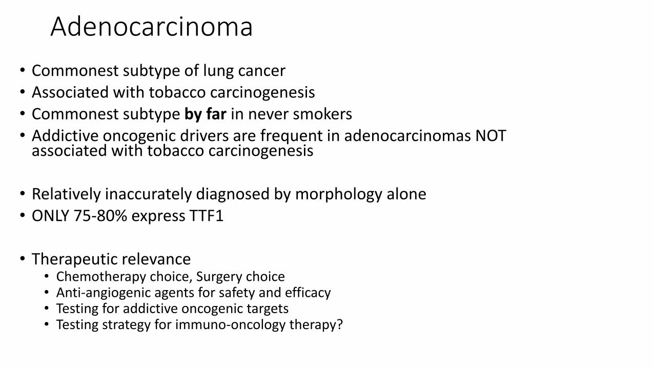
Adenocarcinoma
• Commonest subtype of lung cancer• Associated with tobacco carcinogenesis• Commonest subtype by far in never smokers• Addictive oncogenic drivers are frequent in adenocarcinomas NOT
associated with tobacco carcinogenesis
• Relatively inaccurately diagnosed by morphology alone• ONLY 75-80% express TTF1
• Therapeutic relevance• Chemotherapy choice, Surgery choice• Anti-angiogenic agents for safety and efficacy• Testing for addictive oncogenic targets• Testing strategy for immuno-oncology therapy?

Five histological patterns of adenocarcinoma: Most cases are mixtures, Pure forms are rare
Lepidic
MicropapillaryAcinar
Papillary Solid

Post operative survival vs predominant patternin pulmonary adenocarcinoma
Solid, Micropapillary
AIS, MIALepidic
AcinarPapillary
Yoshizawa A et al. Mod Pathol 2011; 24, 653-664 Stage 1 onlyRussell PA et al. J Thorac Oncol 2011; 6,1496-1504 Stages 1-3Warth A et al. J Clin Oncol 2012; Mar 5 epub Stages 1-4
Tsao MS et al JCO 2015
‘High Grade’ AdenocarcinomaHistology and benefit fromAdjuvant chemotherapy
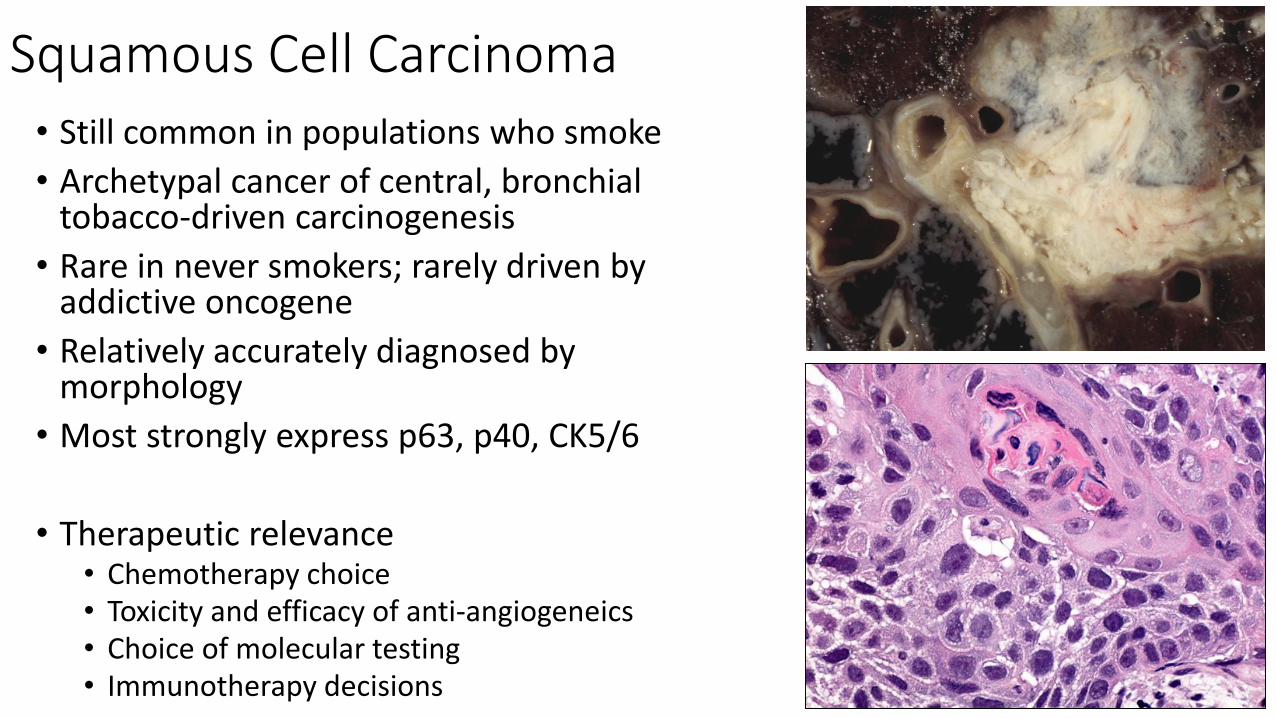
Squamous Cell Carcinoma• Still common in populations who smoke
• Archetypal cancer of central, bronchial tobacco-driven carcinogenesis
• Rare in never smokers; rarely driven by addictive oncogene
• Relatively accurately diagnosed by morphology
• Most strongly express p63, p40, CK5/6
• Therapeutic relevance• Chemotherapy choice• Toxicity and efficacy of anti-angiogeneics• Choice of molecular testing• Immunotherapy decisions

Neuroendocrine tumours other than SCLCLarge Cell Neuroendocrine Carcinoma (LCNEC)
• High grade neuroendocrine carcinoma
• Strongly associated with tobacco carcinogenesis
• Molecularly similar to SCLC
• Generally a diagnosis for surgically resected tumours only, however………..
• Requires immunohistochemistry
• Therapeutic relevance• Chemotherapy choice?
• Uncertainty due to diagnostic problems in advanced disease

Neuroendocrine tumours other than SCLCTypical Carcinoid
Usually central bronchial tumour, Obstructive pneumonia
Paradoxical lesionLow grade, wrong location
Metastatic disease rare10% regional nodes
Distant metastases very rare
Atypical CarcinoidVery rare, Relatively aggressive
Mitoses & necrosis – area dependent: 2mm2
Diagnosis on small samples vs surgical material
Therapeutic relevanceContext important
bronchial polyp or peripheral nodule
Confusion with SCLC in biopsy or cytology

Large Cell Carcinoma
• ONLY diagnosed on surgical resection
• NEVER a diagnosis on small biopsy or cytology
• Most cases (66%) re-assigned as squamous or adenocarcinoma by IHC (WHO 2015)
• Therapeutic relevance• Relatively aggressive tumour
• KRAS mutation dominant

Adenosquamous Carcinoma
• Relatively rare tumour
• Relatively aggressive tumour
• Peripheral or central?
• Requires minority component to comprise at least 10% of the lesion
• A surgical resection diagnosis• Small biopsy or cytology suspicion only
• Morphology vs IHC
• Therapeutic relevance• Manage like adenocarcinoma

Sarcomatoid Carcinoma
• Very rare lesions
• Pleomorphic carcinoma if >10% of lesion shows pleomorphic, spindle or giant cells
• Usually combined with squamous cell or adenocarcinoma
• Surgical resection for definitive diagnosis
• Therapeutic relevance• Chemoresistant
• KRAS mutations relatively frequent
• Found in TKI-recurrent disease
• MET exon14 skipping mutations
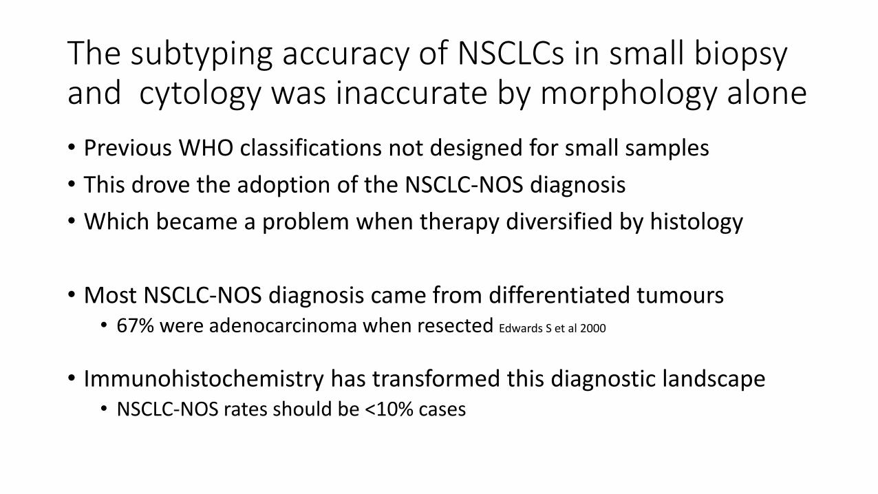
The subtyping accuracy of NSCLCs in small biopsy and cytology was inaccurate by morphology alone
• Previous WHO classifications not designed for small samples
• This drove the adoption of the NSCLC-NOS diagnosis
• Which became a problem when therapy diversified by histology
• Most NSCLC-NOS diagnosis came from differentiated tumours• 67% were adenocarcinoma when resected Edwards S et al 2000
• Immunohistochemistry has transformed this diagnostic landscape• NSCLC-NOS rates should be <10% cases
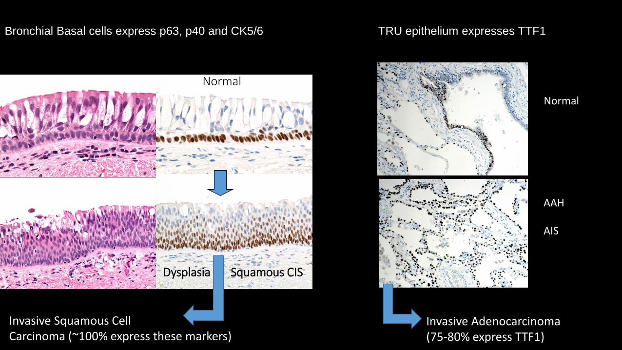
Bronchial Basal cells express p63, p40 and CK5/6 TRU epithelium expresses TTF1
Normal
Dysplasia Squamous CIS
Invasive Squamous CellCarcinoma (~100% express these markers)
Invasive Adenocarcinoma(75-80% express TTF1)
Normal
AAH
AIS
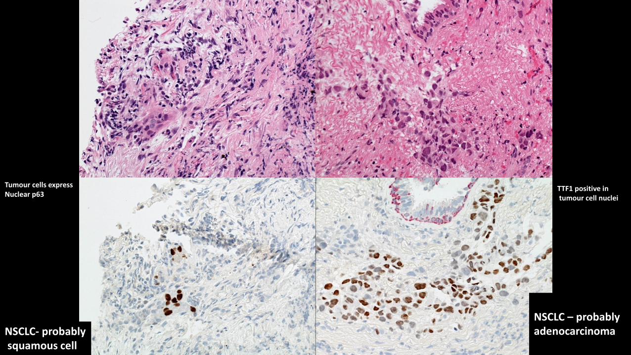
NSCLC – probably adenocarcinoma
TTF1 positive in tumour cell nuclei
Tumour cells express Nuclear p63
NSCLC- probablysquamous cell

Subtyping NSCLC: How good?
➢ Predictive IHC has ‘levelled the playing field’➢ Better diagnosis possible on poorer specimens
25%
40%
6%

Lung Cancer Classification and sample type
WHO 2004 (et prev): intended for, and only applicable to, resected cases
• Small Cell Carcinoma
• Squamous Cell Carcinoma
• Adenocarcinoma
• Large cell carcinomas
• Sarcomatoid carcinomas
• Adenosquamous carcinomas
• Carcinoid tumours
• Salivary-type carcinomas
• Small Cell Carcinoma
• Squamous Cell Carcinoma– Probable Squamous Cell Ca
• Adenocarinoma– Probable Adenocarcinoma
• NSCLC-NOS– NSCLC-NOS (null IHC)
• Carcinoid tumour
• Salivary-type (occasionally)
WHO 2015: a simplified classification intended for small sample diagnosis

Testing algorithm
On average only20% is tumour
Diagnose & subtype
lung cancer
Squamous Adeno
etc
Immuno-Histochemistry
IHCIf required
Biomarker testing
dictated by histology
and protocol
Sections for DNA
extraction
Sections for Biomarker IHC & FISH
EGFR, KRAS, BRAF mutation
(NGS panels)
ALK, ROS1EGFRPD-L1
‘Test tube’ tests
Morphology-basedtests
2 x 1mm tissueFragments
IHC should be used SPARINGLY for diagnosis

pTNM classification (7th edition: adopted by UICC and AJCC)
New proposals for TNM8 by IASLC – from Jan 2017
• Tumour Nodes Metastases
• pTNM based upon pathological examination

Carcinoma in situ: pTis
Squamous CarcinomaIn situ
AdencarcinomaIn situ - AIS

Pulmonary nodule with small solid area surrounded by GGO
TNM8
pT1a(mi)

This focus must be LESSTHAN5mm dia
Most ofthis lesion
isIn situ
disease
Minimally Invasive Adenocarcinoma MIA – pT1a(mi)
Focus ofInvasivePatterns ofAdeno-carcinoma
pT1a(mi)

pT1b>2cm but ≤ 3cm
18mm
26mm
pT1a≤ 2cm
The lesion MUST NOTInvolve a main bronchus
Unless………
9mm
TNM7

The lesion involving the main bronchus is a superficial spreading lesion with invasion
limited to the bronchial wall – pT1a

pT1c>2cm but ≤ 3cm
18mm
26mm
pT1a≤ 1cm
9mm
pT1b>1cm but ≤ 2cm
TNM8T1aT1bT1c
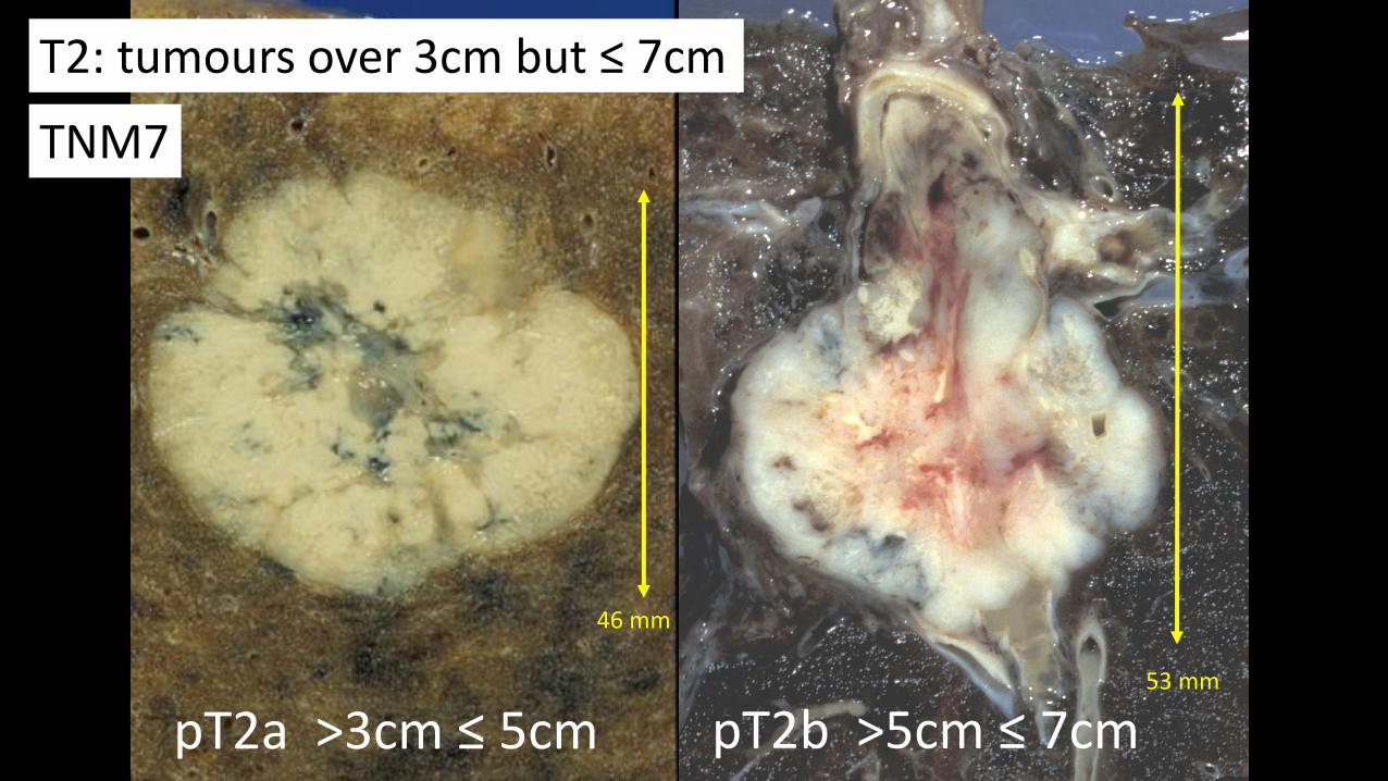
53 mm
46 mm
pT2a >3cm ≤ 5cm pT2b >5cm ≤ 7cm
T2: tumours over 3cm but ≤ 7cm
TNM7
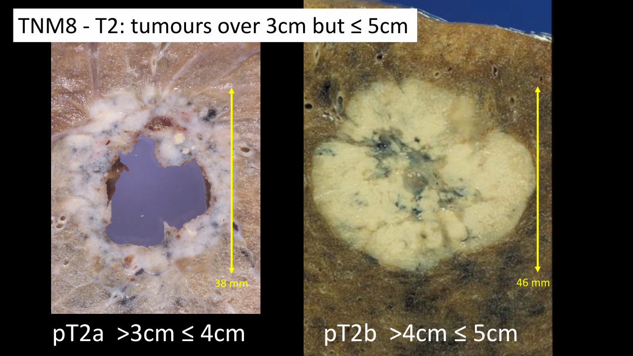
46 mm
pT2b >4cm ≤ 5cm
38 mm
pT2a >3cm ≤ 4cm
TNM8 - T2: tumours over 3cm but ≤ 5cm

Pleural invasion upstages a tumour to pT2a
PL1
PL2
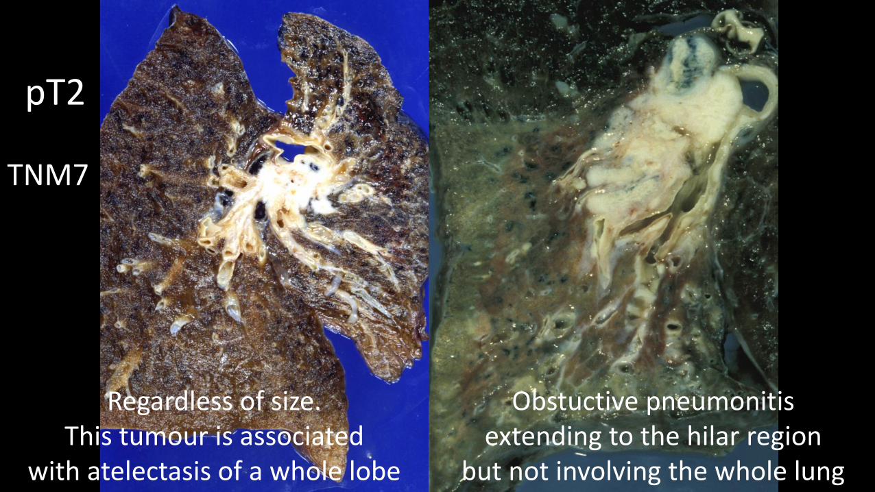
Regardless of size.This tumour is associated
with atelectasis of a whole lobe
Obstuctive pneumonitisextending to the hilar region
but not involving the whole lung
pT2
TNM7

Regardless of size.This tumour is associated
with atelectasis of a whole lobe
Obstuctive pneumonitisextending to the hilar regionOr involving the whole lung
pT2
TNM8

Small tumour but it involves main bronchus
T2a
Tumour > 2cm from carina
TNM7

Small tumour but it involves main bronchus
T2a
Regardless of distance from the Carina as long as it is not involved
TNM8

pT3
> 7cm
85mm
TNM7
53 mm
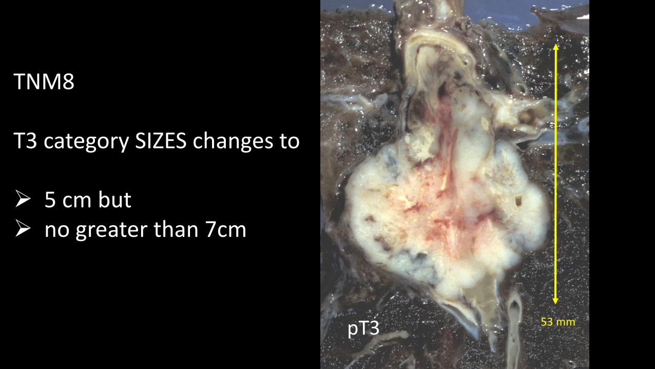
TNM8
T3 category SIZES changes to
➢ 5 cm but ➢ no greater than 7cm
pT3

Chest wall
Chest wall
pT3Invasion of
Mediastinal pleuraChest wall
Superior sulcusPhrenic nerve
DiaphragmParietal pericardium
Main bronchus within 2cm of carina
TNM7

pT3
Atelectasis or Obstructive Pneumonitis
of the entire lung
‘downgraded’ to T2In TNM8
pT3
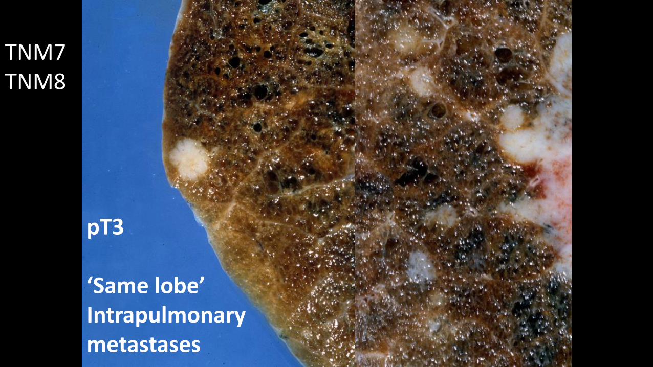
‘Same lobe’Intrapulmonarymetastases
TNM7TNM8
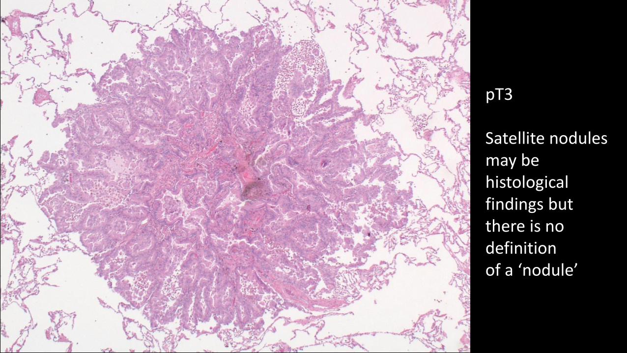
pT3
Satellite nodules may be histological findings but there is no definitionof a ‘nodule’
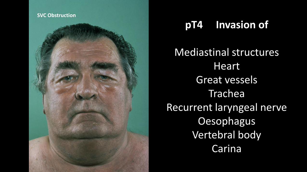
pT4 Invasion of
Mediastinal structuresHeart
Great vesselsTrachea
Recurrent laryngeal nerveOesophagus
Vertebral bodyCarina
SVC Obstruction

pT4‘Different ipsilateral lobe’
Intrapulmonary metastases
Oblique fissure
Left upper lobePrimary tumour
Left lower lobe metastasis
Issues with Pulmonary Metastases versus synchronous Primary tumours

TNM8
>7cmis now
T4
85mmpT4

Which part or element of adenocarcinoma should be measured to determine T status?
Lepidic
growth
Invasive tumourAdencarcinoma-in-situ

pN disease identified by pathological examinationHistology can define node positive disease
N1Stations 10 - 14
N2
N2SubcarinalStation 7
Contralateral mediastinal or hilar nodesScalene or supraclavicular nodes
N3

M1bDistant Metastases
• Liver, Adrenals, Bone, Brain, Skin, etc etc
• Cervical nodes above scalene are also M1M1a
Contralateral lung metastasesPleural nodulesMalignant pleural or pericardial effusion
M1a or M1b defines Stage 4 disease
TNM7

M1cDistant Metastases
• Liver, Adrenals, Bone, Brain, Skin, etc etc
• Cervical nodes above scalene are also M1
M1aContralateral lung metastasesPleural nodulesMalignant pleural or pericardial effusion
M1a or M1b defines Stage IVaM1c defines Stage IVb
M1bSingle extrathoracic metastasis isa single organ (incl lymph node)
TNM8

TNM defines Stage – Stage defines prognosis
TNM7 TNM8
The management of patients with lung cancer is becoming ever more dependant on a knowledge
of the pathology of each patient’s disease
‘Know your enemy’Sun Tzu, The Art of War.