histopathology techniquesndl.ethernet.edu.et/bitstream/123456789/88703/8/chapter 8... · 2020. 5....
TRANSCRIPT

Histopathology Techniques
College of Veterinary Medicine and animal sciences
University of Gondar,
Gondar, Ethiopia
4/22/2020 GB. UoG, CVMAS 1
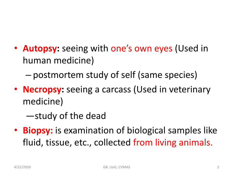
• Autopsy: seeing with one’s own eyes (Used in human medicine)
– postmortem study of self (same species)
• Necropsy: seeing a carcass (Used in veterinary medicine)
―study of the dead
• Biopsy: is examination of biological samples like fluid, tissue, etc., collected from living animals.
4/22/2020 GB. UoG, CVMAS 2
Histopathology Techniques
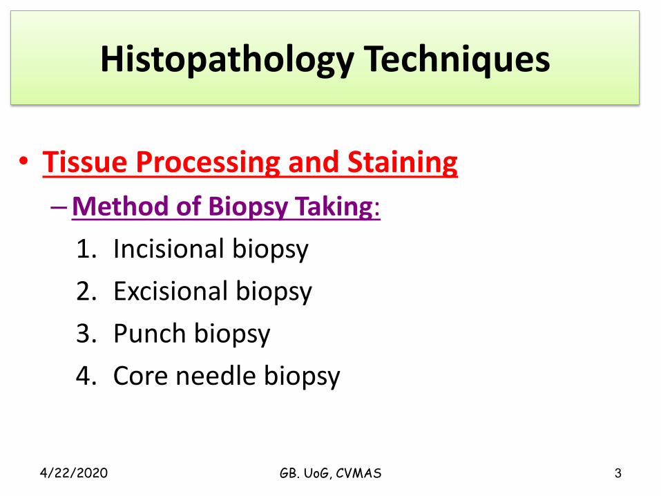
• Tissue Processing and Staining
–Method of Biopsy Taking:
1. Incisional biopsy
2. Excisional biopsy
3. Punch biopsy
4. Core needle biopsy
4/22/2020 3GB. UoG, CVMAS
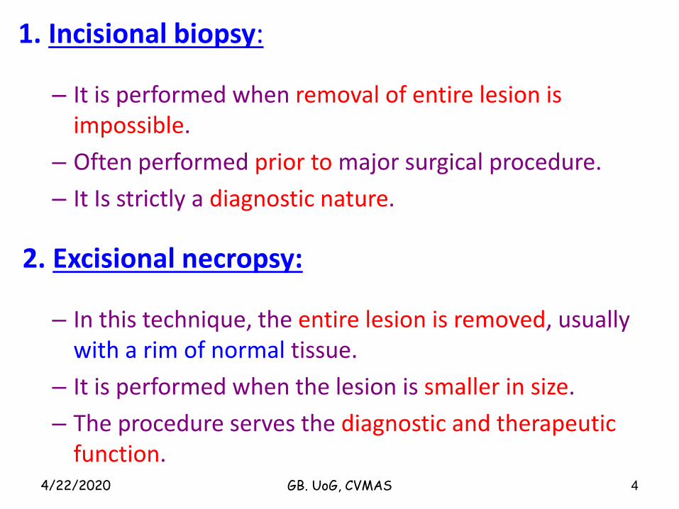
1. Incisional biopsy:
– It is performed when removal of entire lesion is impossible.
– Often performed prior to major surgical procedure.
– It Is strictly a diagnostic nature.
2. Excisional necropsy:
– In this technique, the entire lesion is removed, usually with a rim of normal tissue.
– It is performed when the lesion is smaller in size.
– The procedure serves the diagnostic and therapeutic function.
4/22/2020 4GB. UoG, CVMAS
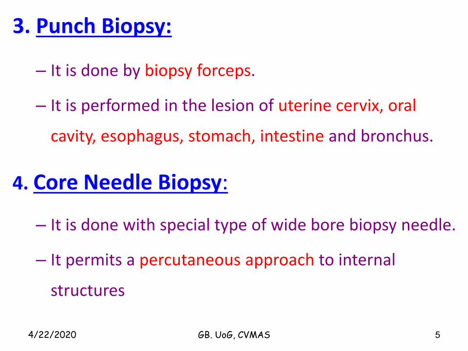
3. Punch Biopsy:
– It is done by biopsy forceps.
– It is performed in the lesion of uterine cervix, oral
cavity, esophagus, stomach, intestine and bronchus.
4. Core Needle Biopsy:
– It is done with special type of wide bore biopsy needle.
– It permits a percutaneous approach to internal
structures
4/22/2020 5GB. UoG, CVMAS
Some General Rules for the biopsy Procedure
• The larger the lesion, the numerous the biopsies that
should be taken from it because of the fact that the
diagnostic areas may be present only focally.
• Necropsy should be taken from the periphery that
includes normal and diseased tissue.
4/22/2020 6GB. UoG, CVMAS
Cont.
• Crushing or squeezing of the tissue with forceps should
be carefully avoided.
• Once the biopsy is obtained, it should be placed
immediately into container with adequate volume of
fixative.
4/22/2020 7GB. UoG, CVMAS
Handling of Specimen
• Specimen should be transported in glass, plastic or metal
container or in a plastic bag in 10%formalin.
– If formalin is not available at hand, place the specimen in
refrigerator at 4oC to slow down autolysis.
• The container should have an opening larger enough so
that the tissue can be removed easily after it has
hardened by fixation.
4/22/2020 8GB. UoG, CVMAS
General Principle of Gross Examination:
Proper identification and orientation of the specimen.
Unlabeled specimen should never be processed.
A properly completed histopathology requisition form containing animal’s ID, age, sex, breed relevant clinical data, surgical findings, nature of operation and name of tissue submitted.
Careful search and examination of all the tissue submitted in order.
4/22/2020 9GB. UoG, CVMAS
General Principle of Gross Examination … cont
Place the specimen on cutting board in an anatomic position and record the following information:
a. Types of specimen b. Structure included.
c. Dimensions c. Weight
e. Shape d. Colour
g. Consistency
h. Surgical margin, whether included or not involved by tumour.
4/22/2020 10GB. UoG, CVMAS
General Principle of Gross Examination …. Cont
Measurements are usually given in centimeter unless
the specimen is very small in which mm can be used.
Endometrial and prostatic tissue should be measured
by aggregate pieces in volume.
4/22/2020 11GB. UoG, CVMAS
Sampling for Histopathological Examination
• Tissue submitted for histopathology must not be more than 3 mm thick and not larger than the diameter of slides used.
• Most specimens from solid tissues are cut in the form of pieces measuring 10 to 15 mm on the slides and 2 to 3 mm in thickness.
• Discrete areas of calcification or ossification should be taken out and should be decalcified in nitric acid/HCl.
• Small fragments of tissue must be wrapped in thin paper.
4/22/2020 12GB. UoG, CVMAS
Histological Technique
• Histological technique deals with the preparation of tissue for microscopic examination.
• The aim of good histological technique to preserve microscopic anatomy of tissue.
• This is achieved by passing through a series of process.– These processes are:
1. Fixation 2. Dehydration3. Cleaning 4. Embedding5. Cutting 6. Staining
4/22/2020 13GB. UoG, CVMAS
Fixation
• This is the process by which the constituents of cells and
tissue are fixed in a physical and a chemical state so that
they will withstand subsequent treatment with various
reagents with minimum loss of architecture.
• This is achieved by exposing the tissue to chemical
compounds, call fixatives
4/22/2020 14GB. UoG, CVMAS
• Mechanism of action of fixatives:
• Most fixatives act by precipitating proteins
• No fixative will penetrate a piece of tissue thicker than 1 cm.
• For dealing with specimen thicker than this, following methods are recommended:
– Solid organ: Cut slices as necessary as but not thicker than 5mm.
– Hollow organ: Either open or fill with fixative or pack lightly with wool soaked in fixative.
– Large specimen: Inject fixative along the vessels or bronchi as in case of lung so that it reaches all parts of the organ
4/22/2020 15GB. UoG, CVMAS
Properties of an Ideal Fixative:
1. Prevents autolysis and bacterial decomposition.
2. Preserves tissue in their natural state and fix all components.
3. Make the cellular components insoluble to reagent used in tissue processing.
4. Preserves tissue volume.
5. Avoid excessive hardness of tissue.
6. Allows enhanced staining of tissue.
7. Should be non-toxic and non-allergic for user.
8. Should not be very expensive.
4/22/2020 16GB. UoG, CVMAS
• Amount of fixative fluid:
– This should be approximately 10-20 times the volume of the specimen.
– Fixative should surround the specimen on all sides.
4/22/2020 17GB. UoG, CVMAS
Factor affecting fixation:
– Size and thickness of piece of tissue.
– Tissue covered by large amount of mucous fix slowly.
– The same applies to tissue covered by blood or organ containing very large amount of blood.
– Fatty and lipomatous tissue fix slowly.
– Fixation is accelerated by agitation.
– Fixation is accelerated by maintaining temperature around 60oc.
4/22/2020 GB. UoG, CVMAS 18

Classification of Fixatives
A. Tissue fixatives
a. Buffered formalin b. Buffered gluteraldehyde
c. Zenker’s formal saline d. Bowen’s fluid
B. Cytological fixatives
a. Ethanol b. Methanol c. Ether
C. Histochemical fixatives
a. Formal saline b. Cold acetone c. Absolute alcohol
4/22/2020 19GB. UoG, CVMAS
Tissue Processing
• Tissue processing is a long procedure and required 24 hours. Tissue processing can be done by manually or mechanically.
• It is done in stages. It can be subdivided into; dehydration, clearing, impregnating and embedding.
• It is important that all specimens are properly labeled before processing is started.
• For labeling, pen containing ordinary ink should not be used. Printed or graphite pencil written, are satisfactory.
4/22/2020 20GB. UoG, CVMAS
A. Dehydration:
– Tissues are dehydrated by using increasing strength of alcohol; e.g. 50%, 70%, 90% and 100%.
– The duration for which tissues are kept in each strength of alcohol depends upon the size of tissue, fixative used and type of tissue.
– The volume of alcohol should be 50 - 100 times that of tissue.
4/22/2020 21GB. UoG, CVMAS
B. Clearing:
• The next step alcohol should be replaced by paraffin wax. • As paraffin wax is not alcohol soluble, we replace alcohol
with a substance in which wax is soluble This step is call clearing.
• Clearing of tissue is achieved by any of the following reagents:– Xylene, Chloroform, Benzene, Carbon tetrachloride and
Toluene
• Xylene is commonly used.
• Small piece of tissue are cleaned in 0.5 – 1 hour; whereas larger (5cm or more thick) are cleaned in 2-4 hours.
4/22/2020 22GB. UoG, CVMAS
C. Impregnation with Wax:
• This is allowed to occur at melting point temperature of paraffin wax, which is 54-60oC.
• Volume of wax should be about 25-30 times the volume of tissues.
• The duration of impregnation depends on size and types of tissues and the clearing agents employed.
• Total duration of 4 hours is sufficient for routine impregnation.
• Types of Wax employed for Impregnation: can be Paraffin wax or Water soluble wax
• Paraffin wax is used routinely. It has hard consistency, so section of 3-4 micron thickness can be cut.
4/22/2020 23GB. UoG, CVMAS
D. Blocking:
• Impregnated tissues are placed in a mould with their labels and then fresh melted wax is poured in it and allowed to settle and solidify.
• Once the block has cooled sufficiently to form a surface skin it should be immersed in cold water to cool it rapidly.
• After the block has completely cooled it is cut into individual blocks and each is trimmed.
• Labels are made to adhere onthe surface of the block. 4/22/2020 24GB. UoG, CVMAS

Staining
• Staining is a process by which we give colour to a section.
• There are hundreds of stains available.
• Generally the stains are classified as:
– Acid stains
– Basic stains
– Neutral stains
4/22/2020 25GB. UoG, CVMAS
Classification of Stains:• Acid Dyes:
– In an acid dye the basic component is coloured and the acid component is colourless.
– Acid dyes stain basic components • e.g. eosin stains cytoplasm red.
• Basic Dyes:– In a basic dye the acid component is coloured and the
basic component is colourless. – Basic dyes stain acidic components
• e.g. basic fuchsin stains nucleus blue.
4/22/2020 26GB. UoG, CVMAS
• Neutral Dyes:– When an acid dye is combined with a basic dye a
neutral dye is formed. – As it contains both coloured radicals, it gives
different colours to cytoplasm and nucleus simultaneously.
– This is the basis of Leishman stain.
4/22/2020 GB. UoG, CVMAS 27
• Procedure of staining
– Every stain is to be used according to a specified method.
– Staining can be done either manually or in an automatic stainer.
– Haematoxylin and Eosin staining: It is the most common used routine stain
4/22/2020 28GB. UoG, CVMAS
Special Stains:
1.PAS (Periodic Acid Schiff) stain: This stain demonstrates glycogen
2.Stains for micro-organism:a. Gram-stain: b. Ziehl_Neelsen stain: This stain detect acid fast bacilli.c. PAS stain: It is used for fungi, amoeba and Tricomonas.d. Modified Giemsa (2% Giemsa in water):For Helicobacter pylori.
4/22/2020 29GB. UoG, CVMAS
3.Congo-red: It is used for identification of amyloid.
4.Sudan-Black: It is used for fat staining.
5.Masson’s Trichrome: It is used for differentiation of connective tissue
4/22/2020 GB. UoG, CVMAS 30
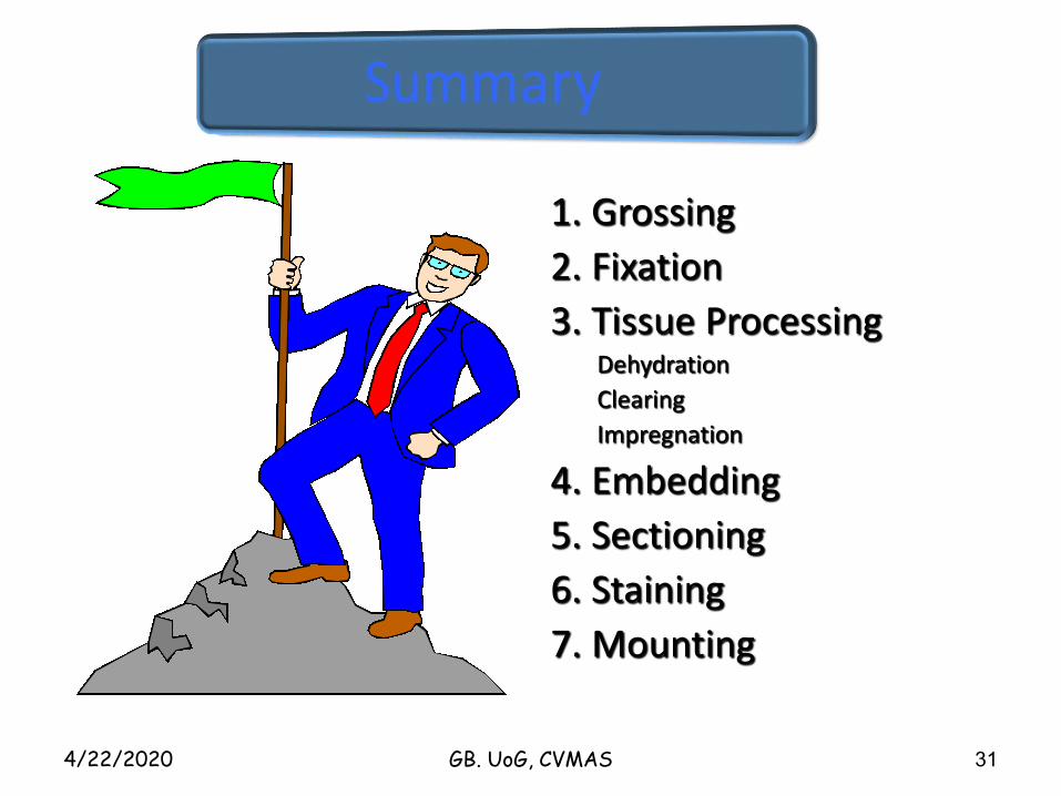
1. Grossing
2. Fixation
3. Tissue ProcessingDehydration
Clearing
Impregnation
4. Embedding
5. Sectioning
6. Staining
7. Mounting
4/22/2020 GB. UoG, CVMAS 31