histomorphology, ultrastructure and fatty acid composition ... · of pansteatitis on the structure...
TRANSCRIPT

RESEARCH Open Access
Histomorphology, ultrastructure and fattyacid composition of the adipose tissue inpansteatitis, the potentials in understandingthe underlying mechanism and diagnosisof pansteatitis in the Nile crocodileO. I. Azeez1,4*, J. G. Myburgh3, R. A. Meintjes1, M. C. Oosthuizen2 and J. P. Chamunorwa1
Abstract
Background: In an effort to characterize the fat body and other adipose tissue in the Nile crocodile and the effectsof pansteatitis on the structure and composition of the adipose tissue, we evaluated the regional variation instructure and fatty acid composition of healthy farmed crocodiles and those affected by pansteatitis.
Methods: Adipose tissue samples were collected from the subcutaneous, visceral and intramuscular fat and theabdominal fat body of ten 4-year old juvenile crocodiles from Izinthaba Crocodile Farm, Pretoria, South Africa whilepansteatitis samples were collected from visceral and intramuscular fat of crocodiles that had died of pansteatitis atthe Olifant River, Mpumalanga, also in South Africa. Histomorphology, ultrastrustucture and fatty acid compositionby fatty acid methyl ester (FAME) analysis were conducted.
Results: Histological examination showed regional variations in the adipose tissue especially in the collagencontent of the ECM, tissue perfusion and division into lobes and lobules by fibrous capsule. Considerable fibrosis,mononuclear cell infiltration especially by macrophages and lymphocytes and toxic changes in the nucleus wereobserved in the pansteatitis samples.Regional variation in lipid composition especially in Myristoleic (C14:1), Erucic acid (C22:1n9), and Docosadienoicacid (C22:2n6) was observed. Most of the saturated and trans fatty acids were found in significant quantities in thepansteatitis samples, but had very low levels of the cis fatty acid and the essential fatty acids with C18 backbone.
Conclusion: This study demonstrates that there exists some regional variation in histomorphology and fatty acidcomposition in the healthy adipose tissue of the Nile crocodile. It also showed that pansteatitis in the Nile crocodilemight have been triggered by sudden change in energy balance from consumption of dead fish; and probableexposure to toxic environmental conditions with the evidence of up scaled monounsaturated long chain fatty acidscomposition and toxic changes in the leucocytes observed in pansteatitis in the present study.
Keywords: Adipose tissue, Histomorphology, Long chain fatty acid, Pansteatitis, Nile crocodile
* Correspondence: [email protected] and Physiology Department, Faculty of Veterinary Science,University of Pretoria, Onderstepoort 0110, Pretoria, South Africa4Department of Veterinary Physiology, Biochemistry and Pharmacology,University of Ibadan, Ibadan, NigeriaFull list of author information is available at the end of the article
© The Author(s). 2017 Open Access This article is distributed under the terms of the Creative Commons Attribution 4.0International License (http://creativecommons.org/licenses/by/4.0/), which permits unrestricted use, distribution, andreproduction in any medium, provided you give appropriate credit to the original author(s) and the source, provide a link tothe Creative Commons license, and indicate if changes were made. The Creative Commons Public Domain Dedication waiver(http://creativecommons.org/publicdomain/zero/1.0/) applies to the data made available in this article, unless otherwise stated.
Azeez et al. Lipids in Health and Disease (2017) 16:47 DOI 10.1186/s12944-016-0405-2

BackgroundThe quest to unravel the mechanisms behind pansteatitis(inflammation of adipose tissue) and crocodile die offs inthe Olifants River and Loskop Dam in Mpumalangaprovince of South Africa is still ongoing. Several sugges-tions have been made as predisposing factors and prob-able aetiology, including consumption of fish that hadpreviously died as a result of environmental pollution ofthe Olifants River and its tributaries [24, 27]. The riverdrains some of the industrial effluents, agricultural run-off water and human sewage as well as acid mine drain-age (AMD) water from abandoned coal mines aroundMiddleburg Colliery, Witbank [3]. Others have alsosuggested that there is an association between vitamin Edeficiency and pansteatitis following excessive consump-tion of unsaturated fatty acid or oxidized fat that coulddeplete vitamin E [12]. It is believed that lack of vitaminE or other antioxidants may predispose the animals toaccumulated reactive oxygen radicals and lipid peroxida-tion [28].In a further study on the probable direct impact of en-
vironmental pollution and heavy metals from AMD wa-ters which seeps into Olifants River from Blesboakstream at a pH of 2.1 on the pathogenesis of pansteatitis,Oberholster et al. [27] reported an association betweenaccumulation of heavy metals especially aluminium andiron and development of yellow fats in Oreochromismossambicus (Tilapia fish) and bioaccumulation of Aland Fe in filamentous algae, Spirogyra fluviatilis and S.adanata that are often consumed by the fish. They sug-gested that the yellowness of the fat in O. mossambicusmight be as a result of membrane lipid peroxidation bythe pro-oxidant action of aluminium as previously sug-gested by Yoshino et al. [36].Adipose tissues in vertebrates generally are storage
sites for lipids for release of energy via lipolysis to acyl-CoA, β-oxidation to acetyl-CoA for energy productionduring fasting, starvation or hibernation and estivationin some animals. It has also been noted to be an endo-crine organ producing leptin and adiponectin; several in-flammatory cytokines and renin-angiotensin system [19];nutrient and energy sensing and mediator of inflamma-tion and immune cells signalling [17, 29]. The structureand composition is also variable as it undergoes constantremodelling, adapting the cell size and numbers to nutri-ent availability and hormonal influences as has beenstudied in humans [33]. Despite the role of adipose tis-sue in inflammation, generalized inflammation involvingthe adipose tissues is not a common occurrence andquite difficult to reproduce. It has also been reportedthat regional variation in structure of white adipose tis-sue in human is also responsible for their activities andinflammatory cytokines. For example, visceral adiposetissue from the mesenteric, omental and retroperitoneal
fat produce more pro-inflammatory cytokines whilethe subcutaneous fats produces more leptin and adi-ponectin [33]. Even subcutaneous fats show some de-gree of regional variation in structure, compositionand functions [30].This therefore calls for another outlook on the struc-
ture and composition of the adipose tissues in the Nilecrocodile in healthy tissues and during pansteatitis forbetter understanding of the factors that might have pre-disposed the tissue to pansteatitis, and determine whichof the fatty acid component might be affected in the invivo esterification/peroxidation that might be respon-sible for the hardening of all adipose tissue in the af-fected crocodiles. Previous study on the lipid propertiesof crocodiles affected by pansteatitis [28] did not takecognizance of possible regional variation in the struc-ture, lipid composition as it affects the release of media-tors of inflammation by adipose tissue.This part of the study is therefore aimed at evaluating
the histology, ultrastructure and lipid composition of theadipose tissue of the Nile crocodile from various regionsof the body in healthy farm bred crocodiles, comparedwith pansteatitis samples from crocodiles that had diedof pansteatitis.
MethodsSample collectionFourty adipose tissue samples were collected from thesubcutaneous, viscera (omental fat around the stomachand the liver), abdominal fat body, and intramuscular fatfrom between the muscles in the tail, and ten liver sam-ples, from ten randomly selected, 4 years old juvenileNile crocodiles. The samples were collected fromIzinthaba Crocodile Farm (Pty) Ltd, Farm 59 (435JQ)Vissershoek, De Wildt area, Pretoria, South Africa. Tenpansteatitis samples from the visceral and intramuscularadipose tissues were collected from frozen adipose tissueand body parts of Nile crocodiles that had died ofpansteatitis.Samples for histology and ultrastructural studies were
collected into 10% neutral buffered formalin (10% for-malin in 0.08 M Sodium phosphate at pH 7.4) whilesamples for lipid profile analysis were collected intohomogenizing buffer consisting of 50 mM Tris–HCl,1.15% KCl at pH 7.4 in distilled water and stored frozenuntil the analysis time.
HistomorphometrySamples were prepared for histology using haematoxylinand eosin (H and E) stain by fixation in 10% bufferedformalin, dehydration in graded alcohol and embeddedin paraffin at 60 °C inside labelled embedding mould orcassettes. The embedded tissues were then sectionedusing a microtome at 5 μm thickness and each section
Azeez et al. Lipids in Health and Disease (2017) 16:47 Page 2 of 19

floated in 45 °C water bath to allow crinkled part tospread before being floated on the glass microscope slidefor proper adherence unto the glass slide. The slideswere stained with haematoxylin and counterstained witheosin using standard protocol. Slides were examinedusing Olympus BX 63 light microscope (Olympus Cor-poration, Tokyo, Japan) at × 10, × 40 and × 100 (in oilimmersion) magnifications.
Transmission electron microscopyParaffin embedded tissue blocks previously used for hist-ology were used for the transmission electron micro-scope study. Samples were cleared with propylene oxidefor 20 minutes and embedded in epoxy resin (TAAB 812resin; TAAB Laboratories, Berkshire, England). Thin sec-tions were cut with Reichert-Jung Ultracut (C. ReichertAG, Vienna, Austria) ultra-microtome using a diamondknife, and stained with lead citrate and uranyl acetate aspreviously described [10]. TEM viewing was done onPhilips CM10 transmission electron microscope at80 kV (Philips Electron Optical Division, Eindhoven,Netherlands).
Long chain fatty acid analysisFatty acids are obtained by a saponification procedure[6] with modifications [7] while methyl esters are pre-pared by the addition of 14% Boron triflouride-methanol(BF3/CH3OH).Fatty acids were identified using a Shi-madzu Tracera GC 2010 with Restek 2560 (fused sil-ica) column. Column temperature was 140–240 °C,while injector temperature was at 250 °C and BID(barrier discharge ionization detector) at 280 °C. Thecarrier gas employed by the instrument was Helium,at linear velocity flow rate of 25 cm/sec. Fatty acidswere identified by comparison with the retentiontimes of fatty Acid methyl ester peaks (Supelco 37Fame mix standard, Sigma Aldrich, USA). Calibrationand quantification of results was done using LabSolu-tions Software, results taken as quantified by the soft-ware in mg/dL and percentage of the total fatty acidcomposition.
Statistical analysisValues are presented as means ± sem. Mean values ofthe fatty acid composition were compared by One-wayANOVA, with Tukey’s post-test for multiple compari-sons between groups, using GraphPad Prism statisticalsoftware, version 5.01 for Windows, GraphPad Software,San Diego California USA (www.graphpad.com). Aprobability value of 0.05 and below was taken to besignificant.
ResultsHistologySamples of adipose tissue that were taken from differentregions were evaluated histologically using H and E. Thesamples included the abdominal fat body visceral, intra-muscular and subcutaneous fats.
Visceral adipose tissueThe adipose from this region including the omental andmesenteric fat is bounded by a thick fibrous connectivetissue capsule that is made up of collagen and fibro-blasts, which tends to extend deep into the parenchymato divide the tissue into lobes (Fig. 1a). Major blood ves-sels are seen in pairs with an artery in close proximity toits draining vein, close to the capsule. The adipocytesappear hexagonal in shape with relatively uniform sizesof which appear to increase gradually and systematicallyas the cells get farther away from the capsule and themajor blood vessels (Fig. 1b). The cells are filled with asingle vacuole of lipids that takes about 98% of the cellvolume. The nucleus is displaced to the periphery, flat-tened and elongated with thick dense chromatin as thelipid vacuole pushes it to the periphery. The extracellularmatrix appears filled with thick dense collagen materialsscattered blood capillaries, immature adipocytes, fibro-blasts and some inflammatory cells especially small lym-phocytes and macrophages (Fig. 1c and d). The ECMalso gives the tissue some sense of looseness because ofits wideness.
Subcutaneous adipose tissueThe subcutaneous adipose tissue is also bound bycollagenous fibrous connective tissue capsule thatoften divide the tissue into small lobes (Fig.2a) unlikethe capsule in the visceral adipose tissue that onlyextends into the parenchyma. The adipose tissueconsists of more arteries and veins located deepwithin the parenchyma and some venous sinusoids(Fig.2d). This is also different from those in the vis-ceral adipose tissue where most arteries are locatedin the periphery, close to the capsule. The adipocytesare round to hexagonal in shape but with less uni-formity in size, and they are more tightly packedwith less ECM spaces. Collagen fibres and fibroblastsalso appear less in the subcutaneous adipose tissue than inthe visceral fat. Also observed are lipid filled adipocyteswith pyknotic nuclei, showing shrunken nuclei with darkhighly condensed nuclear chromatin (Fig. 2f).
Abdominal fat bodyAbdominal fat body in amphibian and reptiles is a singleorgan that is used primarily as a fat storage organ (Fig 3).It is bounded by fibrous connective tissue capsule of colla-gen elastic fibres. As shown in Fig 4, the blood vessels are
Azeez et al. Lipids in Health and Disease (2017) 16:47 Page 3 of 19

all located deep within the organ parenchyma with barelyany close to the periphery near the capsule. The tissue ishighly vascularized with several interspersed capillaries.The adipocytes are hexagonal to round in shapes withvariable sizes and less compacted ECM, unlike that of thesubcutaneous adipose tissue.The ECM is large enough to accommodate blood ca-
pillaries, inflammatory cells (some of which were foundwondering within the ECM, with a lot of collagen fibresand immure adipocytes (preadipocytes).
Intramuscular adipose tissueThe external fibrous connective tissue capsule of theintramuscular adipose tissue is very thin (the thinnest ofall) with sparse fibroblast (Fig. 5a). The cells (adipocytes)are tightly packed, oval to round in shape with high col-lagen fibre content lining the outer surface of the adipo-cytes in the ECM forming a mosaic pattern (Fig. 5b andc), which was not observed in other adipose tissue fromother regions. Inflammatory cells are limited to thehighly compressed blood capillaries (Fig. 5d and e).
Fig. 1 Histology (H and E) of the visceral adipose tissue of the Nile crocodile (Crocodylus niloticus). Note the fibrous connective tissue capsule in a(black arrow head), systematic increase in size of adipocytes from the capsule (b), ECM (red arrow) with collagen, inflammatory cells and bloodcapillaries (c and d), immature adipocytes with dispersed chromatin and prominent nucleolus (black arrow in c) and a mature adipocyte withlipid vacuole and displaced nucleus (f). Bars show magnifications at scales a, 200 μm, b, 100 μm, c – f, 10 μm
Fig. 2 Histology (Hand E) of the subcutaneous adipose tissue of the Nile crocodile (Niloticus crocodylus) showing small lobe, L bounded byfibrous connective tissue a, round to hexagonal shaped adipocytes with variable sizes b, blood vessels and venous sinusoid (vs) in c and d,inflammatory cell (black arrow) and pyknotic nucleus (red arrow). Bars show the magnifications at scales a, 200 μm, b, 50 μm, c, 20 μm, d, 50 μm,e and f, 10 μm
Azeez et al. Lipids in Health and Disease (2017) 16:47 Page 4 of 19

Adipose tissues in pansteatitisGross presentation of pansteatitis in the intramuscularadipose tissue from the tail region of the Nile croco-dile is shown in Fig. 6. Note that the skeletal musclewas not affected by the condition. Histology of theadipose tissue from the visceral and intramuscular fatfrom Nile crocodile that had died of pansteatitis wasalso examined. As shown in Fig. 7, there was a gener-alized disruption of the architecture and expansion ofthe ECM of the adipose tissue with inflammatoryfluid, fibrous connective tissue and cellular infiltrationof the ECM. Macrophages with foaming cytoplasm,small lymphocytes and plasma cells were the main in-flammatory cells observed while fibroblast and colla-gen were also present in large quantities. Giant cellswere also observed while macrophages with toxicchanges in the nucleus could also be seen in the
ECM. Some macrophages in the ECM also show se-vere vacuolation and accumulation of lipids.
Transmission electron microscopyTransmission electron microscope evaluation of the adi-pose tissue in healthy visceral, subcutaneous and intra-muscular as well as pansteatitis samples was also carriedout in this study. No significant regional variations wereobserved in the visceral, subcutaneous, abdominal fatbody and intramuscular adipose tissue cells. The cellswere filled with a single lipid vacuole with thin rim ofcytoplasm and the nuclei displaced to the periphery(Fig. 8). The nuclei are mostly flattened with one prom-inent nucleolus, while the extracellular matrix shows thepresence of collagen on the outer surface of the plasmamembrane. Macrophages are seen in the ECM (Fig. 9).In a manner similar to the observation under the light
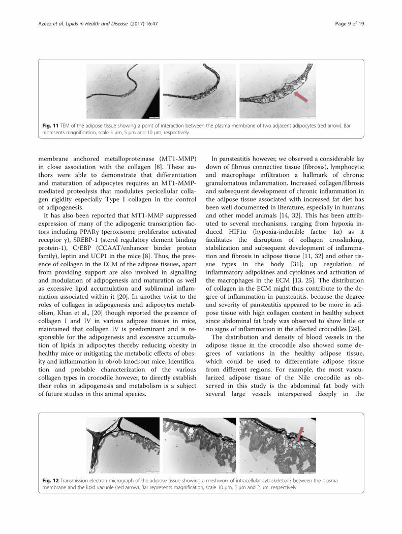
microscope (H and E), the TEM of the adipose tissue inpansteatitis is devoid of regular architecture, with exag-gerated ECM filled with inflammatory cell, fluid and celldebris (Fig. 10).As shown in Fig. 11, we observed a point of inter-
action or communication between the plasma mem-brane of two adjacent adipocytes that appears differentfrom previously reported junctional complexes. It mayalso be an initial point of ECM expansion as inflamma-tory fluid begins to accumulate in the extracellularmatrix.Similarly, we also observe the presence of a meshwork
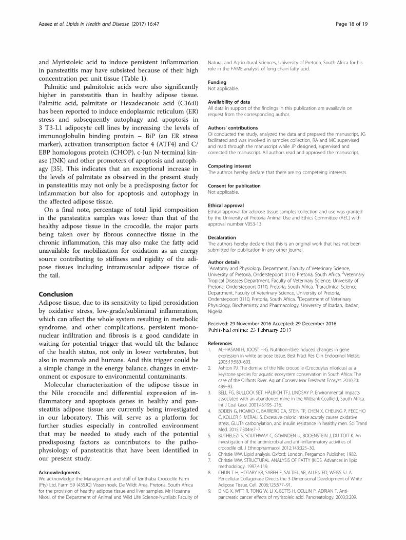
or trabeculae of intracellular myofibrils between themembrane of the lipid vacuole and the plasmamembrane (Fig. 12).
Fig. 3 Gross structure of the abdominal fat body of the Nilecrocodile. Note the white fibrous connective tissue capsule(black arrow)
Fig. 4 Histology (H and E) of the abdominal fat body, showing the fibrous connective tissue capsule (black arrows), blood vessels (bv) deep inthe parenchyma, preadipocytes (p) and inflammatory cells, eosinophil and lymphocytes (e) and monocytes (f) (red arrows). Bars representmagnification at scales a & b, 200 μm, c – f, 10 μm
Azeez et al. Lipids in Health and Disease (2017) 16:47 Page 5 of 19

Long chain fatty acid analysisThe long chain fatty acid composition of the adipose tis-sue was determined using LC/MS (chromatogram samplesare shown in Fig 13) and analysed according to regions aswell as in pansteatitis as shown in Table 1 and 2 and com-pared with liver samples from healthy crocodiles. Of allthe 37 fatty acids in the FAME standard that was used inthis study, 31 were found represented in all our samples.However, Lauric acid (C12:0), Tridecanoic acid (C13:0),cis −10- Pentadecenoic acid (C15: 1) and Linolelaidic acid(C18: 2n6t) were found in trace amounts in pansteatitissamples while Henecosanoic acid (C21:0) was found intrace amount in the liver (Table 2).
Regional variation in the fatty acid composition ofadipose tissueAs shown in Table 2, Myristoleic acid (C14: 1) wasfound only in the abdominal fat body, intramuscularand subcutaneous fat as well as in pansteatitis sam-ples. It was not detected in the visceral fat and theliver. Palmitic acid (C16:0) was found to be signifi-cantly higher (P < 0.001) in the liver than the com-position in the intramuscular fat, while Elaidic acid(C18: 1n9t) was only found in the liver and panstea-titis, with the composition being significantly higher(P < 0.001) in the liver than that of the pansteatitisfat. In a similar manner, Erucic acid (C22: 1n9) wasfound only in the abdominal fat body, intramuscularfat and in pansteatitis samples - which was signifi-cantly higher than the others (P < 0.01), none wasfound in the visceral fat, subcutaneous fat and in theliver. Docosadienoic acid (C22: 2n6) was also absentin the abdominal fat body, visceral fat and the liverwhile trace amount was found in the intramuscularand subcutaneous fats. It was however found in sig-nificant amount in the pansteatitis fat and the com-position was higher (P < 0.01) than those of theintramuscular and subcutaneous fats. Finally, signifi-cant amount of Lignoceric acid (C24:0) was found inthe liver with its percentage composition in the ab-dominal fat body, visceral fat, intramuscular and pan-steatitis fat being significantly lower (P < 0.01) thanthat of the liver, while none was found in subcutane-ous fat.As shown in Fig. 14, the intramuscular fat has the
highest content of fatty acid; it was significantly higher(P < 0.001) than the total fatty acid in the abdominal fat
Fig. 5 Histology of the intramuscular adipose tissue of the Nile crocodile, showing the thin fibrous connective tissue capsule – black arrow (a),thick collagen fibers in the ECM forming a mosaic pattern (m) in b and c. Inflammatory cells (red arrow), monocyte (c), small lymphocyte (d),eosinophil (e), large lymphocyte (f) and preadipocytes (p) are seen in the highly compressed blood vessel. Bars represent the magnifications atscales a, 200 μm, b, 20 μm, c – f, 10 μm
Fig. 6 Gross presentation of pansteatitis in the intramuscularadipose tissue from the tail region of the Nile crocodile
Azeez et al. Lipids in Health and Disease (2017) 16:47 Page 6 of 19

body, liver and pansteatitis samples. While the liver has thelowest long change fatty acid per unit mass and in percent-ages. Surprisingly however, the total percentage fatty acidcomposition in pansteatitis was lower than that of thehealthy adipose tissue storage in the crocodile i.e., the ab-dominal fat body, visceral, intramuscular and subcutaneousadipose tissue storage. It was however higher (P < 0.0001)than that of the liver composition. The total saturated fattyacid, however, did not show any significant variation acrossthe adipose tissue types and even in the liver (Fig 15).
Long chain fatty acid in pansteatitisAs can be observed in Table 2, with the exception ofHeneicosanoic acid, (C21:0), all the long chain fatty acidthat were found in adipose tissue in this study are repre-sented in the pansteatitis samples and the percentagecomposition of each being several folds higher thanthose of the healthy adipose tissues and liver, except incertain instances where the composition in pansteatitiswas considerably lower than those of the healthy tissues.These include stearic acid (C18:0), Oleic acid
Fig. 7 Histology of the visceral and intramuscular adipose tissue of the Nile crocodile in pansteatitis. Note the disruption of the generalarchitecture; cellular infiltration (a & b) and expansion of the ECM with inflammatory fluid and collagen (black arrow) in slide b as well ascongestion in the capillaries (d). Some plasma cells could be seen (c) red arrow and macrophages with foaming cytoplasm (e) with toxic changesin the nucleus (see the red arrow in slide f). Also note the presence of preadipocytes in the ECM (g) fibrous connective tissue lay down withfibroblast and macrophages (h) and more macrophages (i). Horizontal Bars represent magnifications at scales a, 100 μm, b, 20 μm, c – i, 10 μm
Fig. 8 Transmission electron micrograph of the visceral adipose tissue showing the nucleus (N) and a prominent nucleolus, single lipid vacuole(LV), thin rim of cytoplasm (black arrow) and collagen fibers in the ECM. Bar represents magnification, scale 5 μm. Bar represents magnification,scale at 5 μm
Azeez et al. Lipids in Health and Disease (2017) 16:47 Page 7 of 19

(C18:1n9c), Linoleic acid (C18:2n6c), γ-Linoleic acid(C18:3n6) that were observed to be significantly lower(P < 0.001) in pansteatitis than in the other adipose tis-sues and the liver; while Elaidic acid (C18: 1n9t), Behe-nic acid (C22:0), Eicosatrienoic acid (C20:3n6),Arachidonic acid (C20:4n6), Lignoceric acid (C24:0) andNervonic acid (C24:1) composition of the pansteatitissamples were lower than those in the liver. On the con-trary, therefore, the levels/composition of Myristic acid(C14:0), Pentadecanoic acid (C15:0), Palmitic acid(C16:0), Palmitoleic acid (C16:1), Heptadecanoic acid(C17:0), Linolenic acid (C18;3n3), Arachidic acid(C20:0), Eicosaenoic acid (C20:1n9), Eicosadienoic acid(C20:2n6) as well as Erucic acid (C22:1n9), Docosadienoicacid (C22:2n6), Eicosapentaenoic acid (C20:5n3) and Doco-sahexaenoic acid (C22:6n3) in the pansteatitic fat werefound to be significantly higher than those of the healthy
adipose tissue from the abdominal fat body, visceral, intra-muscular and subcutaneous fats (Table 1 and 2).Furthermore and contrary to our expectation, the per-
centage total lipid in the pansteatitis samples was foundto be 10.80 ± 0.83%, which was significantly lower (P <0.0001) than the total percentage lipid of 20.13 ± 1.01%,22.02 ± 0.91%, 23.25 ± 0.54% and 18.96 ± 0.46% in the ab-dominal fat body, visceral, intramuscular and subcutane-ous fats respectively (Fig. 14). The lowest fatty acidcomposition percentage per tissue mass was howeverfound in the liver at 1.24 ± 0.17%, which was significantlylower than any of the adipose tissue samples mentionedearlier. Although, total saturated fatty acid were similarin all samples (Fig. 15), the total monounsaturated(Fig. 16), total omega 6 and 9 fatty acids (Figs. 17 and 18)were lower in pansteatitis while total omega 3 fatty acidwas higher significantly in the pansteatitis sample thanthose of the other adipose tissue and the liver (Fig. 19).The total polyunsaturated fatty acid was also lower in thepansteatitis samples than in the other samples evaluated,although non-significantly (Fig. 20).
DiscussionHistomorphology and ultrastructureComparative evaluation of the adipose tissues in the Nilecrocodile showed considerable regional variation in thehistological structure. Visceral adipose tissue was foundto be lobated with the fibrous connective tissue capsuleextending in various degrees into the parenchyma, butwithout total division into lobules, whereas the subcuta-neous adipose tissue was observed to be lobular or lobu-lated. The abdominal fat body did not show anypenetration of the connective tissue capsule into the par-enchyma, thus forming a single non-divisible lipid stor-age organ irrespective of the size. The intramuscularadipose tissue is characterized by the enormous accumu-lation of collagen fibre in the extracellular matrix in amosaic like appearance that is found only in this tissue.This is a type 3 or fibrous adipose tissue as enumeratedby Sbarbati et al. [30], they are reportedly found in areasthat are more subject to mechanic stress, hence a moredeveloped periadipocyte collagens basket. Based on thiscollagen composition, visceral adipose as well as the ab-dominal fat body can be grouped as type 2 or stromalwhite adipose tissue because of the less collagen thanthat of the intramuscular adipose tissue, while the sub-cutaneous adipose tissue with the least amount of colla-gen could be described as storage or type 1 adipose.Although, this division may be influenced by the age ofthe subject, as age affects lay down of collagen [30], itinadvertently affects the development of the adipose tis-sue involved as it influences the differentiation of thepreadipocytes, proliferation of adipocytes and subse-quent accumulation of lipids through the influence of
Fig. 9 Transmission electron micrograph of the adipose tissue inpansteatitis, showing the ECM with macrophages (M) in theextracellular matrix. Bar represents magnification, scale 5 μm
Fig. 10 Transmission electron micrograph of the adipose tissue inpansteatitis. Note the disruption and increase in size/volume of theECM, filled with inflammatory fluid and debris. Bar representsmagnification, scale 10 μm
Azeez et al. Lipids in Health and Disease (2017) 16:47 Page 8 of 19
membrane anchored metalloproteinase (MT1-MMP)in close association with the collagen [8]. These au-thors were able to demonstrate that differentiationand maturation of adipocytes requires an MT1-MMP-mediated proteolysis that modulates pericellular colla-gen rigidity especially Type I collagen in the controlof adipogenesis.It has also been reported that MT1-MMP suppressed
expression of many of the adipogenic transcription fac-tors including PPARγ (peroxisome proliferator activatedreceptor γ), SREBP-1 (sterol regulatory element bindingprotein-1), C/EBP (CCAAT/enhancer binder proteinfamily), leptin and UCP1 in the mice [8]. Thus, the pres-ence of collagen in the ECM of the adipose tissues, apartfrom providing support are also involved in signallingand modulation of adipogenesis and maturation as wellas excessive lipid accumulation and subliminal inflam-mation associated within it [20]. In another twist to theroles of collagen in adipogenesis and adipocytes metab-olism, Khan et al., [20] though reported the presence ofcollagen I and IV in various adipose tissues in mice,maintained that collagen IV is predominant and is re-sponsible for the adipogenesis and excessive accumula-tion of lipids in adipocytes thereby reducing obesity inhealthy mice or mitigating the metabolic effects of obes-ity and inflammation in ob/ob knockout mice. Identifica-tion and probable characterization of the variouscollagen types in crocodile however, to directly establishtheir roles in adipogenesis and metabolism is a subjectof future studies in this animal species.
In pansteatitis however, we observed a considerable laydown of fibrous connective tissue (fibrosis), lymphocyticand macrophage infiltration a hallmark of chronicgranulomatous inflammation. Increased collagen/fibrosisand subsequent development of chronic inflammation inthe adipose tissue associated with increased fat diet hasbeen well documented in literature, especially in humansand other model animals [14, 32]. This has been attrib-uted to several mechanisms, ranging from hypoxia in-duced HIF1α (hypoxia-inducible factor 1α) as itfacilitates the disruption of collagen crosslinking,stabilization and subsequent development of inflamma-tion and fibrosis in adipose tissue [11, 32] and other tis-sue types in the body [31]; up regulation ofinflammatory adipokines and cytokines and activation ofthe macrophages in the ECM [13, 25]. The distributionof collagen in the ECM might thus contribute to the de-gree of inflammation in pansteatitis, because the degreeand severity of pansteatitis appeared to be more in adi-pose tissue with high collagen content in healthy subjectsince abdominal fat body was observed to show little orno signs of inflammation in the affected crocodiles [24].The distribution and density of blood vessels in the
adipose tissue in the crocodile also showed some de-grees of variations in the healthy adipose tissue,which could be used to differentiate adipose tissuefrom different regions. For example, the most vascu-larized adipose tissue of the Nile crocodile as ob-served in this study is the abdominal fat body withseveral large vessels interspersed deeply in the
Fig. 11 TEM of the adipose tissue showing a point of interaction between the plasma membrane of two adjacent adipocytes (red arrow). Barrepresents magnification, scale 5 μm, 5 μm and 10 μm, respectively
Fig. 12 Transmission electron micrograph of the adipose tissue showing a meshwork of intracellular cytoskeleton? between the plasmamembrane and the lipid vacuole (red arrow). Bar represents magnification, scale 10 μm, 5 μm and 2 μm, respectively
Azeez et al. Lipids in Health and Disease (2017) 16:47 Page 9 of 19

Table
1Con
centratio
nsof
long
chainfattyacid
inadiposetissueof
theNile
crocod
ile(Crocodylusniloticus)with
andwith
outpansteatitis,comparedwith
that
oftheliver.Values
expressedas
mean±SEM
inmg/dl
Fattyacids(m
g/dl)
Abd
ominalfatbo
dyn=10
Visceralfat
n=10
Intram
uscularfat
n=10
Subcutaneo
usfat
n=8
Liver
n=10
Pansteatitis
fat
n=10
Pvalues
C13:0
Methyltridecanoate
(Trid
ecanoicacid)
Nil
Nil
Nil
Nil
Nil
0.40
±0.20
C14:0
Myristic
acid
(Tetrade
cano
icacid)
13.53±1.44
14.15±1.63
16.17±1.63
10.96±1.36
3.41
±1.19
126.8±12.23
P<0.0001
C14:1
Myristoleicacid
(Tetrade
ceno
icacid)
Nil
0.30
±0.15
1.70
±1.38
0.25
±0.16
0.62
±0.35
1.00
±0.47
ns
C15:0
Pentadecylicacid
2.80
±1.15
1.94
±0.17
2.50
±0.13
1.44
±0.17
4.10
±1.30
25.45±2.76
P<0.0001
C16:0
Palm
iticacid
(Hexadecanoicacid)
274.80
±20.57
301.7±16.38
309.3±7.82
249.2±8.23
101.4±11.15
741.0±62.94
P<0.0001
C16:1
Palm
itoleicacid
(cis-Δ9 -Hexadecen
oicacid)
48.47±4.76
56.61±3.92
55.81±2.46
49.04±6.99
8.56
±1.95
214.70
±27.59
P<0.0001
C17:0
Margaric
acid
2.67
±0.41
3.95
±0.35
4.03
±0.39
2.76
±0.51
1.55
±0.37
21.52±1.82
P<0.0001
C17:1
Hep
tade
ceno
icacid
0.03
±0.15
2.20
±0.78
0.50
±0.0.16
0.75
±0.41
Nil
18.60±2.12
P<0.0001
C18:0
Stearic
acid
(Octadecanoicacid)
76.86±3.99
42.93±14.66
96.55±2.18
68.81±9.94
23.34±7.21
107.3±10.83
P<0.0001
C18:1n9
cOleicacid
(cis-Δ9 –Octadecen
oicacid)
381.3±17.93
455.3±30.14
471.3±17.4
390.0±47.11
107.4±14.17
582.1±66.7
P<0.0001
C18:1n9
tElaidicacid
(tran
s-Δ9 -Octadecen
oicacid)
Nil
Nil
Nil
Nil
2.34
±3.49
Nil
C18:2n6
cLino
leicacid
(Methyllineo
leate)
207.3±13.31
255.8±14.10
249.1±7.93
236.7±16.85
64.88±8.89
76.18±11.18
P<0.0001
C20:0
Arachidicacid
(Eicosanoicacid)
0.90
±.34
1.00
±0.39
1.20
±0.41
22.25±21.42
Nil
6.47
±0.73
ns
C18:3n6
γLino
lenicacid
4.10
±1.25
4.70
±1.20
4.39
±1.15
3.34
±1.37
3.43
±1.13
6.79
±0.69
ns
C18:3n3
αLino
lenicacid
7.87
±0.73
9.65
±0.80
10.0±0.60
8.14
±1.07
0.90
±0.38
48.11±8.30
P<0.0001
C20:1n9
Eicosaen
oicacid
4.32
±0.59
4.71
±0.34
4.78
±0.38
3.86
±0.64
0.50
±0.22
18.46±3.55
P<0.0001
C20:2
Eicosadien
oicacid
1.00
±0.42
2.32
±0.59
2.03
±0.65
2.14
±0.92
0.80
±0.36
87.03±17.78
P<0.0001
C22.0
Behe
nicacid
(Docosanoicacid)
0.20
±0.13
0.40
±0.22
0.50
±0.16
0.37
±0.81
1.00
±0.39
Nil
ns
C20:3n6
Eicosatrieno
icacid
3.06
±0.70
4.35
±0.90
4.26
±0.93
2.94
±1.19
6.70
±0.81
8.83
±1.70
P<0.01
C20:3n3
Eicosatrieno
icacid
Nil
Nil
Nil
Nil
Nil
4.30
±0.88
C20:4n6
Arachidon
icacid
5.18
±0.85
6.34
±1.18
5.94
±0.97
4.72
±1.42
12.02±2.00
18.52±3.57
P<0.0001
C24:0
Lign
ocericacid
(Tetracosano
icacid)
Nil
Nil
Nil
2.00
±1.68
Nil
47.00±13.14
Azeez et al. Lipids in Health and Disease (2017) 16:47 Page 10 of 19

Table
1Con
centratio
nsof
long
chainfattyacid
inadiposetissueof
theNile
crocod
ile(Crocodylusniloticus)with
andwith
outpansteatitis,comparedwith
that
oftheliver.Values
expressedas
mean±SEM
inmg/dl
(Con
tinued)
C24:1
Nervonicacid
(cis-15-tetracoseno
icacid)
0.80
±0.20
1.00
±0.0
1.00
±0.0
0.75
±0.25
1.81
±0.24
1.20
±0.20
P<0.01
C20:5n3
Eicosape
ntaeno
icacid
5.24
±0.98
6.96
±1.59
7.88
±1.26
5.50
±1.43
2.54
±0.73
94.81±18.64
P<0.0001
C22:6n3
Docoh
exaeno
icacid
8.60
±0.97
8.67
±1.55
10.03±1.11
5.99
±1.38
3.06
±0.61
300.7±57.57
P<0.0001
TotalLipid
1033.0±54.7
1217.0±67.6
1255.0±30.1
1059.0±29.1
354±42.3
2551
±289.3
P<0.0001
Azeez et al. Lipids in Health and Disease (2017) 16:47 Page 11 of 19

Table
2Long
chainfattyacid
compo
sitio
nof
theadiposetissueof
theNile
crocod
ile(Crocodylusniloticus)with
andwith
outpansteatitis,comparedwith
that
oftheliver.Values
expressedas
mean±SEM
in%
compo
sitio
nof
thetotallipid
Fattyacids(%)
Abd
ominalfatbo
dyn=10
Visceralfat
n=10
Intram
uscularfat
n=10
Subcutaneo
usfat
n=8
Liver
n=10
Pansteatitis
fat
n=10
Pvalues
C12:0
Lauricacid
Nil
Nil
Nil
Nil
Nil
0.00387±0.0038
ND
C13:0
Methyltridecanoate
(Trid
ecanoicacid)
Nil
Nil
Nil
Nil
Nil
0.00634±0.0038
ND
C14:0
Myristic
acid
(Tetrade
cano
icacid)
1.30
±0.13
a1.16
±0.12
b1.22
±0.148c
1.00
±0.11
d0.77
±0.27
e5.10
±0.21
abcd
eP<0.0001
C14:1
Myristoleicacid
(Tetrade
ceno
icacid)
0.006±0.004a
Nil
0.024±0.009b
0.018±0.007c
Nil
0.054±0.01
abc
P<0.0001
C15:0
Pentadecanoicacid
0.14
±0.02
a0.16
±0.01
b0.15
±016c
0.13
±0.01
d0.15
±0.05
e1.04
±0.08
abcd
eP<0.0001
C15:1
cis-10-Pen
tade
ceno
icacid
Nil
Nil
Nil
Nil
Nil
0.052±0.023
P<0.01
C16:0
Palm
iticacid
(Hexadecanoicacid)
26.460
±0.97
24.83±1.908
22.82±1.91
ab23.74±0.193c
29.48±1.590a
30.30±1.63
bc
P<0.0001
C16:1
Palm
itoleicacid
(cis-Δ9 -Hexadecen
oicacid)
4.67
±0.34
ab4.65
±0.15
cd4.10
±0.36
ef
4.91
±0.25
gh
2.14
±0.31
acegi
8.32
±0.40
bdfhi
P<0.0001
C17:0
Margaric
acid
0.27
±0.04
a0.33
±0.02
b0.31
±0.04
c0.25
±0.04
d0.37
±0.11
e0.84
±0.11
abcd
eP<0.0001
C17:1
Hep
tade
ceno
icacid
0.022±0.01
a0.0076
±0.026b
0.042±0.014c
0.0413
±0.018d
Nil
0.74
±0.03
abcd
P<0.0001
C18:0
Stearic
acid
(Octadecanoicacid)
7.48
±0.21
ab7.40
±0.34
cd7.15
±0.62
ef
7.22
±0.14
gh
13.20±0.97
acegi
4.39
±0.30
bdfhi
P<0.0001
C18:1n9
cOleicacid
(cis-Δ9 –Octadecen
oicacid)
37.09±0.94
ab37.34±0.91
cd35.16±3.14
e40.68±0.98
fg30.05±0.99
aefh
22.92±0.99
bdegh
P<0.0001
C18:1n9
tElaidicacid
(tran
s-Δ9 -Octadecen
oicacid)
Nil
Nil
Nil
Nil
0.67
±0.08
a0.02
±0.013a
P<0.001
C18:2n6
tLino
lelaidicacid
Nil
Nil
Nil
Nil
Nil
0.0026
±0.0026
ND
C18:2n6
cLino
leicacid
(Methyllineo
leate)
20.07±0.69
a21.14±0.82
b18.33±1.51
c19.85±0.32
d18.40±0.87
e2.87
±0.25
abcd
eP<0.0001
C20:0
Arachidicacid
(Eicosanoicacid)
0.0045
±0.062a
0.029±0.044b
0.95
±0.032c
0.05
±0.023d
0.008±0.008e
0.26
±0.024a
bcd
eP<0.0001
C18:3n3
αLino
lenicacid
0.76
±0.06
ab0.79
±0.05
cd0.76
±0.08
ef
0.78
±0.07
gh
0.20
±0.07
acegi
1.75
±0.21
bdfhi
P<0.0001
C18:3n6
γLino
lenicacid
0.39
±0.13
a0.38
±0.10
b0.34
±0.96
c0.37
±0.13
d1.50
±0.33
abcd
e0.28
±0.026e
P<0.0001
C20:1n9
Eicosaen
oicacid
0.41
±0.05
ab0.40
±0.03
cd0.35
±0.04
e0.32
±0.04
f0.12
±0.04
acg
0.75
±0.13
bdefg
P<0.0001
C20:2
Eicosadien
oicacid
0.07
±0.05
a0.18
±0.04
b0.15
±0.05
c0.11
±0.03
d0.20
±0.09
e3.18
±0.38
abcd
eP<0.0001
C21:0
Hen
eicosano
icacid
Nil
Nil
Nil
Nil
0.004±0.004
Nil
ND
C22.0
Behe
nicacid
(Docosanoicacid)
0.017±0.01
a0.023±0.012b
0.039±0.013c
0.018±0.009d
0.27
±0.012a
bcd
e0.002±0.002e
P<0.01
C20:3n6
Eicosatrieno
icacid
0.30
±0.07
0.35
±0.07
0.33
±0.08
0.34
±0.11
1.89
±0.18
abcd
e0.39
±0.08
P<0.0001
Azeez et al. Lipids in Health and Disease (2017) 16:47 Page 12 of 19

Table
2Long
chainfattyacid
compo
sitio
nof
theadiposetissueof
theNile
crocod
ile(Crocodylusniloticus)with
andwith
outpansteatitis,comparedwith
that
oftheliver.Values
expressedas
mean±SEM
in%
compo
sitio
nof
thetotallipid
(Con
tinued)
C22:1n9
Erucicacid
0.001±0.001a
00.002±0.001
00
0.009±0.004a
P<0.01
C20:3n3
Eicosatrieno
icacid
Nil
Nil
Nil
Nil
Nil
0.15
±0.03
ND
C20:4n6
Arachidon
icacid
0.51
±0.08
a0.52
±0.08
b0.46
±0.08
c0.53
±0.11
d3.59
±0.61
abcd
e0.67
±0.09
eP<0.0001
C22:2n6
Docosadieno
icacid
00
0.003±0.003a
0.008±0.008a
b0
1.90
±0.17
P<0.0001
C24:0
Lign
ocericacid
(Tetracosano
icacid)
0.004±0.003a
0.002±0.001b
0.004±0.002c
00.10
±0.05
abcd
0.0003
±0.0003
dP<0.01
C24:1
Nervonicacid
(cis-15-tetracoseno
icacid)
0.03
±0.01
a0.03
±0.01
b0.04
±0.01
c0.03
±0.10
d0.60
±0.11
abcd
e0.02
±0.008e
P<0.0001
C20:5n3
Eicosape
ntaeno
icacid
0.50
±0.09
a0.57
±0.13
b0.62
±0.12
c0.54
±0.11
d0.63
±0.18
e3.46
±0.42
abcd
eP<0.0001
C22:6n3
Docoh
exaeno
icacid
0.83
±0.09
a0.71
±0.12
b0.76
±0.12
c0.60
±0.11
d0.87
±0.16
e10.98±1.28
abcd
eP<0.0001
TotalLipid
20.13±1.01
abc
22.02±0.91
de
23.25±0.54
afgh
18.96±0.46
fij
1.24
±0.17
bdgik
10.80±0.82
cehjk
P<0.0001
TotalSaturated
Lipids
4.77
±1.04
4.53
±0.97
3.97
±0.88
4.32
±1.01
4.94
±1.03
4.66
±1.00
ns
TotalM
onou
nsaturated
Lipids
6.03
±1.55
6.07
±1.55
5.67
±1.52
7.70
±2.17
a5.60
±1.44
3.65
±0.78
ans
TotalP
olyunsaturated
Lipids
2.93
±0.73
3.08
±0.78
2.97
±0.78
2.57
±0.73
3.51
±0.70
2.56
±0.34
ns
TotalO
meg
a3
0.70
±0.05
a0.69
±0.06
b0.69
±0.08
c0.63
±0.06
d0.57
±0.10
e4.08
±0.74
abcd
eP<0.0001
TotalO
meg
a6
5.32
±1.38
5.60
±1.45
3.89
±1.07
4.09
±1.31
6.88
±1.25
a1.22
±0.16
aP<0.01
TotalO
meg
a9
12.50±3.24
12.58±3.27
11.84±3.22
20.50±5.23
ab10.28±2.62
a5.93
±1.59
bP<0.01
Values
with
thesamesupe
rscriptedalph
abetsaresign
ificantly
differen
tP<0.00
1NDmeans
notde
term
ined
nsmeans
notsign
ificant
Azeez et al. Lipids in Health and Disease (2017) 16:47 Page 13 of 19

parenchyma and more capillaries in the ECM whilethe visceral adipose tissue has the least blood vesseldensity within the parenchyma with most of themajor vessels located close to the capsule. This mightbe an important factor as adipose tissue lay down in-creases either with increase nutrition only or in
combination with increase in age as the rate of angio-genesis and tissue perfusion might not be able tocatch up with the demand by the adipose tissue. Thusresulting in hypoxia and up regulation of inflamma-tory genes including IL-6, macrophage inflammationfactor 1 (MIF1) through HIF1α induction [32].
Fig. 13 Sample of LC/MS chromatogram showing the spectrum for long chain fatty acid Composition. a – reference standard, b – healthyvisceral fat and c – pansteatitic visceral fat
Fig. 14 Percentage total lipid composition in healthy liver and the adipose tissue of the Nile crocodile with and without pansteatitis. Valuespresented as mean ± sem. Values with the same superscript alphabets are significantly different, P < 0.0001
Azeez et al. Lipids in Health and Disease (2017) 16:47 Page 14 of 19

Sudden exposure of the crocodiles to fish and over-feeding as a result of fish die offs in and along the Oli-fants River gorge, Kwazulu Natal Province where thecondition was identified [2] might have led to adiposetissue hypertrophy, serving as a trigger for chronic in-flammation in the affected animals. This has been ob-served in acute overfeeding with high caloric energybalance resulting in oxidative stress in the adipose tissue[4]. More chronic over nutrition however is morefavourable to inflammation and fibrosis in adipose tissueas a result of persistent endoplasmic reticulum (ER)stress and increased macrophage chemoattractant pro-tein 1 (MCP-1), a proinflammatory cytokine that hasbeen isolated in adipose tissue. It is found in associationwith hypertrophic adipocytes and subsequent macro-phage infiltration, activation and production of inflam-matory cytokines just after 3 weeks of high fat diet inmice [23].Apart from the various predisposing factors such as
reduced tissue perfusion especially, deep within theparenchyma, presence of mononuclear cells and colla-gen in the ECM, we also observed the presence oftoxic changes within the nuclei of some of the mono-nuclear phagocytic cells in pansteatitis samples(Fig. 5f ). Toxic nuclei as the name implies arechanges observable in blood cells exposed to chemical
contaminants resulting in clumping of chromatins inthe nucleus. This has been used in the diagnosis ofpoisoning. The presence of these toxic changes maybe an indicator that the animals might have been ex-posed to toxic chemicals or compounds in the envir-onment, more so that the presence of heavy metal inAMD water and POPs (persistent organophosphates)contamination has been reported in Olifants Riverand their presence correlated with the developmentof pansteatitis in the Tilapia [27]. Acid mine drainagewater and POPs have also been reported to persist inthe environment especially in algae and phytoplank-ton in the area [26].
Lipids analysisFatty acid methyl ester (FAME) analysis of the longchain fatty acid of the adipose tissue in healthy adiposetissue of the Nile crocodile also showed some degree ofregional variation in the lipid composition, though moreconsiderably differences were observed when comparedwith lipids in the liver and pansteatitis samples. For ex-ample, Myristoleic acid (C14:1) was absent in the vis-ceral fat and in the liver, Erucic acid (C22:1n9) wasabsent in visceral and subcutaneous fats and in the liverwhile Oleic acid (C18:1n9c) was found only in the liverand pansteatitis fat with the percentage composition of
Fig. 15 Percentage total saturated fatty acid in healthy liver and the adipose tissue of the Nile crocodile with and without pansteatitis. Valuespresented as mean ± sem. Values with the same superscript alphabets are significantly different, P < 0.0001
Fig. 16 Percentage total monounsaturated fatty acid in healthy liver and the adipose tissue of the Nile crocodile with and without pansteatitis.Values presented as mean ± sem. Values with the same superscript alphabets are significantly different, P < 0.0001
Azeez et al. Lipids in Health and Disease (2017) 16:47 Page 15 of 19

oleic acid in the liver being significantly higher than thatof pansteatitis. Cis pentadecenoic acid (C15:1), Linolelai-dic acid, Eicosatrienoic acid (C20:3n3) were only foundin pansteatitis while Heneicosanoic acid (C21:0) wasfound only in the liver.Myristoleic acid (C14:1), or 9-tetradecenoic acid, is an
omega-5 fatty acid with 14 carbon atoms and a doublebond. It is synthesized from myristic acid (C14:0) by theenzyme delta-9 desaturase. It is found in extract of thesea cucumber, (Cucumaria frondosa) with potent anti-cancer activity and lipoxygenase inhibitory activity [9]. Itwas also reported as the cytotoxic component of the ex-tract of Serenoa repens responsible for apoptosis and ne-crosis in LNCaP cells. It has thus been developed as atool in the treatment of prostate cancer [18]. Myristoleicacid and its derivative cetyl myristoleate, have also beenpatented in relieving the pain of rheumatoid arthritisand osteoarthritis [15, 22]. This compound being a cyto-toxic compound that is capable of inducing apoptosisand necrosis, begs the question: could the presence ofmyristoleic acid in healthy intramuscular and subcutane-ous fat as well as abdominal fat body be a risk factor intissue damage in pansteatitis as the composition
increases in pansteatitis? It is also not known whether itis being extracted in crocodile fat for the purpose of an-ticancer and antiarthritis therapy.The use of crocodile oil though, has been documented
in traditional healing in various ethnic groups in Africa,including South Africa, Madagascar and even China [5].Antimicrobial effects and anti-inflammatory effects ofcrocodile oil has also been reported [5], but there is pau-city of information in literature on the use of crocodilefats. Blood extracts of Crocodylus siamensis has also beenreported to show considerable anti-inflammatory activities[21]. These authors observed anti-inflammatory effectof crocodile blood extracts by examining their inhibi-tory effects on pro-inflammatory mediators in lipo-polysaccharide (LPS)-stimulated murine macrophageRAW 264.7 cells. They further reported that bothplasma and crude leukocyte extract significantlyinhibited the anti-inflammatory Nitrogen oxide (NO)production and exhibited cytotoxicity in all the testedconcentrations.We observed that although, the percentage compos-
ition of many saturated and monounsaturated fatty acidas well as some polyunsaturated fatty acid was higher in
Fig. 17 Percentage total polyunsaturated fatty acid in healthy liver and the adipose tissue of the Nile crocodile with and without pansteatitis.Values presented as mean ± sem. Values with the same superscript alphabets are significantly different, P < 0.0001
Fig. 18 Percentage total omega 3 fatty acid in healthy liver and the adipose tissue of the Nile crocodile with and without pansteatitis. Valuespresented as mean ± sem. Values with the same superscript alphabets are significantly different, P < 0.0001
Azeez et al. Lipids in Health and Disease (2017) 16:47 Page 16 of 19

pansteatitis than in healthy adipose tissues, the percent-age composition of all the fatty acid with C18 backbone(from stearic acid (C18:0), Oleic acid (C18:1n9c), Elaidicacid (C18:1n9t), Linoleic acid (C18:2n6c), Linolelaidicacid (C18:2n6t), to γ Linolenic acid (C18:3n6) exceptLinolenic acid (C18:3n3)) were lower in the pansteatitissamples than in the liver and all the healthy adipose tis-sue samples, irrespective of the region. Arachidonic acidC20:4n6) was also lower. This shows that the crocodileswith pansteatitis, though has large chunk of adiposetissue lack the major essential fatty acids, and theyare instead laden with EPA and DHA, (Eicosapente-noic acid and Docosahexaenoic acid) found almostexclusively in high fat fish and marine animals, whichare precursors for the synthesis of lipid derived mod-ulators of cell signaling and inflammatory cytokines.They are also the main substrate for cyclooxygenases,lipoxygenases, Cytochrome P450 mono-oxygenases that
give rise to the production of signalling molecules xin-cluding leukotrienes, prostanoids, thromboxanes that areimplicated in various biological processes such as inflam-mation to PO regarding the new manuscript provided inCORRECTION.zip on how to proceed.error parsing dueto changing in author group and chemotaxis [1]. This,coupled with high concentration of C18 desaturated lipidssuch as Stearic and Oleic acid (Table 1), which are part ofthe hallmark of obesity and metabolic syndrome in higheranimals including humans and strong inducer of inflam-mation in the adipose tissue of obese animals [34] appearsto be predisposing factors to pansteatitis in the crocodile.Although some authors have hypothesized some de-
gree of anti-inflammatory roles to EPA and DHA, espe-cially against lipopolysaccharide (LPS) induced activationof Toll-like receptor 4 (TLR4) in the downstream activa-tion and pro-inflammatory roles of TNFα and IL-6, [16]the overwhelming effects of other saturated fatty acid
Fig. 19 Percentage total omega 6 fatty acid in healthy liver and the adipose tissue of the Nile crocodile with and without pansteatitis. Valuespresented as mean ± sem. Values with the same superscript alphabets are significantly different, P < 0.0001
Fig. 20 Percentage total omega 9 fatty acid in healthy liver and the adipose tissue of the Nile crocodile with and without pansteatitis. Valuespresented as mean ± sem. Values with the same superscript alphabets are significantly different, P < 0.0001
Azeez et al. Lipids in Health and Disease (2017) 16:47 Page 17 of 19
and Myristoleic acid to induce persistent inflammationin pansteatitis may have subsisted because of their highconcentration per unit tissue (Table 1).Palmitic and palmitoleic acids were also significantly
higher in pansteatitis than in healthy adipose tissue.Palmitic acid, palmitate or Hexadecanoic acid (C16:0)has been reported to induce endoplasmic reticulum (ER)stress and subsequently autophagy and apoptosis in3 T3-L1 adipocyte cell lines by increasing the levels ofimmunoglobulin binding protein – BiP (an ER stressmarker), activation transcription factor 4 (ATF4) and C/EBP homologous protein (CHOP), c-Jun N-terminal kin-ase (JNK) and other promoters of apoptosis and autoph-agy [35]. This indicates that an exceptional increase inthe levels of palmitate as observed in the present studyin pansteatitis may not only be a predisposing factor forinflammation but also for apoptosis and autophagy inthe affected adipose tissue.On a final note, percentage of total lipid composition
in the pansteatitis samples was lower than that of thehealthy adipose tissue in the crocodile, the major partsbeing taken over by fibrous connective tissue in thechronic inflammation, this may also make the fatty acidunavailable for mobilization for oxidation as an energysource contributing to stiffness and rigidity of the adi-pose tissues including intramuscular adipose tissue ofthe tail.
ConclusionAdipose tissue, due to its sensitivity to lipid peroxidationby oxidative stress, low-grade/subliminal inflammation,which can affect the whole system resulting in metabolicsyndrome, and other complications, persistent mono-nuclear infiltration and fibrosis is a good candidate inwaiting for potential trigger that would tilt the balanceof the health status, not only in lower vertebrates, butalso in mammals and humans. And this trigger could bea simple change in the energy balance, changes in envir-onment or exposure to environmental contaminants.Molecular characterization of the adipose tissue in
the Nile crocodile and differential expression of in-flammatory and apoptosis genes in healthy and pan-steatitis adipose tissue are currently being investigatedin our laboratory. This will serve as a platform forfurther studies especially in controlled environmentthat may be needed to study each of the potentialpredisposing factors as contributors to the patho-physiology of pansteatitis that have been identified inour present study.
AcknowledgmentsWe acknowledge the Management and staff of Izinthaba Crocodile Farm(Pty) Ltd, Farm 59 (435JQ) Vissershoek, De Wildt Area, Pretoria, South Africafor the provision of healthy adipose tissue and liver samples. Mr HosannaNkosi, of the Department of Animal and Wild Life Science-Nutrilab: Faculty of
Natural and Agricultural Sciences, University of Pretoria, South Africa for hisrole in the FAME analysis of long chain fatty acid.
FundingNot applicable.
Availability of dataAll data in support of the findings in this publication are avaailavle onrequest from the corresponding author.
Authors' contributionsOI conducted the study, analyzed the data and prepared the manuscript, JGfacilitated and was involved in samples collection, RA and MC supervisedand read through the manuscript while JP designed, supervised andcorrected the manuscript. All authors read and approved the manuscript.
Competing interestThe authros hereby declare that there are no competeing interests.
Consent for publicationNot applicable.
Ethical approvalEthical approval for adipose tissue samples collection and use was grantedby the University of Pretoria Animal Use and Ethics Committee (AEC) withapproval number V053-13.
DecalarationThe authors hereby declare that this is an original work that has not beensubmitted for publication in any other journal.
Author details1Anatomy and Physiology Department, Faculty of Veterinary Science,University of Pretoria, Onderstepoort 0110, Pretoria, South Africa. 2VeterinaryTropical Diseases Department, Faculty of Veterinary Science, University ofPretoria, Onderstepoort 0110, Pretoria, South Africa. 3Paraclinical ScienceDepartment, Faculty of Veterinary Science, University of Pretoria,Onderstepoort 0110, Pretoria, South Africa. 4Department of VeterinaryPhysiology, Biochemistry and Pharmacology, University of Ibadan, Ibadan,Nigeria.
Received: 29 November 2016 Accepted: 29 December 2016
References1. AL-HASANI H, JOOST H-G. Nutrition-/diet-induced changes in gene
expression in white adipose tissue. Best Pract Res Clin Endocrinol Metab.2005;19:589–603.
2. Ashton PJ. The demise of the Nile crocodile (Crocodylus niloticus) as akeystone species for aquatic ecosystem conservation in South Africa: Thecase of the Olifants River. Aquat Conserv Mar Freshwat Ecosyst. 2010;20:489–93.
3. BELL FG, BULLOCK SET, HÄLBICH TFJ, LINDSAY P. Environmental impactsassociated with an abandoned mine in the Witbank Coalfield, South Africa.Int J Coal Geol. 2001;45:195–216.
4. BODEN G, HOMKO C, BARRERO CA, STEIN TP, CHEN X, CHEUNG P, FECCHIOC, KOLLER S, MERALI S. Excessive caloric intake acutely causes oxidativestress, GLUT4 carbonylation, and insulin resistance in healthy men. Sci TranslMed. 2015;7:304re7–7.
5. BUTHELEZI S, SOUTHWAY C, GOVINDEN U, BODENSTEIN J, DU TOIT K. Aninvestigation of the antimicrobial and anti-inflammatory activities ofcrocodile oil. J Ethnopharmacol. 2012;143:325–30.
6. Christie WW. Lipid analysis. Oxford: London, Pergamon Publisher; 1982.7. Christie WW. STRUCTURAL ANALYSIS OF FATTY {KIDS. Advances in lipid
methodology. 1997;4:119.8. CHUN T-H, HOTARY KB, SABEH F, SALTIEL AR, ALLEN ED, WEISS SJ. A
Pericellular Collagenase Directs the 3-Dimensional Development of WhiteAdipose Tissue. Cell. 2006;125:577–91.
9. DING X, WITT R, TONG W, LI X, BETTS H, COLLIN P, ADRIAN T. Anti-pancreatic cancer effects of myristoleic acid. Pancreatology. 2003;3:209.
Azeez et al. Lipids in Health and Disease (2017) 16:47 Page 18 of 19

10. DU PLESSIS L, SOLEY JT. A re-evaluation of sperm ultrastructure in the emu,Dromaius novaehollandiae. Theriogenology. 2014;81:1073–84.
11. FUJISAKA S, USUI I, IKUTANI M, AMINUDDIN A, TAKIKAWA A, TSUNEYAMA K,MAHMOOD A, GODA N, NAGAI Y, TAKATSU K, TOBE K. Adipose tissuehypoxia induces inflammatory M1 polarity of macrophages in an HIF-1α-dependent and HIF-1α-independent manner in obese mice. Diabetologia.2013;56:1403–12.
12. FYTIANOU A, KOUTINAS A, SARIDOMICHELAKIS M, KOUTINAS C. Blood α-tocopherol, selenium, and glutathione peroxidase changes and adiposetissue fatty acid changes in kittens with experimental steatitis (yellow fatdisease). Biol Trace Elem Res. 2006;112:131–43.
13. HENEGAR C, TORDJMAN J, ACHARD V, LACASA D, CREMER I, GUERRE-MILLOM, POITOU C, BASDEVANT A, STICH V, VIGUERIE N, LANGIN D, BEDOSSA P,ZUCKER J-D, CLEMENT K. Adipose tissue transcriptomic signature highlightsthe pathological relevance of extracellular matrix in human obesity.Genome Biol. 2008;9:1–32.
14. HENNINGER AJ, ELIASSON B, JENNDAHL LE, HAMMARSTEDT A. Adipocytehypertrophy, inflammation and fibrosis characterize subcutaneous adiposetissue of healthy, non-obese subjects predisposed to type 2 diabetes. PLoSOne. 2014;9:e105262.
15. HESSLINK R, ARMSTRONG D, NAGENDRAN M, SREEVATSAN S, BARATHUR R.Cetylated fatty acids improve knee function in patients with osteoarthritis.J Rheumatol. 2002;29:1708–12.
16. HONDA KL, LAMON-FAVA S, MATTHAN NR, WU D, LICHTENSTEIN AH. EPAand DHA Exposure Alters the Inflammatory Response but not the SurfaceExpression of Toll-like Receptor 4 in Macrophages. Lipids. 2014;50:121–9.
17. Hotamisligil GS. Inflammation and metabolic disorders. Nature. 2006;444:860–7.
18. IGUCHI K, OKUMURA N, USUI S, SAJIKI H, HIROTA K, HIRANO K. Myristoleic acid,a cytotoxic component in the extract from Serenoa repens, induces apoptosisand necrosis in human prostatic LNCaP cells. Prostate. 2001;47:59–65.
19. KERSHAW EE, FLIER JS. Adipose tissue as an endocrine organ. J ClinEndocrinol Metab. 2004;89:2548–56.
20. KHAN T, MUISE ES, IYENGAR P, WANG ZV, CHANDALIA M, ABATE N, ZHANGBB, BONALDO P, CHUA S, SCHERER PE. Metabolic Dysregulation andAdipose Tissue Fibrosis: Role of Collagen VI. Mol Cell Biol. 2009;29:1575–91.
21. KOMMANEE J, PHOSRI S, DADUANG S, TEMSIRIPONG Y, DHIRAVISIT A,THAMMASIRIRAK S. Comparisons of anti-inflammatory activity of crocodile(Crocodylus siamensis) blood extract. Chiang Mai J Sci. 2014;41:627–34.
22. Leonard EC. Antioxidant; cellular proliferation inhibition, Google Patents. 2004.23. LIONETTI L, MOLLICA MP, LOMBARDI A, CAVALIERE G, GIFUNI G, BARLETTA
A. From chronic overnutrition to insulin resistance: the role of fat-storingcapacity and inflammation. Nutr Metab Cardiovasc Dis. 2009;19:146–52.
24. MYBURGH, J. & BOTHA, A. 2009. Decline in herons along the lower OlifantsRiver–could pansteatitis be a contributing factor. VETnuus March, 20–23.
25. O’ROURKE RW. Inflammation in obesity-related disease. Surgery. 2009;145:255.26. OBERHOLSTER PJ, HILL L, JAPPIE S, TRUTER JC, BOTHA A-M. Applying
genotoxicology tools to identify environmental stressors in support of rivermanagement. Chemosphere. 2016;144:319–29.
27. OBERHOLSTER PJ, MYBURGH JG, ASHTON PJ, COETZEE JJ, BOTHA A-M.Bioaccumulation of aluminium and iron in the food chain of Lake Loskop,South Africa. Ecotoxicol Environ Saf. 2012;75:134–41.
28. OSTHOFF G, HUGO A, BOUWMAN H, BUSS P, GOVENDER D, JOUBERT CC,SWARTS JC. Comparison of the lipid properties of captive, healthy wild, andpansteatitis-affected wild Nile crocodiles (Crocodylus niloticus). CompBiochem Physiol A Mol Integr Physiol. 2010;155:64–9.
29. OZATO K, TSUJIMURA H, TAMURA T. Toll-like receptor signaling andregulation of cytokine gene expression in the immune system.Biotechniques. 2002;33:S66–75.
30. SBARBATI A, ACCORSI D, BENATI D, MARCHETTI L, ORSINI G, RIGOTTI G,PANETTIERE P. Subcutaneous adipose tissue classification. Eur J Histochem.2010;54:48.
31. SCHWARTZ RS, ELTZSCHIG HK, CARMELIET P. Hypoxia and inflammation. NEngl J Med. 2011;364:656–65.
32. SUN K, TORDJMAN J, CLÉMENT K, SCHERER PHILIPPE. Fibrosis and AdiposeTissue Dysfunction. Cell Metab. 2013;18:470–7.
33. WRONSKA A, KMIEC Z. Structural and biochemical characteristics of variouswhite adipose tissue depots. Acta Physiol. 2012;205:194–208.
34. YEW TAN C, VIRTUE S, MURFITT S, ROBERT LD, PHUA YH, DALE M, GRIFFINJL, TINAHONES F, SCHERER PE, VIDAL-PUIG A. Adipose tissue fatty acid chain
length and mono-unsaturation increases with obesity and insulin resistance.Scientific Reports. 2015;5:18366.
35. YIN J, WANG Y, GU L, FAN N, MA Y, PENG Y. Palmitate induces endoplasmicreticulum stress and autophagy in mature adipocytes: Implications forapoptosis and inflammation. Int J Mol Med. 2015;35:932–40.
36. YOSHINO M, ITO M, HANEDA M, TSUBOUCHI R, MURAKAMI K. Prooxidantaction of aluminum ion–stimulation of iron-mediated lipid peroxidation byaluminum. Biometals. 1999;12:237–40.
• We accept pre-submission inquiries
• Our selector tool helps you to find the most relevant journal
• We provide round the clock customer support
• Convenient online submission
• Thorough peer review
• Inclusion in PubMed and all major indexing services
• Maximum visibility for your research
Submit your manuscript atwww.biomedcentral.com/submit
Submit your next manuscript to BioMed Central and we will help you at every step:
Azeez et al. Lipids in Health and Disease (2017) 16:47 Page 19 of 19